Catecholamines-evoked cytosolic ca2+rise in endothelial cells from bovine adrenal medulla
11
Molecular and Cellular Biochemistry 203: 53–58, 2000. © 2000 Kluwer Academic Publishers. Printed in the Netherlands. Catecholamines-evoked cytosolic Ca 2+ rise in endothelial cells from bovine adrenal medulla Raul Vinet, 1 Fernando Rojas, 1 Mario Luxoro, 2 FernandoVargas 3 and Magdalena Cortés 1 1 Escuela de Quimica y Farmacia, Facultad de Medicina, Universidad de Valparaiso; 2 Departamento de Biologia, Facultad de Ciencias, Universidad de Chile, Santiago, Chile. 3 Department of Human Physiology, School of Medicine, University of California, Davis, CA, USA Received 4 December 1998; accepted 8 July 1999 Abstract The effects of catecholamines on intracellular Ca 2+ concentrations ([Ca 2+ ] i ) in single acutely dissociated bovine adrenal medulla endothelial cells (BAMECs) were measured using the intracellular fluorescent probe Fluo–3 AM. 100 μm epinephrine or norepinephrine induced a biphasic [Ca 2+ ] i rise with an initial peak followed by a delayed phase. 10 μm phenylephrine (α 1 -adrenergic agonist) caused a [Ca 2+ ] i rise similar to that evoked by catecholamines. The increase in [Ca 2+ ] i induced by 10 μm phenylephrine was reverted by 10 μm phenoxybenzamine (α-adrenergic antagonist). Neither isoproterenol (β-adrenergic agonist) nor clonidine (α 2 -adrenergic agonist) induced [Ca 2+ ] i rise. The initial peak was insensitive to zero external Ca 2+ and it was abolished after Ca 2+ internal storages were emptied by 10 mM caffeine. The delayed phase was reduced to near zero by external Ca 2+ removal. These results indicate that BAMECs possess α 1 -adrenergic receptors associated to both the release of caffeine-sensitive intracellular Ca 2+ stores and the entry of extracellular Ca 2+ . We suggest that chromaffin cell secretion may activate BAMECs in vivo through an increase in [Ca 2+ ] i which could induce the secretion of vasoactive factors allowing a rapid entry of hormones into the circulation. (Mol Cell Biochem 203: 53–58, 2000) Key words: adrenergic receptors, catecholamines, endothelial cells, fluo-3 Introduction Adrenal glands receive a rich blood supply from a number of arteries; chromaffin cells lie adjacent to endothelial cells (ECs) from capillaries of the medullary plexus [1]. The main function of chromaffin cells is to secrete epinephrine, norepinephrine [2] and ATP [3], which probably bathe surrounding ECs at high concentrations. True capillaries are not surrounded by smooth muscles and, therefore, one should not expect that catecholamines act on them reducing their diameter. But venules which follow and small arteries that precede them do have smooth muscle cells around them. Accordingly, if only an increase of catecholamines were produced one might expect a kind of arterio and venulo-constriction which could impair the incorporation to the rest of the body of the secreted catecholamines. On the other hand, ATP has been shown in BAMECs to both increase [Ca 2+ ] i [4] and to induce the release of the potent vasodilator prostacyclin (PGI 2 ) [5]. Thus, ATP has been proposed to participate in the regulation of blood flow in adrenal medulla [5]. Endothelial cells (ECs) in general, not only serve as a physical barrier between the circulating blood and the underlying interstitial fluid but also perform important physiological functions in the control of vascular tone [6]. This is due to the release of potent vasodilators such as prostacyclin (PGI 2 ) [7] and endothelium-derived relaxing factor (EDRF) [8], identified as nitric oxide (NO) [9]. The intracellular signal for such releases is an increment of [Ca 2+ ] i [10]. It has been shown that binding Address for offprints: R. Vinet. Escuela de Quimica y Farmacia, Universidad de Valparaiso, Casilla 5001, Valparaiso, Chile.
Transcript of Catecholamines-evoked cytosolic ca2+rise in endothelial cells from bovine adrenal medulla

53
Molecular and Cellular Biochemistry 203: 53–58, 2000.© 2000 Kluwer Academic Publishers. Printed in the Netherlands.
Catecholamines-evoked cytosolic Ca2+ rise inendothelial cells from bovine adrenal medulla
Raul Vinet,1 Fernando Rojas,1 Mario Luxoro,2 FernandoVargas3 andMagdalena Cortés1
1Escuela de Quimica y Farmacia, Facultad de Medicina, Universidad de Valparaiso; 2Departamento de Biologia, Facultadde Ciencias, Universidad de Chile, Santiago, Chile. 3Department of Human Physiology, School of Medicine, University ofCalifornia, Davis, CA, USA
Received 4 December 1998; accepted 8 July 1999
Abstract
The effects of catecholamines on intracellular Ca2+ concentrations ([Ca2+]i) in single acutely dissociated bovine adrenal medulla
endothelial cells (BAMECs) were measured using the intracellular fluorescent probe Fluo–3 AM. 100 µm epinephrine ornorepinephrine induced a biphasic [Ca2+]
i rise with an initial peak followed by a delayed phase. 10 µm phenylephrine (α
1-adrenergic
agonist) caused a [Ca2+]i rise similar to that evoked by catecholamines. The increase in [Ca2+]
i induced by 10 µm phenylephrine
was reverted by 10 µm phenoxybenzamine (α-adrenergic antagonist). Neither isoproterenol (β-adrenergic agonist) nor clonidine(α
2-adrenergic agonist) induced [Ca2+]
i rise. The initial peak was insensitive to zero external Ca2+ and it was abolished after Ca2+
internal storages were emptied by 10 mM caffeine. The delayed phase was reduced to near zero by external Ca2+ removal. Theseresults indicate that BAMECs possess α
1-adrenergic receptors associated to both the release of caffeine-sensitive intracellular
Ca2+ stores and the entry of extracellular Ca2+. We suggest that chromaffin cell secretion may activate BAMECs in vivo throughan increase in [Ca2+]
i which could induce the secretion of vasoactive factors allowing a rapid entry of hormones into the circulation.
(Mol Cell Biochem 203: 53–58, 2000)
Key words: adrenergic receptors, catecholamines, endothelial cells, fluo-3
Introduction
Adrenal glands receive a rich blood supply from a number ofarteries; chromaffin cells lie adjacent to endothelial cells (ECs)from capillaries of the medullary plexus [1]. The main functionof chromaffin cells is to secrete epinephrine, norepinephrine[2] and ATP [3], which probably bathe surrounding ECs athigh concentrations. True capillaries are not surrounded bysmooth muscles and, therefore, one should not expect thatcatecholamines act on them reducing their diameter. Butvenules which follow and small arteries that precede them dohave smooth muscle cells around them. Accordingly, if onlyan increase of catecholamines were produced one mightexpect a kind of arterio and venulo-constriction which could
impair the incorporation to the rest of the body of the secretedcatecholamines. On the other hand, ATP has been shown inBAMECs to both increase [Ca2+]
i [4] and to induce the release
of the potent vasodilator prostacyclin (PGI2) [5]. Thus, ATP
has been proposed to participate in the regulation of bloodflow in adrenal medulla [5].
Endothelial cells (ECs) in general, not only serve as a physicalbarrier between the circulating blood and the underlyinginterstitial fluid but also perform important physiologicalfunctions in the control of vascular tone [6]. This is due to therelease of potent vasodilators such as prostacyclin (PGI
2) [7]
and endothelium-derived relaxing factor (EDRF) [8], identifiedas nitric oxide (NO) [9]. The intracellular signal for such releasesis an increment of [Ca2+]
i [10]. It has been shown that binding
Address for offprints: R. Vinet. Escuela de Quimica y Farmacia, Universidad de Valparaiso, Casilla 5001, Valparaiso, Chile.

54
of many agonists – such as acetylcholine, bradykinin, ATP,histamine and thrombine- to their specific receptors on theECs plasma membrane induce the elevation of [Ca2+]
i [10].
These agonist-induced elevations of [Ca2+]i are biphasic in
nature, consisting of a large initial transient followed by adelayed rise of [Ca2+]
i. An important mechanism for the
increase of [Ca2+]i is the extracellular Ca2+ entry mediated by
voltage-dependent Ca2+ channels (VDCC). However, thesechannels are absent in ECs, with the only exception ofmicrovascular ECs from BAMECs [11] and the rat brain [12].Gating and pharmacological characteristics of BAMECsVDCC indicate that the underlying channels are of T- and L-types, resembling those found in endocrine secretory cells[13].
Since ECs that form the walls of adrenomedullary capillariesare thought to play an important role in the export of secretedcatecholamines to the bloodstream, it may be of interest toknow of any interactions between the physiology of thesecells and the substances secreted by the chromaffin cells. Inthis work we have investigated changes in [Ca2+]
i in response
to adrenergic agonists and antagonists in fluo–3 loadedcultured BAMECs. We provide evidence that catecholaminesinduce an increase in BAMECs [Ca2+]
i which is mediated by
α1-adrenergic receptors. We discuss the possible implication
of this finding.
Material and methods
BAMECs culture
Bovine adrenal glands were obtained from the local slaughterhouse, extensively washed and perfused through the adrenalvein with Locke’s solution of the following composition: 154mM NaCl, 5.6 mM KCl, 5.0 mM NaHCO
3, 5.6 mM glucose, 5.0
mM Na-HEPES, 100 U/mL penicillin, 100 µg/mL streptomicin,0.5% albumin, pH 7.2. They were then perfused with Locke’ssolution containing 0.25% collagenase (Böehringer Mannheim)and 0.01% trypsin inhibitor (Sigma) and incubated at 37°C for40 min. Cells dissociated by this procedure were suspended inPercoll (Pharmacia) and centrifuged at 20,000 g for 20 min. Theband containing the highest density of endothelial cells wasplated (cell density 3 x 10⁵ cells per dish) onto circular glasscover slips (Thomas Scientific) placed in dishes (Nunc) in amedium that contained Dulbecco‘s Modified Eagle‘s MediumNutrient Mixture F12 Ham (Sigma) supplemented with 10%fetal calf serum (Biofluids), 50 U/ml penicillin, and 50 µg/mlstreptomycin, pH 7.2. BAMECs relative density in the cellmixture was increased by differential plating, which takesadvantage of BAMECs strong adhesion to glass to removechromaffin and other cells by shaking the culture dish andwashing with culture medium 2–4 h after plating [14]. The cellswere incubated at 37°C in a high humidity atmosphere of 95%
air plus 5% CO2. Experiments were run as early as 12 h, and
not later than 3–4 days after plating.BAMECs were identified by their morphological char-
acteristics, which have been described in great detail usinglight and electron-microscopy [15, 16]. We also checked themorphological identification by measuring BAMECs acety-lated LDL uptake as previously described for endothelial cells[16]. Briefly, BAMECs were incubated with fluorescent Di-I-labeled acetylated LDL for 4 h at 37°C. Cell fluorescence wasexcited at 540 nm and the emission was directly observedthrough a 580 nm filter with an epifluorescent microscope.
[Ca2+]i measurement
[Ca2+]i was measured in isolated cells using the fluorescent
indicator Fluo–3. BAMECs attached to cover slips wereincubated with 7 µM fluo–3 AM (Molecular Probes) and0.094% pluronic acid in modified Krebs solution (in mM: 140NaCl, 5 KCl, 2.6 CaCl
2, 1 MgCl
2, 2.5 NaHCO
3 , 10 glucose, 5
Na-HEPES, pH 7,4) for 20 min. Then, cells were washed withKrebs solution and mounted in a perfusion chamber whichwas placed on the stage of a microscope (Leitz Wetzlar)provided with the appropiate filters (excitation 490 nm;emission 530 nm). The light emitted by Fluo–3 within the cellwas continuosly measured using a photomultiplier (Hamamatsu)and digitalized (3 Hz) employing an analogue converter board(Data Translation) installed in a PC. The data were analyzedusing the software Global Lab (Data Translation). Theconversion of the fluorescent signals to [Ca2+]
i was accom-
plished as described elsewhere [17, 18]. All experiments werecarried out at least four times with consistent results.
Drugs
Epinephrine, norepinephrine, isoproterenol and thapsigarginwere obtained from Sigma. Phenylephrine, phenoxybenzamine,clonidine, propranolol and caffeine were obtained from RBI.
Statistical analysis
Where appropriate, the results are expressed as the mean ±standard error, SEM. Statistical differences were assessed withStudent’s t test, considering values of p < 0.05 as significant.‘n’ represents the number of glass slips examined.
Results
Superfusion of 100 µM epinephrine induced a rise in [Ca2+]i
characterized by an initial transient phase followed by a

55
delayed phase (n = 12) (Fig. 1A). While, the initial phase wascomposed by a simple [Ca2+]
i spike, the delayed phase was
usually described by several [Ca2+]i oscillations. At the time
when epinephrine superfusion was ended, the [Ca2+]i returned
to baseline levels within 1–2 min. As with epinephrine, 100µΜ norepinephrine induced a similar effect on BAMECs[Ca2+]
i (n = 8) (not shown).
[Ca2+]i rise is associated with stimulation of alfa
1-adrenergic
receptors. To identify the receptor involved in the cate-cholamine-induced [Ca2+]
i rise in BAMECs, we evaluated the
effect of adrenergic agonists and antagonists on [Ca2+]i levels.
The [Ca2+]i rise induced by epinephrine (Fig. 1B) was totally
blocked by 10 µM phenoxybenzamine (α-adrenergic anta-gonist), but it was not affected by 10 µM propranolol (β-adrenergic antagonist) (n = 4). Similarly, 10 µM phenylephrine(α
1-adrenergic agonist) (Fig. 2A) reproduced the biphasic
changes in BAMECs [Ca2+]i (n = 37) previously described for
epinephrine and norepinephrine. Neither 10 µM isoproterenol(β-adrenergic agonist) (n = 4) (Fig. 2B), nor 10 µM clonidine(α
2-adrenergic agonist) (n = 4) (not shown) were able to
produce a [Ca2+]i increase. Additionally, 10 µM phenoxy-
benzamine (α-adrenergic antagonist) abolished the phen-ylephrine-induced [Ca2+]
i rise (n = 4) (Fig. 2C).
Sources of Ca2+ for phenylephrine-induced [Ca2+]i rise. In
order to investigate the participation of external Ca2+ in theresponse to phenylephrine, we used Ca2+-free external solu-tions. Superfusion of Ca2+-free Krebs abolished the delayedphase of the phenylephrine-induced [Ca2+]
i rise, indicating the
dependence of this phase on external Ca2+ availability (n = 8)(Fig. 3A). The magnitude of this component was dependenton extracellular Ca2+ concentration as shown in Fig. 3B (n =4).
No inhibitory effect on the delayed phase of the phenyle-phrine-evoked response was observed when 1 µM nitrendipinewas assayed. However, when 10 µM nitrendipine was used,
Fig. 1. Effect of epinephrine on BAMECs [Ca2+]i levels. (A)Representative recording of [Ca2+]i changes induced by 100 µMepinephrine as a function of time in cultured BAMECs. Fluo–3fuorescence was recorded from single cells stimulated by epinephrineduring 1–2 minutes. These experiments are representative of 12 donein BAMECs. One sees that the decline in the first phase is changedinto the rise of the second phase after 30 sec of the application ofepinephrine, which indicates that the delayed phase is initiated earlier.(B) Rise in [Ca2+]i induced by 100 µM epinephrine is totally blockedby 10 µM phenoxybenzamine. Record representative of 4 experimentsdone in BAMECs. Similar results were obtained with norephinephrine.
Fig. 2. Effect of adrenergic agonists and antagonists on [Ca2+]i levels.BAMECs loaded with fluo–3 were exposed to: (A) 10 µM phenylephrine(α1-adrenergic agonist). These experiments are representative of 37done in BAMECs. (B) 10 µM isoproterenol (β-adrenergic agonist).(C) 10 µM phenoxybenzamine (α-adrenergic antagonist) in thepresence of 10 µM phenylephrine Experiments (B) and (C) arerepresentative of 4 done in BAMECs.
56
the delayed component of the phenylephrine-induced [Ca2+]i
rise was abolished (Fig. 4). It is interesting to notice that 10 µM
nitrendipine induced a transient elevation in the [Ca2+]i.
Similarly, nitrendipine (0.1 to 100 µM) led to a significant,concentration-dependent, monophasic increase in [Ca2+]
i in
suspended bovine aortic endothelial cells [19] which wasmainly dependent on the extracellular Ca2+.
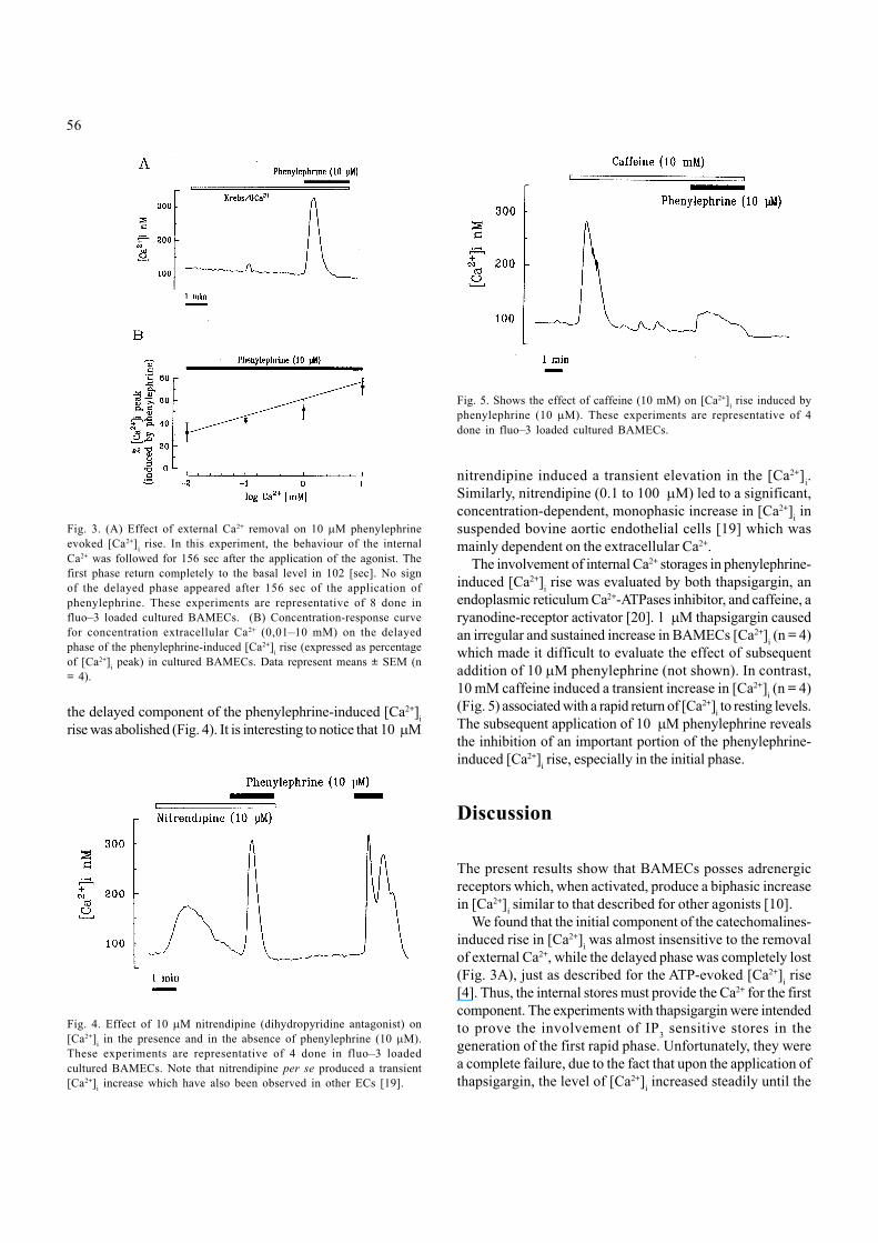
The involvement of internal Ca2+ storages in phenylephrine-induced [Ca2+]
i rise was evaluated by both thapsigargin, an
endoplasmic reticulum Ca2+-ATPases inhibitor, and caffeine, aryanodine-receptor activator [20]. 1 µM thapsigargin causedan irregular and sustained increase in BAMECs [Ca2+]
i (n = 4)
which made it difficult to evaluate the effect of subsequentaddition of 10 µM phenylephrine (not shown). In contrast,10 mM caffeine induced a transient increase in [Ca2+]
i (n = 4)
(Fig. 5) associated with a rapid return of [Ca2+]i to resting levels.
The subsequent application of 10 µM phenylephrine revealsthe inhibition of an important portion of the phenylephrine-induced [Ca2+]
i rise, especially in the initial phase.
Discussion
The present results show that BAMECs posses adrenergicreceptors which, when activated, produce a biphasic increasein [Ca2+]
i similar to that described for other agonists [10].
We found that the initial component of the catechomalines-induced rise in [Ca2+]
i was almost insensitive to the removal
of external Ca2+, while the delayed phase was completely lost(Fig. 3A), just as described for the ATP-evoked [Ca2+]
i rise
[4]. Thus, the internal stores must provide the Ca2+ for the firstcomponent. The experiments with thapsigargin were intendedto prove the involvement of IP
3 sensitive stores in the
generation of the first rapid phase. Unfortunately, they werea complete failure, due to the fact that upon the application ofthapsigargin, the level of [Ca2+]
i increased steadily until the
Fig. 3. (A) Effect of external Ca2+ removal on 10 µM phenylephrineevoked [Ca2+]i rise. In this experiment, the behaviour of the internalCa2+ was followed for 156 sec after the application of the agonist. Thefirst phase return completely to the basal level in 102 [sec]. No signof the delayed phase appeared after 156 sec of the application ofphenylephrine. These experiments are representative of 8 done influo–3 loaded cultured BAMECs. (B) Concentration-response curvefor concentration extracellular Ca2+ (0,01–10 mM) on the delayedphase of the phenylephrine-induced [Ca2+]
i rise (expressed as percentage
of [Ca2+]i peak) in cultured BAMECs. Data represent means ± SEM (n= 4).
Fig. 4. Effect of 10 µM nitrendipine (dihydropyridine antagonist) on[Ca2+]i in the presence and in the absence of phenylephrine (10 µM).These experiments are representative of 4 done in fluo–3 loadedcultured BAMECs. Note that nitrendipine per se produced a transient[Ca2+]i increase which have also been observed in other ECs [19].
Fig. 5. Shows the effect of caffeine (10 mM) on [Ca2+]i rise induced byphenylephrine (10 µM). These experiments are representative of 4done in fluo–3 loaded cultured BAMECs.

57
cells deteriorated. On the other hand, our results show thatthe caffeine-sensitive stores are involved in this phase.Accordingly, if these stores are depleted previously to thetreatment with the agonist, a substantial inhibition of the[Ca2+]
i rise, especially in the initial phase, is shown.
The second, delayed phase in the rise of [Ca2+]i is fully
dependent on the presence of extracellular Ca2+ (Fig. 3). Theroute for the Ca2+entry in ECs is a matter of debate. Evidenceshows that the delayed phase of agonist-induced [Ca2+]
i rise
often involves nonselective cation channels [10]. It has alsobeen postulated a mechanism of Ca2+ entry into the cells whichis triggered by the depletion of intracellular Ca2+ stores [21].This does not seem to be the case, at least for caffeine-sensitive stores (Fig. 5). However, in BAMECs an additionalpathway for Ca2+ entry should be considered. That is to say,VDCCs which were demonstrated in these cells by Bossu etal [11] and further studied by Vinet et al [13]. The laterdemonstrated the presence of T- and L-type VDCCs whichwere shown to resemble those channels in endocrine secretorycells. Since L-type VDCCs are mainly involved in [Ca2+]
i
increase, we aimed to evaluate the participation of thesechannels in the generation of the later phase. For this purposewe used the drug nitrendipine a well known inhibitor of L-channels. Fig. 4 shows that nitrendipine suppressed thedelayed phase of [Ca2+]
i rise which suggest that L-type VDCCs
are involved in the generation of this later component.Nontheless, we cannot discard the possibility of some non-specific action of nitrendipine [22] at the high concentrationused (10 µM), which was necessary to completely inhibit theBAMECs delayed phase.
Our data suggest that the adrenergic receptors involvedare of the α
1-type. Indeed, the biphasic response elicited by
catecholamines is mimicked by the α1-agonist phenylephrine.
The response to phenylephrine and to catecholamines wasblocked by the α-antagonist phenoxybenzamine. Neitherisoproterenol (β-adrenargic agonist) nor clonidine (α
2-ad-
renergic agonist) were able to induce a rise in [Ca2+]i. The effects
of α- and β-adrenergic receptors antagonists on epinephrineand norepinephrine induced [Ca2+]
i rise, also confirm the α
1-
nature of the involved receptors.The [Ca2+]
i rise provoked by catecholamines on BAMECs
suggest that the ECs in the intact gland may respond tocatecholamines released from neighboring chromaffin cells.Since the distance between chromaffin cells and the vascularendothelium is as little as 100–200 nm with only a thin basementmembrane separating these two cell types [23], one mightpresume that catecholamines released from chromaffin cellsbathes the adjacent ECs at high concentrations and thatBAMECs are so activated. The precedent assumption needsa prove which, at present, is beyond the technical possibilitiesof our laboratory. The authors think that a cocultivation ofchromaffin cells with BAMECs might allow them to activatethe chromaffin cells (by means of patch-clamping) and
simultaneously detect the [Ca2+]i rise in the ECs, a project
which is now under consideration. Following with ourspeculation, if an increase in [Ca2+]
i in BAMECs is admitted,
then it follows that a cellular signal for the production ofvasoactive substances such us PGI
2 [24, 25, 26] and NO [24,
27, 28] (which tend to vasodilatation) is generated. Truecapillaries are not surrounded by smooth muscles and,therefore, one should not expect that the secreted cate-cholamines act on them reducing their diameters. But venules,which follow, do have smooth muscle cells around them.Therefore, if only an increase in catecholamines were produced,one might expect a kind of venulo-constriction which wouldimpair the distribution throughout the whole body of thesecreted catecholamines. Opposing to this potential negativeeffect and perhaps overcompensating it may act the activationof the receptors described in this work, thus facilitating theincorporation of the secreted catecholamines into the mainblood stream.
Acknowledgments
This work was supported by grant 1960302 from Fondecyt,Chile and by grant DIPUV–21/99 from Universidad deValparaiso, Chile. We wish to thank Dr. Maritza Rebolledofor provide the adrenal glands.
References
1. Coupland RE, Selby JE: The blood supply of the mammaliamadrenal medulla: A comparative study. J Anat 122: 539–551,1976
2. Ungar A, Phillips JH: Regulation of the adrenal medulla. PhysiolRev 63: 787–843, 1983
3. Rojas E, Pollard HB, Heldman E: Real-time measurements ofacetylcholine-induced release of ATP from bovine medullarychromaffin cells FEBS Lett 185: 323–327, 1985
4. Castro E, Tomé AR, Miras-Portugal MT, Rosário LM: Single-cellfura–2 microfluorometry reveals different purinoceptor subtypescoupled to Ca2+ influx and intracellular Ca2+ release in bovineadrenal chromaffin and endothelial cells. Pflügers Archiv 426:524–533, 1994
5. Forsberg EJ, Feuerstein G, Shohami E, Pollard HB: Adenosinetriphosphate stimulates inositol phospholipid metabolism andprostacyclin formation in adrenal madullary endothelial cells bymeans of P2-purinergic receptors. Proc Natl Acad Sci USA 84:5630–5634, 1987
6. Newby A, Henderson A: Stimulus-secretion coupling in vascularendothelial cells. Annu Rev Physiol 52: 661–674, 1990
7. Moncada S, Gryglewsky R, Bunting S, Vane JR: An enzyme isolatedfrom arteries transforms prostaglandin endoperoxides to anunstable substance that inhibits platelet aggregation. Nature 263:663–665, 1976
8. Furchgott RF, Zawadzki JV: The obligatory role of endothelialcells in the relaxation of arterial smooth muscle by acetylcholine.Nature 288: 373–376, 1980

58
9 . Palmer RM, Ferridge AG, Moncada S: Nitric oxide release accountsfor the biological activity of endothelium-derived relaxing factor.Nature 327: 524–526, 1987
10. Himmel H, Whorton R, Strauss H: Intracellular calcium, currents,and stimulus-response coupling in endothelial cells. Hypertension21: 112–127, 1993
11. Bossu JL, Feltz A, Rodeau JL, Tanzi F: Voltage-dependenttransient calcium currents in freshly dissociated capillaryendothelial cells. FEBS Lett 255: 377–380, 1989
12. Delpiano M, Altura B: Modulatory effect of extracellular Mg2+
ions on K+ and Ca2+ currents of capillary endothelial cell from ratbrain. FEBS Letters 394: 335–339, 1996
13. Vinet R, Vargas FF: L- and T-type voltage-gated Ca2+currents inadrenal medulla endothelial cells. Am J Physiol 276: H1313–1322, 1999
14. Banerjee DK, Ornberg RL, Youdim MB, Heldman E, Pollard HB:Endothelial cells from bovine adrenal medulla develop capillary-like growth patterns in culture. Proc Natl Acad Sci USA 82: 4702–4706, 1985
15. Hoyer LW, de los Santos RP, Hoyer JL: Antihemophilic factorantigen: localization in endothelial cells by immunofluorescentmicroscopy. J Clin Invest 52: 2737–2744, 1973
16. Voyta JC, Via DP, Butterfield CE, Zetter BR. Identification andisolation of endothelial cells based on their increased uptake ofacetylated-low density lipoprotein. J Cell Biol 99: 2034–2040,1984
17. Kao JP, Harootunian AT, Tsien RY: Photochemically generatedcytosolic calcium pulses and their detection by fluo-3. J BiolChem 264: 8171–8178, 1989
18 . Luxoro M, Nassar-Gentina V, Rojas E: Deprivation of Na+,Ca2+ and Mg2+ from the extracellular solution increases cytosolicCa2+ and stimulates catecholamines secretion from culturedbovine adrenal chromaffin cells. Mol Cell Biochem 170: 65–74, 1997
19. Salameh A, Schomecker G, Breitkopf K, Dhein S, Klaus W: The
effect of calciun-antagonist nitrendipine on intracellular calciumconcentration in endothelial cells. Br J Pharmacol 118: 1899–1904, 1996
20. Pozzan T, Rizzuto R, Volpe P, Meldolesi J: Molecular and cellularphysiology of intracellular calcium stores. Physiol Rev 74: 595–636, 1994
21. Gibson A, McFadzean I, Wallace P, Wayman CP: CapacitativeCa2+ entry and the regulation of smooth muscle tone. TrendsPharmacol Sci 19: 266–269, 1998
22. Taylor CW, Broad LM: Pharmacological analysis of intracellularCa2+signalling: problems and pitfalls. Trend Pharmacol Sci 19:370–375, 1998
23. Kikuta A, Murakami T: Relationship between chromaffin cellsand blood vessels in the rat adrenal medulla: a transmission electronmicroscopic study combined with blood vessel reconstructions.Am J Anat 170: 73–81, 1984
24. Lückhoff A, Pohl U, Mülsch A, Busse R: Differential role ofextra- and intracellular calcium in the release of EDRF andprostacyclin from cultured endothelial cells. Br J Pharmacol 95:189–196, 1988
25. Watanabe K, Lan G, Jaffe EA: The correlation between rises inintracellular calcium and PGI
2 production in cultured vascular
endothelial cells. Prostaglandins 46: 211–214, 199226. Hallam TJ, Pearson JD, Needham L: Thrombin stimulated
elevation of human endothelial cell cytoplasmic free calciumconcentration causes prostacyclin production. Biochem J 251:243–249, 1988
27. Lückhoff A, Busse R: Calcium influx into endothelial cells andformation of endothelium-derived relaxing factor is controlledby the membrane potential. Pflügers Arch 416: 305–311, 1990
28. Laskey R, Adams D, Johns A, Rubanyi G, van Breemen C:Membrane potential and N+-K+ pump activity modulate restingand bradykinin-stimulated changes in cytosolic free calcium incultured endothelial cells from bovine atria. J Biol Chem 265:2613–2619, 1990

54
of many agonists – such as acetylcholine, bradykinin, ATP,histamine and thrombine- to their specific receptors on theECs plasma membrane induce the elevation of [Ca2+]
i [10].
These agonist-induced elevations of [Ca2+]i are biphasic in
nature, consisting of a large initial transient followed by adelayed rise of [Ca2+]
i. An important mechanism for the
increase of [Ca2+]i is the extracellular Ca2+ entry mediated by
voltage-dependent Ca2+ channels (VDCC). However, thesechannels are absent in ECs, with the only exception ofmicrovascular ECs from BAMECs [11] and the rat brain [12].Gating and pharmacological characteristics of BAMECsVDCC indicate that the underlying channels are of T- and L-types, resembling those found in endocrine secretory cells[13].
Since ECs that form the walls of adrenomedullary capillariesare thought to play an important role in the export of secretedcatecholamines to the bloodstream, it may be of interest toknow of any interactions between the physiology of thesecells and the substances secreted by the chromaffin cells. Inthis work we have investigated changes in [Ca2+]
i in response
to adrenergic agonists and antagonists in fluo–3 loadedcultured BAMECs. We provide evidence that catecholaminesinduce an increase in BAMECs [Ca2+]
i which is mediated by
α1-adrenergic receptors. We discuss the possible implication
of this finding.
Material and methods
BAMECs culture
Bovine adrenal glands were obtained from the local slaughterhouse, extensively washed and perfused through the adrenalvein with Locke’s solution of the following composition: 154mM NaCl, 5.6 mM KCl, 5.0 mM NaHCO
3, 5.6 mM glucose, 5.0
mM Na-HEPES, 100 U/mL penicillin, 100 µg/mL streptomicin,0.5% albumin, pH 7.2. They were then perfused with Locke’ssolution containing 0.25% collagenase (Böehringer Mannheim)and 0.01% trypsin inhibitor (Sigma) and incubated at 37°C for40 min. Cells dissociated by this procedure were suspended inPercoll (Pharmacia) and centrifuged at 20,000 g for 20 min. Theband containing the highest density of endothelial cells wasplated (cell density 3 x 10⁵ cells per dish) onto circular glasscover slips (Thomas Scientific) placed in dishes (Nunc) in amedium that contained Dulbecco‘s Modified Eagle‘s MediumNutrient Mixture F12 Ham (Sigma) supplemented with 10%fetal calf serum (Biofluids), 50 U/ml penicillin, and 50 µg/mlstreptomycin, pH 7.2. BAMECs relative density in the cellmixture was increased by differential plating, which takesadvantage of BAMECs strong adhesion to glass to removechromaffin and other cells by shaking the culture dish andwashing with culture medium 2–4 h after plating [14]. The cellswere incubated at 37°C in a high humidity atmosphere of 95%
air plus 5% CO2. Experiments were run as early as 12 h, and
not later than 3–4 days after plating.BAMECs were identified by their morphological char-
acteristics, which have been described in great detail usinglight and electron-microscopy [15, 16]. We also checked themorphological identification by measuring BAMECs acety-lated LDL uptake as previously described for endothelial cells[16]. Briefly, BAMECs were incubated with fluorescent Di-I-labeled acetylated LDL for 4 h at 37°C. Cell fluorescence wasexcited at 540 nm and the emission was directly observedthrough a 580 nm filter with an epifluorescent microscope.
[Ca2+]i measurement
[Ca2+]i was measured in isolated cells using the fluorescent
indicator Fluo–3. BAMECs attached to cover slips wereincubated with 7 µM fluo–3 AM (Molecular Probes) and0.094% pluronic acid in modified Krebs solution (in mM: 140NaCl, 5 KCl, 2.6 CaCl
2, 1 MgCl
2, 2.5 NaHCO
3 , 10 glucose, 5
Na-HEPES, pH 7,4) for 20 min. Then, cells were washed withKrebs solution and mounted in a perfusion chamber whichwas placed on the stage of a microscope (Leitz Wetzlar)provided with the appropiate filters (excitation 490 nm;emission 530 nm). The light emitted by Fluo–3 within the cellwas continuosly measured using a photomultiplier (Hamamatsu)and digitalized (3 Hz) employing an analogue converter board(Data Translation) installed in a PC. The data were analyzedusing the software Global Lab (Data Translation). Theconversion of the fluorescent signals to [Ca2+]
i was accom-
plished as described elsewhere [17, 18]. All experiments werecarried out at least four times with consistent results.
Drugs
Epinephrine, norepinephrine, isoproterenol and thapsigarginwere obtained from Sigma. Phenylephrine, phenoxybenzamine,clonidine, propranolol and caffeine were obtained from RBI.
Statistical analysis
Where appropriate, the results are expressed as the mean ±standard error, SEM. Statistical differences were assessed withStudent’s t test, considering values of p < 0.05 as significant.‘n’ represents the number of glass slips examined.
Results
Superfusion of 100 µM epinephrine induced a rise in [Ca2+]i
characterized by an initial transient phase followed by a

55
delayed phase (n = 12) (Fig. 1A). While, the initial phase wascomposed by a simple [Ca2+]
i spike, the delayed phase was
usually described by several [Ca2+]i oscillations. At the time
when epinephrine superfusion was ended, the [Ca2+]i returned
to baseline levels within 1–2 min. As with epinephrine, 100µΜ norepinephrine induced a similar effect on BAMECs[Ca2+]
i (n = 8) (not shown).
[Ca2+]i rise is associated with stimulation of alfa
1-adrenergic
receptors. To identify the receptor involved in the cate-cholamine-induced [Ca2+]
i rise in BAMECs, we evaluated the
effect of adrenergic agonists and antagonists on [Ca2+]i levels.
The [Ca2+]i rise induced by epinephrine (Fig. 1B) was totally
blocked by 10 µM phenoxybenzamine (α-adrenergic anta-gonist), but it was not affected by 10 µM propranolol (β-adrenergic antagonist) (n = 4). Similarly, 10 µM phenylephrine(α
1-adrenergic agonist) (Fig. 2A) reproduced the biphasic
changes in BAMECs [Ca2+]i (n = 37) previously described for
epinephrine and norepinephrine. Neither 10 µM isoproterenol(β-adrenergic agonist) (n = 4) (Fig. 2B), nor 10 µM clonidine(α
2-adrenergic agonist) (n = 4) (not shown) were able to
produce a [Ca2+]i increase. Additionally, 10 µM phenoxy-
benzamine (α-adrenergic antagonist) abolished the phen-ylephrine-induced [Ca2+]
i rise (n = 4) (Fig. 2C).
Sources of Ca2+ for phenylephrine-induced [Ca2+]i rise. In
order to investigate the participation of external Ca2+ in theresponse to phenylephrine, we used Ca2+-free external solu-tions. Superfusion of Ca2+-free Krebs abolished the delayedphase of the phenylephrine-induced [Ca2+]
i rise, indicating the
dependence of this phase on external Ca2+ availability (n = 8)(Fig. 3A). The magnitude of this component was dependenton extracellular Ca2+ concentration as shown in Fig. 3B (n =4).
No inhibitory effect on the delayed phase of the phenyle-phrine-evoked response was observed when 1 µM nitrendipinewas assayed. However, when 10 µM nitrendipine was used,
Fig. 1. Effect of epinephrine on BAMECs [Ca2+]i levels. (A)Representative recording of [Ca2+]i changes induced by 100 µMepinephrine as a function of time in cultured BAMECs. Fluo–3fuorescence was recorded from single cells stimulated by epinephrineduring 1–2 minutes. These experiments are representative of 12 donein BAMECs. One sees that the decline in the first phase is changedinto the rise of the second phase after 30 sec of the application ofepinephrine, which indicates that the delayed phase is initiated earlier.(B) Rise in [Ca2+]i induced by 100 µM epinephrine is totally blockedby 10 µM phenoxybenzamine. Record representative of 4 experimentsdone in BAMECs. Similar results were obtained with norephinephrine.
Fig. 2. Effect of adrenergic agonists and antagonists on [Ca2+]i levels.BAMECs loaded with fluo–3 were exposed to: (A) 10 µM phenylephrine(α1-adrenergic agonist). These experiments are representative of 37done in BAMECs. (B) 10 µM isoproterenol (β-adrenergic agonist).(C) 10 µM phenoxybenzamine (α-adrenergic antagonist) in thepresence of 10 µM phenylephrine Experiments (B) and (C) arerepresentative of 4 done in BAMECs.

56
the delayed component of the phenylephrine-induced [Ca2+]i
rise was abolished (Fig. 4). It is interesting to notice that 10 µM
nitrendipine induced a transient elevation in the [Ca2+]i.
Similarly, nitrendipine (0.1 to 100 µM) led to a significant,concentration-dependent, monophasic increase in [Ca2+]
i in
suspended bovine aortic endothelial cells [19] which wasmainly dependent on the extracellular Ca2+.
The involvement of internal Ca2+ storages in phenylephrine-induced [Ca2+]
i rise was evaluated by both thapsigargin, an
endoplasmic reticulum Ca2+-ATPases inhibitor, and caffeine, aryanodine-receptor activator [20]. 1 µM thapsigargin causedan irregular and sustained increase in BAMECs [Ca2+]
i (n = 4)
which made it difficult to evaluate the effect of subsequentaddition of 10 µM phenylephrine (not shown). In contrast,10 mM caffeine induced a transient increase in [Ca2+]
i (n = 4)
(Fig. 5) associated with a rapid return of [Ca2+]i to resting levels.
The subsequent application of 10 µM phenylephrine revealsthe inhibition of an important portion of the phenylephrine-induced [Ca2+]
i rise, especially in the initial phase.
Discussion
The present results show that BAMECs posses adrenergicreceptors which, when activated, produce a biphasic increasein [Ca2+]
i similar to that described for other agonists [10].
We found that the initial component of the catechomalines-induced rise in [Ca2+]
i was almost insensitive to the removal
of external Ca2+, while the delayed phase was completely lost(Fig. 3A), just as described for the ATP-evoked [Ca2+]
i rise
[4]. Thus, the internal stores must provide the Ca2+ for the firstcomponent. The experiments with thapsigargin were intendedto prove the involvement of IP
3 sensitive stores in the
generation of the first rapid phase. Unfortunately, they werea complete failure, due to the fact that upon the application ofthapsigargin, the level of [Ca2+]
i increased steadily until the
Fig. 3. (A) Effect of external Ca2+ removal on 10 µM phenylephrineevoked [Ca2+]i rise. In this experiment, the behaviour of the internalCa2+ was followed for 156 sec after the application of the agonist. Thefirst phase return completely to the basal level in 102 [sec]. No signof the delayed phase appeared after 156 sec of the application ofphenylephrine. These experiments are representative of 8 done influo–3 loaded cultured BAMECs. (B) Concentration-response curvefor concentration extracellular Ca2+ (0,01–10 mM) on the delayedphase of the phenylephrine-induced [Ca2+]
i rise (expressed as percentage
of [Ca2+]i peak) in cultured BAMECs. Data represent means ± SEM (n= 4).
Fig. 4. Effect of 10 µM nitrendipine (dihydropyridine antagonist) on[Ca2+]i in the presence and in the absence of phenylephrine (10 µM).These experiments are representative of 4 done in fluo–3 loadedcultured BAMECs. Note that nitrendipine per se produced a transient[Ca2+]i increase which have also been observed in other ECs [19].
Fig. 5. Shows the effect of caffeine (10 mM) on [Ca2+]i rise induced byphenylephrine (10 µM). These experiments are representative of 4done in fluo–3 loaded cultured BAMECs.
57
cells deteriorated. On the other hand, our results show thatthe caffeine-sensitive stores are involved in this phase.Accordingly, if these stores are depleted previously to thetreatment with the agonist, a substantial inhibition of the[Ca2+]
i rise, especially in the initial phase, is shown.
The second, delayed phase in the rise of [Ca2+]i is fully
dependent on the presence of extracellular Ca2+ (Fig. 3). Theroute for the Ca2+entry in ECs is a matter of debate. Evidenceshows that the delayed phase of agonist-induced [Ca2+]
i rise
often involves nonselective cation channels [10]. It has alsobeen postulated a mechanism of Ca2+ entry into the cells whichis triggered by the depletion of intracellular Ca2+ stores [21].This does not seem to be the case, at least for caffeine-sensitive stores (Fig. 5). However, in BAMECs an additionalpathway for Ca2+ entry should be considered. That is to say,VDCCs which were demonstrated in these cells by Bossu etal [11] and further studied by Vinet et al [13]. The laterdemonstrated the presence of T- and L-type VDCCs whichwere shown to resemble those channels in endocrine secretorycells. Since L-type VDCCs are mainly involved in [Ca2+]
i
increase, we aimed to evaluate the participation of thesechannels in the generation of the later phase. For this purposewe used the drug nitrendipine a well known inhibitor of L-channels. Fig. 4 shows that nitrendipine suppressed thedelayed phase of [Ca2+]
i rise which suggest that L-type VDCCs
are involved in the generation of this later component.Nontheless, we cannot discard the possibility of some non-specific action of nitrendipine [22] at the high concentrationused (10 µM), which was necessary to completely inhibit theBAMECs delayed phase.
Our data suggest that the adrenergic receptors involvedare of the α
1-type. Indeed, the biphasic response elicited by
catecholamines is mimicked by the α1-agonist phenylephrine.
The response to phenylephrine and to catecholamines wasblocked by the α-antagonist phenoxybenzamine. Neitherisoproterenol (β-adrenargic agonist) nor clonidine (α
2-ad-
renergic agonist) were able to induce a rise in [Ca2+]i. The effects
of α- and β-adrenergic receptors antagonists on epinephrineand norepinephrine induced [Ca2+]
i rise, also confirm the α
1-
nature of the involved receptors.The [Ca2+]
i rise provoked by catecholamines on BAMECs
suggest that the ECs in the intact gland may respond tocatecholamines released from neighboring chromaffin cells.Since the distance between chromaffin cells and the vascularendothelium is as little as 100–200 nm with only a thin basementmembrane separating these two cell types [23], one mightpresume that catecholamines released from chromaffin cellsbathes the adjacent ECs at high concentrations and thatBAMECs are so activated. The precedent assumption needsa prove which, at present, is beyond the technical possibilitiesof our laboratory. The authors think that a cocultivation ofchromaffin cells with BAMECs might allow them to activatethe chromaffin cells (by means of patch-clamping) and
simultaneously detect the [Ca2+]i rise in the ECs, a project
which is now under consideration. Following with ourspeculation, if an increase in [Ca2+]
i in BAMECs is admitted,
then it follows that a cellular signal for the production ofvasoactive substances such us PGI
2 [24, 25, 26] and NO [24,
27, 28] (which tend to vasodilatation) is generated. Truecapillaries are not surrounded by smooth muscles and,therefore, one should not expect that the secreted cate-cholamines act on them reducing their diameters. But venules,which follow, do have smooth muscle cells around them.Therefore, if only an increase in catecholamines were produced,one might expect a kind of venulo-constriction which wouldimpair the distribution throughout the whole body of thesecreted catecholamines. Opposing to this potential negativeeffect and perhaps overcompensating it may act the activationof the receptors described in this work, thus facilitating theincorporation of the secreted catecholamines into the mainblood stream.
Acknowledgments
This work was supported by grant 1960302 from Fondecyt,Chile and by grant DIPUV–21/99 from Universidad deValparaiso, Chile. We wish to thank Dr. Maritza Rebolledofor provide the adrenal glands.
References
1. Coupland RE, Selby JE: The blood supply of the mammaliamadrenal medulla: A comparative study. J Anat 122: 539–551,1976
2. Ungar A, Phillips JH: Regulation of the adrenal medulla. PhysiolRev 63: 787–843, 1983
3. Rojas E, Pollard HB, Heldman E: Real-time measurements ofacetylcholine-induced release of ATP from bovine medullarychromaffin cells FEBS Lett 185: 323–327, 1985
4. Castro E, Tomé AR, Miras-Portugal MT, Rosário LM: Single-cellfura–2 microfluorometry reveals different purinoceptor subtypescoupled to Ca2+ influx and intracellular Ca2+ release in bovineadrenal chromaffin and endothelial cells. Pflügers Archiv 426:524–533, 1994
5. Forsberg EJ, Feuerstein G, Shohami E, Pollard HB: Adenosinetriphosphate stimulates inositol phospholipid metabolism andprostacyclin formation in adrenal madullary endothelial cells bymeans of P2-purinergic receptors. Proc Natl Acad Sci USA 84:5630–5634, 1987
6. Newby A, Henderson A: Stimulus-secretion coupling in vascularendothelial cells. Annu Rev Physiol 52: 661–674, 1990
7. Moncada S, Gryglewsky R, Bunting S, Vane JR: An enzyme isolatedfrom arteries transforms prostaglandin endoperoxides to anunstable substance that inhibits platelet aggregation. Nature 263:663–665, 1976
8. Furchgott RF, Zawadzki JV: The obligatory role of endothelialcells in the relaxation of arterial smooth muscle by acetylcholine.Nature 288: 373–376, 1980

58
9 . Palmer RM, Ferridge AG, Moncada S: Nitric oxide release accountsfor the biological activity of endothelium-derived relaxing factor.Nature 327: 524–526, 1987
10. Himmel H, Whorton R, Strauss H: Intracellular calcium, currents,and stimulus-response coupling in endothelial cells. Hypertension21: 112–127, 1993
11. Bossu JL, Feltz A, Rodeau JL, Tanzi F: Voltage-dependenttransient calcium currents in freshly dissociated capillaryendothelial cells. FEBS Lett 255: 377–380, 1989
12. Delpiano M, Altura B: Modulatory effect of extracellular Mg2+
ions on K+ and Ca2+ currents of capillary endothelial cell from ratbrain. FEBS Letters 394: 335–339, 1996
13. Vinet R, Vargas FF: L- and T-type voltage-gated Ca2+currents inadrenal medulla endothelial cells. Am J Physiol 276: H1313–1322, 1999
14. Banerjee DK, Ornberg RL, Youdim MB, Heldman E, Pollard HB:Endothelial cells from bovine adrenal medulla develop capillary-like growth patterns in culture. Proc Natl Acad Sci USA 82: 4702–4706, 1985
15. Hoyer LW, de los Santos RP, Hoyer JL: Antihemophilic factorantigen: localization in endothelial cells by immunofluorescentmicroscopy. J Clin Invest 52: 2737–2744, 1973
16. Voyta JC, Via DP, Butterfield CE, Zetter BR. Identification andisolation of endothelial cells based on their increased uptake ofacetylated-low density lipoprotein. J Cell Biol 99: 2034–2040,1984
17. Kao JP, Harootunian AT, Tsien RY: Photochemically generatedcytosolic calcium pulses and their detection by fluo-3. J BiolChem 264: 8171–8178, 1989
18 . Luxoro M, Nassar-Gentina V, Rojas E: Deprivation of Na+,Ca2+ and Mg2+ from the extracellular solution increases cytosolicCa2+ and stimulates catecholamines secretion from culturedbovine adrenal chromaffin cells. Mol Cell Biochem 170: 65–74, 1997
19. Salameh A, Schomecker G, Breitkopf K, Dhein S, Klaus W: The
effect of calciun-antagonist nitrendipine on intracellular calciumconcentration in endothelial cells. Br J Pharmacol 118: 1899–1904, 1996
20. Pozzan T, Rizzuto R, Volpe P, Meldolesi J: Molecular and cellularphysiology of intracellular calcium stores. Physiol Rev 74: 595–636, 1994
21. Gibson A, McFadzean I, Wallace P, Wayman CP: CapacitativeCa2+ entry and the regulation of smooth muscle tone. TrendsPharmacol Sci 19: 266–269, 1998
22. Taylor CW, Broad LM: Pharmacological analysis of intracellularCa2+signalling: problems and pitfalls. Trend Pharmacol Sci 19:370–375, 1998
23. Kikuta A, Murakami T: Relationship between chromaffin cellsand blood vessels in the rat adrenal medulla: a transmission electronmicroscopic study combined with blood vessel reconstructions.Am J Anat 170: 73–81, 1984
24. Lückhoff A, Pohl U, Mülsch A, Busse R: Differential role ofextra- and intracellular calcium in the release of EDRF andprostacyclin from cultured endothelial cells. Br J Pharmacol 95:189–196, 1988
25. Watanabe K, Lan G, Jaffe EA: The correlation between rises inintracellular calcium and PGI
2 production in cultured vascular
endothelial cells. Prostaglandins 46: 211–214, 199226. Hallam TJ, Pearson JD, Needham L: Thrombin stimulated
elevation of human endothelial cell cytoplasmic free calciumconcentration causes prostacyclin production. Biochem J 251:243–249, 1988
27. Lückhoff A, Busse R: Calcium influx into endothelial cells andformation of endothelium-derived relaxing factor is controlledby the membrane potential. Pflügers Arch 416: 305–311, 1990
28. Laskey R, Adams D, Johns A, Rubanyi G, van Breemen C:Membrane potential and N+-K+ pump activity modulate restingand bradykinin-stimulated changes in cytosolic free calcium incultured endothelial cells from bovine atria. J Biol Chem 265:2613–2619, 1990


![Separate [3H]-nitrendipine binding sites in mitochondria and plasma membranes of bovine adrenal medulla](https://static.fdokumen.com/doc/165x107/63443fe2df19c083b10781df/separate-3h-nitrendipine-binding-sites-in-mitochondria-and-plasma-membranes-of.jpg)