Correlates of smoking quit attempts: Florida Tobacco Callback Survey, 2007
Upload
independentCategory
view
4download
0
Schizophrenia Research 125 (2011) 30–40
Contents lists available at ScienceDirect
Schizophrenia Research
j ourna l homepage: www.e lsev ie r.com/ locate /schres
Suicidal attempts and increased right amygdala volume in schizophrenia
Ilaria Spoletini a, Fabrizio Piras a, Sabrina Fagioli a, Ivo Alex Rubino b, Giovanni Martinotti c,Alberto Siracusano b, Carlo Caltagirone a,b, Gianfranco Spalletta a,⁎a IRCCS Santa Lucia Foundation, Rome, Italyb “Tor Vergata” University, Dept. of Neuroscience, Rome, Italyc Catholic University of the Sacred Heart, Institute of Psychiatry, Rome, Italy
a r t i c l e i n f o
⁎ Corresponding author. IRCCS Santa Lucia FounClinical and Behavioural Neurology, Via Ardeatina, 30Tel.: +39 06 51501575; fax: +39 06 51501575.
E-mail address: [email protected] (G. Spall
0920-9964/$ – see front matter © 2010 Elsevier B.V.doi:10.1016/j.schres.2010.08.023
a b s t r a c t
Article history:Received 14 January 2010Received in revised form 30 July 2010Accepted 12 August 2010Available online 25 September 2010
Suicide is amajor cause of death in schizophrenia. Neurobiological studies suggest that suicidalityis associated with abnormal brain structure and connectivity in fronto-temporo-limbic regions.However, it is still unclear whether suicidality in schizophrenia is related to volumetricabnormalities in subcortical structures that play a key role in emotion regulation, aggressionand impulse control. Therefore, we aimed to examine whether the volume of selected subcorticalregions is associated with previous suicidal attempts and self-aggression in schizophrenia.For this cross-sectional study, we recruited 50 outpatients with schizophrenia and 50 healthycontrols (HC) matched for age and gender. Fourteen patients had a history of one or moresuicide attempts. Different forms of aggression were assessed using the Modified OvertAggression Scale. All participants underwent structural MR imaging at 3 Tesla. Physicalvolumetric measures were calculated for the lateral ventricles, thalamus, hippocampus,amygdala, caudate, putamen, pallidum and accumbens using an automatic segmentationmethod on T1-weighted high-resolution (voxel size 1×1×1 mm3) images.Multivariate and follow-up univariate ANOVAs revealed a selective increase in volume in theright amygdala of patients with a history of suicidality compared both to patients without sucha history and HC. Moreover, in the entire patient group increased right amygdala volume wasrelated to increased self-aggression.Our findings suggest that right amygdala hypertrophy may be a risk factor for suicide attemptsin patients with schizophrenia and this could be relevant for suicide prevention.
© 2010 Elsevier B.V. All rights reserved.
Keywords:SchizophreniaSubcortical structuresAmygdalaSuicideSelf-aggressionVolumetry
1. Introduction
Suicide is a complex multifactorial phenomenon oftenassociated with psychiatric disorders (Beautrais et al., 1996).Indeed, about 90% of all individuals who kill themselves havediagnosable mental illnesses, particularly mood disorders(Health, 1998). Furthermore, suicide is a major cause of deathin patients with schizophrenia who are at risk for committingsuicide. This risk, which is eightfold that in the general
dation, Laboratory o6. 00179 Rome, Italy
etta).
All rights reserved.
f.
population (Harris and Barraclough, 1998), remains elevatedacross the entire course of the illness (Heila et al., 1997).
Notably, about 10% of patients with a schizophreniadiagnosis die from suicide and up to 40% attempt to commitsuicide (Pompili et al., 2007). Suicide attempts are a far morecommon phenomenon in schizophrenia than completedsuicides. A history of previous attempt(s) is considered one ofthe strongest predictors of completed suicide in schizophreniapatients (Hawton et al., 2005).
A suicide attempt can be defined as a self-aggressivebehavior associated with the intent of dying (Turecki, 2005).Indeed, aggressiveness, impulsivity and suicidality are stronglyrelated (Hong et al., 2004; Rusch et al., 2008). Therefore,impulsive/self-aggressive traits, as well as depression (Hawton

31I. Spoletini et al. / Schizophrenia Research 125 (2011) 30–40
et al., 2005), appear to be relevant clinical variables associatedwith suicidality (Giegling et al., 2009; Turecki, 2005).
However, the clinical correlates of suicide are heteroge-neous and are present in a large number of patients who willnot commit suicide (Aguilar et al., 2003). Thus, a neurobio-logical approach has been proposed to improve our under-standing of the pathogenesis of suicidality and self-aggressionin schizophrenia. The aim of this approach is to improveaccuracy in detecting high-risk patients (Naudts and Hodgins,2006). In this view, suicidal behaviors are thought to berooted in an underlying neurobiological susceptibility we arejust beginning to understand, with preliminary informationarising from post-mortem and neuroimaging studies. Partic-ularly, post-mortem studies have indicated a potential role ofneuroinflammatory processes in brain areas such as thedorsolateral prefrontal cortex and other cortical and subcor-tical structures in suicidal patients (Bernstein et al., 2009;Steiner et al., 2008). Consistently, brain imaging studies havepointed out the role of the prefrontal cortices as mediators ofbehavioral impulsivity (Mann, 2003). Indeed, current neuro-anatomic circuit models for aggression, impulsivity andmoodregulation focus on pathways connecting the frontal corticeswith subcortical structures including the amygdala, basalganglia, thalamus, and others (Mega and Cummings, 1994).Accordingly, reduced white matter integrity in the inferiorfrontal areas and fronto-temporo-limbic circuits associatedwith increased impulsivity (Hoptman et al., 2002, 2004) hasbeen described in schizophrenia. Also, lower connectivity inthe right amygdala/ventral prefrontal circuitry has beenfound to be associated with greater aggression (Hoptmanet al., 2009). Volumetric studies show that individuals with ahistory of violent offences have reduced amygdala volume(Wong et al., 1997) and patients with schizophrenia whohave a violent history display reduced hippocampal andincreased putamen volumes (Barkataki et al., 2006). Finally, ithas been found that a higher level of aggression is related toincreased orbito-frontal gray and white matter volume(Hoptman et al., 2005) and increased caudate volume(Hoptman et al., 2006).
In a previous study on suicide, we (Rusch et al., 2008)found significantly larger inferior frontal white mattervolume bilaterally in patients with a history of suicideattempts and a direct relationship between this white matterarea and level of self-aggression. In another study, Aguilaret al. (2008) found reduced gray matter density in leftsuperior temporal and orbitofrontal cortices in suicide-attempt patients compared to non-suicidal patients. There-fore, converging evidence points to the role of the frontalcortices and their connections in this extremely dangerousbehavior. However, the direction of this gray-white matterabnormality is not clear.
As subcortical abnormalities are well-described inschizophrenia (Ananth et al., 2002; Spoletini et al., 2009),it is surprising that the extent to which the subcorticalstructures are related to suicidality is still unexplored,particularly in light of their key role in emotion regulationand impulse control due to their connectionswith the frontalcortices. Indeed, in schizophrenia altered subcortical struc-tures are implicated in aggression (Hoptman et al., 2006,2009), violence (Barkataki et al., 2006; Wong et al., 1997)and impulsivity (Hoptman et al., 2002, 2004); however, as
yet no studies have explored the issue of suicidality. Inparticular, the amygdala and the hippocampus are keycomponents in the neural circuitry that mediates stressresponses. This is highly relevant in suicide, as demonstratedin schizophrenia (Pompili et al., 2007; Steiner et al., 2008).Therefore, the limbic system might be implicated inschizophrenia suicides.
With these considerations in mind, we designed a study todetect whether volumes of selected subcortical structures arerelated to past suicidality and lifetime self-aggression inschizophrenia. Based on the hypothesis that structural changesin fronto-temporo-limbic circuits are crucial for emotionregulation, impulse control and suicidality (Aguilar et al.,2008; Rusch et al., 2008), we expected that volumetric changesin limbic structures, suchas the amygdala and thehippocampus,would characterize patients with a history of suicide attemptsbut not patients without such a history and healthy controlsubjects (HC). As a secondary aim, we assessed the correlationsbetween the subcortical volumes found to be different amonggroups and four different types of aggression (i.e. self-aggression, verbal aggression, aggression against property andaggression against others) in the whole schizophrenic group.Based on our previous work (Rusch et al., 2008), we expectedthat the subcortical volumes found tobedifferent in suicidal andnon-suicidal patients would be associated with the level of self-aggression in the whole schizophrenia group.
2. Materials and methods
2.1. Subjects
Sixty-five consecutive patients with a diagnosis of schizo-phrenia according to the DSM-IV-TR (APA, 2000) wereinitially recruited from two outpatient clinics in centralItaly. Specifically, two clinicians (I.A.R. and G.M.) who weretreating the patients and knew their clinical history made apreliminary diagnosis of schizophrenia. The clinicians wereblind to the aims of the study. All diagnoses were thenconfirmed or not on the day of image acquisition by anotherclinical psychiatrist (G.S.) using the structured clinicalinterview for DSM-IV-TR (First et al., 2002a). The thirdpsychiatrist also screened the patients to see whether theyfulfilled the inclusion or the exclusion criteria of the study. Inthe case of disagreement between the three clinicians, moredata were requested to help resolve differences, and thediagnostic process continued until a final consensus diagnosiswas assigned. If there was still disagreement among the threediagnosticians, the patient was removed from the sample. Theinterrater reliability of the three psychiatrists was more than0.80 for the diagnosis of schizophrenia.
Informationon clinical history andprevious suicide attemptswas obtained from various sources: the patients, their relatives,their physicians and psychiatrists and their clinical charts.
Age at onset was defined as age at first hospitalization or,where possible, age at onset of positive or negative symptomspreceding the first hospitalization. All patients included werebetween 18 and 65 years of age, were right-handed and hadcompleted at least 5 years of schooling. The inclusioncriterion was a Mini Mental State Examination (MMSE)(Folstein et al., 1975) score equal to or higher than 24 to avoidthe inclusion of patients with global cognitive deterioration,

Table1
Sociod
emog
raph
ican
dglob
alco
gnitivech
aracteristicsof
14pa
tien
tswithschizo
phreniaan
dprev
ious
suicideattempts,36
patien
tswithschizo
phreniawitho
utprev
ious
suicideattempts,an
d50
healthyco
ntrolp
articipa
nts.
Characteristics
SZawithprev
ious
suicideattempts(N
=14
)SZ
witho
utprev
ious
suicideattempts(N
=36
)HC
b(N
=50
)ANOVA
Sche
ffépo
st-h
oc
Mea
n(SD)c
Mea
n(SD)
Mea
n(SD)
F(d
fd:2,97
)p
SZwithprev
ious
suicideattemptsvs
HC
SZwithprev
ious
suicide
attemptsvs
SZwitho
utprev
ious
suicideattempts
SZwitho
utprev
ious
suicideattemptsvs
HC
Age
42.9
(11.3)
39.8
(11.4)
40.0
(16.6)
0.26
70.26
7–
––
Educ
ationa
lleve
l(ye
ars)
12.1
(3.5)
12.2
(2.5)
14.0
(3.2)
4.56
30.01
280.11
270.98
780.03
00
MMSE
escore
28.2
(1.7)
28.4
(1.4)
29.6
(0.7)
13.859
b0.00
010.00
090.85
48b0.00
01
N(%
)N
(%)
N(%
)χ2
(df:2)
p
Gen
der(m
ale)
8(5
7)21
(58)
26(5
2)0.36
90.83
14
Sign
ificant
p-va
lues
(pb0.05
)areindicatedin
bold.
aSchizo
phreniapa
tien
ts.
bHea
lthy
controls.
cStan
dard
deviation.
dDeg
rees
offree
dom.
eMiniM
entalS
tate
Exam
ination.
32 I. Spoletini et al. / Schizophrenia Research 125 (2011) 30–40
in accordance with normative data in the Italian population(Measso et al., 1993).
Exclusion criteria were: history of alcohol or drugdependence or traumatic head injury (identified by conduct-ing interviewswith themedical staff, andwith relatives whenavailable, as well as by consulting the medical charts); anypast or present major medical or neurological illness; anyadditional psychiatric disorder; any brain pathology identi-fied on T1-, T2- and FLAIR-scans; and mental retardationdefined by using DSM-IV-TR criteria. All patients werereceiving stable oral doses of one or more atypical antipsy-chotic drugs such as risperidone, quetiapine, and olanzapine.Antipsychotic dosages were converted to estimated equiva-lent dosages of olanzapine (Oquendo et al., 2003).
From the 65 patients of the initial sample, 15 patientswere excluded based on the above-mentioned exclusioncriteria. Specifically, 8 patients were cognitively impaired, 4patients had a history of alcohol and/or psychoactive drugdependence, and 3 patients were removed from subsequentanalyses because of significant movement artifacts during theMRI procedure. Thus, the final sample consisted of 50schizophrenic patients.
Fourteen patients (28%) had a lifetime history of one ormore suicide attempts, whereas 36 (72%) had never tried tocommit suicide. Specifically, 5 patients (36%) had made onesuicide attempt and 9 patients (64%) had made more thanone suicide attempt (6 patients had attempted suicide twice;1 patient had committed three suicide attempts; 2 patientshad attempted suicide six times). Of the 9 patients who hadmade more than one suicide attempt, five patients had usedonly one suicide method repeatedly, while four patients hadused different methods. Methods included overdosing ofmedication, jumping and wrist cutting. All suicide attemptswere made in adulthood after the onset of schizophrenia, butin every case over 6 months before image acquisition.
Fifty HC subjects were recruited in the same geographicarea and were rigorously matched with the schizophrenicpatients for age and gender (Table 1). All HC were carefullyscreened for a current or past diagnosis of axis I or II disordersusing SCID-I and SCID-II (First et al., 1997, 2002b). Schizo-phrenia or any othermental disorder diagnosis in first-degreerelatives was also an exclusion criterion, as well as the otherabove-mentioned exclusion criteria for patients.
The studywas approved and undertaken in accordancewiththe guidelines of the Santa Lucia Foundation Ethics Committee.Written consent was obtained from all participants after theyhad received a complete explanation of the study procedures.
2.2. Clinical assessment
A clinical psychiatrist (G.S.) rated lifelong self-aggressionbehaviors by administering the self-aggression subscale of theModified Overt Aggression Scale (MOAS) (Kay et al., 1988).Based on the Yudofsky scale, MOASwas developed to assess thenature and prevalence of aggression in a psychiatric population.This scale comprises three additional subscales: verbal aggres-sion, aggression against property and aggression toward others.Each subscale yields a score between 0 and 4, where zerorepresents no aggression and four is the maximum aggressionscore for each subscale. The MOAS measures patient's life-timeaggressive behaviors, classifying aggression by type, level of

33I. Spoletini et al. / Schizophrenia Research 125 (2011) 30–40
severity and frequency of each behavior. In order to rate theaggressive behaviors, the following sources of informationwereused: the interviewwith thepatient and themedical staff and/orrelatives, and the clinical charts.
Psychopathological symptoms were measured using thePositive and Negative Syndrome Scale (PANSS) (Kay et al.,1987), which is a 30-item seven-point rating instrument thatyields scores of positive (7 items, with a total score rangingfrom 7 to 49) and negative (7 items, with a total score rangingfrom 7 to 49) symptoms, and of general psychopathology (16items, with a total score ranging from 16 to 112).
2.3. Image acquisition and processing
The 100 participants underwent the same imagingprotocol, which included 3D T1-weighted, T2-weighted andFLAIR sequences using a 3T Allegra MR imager (Siemens,Erlangen, Germany) with a standard quadrature head coil.
Whole-brain T1-weighted images were obtained in thesagittal plane using a modified driven equilibrium Fouriertransform (MDEFT) sequence (TE/TR=2.4/7.92 ms, flip angle15°, voxel size 1×1×1 mm3). T2 and FLAIR sequences wereacquired to screen for brain pathology.
All image processing was performed using FSL 4.1software with procedures similar to those described in aprevious work (Cherubini et al., 2009). Anatomical T1-weighted images were processed with the segmentationtool FIRST 1.2 (Patenaude et al., 2007) which does not requiremanual intervention. This is an automated model-basedsegmentation/registration tool which has been trained tosegment several structures (implemented in the tool) thathave been previously manually segmented by the Center forMorphometric Analysis (CMA), MGH, Boston, MA. Suchstructures, were originally constructed from 336 subjects,consisting of children and adults, normals and subjects withpathologies. They were subsequently parameterized assurface meshes and modeled as a point distribution model.Deformable surfaces are used to automatically parameterizethe volumetric labels in terms of meshes; the deformablesurfaces are constrained to preserve vertex correspondenceacross the training data. Furthermore, normalized intensitiesalong the surface normals are sampled and modeled. Theshape and appearance model is based on multivariateGaussian assumptions. Shape is then expressed as a meanwith modes of variation (principal components). Based onthe learned models, FIRST searches through linear combina-tions of shape modes of variation for the most probable shapeinstance given the intensities observed in the T1 image. Inother words, this tool is optimized to find the optimal borderand extent of the structures considered, modeling thesestructures as surfaces. This method of segmentation isparticularly useful for structures with a low contrast-to-noise ratio and recentmethodological works that compared itwithmanual tracing and other softwares have shown that theperformance of the tool in the segmentation of deep graymatter structures is highly reliable (Babalola et al., 2009;Morey et al., 2009). In each subject the lateral ventricles,thalamus, caudate, putamen, pallidum, hippocampus, amyg-dala and accumbens were segmented. Segmentation resultsfor each participant were visually assessed by a trainedneuroradiologist. As final output, for each subject and each
hemisphere, FIRST calculates the volumes of the eight above-mentioned segmented deep gray matter structures. Beforestatistical comparison, individual volume values were multi-plied by a normalization factor obtained with the SIENAX toolfrom the corresponding T1-weighted image. Briefly, SIENAXstarts by extracting brain and skull images from the single T1-weighted image. The brain image is then affine-registered toMNI152 space using the skull image to determine theregistration scaling; this is primarily in order to obtain thevolumetric scaling factor, to be used as a normalisation forhead size. The scaling factor represents the whole-brainvolume expansion (or contraction) required to register eachindividual to the template. Importantly, when we calculatedthe affine transform to the template we used the skull imagesto constrain scaling and skew.
2.4. Statistical analyses
Data from the 50 patients and 50 HC were included in thestatistical analyses. Patients were divided into two groups:patients with a history of at least one suicide attempt (SA:n=14) and patients without such a history (NSA: n=36).Comparisons among the three groups (i.e. SA, NSA, and HC)on continuous variables (i.e. age, educational level and MMSEscore) were performed using a series of ANOVA. Comparisonsbetween the two patient groups (i.e. SA and NSA) wereperformed using t-tests for continuous variables and the chi-square test for categorical variables (i.e. gender).
To minimize the likelihood of a type-I error, the series ofunivariate ANOVAs was preceded by overall MultivariateAnalyses of Variance (MANOVA) using all subcortical volumevalues as dependent variables. Consequently, one-wayANOVAs (between groups) were run for each subcorticalbrain structure. Post-hoc comparisons were performed usingScheffè's test. A level of significance of pb0.05 was used forcomparative measurements.
Finally, we performed a series of univariate correlationanalyses (Pearson's correlation) between the subcorticalvolume values and the MOAS subscale scores assessingdifferent types of aggression.
3. Results
As expected from the matching procedure, the wholegroup of schizophrenic patients and the HC did not differsignificantly for age (t-value=−0.218, df=98, p=0.8278)or gender (χ2=0.364; df=1, p=0.5465). However, the twogroups differed for educational level (t-value=3.032,df=98, p=0.0031).
Sociodemographic characteristics of the SA, NSA and HCgroup are reported in Table 1. The SA and NSA did not differsignificantly for age, gender, educational level or MMSE score.
Clinical characteristics of SA and NSA are reported in Table2. As expected, the SA obtained a significantly higher score onthe MOAS self-aggression subscale than the NSA. Interest-ingly, the SA also obtained a significantly higher score on theMOAS aggression toward other people subscale than the NSA.Notably, no differences in the other MOAS subscales (i.e.verbal aggression and aggression against property) or in thePANSS scales (i.e. positive, negative and general psychopa-thology) were observed between SA and NSA.

Table 2Clinical characteristics of 14 patients with schizophrenia and previous suicide attempts and 36 patients with schizophrenia without previous suicide attempts.
SZ a with previous suicide attempts(N=14) mean (SD b)
SZ without previous suicide attempts(N=36) mean (SD)
t-value (df c: 48) p
Olanzapine equivalents (mg/day) 31.3 (54.0) 15.1 (15.3) −1.663 0.1028Duration of illness (years) 18.7 (11.1) 12.4 (9.7) −1.991 0.0522MOAS d self-aggression subscale 3.714 (1.069) 0.111 (0.465) −16.741 b0.0001MOAS verbal aggression subscale 1.571 (1.016) 1.139 (1.199) −1.192 0.2392MOAS aggression against property subscale 0.714 (0.994) 0.611 (0.964) −0.337 0.7378MOAS aggression toward other people subscale 1.714 (1.637) 0.639 (1.099) −2.693 0.0097PANSS e positive scale 22.286 (5.483) 22.194 (7.479) −0.041 0.9671PANSS negative scale 20.929 (8.489) 21.889 (7.042) 0.409 0.6846PANSS general psychopathology scale 51.500 (13.916) 47.944 (11.074) −0.948 0.3480
Significant p-values (pb0.05) are indicated in bold.a Schizophrenia patients.b Standard deviation.c Degrees of freedom.d Modified Overt Aggression Scale.e Positive and Negative Syndrome Scale.
34 I. Spoletini et al. / Schizophrenia Research 125 (2011) 30–40
3.1. Volumetric data
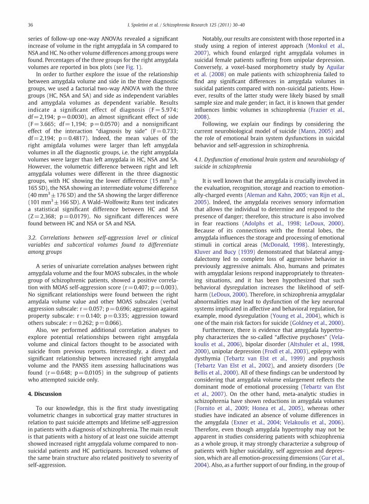
As shown in Fig. 1, an automatic segmentation method(Patenaude et al., 2007) was used to delineate the lateralventricles and the seven subcortical structures (thalamus,
Fig. 1. Regions of interest mapped onto a subject's image. (A) Three-dimensional rethe subcortical structures segmented on the T1-weighted images. The nucleus accumright amygdala volumes of HC, patients with SA and patients without SA are prese
hippocampus, caudate, putamen, pallidum, amygdala, andaccumbens) of each participant.
Table 3 reports volume values of all subcortical structures.A MANOVA using all volume scores as dependent variables(Wilks' λ=0.589; F=1.817; df=28,168; p=0.0114) and a
construction of amygdala volumes; (B) coronal and (C) sagittal slice showingbens does not appear in the selected slices. On the upper left side, box plots onted. HC: healthy controls; SZ: schizophrenia; SA: suicide attempts.
f

Table 3Normalized volumes [mm3] of the subcortical areas in the schizophrenic groups with and without suicide attempts and in the healthy comparison participants.
Area Side SZ a with previoussuicide attempts(N=14)
SZ without previoussuicide attempts(N=36)
HC b (N=50) ANOVA Scheffé post-hoc test
Mean (SD c) Mean (SD) Mean (SD) F(df d: 2,97) p SZ with previous suicideattempts vs HC
SZ with previous suicideattempts vs SZ withoutprevious suicide attempts
SZ without previoussuicide attempts vs HC
Amygdala ⁎ Right 916 (164) 757 (145) 788 (174) 4.858 0.0098 0.0389 0.0106 0.6883Left 815 (139) 717 (150) 773 (191) 1.990 0.1423 – – –
Thalamus Right 5879 (710) 6127 (774) 6297 (687) 1.962 0.1461 – – –
Left 5594 (479) 5739 (764) 5985 (732) 2.211 0.1150 – – –
Hippocampus ⁎⁎ Right 2589 (420) 2437 (394) 2620 (305) 2.866 0.0618 – – –
Left 2384 (345) 2339 (282) 2437 (328) 1.032 0.3601 – – –
Caudate Right 2777 (367) 2643 (421) 2621 (464) 0.706 0.4963 – – –
Left 2645 (368) 2600 (473) 2590 (452) 0.083 0.9208 – – –
Putamen Right 3776 (464) 3659 (512) 3681 (427) 0.579 0.5622 – – –
Left 3491 (461) 3664 (511) 3602 (487) 0.629 0.5354 – – –
Pallidum Right 981 (206) 1015 (240) 1068 (182) 1.255 0.2897 – – –
Left 1005 (213) 1034 (194) 1052 (151) 0.413 0.6625 – – –
Accumbens Right 245 (65) 220 (76) 209 (78) 1.275 0.2841 – – –
Left 313 (103) 313 (99) 282 (89) 1.284 0.2816 – – –
Lateral ventricles ⁎ Right 7714 5032 6278 (3177) 5314 (3281) 2.703 0.0720 – – –
Left 7346 (3783) 6538 (3455) 5392 (2793) 2.638 0.0766 – – –
a Schizophrenia patients.b Healthy controls.c Standard deviation.d Degrees of freedom.⁎ Significant p-values (pb0.05) are indicated in bold.⁎⁎ Trend-level significant p-values (pb0.1) are indicated in italics.
35I.Spoletini
etal./
SchizophreniaResearch
125(2011)
30–40
36 I. Spoletini et al. / Schizophrenia Research 125 (2011) 30–40
series of follow-up one-way ANOVAs revealed a significantincrease of volume in the right amygdala in SA compared toNSA and HC. No other volume differences among groupswerefound. Percentages of the three groups for the right amygdalavolumes are reported in box plots (see Fig. 1).
In order to further explore the issue of the relationshipbetween amygdala volume and side in the three diagnosticgroups, we used a factorial two-way ANOVA with the threegroups (HC, NSA and SA) and side as independent variablesand amygdala volumes as dependent variable. Resultsindicate a significant effect of diagnosis (F=5.974;df=2,194; p=0.0030), an almost significant effect of side(F=3.665; df=1,194; p=0.0570) and a nonsignificanteffect of the interaction “diagnosis by side” (F=0.733;df=2,194; p=0.4817). Indeed, the mean values of theright amigdala volumes were larger than left amygdalavolumes in all the diagnostic groups, i.e. the right amygdalavolumes were larger than left amygdala in HC, NSA and SA.However, the volumetric difference between right and leftamygdala volumes were different in the three diagnosticgroups, with HC showing the lower difference (15 mm3±165 SD), the NSA showing an intermediate volume difference(40 mm3±176 SD) and the SA showing the larger difference(101 mm3±166 SD). A Wald–Wolfowitz Runs test indicatesa statistical significant difference between HC and SA(Z=2,368; p=0.0179). No significant differences werefound between HC and NSA or SA and NSA.
3.2. Correlations between self-aggression level or clinicalvariables and subcortical volumes found to differentiateamong groups
A series of univariate correlation analyses between rightamygdala volume and the four MOAS subscales, in the wholegroup of schizophrenic patients, showed a positive correla-tion with MOAS self-aggression score (r=0.407; p=0.003).No significant relationships were found between the rightamydala volume value and other MOAS subscales (verbalaggression subscale: r=0.057; p=0.696; aggression againstproperty subscale: r=0.140; p=0.335; aggression towardothers subscale: r=0.262; p=0.066).
Also, we performed additional correlation analyses toexplore potential relationships between right amygdalavolume and clinical factors thought to be associated withsuicide from previous reports. Interestingly, a direct andsignificant relationship between increased right amygdalavolume and the PANSS item assessing hallucinations wasfound (r=0.648; p=0.0105) in the subgroup of patientswho attempted suicide only.
4. Discussion
To our knowledge, this is the first study investigatingvolumetric changes in subcortical gray matter structures inrelation to past suicide attempts and lifetime self-aggressionin patients with a diagnosis of schizophrenia. The main resultis that patients with a history of at least one suicide attemptshowed increased right amygdala volume compared to non-suicidal patients and HC participants. Increased volumes ofthe same brain structure also related positively to severity ofself-aggression.
Notably, our results are consistent with those reported in astudy using a region of interest approach (Monkul et al.,2007), which found enlarged right amygdala volumes insuicidal female patients suffering from unipolar depression.Conversely, a voxel-based morphometry study by Aguilaret al. (2008) on male patients with schizophrenia failed tofind any significant differences in amygdala volumes insuicidal patients compared with non-suicidal patients. How-ever, results of the latter study were likely biased by smallsample size and male gender; in fact, it is known that genderinfluences limbic volumes in schizophrenia (Frazier et al.,2008).
Following, we explain our findings by considering thecurrent neurobiological model of suicide (Mann, 2005) andthe role of emotional brain system dysfunctions in suicidalbehavior and self-aggression in schizophrenia.
4.1. Dysfunction of emotional brain system and neurobiology ofsuicide in schizophrenia
It is well known that the amygdala is crucially involved inthe evaluation, recognition, storage and reaction to emotion-ally-charged events (Aleman and Kahn, 2005; van Rijn et al.,2005). Indeed, the amygdala receives sensory informationthat allows the individual to determine and respond to thepresence of danger; therefore, this structure is also involvedin fear reactions (Adolphs et al., 1998; LeDoux, 2000).Because of its connections with the frontal lobes, theamygdala influences the storage and processing of emotionalstimuli in cortical areas (McDonald, 1998). Interestingly,Kluver and Bucy (1939) demonstrated that bilateral amyg-dalectomy led to complete loss of aggressive behavior inpreviously aggressive animals. Also, humans and primateswith amygdalar lesions respond inappropriately to threaten-ing situations, and it has been hypothesized that suchbehavioral dysregulation increases the likelihood of self-harm (LeDoux, 2000). Therefore, in schizophrenia amygdalarabnormalities may lead to dysfunction of the key neuronalsystems implicated in affective and behavioral regulation, forexample, mood dysregulation (Young et al., 2004), which isone of the main risk factors for suicide (Goldney et al., 2000).
Furthermore, there is evidence that amygdala hypertro-phy characterizes the so-called “affective psychoses” (Vela-koulis et al., 2006), bipolar disorder (Altshuler et al., 1998,2000), unipolar depression (Frodl et al., 2003), epilepsy withdysthymia (Tebartz van Elst et al., 1999) and psychosis(Tebartz Van Elst et al., 2002), and anxiety disorders (DeBellis et al., 2000). All of these findings can be understood byconsidering that amygdala volume enlargement reflects thedominant mode of emotional processing (Tebartz van Elstet al., 2007). On the other hand, meta-analytic studies inschizophrenia have shown reductions in amygdala volumes(Fornito et al., 2009; Honea et al., 2005), whereas otherstudies have indicated an absence of volume differences inthe amygdala (Exner et al., 2004; Velakoulis et al., 2006).Therefore, even though amygdala hypertrophy may not beapparent in studies considering patients with schizophreniaas a whole group, it may strongly characterize a subgroup ofpatients with higher suicidality, self aggression and depres-sion, which are all emotion-processing dimensions (Gur et al.,2004). Also, as a further support of our finding, in the group of

37I. Spoletini et al. / Schizophrenia Research 125 (2011) 30–40
patients who attempted suicide, right amygdala hypertrophywas directly related to severity of hallucinations, which are arecognized risk factor of suicide in schizophrenia (Lui, 2009;Montross et al., 2005).
It could be argued that increased amygdala volume maybe linked to suicidality also on the basis of f-MRI studiesshowing a direct relationship between increased amygdalaactivation and suicidal behaviors (Young et al., 2004; Alemanand Kahn, 2005). The link between amygdala and suicide isfurther substantiated by evidence suggesting that theamygdala-frontal serotonergic system is implicated in sui-cidal behaviors because of the altered modulation of theanterior limbic network by the ventral prefrontal cortex, witha “breakdown” in the inhibitory function of the latter (Mann,2003, 2005; Siever, 2008). Therefore, in schizophrenianeurochemical disturbances in fronto-limbic areas thatmodulate impulsivity may lead to impaired evaluation ofbehavioral consequences and therefore greater likelihood ofengaging in suicidal (Anisman et al., 2008; Rusch et al., 2008)and aggressive behaviours (Aguilar et al., 2008; Hoptmanet al., 2002, 2004, 2005, 2006, 2009; Rusch et al., 2008;Spalletta et al., 2001). Based on the results of neurobiologicalstudies on suicide, amygdala hypertrophy seems to representa structural hallmark for suicidality and self-aggression inschizophrenia.
4.2. Right-sided amygdala volume and suicidality/self-aggression in schizophrenia: a lateralized finding
Our finding that increased amygdala volume associatedwith suicidality and self-aggression is right-sided, is in linewith Monkul et al.'s (2007) findings in patients withdepression. This result can be explained by consideringMitchell and Crow's (2005) model, which emphasizes theimportance of the right hemisphere in emotional and social-cognitive sub-dimensions in schizophrenia. The importanceof the right human amygdala in the storage of emotionalmemories linked to dysphoric affective responses has beenpointed out (Cahill, 1997). Furthermore, animal studies andstudies on patients with post-traumatic stress disordersuggest that the right hemisphere plays a key role inpersistent negative affective states during threatening situa-tions (Adamec, 2000b). Specifically, the right amygdalaseems to be implicated in dysphoric affective responsesfollowing traumatic stress (Adamec, 2000a). Consistent withour finding, right-sided connections between the amygdalaand the frontal regions have been found to be associated withaggression and impulsivity in schizophrenia (Hoptman et al.,2002, 2009).
4.3. What is the neurobiological meaning of right amygdalahypertrophy?
The functional bases of hypertrophy in cortical andsubcortical structures are not fully understood. Differentputative mechanisms have been proposed including chronicstress, increased vascular perfusion and metabolism, in-creased size or number of neurons or glia, increasedconnective tissue, increased intercellular fluid and increaseddopaminergic transmission (Altshuler et al., 2000; Drevetset al., 2002).
In particular, hypertrophy has been linked to increasedneuronal size (Vogels et al., 1990). It is well known that cellsenlarge if metabolic or transcriptional demands are increased(Allendoerfer and Shatz, 1994). Interestingly, increasedamygdala activity, which occurs together with enhancedblood flow, can lead to increased amygdala volumes. Thiscould be due either to an increase in vascular volume or to theinduction of neuroplastic changes, such as dendritic out-growth or synaptogenesis (Frodl et al., 2002).
Also, cell enlargement has been described in relation toperinatal injury, and may lead to the pathophysiology ofschizophrenia. Indeed, evidence of neurodevelopmentalimpairments is fairly well-documented in patients withschizophrenia (Jones et al., 1994). Cellular enlargementsubsequent to disrupted cortical differentiation may occurat key times in development. Hypoxia, infection andhemorrhage may affect cortical neurons as well as moredistant neurons (Marin-Padilla, 1999). Therefore, cell hyper-trophy may occur as a result of neuronal damage. Notably, ithas been proposed (Frodl et al., 2002) that a preformedincreased amygdala volume could predispose to the devel-opment of a depressive episode. Stressful life situations orother biological events that influence neuronal development(such as pre-, peri-, or postnatal infections) might change thestructure of the amygdala in such a way that the individualbecomes more vulnerable to the development of depressivediseases.
Neurons may enlarge if they have undergone abnormalearly stage migration or differentiation (Barth, 1987). Inschizophrenia, reelin, a glycoprotein that plays severalimportant roles during embryonic development of thebrain, such as guiding the normal migration of neurons andthe correct lamination of the cortex, was found to besignificantly reduced (Fatemi et al., 2000).
Finally, the effects of stress on cortisol level and HPA axisfunction may provide an explanation for the observedstructural changes, particularly considering the role of stresshormones in amygdala volume enlargement in animal studies(Vyas et al., 2002).
However, the pathophysiological mechanisms underlyingenlarged amygdala volumes and the relationship of the latterwith suicidality and self-aggression in schizophrenia muststill be elucidated.
4.4. Limitations of the present study and suggestions forupcoming research
Some limitations of this studymust be pointed out. First, thesubgroup of patients with a history of suicidality was relativelysmall. Although this does not limit the power of the presentstudy to detect the relationship between the amygdala andsuicide attempts, volumetric alterations of other subcorticalstructures associated with suicidality in schizophrenia cannotbe ruled out. Therefore, upcoming studies using larger sampleswould help clarify whether broader gray matter subcorticalabnormalities play a role in suicidal behavior in patients with adiagnosis of schizophrenia.
Second, we did not have a psychiatric comparison group,that is, a group of patients with depression and a history ofsuicide attempts. Therefore, it cannot be concluded from ourstudy that the associations between amygdalar hypertrophy

38 I. Spoletini et al. / Schizophrenia Research 125 (2011) 30–40
and suicidality and self-aggression are specific to schizophre-nia, also considering the aforementioned evidence arisingfrom other psychiatric disorders, particularly depression(Monkul et al., 2007). Thus, amygdala enlargement could bea non-specific marker of the presence of depression and,subsequently, suicide attempts. Upcoming research ondifferent psychiatric groups should clarify whether neurobi-ological events leading to suicidal behavior differ forschizophrenia, mood disorder and other psychiatric distur-bances. In any case, amygdala enlargement seems to beeffective in differentiating suicidal and non-suicidal patients,regardless of diagnosis (Monkul et al., 2007).
Third, additional sources of information to assess theseverity of aggression in suicidal and non-suicidal patientswould be helpful. Indeed, aggression is a multi-dimensionalphenomenon, and it has been argued that a single ratingscale, such as the MOAS, although valid and reliable, may notbe adequate to assess aggression in depth (Soyka, 2002;Swanson et al., 2000).
Finally, it could be argued that demographic and cognitivefactors may have an influence on subcortical volumes. Thus,we performed a series of correlation analyses in order toexplore potential relationships between age, educationalattainment and MMSE score and the subcortical volumes inthe schizophrenia group. No significant correlations werefound between subcortical volumes and educational attain-ment and MMSE (data available upon request). Significantcorrelations were found between age and bilateral thalamicvolumes and bilateral lateral ventricles volumes, whichremained significant after Bonferroni correction. Thus, weperformed a series of ANCOVAs with age as covariate andfound that right amygdala was the only structure differenti-ating among groups in terms of volume, therefore confirmingour finding.
In conclusion, our findings provide evidence for theinvolvement of amygdalar hypertrophy in suicidality andself-aggression also in schizophrenia. Amygdalar hypertro-phy may play a pivotal role in the pathoetiological circuitsunderlying suicidality in schizophrenia, with potential impli-cations for identification of high-risk patients and suicideprevention. For instance, the use of mood stabilizers, whichreduce limbic irritability, or the use of selective serotoninreuptake inhibitors (SSRIs), which may enhance “top-down”control of self-aggressive action (Siever, 2008), might befurther investigated. Additional research is needed to clarifythe role of amygdalar hypertrophy as a neurobiologicalmarker of suicide and self-aggression in schizophrenia.
Role of funding sourceThis research has been supported by the RC07-08-09/A grants from the
Italian Ministry of Health. The Italian Ministry of Health has had no furtherrole in study design; in the collection, analysis and interpretation of the data;in the writing of the paper; and in the decision to submit the paper forpublication.
ContributorsGianfranco Spalletta, MD, PhD, designed the study, wrote the first draft
of themanuscript and undertook the statistical analyses. Ilaria Spoletini, PhD,wrote the first draft of the manuscript, and took part at the data collectionprocess. Fabrizio Piras, PhD, managed the literature searches and analyses.Ivo Alex Rubino, MD, Giovanni Martinotti, MD, Sabrina Fagioli, PhD, andAlberto Siracusano, MD, took part at the data collection process. Carlo
Caltagirone, MD, wrote the first draft of the manuscript. All authors havecontributed to and have approved the final manuscript.
Dr. Gianfranco Spalletta has full access to all of the data in the study andtakes responsibility for the integrity of the data and the accuracy of the dataanalysis.
Conflict of interestAll authors declare that they have no conflicts of interest.
AcknowledgementEnglish professional style editing of Claire Montagna is gratefully
acknowledged.
References
Adamec, R.E., 2000a. Evidence that long-lasting potentiation in limbiccircuits mediating defensive behaviour in the right hemisphere underliespharmacological stressor (FG-7142) induced lasting increases in anxi-ety-like behaviour: role of benzodiazepine receptors. J. Psychopharma-col. 14 (4), 307–322.
Adamec, R.E., 2000b. Evidence that long-lasting potentiation of amygdalaefferents in the right hemisphere underlies pharmacological stressor(FG-7142) induced lasting increases in anxiety-like behaviour: role ofGABA tone in initiation of brain and behavioural changes. J. Psycho-pharmacol. 14 (4), 323–339.
Adolphs, R., Tranel, D., Damasio, A.R., 1998. The human amygdala in socialjudgment. Nature 393 (6684), 470–474.
Aguilar, E.J., Leal, C., Acosta, F.J., Cejas, M.R., Fernandez, L., Gracia, R., 2003. Apsychopathological study of a group of schizophrenic patients afterattempting suicide. Are there two different clinical subtypes? Eur.Psychiatry 18 (4), 190–192.
Aguilar, E.J., Garcia-Marti, G., Marti-Bonmati, L., Lull, J.J., Moratal, D., Escarti,M.J., Robles, M., Gonzalez, J.C., Guillamon, M.I., Sanjuan, J., 2008. Leftorbitofrontal and superior temporal gyrus structural changes associatedto suicidal behavior in patients with schizophrenia. Prog. Neuropsycho-pharmacol. Biol. Psychiatry 32 (7), 1673–1676.
Aleman, A., Kahn, R.S., 2005. Strange feelings: do amygdala abnormalitiesdysregulate the emotional brain in schizophrenia? Prog. Neurobiol. 77(5), 283–298.
Allendoerfer, K.L., Shatz, C.J., 1994. The subplate, a transient neocorticalstructure: its role in the development of connections between thalamusand cortex. Annu. Rev. Neurosci. 17, 185–218.
Altshuler, L.L., Bartzokis, G., Grieder, T., Curran, J., Mintz, J., 1998. Amygdalaenlargement in bipolar disorder and hippocampal reduction in schizo-phrenia: an MRI study demonstrating neuroanatomic specificity. Arch.Gen. Psychiatry 55 (7), 663–664.
Altshuler, L.L., Bartzokis, G., Grieder, T., Curran, J., Jimenez, T., Leight, K.,Wilkins, J., Gerner, R., Mintz, J., 2000. An MRI study of temporal lobestructures inmenwith bipolar disorder or schizophrenia. Biol. Psychiatry48 (2), 147–162.
Ananth, H., Popescu, I., Critchley, H.D., Good, C.D., Frackowiak, R.S., Dolan, R.J.,2002. Cortical and subcortical gray matter abnormalities in schizophre-nia determined through structural magnetic resonance imaging withoptimized volumetric voxel-based morphometry. Am. J. Psychiatry 159(9), 1497–1505.
Anisman, H., Du, L., Palkovits, M., Faludi, G., Kovacs, G.G., Szontagh-Kishazi, P.,Merali, Z., Poulter, M.O., 2008. Serotonin receptor subtype and p11mRNA expression in stress-relevant brain regions of suicide and controlsubjects. J. Psychiatry Neurosci. 33 (2), 131–141.
APA, 2000. Diagnostic and statistical manual of mental disorders4th ed.American Psychiatric Press, Washington. rev.
Babalola, K.O., Patenaude, B., Aljabar, P., Schnabel, J., Kennedy, D., Crum, W.,Smith, S., Cootes, T., Jenkinson, M., Rueckert, D., 2009. An evaluation offour automatic methods of segmenting the subcortical structures in thebrain. Neuroimage 47 (4), 1435–1447.
Barkataki, I., Kumari, V., Das, M., Taylor, P., Sharma, T., 2006. Volumetricstructural brain abnormalities in men with schizophrenia or antisocialpersonality disorder. Behav. Brain Res. 169 (2), 239–247.
Barth, P.G., 1987. Disorders of neuronal migration. Can. J. Neurol. Sci. 14 (1),1–16.
Beautrais, A.L., Joyce, P.R., Mulder, R.T., Fergusson, D.M., Deavoll, B.J.,Nightingale, S.K., 1996. Prevalence and comorbidity of mental disordersin persons making serious suicide attempts: a case-control study. Am. J.Psychiatry 153 (8), 1009–1014.
Bernstein, H.G., Steiner, J., Bogerts, B., 2009. Glial cells in schizophrenia:pathophysiological significance and possible consequences for therapy.Expert Rev. Neurother. 9 (7), 1059–1071.

39I. Spoletini et al. / Schizophrenia Research 125 (2011) 30–40
Cahill, L., 1997. The neurobiology of emotionally influenced memory.Implications for understanding traumatic memory. Ann. NY Acad. Sci.821, 238–246.
Cherubini, A., Peran, P., Caltagirone, C., Sabatini, U., Spalletta, G., 2009.Aging of subcortical nuclei: microstructural, mineralization andatrophy modifications measured in vivo using MRI. Neuroimage 48(1), 29–36.
De Bellis, M.D., Casey, B.J., Dahl, R.E., Birmaher, B., Williamson, D.E., Thomas,K.M., Axelson, D.A., Frustaci, K., Boring, A.M., Hall, J., Ryan, N.D., 2000. Apilot study of amygdala volumes in pediatric generalized anxietydisorder. Biol. Psychiatry 48 (1), 51–57.
Drevets, W.C., Price, J.L., Bardgett, M.E., Reich, T., Todd, R.D., Raichle, M.E.,2002. Glucose metabolism in the amygdala in depression: relationship todiagnostic subtype and plasma cortisol levels. Pharmacol. Biochem.Behav. 71 (3), 431–447.
Exner, C., Boucsein, K., Degner, D., Irle, E., Weniger, G., 2004. Impairedemotional learning and reduced amygdala size in schizophrenia: a 3-month follow-up. Schizophr. Res. 71 (2–3), 493–503.
Fatemi, S.H., Earle, J.A., McMenomy, T., 2000. Reduction in Reelin immuno-reactivity in hippocampus of subjects with schizophrenia, bipolardisorder and major depression. Mol. Psychiatry 5 (6), 654–663 571.
First, M.B., Gibbon, M., Spitzer, R.L., Williams, J.B.W., Benjamin, L.S., 1997.Structured Clinical Interview for DSM-IV Axis II Personality Disorders(SCID-II). American Psychiatric Press, Washington DC.
First, M.B., Spitzer, R.L., Gibbon, M., Williams, J.B.W., 2002a. StructuredClinical Interview for DSM-IV-TR Axis I Disorders, Research Version,Patient Edition (SCID-I/P). Biometrics Research, New York StatePsychiatric Institute, New York.
First, M.B., Spitzer, R.L., Gibbon, M., Williams, J.B.W., 2002b. StructuredClinical Interview for DSM-IV-TR Axis I Disorders, Research Version,Non-patient Edition (SCID-I/NP). Biometrics Research, New York StatePsychiatric Institute, New York.
Folstein, M.F., Folstein, S.E., McHugh, P.R., 1975. Mini-mental state. Apractical method for grading the cognitive state of patients for theclinician. J. Psychiatr. Res. 12 (3), 189–198.
Fornito, A., Yucel, M., Patti, J., Wood, S.J., Pantelis, C., 2009. Mapping greymatter reductions in schizophrenia: an anatomical likelihood estimationanalysis of voxel-based morphometry studies. Schizophr. Res. 108 (1–3),104–113.
Frazier, J.A., Hodge, S.M., Breeze, J.L., Giuliano, A.J., Terry, J.E., Moore, C.M.,Kennedy, D.N., Lopez-Larson, M.P., Caviness, V.S., Seidman, L.J., Zablotsky,B., Makris, N., 2008. Diagnostic and sex effects on limbic volumes inearly-onset bipolar disorder and schizophrenia. Schizophr. Bull. 34 (1),37–46.
Frodl, T., Meisenzahl, E., Zetzsche, T., Bottlender, R., Born, C., Groll, C., Jager,M., Leinsinger, G., Hahn, K., Moller, H.J., 2002. Enlargement of theamygdala in patients with a first episode of major depression. Biol.Psychiatry 51 (9), 708–714.
Frodl, T., Meisenzahl, E.M., Zetzsche, T., Born, C., Jager, M., Groll, C.,Bottlender, R., Leinsinger, G., Moller, H.J., 2003. Larger amygdala volumesin first depressive episode as compared to recurrent major depressionand healthy control subjects. Biol. Psychiatry 53 (4), 338–344.
Giegling, I., Olgiati, P., Hartmann, A.M., Calati, R., Moller, H.J., Rujescu, D.,Serretti, A., 2009. Personality and attempted suicide. Analysis of anger,aggression and impulsivity. J. Psychiatr. Res. 43 (16), 1262–1271.
Goldney, R.D., Wilson, D., Dal Grande, E., Fisher, L.J., McFarlane, A.C., 2000.Suicidal ideation in a random community sample: attributable risk dueto depression and psychosocial and traumatic events. Aust N Z JPsychiatry 34 (1), 98–106.
Gur, R.E., Kohler, C., Turetsky, B.I., Siegel, S.J., Kanes, S.J., Bilker, W.B., Brennan,A.R., Gur, R.C., 2004. A sexually dimorphic ratio of orbitofrontal toamygdala volume is altered in schizophrenia. Biol. Psychiatry 55 (5),512–517.
Harris, E.C., Barraclough, B., 1998. Excess mortality of mental disorder. Br. J.Psychiatry 173, 11–53.
Hawton, K., Sutton, L., Haw, C., Sinclair, J., Deeks, J.J., 2005. Schizophrenia andsuicide: systematic review of risk factors. Br. J. Psychiatry 187, 9–20.
Health, N., 1998. In harm's way: suicide in America. National Institute ofMental Health, Bethesda, MD.
Heila, H., Isometsa, E.T., Henriksson, M.M., Heikkinen, M.E., Marttunen, M.J.,Lonnqvist, J.K., 1997. Suicide and schizophrenia: a nationwide psycho-logical autopsy study on age- and sex-specific clinical characteristics of92 suicide victims with schizophrenia. Am. J. Psychiatry 154 (9),1235–1242.
Honea, R., Crow, T.J., Passingham, D., Mackay, C.E., 2005. Regional deficits inbrain volume in schizophrenia: a meta-analysis of voxel-based mor-phometry studies. Am. J. Psychiatry 162 (12), 2233–2245.
Hong, C.J., Pan, G.M., Tsai, S.J., 2004. Association study of onset age, attemptedsuicide, aggressivebehavior, andschizophreniawitha serotonin1Breceptor(A-161 T) genetic polymorphism. Neuropsychobiology 49 (1), 1–4.
Hoptman, M.J., Volavka, J., Johnson, G., Weiss, E., Bilder, R.M., Lim, K.O., 2002.Frontal white matter microstructure, aggression, and impulsivity in menwith schizophrenia: a preliminary study. Biol. Psychiatry 52 (1), 9–14.
Hoptman, M.J., Ardekani, B.A., Butler, P.D., Nierenberg, J., Javitt, D.C., Lim, K.O.,2004. DTI and impulsivity in schizophrenia: a first voxelwise correla-tional analysis. NeuroReport 15 (16), 2467–2470.
Hoptman, M.J., Volavka, J., Weiss, E.M., Czobor, P., Szeszko, P.R., Gerig, G.,Chakos, M., Blocher, J., Citrome, L.L., Lindenmayer, J.P., Sheitman, B.,Lieberman, J.A., Bilder, R.M., 2005. Quantitative MRI measures oforbitofrontal cortex in patients with chronic schizophrenia or schizoaf-fective disorder. Psychiatry Res. 140 (2), 133–145.
Hoptman, M.J., Volavka, J., Czobor, P., Gerig, G., Chakos, M., Blocher, J.,Citrome, L.L., Sheitman, B., Lindenmayer, J.P., Lieberman, J.A., Bilder, R.M.,2006. Aggression and quantitative MRI measures of caudate in patientswith chronic schizophrenia or schizoaffective disorder. J. Neuropsychi-atry Clin. Neurosci. 18 (4), 509–515.
Hoptman, M.J., D'Angelo, D., Catalano, D., Mauro, C.J., Shehzad, Z.E., Kelly, A.M.,Castellanos, F.X., Javitt, D.C., Milham, M.P., 2009. Amygdalofrontal func-tional disconnectivity and aggression in schizophrenia. Schizophr. Bull.doi:10.1093/schbul/sbp012
Jones, P., Rodgers, B., Murray, R., Marmot, M., 1994. Child development riskfactors for adult schizophrenia in the British 1946 birth cohort. Lancet344 (8934), 1398–1402.
Kay, S.R., Fiszbein, A., Opler, L.A., 1987. The positive and negative syndromescale (PANSS) for schizophrenia. Schizophr. Bull. 13 (2), 261–276.
Kay, S.R., Wolkenfeld, F., Murrill, L.M., 1988. Profiles of aggression amongpsychiatric patients. I. Nature and prevalence. J. Nerv. Ment. Dis. 176 (9),539–546.
Kluver, H., Bucy, P., 1939. Preliminary analysis of functions of the temporallobe in monkeys. Arch. Neurol. Psychiatry 42, 979–1000.
LeDoux, J.E., 2000. Emotion circuits in the brain. Annu. Rev. Neurosci. 23,155–184.
Lui, S.Y., 2009.Risk factors fordeliberate self-harmandcompletedsuicide inyoungChinese people with schizophrenia. Aust N Z J Psychiatry 43 (3), 252–259.
Mann, J.J., 2003. Neurobiology of suicidal behaviour. Nat. Rev. Neurosci. 4(10), 819–828.
Mann, J.J., 2005. What does brain imaging tell us about the predisposition tosuicidal behavior. Crisis 26 (3), 101–103.
Marin-Padilla, M., 1999. Developmental neuropathology and impact of perinatalbrain damage. III: gray matter lesions of the neocortex. J. Neuropathol. Exp.Neurol. 58 (5), 407–429.
McDonald, A.J., 1998. Cortical pathways to the mammalian amygdala. Prog.Neurobiol. 55 (3), 257–332.
Measso, G., Cavarzeran, F., Zappala, G., Lebowitz, B.D., Crook, T.H., Pirozzolo, F.J.,Amaducci, L.A., Massari, D., Grigoletto, F., 1993. The Mini- Mental StateExamination: normative study of an Italian random sample. Dev.Neuropsychol. 9, 77–85.
Mega, M.S., Cummings, J.L., 1994. Frontal-subcortical circuits and neuropsy-chiatric disorders. J. Neuropsychiatry Clin. Neurosci. 6 (4), 358–370.
Mitchell, R.L., Crow, T.J., 2005. Right hemisphere language functions andschizophrenia: the forgotten hemisphere? Brain 128 (Pt 5), 963–978.
Monkul, E.S., Hatch, J.P., Nicoletti, M.A., Spence, S., Brambilla, P., Lacerda, A.L.,Sassi, R.B., Mallinger, A.G., Keshavan, M.S., Soares, J.C., 2007. Fronto-limbic brain structures in suicidal and non-suicidal female patients withmajor depressive disorder. Mol. Psychiatry 12 (4), 360–366.
Montross, L.P., Zisook, S., Kasckow, J., 2005. Suicide among patients withschizophrenia: a consideration of risk and protective factors. Ann. Clin.Psychiatry 17 (3), 173–182.
Morey, R.A., Petty, C.M., Xu,Y.,Hayes, J.P.,Wagner,H.R. 2nd, Lewis,D.V., LaBar,K.S.,Styner,M.,McCarthy,G., 2009.A comparisonof automatedsegmentationandmanual tracing for quantifying hippocampal and amygdala volumes. Neuro-image 45 (3), 855–866.
Naudts, K., Hodgins, S., 2006. Neurobiological correlates of violent behavioramong persons with schizophrenia. Schizophr. Bull. 32 (3), 562–572.
Oquendo, M.A., Baca-Garcia, E., Kartachov, A., Khait, V., Campbell, C.E.,Richards, M., Sackeim, H.A., Prudic, J., Mann, J.J., 2003. A computeralgorithm for calculating the adequacy of antidepressant treatment inunipolar and bipolar depression. J. Clin. Psychiatry 64 (7), 825–833.
Patenaude, B., Smith, S., Kennedy, D., Jenkinson, M., 2007. FIRST - FMRIB'sIntegrated Registration and Segmentation Tool. Thirteenth AnnualMeeting of the Organization for Human Brain Mapping - HBM. June10-14 Chicago, IL, USA.
Pompili, M., Amador, X.F., Girardi, P., Harkavy-Friedman, J., Harrow, M.,Kaplan, K., Krausz, M., Lester, D., Meltzer, H.Y., Modestin, J., Montross,L.P., Mortensen, P.B., Munk-Jorgensen, P., Nielsen, J., Nordentoft, M.,Saarinen, P.I., Zisook, S., Wilson, S.T., Tatarelli, R., 2007. Suicide risk inschizophrenia: learning from the past to change the future. Ann. Gen.Psychiatry 6, 10.
Rusch, N., Spoletini, I., Wilke, M., Martinotti, G., Bria, P., Trequattrini, A.,Bonaviri, G., Caltagirone, C., Spalletta, G., 2008. Inferior frontal white

40 I. Spoletini et al. / Schizophrenia Research 125 (2011) 30–40
matter volume and suicidality in schizophrenia. Psychiatry Res. 164 (3),206–214.
Siever, L.J., 2008. Neurobiology of aggression and violence. Am. J. Psychiatry165 (4), 429–442.
Soyka, M., 2002. Aggression in schizophrenia: assessment and prevalence. Br.J. Psychiatry 180, 278–279.
Spalletta, G., Troisi, A., Alimenti, S., di Michele, F., Pau, F., Pasini, A.,Caltagirone, C., 2001. Reduced prefrontal cognitive activation associatedwith aggression in schizophrenia. Schizophr. Res. 50 (1–2), 134–135.
Spoletini, I., Cherubini, A., Banfi, G., Rubino, I.A., Peran, P., Caltagirone, C.,Spalletta, G., 2009. Hippocampi, thalami, and accumbens microstructuraldamage in schizophrenia: a volumetry, diffusivity, and neuropsycholog-ical study. Schizophr. Bull. doi:10.1093/schbul/sbp058.
Steiner, J., Bielau, H., Brisch, R., Danos, P., Ullrich, O., Mawrin, C., Bernstein, H.G.,Bogerts, B., 2008. Immunological aspects in the neurobiology of suicide:elevated microglial density in schizophrenia and depression is associatedwith suicide. J. Psychiatr. Res. 42 (2), 151–157.
Swanson, J.W., Swartz, M.S., Borum, R., Hiday, V.A., Wagner, H.R., Burns, B.J.,2000. Involuntary out-patient commitment and reduction of violentbehaviour in persons with severe mental illness. Br. J. Psychiatry 176,324–331.
Tebartz van Elst, L., Woermann, F.G., Lemieux, L., Trimble, M.R., 1999.Amygdala enlargement in dysthymia—a volumetric study of patientswith temporal lobe epilepsy. Biol. Psychiatry 46 (12), 1614–1623.
Tebartz Van Elst, L., Baeumer, D., Lemieux, L., Woermann, F.G., Koepp, M.,Krishnamoorthy, S., Thompson, P.J., Ebert,D., Trimble,M.R., 2002.Amygdalapathology in psychosis of epilepsy: a magnetic resonance imaging study inpatients with temporal lobe epilepsy. Brain 125 (Pt 1), 140–149.
Tebartz van Elst, L., Ebert, D., Hesslinger, B., 2007. Amygdala volume statusmight reflect dominant mode of emotional information processing. Arch.Gen. Psychiatry 64 (2), 251–252 author reply 252–253.
Turecki, G., 2005. Dissecting the suicide phenotype: the role of impulsive–aggressive behaviours. J. Psychiatry Neurosci. 30 (6), 398–408.
van Rijn, S., Aleman, A., Swaab, H., Kahn, R.S., 2005. Neurobiology of emotionand high risk for schizophrenia: role of the amygdala and the X-chromosome. Neurosci. Biobehav. Rev. 29 (3), 385–397.
Velakoulis, D., Wood, S.J., Wong, M.T., McGorry, P.D., Yung, A., Phillips, L.,Smith, D., Brewer, W., Proffitt, T., Desmond, P., Pantelis, C., 2006.Hippocampal and amygdala volumes according to psychosis stage anddiagnosis: a magnetic resonance imaging study of chronic schizophrenia,first-episode psychosis, and ultra-high-risk individuals. Arch. Gen.Psychiatry 63 (2), 139–149.
Vogels, O.J., Broere, C.A., Nieuwenhuys, R., 1990. Neuronal hypertrophy in thehuman supraoptic and paraventricular nucleus in aging and Alzheimer'sdisease. Neurosci. Lett. 109 (1–2), 62–67.
Vyas, A., Mitra, R., Shankaranarayana Rao, B.S., Chattarji, S., 2002. Chronicstress induces contrasting patterns of dendritic remodeling in hippo-campal and amygdaloid neurons. J. Neurosci. 22 (15), 6810–6818.
Wong, T.H., Lumsden, J., Fenton, G.W., Fenwick, P.B., 1997. Neuroimaging inmentally abnormal offenders. Issues of Criminology and Legal Psychol-ogy. 27, 49–58.
Young, L.T., Bezchlibnyk, Y.B., Chen, B., Wang, J.F., MacQueen, G.M., 2004.Amygdala cyclic adenosine monophosphate response element bindingprotein phosphorylation in patients with mood disorders: effects ofdiagnosis, suicide, and drug treatment. Biol. Psychiatry 55 (6), 570–577.
Copyright © 2022 FDOKUMEN