
Electrospun poly(epsilon-caprolactone)/gelatin nanofibrous scaffolds for nerve tissue engineering

Contents
Abstract Materials Required Theory Preparation Testing Conclusion Acknowledgement Literature Cited
Abstract:
The scope of the experiment is widely needed in reducing the dissolution rateof the Bioglass material(prepared in 2013-2014, Report submitted). Thus we aim to prepare a novel crosslinking material that will help in achieving the aforementioned objective. This willsignificantly improve the adoption rateof the Bioglass and allow it to be usedreadily in the field of Medical Diagnosis or Bio-Medical Applciation assuch.
Already there is a surge in developmentof new materials derived from non-renewable sources as mentioned in (1-3). Apart from use in Biomedical
Applicatons, this can be used also in Pharmaceutical and packaging industry as well(4).
The Aim of this project is to study thedemonstrate the existence of different gelatin cross-linking mechanisms by glutaraldehyde at different pH values of the medium.
Materials & Equipments Needed :
1. Pigskin Gelatin Powder2. 25% w/w Glutraldehyde – Water Solution
3. PH Testing Meter4. Milli-Q Water5. Magnetic Stirrer
6. Incubator7. Petridishes and Laboratory Thermometer.
Theory :
Gelatin is a biomacromolecule obtained by the hydrolysis of collagen, the most
abundant protein in the skin, connective tissue, bone, and cartilage of animals. To date, gelatin has been used as the wall material for microcapsules and microspheres, as the sealant for vascular prostheses and wound dressing, or as anadsorbent pad for surgical purposes in clinical applications (5-6). More recently, the possibility to fabricate three-dimensional gelatin-based polymerscaffolds is an appealing approach for tissue-engineering research. Gelatin has also been used in combination with other molecules of biological, synthetic, and inorganic origin throughdifferent techniques. For example, it can be combined with poly(methacrylic acid) to produce interpenetrating polymeric networks (IPN) (7), with DNA to form semi- IPN (7), with methacrylate to generate graft copolymers, and with tetraethoxysilane
to fabricate nanohybrid composites. Theuse of gelatin for the aforementioned purposes is mainly due to its gel-forming properties at temperatures around 35OC. Because of the partial recovery of the collagen triple-helix structure, this temperature may vary depending on parameters such as collagen concentration, type, presence of other molecules, and pH. In additionto the gel-forming property, the gelatin molecule exhibits an excellent versatility due to its amino acid composition. The presence of both positively charged (arginine, lysine, and histidine) and negatively charged (glutamic acid andaspartic acid) amino acids results in its polyampholyte nature, which enablescomplex formation with oppositely charged polymers at specific pH values.Furthermore, the presence of other amino acids with hydrophobic groups

along the gelatin backbone makes hydrophobic interactions with other molecules possible.Biodegradability, biocompatibility, nonimmunogenic properties, and relatively low cost make gelatin an appealing biomacromolecule in the design and development of new functional materials.Despite numerous attempts to fully exploit gelatin-basedbiomaterials, some drawbacks still hinder their applications and marketing. Among them, poor mechanical properties and water sensitivity are generally recognized as the most limiting ones. Although different approaches can be pursued to overcome these hurdles, chemical cross-linking is by far the most widely used technique to improve the thermal, mechanical, and water-sensitive properties of gelatin devices intended
for longterm usages. Thus reactive molecules have been used to modify Gelatin via wide variety of molecules. Gelatin has been cross-linked with chemicals such as glyoxal, epoxides, isocyanates, carbodiimides, and formaldehyde or with natural molecules such as tannin and ferulic acids, glyceraldehyde, and genepin or enzymes such as transglutaminase . However, glutaraldehyde is by far the most widely used cross-linking molecule due to its low cost and excellent efficiency on the stabilization of collagenous materials, which enablesachieving strength and water resistanceof the obtained structure, reducing itscytotoxicity when used at very low concentration.
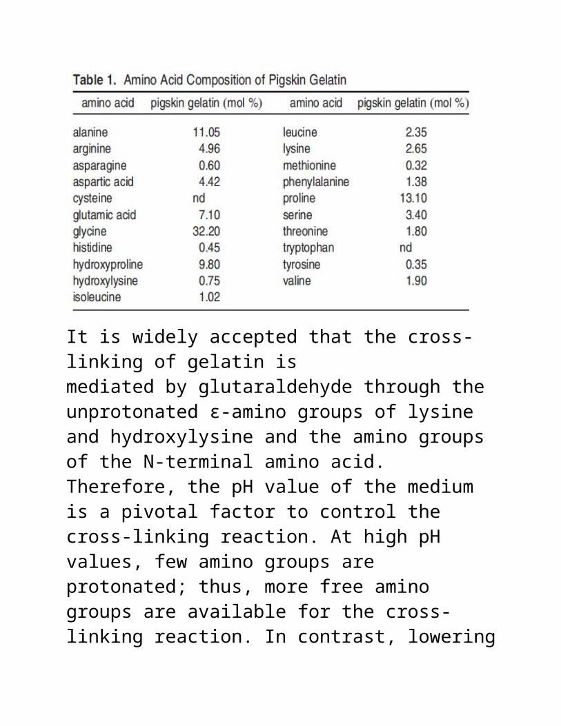
It is widely accepted that the cross-linking of gelatin ismediated by glutaraldehyde through the unprotonated ε-amino groups of lysine and hydroxylysine and the amino groups of the N-terminal amino acid. Therefore, the pH value of the medium is a pivotal factor to control the cross-linking reaction. At high pH values, few amino groups are protonated; thus, more free amino groups are available for the cross-linking reaction. In contrast, lowering
the pH increases the amount of protonatedamino groups; thus, the possibility of cross-linking reactions may be significantly reduced. Otherwise, cross-linking reactions may follow different reaction mechanisms. Nevertheless, the crosslinking of gelatin at pH of approximately 4.5 has been demonstrated, which gives rise to the hypothesis that a different chemicalroute may be involved in this reaction.

Preparation :Firstly a 14% w/w gelatin-water solution was obtained at a native PH of4.5.This was doneby taking 3.5g of Gelatin powder and mixing it in 25ml ofwater. This was done in an environment heated at 60oC for 1 hour. After 1 Hourthe temperature was decreased to 40oC. At this point pure Gelatin films were obtained by spreading some solution(20ml) on the petridishes and storing it into the incubator for the characterization purposes.
The remaining was used for crosslinkning. This was calculated to be done in the ratio of 0.3% w/w Gelatin to 1% w/w Glutradehyde. Although this ration an be varied, but the said ratio is said to produce high

number of schiff bases at the pre-determined PH of 4.5 and thus yield better crosslinking. For this to be done firstly we needed to prepare 0.3% w/w Gelatin, for which we took 1.07ml of Gelatin(14% w/w) and mix it in watermeasuring to 48.93ml. Secondly, we needed to prepare 1% w/w Glutraldehyde(25% w/w), for this we took 2ml of glutraldehyde and mixed it in 48ml of water.
The next step is an essential one wherethe two are mixed and allowed to cross-link which involved dro-by-drop addition of latter to the former under constand stirring. After constant stirring of 1.5hours, the sample was divided into two.
40ml of the crosslinked sample was taken into petridish for the formation
of film while 10ml was stored in a testtube for observational purposes. For the water in the solution it is allowedto be evaporated at 30oC for 24 hours. Afterwards both the samples were storedinside incubator for 48 hours at a constant temperature of 37.6oC.
Before characterization samples were prepared by repeatedly washing them with Milli-Q water and then were followed by air-drying at room temperature for 1 hour. The sample werethen kept in dessicator for 5 days before analyses.
Testing :

1> FTIR2> SEM3> NMR

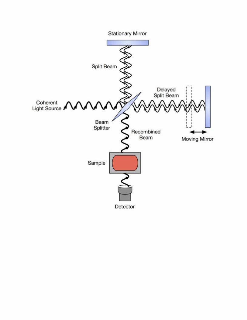
FTIRFourier transform infrared spectroscopy (FTIR) is a technique to obtain infrared spectrum of absorption, emission, photoconductivity or Raman scattering of a solid, liquid or gas. An FTIR spectrometer simultaneously collects spectral data in a wide spectral range. This confers a significant advantage over a dispersive spectrometer which measures intensity over a narrow range of wavelengths at a time. The term Fourier transform infrared spectroscopy originates from the fact that a Fourier transform (a mathematical process) is required to convert the raw data into the actualspectrum.
The is to measure how well a sample absorbs light at each wavelength. Rather than shining a monochromatic beam of light at the sample, this technique shines a beam containing many frequencies of light at once, and measures how much of that beam is absorbed by the sample. Next, the beam is modified to contain a different combination of frequencies, giving a second data point. This process is repeated many
times. Afterwards, a computer takes all these data and works backwards to infer what the absorption is at each wavelength. The beam described above is generated by starting with a broadband light source—one containing the fullspectrum of wavelengths to be measured. As mentioned, computer processing is required to turn the raw data (light absorption for each mirror position) into the desired result (light absorption for each wavelength). The processing required turns out to be a common algorithm called the Fourier transform (hence the name, "Fourier transform spectroscopy"). The raw data is sometimes called an "interferogram".
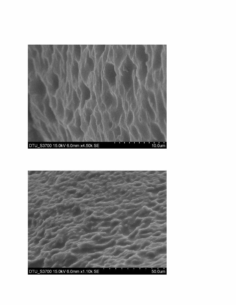
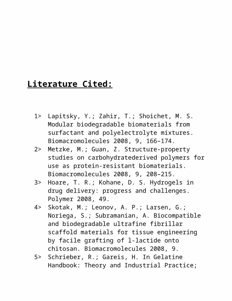
SEM :
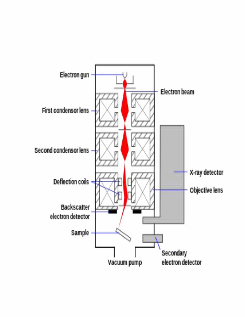
A scanning electron microscope (SEM) is a type of electron microscope that produces images of asample by scanning it with a focused beam of electrons. The electrons interact with atoms in the sample, producing various signals that can be detected and that contain information about the sample's surface topography and composition. The electron beam is generally scanned in a raster scan pattern, and the beam'sposition is combined with the detected signal toproduce an image. SEM can achieve resolution better than 1 nanometer. Specimens can be observed in high vacuum, in low vacuum, in wet conditions (in environmental SEM), and at a widerange of cryogenic or elevated temperatures. Themost common mode of detection is by secondary electrons emitted by atoms excited by the electron beam. On a flat surface, the plume of secondary electrons is mostly contained by the
sample, but on a tilted surface, the plume is partially exposed and more electrons are emitted. By scanning the sample and detecting the secondary electrons, an image displaying thetopography of the surface is created.
The types of signals produced by a SEM include secondary electrons (SE), back-scatteredelectrons (BSE), characteristic X-rays, light (cathodoluminescence) (CL), specimen current andtransmitted electrons. Secondary electron detectors are standard equipment in all SEMs, but it is rare that a single machine would have detectors for all possible signals. The signals result from interactions of the electron beam with atoms at or near the surface of the sample.In the most common or standard detection mode, secondary electron imaging or SEI, the SEM can produce very high-resolution images of a sample surface, revealing details less than 1 nm in size. Due to the very narrow electron beam, SEM micrographs have a large depth of field yieldinga characteristic three-dimensional appearance useful for understanding the surface structure of a sample. This is exemplified by the micrograph of pollen shown above. A wide range of magnifications is possible, from about 10 times (about equivalent to that of a powerful hand-lens) to more than 500,000 times, about 250
times the magnification limit of the best light microscopes.
Back-scattered electrons (BSE) are beam electrons that are reflected from the sample by elastic scattering. BSE are often used in analytical SEM along with the spectra made from the characteristic X-rays, because the intensityof the BSE signal is strongly related to the atomic number (Z) of the specimen. BSE images can provide information about the distribution of different elements in the sample. For the same reason, BSE imaging can image colloidal gold immuno-labels of 5 or 10 nm diameter, whichwould otherwise be difficult or impossible to detect in secondary electron images in biological specimens. Characteristic X-rays are emitted when the electron beam removes an inner shell electron from the sample, causing a higher-energy electron to fill the shell and release energy. These characteristic X-rays are used to identify the composition and measure theabundance of elements in the sample.

NMR :
Nuclear magnetic resonance spectroscopy, most commonly known as NMR spectroscopy, is a research technique that exploits the magnetic properties of certain atomic nuclei. It determines the physical and chemical properties of atoms or the molecules in which they are contained. It relies on the phenomenon of nuclear magnetic resonance and can provide detailed information about the structure, dynamics, reaction state, and chemical environment of molecules. The intramolecular magnetic field around an atom in a molecule changes the resonance frequency, thus giving access to details of the electronic structure ofa molecule.Most frequently, NMR spectroscopy is used by chemists and biochemists to investigate the properties of organic molecules, although it is applicable to any kind of sample that contains nuclei possessing spin. Suitable samples range from small compounds analyzed with 1-dimensional proton or carbon-13 NMR spectroscopy
to large proteins or nucleic acids using 3 or 4-dimensional techniques. The impact of NMR spectroscopy on the sciences has been substantial because of the range of information and the diversity of samples, including solutions and solids.NMR spectra are highly unique, well-resolved, analytically tractable and often highly predictable for small molecules. Thus, in organic chemistry practice, NMR analysis is used to confirm the identity of a substance. Different functional groups are obviously distinguishable, and identical functional groupswith differing neighboring substituents still give distinguishable signals. NMR has largely replaced traditional wet chemistry tests such as color reagents for identification. A disadvantage is that a relatively large amount, 2–50 mg, of a purified substance is required, although it may be recovered. Preferably, the sample should be dissolved in a solvent, becauseNMR analysis of solids requires a dedicated MAS machine and may not give equally well-resolved spectra. The timescale of NMR is relatively long, and thus it is not suitable forobserving fast phenomena, producing only an averaged spectrum. Although large amounts of impurities do show on an NMR spectrum, better
methods exist for detecting impurities, as NMR is inherently not very sensitive.NMR spectrometers are relatively expensive.When placed in a magnetic field, NMR active nuclei (such as 1H or 13C) absorb electromagnetic radiation at a frequency characteristic of the isotope. The resonant frequency, energy of the absorption, and the intensity of the signal are proportional to the strength of the magneticfield. For example, in a 21 Tesla magnetic field, protons resonate at 900 MHz. It is commonto refer to a 21 T magnet as a 900 MHz magnet, although different nuclei resonate at a different frequency at this field strength in proportion to their nuclear magnetic moments.
Upon excitation of the sample with radio frequency pulse, a nuclear magnetic resonance response - a free induction decay (FID) - is obtained. It is a very weak signal, and requiressensitive radio receivers to pick up. A Fourier transform is done to extract the frequency-domain spectrum from the raw time-domain FID.
Results :Due to its uniqueamino acid sequences and numerous functional groups, gelatinis well-suited for producing chemical hydrogels in the form of sheets, films,ormembranes by reacting with small molecules containing reactive functional groups, such as an aldehyde group (42). As mentioned before, it is generally accepted that the mechanism of gelatin cross-linking mediated by glutaraldehyde can be explained throughthe reaction of the aldehyde functionalgroups with free nonprotonated ε-amino groups (-NH2) of lysine or hydroxylysinethrough a nucleophilic addition-type reaction. Although
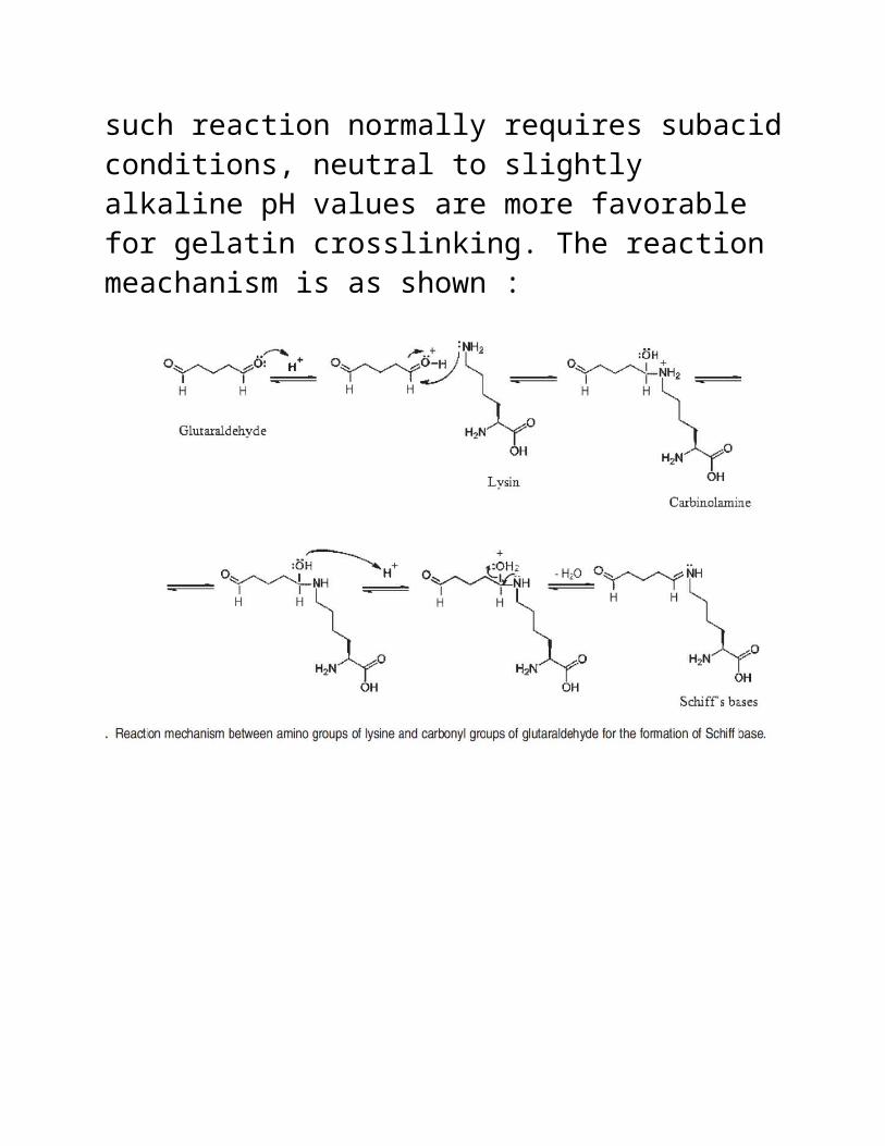
such reaction normally requires subacidconditions, neutral to slightly alkaline pH values are more favorable for gelatin crosslinking. The reaction meachanism is as shown :

More specifically, the pH determines the degree ofprotonation of ε-amino groups, aswell as the presence of negative charges on the carboxylic groups. Therefore, at high pH values, very few amino groups become protonated, and there exists a large amount of free amino groups in gelatin molecules. Conversely, a
decrease in pH leads to an increase in positively charged amino groups, which are unavailable for the crosslinking reaction with glutaraldehyde. In our system, the crosslinking reaction was first carried out at pH 4.5. Because the isoelectric point (pI) of typeAgelatin is approximately 8.5 and the pKa value for the ε-amino group of lysine is 10.53 (45), at this pH, threebasic amino acids in gelatin (i.e., 4% lysine, 1% hydroxylysine, and <1%histidine) are mostly positively charged. Therefore,it is less likely that the cross-linking reaction took placebetween these protonated amino acids and glutaraldehyde molecules.

Above are FTIR Results


Acknowledgement :
I would like to thank Dr. Deenan Santhiya, Dr. D. Kumar, Nidhi for helping me out in completion of this valuable project and for technical assistance.
Literature Cited:
1> Lapitsky, Y.; Zahir, T.; Shoichet, M. S. Modular biodegradable biomaterials from surfactant and polyelectrolyte mixtures. Biomacromolecules 2008, 9, 166–174.
2> Metzke, M.; Guan, Z. Structure-property studies on carbohydratederived polymers foruse as protein-resistant biomaterials. Biomacromolecules 2008, 9, 208–215.
3> Hoare, T. R.; Kohane, D. S. Hydrogels in drug delivery: progress and challenges. Polymer 2008, 49.
4> Skotak, M.; Leonov, A. P.; Larsen, G.; Noriega, S.; Subramanian, A. Biocompatible and biodegradable ultrafine fibrillar scaffold materials for tissue engineering by facile grafting of l-lactide onto chitosan. Biomacromolecules 2008, 9.
5> Schrieber, R.; Gareis, H. In Gelatine Handbook: Theory and Industrial Practice;

Schrieber, R., Gareis, H., Eds.; Wiley-VCH:Weinheim, Germany, 2007; p 163.
6> Lien, S.-M.; Li, W.-T.; Huang, T.-J. Genipin-crosslinked gelatin scaffolds for articular cartilage tissue engineering witha novel crosslinking method. Mater. Sci. Eng., C 2008, 28, 36–43.
7> Zheng, J. P.; Gao, S.; Wang, J. X.; Yao, K.D. Swelling behavior of gelatin-g-methyl methacrylate copolymers. J. Mater. Sci. 2005, 40, 4029–4033.
Copyright © 2022 FDOKUMEN