Lyophilised Biopolymer-Clay Hydrogels for Drug Delivery
9
1 Madridge J Nov Drug Res. ISSN: 2641-5232 Volume 1 • Issue 1 • 1000101 Madridge Journal of Novel Drug Research Research Article Open Access Lyophilised Biopolymer-Clay Hydrogels for Drug Delivery Farnoosh Kianfar 1 , Nicola Dempster 2 , Elsie Gaskell 2 , Matt Roberts 2 and Gillian A Hutcheon 2 * 1 Nemaura Pharma, Loughborough, LE11 3QF, UK 2 School of Pharmacy and Biomedical Sciences, Liverpool John Moores University, Byrom Street, Liverpool, L3 3AF, UK Article Info *Corresponding author: Gillian A Hutcheon School of Pharmacy and Biomedical Sciences Liverpool John Moores University Byrom Street, Liverpool, L3 3AF, UK Tel.: + 44 (0) 151 231 2130 Email: [email protected] Received: June 19, 2017 Accepted: June 22, 2017 Published: June 30, 2017 Citation: Kianfari F, Dempster NM, Gaskell EE, Roberts M, Hutcheon GA. Lyophilised Biopolymer-Clay Hydrogels for Drug Delivery. Madridge J Nov Drug Res. 2017; 1(1): 1-9. doi: 10.18689/mjndr-1000101 Copyright: © 2017 The Author(s). This work is licensed under a Creative Commons Attribution 4.0 International License, which permits unrestricted use, distribution, and reproduction in any medium, provided the original work is properly cited. Published by Madridge Publishers Abstract Clays have previously demonstrated potential as drug delivery carriers for the extended release of a variety of drugs. The objective of this study was to develop and characterise drug-containing clays in combination with natural hydrogels for the preparation of lyophilised xerogels. Sulfathiazole (STH) (a hydrophobic model drug) was intercalated within the interlayer spaces of Laponite® RDS (LAP RDS) or refined montmorillonite (MMT) and then mixed with either carageenen 812 (CAR 812) or hydrohydroxy ethyl cellulose (HEC) hydrogels prior to lyophilisation. The resulting xerogels were characterised visually, using differential scanning calorimetry (DSC) and with scanning electron microscopy (SEM). Optimal geo-polymeric wafers contained 1.5% W/W CAR 812 with 2% LAP RDSand 1% W/W intercalated STH. DSC and SEM results indicated the amorphous form of STH was intercalated inLAP RDS within theleafy structure of CAR 812. This xerogel hydrated up to1700% within 40 minutes and released the STH by Higuchikinetic model. Keywords: Polymer; Clay, Intercalation, Xerogel, Wound delivery, Amorphous, Physicochemical characterisation, Polymers, hydrogel, drug delivery, lyophilised wafer. Abbreviations STH (Sulfathiazole), LAP (Laponite), MMT (montmorillonite), CAR (carrageenan), HEC (hydrohydroxy ethyl cellulose), DSC (differential scanning calorimetry), SEM (scanning electron microscopy), polyethylene glycol (PEG). Introduction Drug-delivery systems composed of natural materials have become increasingly important because of their nontoxicity and biodegradability [1]. Amongst the diverse range of natural materials investigated for this purpose, clays have demonstrated useful properties for pharmaceutical, clinical and general health applications attracting increasing interest over the last decade; although the therapeutic effects of clays have been referred to from prehistoric times [2, 3]. Montmorillonite (MMT) and refined MMT are natural clay minerals with a high internal surface area, high cation exchange capacity, high adsorption ability, and low toxicity [4]. Due to the negatively charged layers, the swelling of these clays in the presence of water is high and positively charged species can be intercalated into the interlayer spaces via electrostatic interactions. Laponite® (LAP), a synthetic layered silicate with the chemical formula NaC 0.7 [(Si 8 Mg 5.5 Li 0.3 ) O 20 (OH) 4 ] ‾K 0.7 is composed of disc-shaped nanoparticles of 25 nm diameter and 1 nm layer thickness [5]. Some octahedral magnesium ions in the octahedral sheets are replaced with lithium ions so ISSN: 2641-5232
-
Upload
khangminh22 -
Category
Documents
-
view
0 -
download
0
Transcript of Lyophilised Biopolymer-Clay Hydrogels for Drug Delivery
1Madridge J Nov Drug Res.ISSN: 2641-5232
Volume 1 • Issue 1 • 1000101
MadridgeJournal of Novel Drug Research
Research Article Open Access
Lyophilised Biopolymer-Clay Hydrogels for Drug DeliveryFarnoosh Kianfar1, Nicola Dempster2, Elsie Gaskell2, Matt Roberts2 and Gillian A Hutcheon2*1Nemaura Pharma, Loughborough, LE11 3QF, UK2School of Pharmacy and Biomedical Sciences, Liverpool John Moores University, Byrom Street, Liverpool, L3 3AF, UK
Article Info*Corresponding author:Gillian A HutcheonSchool of Pharmacy and Biomedical SciencesLiverpool John Moores UniversityByrom Street, Liverpool, L3 3AF, UKTel.: + 44 (0) 151 231 2130Email: [email protected]
Received: June 19, 2017Accepted: June 22, 2017Published: June 30, 2017
Citation: Kianfari F, Dempster NM, Gaskell EE, Roberts M, Hutcheon GA. Lyophilised Biopolymer-Clay Hydrogels for Drug Delivery. Madridge J Nov Drug Res. 2017; 1(1): 1-9.doi: 10.18689/mjndr-1000101
Copyright: © 2017 The Author(s). This work is licensed under a Creative Commons Attribution 4.0 International License, which permits unrestricted use, distribution, and reproduction in any medium, provided the original work is properly cited.
Published by Madridge Publishers
AbstractClays have previously demonstrated potential as drug delivery carriers for the
extended release of a variety of drugs. The objective of this study was to develop and characterise drug-containing clays in combination with natural hydrogels for the preparation of lyophilised xerogels. Sulfathiazole (STH) (a hydrophobic model drug) was intercalated within the interlayer spaces of Laponite® RDS (LAP RDS) or refined montmorillonite (MMT) and then mixed with either carageenen 812 (CAR 812) or hydrohydroxy ethyl cellulose (HEC) hydrogels prior to lyophilisation. The resulting xerogels were characterised visually, using differential scanning calorimetry (DSC) and with scanning electron microscopy (SEM). Optimal geo-polymeric wafers contained 1.5% W/W CAR 812 with 2% LAP RDSand 1% W/W intercalated STH. DSC and SEM results indicated the amorphous form of STH was intercalated inLAP RDS within theleafy structure of CAR 812. This xerogel hydrated up to1700% within 40 minutes and released the STH by Higuchikinetic model.
Keywords: Polymer; Clay, Intercalation, Xerogel, Wound delivery, Amorphous, Physicochemical characterisation, Polymers, hydrogel, drug delivery, lyophilised wafer.
AbbreviationsSTH (Sulfathiazole), LAP (Laponite), MMT (montmorillonite), CAR (carrageenan), HEC
(hydrohydroxy ethyl cellulose), DSC (differential scanning calorimetry), SEM (scanning electron microscopy), polyethylene glycol (PEG).
IntroductionDrug-delivery systems composed of natural materials have become increasingly
important because of their nontoxicity and biodegradability [1]. Amongst the diverse range of natural materials investigated for this purpose, clays have demonstrated useful properties for pharmaceutical, clinical and general health applications attracting increasing interest over the last decade; although the therapeutic effects of clays have been referred to from prehistoric times [2, 3].
Montmorillonite (MMT) and refined MMT are natural clay minerals with a high internal surface area, high cation exchange capacity, high adsorption ability, and low toxicity [4]. Due to the negatively charged layers, the swelling of these clays in the presence of water is high and positively charged species can be intercalated into the interlayer spaces via electrostatic interactions. Laponite® (LAP), a synthetic layered silicate with the chemical formula NaC0.7 [(Si8Mg5.5Li0.3) O20 (OH) 4] ‾K0.7 is composed of disc-shaped nanoparticles of 25 nm diameter and 1 nm layer thickness [5]. Some octahedral magnesium ions in the octahedral sheets are replaced with lithium ions so
ISSN: 2641-5232
Madridge Journal of Novel Drug Research
2Volume 1 • Issue 1 • 1000101Madridge J Nov Drug Res.ISSN: 2641-5232
negatively charged layers are present which are compensated for with exchangeable sodium ions in the interlayer space. The film forming ability of these materials is a desirable property for the development of mucosal films for novel drug delivery systems [6]. Tixogel VZ and VP are synthetic organophilic clays with high thixotropic properties and can be used as anti-settling agents in low polarity systems. They can be dispersed in alcohols or ketones following a high agitation process [7].
The use of clay minerals for human health is not restricted to pharmaceutical excipients and cosmetics [8] as recently clays have demonstrated potential as carriers for the delivery and extended release of a variety of drugs [9]. Studies on the mobility of chemical elements from healing clays to the human body during topical applications and ingestion were under taken by [6, 10, 11]. Many formulations are based on the intercalation of molecules between the interlayer spaces of the clay enabling the incorporation of either hydrophilic or hydrophobic drugs via the exchange of small two dimensional organic molecules, inorganic and organic ions or polymeric ions with the ions originally present within the clay structure [12, 13]. For example Beak, et al. [14] intercalated glutathione in montmorillonite for antioxidant delivery with the aim of protecting the drug molecules from chemical and enzymatic hydrolysis following oral ingestion. Ibuprofen, the most common medicine for rheumatoid arthritis, osteoarthritis and moderate pain treatment, was intercalated in montmorillonite to minimize side effects [15]. The intercalated formulation controls the drug release and minimizes frequent side effects within the gastrointestinal tract and the ulcerogenic effect [16].
Clay:polymer nanocomposites are widely used in the materials industry where the addition of clay can be used to enhance the physical and engineering properties of a material [17] and there is increasing interest in the use of clay-polymer composites for pharmaceutical applications such as tissue engineering and drug delivery [9, 18]. Although individually, both biopolymers and clays are present in many drug delivery systems a hybrid of the two can provide enhanced properties to a drug delivery matrix such as improved water uptake, increased mechanical strength, improved rheology and modified drug release.
Carrageenan and hydroxyethyl cellulose (HEC) are both natural products commonly used for the formulation of drugs. More recently they have been used to prepare buccal [19] and lyophilised xerogels [20] for drug delivery and wound healing [20, 21]. Lyophilised xerogels are shaped hydrophilic polymer glasses that adhere to the wound, adsorb fluid and form a viscous gel releasing any drugs contained within.
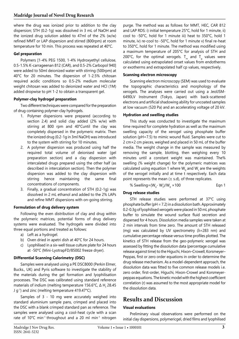
In this present study a model drug, Sulfathiazole (STH, figure 1), was intercalated into various clays(LAP, MMT, refined MMT, Tixogel VZ and Tixogel VP) and thenincorporated into a natural hydrogel consisting of either carrageenan or hydroxyethyl cellulose. The gels were subsequently lyophilised to form a dry xerogel. The composition of the xerogel was varied in terms of type and quantity of clay, polymer and drug to produce a matrix with optimal properties for wound healing and drug delivery applications. STH (pKa=7.2), an antimicrobial
drug, is a weak acid with extremely low solubility in water (LogP=0.35). Formulation methodology was developed and optimal conditions (hydration time, pH, and clay concentration) for an even distribution of the drugs and clays within the aqueous polymeric matrix were realised by intercalating STH into the interlayers of refined MMT and various grades of LAP. Figure 1
Figure 1. Chemical structure of Sulfathiazole (STH).
Experimental and MethodsMaterials
MMT, refined MMT, various Tixogels (VP & VZ; CAS: 67479-91-8) and all LAP grades (CAS: 227605-22-3, XL21, RDS, RD and WXFP) were kindly gifted from BYK(Widnes, UK). Sulfathiazole (STH; CAS: 72-14-0, batch number: 053K3451) and Chitosan medium molecular weight (CAS: 9012-76-4, batch number: MKBH1108V) were purchased from Sigma Aldrich. The PEG 1500 (CAS: 25322-68-3, batch number: ZA3726628) was purchased from BDH and Hydroxethylcellulose (CAS: 9004-62-0, batch number: P-0189) from Aqualon Hercules. Carrageenan 812(CAS: 9000-07-1 232, Gelcarine) was kindly gifted from FMC BioPolymer company.
Dispersion of claysA gradual ratio of MMT, refine MMT, Tixogels or various
LAP grades [0.5-20%(w/w)] were added to 50 mL deionized water in separate beakers (200 mL) and stirred vigorously at 800rpm for 30 minutes at room temperature. The visual properties were inspected and initial pH determined before the drop wise addition of 1M NaOH or HCl while recording the pH accordingly. In order to enhance the solubility and obtain an even dispersion of 0.5-1.5% (w/w) MMT and Tixogels within the system, a gradual volume of ethanol (2-20 mL) was added while the mixture was constantly agitated. Furthermore, 0.5-1.5% (w/w) MMT or Tixogels were dispersed in 20 mL ethanol or propanol and the physical properties evaluated.
Intercalation of STHinto MMT and LAPA 2% dispersion of clay (LAPs and MMT) was produced by
adding the clay to deionized water while agitating vigorously (800rpm) at room temperature for 30 minutes. Then STH (0.2-1g) was slowly added to this dispersion with the gradual addition of 3 mL NaOH to ionize the drug with on-going agitation for a further 45 minutes. To reduce the time required for the intercalation process a second technique was employed
Madridge Journal of Novel Drug Research
3Volume 1 • Issue 1 • 1000101Madridge J Nov Drug Res.ISSN: 2641-5232
where the drug was ionized prior to addition to the clay dispersion; STH (0.2-1g) was dissolved in 3 mL of NaOH and the ionized drug solution added to 47ml of the 2% (w/w) refined MMT or LAP dispersion and stirred (800rpm) at room temperature for 10 min. This process was repeated at 40°C.
Gel preparationPolymers [1-4% PEG 1500, 1-4% Hydroxyethyl cellulose,
0.5-1.5% Ƙ-carrageenan 812 (CAR), and 0.5-2% Carbopol 940] were added to 50ml deionized water with stirring (500rpm) at 40°C for 20 minutes. The dispersion of 1-2.5% chitosan required acidic conditions so 0.5-2% medium molecular weight chitosan was added to deionized water and HCl (1M) added dropwise to pH 1-2 to obtain a transparent gel.
Polymer-clay hydrogel preparationTwo different techniques were compared for the preparation
of drug containing polymer-clay hydrogels.1. Polymer dispersions were prepared (according to
section 2.4) and solid clay added (2% w/w) with stirring at 800 rpm and 40°Cuntil the clay was completely dispersed in the polymeric matrix. Then the ionized drug (0.2-1g in 3ml NaOH) was introduced to the system with stirring for 10 minutes.
2. A polymer dispersion was produced using half the required total volume of deionised water (gel preparation section) and a clay dispersion with intercalated drugs prepared using the other half (as described in intercalation section). Then the polymer dispersion was added to the clay dispersion with stirring hence maintaining the same final concentrations of components.
3. Finally, a gradual concentration of STH (0.2-1g) was dissolved in 2 mL ethanol and added to the 2% LAPs and refine MMT dispersions with on-going stirring.
Formulation of drug delivery systemFollowing the even distribution of clay and drug within
the polymeric matrices, potential forms of drug delivery systems were evaluated. The hydrogels were divided into three equal portions and treated as follows:
a) Left as a hydrogel.b) Oven dried in apetri dish at 40°C for 24 hours.c) Lyophilised in a six-well tissue culture plate for 24 hours
at -50°C (Mini-LyotrapFD/85002 freeze dryer).
Differential Scanning Calorimetry (DSC)Samples were analysed using a PE DSC8000 (Perkin Elmer,
Bucks., UK) and Pyris software to investigate the stability of the materials during the gel formation and lyophilisation processes. The DSC was calibrated using standard reference materials of indium (melting temperature 156.6°C, Δ Hf 28.45 J g-1) and zinc (melting temperature 419.47°C).
Samples of 3 - 10 mg were accurately weighed into standard aluminium sample pans, crimped and placed into the DSC with a blank crimped standard pan as reference. The samples were analysed using a cool-heat cycle with a scan rate of 10°C min-1 throughout and a 20 ml min-1 nitrogen
purge. The method was as follows for MMT, HEC, CAR 812 and LAP RDS: i) initial temperature 25°C, hold for 1 minute, ii) cool to -50°C, hold for 1 minute iii) heat to 350°C, hold 1 minute, iv) re-cool to -50°C, hold for 1 minute v) final re-heat to 350°C, hold for 1 minute. The method was modified using a maximum temperature of 205°C for analysis of STH and 200°C, for the optimal xerogels. Tm and Tg values were calculated using extrapolated onset values from endotherms or exotherms and extrapolated half cp values, respectively.
Scanning electron microscopyScanning electron microscopy (SEM) was used to evaluate
the topographic characteristics and morphology of the xerogels. The analyses were carried out using a JeolJSM-6490LV Instrument (Tokyo, Japan) with back-scattered electrons and artificial shadowing ability for uncoated samples at low vacuum (520 Pa) and an accelerating voltage of 20 kV.
Hydration and swelling studies This study was conducted to investigate the maximum
time required for complete hydration as well as the maximum swelling capacity of the xerogel using phosphate buffer solution (pH=7.5) to mimic wound fluid. Samples were cut to 2 cm×2 cm pieces, weighed and placed in 50 mL of the buffer media. The weight change in the sample was measured by removing the sample, blotting, then weighing every 10 minutes until a constant weight was maintained. The% swelling (% weight change) for the polymeric matrices was calculated using equation 1 where W0 and Wt are the weights of the xerogel initially and at time t respectively. Each data point represents the mean (± s.d), of three replicates.
% Swelling=(W0- Wt)/W0 ×100 Eqn 1
Drug release studies STH release studies were performed at 37°C using
phosphate buffer (pH = 7.2) in a dissolution bath. Approximately 0.2-0.3g of lyophilised xerogels were placed in 50 mL phosphate buffer to simulate the wound surface fluid secretion and dispersed for 4 hours. Dissolution media samples were taken at 2 min intervals from time zero. The amount of STH released (mg) was calculated by UV spectrometry (λ=283 nm) and cumulative percentage release versus time profiles plotted. The kinetics of STH release from the geo-polymeric xerogel was assessed by fitting the dissolution data (percentage cumulative release against time) to the Higuchi, Hixon–Crowell, Korsmeyer–Peppas, first or zero order equations in order to determine the drug release mechanism. As a model-dependent approach, the dissolution data was fitted to five common release models i.e. zero order, first-order, Higuchi, Hixon-Crowel and Korsmeyer-peppas equations. The kinetic model with the highest coefficient correlation (r) was assumed to the most appropriate model for the dissolution data.
Results and DiscussionVisual evaluations
Preliminary visual observations were performed on the initial clay dispersions, polymericgel, dried films and lyophilised
Madridge Journal of Novel Drug Research
4Volume 1 • Issue 1 • 1000101Madridge J Nov Drug Res.ISSN: 2641-5232
xerogels to evaluate them in terms of; ease of pouring, stability, flexibility and the even distribution of the components within the matrix with no air bubbles in the films. The fragility and brittleness of a matriximpacts on the physical and mechanical stability during handling and storage. For wound healing applications, a xerogel must also be soft and flexible to provide the desirable mechanical strength during handling and comfort upon the application without potential irritation [22]. In addition, an ideal xerogel should have an optimum thickness of less than 1mm as thicker preparations maybe difficult to apply without disturbing the wound surface. Thickness also affects both the rate of hydration and the diffusion distance of the drug through the resulting swollen gel which can have significant effects on drug release profiles [20, 21].
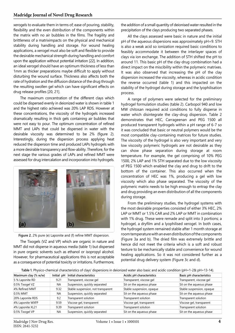
The maximum concentration of the different clays which could be dispersed evenly in deionized water is shown in table 1 and the highest ratio achieved was 20% LAP RDS. However at these concentrations, the viscosity of the hydrogels increased dramatically resulting in thick gels containing air bubbles that were not easy to pour. The optimum concentration of refined MMT and LAPs that could be dispersed in water with the desirable viscosity was determined to be 2% (figure 2). Interestingly, during the dispersion process applying heat reduced the dispersion time and produced LAPs hydrogels with a more desirable transparency and flow-ability. Therefore, for the next stage the various grades of LAPs and refined MMT were assessed for drug intercalation and incorporation into hydrogels.
Figure 2. 2% pure (e) Laponite and (f) refine MMT dispersion.
The Tixogels (VZ and VP) which are organic in nature and MMT did not disperse in aqueous media (table 1) but dispersed in pure organic solvents such as ethanol or isopropyl alcohol. However, for pharmaceutical applications this is not acceptable as a consequence of potential toxicity or irritations. Furthermore,
the addition of a small quantity of deionised water resulted in the precipitation of the clays producing two separated phases.
All the clays assessed were basic in nature and the initial pH of the aqueous dispersions was approximately pH=9. STH is also a weak acid so ionization required basic conditions to feasibly accommodate it between the interlayer spaces of clays via ion exchange. The addition of STH shifted the pH to around 11. This basic pH of the clay-drug combination had a direct impact on the miscibility within the polymeric matrixes. It was also observed that increasing the pH of the clay dispersion increased the viscosity, whereas in acidic condition the reverse occurred (table 1) and this impacted on the stability of the hydrogel during storage and the lyophilisation process.
A range of polymers were selected for the preliminary hydrogel formulation studies (table 2). Carbopol 940 and low MW chitosan required acid conditions to fully disperse in water which disintegrate the clay-drug dispersion. Table 2 demonstrates that HEC, Carrageenan and PEG 1500 all produced transparent hydrogels within a pH range of 6-7 so it was concluded that basic or neutral polymers would be the most compatible clay-containing matrices for future studies. The viscosity of the hydrogel is also very important and very low viscosity polymeric hydrogels are not desirable as they can show phase separation during storage at room temperature. For example, the gel comprising of 10% PEG 1500, 2% LAP and 1% STH separated due to the low viscosity of PEG 1500 which enabled the clay and drug to drift to the bottom of the container. This also occurred when the concentration of HEC was 1%, producing a gel with low viscosity which also phase separated. The viscosity of the polymeric matrix needs to be high enough to entrap the clay and drug providing an even distribution of all the components during storage.
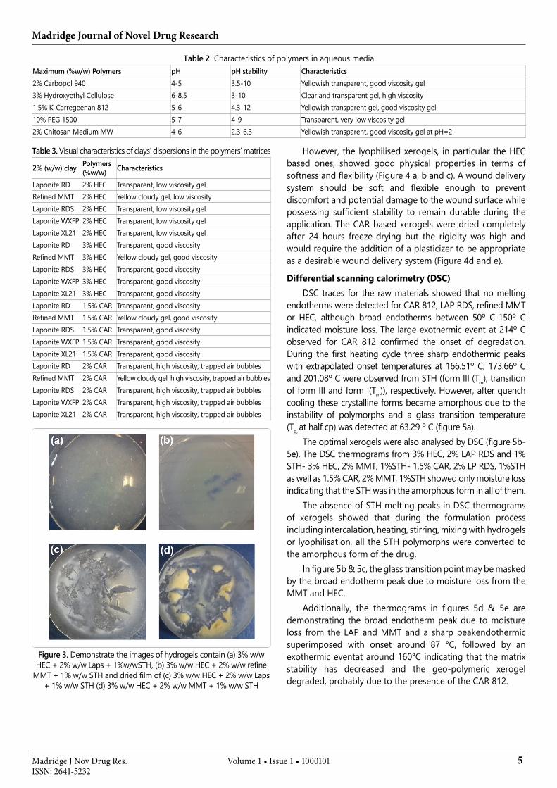
From the preliminary studies, the hydrogel systems with the most desirable properties consisted of either 3% HEC, 2% LAP or MMT or 1.5% CAR and 2% LAP or MMT in combination with 1% drug. These were remade and split into 3 portions; a hydrogel, a dryfilm and a lyophilised xerogel. In both cases, the hydrogel system remained stable after 1 month storage at room temperature with an even distribution of the components (Figure 3a and b). The dried film was extremely brittle and hence did not meet the criteria which is a soft and robust texture to be mechanically stable and convenience for wound healing applications. So it was not considered further as a potential drug delivery system (Figure 3c and d).
Table 1. Physico-chemical characteristics of clays’ dispersions in deionized water also basic and acidic condition (pH=1-2& pH=13-14).Maximum clay (% w/w) Initial pH Initial characteristics Acidic pH characteristics Basic pH characteristics5 % Laponite RD 9.21 Transparent, viscose gel Transparent, viscose gel Transparent, viscose gel0.5% Tixogel VZ NA Suspension, quickly separated Sit on the aqueous phase Sit on the aqueous phase4% Refined MMT 9.32 Stable suspension, not transparent Stable suspension, opaque Stable suspension, opaque0.5% MMT NA Suspension, quickly separated Sit on the aqueous phase Sit on the aqueous phase20% Laponite RDS 9.2 Transparent solution Transparent solution Transparent solution4% Laponite WXFP 9.59 Viscose gel, transparent Viscose gel, transparent Viscose gel, transparent4% Laponite XL21 9.39 Transparent solution Transparent solution Transparent solution0.5% Tixogel VP NA Suspension, quickly separated Sit on the aqueous phase Sit on the aqueous phase
Madridge Journal of Novel Drug Research
5Volume 1 • Issue 1 • 1000101Madridge J Nov Drug Res.ISSN: 2641-5232
Table 3. Visual characteristics of clays’ dispersions in the polymers’ matrices
2% (w/w) clay Polymers (%w/w) Characteristics
Laponite RD 2% HEC Transparent, low viscosity gel Refined MMT 2% HEC Yellow cloudy gel, low viscosity Laponite RDS 2% HEC Transparent, low viscosity gel Laponite WXFP 2% HEC Transparent, low viscosity gel Laponite XL21 2% HEC Transparent, low viscosity gel Laponite RD 3% HEC Transparent, good viscosity Refined MMT 3% HEC Yellow cloudy gel, good viscosity Laponite RDS 3% HEC Transparent, good viscosity Laponite WXFP 3% HEC Transparent, good viscosity Laponite XL21 3% HEC Transparent, good viscosity Laponite RD 1.5% CAR Transparent, good viscosity Refined MMT 1.5% CAR Yellow cloudy gel, good viscosity Laponite RDS 1.5% CAR Transparent, good viscosity Laponite WXFP 1.5% CAR Transparent, good viscosity Laponite XL21 1.5% CAR Transparent, good viscosity Laponite RD 2% CAR Transparent, high viscosity, trapped air bubbles Refined MMT 2% CAR Yellow cloudy gel, high viscosity, trapped air bubbles Laponite RDS 2% CAR Transparent, high viscosity, trapped air bubblesLaponite WXFP 2% CAR Transparent, high viscosity, trapped air bubbles Laponite XL21 2% CAR Transparent, high viscosity, trapped air bubbles
Figure 3. Demonstrate the images of hydrogels contain (a) 3% w/w HEC + 2% w/w Laps + 1%w/wSTH, (b) 3% w/w HEC + 2% w/w refine
MMT + 1% w/w STH and dried film of (c) 3% w/w HEC + 2% w/w Laps + 1% w/w STH (d) 3% w/w HEC + 2% w/w MMT + 1% w/w STH
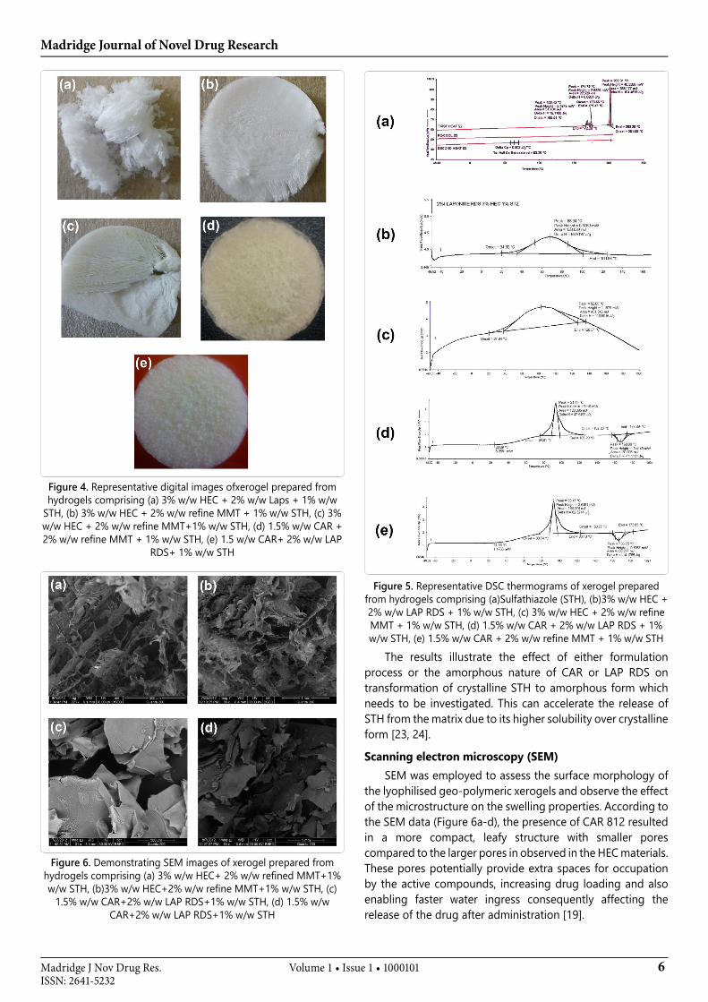
However, the lyophilised xerogels, in particular the HEC based ones, showed good physical properties in terms of softness and flexibility (Figure 4 a, b and c). A wound delivery system should be soft and flexible enough to prevent discomfort and potential damage to the wound surface while possessing sufficient stability to remain durable during the application. The CAR based xerogels were dried completely after 24 hours freeze-drying but the rigidity was high and would require the addition of a plasticizer to be appropriate as a desirable wound delivery system (Figure 4d and e).
Differential scanning calorimetry (DSC)DSC traces for the raw materials showed that no melting
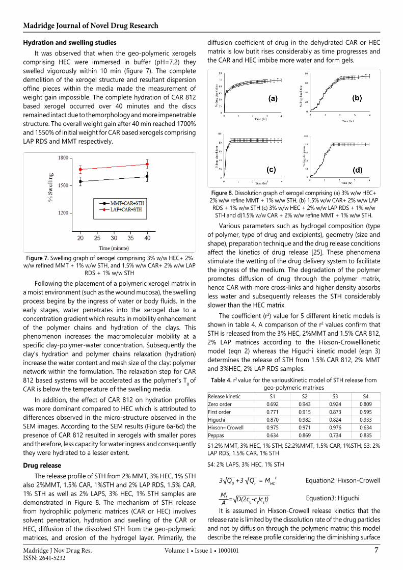
endotherms were detected for CAR 812, LAP RDS, refined MMT or HEC, although broad endotherms between 50º C-150º C indicated moisture loss. The large exothermic event at 214º C observed for CAR 812 confirmed the onset of degradation. During the first heating cycle three sharp endothermic peaks with extrapolated onset temperatures at 166.51º C, 173.66º C and 201.08º C were observed from STH (form III (Tm), transition of form III and form I(Tm)), respectively. However, after quench cooling these crystalline forms became amorphous due to the instability of polymorphs and a glass transition temperature (Tg, at half cp) was detected at 63.29 º C (figure 5a).
The optimal xerogels were also analysed by DSC (figure 5b- 5e). The DSC thermograms from 3% HEC, 2% LAP RDS and 1% STH- 3% HEC, 2% MMT, 1%STH- 1.5% CAR, 2% LP RDS, 1%STH as well as 1.5% CAR, 2% MMT, 1%STH showed only moisture loss indicating that the STH was in the amorphous form in all of them.
The absence of STH melting peaks in DSC thermograms of xerogels showed that during the formulation process including intercalation, heating, stirring, mixing with hydrogels or lyophilisation, all the STH polymorphs were converted to the amorphous form of the drug.
In figure 5b & 5c, the glass transition point may be masked by the broad endotherm peak due to moisture loss from the MMT and HEC.
Additionally, the thermograms in figures 5d & 5e are demonstrating the broad endotherm peak due to moisture loss from the LAP and MMT and a sharp peakendothermic superimposed with onset around 87 °C, followed by an exothermic eventat around 160°C indicating that the matrix stability has decreased and the geo-polymeric xerogel degraded, probably due to the presence of the CAR 812.
Table 2. Characteristics of polymers in aqueous mediaMaximum (%w/w) Polymers pH pH stability Characteristics2% Carbopol 940 4-5 3.5-10 Yellowish transparent, good viscosity gel 3% Hydroxyethyl Cellulose 6-8.5 3-10 Clear and transparent gel, high viscosity1.5% Ƙ-Carregeenan 812 5-6 4.3-12 Yellowish transparent gel, good viscosity gel 10% PEG 1500 5-7 4-9 Transparent, very low viscosity gel2% Chitosan Medium MW 4-6 2.3-6.3 Yellowish transparent, good viscosity gel at pH=2
Madridge Journal of Novel Drug Research
6Volume 1 • Issue 1 • 1000101Madridge J Nov Drug Res.ISSN: 2641-5232
Figure 4. Representative digital images ofxerogel prepared from hydrogels comprising (a) 3% w/w HEC + 2% w/w Laps + 1% w/w
STH, (b) 3% w/w HEC + 2% w/w refine MMT + 1% w/w STH, (c) 3% w/w HEC + 2% w/w refine MMT+1% w/w STH, (d) 1.5% w/w CAR + 2% w/w refine MMT + 1% w/w STH, (e) 1.5 w/w CAR+ 2% w/w LAP
RDS+ 1% w/w STH
Figure 6. Demonstrating SEM images of xerogel prepared from hydrogels comprising (a) 3% w/w HEC+ 2% w/w refined MMT+1% w/w STH, (b)3% w/w HEC+2% w/w refine MMT+1% w/w STH, (c)
1.5% w/w CAR+2% w/w LAP RDS+1% w/w STH, (d) 1.5% w/w CAR+2% w/w LAP RDS+1% w/w STH
Figure 5. Representative DSC thermograms of xerogel prepared from hydrogels comprising (a)Sulfathiazole (STH), (b)3% w/w HEC + 2% w/w LAP RDS + 1% w/w STH, (c) 3% w/w HEC + 2% w/w refine MMT + 1% w/w STH, (d) 1.5% w/w CAR + 2% w/w LAP RDS + 1% w/w STH, (e) 1.5% w/w CAR + 2% w/w refine MMT + 1% w/w STH
The results illustrate the effect of either formulation process or the amorphous nature of CAR or LAP RDS on transformation of crystalline STH to amorphous form which needs to be investigated. This can accelerate the release of STH from the matrix due to its higher solubility over crystalline form [23, 24].
Scanning electron microscopy (SEM)SEM was employed to assess the surface morphology of
the lyophilised geo-polymeric xerogels and observe the effect of the microstructure on the swelling properties. According to the SEM data (Figure 6a-d), the presence of CAR 812 resulted in a more compact, leafy structure with smaller pores compared to the larger pores in observed in the HEC materials. These pores potentially provide extra spaces for occupation by the active compounds, increasing drug loading and also enabling faster water ingress consequently affecting the release of the drug after administration [19].
Madridge Journal of Novel Drug Research
7Volume 1 • Issue 1 • 1000101Madridge J Nov Drug Res.ISSN: 2641-5232
Hydration and swelling studies It was observed that when the geo-polymeric xerogels
comprising HEC were immersed in buffer (pH=7.2) they swelled vigorously within 10 min (figure 7). The complete demolition of the xerogel structure and resultant dispersion offine pieces within the media made the measurement of weight gain impossible. The complete hydration of CAR 812 based xerogel occurred over 40 minutes and the discs remained intact due to themorphology and more impenetrable structure. The overall weight gain after 40 min reached 1700% and 1550% of initial weight for CAR based xerogels comprising LAP RDS and MMT respectively.
Figure 7. Swelling graph of xerogel comprising 3% w/w HEC+ 2% w/w refined MMT + 1% w/w STH, and 1.5% w/w CAR+ 2% w/w LAP
RDS + 1% w/w STH
Following the placement of a polymeric xerogel matrix in a moist environment (such as the wound mucosa), the swelling process begins by the ingress of water or body fluids. In the early stages, water penetrates into the xerogel due to a concentration gradient which results in mobility enhancement of the polymer chains and hydration of the clays. This phenomenon increases the macromolecular mobility at a specific clay-polymer-water concentration. Subsequently the clay’s hydration and polymer chains relaxation (hydration) increase the water content and mesh size of the clay: polymer network within the formulation. The relaxation step for CAR 812 based systems will be accelerated as the polymer’s Tg of CAR is below the temperature of the swelling media.
In addition, the effect of CAR 812 on hydration profiles was more dominant compared to HEC which is attributed to differences observed in the micro-structure observed in the SEM images. According to the SEM results (Figure 6a-6d) the presence of CAR 812 resulted in xerogels with smaller pores and therefore, less capacity for water ingress and consequently they were hydrated to a lesser extent.
Drug release The release profile of STH from 2% MMT, 3% HEC, 1% STH
also 2%MMT, 1.5% CAR, 1%STH and 2% LAP RDS, 1.5% CAR, 1% STH as well as 2% LAPS, 3% HEC, 1% STH samples are demonstrated in Figure 8. The mechanism of STH release from hydrophilic polymeric matrices (CAR or HEC) involves solvent penetration, hydration and swelling of the CAR or HEC, diffusion of the dissolved STH from the geo-polymeric matrices, and erosion of the hydrogel layer. Primarily, the
diffusion coefficient of drug in the dehydrated CAR or HEC matrix is low butit rises considerably as time progresses and the CAR and HEC imbibe more water and form gels.
Figure 8. Dissolution graph of xerogel comprising (a) 3% w/w HEC+ 2% w/w refine MMT + 1% w/w STH, (b) 1.5% w/w CAR+ 2% w/w LAP RDS + 1% w/w STH (c) 3% w/w HEC + 2% w/w LAP RDS + 1% w/w STH and d)1.5% w/w CAR + 2% w/w refine MMT + 1% w/w STH.
Various parameters such as hydrogel composition (type of polymer, type of drug and excipients), geometry (size and shape), preparation technique and the drug release conditions affect the kinetics of drug release [25]. These phenomena stimulate the wetting of the drug delivery system to facilitate the ingress of the medium. The degradation of the polymer promotes diffusion of drug through the polymer matrix, hence CAR with more cross-links and higher density absorbs less water and subsequently releases the STH considerably slower than the HEC matrix.
The coefficient (r2) value for 5 different kinetic models is shown in table 4. A comparison of the r2 values confirm that STH is released from the 3% HEC, 2%MMT and 1.5% CAR 812, 2% LAP matrices according to the Hixson-Crowellkinetic model (eqn 2) whereas the Higuchi kinetic model (eqn 3) determines the release of STH from 1.5% CAR 812, 2% MMT and 3%HEC, 2% LAP RDS samples.
Table 4. r2 value for the variousKinetic model of STH release from geo-polymeric matrixes
Release kinetic S1 S2 S3 S4Zero order 0.692 0.943 0.924 0.809First order 0.771 0.915 0.873 0.595Higuchi 0.870 0.982 0.824 0.933Hixson– Crowell 0.975 0.971 0.976 0.634Peppas 0.634 0.869 0.734 0.835
S1:2% MMT, 3% HEC, 1% STH; S2:2%MMT, 1.5% CAR, 1%STH; S3: 2% LAP RDS, 1.5% CAR, 1% STH
S4: 2% LAPS, 3% HEC, 1% STH
3 Q0 +3 Qt = MHCt Equation2: Hixson-Crowell
Mt
A = D(2c0-cs)cst) Equation3: Higuchi
It is assumed in Hixson-Crowell release kinetics that the release rate is limited by the dissolution rate of the drug particles and not by diffusion through the polymeric matrix; this model describe the release profile considering the diminishing surface
Madridge Journal of Novel Drug Research
8Volume 1 • Issue 1 • 1000101Madridge J Nov Drug Res.ISSN: 2641-5232
of the drug particles during the dissolution [26]. The Hixson-Crowell cube root law describes the release of the STH from xerogels where the surface area and diameter of delivery system is critical. The dissolution rate of STH from the surface of geo-polymeric systems is proportionally to diminishing polymers (CAR or HEC), in such a manner that the initial geometrical form remains constant [27].
The Higuchi kinetic model however describes the STH release as a diffusion process based in the Fick’s law, square root time dependent [28]. Fickian diffusion refers to the STH transport process where the polymer relaxation time (tr) is much greater than the characteristic solvent diffusion time (td).This equation has been used successfully to describes the experimentally observed in vitro release STH from polymers (HEC and LAP RDS or CAR 812 and MMT) where erosion or swelling of the polymeric matrix does not contribute to drug release during the short time period [29, 30].
Overall, in both kinetic models, the release of STH from CAR and HEC involves water diffusion and chain disentanglement at the final stage. Though the polymer dissolution does not result in the scission of polymer chains, it results in the loss of bulk material. CAR 812 and HEC experience dissolution in the aqueous medium due to water penetration effect, swelling, and polymer chain disentanglement and relaxation. The fractional release of STH from xerogels consisting of HEC and CAR 812 matrices corresponded well with the fractional release of HEC and CAR812, indicating drug release is primarily driven by dissolution and followed by erosion [31].
Conclusion These results demonstrate that due to strong cross-linked
polymer chains CAR 812 sustains the release of STH from the geo-polymeric matrices for up to 2 h due to the prolonged hydration of the matrix. This favours the delivery of STH to wound surfaces from CAR 812 matrices over the extremely fast hydration and drug release from the HEC based geo-polymeric matrices which would also be displaced from the wound surfaces due to the generation of a slippery residue following the fast polymer degradation.
Overall, a comparison of the four optimized xerogels demonstrated that the matrix comprising 1.5% CAR 812, 2% LAP RDS and 1% STH possessed the most desirable stability, swelling and drug release for wound deliver applications. The DSC results confirmed that any STH present was in an amorphous form, which supports data from dissolution studies that confirmed the effect of the drug dissolution rate on release kinetic. The presence of the STH in amorphous form increased the dissolution rate which can be advantageous where the purpose of the application is obtaining the fast release of antibiotic from the wound delivery system. However it might be not desirable if the drug release should be prolonged and over a longer period of time. Ideally, the most efficient system in order to deliver the antibiotic to the wound surfaces should be capable to release the drug rapidly after the application followed by a gradual release over the time [32]. This will be obtained by designing the most optimised combination of hydrogel polymer and clay to develop a stabilized geo-polymeric matrix.
Acknowledgment We would like to announce special gratitude to BYK
additive Ltd Company for sponsoring this project.
References1. Rodrigues LA, Figueiras A, Veiga F, et al. The systems containing clays
and clay minerals from modified drug release: a review. Colloids Surf B. 2013; 103: 642-51. doi: 10.1016/j.colsurfb.2012.10.068
2. Carretero MI, Gomes CSF, Tateo F. Clays and human health. In: Bergaya F, Theng BKG, Lagaly G, edtors. Handbook of Clay Science. Developments in Clay Science. Amsterdam Elsevier. 2006; 1: 717-741.
3. Gomes CSF, Pereira Silva JB. Minerals and clay minerals in medical geology. Appl Clay Sci. 2007; 36: 4-21. doi: 10.1016/j.clay.2006.08.006
4. Nuruzzaman MD, Rahman MM, Liu Y, Naidu R. Nanoencapsulation, Nano-guard for Pesticides: A New Window for Safe Application. J Agric Food Chem. 2016; 64 (7): 1447-1483. doi: 10.1021/acs.jafc.5b05214
5. Kroon M, Vos WL, Wegdam GH. Structure and formation of a gel of colloidal disks. Phys Rev E. 1998; 57: 1962–1970. doi: 10.1103/PhysRevE.57.1962
6. Park TU, Jung H, Kim HM, Choy JH, Lee CW. Hybrid of itraconazole, cyclosporine of carvedilol with a layered silicate and a process for preparing the same. 2004; WO Patent 2004/009120.
7. http://www.byk.com/en/additives/additives-by-name/cloisite.php.
8. López-Galindo A, Viseras C, Cerezo P. Compositional, technical and safety specifications of clays to be used as pharmaceutical and cosmetic products. Appl Clay Sci. 2007; 36: 51-63. doi: 10.1016/j.clay.2006.06.016
9. Viseras C, Aguzzi C, Cerezo P, Lopez-Galindo A. Uses of clay minerals in semisolid health care and therapeutic products. Appl Clay Sci. 2007; 36 (1–3): 37-50. doi: 10.1016/j.clay.2006.07.006
10. Tateo F, Summa V. Element mobility in clays for healing use. Appl Clay Sci. 2007; 36: 64-76. doi: 10.1016/j.clay.2006.05.011
11. Jung H, Kimc HM, Choy YB, Hwang SJ, Choy JH. Laponite-based nanohybrid for enhanced solubility and controlled release of itraconazole. Int J Pharm. 2008; 349: 283-290. doi: 10.1016/j.ijpharm.2007.08.008
12. Liu B, Luo J, Wang X, Lu J, Deng H and Sun R. Alginate/quaternized carboxymethyl chitosan/clay nanocomposite microspheres: preparation and drug-controlled release behavior.J. Biomater. Sci Polym Ed. 2013; 24(5): 589-605. doi: 10.1080/09205063.2012.701160
13. Paek SM, Jung H, Lee YJ, Park M, Hwang SJ, Choy JH. Exfoliation and reassembling route to mesoporous titania nanohybrids. Chem Mater. 2006; 18: 1134-1140. doi: 10.1021/cm052201d
14. Baek M, Choyb JH, Choia SJ. Montmorillonite intercalated with glutathione for antioxidant delivery: Synthesis, characterization, and bioavailability evaluation. Int J Pharm. 2012; 425: 29-34. doi: 10.1016/j.ijpharm.2012.01.015
15. Zheng JP, Luan L, Wang HY, Xi LF, Yao KD. Study on ibuprofen/montmorillonite intercalation composites as drug release system. Appl Clay Sci. 2007; 36: 297-301. doi: 10.1016/j.clay.2007.01.012
16. Abdeen R and Salahuddin N. Modified Chitosan-Clay Nanocomposite as a Drug Delivery System Intercalation and In Vitro Release of Ibuprofen. JChemistry. 2013. doi.org/10.1155/2013/576370.
17. Gao F. Clay/polymer composites: the story. Mar today. 2004; 7 (11): 50-55. doi: 10.1016/S1369-7021(04)00509-7
18. Kevadiya BD, Joshi GV, Bajaj HC. Layered bio-nanocomposites as carrier for procainamide. Int J Pharm. 2010; 388: 280-286. doi: 10.1016/j.ijpharm.2010.01.002
19. Kianfar F, Antonijevic M, Chowdhry B, Boateng JS. Lyophilized wafers comprising κ-carrageenan &pluronic acid for buccal drug delivery using model soluble and insoluble drugs. Colloids Surf B. 2013; 101: 99-106. doi: 10.1016/j.colsurfb.2012.10.006
20. Matthews KH, Stevens HNE, Auffret AD, Humphrey MJ, Eccleston GM. Lyophilised wafers as a drug delivery system for wound healing containing methylcellulose as a viscosity modifier. International Journal of Pharmaceutics. 2005; 289 (1–2): 51–62. doi: 10.1016/j.ijpharm.2004.10.022
Madridge Journal of Novel Drug Research
9Volume 1 • Issue 1 • 1000101Madridge J Nov Drug Res.ISSN: 2641-5232
21. Boateng JS, Matthews KH, Stevens HNE, Eccleston GM. Wound healing dressings and drug delivery systems: a review. J Pharm Sci. 2008; 97 (8): 2892-2923. doi: 10.1002/jps.21210
22. Kim GH, Kang YM, Kang KN, et al. Wound Dressings for Wound Healing and Drug Delivery. Tissue Eng Regen Med. 2011; 8 (1): 1-7.
23. Murdande SB, Pikal MJ, Shanker RM, Bogner RH. Aqueous solubility of crystalline and amorphous drugs: Challenges in measurement. Pharm Dev Technol. 2011; 16(3): 187-200. doi: 10.3109/10837451003774377
24. Babu JN, Nangia A. Solubility Advantage of Amorphous Drugs and Pharmaceutical Co-crystals. Cryst Growth Des. 2011; 11(7): 2662-2679. doi: 10.1021/cg200492w
25. Ebewele RO. Polymer science and technology. 2000. CRC Press LLC.
26. Siepmann J, Siepmann F. Mathematical modelling of drug delivery. Int J Pharm. 2008; 364: 328-343. doi: 10.1016/j.ijpharm.2008.09.004
27. Dash S, Murthy PM, Nath L, Chowdhury P. Kinetic modelling on drug release from controlled drug delivery systems. Acta Pol Pharm. 2010; 67(3): 217-223.
28. Siepmann J, Siepmann F. Higuchi equation: derivation, applications, use and misuse. Int J Pharm. 2011; 418 (1): 6-12. doi: 10.1016/j.ijpharm.2011.03.051
29. Goyal A, Shukla P, Srivastava AK. Factors influencing drug release characteristic from hydrophilic polymer matrix tablet. Asian J Pharm Clin Res. 2009; 2 (1): 93-98.
30. Carretero MI, Pozo M, Sánchez C, Garcia FJ, Medina JA, Bernabé JM. Comparison of saponite and montmorillonite behavior during static and stirring maturation with sea water for pelotherapy. Appl Clay Sci. 2007; 36: 161-173. doi: 10.1016/j.clay.2006.05.010
31. Fu Y, Kao W. Drug Release Kinetics and Transport Mechanisms of Nondegradable. Expert Opin Drug Deliv. 2010; 7(4): 429-444. doi: 10.1517/17425241003602259
32. Shiow-Fern Ng, Jumaat N. Carboxymethyl cellulose wafers containing antimicrobials: a modern drug delivery system for wound infections. European Journal of Pharmaceutical Sciences. 2014; 51: 173-179. doi: 10.1016/j.ejps.2013.09.015