Surface imprinted beads for the recognition of human serum albumin
30
1 Biosensors & Bioelectronics 22, 2322-2328 2007 “Surface imprinted beads for the recognition of human serum albumin” Bonini Francesca 1 , Piletsky Sergey 2 , Turner Anthony P.F. 2 , Bossi Alessandra* 1 1 Department of Science and Technology, University of Verona, Strada le Grazie 15, 37134 Verona, Italy. 2 Cranfield Health, Cranfield University, Silsoe, Bedfordshire, MK 45 4DT, UK. *Corresponding author: Email: [email protected], Fax: +39-045-8027929. Keywords: Molecularly Imprinted Polymers, Human Serum Albumin, 3- aminophenylboronic acid, serum depletion, proteome
-
Upload
independent -
Category
Documents
-
view
3 -
download
0
Transcript of Surface imprinted beads for the recognition of human serum albumin

1
Biosensors & Bioelectronics 22, 2322-2328 2007
“Surface imprinted beads for the recognition of human serum albumin”
Bonini Francesca1, Piletsky Sergey 2, Turner Anthony P.F. 2, Bossi Alessandra*1
1Department of Science and Technology, University of Verona, Strada le Grazie 15,
37134 Verona, Italy.
2 Cranfield Health, Cranfield University, Silsoe, Bedfordshire, MK 45 4DT, UK.
*Corresponding author: Email: [email protected], Fax: +39-045-8027929.
Keywords: Molecularly Imprinted Polymers, Human Serum Albumin, 3-
aminophenylboronic acid, serum depletion, proteome

2
Abstract
The synthesis of poly-aminophenylboronic acid (ABPA) imprinted beads for the
recognition of the protein human serum albumin (HSA) is here reported. In order to
create homogeneous recognition sites the covalent immobilisation of the template HSA
was exploited. Imprinted beads resulted selective for HAS. The indirect imprinting
factor (IF) calculated from supernatant was of 1.6 and the direct IF, evaluated from the
protein recovered from the beads, was 1.9. The binding capacity was 1.4 mg/g,
comparable to commercially available affinity materials. The specificity of the HSA
recognition was evaluated with competitive experiments, indicating a molar ratio 4.5/1
of competitor was necessary to displace half of the bound HSA. The recognition and
binding of the imprinted beads was tested also with a complex sample, the human
serum sample, showing a targeted removal of HSA, without a loss of the other protein
components. The easy preparation protocol of derivatised beads and a good protein
recognition make the approach attractive to resolve analytical and bio-analytical
problems in the field of biotechnology.

3
Introduction
Molecularly imprinted polymers (MIPs) are polymers prepared in presence of a
template, that serves as a mould for the formation of template-complementary binding
sites [Mosbach, 1994; Sellergren, 2001]. Thus, MIPs can be created to recognise a
large variety of target molecules often with affinity and selectivity comparable to those
exhibited by poly- or mono-clonal antibodies. MIPs are less expensive and quicker to
prepare than biological receptors. Additionally, they are capable of withstanding much
harsher conditions, such as high temperature, pressure, extreme pH, and organic
solvents as compared to antibodies. These properties have made them extremely
attractive for solving problems in the fields of preparative chemical separations
(Sellergren, 2001 a-b), solid phase extraction (Lanza and Sellengren, 2001; Andersson
and Schweitz, 2003) and sensing (Chianella et al., 2003; Haupt and Mosbach, 2000), or
for the removal of specific molecular targets from food (Whitcombe et al., 1997;
Ramstrom et al., 2001).
MIPs have been prepared using as template molecule polypeptides (Kempe et
al.,1995; Andersson et al.,1995; Minoura and Rachkov, 2000), bacteria (Dickert and
Hayden, 2002), low molecular mass compounds (Katz and Davis, 2000; Chianella et
al., 2003) and proteins (Burow and Minoura, 1996; Bossi et al., 2001; Guo et al.,
2004).
In the case of proteins, however, only some modest results have been obtained. The
main problem is strictly dependent on the specific properties of these templates. First,
all proteins are water-soluble compounds that are not always compatible with
mainstream MIP technologies, which relies on the use of organic solvents for the
polymer preparation. Secondly, proteins have a large amount of functional groups
available for the interaction with functional monomers. Thirdly, proteins have a flexible

4
structure and conformation, which can be easily affected by changes in environmental
temperature, or in dependence to the solvent. Thus, from a thermodynamic and
practical standpoint, it is difficult to develop successful imprints for such molecules.
In the last ten years, some new approaches have been proposed. These approaches
follows both the covalent and non covalent approaches [Wulff, 1993; Mosbach and
Ramstrom, 1996]. The immobilisation of the protein template on a supporting surface
[Shi et al., 1999; Yilmaz et al., 2000, Shiomi et al., 2005], gives a number of
advantages, i.e. allows the imprinting of templates independently to their solubility into
the polymerisation mix, it minimises protein aggregation, and it creates more
homogeneous binding sites.
In the present contribution, we report the preparation of imprinted beads for the
recognition of Human Serum Albumin (HSA). The protein template is immobilised at the
surface of the beads, with an approach modified from Shiomi and co-workers in 2005
[Shiomi et al., 2005], in which covalent immobilisation was achieved by derivatising
silica beads with aldehyde groups, exploited next for forming imine bonds with the
amino groups of lysine of HSA. The polymer was made of 3-aminophenyl boronic acid
(APBA) [Piletsky et al., 2001; Bossi et al., 2001; Pribyl and Skladal, 2005] with a
polymerisation process was described earlier by our group [Bossi et al., 2001].
The beads thus obtained, were characterised in order to evaluate the polymer
derivatisation percentage, the binding capacity for HSA, the binding kinetics, the
specificity of the HSA binding and the recovery of bound protein.
Finally, the imprinted beads were applied to a real biological fluid, human serum, in
which HSA is the most abundant protein (ca. 60% of the total proteins) [Anderson and
Anderson, 2002; Tirumalai et al., 2003]. The high quantity of HSA in serum is
considered as a drawback, since it seriously hampers the detection of low abundant
proteins, which are often marker of diseases. It has been demonstrated that if serum is

5
selectively depleted of albumin, it would facilitate the analysis of such low-abundant
proteins. Albumin removal has been achieved through adsorption to immobilized dyes
[Ahmed N., et al., 2003], or immuno-affinity extraction [Wang et al., 2003], or affinity
capture by immobilized phage-derived peptides [Sato et al., 2002], but to date no
reports concern the use of molecularly imprinted polymers. In the present case, we
attempt the use of a synthetic receptor, the imprinted beads, for the specific removal of
HSA from serum.
Materials and Methods.
Materials and reagents
Acetic Acid, Aminopropyl silica (particle size 15-40 um, mean pore size 60 A), 3-Amino
propyl trimethoxysilane (APTMS), Ammonium persulfate (APS), Bradford Assay, [3-
cholamidopropyl dimethylammonio]-1-propanesulfonate (CHAPS), Ethanol, Glycine,
Glutaraldehyde, Human Serum Albumin (HSA) (Mr 66 kDa), Methilic Acid, 2 N-
Morpholine Ethane Sulfonic Acid (MES), Oxalic Acid, Propyl Triethoxy Silane, Sodium
Chloride (NaCl), SDS, Sypro Ruby, Thiourea, Trizma, Urea, were obtained from Sigma
Aldrich Chemie GmbH (Steinheim, Germany).
Acrylamide, N,N-methylenebisacrylamide, N,N,N,N tetramethyl-ethylenediamine
(TEMED), dithiothreitol (DTT), linear Immobiline dry strips pH gradient 3-10 (7 cm) were
obtained from Bio-rad (Hercules, CA, USA).
Preparation of Human Serum Albumin (HSA)-imprinted silica using immobilized
templates.

6
A 1 g of silica beads were derivatised with 10% (w/v) APTMS in methanol, in order to
introduce NH2 functional groups. Afterwards, the beads were washed three times with
ethanol and three times with deionised water, after that they were dried at 50°C for 24
hours. They were then incubated with 10 mM 2-N-Morpholino Ethane Sulfonic Acid
(MES) buffer pH 5.5 containing 1% glutaraldehyde, and the mixture was incubated for
12 hours at room temperature, in order to introduce aldehyde groups. The product was
washed repeatedly with deionised water, and finally 1 ml of a 10 mM 3-N-Morpholino
Propane Sulfonic Acid (MOPS) solution (pH 7.0) containing 2.0 mg/ml Human Serum
Albumin (HSA) and 0.1 M NaCl was admixed as the template for 3 h at 4°C, in order to
bind covalently the protein on the aldehyde groups. After, the beads were incubated with
1 M Tris, in order to block the un-reacted aldehyde groups. Washes with deionised
water followed, and finally 50 mM ABPA water solution was added to the beads. After
45 min incubation, 25 mM ammonium persulfate (APS) water solution was added in
order to start the polymerisation reaction. The polymerisation was carried out at room
temperature for 1 h, after that the beads were washed again with deionised water.
Finally, 1 ml of 1 M oxalic acid was added in order to remove the template. This step
was carried out at room temperature for 12 h. The beads were finally conditioned with
10 mM phosphate buffer pH 8.0, in order to increase the pH and remove the free HSA in
solution produced by the treatment with 1 M oxalic acid.
Evaluation percentage of derivatisation
In order to evaluate the percentage of silica beads derivatisation, 0.500 g of beads were
derivatised (with APTMS, glutaraldehyde, APBA, as previously described). After each
step, the beads were dried overnight at 87°C and they were weighted. The percentage
of derivatisation was calculated as (mg of “polymer” bound/g beads)*100.

7
Calculation of the binding capacity.
In order to evaluate the binding capacity, a 0.500 g of beads were conditioned with 10
mM phosphate buffer pH 8.0 and incubated with 300 ul of HSA solution at different
concentrations (0.036, 0.058, 0.073, 0.18, 0.3, 1.0, 2.6, 2.74 and 4.08 ug/ul) for different
times (3, 5, 20, 60 minutes). The beads were centrifuged at 6000 rpm for 3 minutes at
room temperature, then the supernatant was transferred in new tubes and it was
quantified by Bradford Assay at 595 nm. The protein bound was expressed as the
difference between the total micrograms of HSA loaded and micrograms of HSA in
solution after the binding.
The binding kinetic.
In order to evaluate the binding kinetic, a 0.500 g of control or MIP beads, conditioned
with 10 mM phosphate buffer pH 8.0, were incubated with 300 ul of HSA solution at
different concentrations (0.036, 0.058, 0.073, 0.18, 0.3, 1.0, 2.6, 2.74 and 4.08 ug/ul) for
different times (3, 5, 20, 60 minutes). The quantity of bound protein was evaluated as
described in the previous paragraph.
The protocol for HSA elution from the beads
In order to evaluate the quantity of protein bound from beads, a 15 mg of control or MIP
beads, conditioned with 10 mM phosphate buffer pH 8.0, were incubated with 300 ul of
0.1 ug/ul HSA solution for 5 minutes. The beads were washed once with 10 mM
phosphate buffer pH 8.0. The supernatant was discarded and the beads were incubated
with a solution of 7% acetic acid and 0.1% Tween-20. After 10 minutes the beads were
centrifuged and the supernatant was recovered, and the proteins were precipitated with
8 volumes of cold acetone and analysed through SDS-PAGE.
8
Binding experiment with human serum
In order to evaluate the binding of HSA from a real complex sample, 0.300 g of MIP
beads, conditioned with 10 mM phosphate buffer pH 8.0, were incubated with 300 ul of
0.1 ug/ul human serum solution for 5 minutes. The beads were washed as previously
described. The supernatant was recovered and the proteins were precipitated and
analysed through 2D-PAGE.
SDS-PAGE and 2D PAGE
Electrophoresis of proteins was performed using regular SDS PAGE with 12%
polyacrylamide gel, with a Tris-Glycine cathode buffer (192 mM glycine, 0.1% SDS and
40 mM Tris to pH 8.3). The run was performed by applying 15 mA/gel for 20 min
followed by 25 mA/gel until the dye front reached the bottom of the gel.
For 2D-maps, instead, the desired volume of each sample was subjected to protein
precipitation in a cold mixture of acetone and methanol (v/v ratio 8:1), for removing lipids
and salts and for regulating the concentration of protein samples, for 2 hours at -20°C;
the solution was then centrifuged at 10 000 g for 20 minutes. The pellet was solubilised
in the 2-D sample buffer (7M urea, 2M thiourea, 3% 3-[(3-cholamidopropyl)
dimethylammonio]-1-propanesulfonate, 40mM TRIS, 5mM tributylphosphine, and 10mM
acrylamide) and allowed to be alkylated at room temperature for 90 minutes. For
blocking the alkylation reaction, 10 mM dithiothreitol was used, and then 0.5%
Ampholine and bromophenol blue in traces were added to the solution. An aliquot (150
ml) of protein solution was subsequently used for rehydrating 7 cm long immobilized pH
gradient (IPG) strips (Bio-Rad), pH 3 to 10 not linear, for 4 hours. Isoelectric focusing
(IEF) was performed with a Protean IEF Cell (Bio-Rad) at low initial voltage, followed by
a voltage gradient up to 5000 V; the total product time voltage applied was 25000

9
voltage hour for each strip. For the second dimension, the IPGs strips were equilibrated
for 26 minutes in a solution containing 6 mM urea, 2% SDS, 20% glycerol, and 375 mM
Tris-HCl, pH 8.8, under gentle shaking. The IPG strips were then laid on an 8% to 18%
T-gradient SDS–gel slab and cemented in situ with 0.5% agarose in the cathode buffer
(192 mM glycine, 0.1% SDS, and Tris to pH 8.3). The electrophoretic run was performed
by setting a current of 5 mA/gel for 1 hour, followed by 10 mA/gel for 1 hour and 20
mA/gel until run completation.
Finally, for SDS-PAGE and 2D-maps, gels were incubated in a fixing solution containing
40% ethanol and 10% acetic acid for 1 h, followed by overnight staining in a ready-to-
use Sypro Ruby solution. Destaining was performed in 10% methanol and 7% acetic
acid, followed by a rinse of at least 3 h in pure water. The gels were scanned with a
Versa Doc Imaging System (Bio-Rad, Hercules, CA) and analysed with the software
PDQuest Version 7.1 (Bio-Rad, Hercules, CA). A match set was created from the
protein patterns of three replicate gels for each independent samples. A standard gel
was generated out of the image with the highest spot number. Spot quantities of all gels
were normalized to remove non expression-related variations in spot intensity. The
results were evaluated in terms of spot OD (optical density).
Results and discussion
Aminophenylboronate based molecularly imprinted polymers were prepared according
to the scheme depicted in Figure 1. The approach includes at first exploiting silica beads
as supports for the template (HSA) immobilisation, followed by the polymerisation of the
homopolymeric material onto the derivatised beads and finally the cleavage of the
protein-silica covalent bonds resulting in leaving the imprinted cavities freely accessible
for binding.

10
The silica beads were derivatised with 10% APTMS in order to introduce NH2 groups.
Next, a treatment with 1% glutaraldehyde was performed to introduce aldehydic groups.
The immobilisation of the protein on the surface of the silica beads happens through
covalent bond between amino groups of lysine of template and aldehyde groups on
the silica beads.
APBA was used as functional monomer. The polymerisation procedure, is
straightforward: a 1:1 (v/v) ratio of the ammonium persulfate (50 mM) and APBA (100
mM) solution in water were let react together in presence of the protein-derivatised
beads for 1 h, gently stirred at room temperature (Bossi et al., 2001; Bossi et al.,
2004). Study on the polymerisation of monomers of the same family of ABPA (e.g.
aniline) showed that the polymer in solution forms relatively short chains (Mattoso et
al.,1995), flocculating in aggregates, while on the supporting surface, it deposits in a
reasonably thin and ordered film (Mc Quade et al., 2000).
After APBA polymerisation, it is necessary remove the HSA immobilised at the surface
of the beads for getting free binding cavities. Strong acids have to be used in order to
break the silica-protein covalent bonds. Thus, incubation in 1M oxalic acid for 12 hours
was adopted as removing procedure (Shiomi et al., 2005). Subsequently, washing
steps were performed to remove the protein released from the beads and to condition
the beads in a buffer suitable for the binding experiments. In order to remove HSA and
to increase the pH from 1.2 to 8.0, six washes with 10 mM phosphate buffer were
performed. After each washing step, the pH of the solution was measured and the
proteins removed were recovered by acetone precipitation and analysed by SDS
PAGE, followed by quantification of the HSA released through densitometry analysis.
The data were then plotted as a function of the number of the washing steps, as shown
in Figure 2 No free template was detected in solution after seven washing steps.

11
Moreover, after seven washes, the pH of the solution had risen to 8.0, that is the pH
chosen for the binding experiments.
It is probable that a small part of the HSA previously bound to the beads remains
embedded in the polymer matrix. The contribution of such remaining template might be
negative occupying the binding sites of the polymer and thus affecting its rebinding
properties. Nevertheless there are many documents reporting a positive effect of the
non-complete removal of the embedded template. As reported by Hjerten and co-
workers, the residual template can improve the mechanical stability of the polymer
architecture, conferring rigidity to the binding sites (Hjerten et al., 1997).
The quantity of the polymer deposited on the beads was evaluated too. Starting from a
known quantity of beads, the weight of the beads batch was monitored after each
derivatisation step, after drying. The quantity of APTMS bound was 55.3 0.5 mg/g
beads; the quantity of glutaraldehyde was 54.0 0.5 mg/g beads. The quantity of
ABPA deposed around the silica beads was 15.2 0.5 mg of polymer per gram of
beads. It is probable that the treatment with oxalic acid, used to remove the covalently
bound HSA, removes few superficial layers of APBA MIP-polymer too. A qualitative
evaluation of the persistence of the dark brown colour of the APBA-beads suggests
that, in any case, part of ABPA layers resist to the strong acidic treatment.
Binding experiments were performed in order to evaluate the binding capacity for HSA of
both control and imprinted APBA beads. Control and MIP beads (0.500 g), conditioned
with 10 mM phosphate buffer pH 8.0, were incubated with increasing quantities of HSA.
The quantity of bound protein was evaluated by difference, measuring the residual free
protein in the supernatant. The micrograms bound were plotted as a function of the
quantity of HSA used, as shown in Figure 3 No difference in the amount of bound
protein was observed between control and MIP beads when the quantity of protein used
was below 780 ug, while a difference was observed, when the quantity of protein used

12
was above 780 ug. The difference between MIP and ctrl beads correlates with the
quantity of available binding sites. When the protein quantity is not enough to saturate
the unspecific binding, there is no difference between ctrl and MIP beads, but when HSA
is saturating ( 780 ug), the presence of specific binding cavities on MIP becomes
evident. The indirect binding capacities, calculated from the quantity of HSA left
unbound in the supernatant, was 1.4 mg/g for MIP beads and 0.9 mg/g for control.
These values are in accordance to binding capacities reported for commercial affinity
materials.
Kinetic experiments were performed to evaluate the optimum binding time within
the range 0.5 - 60 min. The binding kinetic was evaluated for concentrations of HSA
ranging from 0.073 ug/ul to 4.1 ug/ul. As shown in Figure 4, the best incubation time
was 5 minutes, irrespectively to the HSA concentration used. The result is confirmed by
the profile of the time course of the imprinting factor (IF, ug bound by MIP / ug bound by
ctrl) plotted in Figure 5, showing a maximum IF of 1.6 0.08 for high HSA concentration
at 5 min and a maximum IF of 1.2 0.05 for low, non saturating, concentrations. The
binding profile (Figure 4b and 5) for non saturating HSA concentrations indicates that for
short incubation times (<5 min) the HSA molecules approach the surface and sticks
randomly to it (IF is only slightly in favour of MIPs). When the incubation time rises up to
5 min, the binding to specific cavities is appreciable and maximised (IF reach its
maximum), as if the total quantity of protein bound does not change significantly respect
3 min, but as if a thermodynamic driven rearrangement of the bound proteins took place
and proteins on MIP move to the specific cavities. Finally, for longer incubation time (20-
60 min), the specific binding sites are saturated and proteins get bound un-specifically.
The same does not apply to the high concentration of HSA (4.1 ug/ul), accounting for
enough protein to get a fast saturation of unspecific binding sites. In the case, once the
maximum IF reached at 5 min it maintains almost constant over the time course.

13
The recovery of the bound protein onto the beads was next evaluated. A protocol
for protein elution was optimised. Particularly, after binding, five different conditions for
elution HSA from beads were tried, in order to select the most effective washing
procedure. The first protocol is made of 7 M Urea, 2.2 M Thiourea, 3% CHAPS, 40 mM
Tris, pH 8.0 (TUC buffer), that is a strongly denaturant buffer. It is usually applied to treat
sample before 2D-gel electrophoresis (Righetti et al., 2004). The second one is TUC
buffer, followed by 9 M Urea and 5% acetic acid pH 3.0 which is usually applied for
protein elution from affinity capture beads, as described by Fortis and co-workers (Fortis
et al., 2005 a-b). The third one is 7% Acetic Acid, 0.1% Tween 20. This solution was
reported for washing APBA-MIPs polymers from Bossi and co-workers (Bossi et al.,
2001). The forth one is an acidic buffer supplemented with salt to increase the ionic
strength: 0.1 M glycine pH 3.0, 0.5 M NaCl and finally the last one is a high ionic strength
solution 1 M NaCl. The quantity of HSA eluted was evaluated by SDS-PAGE. As
reported in Table 1, no protein release was observed for alkaline washing, while washing
with acid or high ionic strength allowed to recover the HSA. However, among all the
protocols applied only the protocol number 3 has supplied good results, similar to those
attended from Bradford Assay. The other protocols allowed to recover a little quantity of
protein, thus in all the following experiments, washing with 7% acetic acid 0.1% Tween-
20 was adopted for elution of the protein from beads.
From these results, it was possible to calculate a direct binding capacity for MIP and ctrl
(calculated from the quantity of HSA recovered from the beads), which was 0.696 mg/g
on MIP beads and 0.363 mg/g on ctrl. The advantage of a direct calculation of binding
capacity is the elimination of part of the unspecific contributions. The consequence is
fairly lower values of binding capacities, respect those calculated indirectly, whereas a
significant increase in the IF 1.91 was observed, thus indicating the binding cavities on
the imprinted beads are specific for HSA recognition.

14
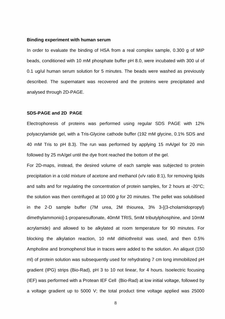
In order to evaluate the specificity of binding, a competitive experiment was
performed (see Figure 6). The beads were assayed for the rebinding with a mixture
made of a fixed concentration of the template and an increasing concentration of a
competing protein. The quantity of the protein bound to the beads was evaluated after
elution from beads and SDS-PAGE. The competitor chosen was Carbonic Anhydrase
(CA), which has approximately the same isoelectric point of HSA but a smaller molecular
mass (31 kDa), (Righetti and Caravaggio, 1976). The shape effect on the binding was
evaluated. In control beads, the HSA binding (approximately 3.5 0.5 ug) is not
perturbed by the addition of CA, accounting for the un-specificity of the binding. In the
MIP, no displacement was observed for CA/HSA molar ratio lower than 3.5/1, while the
addition of the CA competitor at ratio 4.5/1 respect HSA displaces half of the HSA
bound, and CA/HSA ratio above 8/1 give rise to a complete displacement of the HSA
binding, which decreases from 10.3 ug to the unspecific level of 3.5 ug. These
preliminary results indicate that only for relative high concentrations of competitor the
HSA binding is displaced. It follows that samples like human serum, which contains
approximately the 60-80% of albumin, the problem of competition for the imprinted
binding sites might be negligible. For these reasons, the last part of our work was
devoted to the evaluation of the binding performance of imprinted beads on the real
biological fluid human serum. Serum samples are composed of thousands of proteins
and peptides, released from various organs and travelling through the body through the
vessel communication system, accounting for a high complexity of such fluid. Despite
albumin and immunoglobulins constitute the vast majority of the proteic fraction of serum
(ca. 90%), the main interest is focused on the low represented proteins, as their
presence or their quantitative variations are often correlated to pathological states.
Therefore, methods for an efficient removal of HSA are highly welcomed, as they permit
tracing and analysing low abundant proteins, usually masked by serum HSA. In the

15
present research we attempted the use of the imprinted beads for albumin removal.
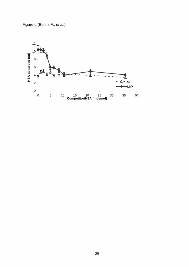
Serum samples were incubated with imprinted beads, the supernatants recovered and
their proteins precipitated with TCA. Recovered proteins were subjected to bi-
dimensional (2D) electrophoresis analysis. The 2D maps of untreated serum and serum
depleted of albumin with imprinted beads were analysed with the software PDquest
(Biorad, Hercules, CA) and are shown in Figure 7. The total number of protein spots
counted for serum was 124 and 116 for MIP-treated serum, indicating again the
imprinted beads are targeted for the binding of HSA and does not remove randomly
other serum components. By analysing the optical density of the albumin spot, a
decrease was reported for serum depleted with imprinted beads, in fact the spot
intensity decreases of a factor 1.7 0.3 upon treatment (i.e. the 58% HSA was
removed). The quantity of albumin removed with the beads was of 183 ug, starting from
311 ug of HSA. It can be concluded that the imprinted beads remove the protein target
also in a complex fluid, leaving nearly unchanged the other proteins. The removal
process is easy and straightforward, being the optimal incubation time of 5 min. Of
course, these preliminary experiments with a complex fluid needs further investigation,
in which the quantity of imprinted beads would be optimised to obtain an almost
complete depletion of HSA from serum.
In conclusion, the results presented here, demonstrate the efficiency of the
immobilisation approach for generating a surface able to recognize a specific protein
target in simple and complex samples. This approach, in the future, may have great
potential applications, in biomaterials for recognition of proteins, in sensing and in
proteome applications.

16
References
Ahmed, N., Barker, G., Oliva, K., Garfin, D., Rice G., 2003. An approach to remove
albumin for the proteomic analysis of low abundance biomarkers in human serum.
Proteomics 3, 1980 –1987.
Andersson, L.I., Muller, R., Vlatakis, G., Mosbach, K., 1995. Mimics of the binding sites
of opioid receptors obtained by molecular imprinting of enkephalin and morphine. Proc.
Natl. Acd. Sci. USA 92 11, 4788-4792.
Andersson, L.I., Schweitz, L., 2003. Solid-phase extraction on molecularly imprinted
polymers. Handb. Anal. 4, 45–71.
Anderson, N.L., Anderson, N.G., 2002. The human plasma proteome: history, character,
and diagnostic prospects. Mol. Cell. Proteomics 1, 845 –867.
Bossi, A., Piletsky, S.A., Piletska, E.V., Righetti, P.G, Turner, A.P.F., 2001. Surface-
grafted molecularly imprinted polymers for protein recognition. Anal. Chem. 73, 5281-
5286.
Bossi, A., Castelletti, L., Piletsky, S.A., Turner, A.P., Righetti, P.G., 2004. Properties of
poly-aminophenylboronate coatings in capillary electrophoresis for the selective
separation of diastereoisomers and glycoproteins. J. Chromatogr A. 1023, 297-303.

17
Burow, M., Minora, N., 1996. Molecular imprinting: synthesis of polymer particles with
antibody-like binding characteristics for glucose oxidase. Biochem. Biophys. Res.
Commun. 227, 419-422.
Chianella, I., Piletsky, S. A., Tothill, I. E., Chen, B., Turner, A. P. F., 2003. Combination
of solid phase extraction cartridges and MIP-based sensor for detection of microcystin-
LR. Biosensors & Bioelectronics 18,119-127.
Dickert, F.L., Hayden, O., 2002. Bio-imprinting of polymers and sol-gel phases.
Selective detection of yeasts with imprinted polymers. Anal. Chem. 74, 1302-1306.
a. Fortis, F., Girot, P., Brieau, O., Castagna, A., Righetti, P.G., Boschetti, E., 2005.
Isoelectric beads for proteome pre-fractionation II: experimental evaluation in a
multicompartment electrolyzer. Proteomics 5, 629-638.
b. Fortis, F., Girot, P., Brieau, O., Castagna, A., Boschetti, E., Righetti, P.G., 2005.
Amphoteric buffering chromatographic beads for proteome prefractionation. I: theoretical
model. Proteomics 5, 620-628.
Guo, T.Y., Xia, Y.Q., Hao, G.J., Song, M.D., Zhang, B.H., 2004. Adsorptive separation
of haemoglobin by molecularly imprinted chitosan beads. Biomaterials 25, 5905-5912.
Haupt, K., Mosbach, K., 2000. Molecularly imprinted polymers and their use in bio-
mimetic sensors. Chem. Rev. 100, 2495–2504.

18
Hjertén, S., Liao, J. L., Nakazato, K., Wang, Y., Zamaratskaia, G., Zhang, H.X., 1997.
Mimicking Antibodies in Their Selective Recognition of Proteins. Chromatographia 44,
227-234.
Katz, A., Davis, M.E., 2000. Molecular imprinting of bulk, microporous silica. Nature 403,
286-289.
Kempe, M., Glad, M., Mosbach, K., 1995. An approach towards surface imprinting using
the enzyme ribonuclease A. Journal of Molecular Recognition 8, 35-39.
Lanza, F., Sellergren, B., 2001. Molecularly imprinted extraction materials for highly
selective sample cleanup and analyte enrichment. Adv. Chromatogr. 3, 41137– 41173.
MacQuade, D.T., Pullen, A.F., Swager, T.M., 2000. Conjugated polymer-based
chemical sensors. Chem. Rew. 14, 2537-2574.
Mattoso, L.H.C., Manohar, S.K., Mac Diamid, A.G., Epstein, A.J., 1995. Studies on the
Chemical Synthesis and on the Characteristics of Polyaniline Derivatives. Polym. Sci.
Pol. Chem. 33, 1227-1230.
Minoura, N., Rachkov, A., 2000. J. Chromatogr. A 889, 111-118.
Mosbach, K., 1994. Molecular Imprinting. Trends in biochemical sciences 19, 9-14.
Mosbach, K., Ramstrom, O., 1996. Biotechnology 14,163-170.

19
Piletsky, S.A., Piletska, E.V., Bossi, A., Karim, K., Lowe, P., Turner, A.P., 2001.
Substitution of antibodies and receptors with molecularly imprinted polymers in enzyme-
linked and fluorescent assays. Biosens Bioelectron. 16, 701-707.
Pribyl, J., Skladal, P., 2005. Development of a combined setup for simultaneous
detection of total and glycated haemoglobin content in blood samples.
Biosens Bioelectron. [Epub ahead of print].
Ramstrom, O., Skudar, K., Haines, J., Patel, P., Bruggemann, O., 2001. Food analyses
using molecularly imprinted polymers. J. Agric Food Chem. 49, 2105-2114.
Righetti, P.G., Caravaggio, T., 1976. Isoelectric points and molecular weights of
proteins. Journal of Chromatography 127,1-28.
Righetti, P.G., Castagna, A., Antonucci, F., Piubelli, C., Cecconi, D., Campostrini, N.,
Antonioli, P., Astner, H., Hamdan, M., 2004. Critical survey of quantitative proteomics in
two-dimensional electrophoretic approaches. J. Chromatogr. A 8, 3-17.
Sato, A.K., Sexton, D.J., Morganelli, L.A., Cohen, E.H., Wu, Q.L., Conley, G.P.,
Streltsova, Z., Lee, S.W., Devlin, M., De Oliveira, D.B., Enright, J., Kent, R.B., Wescott,
C.R., Ransohoff, T.C., Ley, A.C., Ladner, R.C., 2002. Development of mammalian
serum albumin affinity purification media by peptide phage display. Biotechnol.Prog. 18,
182 –192.
a. Sellergren, B., 2001. Molecularly imprinted polymers, man made mimics of
antibodies and their applications in Analytical Chemistry. In: Techniques and
Instrumentation in Analytical Chemistry, vol. 23, Elsevier, Amsterdam.

20
b. Sellergren, B., 2001. Enantiomer separations using designed imprinted chiral
phases. in: G. Subramanian (Ed.), Chiral Separation Techniques, 2nd edition,
Wiley-VCH, Weinheim, pp. 153–184.
Sellergren., B., 2004. Imprinted polymers with memory for small molecules, proteins, or
crystals, Angew. Chem., Int. Ed. 2000: 1031–1037. imprinted polymers, J. Chromatogr.,
B-Anal. Technol. Biomed. Life Sci. 804: 113– 125.
Shi, H.Q., Tsa, W.B.I., Garrison, M.D., Ferrari, S., Ratner, B.D., 1999. Template
imprinted nano-structured surface for protein recognition. Nature 398, 593-597.
Shiomi, T., Matsui, M., Mizukami, F., Sakaguchi, K., 2005. A method for the molecular
imprinting of haemoglobin on silica surfaces using silanes. Biomaterials 27, 5564-5571.
Tirumalai, R.S., Chan, K.C., Da Rue, A.P., Issaq, H.J., 2003. Characterization of the low
molecular weight human serum proteome. Mol. Cell. Proteomics 2, 1096 –1103.
Wang, Y.Y., Cheng, C.P., Chan, D.W., 2003. A simple affinity spin tube filter method
for removing high-abundant common proteins or enriching low-abundant biomarkers for
serum proteomic analysis. Proteomics 3, 243 –248.
Whitcombe, M.J., Alexander, C., Vulfson, E.N., 1997. Smart polymers for the food
industry. Trends Food Sci. Technol. 8, 140–145.

21
Wullff, G., 1993. The role of binding-site interactions in the molecular imprinting of
polymers. Trends Biotechnol. 11, 85-87.
Yilmaz, E., Haupt, K., Mosbach, K., 2000. The Use of Immobilized Templates-A New
Approach in Molecular Imprinting. Angew Chem. Int. Ed. 39, 2115-2118.

22
FIGURE CAPTIONS
Figure 1. Steps of the protein imprinting process with immobilisation of the albumin
template (modified from Shiomi et al., 2005).
Figure 2. Influence of the washing protocol on the pH of the beads and on the removal
of template.
Figure 3. Evaluation of the binding capacity for control (open) and mip beads (solid).
The micrograms bound from beads are plotted as function of micrograms used for
binding.
Figure 4. Binding kinetic. The micrograms of HSA bound are plotted as function of the
time, in saturated condition (a) and no-saturated condition (b).
Figure 5. The IF calculated as micrograms of HSA bound by MIP/ micrograms of HSA
bound by ctrl is plotted as function of the time, both for saturated condition and no-
saturated condition.
Figure 6. Competitive adsorption of HSA in presence of a non template protein,
Carbonic Anhydrase (CA). Rebinding was quantified in ug of HSA adsorbed on MIP and
ctrl beads, after elution of bound proteins and analysis in SDS-PAGE
Figure 7 2D-maps of human serum a) untreated serum sample, b) serum treated with
imprinted beads, for the depletion of HSA.

23
Table 1 (Bonini F., et al.)
Table 1. Recovery of HSA from MIP and control beads.
Solution used for elution ctrl beadsug
MIP beadsug
TUC buffer pH 8.8 0 .3 0.25 0.23 0.20
TUC buffer + 9 M Urea pH 3.0 5.5 0.52 6.0 0.40
7% acetic acid + 0.1% Tween-20 5.8 1.20 9.2 1.50
0.1 M glycine + 0.5 M NaCl, pH 3.0 0.4 0.12 0.24 0.15
1 M NaCl 0.57 0.15 0.85 0.16

24
Figure 1 (Bonini F., et al.)
Introductionaldehydicgroups.
NH2
SSiiOO22
NH2
NH2NH2
NH2
NH2H2N
H2NCHO
CHSSiiOO22
CHO
CHOOHC
CHO
OHC
OHC
SSiiOO22 P
P
P
SSiiOO22 P
P
P
SSiiOO22
Immobilitationprotein
ABPApolymerisation.
Protein template
ABPA used topolymeritation
1 M oxalicacid

25
Figure 2 (Bonini F., et al.)
0
5
10
15
20
25
30
0 2 4 6 8 10
washes
HS
A(u
g)
0
1
2
3
4
5
6
7
8
9
pH
ug
pH

26
Figure 3 (Bonini F., et al.)
0
100
200
300
400
500
600
700
800
0 10,8 17,4 22 54 90 300 540 780 822 1224
HSA loaded (ug)
HS
Aa
ds
orb
ed
(ug
)
ctrl
MIP
ctrl
MIP

27
Figure 4 (Bonini F., et al.)
a)
0
100
200
300
400
500
600
700
800
0 10 20 30 40 50 60
time (min)
HS
Aa
ds
orb
ed
(ug
)
ctrl
MIP
b)
0
2
4
6
8
10
12
0 10 20 30 40 50 60
time (min)
HS
Aa
ds
orb
ed
(ug
)
ctrl
MIP

28
Figure 5 (Bonini F., et al.)
0,9
1,2
1,5
1,8
0 10 20 30 40 50 60
time (min)
Imp
rin
tin
gF
ac
tor
29
Figure 6 (Bonini F., et al.)
0
2
4
6
8
10
12
0 5 10 15 20 25 30 35 40Competitor/HSA (mol/mol)
HS
Aa
ds
orb
ed
(ug
)
ctrl
MIP
30
Figure 7 (Bonini F., et al.)
3 10Mr (kDa)
100
70
50
pI separation
3 10 Mr (kDa)
100
70
50
pI separation