Effect of lyophilization on the structure and phase changes of PEGylated-bovine serum albumin
11
International Journal of Pharmaceutics 304 (2005) 124–134 Effect of lyophilization on the structure and phase changes of PEGylated-bovine serum albumin Virgilio Tattini Jr. a , Duclerc F. Parra b , Bronislaw Polakiewicz a , Ronaldo N.M. Pitombo a,∗ a Department of Biochemical and Pharmaceutical Technology, Pharmaceutical Sciences School, University of S˜ ao Paulo, Av. Prof. Lineu Prestes, 580, Bloco 16, CEP 05508-900 S˜ ao Paulo, SP, Brazil b Nuclear and Energetic Research Institute/University of S˜ ao Paulo, S˜ ao Paulo, Brazil Received 25 February 2005; received in revised form 30 June 2005; accepted 10 August 2005 Available online 26 September 2005 Abstract Poly (ethylene glycol) (PEG) conjugation masks the protein’s surface and increases the molecular size of the polypeptide, thus preventing the approach of antibodies or antigen processing cells and reducing the degradation by proteolytic enzymes. Proteins are readily denatured by numerous stresses arising in solution (e.g., heating, agitation, freezing and pH changes) or by chemical reactions (e.g., hydrolysis and deamidation), many of which are mediated by water. Lyophilization is most commonly used to prepare dehydrated proteins, which, theoretically, should have the desired long-term stability at ambient temperatures. Through Raman spectroscopy, differential scanning calorimetry (DSC) associated with the determination of water content by Karl Fisher titration, it was observed that after the modification of BSA–PEG in a ratio of 1:0.25 showed lower degree of structural alterations and consequently lower variation on the physical–chemical characteristics when it was compared to BSA–PEG (1:0.5). Moreover, the BSA–PEG (1:0.25) optimizes the conditions during the lyophilization process and storage of the protein. © 2005 Elsevier B.V. All rights reserved. Keywords: Freeze–drying; Lyophilization; PEGylation; Bovine serum albumin; Glass transition; Raman spectroscopy 1. Introduction PEGylation is of interest in applied biotechnology because upon modification it masks the protein’s sur- ∗ Corresponding author. Tel.: +55 11 3091 3665; fax: +55 11 3815 6386. E-mail address: [email protected] (R.N.M. Pitombo). face, increases the molecular size of the polypeptide, conveys to molecules its physical–chemical properties and therefore also modifies biodistribution and solu- bility of peptide and non-peptide drugs. This property provides new techniques in biocatalysis and in phar- maceutical technology where many insoluble drugs are solubilized by poly (ethylene glycol) (PEG) conjuga- tion and thus more easily administered (Herman et al., 1995). 0378-5173/$ – see front matter © 2005 Elsevier B.V. All rights reserved. doi:10.1016/j.ijpharm.2005.08.006
-
Upload
independent -
Category
Documents
-
view
1 -
download
0
Transcript of Effect of lyophilization on the structure and phase changes of PEGylated-bovine serum albumin

International Journal of Pharmaceutics 304 (2005) 124–134
Effect of lyophilization on the structure and phase changesof PEGylated-bovine serum albumin
Virgilio Tattini Jr.a, Duclerc F. Parrab, Bronislaw Polakiewicza,Ronaldo N.M. Pitomboa,∗
a Department of Biochemical and Pharmaceutical Technology, Pharmaceutical Sciences School, University of Sao Paulo,Av. Prof. Lineu Prestes, 580, Bloco 16, CEP 05508-900 Sao Paulo, SP, Brazil
b Nuclear and Energetic Research Institute/University of Sao Paulo, Sao Paulo, Brazil
Received 25 February 2005; received in revised form 30 June 2005; accepted 10 August 2005Available online 26 September 2005
Abstract
Poly (ethylene glycol) (PEG) conjugation masks the protein’s surface and increases the molecular size of the polypeptide, thuspreventing the approach of antibodies or antigen processing cells and reducing the degradation by proteolytic enzymes. Proteinsare readily denatured by numerous stresses arising in solution (e.g., heating, agitation, freezing and pH changes) or by chemicalreactions (e.g., hydrolysis and deamidation), many of which are mediated by water. Lyophilization is most commonly used toprepare dehydrated proteins, which, theoretically, should have the desired long-term stability at ambient temperatures. ThroughR arl Fishert lterationsa Moreover,t©
K
1
b
f
tide,rtiesolu-rty
har-area-,
0
aman spectroscopy, differential scanning calorimetry (DSC) associated with the determination of water content by Kitration, it was observed that after the modification of BSA–PEG in a ratio of 1:0.25 showed lower degree of structural and consequently lower variation on the physical–chemical characteristics when it was compared to BSA–PEG (1:0.5).
he BSA–PEG (1:0.25) optimizes the conditions during the lyophilization process and storage of the protein.2005 Elsevier B.V. All rights reserved.
eywords: Freeze–drying; Lyophilization; PEGylation; Bovine serum albumin; Glass transition; Raman spectroscopy
. Introduction
PEGylation is of interest in applied biotechnologyecause upon modification it masks the protein’s sur-
∗ Corresponding author. Tel.: +55 11 3091 3665;ax: +55 11 3815 6386.
E-mail address: [email protected] (R.N.M. Pitombo).
face, increases the molecular size of the polypepconveys to molecules its physical–chemical propeand therefore also modifies biodistribution and sbility of peptide and non-peptide drugs. This propeprovides new techniques in biocatalysis and in pmaceutical technology where many insoluble drugssolubilized by poly (ethylene glycol) (PEG) conjugtion and thus more easily administered (Herman et al.1995).
378-5173/$ – see front matter © 2005 Elsevier B.V. All rights reserved.doi:10.1016/j.ijpharm.2005.08.006

V. Tattini Jr. et al. / International Journal of Pharmaceutics 304 (2005) 124–134 125
Castellanos et al. (2005)have demonstratedthe effectiveness of the covalent modification ofalpha-chymotrypsin with methoxy-PEG to affordits stabilization during encapsulation in poly (lactic-co-glycolic) acid (PLGA) microspheres by asolid-in-oil-in-water method. Alpha-chymotrypsinwas chemically modified with PEG (Mw = 5000) usingmolar ratios of PEG-to-chymotrypsin ranging from 0.4to 96. The results demonstrated that PEG modificationwas able to prevent chymotrypsin aggregation andactivity loss upon solid-in-oil-in-water encapsulationin PLGA microspheres. It is demonstrated that it isessential to optimize the degree of protein modificationto ascertain protein stability upon encapsulation.
The role of PEG in this study was acting as a modi-fier agent where a covalent bond is formed between thePEG polymer and polypeptide drug of choice. Stud-ies of PEG in solution show that each ethylene glycolsub-unit is tightly associated with two or three watermolecules. A binding process with water makes PEGy-lated compounds function as though they are 5–10times larger than a corresponding soluble protein ofsimilar molecular weight. Further, the PEG polymerwith associated water molecules is very mobile, andacts like a shield to protect the attached drug fromenzyme degradation, pH and temperature changes,interactions with cell surface proteins, and providesincreased size to prevent rapid renal filtration and clear-ance. In general, a PEG polymer is first chemically acti-vated in order to react with a polypeptide drug (Katre,1 ntlyl ug.C ighto oft pep-t andi
inp re-f rtero arel clei ccuri s oft oteina ls),l bert vel-
opment of the freeze–drying cycle. Therefore, the glasstransition of a lyophilized product can be studied andapplied to improve processability, quality and stabilityof the product (Chen and Oakley, 1995).
However, infrared spectroscopic studies have docu-mented that the acute freezing and dehydration stressesof lyophilization can induce protein unfolding (Donget al., 1995). The application of Raman spectroscopyto characterize natively unfolded proteins has beenunderdeveloped, even though it has significant tech-nical advantages. The structural changes as a resultof freeze–drying have been investigated, especiallyby Raman spectroscopy. In general, drying resultsin a decrease of�-helix and random structure andan increase in�-sheet structure.Roy and Gupta(2004) studied the case of basic fibroblast growthfactor and�-interferon. The enhanced FTIR showedlarge conformational changes and aggregation dur-ing freeze–drying, which could be prevented by usingsucrose as a lyoprotectant. In the presence of mois-ture, freeze–dried proteins can undergo disulphideinterchange and other reactions, which lead to inac-tivation. Such molecular changes during storage havebeen described for human insulin, tetanus toxoid andinterleukin-2.
The underlying assumption is that IR spectralchanges in the amides I and III regions upon proteindehydration are caused by protein structural changes.However, it has been claimed that amide I IR spec-tral changes could be the result of water removal pers c-t thy-l ousc ehy-d andb o-n n andU d inn t ofH iron-m ate)w Thea ffera ben-z thes ofh thes
993). The activated PEG derivative is then covaleinked to a reactive group on the polypeptide drhanges in the size, structure and molecular wef PEG polymers can affect the biological activity
he attached drug. In general, PEGylation of a polyide lowers its renal clearance, increases its half-lifemproves its biological activity (Harris et al., 2001).
Lyophilization is often used to stabilize proteroducts with limited shelf lives in solution. The
ore, it is not surprising that more than one quaf the therapeutic protein products in the market
yophiles. The determination of the lyophilization cys important because of physical changes that on the solution during freezing and drying phasehe process. Due to the amorphous nature of prnd stabilizer (most commonly sugars and polyo
yophilized formulations often exhibit a glass–rubransition that is an important parameter in the de
e.Al-Azzam et al. (2002)have investigated the struure of horseradish peroxidase (HRP) and poly (eene glycol)-modified HRP (HRP–PEG) under varionditions (in aqueous solution, the amorphous drated state and dissolved/suspended in tolueneenzene) by UV–visible (UV–vis), FTIR and resance Raman spectroscopy. The resonance RamaV–vis spectra of dehydrated HRP–PEG dissolveeat toluene or benzene were very similar to thaRP in aqueous buffer, and thus the heme envent (heme iron spin, coordination and redox stas essentially the same under both conditions.mide I IR spectra of HRP–PEG in aqueous bund of dehydrated HRP–PEG dissolved in neatene and toluene were also very similar, andecondary structure compositions (percentages�-elices and�-sheets) were within the standard errorame.

126 V. Tattini Jr. et al. / International Journal of Pharmaceutics 304 (2005) 124–134
The aim of the current study was to examine theeffects of lyophilization on the structural and phasechanges of PEGylated-BSA. The behavior of the mod-ified protein during each stages of lyophilization pro-cess was evaluated through DSC thermal analysis andRaman spectroscopy.
2. Materials and methods
2.1. Materials
Crystallized bovine serum albumin was purchasedfrom INLAB (Sao Paulo, Brazil).
Methoxy-polyethyleneglycol 5000 (mPEG), aver-age molecular weight 5000, was obtained from SigmaChemical Co. (St. Louis, EUA).
2.2. Methoxy-polyethyleneglycol 5000 synthesis
To obtain the succinyl derivative it was necessary toprovide the following reaction:
The reaction of methoxy-PEG activated complex isprovided below:
Table 1Amount spent in the methoxy-polyethyleneglycol activation reaction
Methoxy-PEG 5000 (g) 15Ethyl acetate (mL) 150N-Hydroxysuccinimide (NHS) (g) 0.86Diciclohexylcarbodiimide (DCC) (g) 1.55
The amount spent in the methoxy-polyethyle-neglycol activation reaction is mentioned inTable 1.
Dehydrated ethyl acetate (standard analytic ethylacetate also fits), methoxy-PEG (already as a succinylderivative), N-hydroxysuccinimide (NHS) and dici-clohexylcarbodiimide (DCC) were poured into a glassreactor with magnetic agitation and controlled heat.The mix rapidly heated till complete reagents solutionand resting for 24 h at 30± 1◦C. At the end of thisperiod, the reaction mass was filtered and left restingfor another 24 h at 5± 1◦C and then filtered to isolatethe solid product that was washed with ice cold ethylacetate. Next, the product was vacuum-dried till reacha constant weight. The material was then dissolved
three times its mass in benzene, and then, the samevolume of petroleum ether was added, the material wascooled and filtered two more times (Clark et al., 1996).
The final methoxy-PEG succinimide yield (obtainedfollowing the above descriptions) was 7.47 g of driedbase.
This material was kept in flask under a nitrogen blan-ket and used in the PEGylation reaction with bovines
2
w ande uffera
pec-t nd1
erum albumin.
.3. Preparation of BSA–PEG solution
A stock solution of 100 mg/mL (150–300�M) BSAas prepared in 5 mM phosphate buffer pH 7.4xhaustively dialyzed overnight against the same bt 4± 1◦C.
It was poured into three reaction vessels, resively: 150 mL of an alkaline phosphate buffer a00 mL of a BSA solution 40 mg/mL.
V. Tattini Jr. et al. / International Journal of Pharmaceutics 304 (2005) 124–134 127
It was added to each vessel, respectively, 1 g(BSA–PEG, 1:0.25), 2 g (BSA–PEG, 1:0.5) and 4 g(BSA–PEG, 1:1) of activated PEG 5000.
The solutions were kept at 30± 1◦C and then trans-ferred to a cold chamber at 8± 1◦C, where they stayed“overnight”. The three samples were purified on aSephadex column G150 calibrated with Tris buffer100 mM, pH 8.6, and eluted with the same buffer (Clarket al., 1996). Then, the modified BSA solutions withPEG 5000 were taken into lyophilization.
2.4. Lyophilization of a BSA–PEG solution
The solutions were lyophilized in a FTS Systems,model TDS-00209-A, microprocessor controlled traydryer (Dura-Stop, Dura-Dry-MP).
A 2 mL of an aqueous solution of BSA–PEG(1:0.25) and (1:0.5) was filled manually into 5 mL glassvials (10 R) and partially closed with rubber stoppers.The freezing step was performed by placing the vialsonto ultra-freezer for 4 h with pre-cooled shelves at
−70± 0.5◦C. Then, the vials were transferred ontothe lyophilizer shelves pre-cooled at−25± 1◦C. Theprimary drying was conducted at−30± 1◦C (producttemperature), chamber pressure at 140± 5 mTorr andcondenser temperature at−90± 1◦C. The secondarydrying was conducted at 25± 1◦C (product tempera-ture), chamber pressure at 50± 5 mTorr and condensertemperature at−90± 1◦C (Fig. 4). The chamber andcondenser pressures were evaluated by a MKS BaratronType 122A Absolute Pressure Transducer. The cham-ber pressure was controlled by a vacuum bleed valvemounted on the inside of the cabinetry on the top ofthe chamber. It runs to the rear of the Tray Dryer toan easily accessible port. The port is equipped witha filter that is an unlaminate 0.2�m PTFE (Teflon)membrane sealed between screen supports. The endof primary and secondary drying was determinedby a Hygrometer Endress + Hauser Hygroguard 2550microprocessor controlled trace moisture analyzer. Alldried samples were stoppered under vacuum pressure at50± 5 mTorr.
), illust .
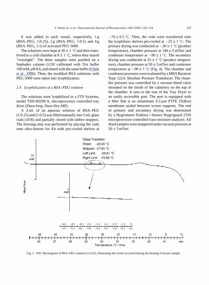
Fig. 1. DSC thermogram of BSA–PEG solution (1:0.25 rating the events occurred during the heating of frozen sample
128 V. Tattini Jr. et al. / International Journal of Pharmaceutics 304 (2005) 124–134
2.5. Determination of glass transition temperature
The glass transition temperature of a BSA–PEGsolutions (T ′
g) and dried samples (Tg) were deter-mined by DSC using a METTLER TOLEDO DSC822 unit. The DSC was calibrated using mercury andindium standards, with onset temperatures of−38.87and 156.6◦C, respectively. For the frozen solutionsthe samples were frozen at−50◦C, and then warmedat 0◦C, both using a cooling/heat flow of 5◦C/minand Nitrogen flow rate of 50 mL/min. In order to pre-vent contamination of the dried samples by moisturein the surrounding air, the lyophilized samples wereweighed into aluminium DSC pans in a dry box filledwith air of equilibrated residual humidity (rh), achievedby the presence of anhydrous phosphorous pentox-ide as a desiccant. A portable hygrometer monitoredhumidity in the dry box. The samples were frozen at0◦C and then warmed at 60◦C, both using a cool-
ing/heat flow of 5◦C/min. TheT ′g andTg values were
measured through five analyses for each BSA–PEGratio.
2.6. Structural analysis
The secondary structure of BSA–PEG solution anddried samples were determined by FT-Raman spec-troscopy using a BRUKER FRA 106/S Raman moduleon an IFS 28/N. All spectra were the average of 200scans for solid samples and 4000 scans for solutions at4 cm−1 resolution. All experiments were performed atroom temperature (25◦C).
The structural analysis was conducted on nativesolution, lyophilized powder and lyophilized materialreconstituted.
For spectral acquisition tubes were filled with:150 mL of solution sample; 30 mg of solid sample;150 mL of rehydrated sample.
), illustr .
Fig. 2. DSC thermogram of BSA–PEG solution (1:0.5 ating the events occurred during the heating of frozen sample
V. Tattini Jr. et al. / International Journal of Pharmaceutics 304 (2005) 124–134 129
Fig. 3. DSC thermogram of BSA–PEG solution (1:1), illustrating the events occurred during the heating of frozen sample.
This procedure was conducted under controlled rel-ative humidity (dry box) achieved by the presence ofanhydrous phosphorous pentoxide as a desiccant. Aportable hygrometer monitored humidity in the drybox.
2.7. Determination of water content
The water content of BSA–PEG freeze–dried sam-ples was determined by Karl Fisher titration using aMETTLER TOLEDO DL 31 Karl Fisher Titrator.
It was added a known amount of anhydrousmethanol with a syringe to the lyophilized BSA–PEGcontainer. The methanol has dissolved the sample.Known amounts of sample and methanol are with-drawn and added to the Karl Fisher titration vessel formoisture determination. The moisture content of theanhydrous methanol is determined as the blank (FDA,1990).
Fig. 4. Comparison of lyophilization process of BSA–PEG (1:0.25)and (1:0.5). The samples presented the same behavior during theprocess.

130 V. Tattini Jr. et al. / International Journal of Pharmaceutics 304 (2005) 124–134
3. Results and discussion
Figs. 1–3represent the DSC thermograms of theBSA–PEG (1:0.25), (1:0.5) and (1:1), respectively.During the rewarming step, all samples presented achange in the heat capacity in the baseline. This base-line shift is related to glass transition temperature (T ′
g).The glass transition temperatures were−28,−11 and−13± 1◦C for the BSA–PEG (1:0.25), (1:0.5) and(1:1), respectively. After theT ′
g an exothermic peak wasobserved for the BSA–PEG (1:0.25) and (1:0.5). Thispeak refers to the recrystallization of some BSA–PEGsolution compounds, as for instance, the phosphatebuffer into the solution and also the polyethylene glycolrecrystallization.
According toRadaev and Sun (2002), during freez-ing PEG has the tendency to crystallize. However, thischaracteristic is dependent to size and concentration ofPEG. On the BSA–PEG (1:1) the solution showed awell-defined glass transition temperature without crys-
tallization peak due to the strongly amorphous charac-teristics during it’s freezing.
Amin et al. (2004)have used the PEG as cosol-vent system and they have observed that addition ofPEG 400 to commonly used bulking agents, such asmannitol, sucrose or polyvinylpyrrolidone, caused asignificant change in the thermal properties of thebulking agents as observed by modulated differentialscanning calorimetry. In addition, PEG 8000 was eval-uated as a bulking agent because it also can functionas a cosolvent in solution and forms an acceptablecake after lyophilization. Addition of PEG 400 to PEG8000 caused negligible changes in the thermogram ofthis bulking agent. Surprisingly, the combination ofPEG 8000 and PEG 400 forms a solid lyophilizedcake. The current system can be best described asthe lyophilization of a miscible solution of PEG 8000and PEG 400 resulting in a lyophile that has a crys-talline structure of PEG 8000, which is able to supportPEG 400.
stratin
Fig. 5. DSC thermogram of BSA–PEG lyophilized (1:0.25) illu g theTg during the heating of the sample. Residual moisture (∼=6.4%).
V. Tattini Jr. et al. / International Journal of Pharmaceutics 304 (2005) 124–134 131
Fig. 6. DSC thermogram of BSA–PEG lyophilized (1:0.5) illustrating theTg during the heating of the sample. Residual moisture (∼=5.3%).
The BSA–PEG (1:0.25) and (1:0.5) lyophilizationwere conducted under the same conditions (tempera-ture, pressure and time).
In Fig. 4, no difference can be seen related tothe BSA–PEG (1:0.25) and (1:0.5) behavior duringlyophilization. However, the continuity of BSA–PEG(1:1) studies was not possible because of the difficul-ties found during the lyophilization of the material.The BSA–PEG (1:1) presented a jelly-kind materialpreventing water from subliming during the primarydrying. It takes more then 50 h to dry and even so at theend of the process the cake had showed collapse signs.
After the lyophilization process it was determinedthe residual moisture of the lyophilized powder byKarl Fisher titration. The BSA–PEG (1:0.25) showedresidual moisture between 4.3 and 6.4% (w/w) andthe BSA–PEG (1:0.5) between 2.3 and 5.3% (w/w).Even the lyophilization cycle were conducted under thesame parameters for both BSA–PEG rates we couldsuppose that as higher the PEGylation rate higherthe hydrophilic groups linked to BSA consequentlysmaller the water retention by the system.
Figs. 5 and 6represent the DSC thermograms ofthe BSA–PEG (1:0.25) and (1:0.5) lyophilized pow-der, respectively. A raise onTg value of the mate-rial can be noted. The glass transition temperatureof lyophilized samples were 22± 1 and 41± 1◦Cfor BSA–PEG (1:0.25) and (1:0.5), respectively. TheBSA–PEG (1:0.25) presented an endothermic peak at57± 1◦C and it is related to the material decomposi-tion during the sample heating. The same event was notobserved during the BSA–PEG (1:0.5) heating.
Structural information is obtained through the anal-ysis of the conformationally sensitive amide I band,which is located between 1600 and 1700 cm−1. Thisband is due to the in-plane CO stretching vibration,weakly coupled with CN stretching and in-plane NHbending. Each type of secondary structure (i.e.,�-helix,�-turn and disordered) gives rise to a different COstretching frequency and, hence, has a characteristicband position, which is designated by wavenumber(cm−1). Band positions are used to determine the sec-ondary structural types present in a protein. An analysisof the infrared bands in the amide I region can pro-

132 V. Tattini Jr. et al. / International Journal of Pharmaceutics 304 (2005) 124–134
Fig. 7. Comparison of Raman spectra of BSA native solution (10%)and BSA–PEG solution (1:0.25).
vide quantitative as well qualitative information aboutthe secondary structure of the protein (Carpenter et al.,1998).
Figs. 7–9compare the Raman spectrum betweennative BSA solutions and different PEGylationrates of BSA–PEG solutions. It was observed thatthe modification of bovine albumin with methoxy-polyethyleneglycol induced structural alterations in theprotein conformation. This fact is related to the differ-ent intensities and localization of the absorbance peaksin the Raman spectrum.
A �-helix structure loss was indicated by inten-sity diminution at 1654 cm−1 band. The BSA–PEG(1:0.25) presented less�-helix (1654 cm−1) structuralvariation when compared to BSA–PEG (1:0.5) and
F 0%)a
Fig. 9. Comparison of Raman spectra of BSA native solution (10%)and BSA–PEG solution (1:1).
(1:1). In the amide III region, the bands between 1230and 1240 cm−1 were attributed to the fact that the�-sheet structures also presented spectral alterations,whenever in position as in the intensity, after PEG con-jugation.
The intensity increase in the�-sheet structuresrelated bands is a characteristic sign of proteinaggregation, inducted by the raise of intermolecularinteractions between the H+ group, because thosepolarized groups need to satisfy the necessity ofhydrogen links through intra or intermolecular inter-actions occasioned by water removal. The nativebovine albumin has in its structure more quantityof hydrophilic groups not available in comparisonwith BSA–PEG. We can observe that the higher the
F izeda
ig. 8. Comparison of Raman spectra of BSA native solution (1nd BSA–PEG solution (1:0.5).
ig. 10. Comparison of Raman spectra of BSA native lyophilnd BSA–PEG (1:0.25) and (1:0.5) lyophilized.

V. Tattini Jr. et al. / International Journal of Pharmaceutics 304 (2005) 124–134 133
Fig. 11. Comparison of Raman spectra of BSA–PEG (1:0.25) nativeand rehydrated solution.
quantity of PEG linked to the BSA, the lower will bethe maintenance of the secondary structure, occurredby the low water retention by hydrophilic groups.
In Fig. 10, we observed that the lyophilizedBSA–PEG (1:0.25) presented a better maintenance ofthe secondary structure of the protein than BSA–PEG(1:0.5). This result can also be related to a lower alter-ation or ruptures of the hydrogen bridges and van derWaals forces.
Figs. 11 and 12showed that the BSA–PEG (1:0.25)presented better results of structural maintenance,after lyophilization and rehydration when comparedto BSA–PEG (1:0.5). Three bands (1620, 1630 and1690 cm−1) attributed to antiparallel�-sheet vibrationssuffered different alterations levels. The amides I and
F ativea
III regions presented structural alterations at differentlevels in relation to the different PEGylation rates.
The maintenance of bovine albumin structural con-formation is directly related to water retention throughthe NH2 groups linked to lysine. The 1000 cm−1 wave-length band related to the�-helix (C C N) practi-cally disappeared after BSA modification with PEGat (1:0.5) ratio.
After rehydration of the lyophilized powder,the intensity distribution on band between 1320–1340 cm−1 related to the aliphatic lateral chains washigher for the BSA–PEG (1:0.25).
The lyophilization-induced spectral alterations inthe conformationally sensitive amide I region are dueto protein unfolding and not simply to the loss ofwater from the protein. The intrinsic effects of waterremoval on the vibrational properties of the peptidebond, and hence protein infrared spectra, were foundto be insignificant byPrestrelski et al. (1993). If thedirect vibrational effects of water removal were respon-sible for drying-induced spectral changes, then theinfrared spectra of all proteins should be altered to thesame degree in the dried solid, which is not the case(Carpenter et al., 1998).
It was observed two different behaviors of BSA–PEG lyophilized powder related with structural unfold-ing:
• the BSA–PEG (1:0.25) regains the native conforma-tion upon rehydration (reversible unfolding);
• re-
s thatp fterr tives
4
ingc
• y-werwers ofand
ig. 12. Comparison of Raman spectra of BSA–PEG (1:0.5) nnd rehydrated solution.
a significant fraction of BSA–PEG (1:0.5) agggates upon rehydration (irreversible unfolding).
It has been documented through several proteinrevention of aggregation and recovery of activity aehydration correlate directly to retention of the natructure in the dried solid (Carpenter et al., 1998).
. Conclusions
The present work allowed us to expose the followonclusions:
After the bovine albumin conjugation with methoxPEG, the BSA–PEG (1:0.25) presented a lodegree of the structural alterations and also a lovariation in the physical–chemical characteristicthe protein when compared to BSA–PEG (1:0.5)(1:1).

134 V. Tattini Jr. et al. / International Journal of Pharmaceutics 304 (2005) 124–134
• The bovine albumin conjugation with methoxy-PEGhas increased the glass transition temperature forthe protein solution as for the lyophilized powder,optimizing the lyophilization process and also thestorage conditions below the collapse temperatureof the material.
• The critical parameters (T ′g, TP, TS, PC andTg) for
the lyophilization process and storage conditions ofthe modified BSA–PEG for the (1:0.25), (1:0.5) and(1:1) rates were determined.
• The results obtained suggest a more profound studyto determine the ideal rate of PEGylation, as themolecular modification of the BSA resulted in dif-ferent levels of structural alterations, a major impor-tance factor for the preservation of biological prod-ucts.
Acknowledgements
The authors acknowledge the financial assistance ofCNPq, CAPES, FAPESP. The authors are grateful toProf. Dr. Reinaldo Guidici and Dr. Marlon Martins dosReis (Chemical Engineering Department, PolytechnicSchool, LSCP Group, University of Sao Paulo) for hisvaluable advice on the use of the Raman spectrome-ter. The authors wish to thank Prof. Dr. Jose AbrahaoNeto (Department of Biochemical and PharmaceuticalTechnology, Pharmaceutical Sciences School, Univer-sity of Sao Paulo) for his assistance on the PEGylationr isa
R
A tzer-lene
glycol)-modified horseradish peroxidase in organic solvents:infrared amide I spectral changes upon protein dehydration arelargely caused by protein structural changes and not by waterremoval per se. Biophys. J. 83, 3637–3651.
Amin, K., Dannenfelser, R.M., Zielinski, J., Wang, B., 2004.Lyophilization of polyethylene glycol mixtures. J. Pharm. Sci.93, 2244–2249.
Carpenter, J.F., Prestrelski, S.J., Dong, A., 1998. Application ofinfrared spectroscopy to development of stable lyophilized pro-tein formulations. Eur. J. Pharm. Biopharm. 45, 231–238.
Castellanos, I.J., Al-Azzam, W., Griebenow, K., 2005. Effect ofthe covalent modification with poly (ethylene glycol) on alpha-chymotrypsin stability upon encapsulation in poly (lactic-co-glycolic) microspheres. J. Pharm. Sci. 94, 327–340.
Chen, T., Oakley, D.M., 1995. Thermal analysis of proteins of phar-maceutical interest. Thermochim. Acta 248, 229–244.
Clark, R., Olson, K., Fuh, G., Marian, M., Mortensen, D., Teshima,G., Chang, S., Chu, H., Mukku, V., Canova-Davis, E., Somers, T.,Cronin, M., Winkler, M., Wells, J.A., 1996. Long-acting growthhormones produced by conjugation with polyethylene glycol. J.Biol. Chem. 271, 21969–21977.
Dong, A., Prestrelski, S.J., Allison, S.D., Carpenter, J.F.,1995. Infrared spectroscopic studies of lyophilization- andtemperature-induced protein aggregation. J. Pharm. Sci. 84,415–424.
FDA (Food and Drug Administration), 1990. Guideline for the Deter-mination of Residual Moisture in Dried Biological Products.Center for Biologics Evaluation and Research (CBER).
Herman, S., Hooftman, G., Schacht, E., 1995. Poly(ethylene glycol)with reactive endgroups: I. Modification of proteins. J. Bioact.Compat. Polym. 10, 145–187.
Harris, J.M., Martin, N.E., Modi, M., 2001. Pegylation a novel pro-cess for modifying pharmacokinetics. Clin. Pharmcokinet. 40,539–551.
Katre, N.V., 1993. The conjugation of proteins with polyethylenes to, 91–
P 1993.and
R erg-
R com-
eaction and Mr. Edson Ghilardi (CQMA-IPEN) for hssistance on the DSC analysis.
eferences
l-Azzam, W., Pastrana, E.A., Ferrer, Y., Huang, Q., SchweiStenner, R., Griebenow, K., 2002. Structure of poly(ethy
glycol and other polymers: altering properties of proteinenhance their therapeutic potential. Adv. Drug Del. Rev. 10114.
restrelski, S.J., Tedeschi, N., Arakawa, T., Carpenter, J.F.,Dehydration-induced conformational changes in proteinstheir inhibition by stabilizers. Biophys. J. 65, 661–671.
oy, I., Gupta, M.N., 2004. Freeze–drying of proteins: some eming concerns. Biotechnol. Appl. Biochem. 39, 165–177.
adaev, S., Sun, P.D., 2002. Crystallization of protein–proteinplexes. J. Appl. Cryst. 35, 674–676.