DETECTION, CHARACTERIZATION, AND CONTROL OF BOVINE ...
124
DETECTION, CHARACTERIZATION, AND CONTROL OF BOVINE VIRAL DIARRHEA VIRUS IN DAIRY HERDS A DISSERTATION SUBMITTED TO THE FACULTY OF THE GRADUATE SCHOOL OF THE UNIVERSITY OF MINNESOTA BY Jeremy Math Schefers IN PARTIAL FULFILLMENT OF THE REQUIREMENTS FOR THE DEGREE OF DOCTOR OF PHILOSOPHY James E. Collins, Co-Advisor Sagar Goyal, Co-Advisor July 2010
-
Upload
khangminh22 -
Category
Documents
-
view
1 -
download
0
Transcript of DETECTION, CHARACTERIZATION, AND CONTROL OF BOVINE ...
DETECTION, CHARACTERIZATION, AND CONTROL OF
BOVINE VIRAL DIARRHEA VIRUS IN DAIRY HERDS
A DISSERTATION
SUBMITTED TO THE FACULTY OF THE GRADUATE SCHOOL
OF THE UNIVERSITY OF MINNESOTA
BY
Jeremy Math Schefers
IN PARTIAL FULFILLMENT OF THE REQUIREMENTS
FOR THE DEGREE OF
DOCTOR OF PHILOSOPHY
James E. Collins, Co-Advisor
Sagar Goyal, Co-Advisor
July 2010

© Jeremy Math Schefers 2009
i
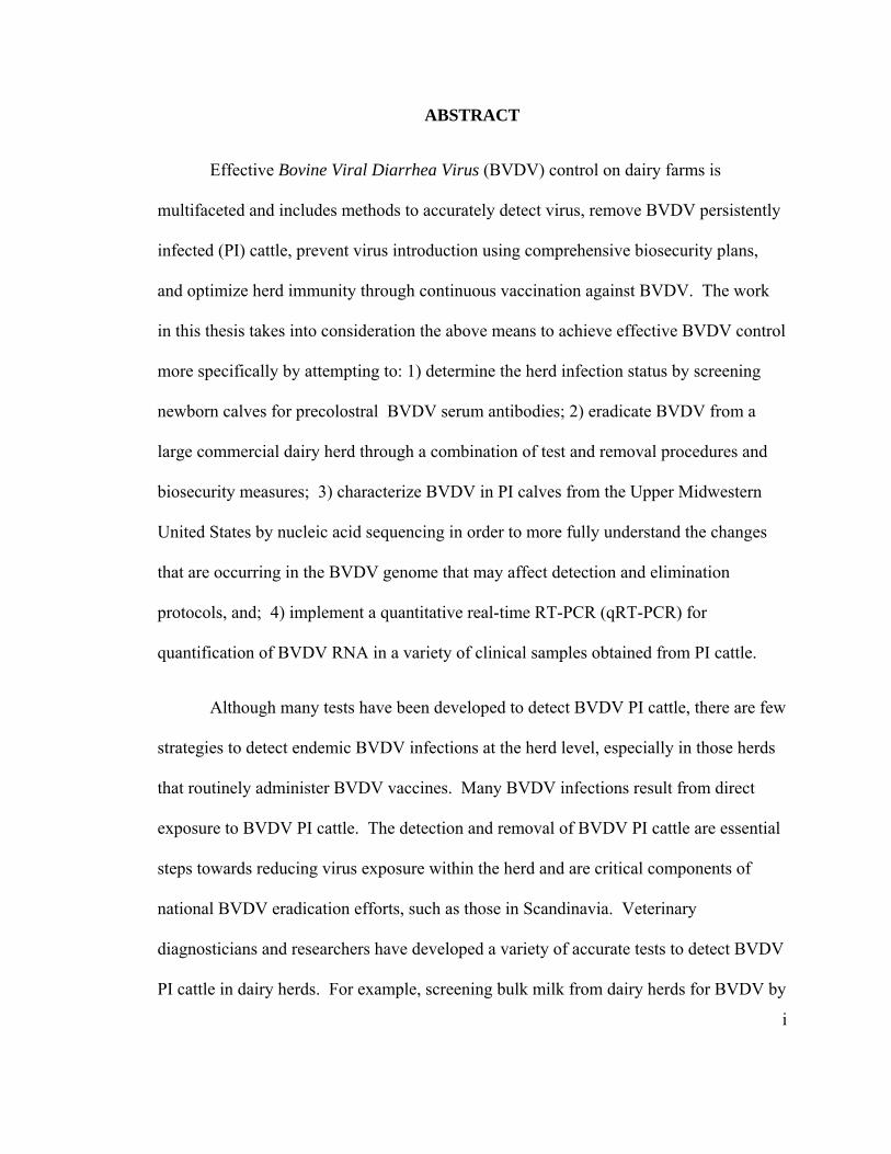
ABSTRACT
Effective Bovine Viral Diarrhea Virus (BVDV) control on dairy farms is
multifaceted and includes methods to accurately detect virus, remove BVDV persistently
infected (PI) cattle, prevent virus introduction using comprehensive biosecurity plans,
and optimize herd immunity through continuous vaccination against BVDV. The work
in this thesis takes into consideration the above means to achieve effective BVDV control
more specifically by attempting to: 1) determine the herd infection status by screening
newborn calves for precolostral BVDV serum antibodies; 2) eradicate BVDV from a
large commercial dairy herd through a combination of test and removal procedures and
biosecurity measures; 3) characterize BVDV in PI calves from the Upper Midwestern
United States by nucleic acid sequencing in order to more fully understand the changes
that are occurring in the BVDV genome that may affect detection and elimination
protocols, and; 4) implement a quantitative real-time RT-PCR (qRT-PCR) for
quantification of BVDV RNA in a variety of clinical samples obtained from PI cattle.
Although many tests have been developed to detect BVDV PI cattle, there are few
strategies to detect endemic BVDV infections at the herd level, especially in those herds
that routinely administer BVDV vaccines. Many BVDV infections result from direct
exposure to BVDV PI cattle. The detection and removal of BVDV PI cattle are essential
steps towards reducing virus exposure within the herd and are critical components of
national BVDV eradication efforts, such as those in Scandinavia. Veterinary
diagnosticians and researchers have developed a variety of accurate tests to detect BVDV
PI cattle in dairy herds. For example, screening bulk milk from dairy herds for BVDV by
ii
RT-PCR is popular due to the ease of sample collection and the large number of animals
that can be screened with one sample. A disadvantage of screening bulk milk is that it
will not detect PI cattle in the non-lactating herd nor in youngstock. Alternatively, non-
vaccinated sentinel calves can be used to detect BVDV PI exposure in youngstock;
however, the sensitivity of sentinel calves in large herds with multiple groups of cattle is
not known.
Chapter 2 of this thesis investigated a novel screening approach to detect BVDV
by screening newborn calves for BVDV serum antibodies prior to colostrum feeding.
Newborn calves that are seropositive for BVDV antibody prior to colostrum feeding
indicate fetal infection during the last two trimesters of gestation. The number of
newborn calves seropositive for BVDV serum antibodies at birth is estimated to be
greater than the number of PI calves. Because the number of BVDV seropositive calves
is greater than the number of BVDV PI calves fewer calves need to be tested by
precolostral serum antibody screening to detect BVDV fetal infections and the
probability of one or more PI cattle in the pregnant herd is likely. In addition to requiring
fewer test animals, precolostral screening detects infections in lactating, non-lactating,
and pregnant youngstock populations and is not confounded by vaccination.
Rapid consolidation of the United States dairy industry has resulted in fewer and
larger dairy herds. Chapter 3 of this thesis describes the elimination of BVDV PI animals
in a large commercial dairy herd with a RT-PCR test. Previous testing in the study herd
indicated that approximately 5% of the calves were born with BVDV precolostral serum
antibodies. The birth of BVDV seropositive calves also roughly coincided with an
iii
increase in post-partum diseases that failed to respond to proven therapies. The herd
owners elected to test all animals for BVDV PI with a serum BVDV RT-PCR test.
Accurate detection of BVDV PI cattle is important in all herds, but less than perfect
sensitivity and the potential of a false negative result are amplified in large herds with PI
cattle. False negative test results would lead to the retention of one or more PI cattle and
ultimately the continued persistence of BVDV within the herd. Serum samples from all
cattle on the premises, and heifer calves born during the following 9 months, were tested
for BVDV by RT-PCR and those determined to be BVDV PI on confirmatory tests were
removed from the herd. Whether or not BVDV persisted in, or was eliminated from, this
herd was determined by monitoring newborn calf precolostral serum antibodies for
BVDV one year after the test and removal of all PI cattle. The chapter describes the
detection of BVDV PI cattle, genetic characterization of BVDV isolated from the PI
cattle, the detection of BVDV acute infections, and the precolostral monitoring results
before and after the removal of PI cattle.
Bovine viral diarrhea virus is a single-stranded RNA virus that lacks a proof-
reading mechanism resulting in mutations and recombination of the viral genome. Point
mutations and recombination of viral RNA can result in novel, unique viruses. Few
animal disease laboratories perform nucleic acid sequencing for BVDV, thus changes in
the BVDV genome are not well described. The objective of chapter 4 was to successfully
sequence a portion of the viral RNA and compare the viral genome sequences of forty PI
cattle detected on dairy farms in the Upper Midwestern United States. The 5’
untranslated region (5’UTR) region of the BVDV genome contains conserved regions
iv
and is commonly used for PCR detection tests. This project described the use of primers
targeting 5’UTR that produce a PCR product for nucleic acid sequence comparisons
between vaccine and field strains and allow for differentiation between subgenotypes
BVDV 1a, BVDV 2a, and BVDV 1b.
Testing many animals for BVDV PI requires appreciable amount of supplies and
labor. The ear notch (skin) sample is a convenient tissue for testing and detecting BVDV
PI animals because it is an easy sample to collect and requires minimal amounts of
supplies and equipment. Ear notch skin samples offer some flexibility because they can
be tested for BVDV by immunohistochemistry (IHC), antigen-capture ELISA (ACE), or
RT-PCR. Pooling ear notch phosphate buffered saline (PBS) supernatant for RT-PCR is
a popular method to screen large numbers of animals at a reduced cost. This method
involves soaking the ear notch in a small amount (~2 ml) of PBS and then pooling the
supernatant. The pooled supernatant is then tested for BVDV by RT-PCR. If the pooled
supernatant is positive, the originally submitted samples can be tested individually to
determine the PI animal. While ear notches have become the sample of choice for PI
testing, there is little information available on the quantity of viral RNA in ear notches
and the PBS supernatant that contains the soaking ear notch. The objective outlined in
Chapter 6 was to implement a quantitative real-time RT-PCR (qRT-PCR) for
quantification of BVDV RNA in a variety of clinical samples obtained from PI cattle.
Serum, whole blood, nasal swabs and skin samples were collected from PI cattle and
analyzed by qRT-PCR. The data derived from qRT -PCR allowed for an estimation of
RNA copies in the variety of samples obtained from PI calves.
v
This thesis will give bovine veterinarians, diagnosticians, and researchers
additional information on the dynamics and manifestations of BVDV in dairy herds. The
precolostral screening method of newborn calves appears feasible and has potential
application in commercial dairy herds. The performance and utility of a highly sensitive
and specific RT-PCR test was assessed and appeared successful in a large commercial
dairy herd. Additionally, nucleic acid sequence analysis of BVDV obtained from PI
dairy cattle were compared to PI cattle from other farms and well-described viruses
strains listed in GenBank. Quantification of BVDV RNA in clinical samples provided
essential information needed to estimate the size of pools and potential variations in
detectable RNA from diagnostics samples.
vi
TABLE OF CONTENTS Abstract i Table of Contents vi List of Tables ix List of Figures x Section A: Literature review Chapter 1: Literature review 1
1.1 Introduction 2 1.1.1 Taxonomy, morphology and virus features 2
1.2 Transplacental and intrauterine infections 4
1.3 Mucosal disease 7
1.4 BVDV and the immune system 8
1.5 BVDV and concurrent infections 9
1.6 Epidemiology and herd screening methods 10
1.7 Detecting BVDV PI cattle 16
1.7.1 Reverse transcription-polymerase chain reaction 18
1.7.2 Immunohistochemistry (IHC) 18
1.7.3 Comparison of tests 18
1.8 Literature review summary 20
vii
Section B: Bovine Viral Diarrhea Virus detection in dairy herds
Chapter 2: Serological evaluation of precolostral serum samples to detect Bovine viral diarrhea virus infections in large commercial dairy herds 21
2.1 Introduction 22
2.2 Materials and Methods 24
2.3 Results 27
2.4 Discussion and Limitations 30
Section C: Detection, characterization and control of Bovine Viral Diarrhea Virus in a dairy herd
Chapter 3: Case Report: Detection, characterization, and control of Bovine Viral Diarrhea Virus in a large commercial dairy herd. 34
3.1 Case Description 35
3.2 Initial precolostral serum sampling 40
3.3 Herd testing for BVDV PI 41
3.4 Follow-up precolostral serum sampling 42
3.5 Molecular characterization of field viruses 43
3.6 Discussion and Limitations 48
Chapter 4: Post mortem examination of aborted fetuses, stillborn calves and non-viable weakborn calves in a large dairy herd endemically infected with Bovine Viral Diarrhea Virus. 55 4.1 Introduction 56 4.2 Materials and Methods 56 4.3 Results 57
4.4 Discussion 60
viii
Section D: Molecular Epidemiology: Characterization of Bovine Viral Diarrhea Virus in persistently infected calves by nucleic acid sequencing
Chapter 5: Bovine Viral Diarrhea Virus: Genetic analysis and signalment of persistently infected dairy calves in the upper Midwestern United States. 65
5.1 Introduction 66
5.2 Materials and Methods 68
5.3 Results 70
5.4 Discussion and Limitations 77
Section E: Quantity of BVDV RNA copies in clinical samples from PI calves using quantitative RT-PCR
Chapter 6: Quantification of Bovine Viral Diarrhea Virus (BVDV) RNA in clinical samples obtained from persistently infected calves using quantitative RT-PCR. 80
6.1 Introduction 81
6.2 Materials and Methods 83
6.3 Results 85
6.4 Discussion 90
Chapter 7: Comparison of two ear notching devices for the detection of Bovine Viral Diarrhea Virus (BVDV) RNA by quantitative RT-PCR. 94
7.1 Introduction 95
7.2 Materials and Methods 95
7.3 Results and Conclusions 99
Concluding Summary: 100
Section F: References 103
ix
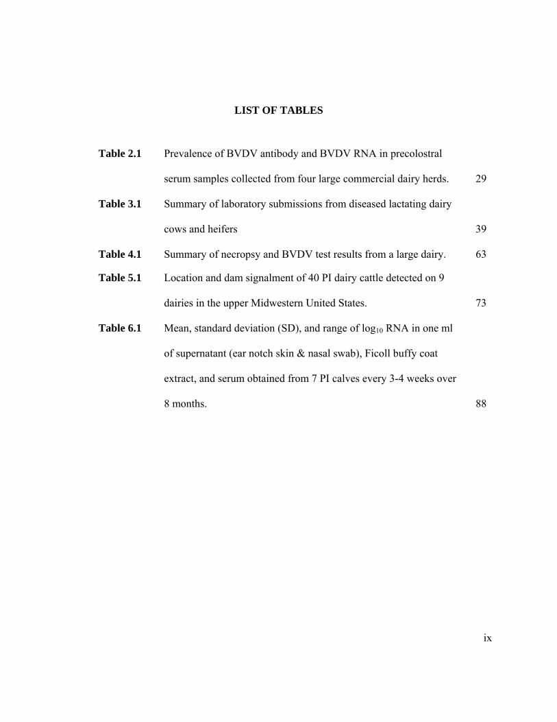
LIST OF TABLES
Table 2.1 Prevalence of BVDV antibody and BVDV RNA in precolostral
serum samples collected from four large commercial dairy herds. 29
Table 3.1 Summary of laboratory submissions from diseased lactating dairy
cows and heifers 39
Table 4.1 Summary of necropsy and BVDV test results from a large dairy. 63
Table 5.1 Location and dam signalment of 40 PI dairy cattle detected on 9
dairies in the upper Midwestern United States. 73
Table 6.1 Mean, standard deviation (SD), and range of log10 RNA in one ml
of supernatant (ear notch skin & nasal swab), Ficoll buffy coat
extract, and serum obtained from 7 PI calves every 3-4 weeks over
8 months. 88
x
LIST OF FIGURES
Figure 2.1 Image of the collection of precolostral blood from a newborn dairy
calf. 27
Figure 3.1 Image of a group of pregnant heifers on a commercial dairy farm
that contained a BVDV PI animal. 36
Figure 3.2 Image of dairy freestall barn containing lactating cows. 36
Figure 3.3 Phylogenic tree analysis of BVDV detected in the PI heifers from a
commercial dairy herd. 45
Figure 3.4 Percent identity chart of BVDV detected in the PI heifers from a
commercial dairy herd. 46
Figure 4.1 Image of an aborted bovine fetus and three stillborn calves
collected from a large commercial dairy farm with endemic BVDV
infections. 64
Figure 5.1 Phylogenetic tree constructed from 40 PI dairy calves and BVDV
reference strains. 72
Figure 5.2 Image of PI Holstein bull calf purchased at a Minnesota auction
facility. 76
Figure 6.1 Comparison of the log10 BVDV RNA in ear notch PBS, buffy coat
extract, serum and nasal swab PBS on 10 collection dates in 7 PI
calves over 8 months. 89
Figure 7.1: Image of skin samples collected from one of seven BVDV PI
calves. 95
xi
Figure 7.2 Prosampler® pliers and collection vials (clip) containing 0.5 mls PBS. 95
Figure 7.3 Chart of the quantity of BVDV RNA in a circular ear sample
(Prosampler®) and a triangular notch in PI calves as determined by
quantitative RT-PCR. 96
1
SECTION A: Literature review
CHAPTER 1: LITERATURE REVIEW
2
1.1 Introduction
In March 1946, Olafson, MacCallum and Fox described an acute, infectious and
contagious disease of cattle. The disease was characterized by leukopenia, high
temperatures, salivation, nasal discharge, diarrhea, depression, anorexia, dehydration and
occasional abortions pregnant cattle. Infected cattle had ulcers of mucous membranes
lining the lips, cheeks, tongue, pharynx and esophagus. In the initial cases, the cattle had
decreased milk production and mortalities ranged from four to eight percent. The cattle
described by Olfason, MacCallum and Fox are thought to be the first described cases of
Bovine Viral Diarrhea Virus (BVDV) infection. Since then, thousands of peer-reviewed
manuscripts and at least three books have been written on BVDV. This review of
literature will cover topics relevant to this dissertation.
1.1.1 Taxonomy, morphology and virus features
Recent taxonomy and classification of BVDV is described in a text entitled; Virus
Taxonomy: Classification and Nomenclature of Viruses: Eighth report of the
International Committee on the Taxonomy of Viruses (van Regenmortel MHV et al.:
2005). The following paragraphs summarize the properties and features of BVDV as
reported in the text.
Bovine viral diarrhea virus (BVDV) is an enveloped, positive sense ssRNA virus
in the Flaviviridae family and Pestivirus genus. The virus is approximately 12.3 kb in
size and most of the viral genome is spanned by a single ORF. Virions are composed of
a nucleocapsid protein (C) and three enveloped glycoproteins, Erns, E1, and E2.
3
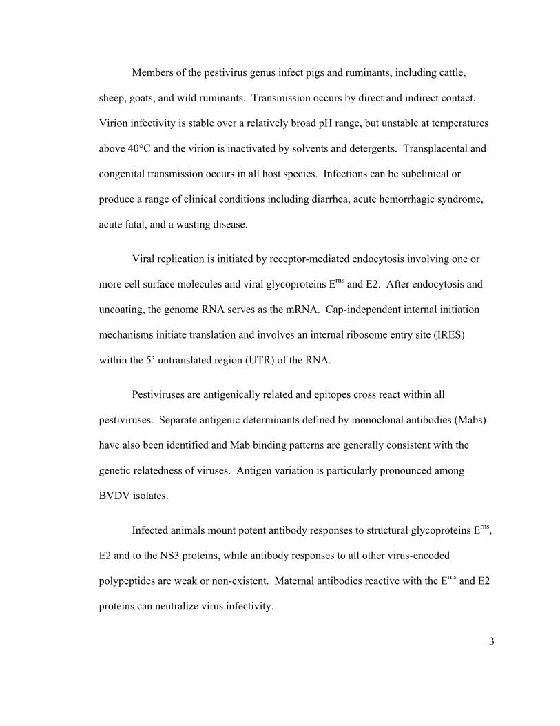
Members of the pestivirus genus infect pigs and ruminants, including cattle,
sheep, goats, and wild ruminants. Transmission occurs by direct and indirect contact.
Virion infectivity is stable over a relatively broad pH range, but unstable at temperatures
above 40°C and the virion is inactivated by solvents and detergents. Transplacental and
congenital transmission occurs in all host species. Infections can be subclinical or
produce a range of clinical conditions including diarrhea, acute hemorrhagic syndrome,
acute fatal, and a wasting disease.
Viral replication is initiated by receptor-mediated endocytosis involving one or
more cell surface molecules and viral glycoproteins Erns and E2. After endocytosis and
uncoating, the genome RNA serves as the mRNA. Cap-independent internal initiation
mechanisms initiate translation and involves an internal ribosome entry site (IRES)
within the 5’ untranslated region (UTR) of the RNA.
Pestiviruses are antigenically related and epitopes cross react within all
pestiviruses. Separate antigenic determinants defined by monoclonal antibodies (Mabs)
have also been identified and Mab binding patterns are generally consistent with the
genetic relatedness of viruses. Antigen variation is particularly pronounced among
BVDV isolates.
Infected animals mount potent antibody responses to structural glycoproteins Erns,
E2 and to the NS3 proteins, while antibody responses to all other virus-encoded
polypeptides are weak or non-existent. Maternal antibodies reactive with the Erns and E2
proteins can neutralize virus infectivity.
4
Pestivirus species differentiation considers several parameters and their
relationship to the type viruses of the currently recognized species. Nucleotide sequence
relatedness is and important criterion for pestivirus species differentiation. Parameters
for differentiation often include: 1) at least 25% difference at the nucleotide level across
the entire genome; 2) at least 10-fold difference in neutralization titre in cross-
neutralization tests with polyclonal immune sera; and 3) differences in host or origin,
range, and disease can assist in species identification.
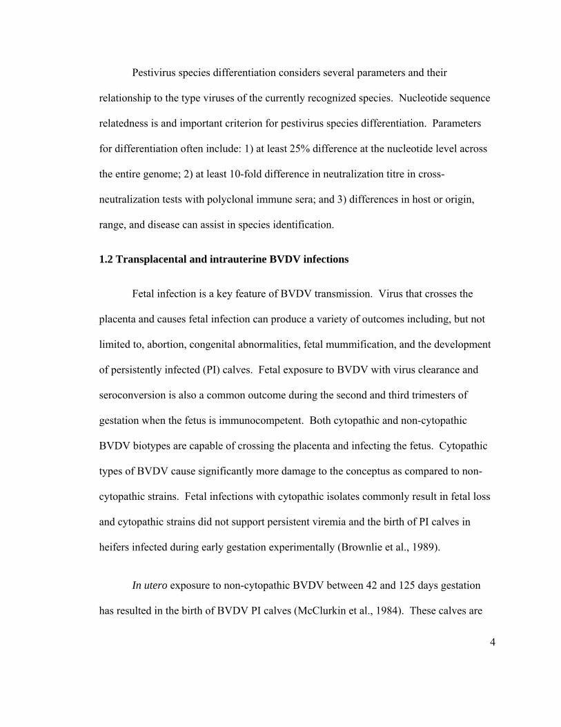
1.2 Transplacental and intrauterine BVDV infections
Fetal infection is a key feature of BVDV transmission. Virus that crosses the
placenta and causes fetal infection can produce a variety of outcomes including, but not
limited to, abortion, congenital abnormalities, fetal mummification, and the development
of persistently infected (PI) calves. Fetal exposure to BVDV with virus clearance and
seroconversion is also a common outcome during the second and third trimesters of
gestation when the fetus is immunocompetent. Both cytopathic and non-cytopathic
BVDV biotypes are capable of crossing the placenta and infecting the fetus. Cytopathic
types of BVDV cause significantly more damage to the conceptus as compared to non-
cytopathic strains. Fetal infections with cytopathic isolates commonly result in fetal loss
and cytopathic strains did not support persistent viremia and the birth of PI calves in
heifers infected during early gestation experimentally (Brownlie et al., 1989).
In utero exposure to non-cytopathic BVDV between 42 and 125 days gestation
has resulted in the birth of BVDV PI calves (McClurkin et al., 1984). These calves are
5
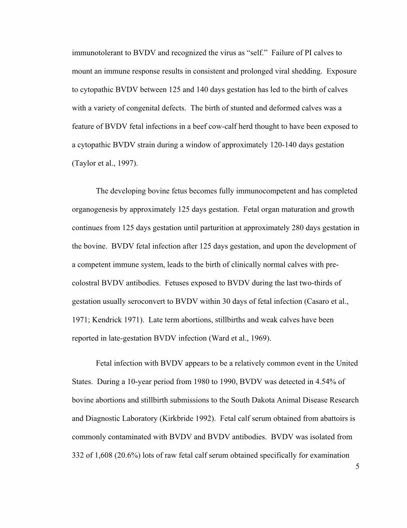
immunotolerant to BVDV and recognized the virus as “self.” Failure of PI calves to
mount an immune response results in consistent and prolonged viral shedding. Exposure
to cytopathic BVDV between 125 and 140 days gestation has led to the birth of calves
with a variety of congenital defects. The birth of stunted and deformed calves was a
feature of BVDV fetal infections in a beef cow-calf herd thought to have been exposed to
a cytopathic BVDV strain during a window of approximately 120-140 days gestation
(Taylor et al., 1997).
The developing bovine fetus becomes fully immunocompetent and has completed
organogenesis by approximately 125 days gestation. Fetal organ maturation and growth
continues from 125 days gestation until parturition at approximately 280 days gestation in
the bovine. BVDV fetal infection after 125 days gestation, and upon the development of
a competent immune system, leads to the birth of clinically normal calves with pre-
colostral BVDV antibodies. Fetuses exposed to BVDV during the last two-thirds of
gestation usually seroconvert to BVDV within 30 days of fetal infection (Casaro et al.,
1971; Kendrick 1971). Late term abortions, stillbirths and weak calves have been
reported in late-gestation BVDV infection (Ward et al., 1969).
Fetal infection with BVDV appears to be a relatively common event in the United
States. During a 10-year period from 1980 to 1990, BVDV was detected in 4.54% of
bovine abortions and stillbirth submissions to the South Dakota Animal Disease Research
and Diagnostic Laboratory (Kirkbride 1992). Fetal calf serum obtained from abattoirs is
commonly contaminated with BVDV and BVDV antibodies. BVDV was isolated from
332 of 1,608 (20.6%) lots of raw fetal calf serum obtained specifically for examination
6
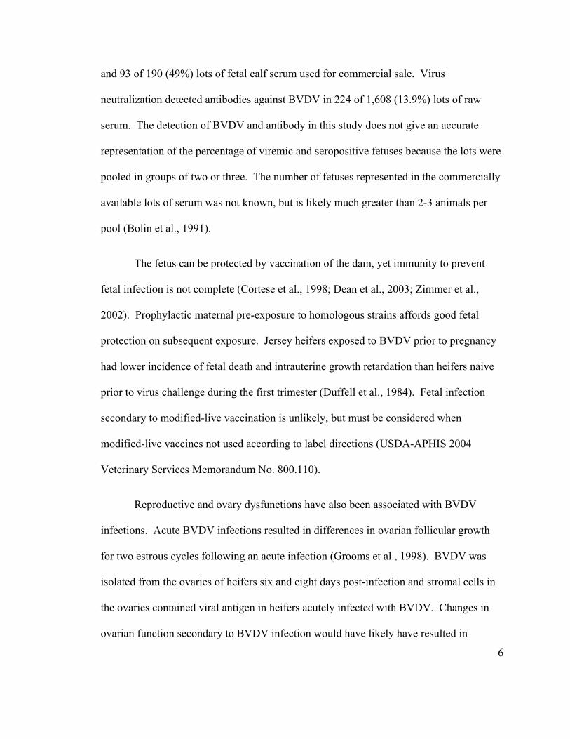
and 93 of 190 (49%) lots of fetal calf serum used for commercial sale. Virus
neutralization detected antibodies against BVDV in 224 of 1,608 (13.9%) lots of raw
serum. The detection of BVDV and antibody in this study does not give an accurate
representation of the percentage of viremic and seropositive fetuses because the lots were
pooled in groups of two or three. The number of fetuses represented in the commercially
available lots of serum was not known, but is likely much greater than 2-3 animals per
pool (Bolin et al., 1991).
The fetus can be protected by vaccination of the dam, yet immunity to prevent
fetal infection is not complete (Cortese et al., 1998; Dean et al., 2003; Zimmer et al.,
2002). Prophylactic maternal pre-exposure to homologous strains affords good fetal
protection on subsequent exposure. Jersey heifers exposed to BVDV prior to pregnancy
had lower incidence of fetal death and intrauterine growth retardation than heifers naive
prior to virus challenge during the first trimester (Duffell et al., 1984). Fetal infection
secondary to modified-live vaccination is unlikely, but must be considered when
modified-live vaccines not used according to label directions (USDA-APHIS 2004
Veterinary Services Memorandum No. 800.110).
Reproductive and ovary dysfunctions have also been associated with BVDV
infections. Acute BVDV infections resulted in differences in ovarian follicular growth
for two estrous cycles following an acute infection (Grooms et al., 1998). BVDV was
isolated from the ovaries of heifers six and eight days post-infection and stromal cells in
the ovaries contained viral antigen in heifers acutely infected with BVDV. Changes in
ovarian function secondary to BVDV infection would have likely have resulted in
7
reduced fertility (Grooms et al., 1998). Cytopathic BVDV was also isolated and detected
in the ovaries from cows and heifers vaccinated with a modified-live vaccine (Grooms et
al., 1998). More recent research indicates that bovine follicular cells and oocytes can be
infected with BVDV at all stages of follicular development and acute infection is
associated with a transient fall in estradiol secretion. Acute BVDV infection is suspected
to reduce fertility by impairment of oocyte quality and disruption of gonadal
steroidogenesis (Fray et al., 2000).
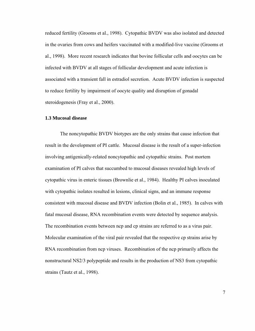
1.3 Mucosal disease
The noncytopathic BVDV biotypes are the only strains that cause infection that
result in the development of PI cattle. Mucosal disease is the result of a super-infection
involving antigenically-related noncytopathic and cytopathic strains. Post mortem
examination of PI calves that succumbed to mucosal diseases revealed high levels of
cytopathic virus in enteric tissues (Brownlie et al., 1984). Healthy PI calves inoculated
with cytopathic isolates resulted in lesions, clinical signs, and an immune response
consistent with mucosal disease and BVDV infection (Bolin et al., 1985). In calves with
fatal mucosal disease, RNA recombination events were detected by sequence analysis.
The recombination events between ncp and cp strains are referred to as a virus pair.
Molecular examination of the viral pair revealed that the respective cp strains arise by
RNA recombination from ncp viruses. Recombination of the ncp primarily affects the
nonstructural NS2/3 polypeptide and results in the production of NS3 from cytopathic
strains (Tautz et al., 1998).
8
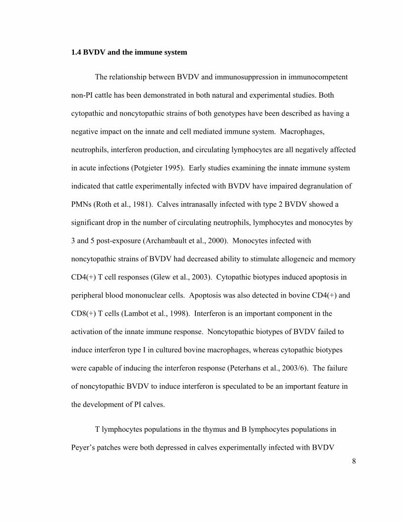
1.4 BVDV and the immune system
The relationship between BVDV and immunosuppression in immunocompetent
non-PI cattle has been demonstrated in both natural and experimental studies. Both
cytopathic and noncytopathic strains of both genotypes have been described as having a
negative impact on the innate and cell mediated immune system. Macrophages,
neutrophils, interferon production, and circulating lymphocytes are all negatively affected
in acute infections (Potgieter 1995). Early studies examining the innate immune system
indicated that cattle experimentally infected with BVDV have impaired degranulation of
PMNs (Roth et al., 1981). Calves intranasally infected with type 2 BVDV showed a
significant drop in the number of circulating neutrophils, lymphocytes and monocytes by
3 and 5 post-exposure (Archambault et al., 2000). Monocytes infected with
noncytopathic strains of BVDV had decreased ability to stimulate allogeneic and memory
CD4(+) T cell responses (Glew et al., 2003). Cytopathic biotypes induced apoptosis in
peripheral blood mononuclear cells. Apoptosis was also detected in bovine CD4(+) and
CD8(+) T cells (Lambot et al., 1998). Interferon is an important component in the
activation of the innate immune response. Noncytopathic biotypes of BVDV failed to
induce interferon type I in cultured bovine macrophages, whereas cytopathic biotypes
were capable of inducing the interferon response (Peterhans et al., 2003/6). The failure
of noncytopathic BVDV to induce interferon is speculated to be an important feature in
the development of PI calves.
T lymphocytes populations in the thymus and B lymphocytes populations in
Peyer’s patches were both depressed in calves experimentally infected with BVDV
9
(Brodersen and Kelling, 1999). There are numerous studies describing the effect of
BVDV on the immune system; however, additional details will not be included in this
review of literature.
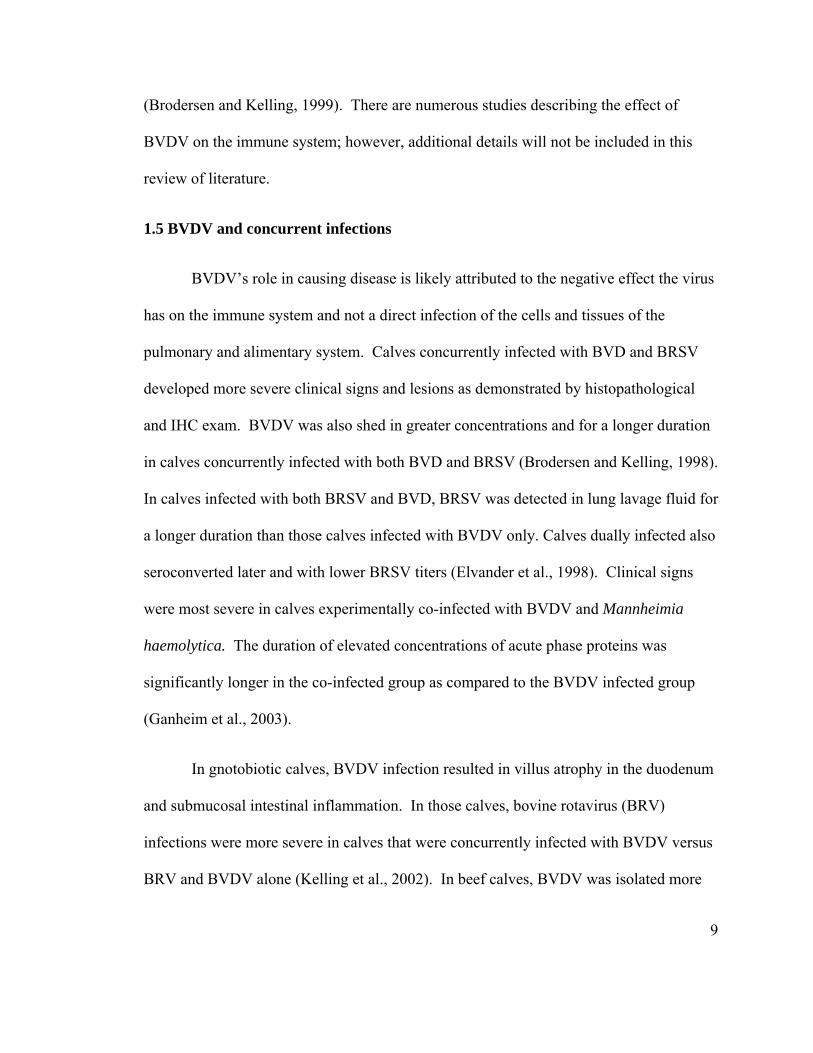
1.5 BVDV and concurrent infections
BVDV’s role in causing disease is likely attributed to the negative effect the virus
has on the immune system and not a direct infection of the cells and tissues of the
pulmonary and alimentary system. Calves concurrently infected with BVD and BRSV
developed more severe clinical signs and lesions as demonstrated by histopathological
and IHC exam. BVDV was also shed in greater concentrations and for a longer duration
in calves concurrently infected with both BVD and BRSV (Brodersen and Kelling, 1998).
In calves infected with both BRSV and BVD, BRSV was detected in lung lavage fluid for
a longer duration than those calves infected with BVDV only. Calves dually infected also
seroconverted later and with lower BRSV titers (Elvander et al., 1998). Clinical signs
were most severe in calves experimentally co-infected with BVDV and Mannheimia
haemolytica. The duration of elevated concentrations of acute phase proteins was
significantly longer in the co-infected group as compared to the BVDV infected group
(Ganheim et al., 2003).
In gnotobiotic calves, BVDV infection resulted in villus atrophy in the duodenum
and submucosal intestinal inflammation. In those calves, bovine rotavirus (BRV)
infections were more severe in calves that were concurrently infected with BVDV versus
BRV and BVDV alone (Kelling et al., 2002). In beef calves, BVDV was isolated more
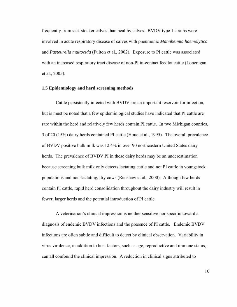
10
frequently from sick stocker calves than healthy calves. BVDV type 1 strains were
involved in acute respiratory disease of calves with pneumonic Mannheimia haemolytica
and Pasteurella multocida (Fulton et al., 2002). Exposure to PI cattle was associated
with an increased respiratory tract disease of non-PI in-contact feedlot cattle (Loneragan
et al., 2005).
1.5 Epidemiology and herd screening methods
Cattle persistently infected with BVDV are an important reservoir for infection,
but is must be noted that a few epidemiological studies have indicated that PI cattle are
rare within the herd and relatively few herds contain PI cattle. In two Michigan counties,
3 of 20 (15%) dairy herds contained PI cattle (Houe et al., 1995). The overall prevalence
of BVDV positive bulk milk was 12.4% in over 90 northeastern United States dairy
herds. The prevalence of BVDV PI in these dairy herds may be an underestimation
because screening bulk milk only detects lactating cattle and not PI cattle in youngstock
populations and non-lactating, dry cows (Renshaw et al., 2000). Although few herds
contain PI cattle, rapid herd consolidation throughout the dairy industry will result in
fewer, larger herds and the potential introduction of PI cattle.
A veterinarian’s clinical impression is neither sensitive nor specific toward a
diagnosis of endemic BVDV infections and the presence of PI cattle. Endemic BVDV
infections are often subtle and difficult to detect by clinical observation. Variability in
virus virulence, in addition to host factors, such as age, reproductive and immune status,
can all confound the clinical impression. A reduction in clinical signs attributed to
11
vaccination and other endemic pathogens such as Salmonellosis, Johne’s disease and
Bovine Respiratory Disease Complex (BRDC) will also confound the clinical impression.
In 52 beef herds suspected of having BVDV PI cattle based on history and clinical signs,
only 10 (19%) actually had BVDV PI cattle (Wittum et al., 2001). In unvaccinated
European herds, PI cattle were detected in 10 of 19 Danish dairy herds with an unknown
status (Houe and Meyling, 1991). Endemic BVDV infections are also masked by slow
transmission rates, as described in a study examining the transmission of BVDV in two
dry-lot dairy youngstock populations (Rush et al., 2001). In those two California dry-lot
dairies with similar management practices, transmission rates ranged from 0.5%/day to
>1.3%/day, and the proportion of calves infected with BVDV by age 9 months was 67%
and 36%, respectively. Although few studies have formally assessed the accuracy of a
veterinarian’s clinical impression, it is likely the accuracy of a practitioner’s clinical
impression is low.
Monitoring dairy herds for PI cattle can be attempted using the following options;
1) monitoring reproduction efficiency, 2) monitoring neonatal and postnatal morbidity
and mortality, 3) serological evaluation of sentinel animals, 4) laboratory examination
aborted fetus and neonatal deaths, 5) attempts to demonstrate seroconversion with acute
and convalescent serology, and 6) screening milk for BVDV by RT-PCR or virus
isolation. If BVDV infections are detected, a whole-herd testing for PI cattle should be
initiated. In addition to testing all ruminants within the herd, all the calves from pregnant
females should be tested for at least 9 months following the entire herd test to detect in
utero PI calves.
12
Monitoring dairy herds for BVDV infections by investigating low reproductive
efficiency is problematic. Many producers and herd managers are constantly striving to
improve reproductive efficiency. The causes of poor reproductive efficiency are
numerous and usually multi-factorial. In herds that artificially inseminate, variations in
fertility can be significantly influenced by human factors. Heat detection efficiency,
artificial insemination technique, routine changes in feedstuffs, and environmental factors
can all have a significant impact on conception rates and reproductive efficiency. Post-
partum metabolic disease and infectious diseases that directly impact the reproductive
tract can result in subsequent losses of reproductive efficiency. Although there are
numerous man-made and environmental factors affecting reproductive efficiency on
dairy herds, endemic BVDV infections will contribute to decreased reproductive
performance; however, endemic BVDV infections are not expected to have a profound,
widespread impact. In one study examining 128 US beef herds, the fall pregnancy rate
was 5% lower in herds with PI calves than those herds without PI calves (Wittum et al.,
2001). Studies examining and comparing the reproductive efficiency in dairy herds with
and without endemic BVDV infections are lacking.
BVDV infections have a negative impact on neonatal and postnatal calf health,
yet detecting the infections can be challenging. Screening calves for BVDV infection can
be accomplished by attempting to demonstrate virus in sick calves by virus isolation, RT-
PCR, or immunohistochemistry. In many cases, testing young calves (< 4 months old)
can only be done by demonstrating virus (VI, RT-PCR, IHC) in acute infections and PI
cattle. Widespread use of vaccines and high levels of serum antibody obtained from
13
colostrum has limited the usefulness of serology in young calves. BVDV antibodies in
colostrum fed to calves from vaccinated dams will circulate for up to 4 months. Calves
are predicted to have a mean antibody titer of 1:32 for type I BVDV and 1:16 for type II
BVDV by three months and congenitally infected calves, i.e. those born with BVDV
antibody and had seroconverted in utero, had higher antibody titers than non-infected
cohorts (Munoz-Zanzi et al., 2002).
Serologic evaluation of unvaccinated sentinel calves has been used to detect the
presence of BVDV PI cattle in dairy and beef herds. The presence of non-vaccinated,
seropositive calves serves as indirect evidence of virus exposure and the likelihood of at
least one PI calf within the herd. In 14 Michigan dairy herds, BVDV PI cattle were
detected when 3 of 5 unvaccinated 6- to 12-month-old heifers had antibody titers > or =
128. In the this herd screening strategy, evaluating 5 sentinel calves for the presence of
BVDV PI calves had a sensitivity of 66% and a specificity of 100%. When virus
isolation was included with SN test to detect PI animals, the sensitivity increased to 83%.
In this study, one of the sentinel calves was PI and because PI calves rarely seroconvert,
the PI sentinel calves was classified as non-exposed (Pillars and Grooms, 2002). In a
more recent study, PI cattle were predicted with high accuracy in Japanese dairy herds by
the use of serum neutralization and virus isolation in three unvaccinated calves 6- to 12-
months-old. When both SN and virus isolation were used, 5 of 20 herds were corrected
identified as containing PI cattle (sensitivity 100%). In herds without PI cattle, the
testing strategy classified 13 or 15 herds as negative (specificity 86%). In the Japanese
study, the SN cut-off titer was 64 and the herd was considered infected if two of three
14
non-PI calves had titers greater than 64. The majority of the herds in this study were
dairy (19/20) and herd size ranged from 23 to 183 animals (Seki et al., 2006).
In beef herds, the use of sentinel samples was also attempt to predict PI cattle.
Three of 10 randomly selected calves had a titer greater than 1:1000 in 53% of the herds
with PI cattle. Unfortunately, at least 3 of 10 calves also had titers greater than 1:1000 in
20% of the herds that did not have a PI calf. Serological evaluation of a subset of beef
calves in Canadian beef cattle herds had a sensitivity of 53% and a specificity of 80%.
Serologic evaluation of calves or cows did not accurately predict the presence of PI cattle
in these beef cattle herds. Determining a herd as infected may be a result of fenceline
contact with other herds or the sharing of pastures with neighboring cattle. The beef cow
herds ranged in size from 54 to 280 cattle (Waldner and Campbell, 2005).
The limited sensitivity of sentinel calves may result from variations in herd size
and low stocking density that limits exposure to PI cattle. Testing large herds with
multiple groups of cattle will result in decreased sensitivity of sentinel calf groups.
Sentinel calves would need prolonged, direct contact with almost all cattle in the herd to
be effective sentinels. Without direct contact with a PI animal, seroconversion in the
sentinel calves will not happen. The probability of direct contact with a PI animal
decreases when there are multiple groups of cattle spread over a larger geographical area.
The sensitivity of sentinel calf groups can be improved by having multiple groups or
repeating groupings over time.
15
The specificity of sentinel calves appears high. In the two dairy herd examples
mentioned previously, the specificity of sentinel calves was 100 and 86%. In the beef
herd study, the arbitrary SN cut-off value resulted in herds being classified as false
positive, which decreased the specificity of the screening approach. Many of the beef
cattle in that study were vaccinated and non-vaccinated sentinel calves were not used.
Acute and convalescent serology is a popular and reliable way to detect exposure
to numerous pathogens in many animal species. Vaccination, especially modified-live
virus vaccines, will produce high titers that can confound interpretation of serum
neutralization SN titers. There is no accurate method of differentiating BVDV antibody
produced by vaccination, field infection, or antibody transferred from dam to offspring
through colostrum. In addition, many SN tests offered at diagnostic laboratories use two
live viruses (genotypes 1a and 2a) to determine cytopathic effects in cell culture. Most of
the BVDV that has been characterized today is BVDV subgenotype 1b, and examination
of how well type 1a and type 2a viruses cross react with 1b antibody is not known. In one
report, comparison of neutralizing antibodies to type 1a, 1b and 2a from experimentally
infected and vaccinated cattle cautioned against using SN titers alone to differentiate
natural infections from vaccination with MLV vaccines (Jones et al., 2001). Cytopathic
BVDV 1b viruses are available, but not currently offered in an SN tests in diagnostic
laboratories.
An antibody response to BVDV infection can be detected 2-3 weeks postinfection
with a plateau 8-10 weeks post infection (Lambot et al., 1997). Timing the collection of
acute and convalescent samples in vaccinated animals is confounded seroconversion
16
rates. Acute and convalescent serology has the additional drawback in that virus
transmission in dairy herds can be slow (Rush et al., 2001). Selecting acute and
convalescent sampling dates are also difficult because many BVDV infections are
subclinical.
1.7 Detecting BVDV PI cattle
Efficient and reliable tests are essential for BVDV control programs. Accurate
detection and elimination of PI cattle is essential for controlling the transmission of virus.
Historically, detection of PI cattle involved cell culture isolation followed by virus
detection through immunofluorescence or immunoperoxidase monolayer assay (IPMA)
methods. Later, immunohistochemistry (IHC) on formalin-fixed skin samples (ear
notches) was added as a routine test for PI detection. More recently, the detection of
BVDV by reverse transcription-polymerase chain reaction (RT-PCR) and antigen capture
enzyme-linked immunosorbent assay (ACE) has show to be more sensitive and rapid than
by cell culture isolation. Currently, a variety of tests are currently used by diagnostic
laboratories to detect PI cattle. These tests often include IHC on skin biopsies (ear
notches), ACE on fresh skin samples, virus isolation (VI), and RT-PCR on a variety of
samples, including blood, serum and ear-notch supernatant (phosphate buffered saline)
1.7.1 Reverse transcription-polymerase chain reaction (RT-PCR):
Numerous clinical samples including serum, blood (buffy coats), tissues, milk,
nasal swab and soaked skin supernatant (PBS) can all be tested by RT-PCR methods.

17
In dairy herds, screening bulk tank milk by RT-PCR is a popular method to detect
BVDV in lactating dairy cattle (Radwan et al., 1995). Bulk milk contains somatic cells
(leukocytes) that can be screened for BVDV by RT-PCR procedures. In one study, a
persistently infected (PI) cow was detected in a bulk milk sample that contained 162
lactating cows. In the same study, 19 other herds were screened with bulk milk and
individual RT-PCR blood testing. A positive bulk milk test was detected in all ten herds
with one lactating PI animals (Drew et al., 1999). In 2000, Renshaw et al., reported that
VI and RT-PCR are both suitable for detection of BVDV in bulk milk samples when used
independently, but to increase the probability of successful detection and to provide
cross-checks against assay contamination, it is desirable to utilize both methods in
parallel (Renshaw et al., 2000). The primary limitation of bulk milk RT-PCR is that the
screening method will not capture infections in nonlactating cows, calves and heifers.
Pooling ear notch phosphate buffered saline (PBS) for RT-PCR is a popular
method to screen many animals at a reduced cost. The pooling method usually involves
soaking the ear notch in a small amount (approximately 2 ml) of PBS and pooling the
supernatant. The pooled supernatant is then tested for BVDV by RT-PCR. If the pooled
supernatant is positive by RT-PCR, the originally submitted samples are tested
individually.
Ear notches are the preferred samples for PI testing because they are easy samples
to collect and require minimal tools. Kennedy et al., examined pooling supernatant from
ear notches and reported that RT-PCR detected a single positive ACE positive ear notch
in pools up to 100 animals (100% , n = 36) (Kennedy et al., 2006). The pooling could
18
provide an initial, rapid, cost-effective method of screening cattle herds for BVDV PI
animals. Although the pooled procedure appeared sensitive, there is no data on the
quantity of viral RNA copies in skin and the PBS fluid.
The major limitation of RT-PCR is the lack of methods standardization between
laboratories that could compromise the consistent identification animals of animals
infected with BVDV.
1.7.2 Immunohistochemistry (IHC)
Immunohistochemical (IHC) staining of skin biopsy samples from cattle has been
used as a method for the early detection of persistent BVDV infection. Initial reports
described pronounced staining in the keratinocytes and in hair follicle epithelium, hair
matrix cells of the hair bulb, and the dermal papilla of PI cattle. Staining in calves
acutely infected, and not BVDV PI, had different and distinct staining confined to small
foci in the nonfollicular epidermis and follicular ostia (Njaa et al., 2000; Baszler et al.,
1995). Another study reported similar data and concluded the staining of skin biopsy
samples is a reliable method for screening neonatal calves for BVDV PI (Grooms and
Keilen, 2002). Skin biopsy represents an effective method for identifying PI cattle.
1.7.3 Comparison of tests
In one study involving 59 PI Angus calves, skin biopsy samples (ear notches) were
collected and IHC and ACE were compared. Both IHC and ACE detected 100% of PI
calves. In that study, RT-PCR and virus isolation were used as the gold standards.
However, IHC and ACE also detected six and eight acutely infected calves, respectively,
19
at initial screening. Both IHC and ACE are accurate at detecting BVDV-infected calves,
but veterinarians and producers should be advised that both tests detect some calves
acutely infected with BVDV. Repeat testing using VI or RT-PCR on buffy coat samples
should be performed at 30 days after initial screening to conclusively discriminate
between acute and PI (Cornish et al. 2005). In 2007, Edmondson et al. evaluated the
diagnostic proficiency of current methods for detecting BVDV in infected cattle using
intra- and inter-laboratory comparisons. Samples were collected from 4 animals more
than 7 months of age (2 BVDV negative animals, a PI animal, and a PI animal that
previously lacked detectable virus in serum as determined by VI). Samples were
submitted to 23 participating diagnostic laboratories using the respective laboratory's
standard submission protocol. Samples collected for submission included: 1) serum for
ACE, RT-PCR, and VI; 2) whole blood for RT-PCR and VI; and 3) skin biopsies for
ACE and IHC. The ACE performed on skin provided the greatest consistency in
detecting positive samples and a perfect level of agreement among laboratories. Reverse
transcription-polymerase chain reaction and IHC performed well by correctly identifying
> or = 85% of samples positive for BVDV. Virus isolation performed on serum yielded
the lowest consistency in detecting positive samples and the lowest level of agreement.
The level of agreement between laboratories for detecting BVDV in persistently infected
cattle ranged from perfect to less than expected by chance. The variation between
laboratories suggests a need for training opportunities in standardized laboratory
protocols and proficiency testing (Edmondson et al., 2007).
20
1.8 Literature review summary
Over sixty years after the initial description, BVDV continues to be an important
disease of cattle worldwide. Decades of research has revealed that the detection and
removal of PI cattle is a critical step in stopping infections. After reviewing the
literature, there appears to be a need for simple, affordable screening methods for large
dairy herds that routinely vaccinate for BVDV. In addition, few case reports have
described the manifestations of BVDV infections in beef and dairy herds, but none have
extensively characterized the infections and documented the control efforts in large (>500
cow) dairy herds. Documenting and reporting the details of a herd control programs will
give veterinarians and producers the confidence to pursue investigations in their clients
herds.
Accurate diagnostic tests are critical for BVDV programs. Screening large groups
of cattle by ear-notching is popular with producers, yet, details on the quantity and
potential variation in pooled skin samples are limited. Additional data are needed to allow
for an estimation and potential variability of detectable BVDV RNA copes in the variety
of samples collected from PI calves.
21
SECTION B: Detection of Bovine Viral Diarrhea Virus in dairy herds
CHAPTER 2
SEROLOGICAL EVALUATION OF PRECOLOSTRAL SERUM SAMPLES TO DETECT BOVINE VIRAL DIARRHEA VIRUS INFECTIONS IN LARGE
COMMERCIAL DAIRY HERDS
Published in: Journal of Veterinary Diagnostic Investigation (2008) 20: 625-628
Jeremy Schefers, Claudia Munoz-Zanzi, James E. Collins, Sagar M. Goyal, Trevor Ames
22
2.1 Introduction
Bovine viral diarrhea virus (BVDV; family Flaviviridae, genes Pestivirus)
continues to be an important pathogen affecting ruminants worldwide. Despite the
widespread use of modified-live and killed vaccines, BVDV persists as a pathogen
causing a wide variety of sub-clinical and clinical infections manifested by respiratory
disease, immunosuppression, and decreased reproductive performance (Brock 2004).
Serological screening strategies to detect herds with endemic BVDV infections
are limited, especially in herds that routinely vaccinate against BVDV. One approach for
detecting endemic BVDV infection involves demonstrating seroconversion in a subset of
animals by testing paired sera (acute and convalescent). The interpretation of this
approach (acute and convalescent serology) is confounded by the widespread use of
BVDV vaccines. Also, seroconversion in a subset of animals is difficult to demonstrate
due to subclinical infections and to the slow intraherd spread of BVDV. Slow intraherd
transmission rates in two California dry-lot dairies with similar management practices
ranged from 0.5%/day to >1.3%/day, and the proportion of calves infected with BVDV
by age 9 months was 67% and 36%, respectively (Rush et al., 2001). The presence of
subclinical infections and slow intraherd transmission creates a diagnostic challenge
when attempting to select the acute and convalescent sampling dates.
Serological evaluation of nonvaccinated sentinel calves has been attempted in
both dairy and beef herds and has been marginally successful (Pillars and Groom, 2002;
Waldner and Campbell, 2005). The presence of nonvaccinated seropositive calves serves

23
as indirect evidence of virus exposure and the likelihood of a BVDV persistently infected
(PI) calf within the herd. In Michigan dairy herds containing less than 200 lactating dairy
cattle, screening nonvaccinated heifers (6–12 months old) had a herd sensitivity of 66%
and a herd specificity of 100% for detecting herds with endemic BVDV infections and
BVDV PI cattle. The limited sensitivity of sentinel calf screening in dairy and beef herds
may be a result of low stocking density and the limited exposure to BVD PI cattle. In
addition, large cattle farms have numerous, variable sized cattle groups that could reduce
exposure of BVD PI cattle to sentinels. Commingling or fence-line contact of cattle to
positive adjacent herds can result in false-positive results and further confound the use of
sentinel calves. Nevertheless, sentinel calves continue to be useful to screen for other
pathogens including infectious bovine rhinotracheitis virus (IBRV) and Leptospira.
Simple, easily interpretable strategies to detect endemic BVDV infections in vaccinated
herds with a wide variety of management practices are necessary to provide herd-level
information essential for a BVDV control program.
Antigen and nucleic acid detection tests for BVDV, such as
immunohistochemistry (IHC) on skin, antigen-capture enzyme-linked immunosorbent
assay (ACE) on skin, and reverse transcription-polymerase chain reaction (RT-PCR) on
various tissues and serum are routinely used for the detection of BVDV PI cattle among
vaccinated and nonvaccinated cattle (Goyal 2005). All three tests have high sensitivity
and specificity for detecting BVDV PI cattle (Cornish et al., 2005; Fulton et al., 2006;
Grooms and Keilen, 2002; Njaa et al., 2000). However, a large number of animals must
be tested to obtain a high level of confidence that the herd is free of BVDV PI cattle
24
because such animals often represent less than 1% of animals within the herd. Pooling
strategies to detect BVDV PI cattle using bulk tank milk (Radwan et al., 1995) or pooled
ear notch supernatants (Kennedy et al., 2006) are popular alternatives because of reduced
testing fees. When sampled correctly, the bulk tank milk–screening test is reliable at
detecting BVDV infections, yet screening bulk milk for BVDV does not capture
infections in nonlactating cows and heifers. Pooling saline from soaked skin samples (ear
notches) is another method to screen many animals and to reduce testing fees; however,
the sensitivity of RT-PCR on pooled ear notch fluids has not been extensively validated
and pooling can result in decreased sensitivity.
Newborn calves can serve as a useful sentinel animal for BVDV infection in a
herd of pregnant cattle. After 125 days gestation, the developing bovine fetus is
immunocompetent and has completed organogenesis. Fetal infection with BVDV after
125 days of gestation (after the development of a competent immune system) usually
leads to the birth of normal calves with precolostral BVDV antibodies (Casaro et al.,
1971; Kendrick 1971). Therefore, detecting fetal infection gives a clear indication that
BVDV is circulating within the herd, crossing the placenta, and causing fetal infections.
2.2 Materials and Methods
The current study was conducted on four large commercial dairy farms of at least
1000 lactating cows each; two herds were in California (herds A and B) and two were in
Minnesota (herds C and D). The data collected from the California herds has been
previously published in an article examining the health impact of natural congenital
25
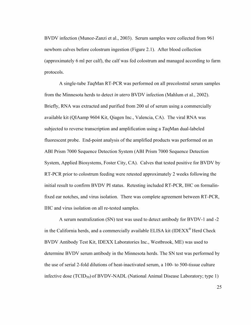
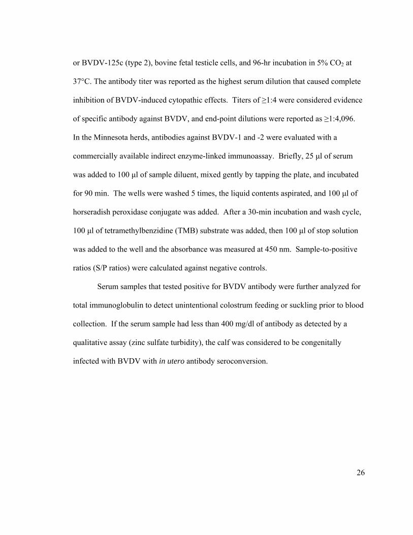
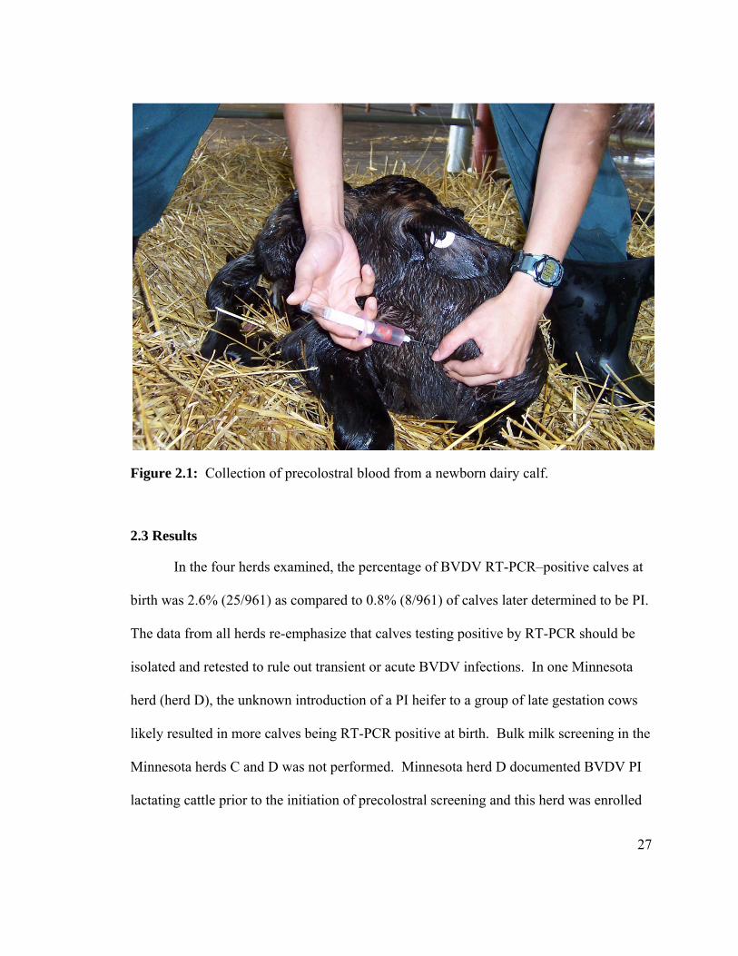
BVDV infection (Munoz-Zanzi et al., 2003). Serum samples were collected from 961
newborn calves before colostrum ingestion (Figure 2.1). After blood collection
(approximately 6 ml per calf), the calf was fed colostrum and managed according to farm
protocols.
A single-tube TaqMan RT-PCR was performed on all precolostral serum samples
from the Minnesota herds to detect in utero BVDV infection (Mahlum et al., 2002).
Briefly, RNA was extracted and purified from 200 ul of serum using a commercially
available kit (QIAamp 9604 Kit, Qiagen Inc., Valencia, CA). The viral RNA was
subjected to reverse transcription and amplification using a TaqMan dual-labeled
fluorescent probe. End-point analysis of the amplified products was performed on an
ABI Prism 7000 Sequence Detection System (ABI Prism 7000 Sequence Detection
System, Applied Biosystems, Foster City, CA). Calves that tested positive for BVDV by
RT-PCR prior to colostrum feeding were retested approximately 2 weeks following the
initial result to confirm BVDV PI status. Retesting included RT-PCR, IHC on formalin-
fixed ear notches, and virus isolation. There was complete agreement between RT-PCR,
IHC and virus isolation on all re-tested samples.
A serum neutralization (SN) test was used to detect antibody for BVDV-1 and -2
in the California herds, and a commercially available ELISA kit (IDEXX® Herd Check
BVDV Antibody Test Kit, IDEXX Laboratories Inc., Westbrook, ME) was used to
determine BVDV serum antibody in the Minnesota herds. The SN test was performed by
the use of serial 2-fold dilutions of heat-inactivated serum, a 100- to 500-tissue culture
infective dose (TCID50) of BVDV-NADL (National Animal Disease Laboratory; type 1)
26
or BVDV-125c (type 2), bovine fetal testicle cells, and 96-hr incubation in 5% CO2 at
37°C. The antibody titer was reported as the highest serum dilution that caused complete
inhibition of BVDV-induced cytopathic effects. Titers of ≥1:4 were considered evidence
of specific antibody against BVDV, and end-point dilutions were reported as ≥1:4,096.
In the Minnesota herds, antibodies against BVDV-1 and -2 were evaluated with a
commercially available indirect enzyme-linked immunoassay. Briefly, 25 μl of serum
was added to 100 μl of sample diluent, mixed gently by tapping the plate, and incubated
for 90 min. The wells were washed 5 times, the liquid contents aspirated, and 100 μl of
horseradish peroxidase conjugate was added. After a 30-min incubation and wash cycle,
100 μl of tetramethylbenzidine (TMB) substrate was added, then 100 μl of stop solution
was added to the well and the absorbance was measured at 450 nm. Sample-to-positive
ratios (S/P ratios) were calculated against negative controls.
Serum samples that tested positive for BVDV antibody were further analyzed for
total immunoglobulin to detect unintentional colostrum feeding or suckling prior to blood
collection. If the serum sample had less than 400 mg/dl of antibody as detected by a
qualitative assay (zinc sulfate turbidity), the calf was considered to be congenitally
infected with BVDV with in utero antibody seroconversion.
27
Figure 2.1: Collection of precolostral blood from a newborn dairy calf.
2.3 Results
In the four herds examined, the percentage of BVDV RT-PCR–positive calves at
birth was 2.6% (25/961) as compared to 0.8% (8/961) of calves later determined to be PI.
The data from all herds re-emphasize that calves testing positive by RT-PCR should be
isolated and retested to rule out transient or acute BVDV infections. In one Minnesota
herd (herd D), the unknown introduction of a PI heifer to a group of late gestation cows
likely resulted in more calves being RT-PCR positive at birth. Bulk milk screening in the
Minnesota herds C and D was not performed. Minnesota herd D documented BVDV PI
lactating cattle prior to the initiation of precolostral screening and this herd was enrolled
28
to document fetal infections in a herd with known endemic BVDV infections. Minnesota
herd D managers did not attempt to detect the lactating PI cattle because they concluded
that additional testing was cost prohibitive. All cattle in Minnesota herd C were tested
for PI by RT-PCR and two heifers were detected in the youngstock population. There
were no lactating PI cattle detected in Minnesota herd C at the time of testing; therefore,
bulk milk testing in Minnesota herd C would likely have been negative at the time of test
and removal. Bulk milk screening in was not attempted in either of the California herds.
No PI calves were born during the sampling period in California herd B.
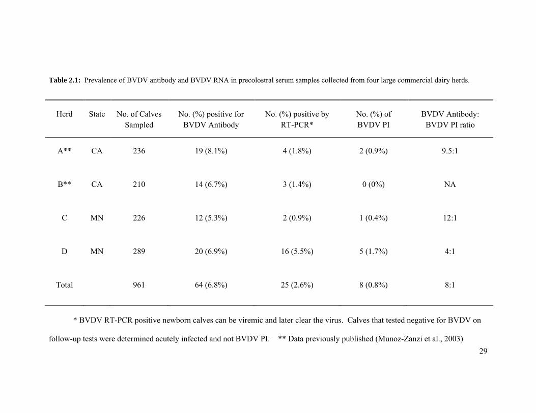
The detection of BVDV antibody in precolostral serum samples correlated with
the birth of PI calves in 3 of 4 herds examined (Table 2.1). BVDV serum antibody was
detected in 6.8% (range 5.3–8.1%) of live newborn calves. The ratio of BVDV antibody
positive to PI in newborn calves across all four herds indicated that for every PI calf born,
there are approximately eight calves congenitally infected and seropositive for BVDV
antibody prior to colostrum feeding.
29
Table 2.1: Prevalence of BVDV antibody and BVDV RNA in precolostral serum samples collected from four large commercial dairy herds.
Herd State No. of Calves Sampled
No. (%) positive for BVDV Antibody
No. (%) positive by RT-PCR*
No. (%) of BVDV PI
BVDV Antibody: BVDV PI ratio
A** CA 236 19 (8.1%) 4 (1.8%) 2 (0.9%) 9.5:1
B** CA 210 14 (6.7%) 3 (1.4%) 0 (0%) NA
C MN 226 12 (5.3%) 2 (0.9%) 1 (0.4%) 12:1
D MN 289 20 (6.9%) 16 (5.5%) 5 (1.7%) 4:1
Total 961 64 (6.8%) 25 (2.6%) 8 (0.8%) 8:1
* BVDV RT-PCR positive newborn calves can be viremic and later clear the virus. Calves that tested negative for BVDV on
follow-up tests were determined acutely infected and not BVDV PI. ** Data previously published (Munoz-Zanzi et al., 2003)
30
2.4 Discussion and Limitations
There is a lack of simple and cost-effective strategies to detect endemic BVDV
infections in large commercial dairy herds that routinely vaccinate for BVDV. The
current “all-antigen” and nucleic acid–detection strategies used to detect BVDV in
vaccinated herds has limitations, and eliminating vaccination to determine seroconversion
is not appealing to producers and veterinarians. The clinical signs associated with BVDV
infection are often subclinical and non-descript. Because of this, livestock producers and
veterinarians are often reluctant to invest the time and resources needed to rule-in or rule-
out an endemic BVDV infection based solely on nonspecific clinical impression.
Hundreds of cattle in a herd of thousands would need to be tested for PI status to achieve
statistical significance that the herd is free of BVDV infections.
In dairy herds with year-round breeding schedules, endemic BVDV infections,
and slow intraherd transmission rates, the risk of fetal BVDV infection would be
expected to be equal across the entire gestational period. The odds of a fetus being
infected during the last half of gestation, a period of approximately 160 days, would be 3
to 4 times greater than the period when fetal infection results in a PI animal
(approximately 50 days). Therefore, screening precolostral serum samples for BVDV
antibodies would theoretically yield 3 to 4 times more BVDV antibody–positive calves
than PI calves. The higher percentage of BVDV seropositive calves than that of BVDV
PI calves reduces the number of animals tested to achieve a high confidence of detecting
31
endemic fetal infection. In the 4 herds examined, there were approximately 8 BVDV
seropositive calves for every calf determined PI.
The relatively high number of calves classified as acutely infected at birth in
Minnesota herd D was unusual and unexpected. The number of acutely infected calves at
birth was twice that of persistently infected calves. Yet, this finding may be explained by
how pregnant cows and heifers are co-mingled on dairy farms during late gestation. On
most dairies, pregnant dry cows (50-60 days prior to calving) and pregnant heifers (50-60
days prior to calving) are first commingled during late gestation. The comingling of
these two groups of cattle shortly before calving is unique to dairy cattle and not beef
cow-calf herds. When these two groups are comingled, a PI dairy heifer or cow has the
potential to infect naïve, late gestating cows or heifers in either group. If these two
groups of cattle were commingled at the beginning of gestation, fewer viremic calves at
birth would be expected, but commingling during late gestation would likely result in
more viremic calves.
Precolostral screening of newborn calves appears to have many advantages.
When newborn calves from first-calf heifers and second lactation and older cows are
screened for precolostral BVDV antibodies, both bred heifers and dry cows are screened
simultaneously. This approach is advantageous over the young stock sentinel program
because it detects virus in pregnant dry cows and first-calf heifers. Herds can continue to
vaccinate nonpregnant cattle with BVDV vaccines because BVDV modified-live virus
32
vaccine has been shown not to shed and infect nonvaccinated animals (Kleiboeker et al.,
2003).
Although precolostral screening does not appear to be confounded by vaccination,
it must be noted that many vaccine companies have received USDA exemptions to use
modified live IBRV and BVDV vaccine in pregnant cows. With the USDA exemption,
modified live virus vaccines can be used in pregnant cows and heifers provided they were
vaccinated, according to label directions, with an approved vaccine prior to breeding. In
regards to BVDV, the USDA exemption study parameters included pre-suckling serum
sampling from at least 400 randomly chosen calves from cattle vaccinated with modified
live vaccinate during the second and third trimester of pregnancy. All pre-suckling serum
samples were tested and found negative for antibodies to types 1 and 2 BVDV, thus
demonstrating lack of fetal exposure to BVDV MLV in utero. Therefore, there is no
evidence to suggest that fetal seroconversion will occur in cattle vaccinated according to
label directions. Improperly administering a BVDV modified-live vaccine to pregnant
cattle during the last two trimesters of pregnancy could result in BVDV fetal infection
and fetal seroconversion that could compromise a precolostral surveillance program.
Therefore, following label directions when vaccinating against BVDV is critical to
maintaining a successful precolostral surveillance program.
Fetal exposure to BVDV and subsequent fetal seroconversion are expected when
late gestation cattle are exposed to BVDV. If BVDV infections are occurring in
nonpregnant cattle, congenital infections will not occur and screening for precolostral
33
BVDV antibody in newborn calves will fail to detect infections in nonpregnant cattle and
cattle in early gestation. Although precolostral screening needs to be validated across
many herds and compared to other screening tests such as bulk tank milk PCR and calf
sentinel programs, preliminary data suggest that screening newborns for BVDV antibody
can reliably detect endemic infections in large dairy herds and be used to monitor the
progress of BVDV control programs over time.
34
SECTION C: Detection, characterization and control of Bovine Viral Diarrhea Virus in a dairy herd
CHAPTER 3
CASE REPORT: DETECTION, CHARACTERIZATION, AND CONTROL OF BOVINE VIRAL DIARRHEA VIRUS IN A LARGE COMMERCIAL DAIRY
HERD
Published in: Canadian Veterinary Journal (2009) 50: 1075-1079
Jeremy Schefers, James E. Collins, Sagar M. Goyal, Trevor Ames
35
3.1 Case description
A large Holstein dairy herd in central Minnesota had been experiencing an
increased rate of post-partum metritis and pneumonia for more than two years. During
the more severe episodes, the post-partum metritis cases approached 40% and the
pneumonia cases approached 30% in post-partum cows and heifers. The pneumonia and
metritis disease episodes did not correlate with known management changes and were
described by the owners and employees as unpredictable events. In addition to metritis
and pneumonia, many post-partum cows and heifers developed diarrhea and Salmonella
montevideo was routinely cultured from the feces of diarrhetic cows. Many diarrhetic
cows were also seropositive for Mycobacterium paratuberculosis antibody by ELISA.
The dairy herd consists of approximately 1,200 lactating cows and 1,800
replacement heifers (Figures 3.1 and 3.2). The herd is similar to many expanding dairy
herds in Minnesota and was originally assembled from small dairy herds, sale-barn
heifers, and heifers from other suppliers. This dairy retains and raises all heifer calves
and does not commingle cattle with other herds. The vaccination protocol includes
vaccinating heifers twice prior to breeding and vaccinating lactating cows once
approximately 30 days post-partum with a commercially available modified-live virus
(MLV) vaccine containing bovine viral diarrhea virus (BVDV), bovine herpes virus
(BHV-1), parainfluenza 3 virus (PI-3), and bovine respiratory syncytial virus (BRSV).
Once pregnant, the cows and heifers are not vaccinated.
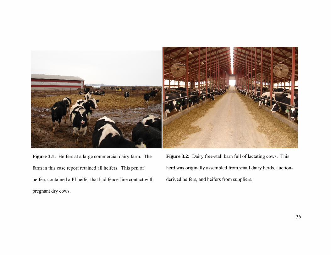
36
Figure 3.1: Heifers at a large commercial dairy farm. The
farm in this case report retained all heifers. This pen of
heifers contained a PI heifer that had fence-line contact with
pregnant dry cows.
Figure 3.2: Dairy free-stall barn full of lactating cows. This
herd was originally assembled from small dairy herds, auction-
derived heifers, and heifers from suppliers.
37
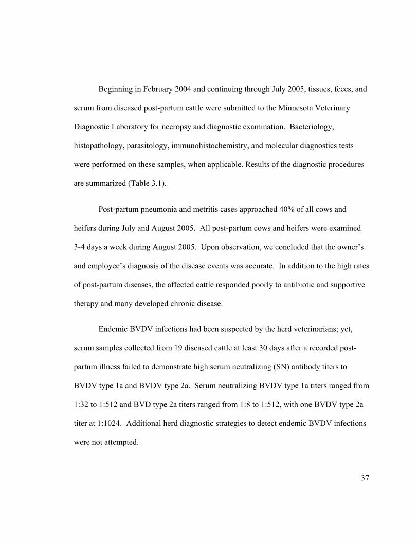
Beginning in February 2004 and continuing through July 2005, tissues, feces, and
serum from diseased post-partum cattle were submitted to the Minnesota Veterinary
Diagnostic Laboratory for necropsy and diagnostic examination. Bacteriology,
histopathology, parasitology, immunohistochemistry, and molecular diagnostics tests
were performed on these samples, when applicable. Results of the diagnostic procedures
are summarized (Table 3.1).
Post-partum pneumonia and metritis cases approached 40% of all cows and
heifers during July and August 2005. All post-partum cows and heifers were examined
3-4 days a week during August 2005. Upon observation, we concluded that the owner’s
and employee’s diagnosis of the disease events was accurate. In addition to the high rates
of post-partum diseases, the affected cattle responded poorly to antibiotic and supportive
therapy and many developed chronic disease.
Endemic BVDV infections had been suspected by the herd veterinarians; yet,
serum samples collected from 19 diseased cattle at least 30 days after a recorded post-
partum illness failed to demonstrate high serum neutralizing (SN) antibody titers to
BVDV type 1a and BVDV type 2a. Serum neutralizing BVDV type 1a titers ranged from
1:32 to 1:512 and BVD type 2a titers ranged from 1:8 to 1:512, with one BVDV type 2a
titer at 1:1024. Additional herd diagnostic strategies to detect endemic BVDV infections
were not attempted.

38
The goal of this case investigation was to detect endemic BVDV infections by
precolostral screening of newborn calves, pursue BVDV control by testing and removing
PI cattle, and genetically characterize BVDV isolates by RNA sequencing techniques.
39
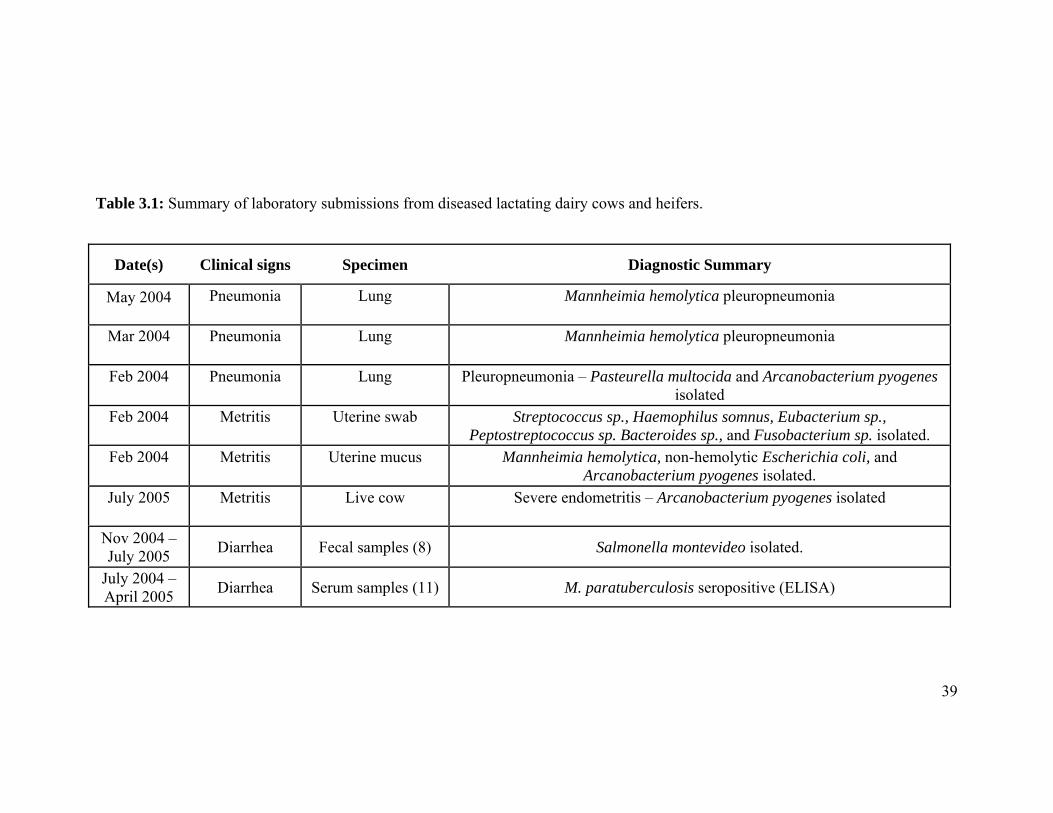
Table 3.1: Summary of laboratory submissions from diseased lactating dairy cows and heifers.
Date(s) Clinical signs Specimen Diagnostic Summary
May 2004 Pneumonia Lung Mannheimia hemolytica pleuropneumonia
Mar 2004 Pneumonia Lung Mannheimia hemolytica pleuropneumonia
Feb 2004 Pneumonia Lung Pleuropneumonia – Pasteurella multocida and Arcanobacterium pyogenes isolated
Feb 2004 Metritis Uterine swab Streptococcus sp., Haemophilus somnus, Eubacterium sp., Peptostreptococcus sp. Bacteroides sp., and Fusobacterium sp. isolated.
Feb 2004 Metritis Uterine mucus Mannheimia hemolytica, non-hemolytic Escherichia coli, and Arcanobacterium pyogenes isolated.
July 2005 Metritis Live cow Severe endometritis – Arcanobacterium pyogenes isolated
Nov 2004 – July 2005
Diarrhea Fecal samples (8) Salmonella montevideo isolated.
July 2004 – April 2005
Diarrhea Serum samples (11) M. paratuberculosis seropositive (ELISA)
40
3.2 Initial precolostral serum sampling
Employees of the dairy were trained to collect a blood sample from newborn
calves prior to colostrum feeding. Trained farm staff collected approximately 6 ml of
blood in Vacutainer® SST tubes (Vacutainer SST, Becton, Dickinson and Company, NJ).
The blood tubes were centrifuged and stored in a refrigerator until bi-weekly delivery to
the Minnesota Veterinary Diagnostic Laboratory. The restraint and collection of blood
from newborn calves was approved by the University of Minnesota Institutional Animal
Use and Care Committee (IACUC).
Serum samples were collected from calves when it was convenient for the
employees and not all calves were sampled. Approximately 75% of live newborn calves
were sampled. During a four month sampling period (September 2005 through December
2005), BVDV antibody was detected in 5.3% (12/226) of newborn calf serum samples
using a commercially available ELISA kit (IDEXX Herdchek BVDV Antibody Test Kit,
Westbrook, ME). Serum samples from calves that tested positive for BVDV antibody
were then tested for total immunoglobulin by zinc sulfate turbidity to rule-out inadvertent
colostrum ingestion. Calves with less that 400 mg/dl IgG were classified as BVDV
congenitally infected with in utero seroconversion. All precolostral serum samples were
tested for BVDV by RT-PCR and 0.8% (2/226) of newborn calves tested positive for
BVDV. One of the calves that tested positive for BVDV died within a day of birth and
the other calf tested negative for BVDV on follow-up tests. None of the 226 calves
sampled during the four month period were confirmed PI.
41
3.3 Herd testing for BVDV PI
Precolostral screening for BVDV antibody in newborn calves gave clear
indication that BVDV was circulating in the pregnant cow and heifer populations. Serum
samples were collected from all animals on the farm (3065 cattle) and tested for BVDV
by RT-PCR. The BVDV RT-PCR procedure, including the primers and protocol, has
been described in a previous publication (Mahlum et al., 2002).
Serum samples from eight of the 3065 cattle tested positive for BVDV by RT-
PCR on the initial test. All cattle that tested positive on the initial test were non-lactating
heifers. Whole blood, serum, and a formalin fixed ear-notch were collected from each of
the eight heifers approximately two weeks following the initial positive RT-PCR test
result. Two of the eight heifers tested positive for BVDV on the follow-up tests. In those
two heifers, follow-up testing detected BVDV antigen in a formalin fixed ear-notch by
immunohistochemistry (IHC), BVDV was again detected in a serum sample by RT-PCR,
and BVDV was isolated from whole blood on bovine turbinate cells. The two heifers that
tested positive on follow-up tests were classified as PI and removed from the herd. The
six remaining heifers that tested negative on follow-up tests were determined as acutely
infected, non-BVDV PI heifers, and were retained in the herd.
The two PI heifers were in the youngstock population and all acute infections
were in heifers with direct contact with the PI heifers. PI cattle and acute BVDV
infections were not detected in lactating cattle. The oldest BVDV PI heifer (#7771) was
16-months-old and was located in a pen with approximately 125 heifers being bred by
42
artificial insemination. The breeding pen containing PI heifer 7771 had direct fence-line
contact with a large pen of approximately 125 dry cows (220-250 days gestation) and a
pen of approximately 80 heifers exposed to a bull. Heifer 7771 had been in the pen for
approximately five months and had direct fenceline contact with more than 300 pregnant
dry cows during the 5 month period. The younger heifer (#8527) was 6-months-old and
was in a treatment pen with 8 other calves. Heifer calf 8527 had developed chronic
pneumonia and died during follow-up testing.
In the nine months following the completion of herd testing, three newborn PI
calves were detected. Two of the three PI calves were bull calves that had persistent
central nervous systems signs including head tremors with caudal neck extension to an
opisthotonic posture, ataxia, and an inability to stand. The affected bull calves had a
bright and alert mentation, but they failed to thrive and were euthanized by intravenous
injection of sodium pentabarbitol. There were no significant lesions noted on necropsy or
histopathological examination of the affected bull calves. Although histopathological
examination of the brain was unremarkable, the neurons in the medulla, pons and
midbrain stained intensely positive for BVDV antigen by IHC, a feature of noncytopathic
PI. Formalin fixed ear notches and tissues from the affected bull calves were also
positive for BVDV by IHC and RT-PCR, respectively.
3.4 Follow-up precolostral serum sampling
Precolostral serum sampling of newborn calves continued for 17 months after the
removal of PI heifers. As expected, BVDV seropositive calves continued to be born
43
during the 5 months following the removal of BVDV PI animals, indicating historical
fetal exposure with in utero seroconversion. In the 12 months following the five month
period of seropositive calves, two of 450 (0.4%) calves tested positive for BVDV
antibody by ELISA. The two calves that tested positive for BVDV by ELISA had less
than 400 mg/dl IgG and were also positive for BVDV antibody by serum neutralization.
All of the 450 precolostral serum samples were negative for BVDV by RT-PCR.
3.5 Molecular characterization of field viruses
To further characterize the virus detected in the PI heifers, the nucleotide
sequence of the 5’ untranslated region (5’ UTR) was analyzed. The 5’ UTR represents a
conserved region of the BVDV genome (Ridpath et al., 1993; Boye et al., 1991).
Comparison of the BVDV 5’ UTR classifies the virus into three genotypes; namely,
BVDV type 1a, BVDV type 1b and BVDV type 2a (Ridpath and Bolin, 1998).
Two sequencing primers were selected from previously published data (Ridpath,
2005). Viral RNA was extracted from the serum using a commercially available RNA
extraction kit (QIAamp Viral RNA Mini Extraction Kit, Qiagen Inc, Valencia, CA), and
RT-PCR was conducted utilizing commercially available kits (Qiagen OneStep RT-PCR
enzyme mix, buffer and dNTPs, Qiagen Inc, Valencia, CA). The reagents, PCR primers
and extracted viral RNA were subjected to thermocycling and a product of approximately
240 base pairs was detected on agarose gel. The RT-PCR products were subjected to
Sanger dye-terminator sequencing and were analyzed using an automated sequence
machine (ABI 3730 DNA Analyzer, Applied Biosystems, Foster City, CA) at the
44
BioMedical Genomics Center, University of Minnesota. Viral nucleotide sequences
were compared to published BVDV sequences in Genbank by using computer software
(Dnastar-Megalign sequence analysis program, Dnastar Inc, Madison,WI).
Phylogenetic tree analysis of the BVDV detected from the PI cattle and BVDV
sequences obtained from Genbank revealed that there were two distinct viruses detected
on this farm. The strain detected in PI heifer 7771 was similar to the subgenotype 1b
Draper reference strain and the strain detected in calf 8527 was similar to the Oregon
C24V type 1a reference strain (Figure 3.3).

45
Figure 3.3: Phylogenic tree analysis of BVDV detected in the PI cattle (bold) and sequences obtained from Genbank. The two
newborn calves and the oldest BVDV PI heifer 7771 are identical to each other and most similar to the Draper subgenotype 1b
reference strain over a 240 base pair segment of the 5’ UTR. The 8527 PI calf (6-month-old heifer) is most similar to the Oregon
C24V type 1a strain over a 240 base pair segment of the 5’ UTR.
Nucleotide Substitutions (x100)
0
15.6
24 681012 14
Oregon C24V 1a 8527 PI Heifer Singer 1a NADL BVD 1aAbnormal Bull Calf Normal Heifer Calf 7771 PI HeiferDraper 1bBVD 2aNew York BVD 2a
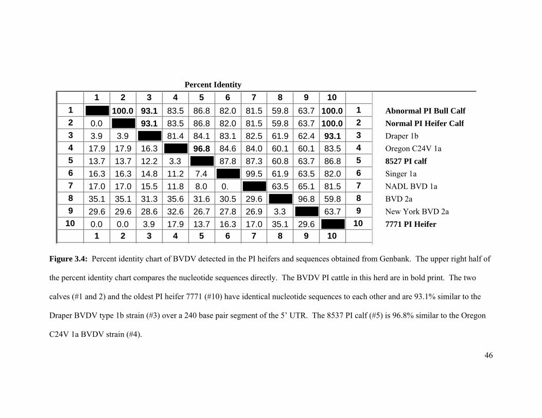
46
Figure 3.4: Percent identity chart of BVDV detected in the PI heifers and sequences obtained from Genbank. The upper right half of
the percent identity chart compares the nucleotide sequences directly. The BVDV PI cattle in this herd are in bold print. The two
calves (#1 and 2) and the oldest PI heifer 7771 (#10) have identical nucleotide sequences to each other and are 93.1% similar to the
Draper BVDV type 1b strain (#3) over a 240 base pair segment of the 5’ UTR. The 8537 PI calf (#5) is 96.8% similar to the Oregon
C24V 1a BVDV strain (#4).
Percent Identity
1 2 3 4 5 6 7 8 9 101 100.0 93.1 83.5 86.8 82.0 81.5 59.8 63.7 100.0 1 Abnormal PI Bull Calf
2 0.0 93.1 83.5 86.8 82.0 81.5 59.8 63.7 100.0 2 Normal PI Heifer Calf
3 3.9 3.9 81.4 84.1 83.1 82.5 61.9 62.4 93.1 3 Draper 1b
4 17.9 17.9 16.3 96.8 84.6 84.0 60.1 60.1 83.5 4 Oregon C24V 1a
5 13.7 13.7 12.2 3.3 87.8 87.3 60.8 63.7 86.8 5 8527 PI calf
6 16.3 16.3 14.8 11.2 7.4 99.5 61.9 63.5 82.0 6 Singer 1a
7 17.0 17.0 15.5 11.8 8.0 0. 63.5 65.1 81.5 7 NADL BVD 1a
8 35.1 35.1 31.3 35.6 31.6 30.5 29.6 96.8 59.8 8 BVD 2a
9 29.6 29.6 28.6 32.6 26.7 27.8 26.9 3.3 63.7 9 New York BVD 2a
10 0.0 0.0 3.9 17.9 13.7 16.3 17.0 35.1 29.6 10 7771 PI Heifer
1 2 3 4 5 6 7 8 9 10
47
The birth and detection of a PI calf is the result of a historical infection. When a
cow or heifer gives birth to known BVDV PI calf, the infection indicates that the dam
was infected with BVDV during the first trimester of pregnancy. Occasionally, the dam
of the PI calf is herself PI, but this is a less frequent observation (Wittum et al., 2001).
Finding the dam of the PI calf and researching the dam’s location during the first
trimester of pregnancy will indicate the probable location of BVDV exposure. In this
herd, both dams of the PI cattle (7771 and 8527) were purchased as pregnant heifers. The
dams of both of the PI cattle tested negative for PI. The location of the dams of BVDV
PI heifers 7771 and calf 8527 during the first trimester of gestation is unknown.
Therefore, BVDV infections that resulted in the birth of heifers 7771 and calf 8527 did
not occur at this dairy. In addition, the BVDV detected in heifers 7771 and calf 8527 had
different viral sequences. The viral sequence in heifer 7771 was similar to genotype 1b
and the viral sequence in calf 8527 was similar to genotype 2a (Figure 3.4).
The three BVDV PI calves born on the dairy after the test and removal of BVDV
PI heifers were born from non-lactating, first-calf heifers. The viral sequence of BVDV
obtained from the three newborn calves indicated that the virus had an identical genetic
sequence to PI heifer 7771. On-farm records indicate that PI heifer 7771 had exposed the
dams of all three newborn PI calves to BVDV during the first trimester of pregnancy in
the heifer breeding pen. PI calf 8527 was approximately 6-months-old and did not have
direct contact with pregnant cows or heifers.
48
3.6 Discussion
Diagnosing subclinical, endemic BVDV infections in large dairy herds is
challenging for bovine veterinarians. Demonstrating BVDV seroconversion in a subset
of dairy cattle with acute and convalescent serology is confounded by the widespread use
of modified-live vaccines, slow intra-herd transmission rates (Rush et al., 2001), and a
wide variety of concurrent infections that can mimic BVDV infections. Screening bulk
tank milk for BVDV by RT-PCR is a popular test to detect BVDV in lactating dairy
cattle (Radwan et al., 1995), but bulk milk RT-PCR will obviously not capture infections
in non-lactating cows and heifers. Non-vaccinated sentinel calves have been used to
detect endemic BVDV infections and PI cattle in small dairy herds (Pillars and Grooms,
2002; Seki et al., 2006), but the sensitivity of this approach in large herds with multiple
animal groups is unknown. Also, many months would pass before sentinel calves would
seroconvert which would delay the diagnosis. Simply testing many animals for PI with
ACE, IHC, or RT-PCR is not appealing to veterinarians, owners, and herdsmen due to
labor and testing fees. In addition to the labor and fees, several cattle in a large herd
would need to be tested for BVDV PI to achieve statistical certainty that the herd is free
of PI cattle. In this dairy herd with 0.07% (2/3065) prevalence of PI, over 800 cattle were
tested before the first BVDV PI heifer was detected.
Precolostral serum sampling of newborn calves was selected as the herd screening
method for the following reasons; 1) detecting fetal infection gave clear evidence that
BVDV was circulating within the herd, crossing the placenta, and infecting fetuses, 2)
49
screening fetuses captured fetal infections in the non-lactating pregnant cow and heifer
populations, 3) precolostral serum sampling is not confounded by the potential shedding
of modified-live vaccine virus from vaccinated animals to non-vaccinated pregnant cattle,
4) there was a higher incidence of precolostral BVDV antibodies compared to those that
are PI, so fewer calves need to be screened to detect an endemic infection, and 5) if
BVDV infections in this herd had a negative impact on post-partum heath, fetal infections
should be occurring simultaneously.
Establishing a cause-effect relationship between the increase in post-partum
diseases and endemic BVDV infections in this herd was problematic. We documented
that PI heifer 7771 had direct fence-line contact with late gestation cows and heifers for 5
months prior to her detection and removal. Precolostral screening of newborn calves
from cows and heifers exposed to PI heifer 7771 indicated fetal exposure,
seroconversion, and in two newborn calves, acute BVDV infection. Additional
diagnostic tests to determine which pregnant cows and heifers may have been exposed in
late gestation were not attempted. In hindsight, serum samples from all post partum
cattle could have been collected and archived for serum neutralization assays using a
BVDV type 1b virus, but the detection and characterization of BVDV type 1b on this
farm was not completed for more than a year after PI cattle were detected and removed.
In addition, there are few reports available on how to interpret BVDV type 1b antibody
titers in cattle vaccinated with MLV vaccines. In one report, comparison of neutralizing
antibodies to type 1a, 1b and 2a from experimentally infected and vaccinated cattle
cautioned against using SN titers alone to differentiate natural infections from
50
vaccination with MLV vaccines (Jones et al., 2001). In the absence of determining which
cows and heifers were infected during late gestation, direct quantification of BVDV-
associated losses could not be measured or estimated. Nevertheless, the owners and staff
of this dairy reported an improvement in post-partum cow and calf health after the
removal of the PI heifers and concluded that the investment in detecting and removing
the PI cattle was justified. The cost of RT-PCR BVDV testing, labor, and supplies for
sampling 3,065 cattle were paid by the dairy. The investigators paid for the initial and
follow-up precolostral screening testing and the genetic sequencing.
The relationship between BVDV and its ability to cause immunosuppression in
immunocompetent non-PI cattle has been demonstrated in both natural and experimental
studies. Both cytopathic and noncytopathic strains of BVDV have been described as
having a negative impact on the innate and cell mediated immune system. Macrophages,
neutrophils, interferon production, and circulating lymphocytes are all negatively affected
in acute BVDV infections (Potgieter 1995). Early studies examining the innate immune
system indicated that cattle experimentally infected with BVDV have impaired
degranulation of polymorphonuclear leukocytes (Roth et al., 1981) and calves
intranasally infected with BVDV type 2 showed a significant drop in the number of
circulating neutrophils, lymphocytes and monocytes by 3 and 5 days post-exposure
(Archambault et al., 2000). Clinical pneumonia was also the most severe in calves
experimentally co-infected with BVDV and Mannheimia hemolytica and the duration of
elevated concentrations of acute phase proteins was significantly longer in the co-infected
group as compared to the BVDV infected group (Ganheim et al., 2003). In this herd,
51
BVDV’s role in causing post partum disease was likely attributed to the negative effect
the virus had on the immune system rather than to a direct infection and pathology of the
pulmonary, reproductive, and gastrointestinal systems.
Cattle that are BVDV PI are an important reservoir of virus (Werdin et al., 1989)
and shed large amounts of virus throughout their lives (Duffell and Harkness, 1985). The
variety of tests used to detect BVDV PI, namely ACE, IHC and RT-PCR, have all
reported high sensitivity and specificity for detecting BVDV PI cattle when tested
individually (Cornish et al., 2005; Grooms and Keilen, 2002; Fulton et al., 2006; Njaa et
al., 2000), but ACE on fresh ear notch appears to be the most robust test for the detection
of BVDV PI across many Veterinary Diagnostic Laboratories (Edmondson et al., 2007).
In this herd, RT-PCR was selected because the author wanted to document acute BVDV
infections. Acute infections were only detected in cattle with direct contact with the PI
heifers. If acute infections were detected in groups without PI cattle, the investigation
would have continued attempting to determine why virus would have persisted without
direct contact with PI cattle.
Accurate detection of PI cattle is important in all herds, but less than perfect
sensitivity and the potential of false negative test result is amplified in large herds with PI
cattle. If RT-PCR would have produced a false negative test result and a PI animal
would have been retained in the herd, fetal infections, as detected by the follow-up
precolostral screening methods, would have been expected to continue. Precolostral
screening of newborn calves continued for 12 months after the birth of the last known PI
52
calf (17 months after test and removal of BVDV PI cattle). Of the 450 calves sampled,
two precolostral serum samples tested positive for BVDV antibody. Both serum samples
had less than 400 mg/dl IgG by the qualitative zinc sulfate turbidity test and both serum
samples were positive for BVDV antibody by serum neutralization. By previous
definition, these two calves were congenitally infected, non-BVDV PI calves with in
utero seroconversion. Although the two newborn calves were seropositive for BVDV
and fit the previous definition of in utero exposure to BVDV, there was a significant
reduction (Fisher’s exact test, p <0.01) from 5.3% (12/226) seropositive calves before
detection and removal of PI heifers to 0.44% (2/450) seropositive calves after the
removal of PI heifers.
The detection of serum antibodies in the two seropositive calves may not have
been a result of fetal exposure and seroconversion to circulating BVDV. If the two
calves unknowingly suckled a small amount of colostrum, but failed to achieve high
levels of immunoglobulin transfer (>400 mg/dl IgG), the precolostral serum sample
would have tested positive for BVDV antibodies. The detection of BVDV serum
antibody in newborn calves prior to colostrum feeding might also occur if the
epitheliochorial bovine placenta did not prevent complete in utero transfer of antibodies
from the dam to the fetus. In rare occurrences, an incomplete placental barrier may result
in the transfer of antibodies from the dam to the fetus. If BVDV antibody from the dam
crosses the placenta to the fetus, the fetal serum will test positive for BVDV antibody
prior to colostrum feeding. Additional evidence to support that fetal infection stopped
53
after the detection and removal of PI heifers includes that BVDV was not detected by
RT-PCR in any of the 450 precolostral samples after the removal of the PI heifers.
Bovine viral diarrhea virus is a single-stranded RNA virus that lacks a proof-
reading mechanism resulting in mutations and recombination of the viral genome. Point
mutations and recombination of viral RNA can result in novel, unique viruses. Few
animal disease laboratories perform nucleic acid sequencing for BVDV; thus, changes in
the BVDV genome are not well described. Tracking viral changes is important for
control and eradication strategies. In this herd, the virus detected in three of four BVDV
PI cattle was genetically sequenced and determined to be closely related to BVDV the
subgenotype 1b Draper reference strain, a virus that is similar to BVDV 1a, but not
included in commercially available vaccines. Many commercial vaccines contain
genotypes 1a and 2a. Field strains of BVDV will likely evolve away from vaccine
strains.
In summary, endemic BVDV fetal infections were documented by a precolostral
screening method, PI heifers were detected and removed, and there was a significant
reduction in fetal infections after the removal of the PI heifers. Although specific health
data were not recorded by the dairy, the owners and herdspersons reported a noticeable
improvement in animal health after the removal of the PI heifers. This herd appears to
have achieved a negative PI status and has implemented a protocol to PI test all new
introductions and the offspring of purchased pregnant cattle. In addition, the herd
managers are aware of the potential risk of fence-line transmission from neighboring

54
herds. Currently, the cattle on this farm do not have direct contact with neighboring
cattle.
55
CHAPTER 4
POSTMORTEM EXAMINATION OF ABORTED FETUSES, STILLBORN CALVES AND NON-VIABLE WEAKBORN CALVES IN A LARGE DAIRY
HERD ENDEMICALLY INFECTED WITH BOVINE VIRAL DIARRHEA VIRUS
Published in: Hoard’s Dairyman Vol 154, No. 1, 2009
56
4.1 Introduction
The percentage of live-born dairy calves in the United States has dropped from
93.4% in 1996 to 88.8% in 2002. In addition, the all-herd abortion rate on dairy farms
increased from 3.5% in 1996 to 4.0% in 2002. Large dairy herds (> 500 cows) had the
highest percentage of abortions at 4.9% (USDA-APHIS NAHMS Dairy 1996 and 2002).
The reported stillbirth rate (calves born dead) in American dairies is 8% (Silvia del Rio et
al., 2007). In another study, the loss of the calf was compounded by a significantly
increased risk of culling/death throughout the lactation and increased days open in the
cows that had stillbirths (Bicalho et al., 2007). This chapter summarizes the post-mortem
examination findings of aborted fetuses, stillborn calves, and non-viable weakborn calves
(death within the first day of life) from a large dairy herd endemically infected with
BVDV. The percentage of BVDV infections in aborted fetuses, stillborn, and non-viable
weakborn calves were compared to live-born BVDV antibody and RT-PCR positive
calves. This study examined if BVDV infections were more common in aborted fetuses,
stillborn, and non-viable calves than live calves in a single large dairy herd. The gross
necropsy finding in all stillborn calves and aborted fetuses were recorded and are
reported.
4.2 Materials and Methods
Sixty stillborn calves and twelve aborted fetuses were collected from a
commercial dairy of approximately 1,200 lactating cows. Aborted fetuses, stillborn, and
57
non-viable weakborn calves (perinatal mortalities) were saved, picked-up, and taken to
the Minnesota Veterinary Diagnostic Laboratory for necropsy exam and BVDV testing.
Abnormal necropsy findings of interest included, but were not limited to, meconium
staining, aspiration of amniotic fluid, aerated lung, congenital anomalies and post-
mortem decomposition. Fresh lung, thymus, spleen, and brain were collected,
homogenized and tested for BVDV by RT-PCR (Mahlum et al., 2003). Thoracic fluid
was tested for BVDV antibodies with a commercially available antibody ELISA kit
(IDEXX® Herd Check BVDV Antibody Test Kit, IDEXX Laboratories Inc., Westbrook,
ME). Precolostral serum samples from clinically normal, live-born calves were
simultaneously collected for testing and with the same tests used on the necropsied calves
and fetuses.
4.3 Results
There were 708 calvings during the 5 month collection period starting in August
and ending in December 2005. The stillborn rate over the five month time period was
8.5% (60/708). Twelve aborted fetuses were identified and collected over the same time
period giving a yearly calculated abortion rate of 2.4%.
Gross exam and necropsy revealed meconium staining in 17% (10/60) of stillborn
calves. Meconium is the fecal material found in the colon of newborn calves. If a calf
experienced hypoxia secondary to distress or a difficult delivery, it will release
meconium staining the surrounding amniotic fluid and hair (Dufty and Sloss, 1977).
Meconium staining is not always fatal because some live-born calves will have staining,
58
yet staining is common in calves that experience hypoxia. The cause of fetal hypoxia is
not always known and can have multiple causes.
If a fetus is deprived of oxygen for an extended period, it will eventually take a
breath. If the nose is submerged in amniotic fluid when it takes a breath, it will aspirate
amniotic fluid into the trachea. In the sixty stillborn calves examined, 9 (15%) had
aspirated amniotic fluid.
Approximately 25% (15/60) of the stillborns were from cattle that recorded an
assisted calving (dystocia). In four of the dystocia stillborns, necropsy revealed traumatic
injuries. Two of the calves had hemothorax and hemoabdomen, one calf had fractured
ribs, and one calf had a torn diaphragm. Many of the dystocia calves that did not have
hemorrhage or fractures had congested and swollen heads and tongues consistent with
prolonged delivery.
The lungs of 17% (10/60) of the stillborn/weakborn calves floated in formalin.
This finding is consistent with respiration and indicates the calf was born live and took a
breath. By definition, these calves are not stillborns, but would be classified as postnatal
mortalities.
Four of the sixty stillborn calves were in various stages of decomposition. Calves
with one or more of the following gross features were classified as decomposed; 1)
cloudy cornea, 2) hair sloughs, 3) generalized edema (anasarca), 4) dehydration, and 5)
malodorous.
59
Bovine viral diarrhea virus infections were not over-represented in the aborted
fetuses, stillborn calves, and non-viable weakborn calves. Only two (2.7%, 2/72) of the
necropsied stillborns had thoracic fluid that was positive for BVDV antibody and only
one of the stillborn calves had tissues that were positive for BVDV by RT-PCR.
Simultaneous screening of live calves indicated that approximately 5.3% (12/226) of the
live-born calves had BVDV antibodies prior to colostrum feeding and approximately
0.9% (2/226) of live-born calves were RT-PCR positive for virus at birth as previously
described in chapter 2. Although BVDV infections had other negative impacts in this
herd, BVDV infections were not statistically higher in the aborted fetuses, stillborn
calves, and non-viable weakborn calves when compared to the live-born calves (Fisher’s
Exact Test, p-value = 0.5).
One stillborn calf had a cleft palate and another had an enlarged heart with a
ventricular septal defect. Both are uncommon, but not unique congenital anomalies of
cattle. There were no significant lesions detected in the remaining stillborn calves (19/60,
32%).
Twelve aborted fetuses were collected and examined. One fetus had
hydrocephalus, one was macerated, and another had midline medial facial cleft. Two of
the aborted fetuses were decomposed and there were no significant lesions detected in the
remaining seven fetuses. None of the aborted fetal tissue tested positive for BVDV by
RT-PCR and none of the available thoracic fluid tested positive for BVDV antibody by
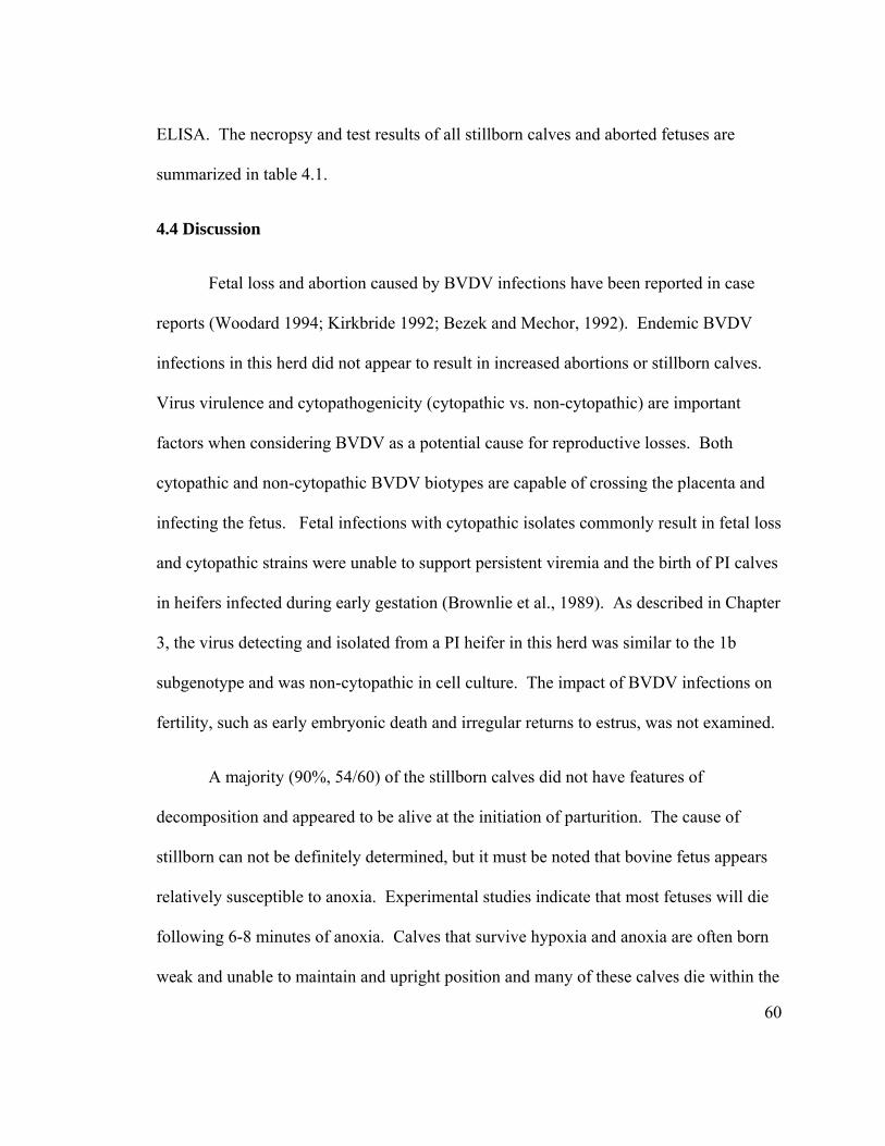
60
ELISA. The necropsy and test results of all stillborn calves and aborted fetuses are
summarized in table 4.1.
4.4 Discussion
Fetal loss and abortion caused by BVDV infections have been reported in case
reports (Woodard 1994; Kirkbride 1992; Bezek and Mechor, 1992). Endemic BVDV
infections in this herd did not appear to result in increased abortions or stillborn calves.
Virus virulence and cytopathogenicity (cytopathic vs. non-cytopathic) are important
factors when considering BVDV as a potential cause for reproductive losses. Both
cytopathic and non-cytopathic BVDV biotypes are capable of crossing the placenta and
infecting the fetus. Fetal infections with cytopathic isolates commonly result in fetal loss
and cytopathic strains were unable to support persistent viremia and the birth of PI calves
in heifers infected during early gestation (Brownlie et al., 1989). As described in Chapter
3, the virus detecting and isolated from a PI heifer in this herd was similar to the 1b
subgenotype and was non-cytopathic in cell culture. The impact of BVDV infections on
fertility, such as early embryonic death and irregular returns to estrus, was not examined.
A majority (90%, 54/60) of the stillborn calves did not have features of
decomposition and appeared to be alive at the initiation of parturition. The cause of
stillborn can not be definitely determined, but it must be noted that bovine fetus appears
relatively susceptible to anoxia. Experimental studies indicate that most fetuses will die
following 6-8 minutes of anoxia. Calves that survive hypoxia and anoxia are often born
weak and unable to maintain and upright position and many of these calves die within the
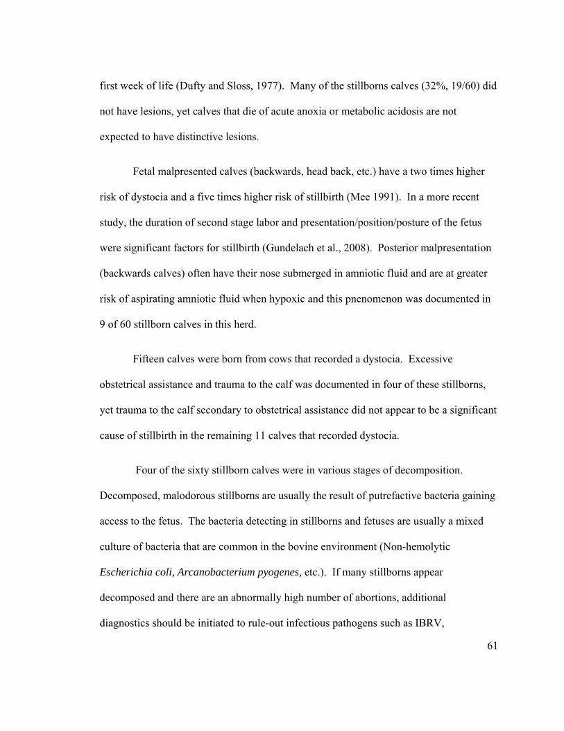
61
first week of life (Dufty and Sloss, 1977). Many of the stillborns calves (32%, 19/60) did
not have lesions, yet calves that die of acute anoxia or metabolic acidosis are not
expected to have distinctive lesions.
Fetal malpresented calves (backwards, head back, etc.) have a two times higher
risk of dystocia and a five times higher risk of stillbirth (Mee 1991). In a more recent
study, the duration of second stage labor and presentation/position/posture of the fetus
were significant factors for stillbirth (Gundelach et al., 2008). Posterior malpresentation
(backwards calves) often have their nose submerged in amniotic fluid and are at greater
risk of aspirating amniotic fluid when hypoxic and this pnenomenon was documented in
9 of 60 stillborn calves in this herd.
Fifteen calves were born from cows that recorded a dystocia. Excessive
obstetrical assistance and trauma to the calf was documented in four of these stillborns,
yet trauma to the calf secondary to obstetrical assistance did not appear to be a significant
cause of stillbirth in the remaining 11 calves that recorded dystocia.
Four of the sixty stillborn calves were in various stages of decomposition.
Decomposed, malodorous stillborns are usually the result of putrefactive bacteria gaining
access to the fetus. The bacteria detecting in stillborns and fetuses are usually a mixed
culture of bacteria that are common in the bovine environment (Non-hemolytic
Escherichia coli, Arcanobacterium pyogenes, etc.). If many stillborns appear
decomposed and there are an abnormally high number of abortions, additional
diagnostics should be initiated to rule-out infectious pathogens such as IBRV,

62
Brucellosis, Campylobacter sp., Listeriosis, Salmonellosis, Neospora caninum, Q fever,
or Leptospirosis.
Congenital defects were rare (0.5%, 4/708) and were observed in both the
stillborn calves and aborted fetuses. The anomalies (heart defect, cleft palate, etc.) are
uncommon, but not unique to cattle. Defects of the heart wall are amongst the most
common heart defects in stillborn calves (Mee 1991). Multiple calves with congenital
defects and a common genetic ancestry should initiate a thorough genetic investigation.
In summary, BVDV infections were not over-represented in the aborted fetuses,
stillborn, or weak born calves in a herd endemically infected with BVDV. The herd’s
stillborn rate of 8.5% was similar to the 8% reported in American dairies (Silvia del Rio
N et al., 2007), and the abortion rate of 2.5% was less than the 4.9% reported by USDA-
APHIS (USDA-APHIS NAHMS Dairy 1996 and 2002). Most of the stillborns appeared
to have been alive at the onset of parturition and necropsy revealed many lesions
consistent with fetal hypoxia or anoxia.
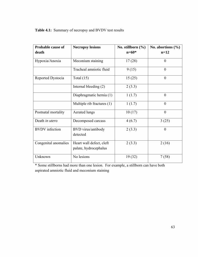
63
Table 4.1: Summary of necropsy and BVDV test results
Probable cause of death
Necropsy lesions No. stillborn (%) n=60*
No. abortions (%) n=12
Hypoxia/Anoxia Meconium staining 17 (28) 0
Tracheal amniotic fluid 9 (15) 0
Reported Dystocia Total (15) 15 (25) 0
Internal bleeding (2) 2 (3.3)
Diaphragmatic hernia (1) 1 (1.7) 0
Multiple rib fractures (1) 1 (1.7) 0
Postnatal mortality Aerated lungs 10 (17) 0
Death in utero Decomposed carcass 4 (6.7) 3 (25)
BVDV infection BVD virus/antibody detected
2 (3.3) 0
Congenital anomalies Heart wall defect, cleft palate, hydrocephalus
2 (3.3) 2 (16)
Unknown No lesions 19 (32) 7 (58)
* Some stillborns had more than one lesion. For example, a stillborn can have both aspirated amniotic fluid and meconium staining
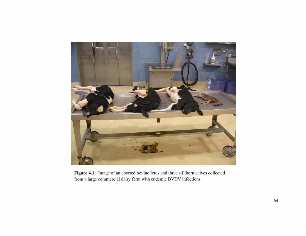
64
Figure 4.1: Image of an aborted bovine fetus and three stillborn calves collected from a large commercial dairy farm with endemic BVDV infections.
65
SECTION D: Molecular Epidemiology: Characterization of BVDV in PI calves by nucleic acid sequencing
CHAPTER 5
BOVINE VIRAL DIARRHEA VIRUS: GENETIC ANALYSIS AND SIGNALMENT OF PERSISTENTLY INFECTED DAIRY CALVES IN THE
UPPER MIDWESTERN UNITED STATES
Submitted for publication in: Journal of Veterinary Diagnostic Investigation
66
5.1 Introduction
Bovine viral diarrhea virus (BVDV; family Flaviviridae, genus Pestivirus) is an
economically important virus affecting ruminants worldwide. The reproductive,
respiratory, gastrointestinal, circulatory, immunologic, lymphatic, musculoskeletal,
integumentary and central nervous systems have all been negatively affected by BVDV
(Brock, 2004). Cattle persistently infected (PI) with BVDV are an import reservoir for
the virus and BVDV control and eradication strategies focus on the detection and
removal of PI cattle.
PI cattle occur when a fetus is infected with noncytopathic BVDV during the first
trimester of pregnancy (Brownlie et al., 1989; Liess et al., 1987; McClurkin et al., 1984).
Fetuses that survive a noncytopathic BVDV infection during the first trimester are
immunotolerent to, and persistently infected with, BVDV. If a PI calf is detected and
confirmed, the dam of the calf should be tested for persistent infection. If the dam of a PI
calf is not herself PI, researching the signalment of the dam can provide additional
information about BVDV infections within the herd. Determining the location of the
dam of the PI calf during the first trimester of pregnancy will reveal the time and location
of fetal infection. When a PI calf is detected, the time and location of BVDV infection of
the dam can be retrospectively calculated to a time approximately 5 to 9 months prior to
the birth date of the PI calf. The “point-in-time” infection that results in the birth of a PI
animal provides information that cannot always be obtained when documenting acute
infections by seroconversion in non-PI cattle.
67
The manifestations of BVDV exposure and infection are expected to be different
on dairy farms than on beef farms. In addition to year-round breeding schedules, the
dairy industry is experiencing rapid consolidation and specialization. It is not uncommon
for a dairy calf to be born, raised, and bred on one or more farms prior to lactation. Off-
site calf ranches are becoming more common as dairy farms become larger and these
specialized farms often commingle and raise cattle from more than one farm. According
to 2007 NAHMS data, at least 22.5% of heifers from large herds (500 or more) were bred
off-site and nearly two-thirds (63.8 percent) of farms sent heifers to a raiser where the
cattle had contact with cattle from other farms (USDA-APHIS 2007). Failure to separate
cattle according to farm origin can result in an increased risk of disease transmission
among the commingled animals. The development of large heifer raising facilities will
likely lead to an increased risk of dairies purchasing PI replacement heifers or pregnant
non-PI heifers gestating PI fetuses (Larson 2005).
Dairy cattle persistently infected with BVDV are rare. Data in chapter 2 revealed
that, 8 of 961 (0.8%) dairy calves born on four large dairy farms in Minnesota and
California were PI. Obtaining as much information from PI cattle can provide important
information in BVDV control strategies. In addition to determining the location of the PI
calf’s dam during the first trimester, the virus shed by the PI animal can be sequenced
and compared to those obtained from other PI cattle and those detected in acute
infections. Yet, few animal disease laboratories routinely perform nucleic acid
sequencing for BVDV and sequences are rarely documented and compared to PI cattle
and acute infections within and between farms.
68
The purpose of this study was to detect and confirm PI dairy cattle, research the
dam’s location during the first trimester of gestation, and genetically sequence a portion
of the 5’ untranslated (UTR) region of viral RNA to determine the prevalence of different
genotypes of PI dairy cattle in the upper Midwest.
5.2 Materials and Methods
Only dairy cattle that were confirmed BVDV PI were included in this study.
Persistently infected cattle reported in this study had at least two positive test results, at
least 3 weeks apart, and were tested with either antigen-capture enzyme-linked
immunosorbent assay (ACE) on skin or reverse transcription-polymerase chain reaction
(RT-PCR) on skin or serum. The ACE was performed on phosphate-buffered saline
(PBS) containing a soaking skin (ear notch) sample. The ACE test was performed
according to the instructions outlined in the test-kit package insert provided by the
manufacturer (Bovine Virus Diarrhea Antigen Test Kit®, IDEXX Laboratories,
Westbrook, ME). The single-tube TaqMan RT-PCR test has been described previously
(Mahlum et al., 2002). Briefly, RNA was extracted and purified from 200 ul of serum or
PBS soak using a commercially available kit (QIAamp 9604 Kit, Qiagen Inc., Valencia,
CA). The viral RNA was subjected to reverse transcription and amplification using a
TaqMan dual-labeled fluorescent probe. Endpoint analysis of the amplified products was
performed on an ABI Prism 7000 Sequence Detection System (ABI Prism 7000
Sequence Detection System, Applied Biosystems, Foster City, CA).
69
Once an animal was determined PI, the farm records were examined to determine
the dam of the PI. When available, the dam was tested for PI. The dam’s lactation
number (1st calf heifer, 2nd lactation cow, etc.) when she bore the PI calf was obtained
from farm records.
To further characterize the virus detected in BVDV PI cattle, a 270 base pair
segment of the 5’ untranslated region (5’ UTR) was analyzed. The two sequencing
primers were selected from previously published data (Ridpath 2005). The 5’ forward
primer (5’ – CATGCCCTTAGTAGGAC – 3’) extends from position 109-125 and the 3’
primer (5’ – CTCCATGTGCCATGRACAG – 3’) extends from position 381-399 of the
5’ UTR BVDV1-SD-1 genome. The 5’ UTR represents a conserved region of the BVDV
genome (Boye et al., 1991; Ridpath et al., 1993). Comparison of the 5’ UTR classified
the virus into three genotypes; namely, BVDV type 1a, BVDV type 1b and BVDV type
2a (Ridpath 1998). Phylogenetic analysis of BVDV isolates using the 5’ UTR region of
the genome generates results similar to those using the E2, NS2/3 and Npro genes (Becher
et al., 1999; Evermann and Ridpath 2002; Fulton et al., 2000; Pellerin et al., 1994;
Ridpath et al., 1994; Stokstad et al., 2004; van Rijn et al., 1997).
Viral RNA for sequencing was extracted from the PBS containing the soaking ear
notch or the serum using a commercially available RNA extraction kit (QIAamp Viral
RNA Mini Extraction Kit, Qiagen Inc, Valencia, CA), and RT-PCR was conducted
utilizing commercially available kits (Qiagen OneStep RT-PCR enzyme mix, buffer and
dNTPs, Qiagen Inc, Valencia, CA). Sequencing was performed directly from the ear
70
notch PBS, serum, and one paraffin-embedded formalin-fixed ear notch (PI calf 31).
None of the sequences were performed on viral isolates. The reagents, PCR primers and
extracted viral RNA were subjected to thermocycling and a product of approximately 240
base pairs was detected on agarose gel. The RT-PCR products were subjected to Sanger
dye-terminator sequencing and were analyzed using an automated sequence machine
(ABI 3730 DNA Analyzer, Applied Biosystems, Foster City, CA) at the BioMedical
Genomics Center, University of Minnesota. Nucleotide sequences were compared
among each other and with published NADL, C24V, Singer and Draper BVDV reference
sequences obtained from Genbank by using computer software (Dnastar-Megalign
sequence analysis program, Dnastar Inc, Madison, WI).
5.3 Results
Almost all of the PI calves (36/40, 90%) included in this study were shedding a
virus similar to the BVDV 1b subgenotype and all 36 1b sequences were greater than
95% similar to each other (Figure 4.1). Three PI calves (4, 15 and 20) were shedding
strains similar to genotype 1a, and calf 39 from dairy F was shedding a strain similar to
genotype 2a.
Across all dairies, a majority (23/37, 62%) of the BVDV PI dairy calves were
offspring of first calf heifers exposed to other growing heifers, and presumably one or
more PI heifers, within the non-lactating youngstock population. A minority of PI calves
(14/37 38%) were calves from lactating dairy cows in a second or greater lactation where
the fetal infection would have occurred in the herd of lactating cows (Table 4.1).
71
At the herd level, 5 of the 6 dairies with available dam data had PI calves that
were the offspring of 1st lactation heifers, 4 of the 6 dairies had PI calves that were
offspring of lactating cows, and dairies D, E, and F had PI calves born from both heifers
and cows.
Three PI calves were confirmed in dairy A. Viruses from calf 7 and heifer 5 in
dairy A had identical sequences over a 240 base pair segment of the 5’ UTR and were
most similar to the reference Draper 1b subgenotype. On farm records indicated that the
dam of PI calf 7 was a heifer of the same age of PI heifer 5 and both the PI heifer and the
dam of PI calf 7 were in the same pen during the first trimester of gestation. Therefore,
the development of PI calf 7 was the result of direct exposure to PI heifer 5. Calf 4 from
dairy A was a 6-month-old calf that was shedding a virus similar to the 1a genotype
reference strains.
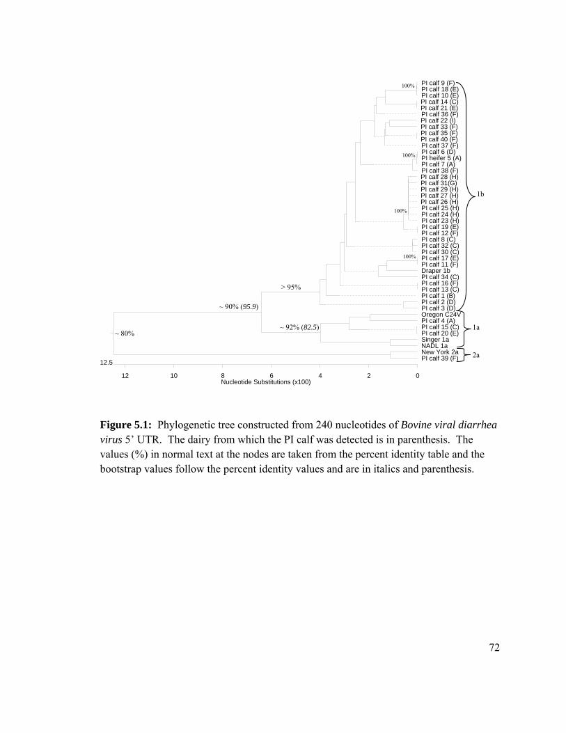
72
Figure 5.1: Phylogenetic tree constructed from 240 nucleotides of Bovine viral diarrhea virus 5’ UTR. The dairy from which the PI calf was detected is in parenthesis. The values (%) in normal text at the nodes are taken from the percent identity table and the bootstrap values follow the percent identity values and are in italics and parenthesis.
~ 90% (95.9)
1a
2a
Nucleotide Substitutions (x100)0
12.5
24681012
PI calf 9 (F)PI calf 18 (E)PI calf 10 (E)PI calf 14 (C)PI calf 21 (E)PI calf 36 (F)PI calf 22 (I)PI calf 33 (F)PI calf 35 (F)PI calf 40 (F)PI calf 37 (F)PI calf 6 (D)PI heifer 5 (A)PI calf 7 (A)PI calf 38 (F)PI calf 28 (H)PI calf 31(G)PI calf 29 (H)PI calf 27 (H)PI calf 26 (H)PI calf 25 (H)PI calf 24 (H)PI calf 23 (H)PI calf 19 (E)PI calf 12 (F)PI calf 8 (C)PI calf 32 (C)PI calf 30 (C)PI calf 17 (E)PI calf 11 (F)Draper 1bPI calf 34 (C)PI calf 16 (F)PI calf 13 (C)PI calf 1 (B)PI calf 2 (D)PI calf 3 (D)Oregon C24VPI calf 4 (A)PI calf 15 (C)PI calf 20 (E)Singer 1a NADL 1aNew York 2aPI calf 39 (F)
1b
100%
100%
100%
> 95%
~ 80% ~ 92% (82.5)
100%
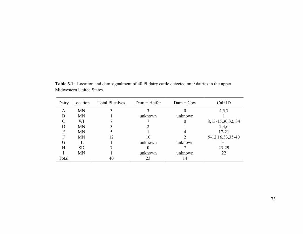
73
Table 5.1: Location and dam signalment of 40 PI dairy cattle detected on 9 dairies in the upper Midwestern United States.
Dairy Location Total PI calves Dam = Heifer Dam = Cow Calf ID
A MN 3 3 0 4,5,7 B MN 1 unknown unknown 1 C WI 7 7 0 8,13-15,30,32, 34 D MN 3 2 1 2,3,6 E MN 5 1 4 17-21 F MN 12 10 2 9-12,16,33,35-40 G IL 1 unknown unknown 31 H SD 7 0 7 23-29 I MN 1 unknown unknown 22
Total 40 23 14
74
Seven PI calves were detected on dairy C. Three of the PI calves (8, 30, 32) were
shedding stains similar to the 1b subgenotype that were more than 99.6% similar to each
other. Three of the PI calves in dairy C had sequences that were identical to those
detected in PI calves from dairies E and F. Calf 14 was shedding a 1b strain that was
identical to PI calf 21 in herd E, PI calf 15 was shedding a 1a strain that was identical to
calf 20 in herd E, and calf 13 was shedding a strain that was identical to a PI calf 16 in
herd F. Dairy C purchased pregnant heifers from multiple sources over multiple years
and the dams of the PI calves in dairy C were all first-calf heifers. All newborn calves in
dairy C were tested for PI with ACE over three years and none of the lactating cows in
dairy C produced a PI calf. Therefore, there was no evidence of BVDV infections within
the lactating population of dairy C.
The three PI calves in dairy D were all shedding stains similar to the 1b
subgenotype and PI calves 2 and 3 were similar (98.9%) to each other. The virus
sequence in PI calf 6 in dairy D was identical to viruses from heifer 7 and calf 5 in herd
A. Dairies D and A were both in central Minnesota and had purchased pregnant heifers
from suppliers, but neither dairy used a common heifer raiser.
Dairy E had five confirmed PI calves. Four of the PI calves were shedding strains
similar to the 1b genotype and one PI calf was shedding a strain similar to the 1a
genotype. Three of the four 1b strains were unique, but three viral sequences from PI
calves in dairy E were identical to sequences from PI calves detected in dairy F. Viral
sequences from calves 10 and 18 were identical to sequences from calf 9 in dairy F. Calf
75
19 had an identical sequence to calf 12 in dairy F, and calf 17 had a sequence identical to
calf 11 in dairy F. Both dairies E and F had many of their heifers raised at multiple
custom heifer farms throughout Minnesota.
Dairy F was the largest dairy and had 12 confirmed PI calves. This dairy had
multiple heifer raisers and also purchased cattle from multiple sources in multiple states.
Eleven of the 12 PI calves were shedding stains similar to the 1b subgenotype and one PI
calf was shedding a strain similar to the 2a genotype. Only calves 35 and 40 in dairy F
had identical genetic sequences and the 10 remaining 1b strains had sequence identities
that ranged in similarity from 96.0 to 99.0% (percent identity table not provided).
All seven PI calves in dairy H had identical sequences over the 270 base pair
segment of the 5” UTR and all seven calves were born within three weeks of each other
at a South Dakota dairy. The dams of all the PI calves were cows that were purchased as
dry cows when the dairy was constructed and multiple cattle were purchased and
assembled.
Dairy B had one PI calf that was unique to the other PI cattle and was more the
2.5% different than the remaining strains. The PI calf in dairy B (Figure 4.3) was
purchased from a central Minnesota sale-barn and the only information available was that
the calf was from a small dairy.
One PI calf was detected in herd I and it had a viral sequence identical to a PI calf
37 in herd F. Herd I was a small dairy in north central Minnesota and would occasionally
buy pregnant heifers.
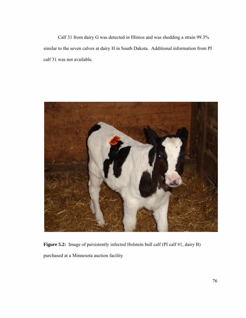
76
Calf 31 from dairy G was detected in Illinios and was shedding a strain 99.3%
similar to the seven calves at dairy H in South Dakota. Additional information from PI
calf 31 was not available.
Figure 5.2: Image of persistently infected Holstein bull calf (PI calf #1, dairy B)
purchased at a Minnesota auction facility
77
5.4 Discussion
Genetic sequencing data showed that most PI cattle (36/40, 90%) from these
dairies are infected with BVDV strains similar to the 1b subgenotype. This is consistent
with previous reports on PI beef cattle in central plains feedlots (Fulton et al., 2006;
Fulton et al., 2002). In another study, analysis of 53 BVDV isolates from dairy herds
between 1998 and 2001 showed that 40% of the field isolates were genotype 2 and 60%
were genotype 1. Of the genotype 1 isolates, 81% were subgenotype 1b (Tajima and
Dubovi, 2005). Strains most similar to BVDV genotype 1b appear to persist despite the
widespread use of vaccines and most commercially available vaccines do not contain
virus similar to the 1b subgenotype.
Like most RNA viruses, BVDV lacks a proof-reading mechanism, resulting in
mutations and recombination of the viral genome. Point mutations and recombination of
viral RNA can result in novel, unique viruses. Tracking and documenting changes in the
virus can provide important information for BVDV control and eradication programs.
Genetic sequencing of the highly conserved 5” UTR can also be a useful tool at the farm
level when attempting to eliminate BVDV PI cattle from a herd. If the herd tests and
removes all PI cattle but infections are discovered at a later date, the viral sequences of
the 5’ conserved region of the genome can be analyzed to determine if the original strain
persisted, or if a new virus was introduced in the herd. The 5” UTR region appears
highly conserved. In dairy A, heifer 7 had infected the dam (cohort heifer) of PI calf 5
78
and the genetic sequence remained identical throughout the entire gestation and birth of
PI calf 5.
Genetic sequencing can also be used to determine if a farm has more than one
genotype of the virus. Genetic analysis of the 40 PI cattle on these dairies revealed that if
a farm had more than 1 PI calf, they likely had calves that were shedding unique viruses.
Dairy H was the only dairy where all seven calves had identical genetic sequences over a
240 base pair segment of the 5’ UTR. Not all cattle were tested in dairy H leaving open
the possibility that there may have been other virus subtypes.
The dams of most of the PI calves (23/37, 62%) were heifers and dairies C, D, E
and F had used one or more custom heifer raisers to raise and breed heifers. In addition to
using heifer raisers, all four dairies had reported buying pregnant heifers from suppliers.
Dairies C, D, E and F reported that the heifer raisers had commingled cattle from other
farms. Commingling animals from multiple sources is not an unusual practice for heifer
raisers. One publication reported that off-site heifer facilities rarely separated heifers
according to farm of origin during the rearing period (Wolf 2003). This information
suggests that the risk of exposure to BVDV, presumably from another PI animal, is
greater in a youngstock heifer population than in a lactating cow population.
When heifer raising farms commingle cattle from two or more farms and they do
not have a PI testing protocol, a lactating dairy farm can potentially send a non-PI calf to
a heifer raiser for rearing and breeding where she can then become pregnant, infected,
and gestate a PI fetus. If an original lactating dairy was free of PI cattle, the calves from
79
heifers raised at an off-site farm can be a source of PI calves. If the dairy does not test
for PI and retains ownership of a PI calf, a PI calf has the potential to return to a heifer
rearing facility, become pregnant, and enter the lactating herd. Even if the PI calf fails to
become pregnant and lactate, it has the potential to sustain infections within the
youngstock population by exposing early gestation heifers to BVDV and making more PI
calves within the pregnant heifer population. In this manner, PI calves would need to
only live to breeding age to sustain infection within the youngstock population resulting
in the birth of more PI calves.
In summary, most PI calves detected on these large Midwest dairies shed a
BVDV similar to the 1b subgenotype and the highest risk for infection and the
subsequent birth of PI calves was in the youngstock heifer population. Genetic
sequencing documented that many PI calves had highly similar or, in 8 separate sets of
calves on 5 different dairies, identical sequences over the conserved region of the 5’ UTR
of the BVDV genome.
80
SECTION E: Quantification of BVDV RNA copies in clinical samples obtained from PI calves using quantitative RT-PCR
CHAPTER 6
QUANTITY OF BOVINE VIRAL DIARRHEA VIRUS RNA IN CLINICAL SAMPLES OBTAINED FROM A COHORT OF NATURALLY EXPOSED PERSISTENLY INFECTED CALVES USING QUANTITATIVE RT-PCR
81
Introduction:
An ear notch skin sample is a convenient tissue for detection of Bovine viral
diarrhea virus (BVDV) persistently infected (PI) cattle because it is easy to collect and
requires minimal supplies and equipment. Ear notch skin also offers the flexibility of
testing by immunohistochemistry (IHC), reverse-transcriptase polymerase chain reaction
(RT-PCR), and antigen-capture ELISA (ACE). All three tests have high sensitivity and
specificity for detecting PI cattle (Cornish et al., 2005; Fulton et al., 2006; Grooms and
Keilen 2002; Njaa et al., 2000), and none appear to be negatively affected by passively
acquired antibodies. ACE on fresh ear notch appears to be the most robust test for PI
detection across many Veterinary Diagnostic Laboratories (Edmondson et al., 2007).
Pooling ear notch phosphate-buffered saline (PBS) supernatant for RT-PCR is a
popular method to screen many cattle at a reduced cost. The pooling method usually
involves soaking the ear notch skin samples in a small amount (approximately 1-2 ml) of
PBS and pooling a portion of the supernatant for RT-PCR testing (Kennedy et al., 2006).
If the pooled supernatant tests positive by RT-PCR, the samples are then tested
individually. While ear notches have become the sample of choice for PI testing, there is
minimal information on the quantity of viral RNA copies in the PBS supernatant
containing the soaking skin and how the skin sample compares to blood, serum, or a
nasal swab from the same animal. Constructing pools without data on the quantity of
RNA copies in clinical samples is a significant limitation when attempting to determine
pool size.
82
Many conventional RT-PCR tests are only qualitative. Quantitative RT-PCR
assays can be used to determine the initial template concentration by monitoring the
progress of the reaction in real-time. The TaqMan real-time system detects PCR products
using the 5’ nuclease activity of Taq DNA polymerase on fluorogenic DNA probes
during each extension cycle. The TaqMan probe contains a fluorescent reporter dye at
the 5’ end and a quencher dye at the 3’ end. The quencher dye in the intact probe
prevents the emission of the reporter dye. If the target sequence is present, the probe
anneals to the target and is cleaved by the 5’ nuclease activity of Taq DNA polymerase
during primer extension. Cleavage of the probe separates the reporter from the quencher
dye and reporter dye fluorescence increases as a function of cycle number. The cycle
number at which the reporter dye fluorescence passes a fixed threshold above baseline is
the threshold cycle (Ct). The greater the initial concentration of the target RNA (or
DNA), the sooner a significant increase in fluorescence is observed (McBride et al.,
1998; Reischl and Kochanowshi 1999; Williams et al., 1998). Using the standard curve
generated with a target RNA sequence of known copy number, the starting amount of
target RNA in an unknown sample can be calculated. With this in mind, quantitative RT-
PCR has two prerequisites: (1) a standard curve demonstrates the range over which PCR
product yield provides a reliable measure of mRNA input, and (2) testing of numerous
samples allows for statistical analysis of differences in PCR product yield (Halford
1999).
The purpose of this study is to develop a quantitative real-time RT-PCR (qRT-
PCR) for quantification of BVDV RNA in four diagnostic samples obtained from a
83
cohort of naturally exposed PI cattle. Data obtained from these calves will give an
estimation of the quantity of detectable RNA in clinical samples. Serum, whole blood,
nasal swabs and ear notch skin samples were collected from a cohort of live PI cattle over
8 months, extracted within 2 days of collection, and analyzed by qRT-PCR at the
completion of sample collection. The data collected from this study provides an
estimation of RNA copies in clinical specimens obtained from PI calves and attempts to
document changes in virus quantity over an 8 month sampling period.
Materials and Methods:
A DNA product of 199 base pairs was generated from a BVDV isolate obtained
from National Veterinary Services Laboratories (NADL BVDV type 1a) by a single-step
RT-PCR reaction. The PCR product was confirmed and visualized on an ethidium
bromide-stained agarose gel and purified with a commercially available kit (Qiagen
QIAquick PCR purification Kit Cat. No. 2806). The purified RT-PCR product was
cloned in to a plasmid vector PCRII-TOPO according to manufacture’s specifications
(Invitrogen TOPO-TA cloning kit for sequencing; Invitrogen). The recombinant plasmid
was incubated in and propagated from competent Escherichia coli TOP10F’ cells.
The recombinant plasmid DNA was purified from the TOP10 F’ Escherichia cells
using a commercial kit (Qiagen QIAprep Spin Miniprep Kit Cat. No. 27106). The copy
number per volume for the purified plasmid was calculated from the concentration of
extracted DNA quantified by spectrophotometry (NanoDrop 1000). A standard curve
was calculated from 10-fold serial dilutions of recombinant purified standard plasmid
84
DNA in an estimated range of 10-1 to 106 copies/ul. The minimum detection limit using
the plasmid template was defined as the lowest plasmid copy number producing a
threshold cycle significantly different from reactions with no template DNA. The
minimum detection limit was determined to be approximately 50 – 100 copies/ml of PBS
supernatant, buffy coat extract and serum.
Seven PI dairy calves obtained and isolated from a dairy were sampled 10 times
from February 9 through October 18, 2009. The seven PI heifer calves were born within
2 weeks of each other. The samples were collected approximately every 3 to 4 weeks
and the calves were 6 months old on the first sample date and 14 months old at the
completion of the study. The virus shed by all calves had identical nucleotide sequences
over a portion of the 5’ UTR. The virus sequence was compared to BVDV reference
strains obtained from GenBank and was >95% similar to the Draper BVDV 1b
subgenotype reference strain.
Samples collected from the PI calves included a whole blood sample collected in
an EDTA blood tube, a blood sample that was allowed to clot and centrifuged for serum,
a triangular ear notch approximately 5 mm from the base of the haired ear margin to the
cut tip obtained from the lower ear margin, and a cotton nasal swab collected from one
nostril. The ear notch skin and cotton nasal swab were placed in 1 ml of PBS and all
samples were packaged in a cooler with frozen ice packs and taken to the Minnesota
Veterinary Diagnostic Laboratory (MVDL) for RNA extraction and storage at -80 C.
Buffy coats were removed from the whole blood samples by Ficoll separation and 200 ul
85
of each sample was extracted using a commercially available kit (QIAamp 9604 Kit,
Qiagen Inc., Valencia, CA). All samples were stored at -80 °C until tested by qRT-PCR.
A single-tube TaqMan RT-PCR (Mahlum et al., 2002) was performed on all samples
during one week. Briefly, the viral RNA was subjected to reverse transcription and
amplification using a TaqMan dual-labeled fluorescent probe. Analysis of the amplified
products was performed on an ABI Prism 7000 Sequence Detection System (ABI Prism
7000 Sequence Detection System, Applied Biosystems, Foster City, CA). Absolute
quantitative values were converted to the log10 scale and analyzed with statistical
software (SAS MIXED PROC).
Additional validation on pooling was accomplished by collecting supernatant
from negative ear notch skin samples routinely submitted to the MVDL. Twenty five ul
of ear notch supernatant from the PI calves was added to 1.25, 2.50, and 5.0 ml of
supernatant from test negative calves to generate pools of 1:50, 1:100, and 1:200,
respectively.
Results:
All 70 buffy coats, ear notch and nasal swab supernatant samples tested positive
by qRT-PCR (100%, 95% confidence interval .95 – 1.0). Sixty nine of the seventy serum
samples tested positive by qRT-PCR (99%, 95% confidence interval .93 – .99). The
mean log10 RNA copies in one ml of ear notch, nasal swab supernatant, and buffy coat
extract were 4.67, 5.20 and 4.93, respectively. The mean log10 RNA quantity in serum
was 4.48. There were no cattle that served as negative controls. The negative control for
86
RNA extraction was fetal horse serum and the negative control for qRT-PCR was
purified water. All negative controls used in the extraction methods or qRT-PCR tested
negative.
Calves 1 and 2 had significantly lower (p<0.01) log10 RNA copies in the ear notch
supernatant and buffy coat samples than calves 3 – 7 (Table 6.1). The mean log10 RNA
copies in the ear notch supernatant were 3.92 and 3.78 in calves 1 and 2, respectively, and
the mean log10 RNA copies in the ear notch supernatant from the other five calves ranged
from 4.76 to 5.35. The mean log10 RNA copies in buffy coat extract from calves 1 and 2
were also significantly lower (p<0.01) at 3.88 and 4.25, respectively. Buffy coat extract
mean log10 RNA copies in the five other calves ranged from 5.15 to 5.39. The mean
standard deviations across all calves and all samples were highest in the buffy coat
extracts (±0.84) followed by ear notch supernatant (±0.78), nasal swab supernatant
(±0.75), and serum (±0.63) (Table 6.1).
During the 8 month sampling period, there was a slight, statistically significant
decrease in viral RNA in the ear notch supernatant and buffy coat extract across all
calves. Ear notch supernatant RNA decreased 0.05 log10 RNA (p = 0.01) and buffy coat
extract decreased 0.10 log10 RNA (p < 0.0001). There was no significant increase or
decrease of viral RNA in the serum or nasal swab samples over the 8 months. Serum
RNA decreased 0.014 log10 (p = 0.38) and nasal swab decreased 0.01 log10 RNA (p =
0.62) (Figure 6.1).
87
Ear notch, nasal swab supernatant, and buffy coat samples produced higher mean
log10 RNA copies, wider ranges and higher standard deviations than serum. The range
for ear notch supernatant was 2.23 to 5.89 log10 RNA copies / ml, the range for nasal
swab supernatant was 2.95 to 6.37 log10 RNA copies / ml, and the buffy coat samples
ranged from 3.42 to 6.60 log10 RNA copies / ml. The range in 69 of the 70 positive
serum samples was 3.93 to 5.30 log10 RNA copies per ml. A serum sample from calf 4
collected on the test collection date tested negative.
Pools generated with negative ear notch supernatant and spiked samples from the
PI calves produced positive results in 209 of 210 of pools. All seventy pools of 1:100
and 1:200 tested positive. Sixty nine of 70 pools at 1:50 tested positive by RT-PCR. One
pool of 1:50 tested negative.
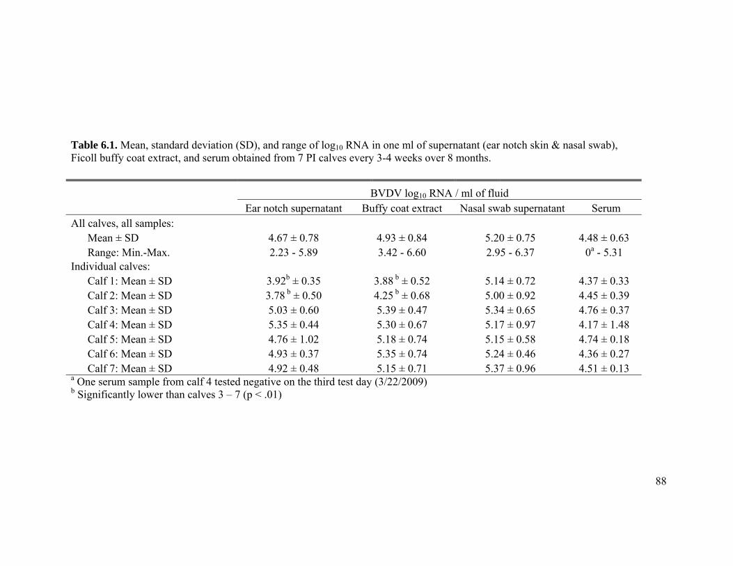
88
Table 6.1. Mean, standard deviation (SD), and range of log10 RNA in one ml of supernatant (ear notch skin & nasal swab), Ficoll buffy coat extract, and serum obtained from 7 PI calves every 3-4 weeks over 8 months.
BVDV log10 RNA / ml of fluid Ear notch supernatant Buffy coat extract Nasal swab supernatant Serum All calves, all samples: Mean ± SD 4.67 ± 0.78 4.93 ± 0.84 5.20 ± 0.75 4.48 ± 0.63 Range: Min.-Max. 2.23 - 5.89 3.42 - 6.60 2.95 - 6.37 0a - 5.31 Individual calves: Calf 1: Mean ± SD 3.92b ± 0.35 3.88 b ± 0.52 5.14 ± 0.72 4.37 ± 0.33 Calf 2: Mean ± SD 3.78 b ± 0.50 4.25 b ± 0.68 5.00 ± 0.92 4.45 ± 0.39 Calf 3: Mean ± SD 5.03 ± 0.60 5.39 ± 0.47 5.34 ± 0.65 4.76 ± 0.37 Calf 4: Mean ± SD 5.35 ± 0.44 5.30 ± 0.67 5.17 ± 0.97 4.17 ± 1.48 Calf 5: Mean ± SD 4.76 ± 1.02 5.18 ± 0.74 5.15 ± 0.58 4.74 ± 0.18 Calf 6: Mean ± SD 4.93 ± 0.37 5.35 ± 0.74 5.24 ± 0.46 4.36 ± 0.27 Calf 7: Mean ± SD 4.92 ± 0.48 5.15 ± 0.71 5.37 ± 0.96 4.51 ± 0.13 a One serum sample from calf 4 tested negative on the third test day (3/22/2009) b Significantly lower than calves 3 – 7 (p < .01)
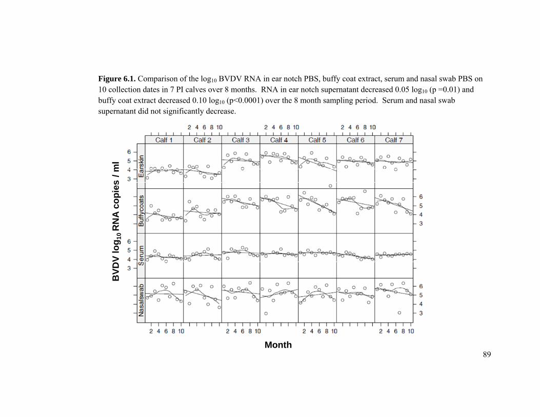
89
Figure 6.1. Comparison of the log10 BVDV RNA in ear notch PBS, buffy coat extract, serum and nasal swab PBS on 10 collection dates in 7 PI calves over 8 months. RNA in ear notch supernatant decreased 0.05 log10 (p =0.01) and buffy coat extract decreased 0.10 log10 (p<0.0001) over the 8 month sampling period. Serum and nasal swab supernatant did not significantly decrease.
BVD
V lo
g 10R
NA
copi
es/ m
l
Month
90
Discussion:
Viral quantification can provide valuable information both in viral diagnostics and
pathogenesis research. In this study, the goal was to estimate the quantity of BVDV
RNA copies in four diagnostic samples collected from seven PI calves over 8 months. In
addition to diagnostic and pathogenesis research, estimating the quantity of RNA copies
in diagnostic samples can help determine the limitations of pooling diagnostic samples.
In these BVDV PI calves, the quantity of log10 RNA across all samples ranged from 2.23
to 6.60 and was higher, but more variable, in the cell-rich heterogeneous ear notch, nasal
swab supernatant and buffy coat extract when compared to serum.
Only a few studies have documented the quantity and variability of virus in
clinical samples obtained from PI cattle. Variation of virus quantity in serum from 7 PI
cattle ranged from 5 x 10³ to 5 x 10⁵ and decreased by 1 to 10-fold over at least a 2-year
period (Brock et al., 1998). The decrease in serum virus in that study was associated
with the development of virus neutralizing antibody. The development of virus
neutralizing or passively acquired antibodies in BVDV PI cattle and its effect on viral
BVDV RNA copies in skin samples is likely negligible on the assay because RT-PCR is
not affected by the presence of antibody. Yet, the quantity and attendant variability of
virus in skin, whole blood and nasal swabs and the effect antibody might have on the
overall virus burden was not examined.
91
The diagnostic samples collected from the seven PI calves in this study
consistently produced high quantities of detectable RNA. Variability in virus quantity in
the ear notch, buffy coat and nasal swab samples was expected due to variability when
pipetting small aliquots (200 ul) of a heterogeneous sample for extraction. Variation in
the proportions of pipetted blood, epithelial cells, mucus and bacteria in the diagnostic
sample will result in variability of detectable RNA. Endogenous and exogenous
substances in ear notch supernatant, such as hair, enzymes, ribonucleases (RNases),
feces, and bacteria can also affect RT-PCR efficiency and generate variability (Akane et
al., 1994; Izraeli et al., 1991). Also, the use of a plasmid vector clone and the dilution
series used to generate a standard curve gives a good estimate of the efficiency of the
PCR assay, but, more importantly, the generation of a standard curve does not tell us
anything about the effect of the matrix of the diagnostic sample. Laboratory materials,
such as EDTA and Ficoll reagents used to extract buffy coats from whole blood, are
complex and may contain inhibitory substances that were not present in the purified
plasmid template used to generate the standard curve. Variation was expected because
absolute qRT-PCR tests suffer from inter-tube variations and qRT-PCR data should be
interpreted as semi-quantitative (MacKay et al., 2002).
Although all calves were from the same farm, of similar age, and shed a virus that
was identical over a portion of the 5’ UTR, calves 1 and 2 had an approximately 1 log10
lower mean RNA levels in the ear notch and nasal swab supernatant than the 5 other
92
calves. The reason for lower ear notch supernatant and buffy coat extract in calves 1 and
2 is not known, but one could speculate that the time of fetal infection and fetal
immunocompetence may impact viral persistence. If the time of fetal infection and
immunocompetence influence viral load in PI calves, we would expect all samples
collected from PI calves to be lower. Yet, the quantity of viral RNA in serum and nasal
swab supernatant was similar across all calves.
In addition to lower RNA in the ear notch supernant and buffy coat extract in
calves 1 and 2, the quantity of RNA in ear notch supernatant and buffy coat extract
decreased in all calves over the 8 month sampling period. The drop in detectable virus in
these two samples was small (0.05 and 0.10 log10 RNA) and has no clear explanation at
this time. Nevertheless, the samples collected from these PI calves routinely produced
high levels of detectable RNA. When compared to a few other studies, the quantity of
BVDV in serum collected from PI cattle was similar using CCID50 virus isolation. In one
study of 7 PI cattle, the quantity of serum virus titers ranged from 103 - 105 / ml (Brock et
al., 1998). Variation in viral serum concentration has also been reported in other studies
and ranged from of 104 – 106 CCID50/ml (Brock et al., 1990; Palfi et al., 1993; Paton et
al., 1994). To the author’s knowledge, this is the only study that quantified detectable
RNA in four routinely collected diagnostic samples obtained from a cohort of naturally
exposed PI calves shedding a similar virus.
In summary, buffy coats, serum, ear notch and nasal swab supernatant contains,
on average, 104 to 105 BVDV RNA copies / ml and is much higher than the minimal
93
detection limit of this qRT-PCR assay (101 RNA copies /ml). The buffy coat, ear notch
and nasal swab samples are heterogeneous diagnostic samples and contain endogenous
and exogenous substances that appear to produce wider variations in quantitative results
when compared to serum. Nevertheless, the seven PI calves in this study consistently
produced high levels of RNA in routine diagnostic samples that can be consistently
detected individually and when pooled.
94
CHAPTER 7
COMPARISON OF TWO EAR NOTCHING DEVICES FOR THE DETECTION OF BOVINE VIRAL DIARRHEA VIRUS RNA BY QUANTITATIVE RT-PCR
Presented as a poster at the 4th U.S. BVDV Symposium: BVDV Variability: Impact on Virulence, Host Range and Control
Phoenix, Arizona, January 2009
95
7.1 Introduction:
Skin from an ear notch is a convenient tissue for detection of BVDV PI cattle
because it is easy to collect and requires minimal supplies and equipment. Pooling ear
notch phosphate-buffered saline (PBS) supernatant for RT-PCR testing is a popular
method to screen groups of animals at a reduced cost. The pooling method involves
soaking the ear notch in PBS, pooling the supernatant, and testing the pooled supernatant
for BVDV by RT-PCR (Kennedy et al., 2006). If the pool is positive, the originally
submitted samples can be tested individually to determine the PI animal.
While ear notches have become the sample of choice for PI testing, there is
minimal information on the quantity of viral RNA in skin (ear notches) and the PBS
supernatant that contains the soaking sample. Pooling procedures have not been
extensively validated and are noted to be different among laboratories. The purpose of
this study was to compare two sampling devices and quantify BVDV RNA in the PBS
supernatant containing the skin sample. All samples were also individually tested by
antigen-capture ELISA (ACE).
7.2 Materials and Methods:
A quantitative RT-PCR (qrt-PCR) was developed. Briefly, a DNA product of
approximately 199 base pairs was generated from a BVDV type 1a isolate (NADL) from
a single-step RT-PCR reaction (Mahlum et al., 2002). The purified RT-PCR product was
96
then cloned to a plasmid vector according to manufacturer specifications (TOPO-TA
cloning kit for sequencing; Invitrogen). After incubation, propagation, and purification, a
copy number of the purified plasmid was calculated from the concentration of extracted
DNA by spectrophotometry (NanoDrop). A standard curve was calculated from 10-fold
serial dilutions of plasmid DNA in an estimated range of 10-1 to 106 copies/ul. The
minimum detection limit using the plasmid template was defined as the lowest plasmid
copy number producing a threshold cycle significantly different from reactions with no
template DNA.
Ear notches were collected from seven PI calves on three dates, every three weeks
(Figure 7.1). The circular skin sample (Figure 7.2) was harvested from the upper ear
margin and placed in 0.5 ml of PBS. The triangular skin sample was harvested from the
lower ear margin and placed in a sterile glass tube containing 2 ml PBS. The
circumference and length of fresh cut skin for the circular and triangular notch were 1.99
cm and 2.60 cm, respectively. The ratio of fresh cut skin to PBS in the circular and
triangular notch were 3.99 and 1.3 cm of fresh cut in one ml of PBS, respectively. The
skin samples were soaked in PBS for 24 hours after which 200 ul of PBS was extracted
and analyzed by qrt-PCR.
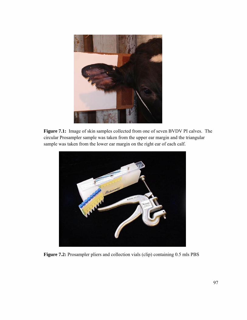
97
Figure 7.1: Image of skin samples collected from one of seven BVDV PI calves. The circular Prosampler sample was taken from the upper ear margin and the triangular sample was taken from the lower ear margin on the right ear of each calf.
Figure 7.2: Prosampler pliers and collection vials (clip) containing 0.5 mls PBS
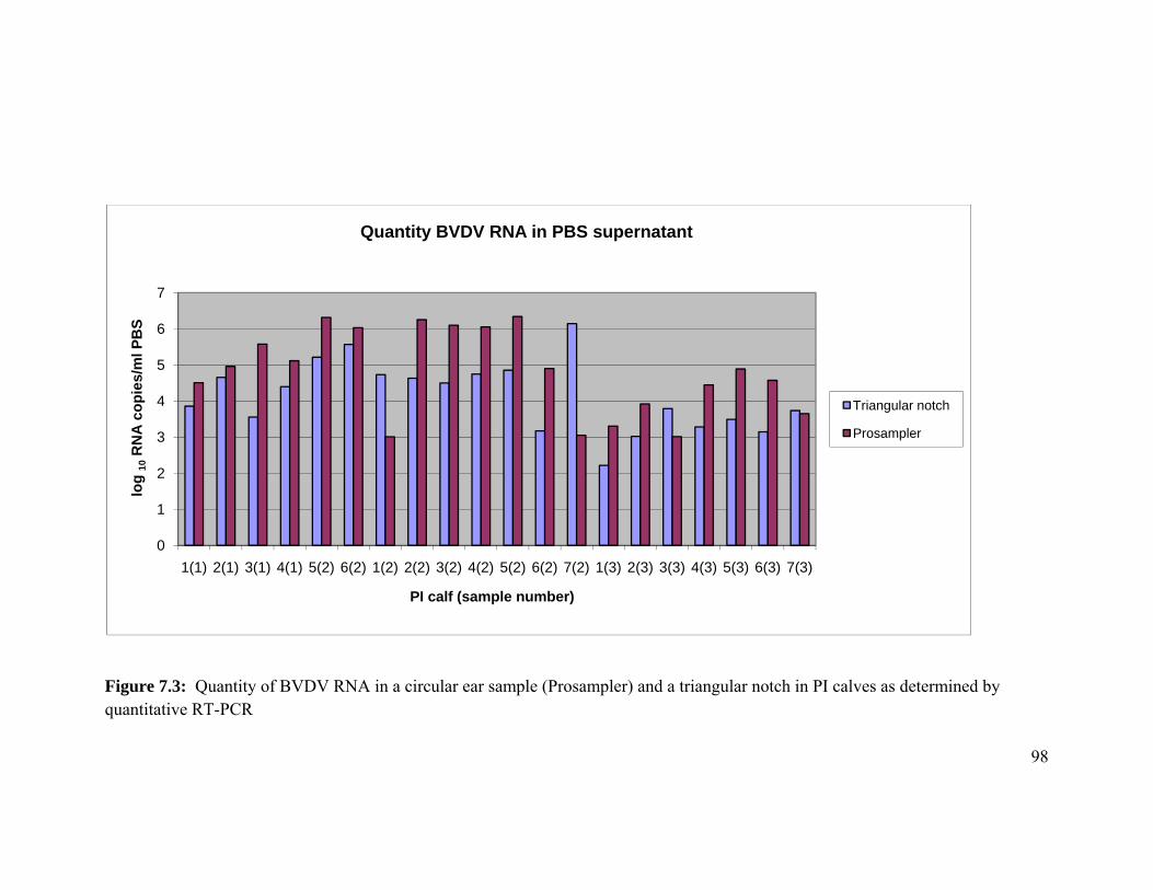
98
Figure 7.3: Quantity of BVDV RNA in a circular ear sample (Prosampler) and a triangular notch in PI calves as determined by quantitative RT-PCR
0
1
2
3
4
5
6
7
1(1) 2(1) 3(1) 4(1) 5(2) 6(2) 1(2) 2(2) 3(2) 4(2) 5(2) 6(2) 7(2) 1(3) 2(3) 3(3) 4(3) 5(3) 6(3) 7(3)
log
10R
NA
cop
ies/
ml P
BS
PI calf (sample number)
Quantity BVDV RNA in PBS supernatant
Triangular notch
Prosampler
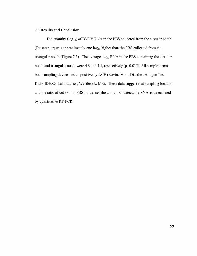
99
7.3 Results and Conclusion
The quantity (log10) of BVDV RNA in the PBS collected from the circular notch
(Prosampler) was approximately one log10 higher than the PBS collected from the
triangular notch (Figure 7.3). The average log10 RNA in the PBS containing the circular
notch and triangular notch were 4.8 and 4.1, respectively (p=0.015). All samples from
both sampling devices tested positive by ACE (Bovine Virus Diarrhea Antigen Test
Kit®, IDEXX Laboratories, Westbrook, ME). These data suggest that sampling location
and the ratio of cut skin to PBS influences the amount of detectable RNA as determined
by quantitative RT-PCR.
100
Concluding Summary:
Rapid consolidation of the dairy industry will increase the odds that a dairy farm,
or farms, will purchase BVDV PI cattle. One challenge for bovine veterinarians and
livestock producers is that there is a lack of simple, cost-effective strategies to determine
endemic BVDV infections in large dairy herds that routinely vaccinate for BVDV.
Attempting to demonstrate seroconversion in a subset of cattle by testing paired sera
(acute and convalescent) is confounded by vaccination. Non-vaccinated sentinel calves
have been used with marginal success and require that a every animal on the farm have
direct contact with the nonvaccinated sentinel to detect infection. Using highly sensitive
antigen or nucleic acid detection tests to detect PI cattle can be used to determine herd
status, but many cattle would have to be tested to achieve a high level of confidence that
the herd is free of BVDV infections and PI cattle because such animals often represent
less that 1% of the cattle in a the herd. Newborn calves can serve as useful sentinels
when sampled for BVDV antibodies prior to colostrum feeding and the data in chapter 2
suggests that there are approximately 8 calves seropositive for BVDV as compared to
those that are BVDV PI. Screening newborn calves and documenting fetal
seroconversion is a much more efficient method than screening for PI when attempting to
determine herd status.
Chapter 3 describes natural BVDV infections, transmission and test performance
in a large commercial dairy herd. This chapter is only one of a few case reports that
described the use of precolostral screening, RT-PCR, and nucleotide sequences to
101
describe and document natural BVDV infections in a large commercial dairy herd.
Detection and characterization of viral infections in this herd were consistent with
previous research describing BVDV pathogenesis and control. This chapter was found to
be a useful example of BVDV detection and control for veterinarians and producers who
have implemented, or are considering, BVDV control programs.
An apparent increase in stillborn dairy calves throughout the dairy industry
prompted an investigation into the stillborn calves obtained from the dairy herd described
in chapter 3. Although endemic BVDV infections and PI cattle were documented in the
herd, fetal infections were not overrepresented in the aborted fetuses or stillborn calves.
A majority of the stillborn calves in this herd had features of anoxia (aspirated amniotic
fluid, meconium staining, etc). In this herd, endemic BVDV infections did not result in
increased fetal loss.
Like most RNA viruses, BVDV lacks a proof-reading mechanism and mutations
and recombination of the viral genome is expected. Tracking the changes is important
for BVDV control and eradication programs. In the Midwest, most of the BVDV PI
calves are shedding a subgenotype similar to the Draper 1b reference strain and most PI
calves are the offspring of heifers indicating that BVDV infections appear more common
in heifer youngstock populations than adult lactating cow populations. Investigating the
possible inclusion of a 1b subgenotype stain in a vaccination program appears justified.
Because PI cattle are rare and the odds of finding a PI animal in a group of cattle
is unlikely, the fees associated with testing are often not justified by producers and
veterinarians. One method to reduce fees is to pool samples and test the pool for BVDV
102
with RT-PCR. Although a few studies have shown that pooling will efficiently detected
PI cattle, there is a lack of information on the quantity of viral RNA in clinical samples.
The data reported in chapter 6 quantified and described the amount of detectable RNA in
a cohort of seven, naturally exposed PI cattle. In that group of PI calves, ear notch skin,
buffy coats, serum and nasal swabs generate large amounts of RNA that was easily
detected by the qRT-PCR test when tested individually and pooled.
In summary, the bovine industry has many methods and assays to detected BVDV
infections and BVDV PI cattle. When used corrected, they appear highly effective and
appear to complement each other. As farm consolidation continues, we will need these
diagnostic tools and methods to control, and potentially eliminate, BVDV from the North
American cattle herd.
103
SECTION F: REFERENCES
Akane A, Matsubara H, Nakamura S et al: 1994, Identification of the heme compound copurified with deoxyribonucleic acid (DNA) from bloodstains, a major inhibitor of polymerase chain reaction (PCR) amplification. J Forensic Science 39:362-371.
Archambault D, Beliveau C, Couture Y, Carman S: 2000, Clinical response and immunomodulation following experimental challenge of calves with type 2 noncytopathogenic bovine viral diarrhea virus. Vet Res 31:215-227.
Baszler TV, Evermann JF, Kaylor PS et al: 1995, Diagnosis of naturally occurring bovine viral diarrhea virus infections in ruminants using monoclonal antibody-based immunohistochemistry. Vet Pathol 32:609-618.
Becher P, Orlich M, Kosmidou A et al: 1999, Genetic Diversity of Pestiviruses: Identification of Novel Groups and Implications for Classification. Virology 262:64-71.
Bezek DM, Mechor GD: 1992, Identification and eradication of bovine viral diarrhea virus in a persistently infected dairy herd. J Am Vet Med Assoc 201:580-586.
Bicalho RC, Galvao KN, Cheong SH et al: 2007, Effect of stillbirths on dam survival and reproduction performance in Holstein dairy cows. J Dairy Sci 90:2797–2803.
Bolin SR, Matthews PJ, Ridpath JF: 1991, Methods for detection and frequency of contamination of fetal calf serum with bovine viral diarrhea virus and antibodies against bovine viral diarrhea virus. J Vet Diagn Invest 3:199-203.
Bolin SR, McClurkin AW, Cutlip RC, Coria MF: 1985, Severe clinical disease induced in cattle persistently infected with noncytopathic bovine viral diarrhea virus by superinfection with cytopathic bovine viral diarrhea virus. Am J Vet Res 46:573-576.
Boye M, Kamstrup S, Dalsgaard K: 1991, Specific sequence amplification of bovine virus diarrhea virus (BVDV) and hog cholera virus and sequencing of BVDV nucleic acid. Vet Microbiol 29:1-13.
Brock KV: 2004, Strategies for the control and prevention of bovine viral diarrhea virus. Vet Clin North Am Food Anim Pract. 20:171-180.
Brock KV, Grooms DL, Ridpath J, Bolin SR: 1998, Changes in levels of viremia in cattle persistently infected with bovine viral diarrhea virus. J Vet Diagn Invest 10:22-26.
104
Brock KV, Redman DR, Vickers ML, Irvine NE: 1990, Quantitation of bovine viral diarrhea virus in embryo transfer flush fluids collected from a persistently infected heifer. J Vet Diagn Invest 3:99–100.
Brodersen BW, Kelling CL: 1999, Alteration of leukocyte populations in calves concurrently infected with bovine respiratory syncytial virus and bovine viral diarrhea virus. Viral Immunol 12:323-334.
Brodersen BW, Kelling CL: 1998, Effect of concurrent experimentally induced bovine respiratory syncytial virus and bovine viral diarrhea virus infection on respiratory tract and enteric diseases in calves. Am J Vet Res 59:1423-1430.
Brownlie J, Clarke MC, Howard CJ: 1989, Experimental infection of cattle in early pregnancy with a cytopathic strain of bovine virus diarrhoea virus. Res Vet Sci 46:307-311.
Brownlie J, Clarke MC, Howard CJ: 1984, Experimental production of fatal mucosal disease in cattle. Vet Rec 114:535-536.
Casaro AP, Kendrick JW, Kennedy PC: 1971, Response of the bovine fetus to bovine viral diarrhea-mucosal disease virus. Am J Vet Res 32:1543-1562.
Cornish T, van Olphen A, Cavender J et al: 2005, Comparison of ear notch immunohistochemistry, ear notch antigen-capture ELISA, and buffy coat virus isolation for detection of calves persistently infected with bovine viral diarrhea virus. J Vet Diagn Invest 17:110-117.
Cortese VS, Grooms DL, Ellis J et al: 1998, Protection of pregnant cattle and their fetuses against infection with bovine viral diarrhea virus type 1 by use of a modified-live virus vaccine. Am J Vet Res 59:1409-1413.
Dean HJ, Hunsaker BD, Bailey OD, Wasmoen T: 2003, Prevention of persistent infection in calves by vaccination of dams with noncytopathic type-1 modified-live bovine viral diarrhea virus prior to breeding. Am J Vet Res 64:530-537.
Drew TW, Yapp F, Paton DJ: 1999, The detection of bovine viral diarrhoea virus in bulk milk samples by the use of a single-tube RT-PCR. Vet Microbiol 64:145-154.
Duffell SJ, Harkness JW: 1985, Bovine virus diarrhoea-mucosal disease infection in cattle. Vet Rec 117:240-245.
105
Duffell SJ, Sharp MW, Winkler CE et al: 1984, Bovine virus diarrhoea-mucosal disease virus-induced fetopathy in cattle: Efficacy of prophylactic maternal pre-exposure. Vet Rec 114:558-561.
Dufty JH, Sloss V: 1977, Anoxia in the bovine foetus. Aust Vet J 53:262-267.
Edmondson MA, Givens MD, Walz PH et al: 2007, Comparison of tests for detection of bovine viral diarrhea virus in diagnostic samples. J Vet Diagn Invest 19:376-381.
Elvander M, Baule C, Persson M et al: 1998, An experimental study of a concurrent primary infection with bovine respiratory syncytial virus (BRSV) and bovine viral diarrhoea virus (BVDV) in calves. Acta Vet Scand 39:251-264.
Evermann JF, Ridpath JF: 2002, Clinical and epidemiologic observations of bovine viral diarrhea virus in the northwestern United States. Vet Microbiol 89:129-139.
Fray MD, Mann GE, Clarke MC, Charleston B: 2000, Bovine viral diarrhoea virus: its effects on ovarian function in the cow. Vet Microbiol 77:185-194.
Fulton RW, Briggs RE, Ridpath JF et al: 2005, Transmission of bovine viral diarrhea virus 1b to susceptible and vaccinated calves by exposure to persistently infected calves. Can J Vet Res 69:161-169.
Fulton RW, Saliki JT, Confer AW et al: 2000, Bovine viral diarrhea virus cytopathic and noncytopathic biotypes and type 1 and 2 genotypes in diagnostic laboratory accessions: clinical and necropsy samples from cattle. J Vet Diagn Invest 12:33-38.
Fulton RW, Hessman B, Johnson BJ et al: 2006, Evaluation of diagnostic tests used for detection of bovine viral diarrhea virus and prevalence of subtypes 1a, 1b, and 2a in persistently infected cattle entering a feedlot. J Am Vet Med Assoc 228:578-584.
Fulton RW, Ridpath JF, Saliki JT et al: 2002, Bovine viral diarrhea virus (BVDV) 1b: predominant BVDV subtype in calves with respiratory disease. Can J Vet Res 66:181-190.
Ganheim C, Hulten C, Carlsson U et al: 2003, The acute phase response in calves experimentally infected with bovine viral diarrhoea virus and/or Mannheimia haemolytica. J Vet Med B Infect Dis Vet Public Health 50:183-190.
Glew EJ, Carr BV, Brackenbury LS et al: 2003, Differential effects of bovine viral diarrhoea virus on monocytes and dendritic cells. J Gen Virol 84:1771-1780.
106
Goyal SM: 2005, Diagnosis. In: Bovine viral diarrhea virus: diagnosis, management, and control, ed. Goyal SM, Ridpath JF, 1st ed., pp. 197–208. Blackwell, Ames, IA.
Grooms DL, Keilen ED: 2002, Screening of neonatal calves for persistent infection with bovine viral diarrhea virus by immunohistochemistry on skin biopsy samples. Clin Diagn Lab Immunol 9:898-900.
Grooms DL, Brock KV, Pate JL, Day ML: 1998, Changes in ovarian follicles following acute infection with bovine viral diarrhea virus. Theriogenology 49:595-605.
Grooms D, Brock K, Ward L: 1998, Detection of bovine viral diarrhea virus in the ovaries of cattle acutely infected with bovine viral diarrhea virus. J Vet Diagn Invest 10:125-129.
Grooms D, Brock K, Ward L: 1998, Detection of cytopathic bovine viral diarrhea virus in the ovaries of cattle following immunization with a modified live bovine viral diarrhea virus vaccine. J Vet Diagn Invest 10:130-134.
Gundelach Y, Essmeyer K, Teltscher MK, Hoedemaker M: 2008, Risk factors for perinatal mortality in dairy cattle; Cow and foetal factors, calving process. Theriogenology 71: 901-909.
Halford WP: 1999, The essential prerequisites for quantitative RT-PCR. Nat Biotechnol 17:835.
Houe H, Meyling A: 1991/8, Prevalence of bovine virus diarrhoea (BVD) in 19 Danish dairy herds and estimation of incidence of infection in early pregnancy. Prev Vet Med 11:9-16.
Houe H, Baker JC, Maes RK et al: 1995, Prevalence of cattle persistently infected with bovine viral diarrhea virus in 20 dairy herds in two counties in central Michigan and comparison of prevalence of antibody-positive cattle among herds with different infection and vaccination status. J Vet Diagn Invest 7:321-326.
Izraeli S, Pfleiderer C, Lion T: 1991, Detection of gene expression by PCR amplification of RNA derived from frozen heparinized whole blood. Nucleic Acids Res 19:6051.
Jones L, Van Campen H, Xu Zhi-Chang, Schnackel JA: 2001, Comparison of neutralizing antibodies to type 1a, 1b and 2 bovine viral diarrhea virus from experimentally infected and vaccinated cattle. Bov Pract 35:137-140.
107
Kelling CL, Steffen DJ, Cooper VL et al: 2002, Effect of infection with bovine viral diarrhea virus alone, bovine rotavirus alone, or concurrent infection with both on enteric disease in gnotobiotic neonatal calves. Am J Vet Res 63:1179-1186.
Kendrick JW: 1971, Bovine viral diarrhea-mucosal disease virus infection in pregnant cows. Am J Vet Res 32:533-544.
Kennedy JA, Mortimer RG, Powers B: 2006, Reverse transcription-polymerase chain reaction on pooled samples to detect bovine viral diarrhea virus by using fresh ear-notch-sample supernatants. J Vet Diagn Invest 18:89-93.
Kirkbride CA: 1992, Viral agents and associated lesions detected in a 10-year study of bovine abortions and stillbirths. J Vet Diagn Invest 4:374-379.
Kleiboeker SB, Lee SM, Jones CA, Estes DM: 2003, Evaluation of shedding of bovine herpesvirus 1, bovine viral diarrhea virus 1, and bovine viral diarrhea virus 2 after vaccination of calves with a multivalent modified-live virus vaccine. J Am Vet Med Assoc 222:1399-1403.
Lambot M, Douart A, Joris E et al: 1997, Characterization of the immune response of cattle against non- cytopathic and cytopathic biotypes of bovine viral diarrhoea virus. J Gen Virol 78:1041-1047.
Lambot M, Hanon E, Lecomte C et al: 1998, Bovine viral diarrhoea virus induces apoptosis in blood mononuclear cells by a mechanism largely dependent on monocytes. J Gen Virol 79:1745-1749.
Larson RL: 2005, Management Systems and Control Programs. In: Bovine viral diarrhea virus: diagnosis, management and control, ed. Goyal SM, Ridpath JF, 1st ed., pp. 223-238. Blackwell, Ames, IA.
Liess B, Orban S, Frey HR, Trautwein G: 1987, Consequences of the transplacental transmission of BVD virus to cattle fetuses. Dtsch Tierarztl Wochenschr 94:585-587.
Loneragan GH, Thomson DU, Montgomery DL et al: 2005, Prevalence, outcome, and health consequences associated with persistent infection with bovine viral diarrhea virus in feedlot cattle. J Am Vet Med Assoc 226:595-601.
Mackay IM, Arden KE, Nitsche A: 2002, Real-time PCR in virology. Nucleic Acids Res 30:1292-1305.
108
Mahlum CE, Haugerud S, Shivers JL et al: 2002, Detection of bovine viral diarrhea virus by TaqMan reverse transcription polymerase chain reaction. J Vet Diagn Invest 14:120-125.
McClurkin AW, Littledike ET, Cutlip RC et al: 1984, Production of cattle immunotolerant to bovine viral diarrhea virus. Can J Comp Med 48:156-161.
McBride L, Livak K, Lucero M, et al: 1998, Quantitative PCR technology. In: Gene quantification, ed. Ferre F, pp. 97-110. Birkhauser, Boston, MA.
Mee JF: 1991, Bovine perinatal mortality and parurient problems in Irish dairy herds. Ph.D. dissertation, National University of Ireland. pp 1-365.
Munoz-Zanzi CA, Hietala SK, Thurmond MC, Johnson WO: 2003, Quantification, risk factors, and health impact of natural congenital infection with bovine viral diarrhea virus in dairy calves. Am J Vet Res 64:358-365.
Munoz-Zanzi CA, Thurmond MC, Johnson WO, Hietala SK: 2002, Predicted ages of dairy calves when colostrum-derived bovine viral diarrhea virus antibodies would no longer offer protection against disease or interfere with vaccination. J Am Vet Med Assoc 221:678-685.
Njaa B, Clark E, Janzen E et al: 2000, Diagnosis of persistent bovine viral diarrhea virus infection by immunohistochemical staining of formalin-fixed skin biopsy specimens. J Vet Diagn Invest 12:393-399.
Palfi V, Houe H, Philipsen J: 1993, Studies on the decline of bovine virus diarrhoea virus (BVDV) maternal antibodies and detectability of BVDV in persistently infected calves. Acta Vet Scand 34:105–107.
Paton DJ, Lowings JP, Ramirez GC: 1994, Stability of the gp53 gene of a bovine viral diarrhea virus isolated at different times from a persistently infected steer. Br Vet J 150:603–607.
Pellerin C, van den Hurk J, Lecomte J, Tussen P: 1994, Identification of a new group of bovine viral diarrhea virus strains associated with severe outbreaks and high mortalities. Virology 203:260-268.
Peterhans E, Jungi TW, Schweizer M: 2003/6, BVDV and innate immunity. Biologicals 31:107-112.
109
Pillars RB, Grooms DL: 2002, Serologic evaluation of five unvaccinated heifers to detect herds that have cattle persistently infected with bovine viral diarrhea virus. Am J Vet Res 63:499-505.
Potgieter LN: 1995, Immunology of bovine viral diarrhea virus. Vet Clin North Am Food Anim Pract 11:501-520.
Radwan GS, Brock KV, Hogan JS, Smith KL: 1995, Development of a PCR amplification assay as a screening test using bulk milk samples for identifying dairy herds infected with bovine viral diarrhea virus. Vet Microbiol 44:77-91.
Reischl U, Kochanowski B: 1999, Quantitative PCR: a survey of the present technology. In: Quantitative PCR protocols, ed. Kochanowski B, Reischl U, pp. 3-30. Humana Press, Totowa, NJ.
Renshaw R, Ray R, Dubovi E: 2000, Comparison of virus isolation and reverse transcription polymerase chain reaction assay for detection of bovine viral diarrhea virus in bulk milk tank samples. J Vet Diagn Invest 12:184-186.
Ridpath JF: 2005, Classification and Molecular Biology. In: Bovine viral diarrhea virus: diagnosis, management and control, ed. Goyal SM, Ridpath JF, 1st ed., pp. 65-80. Blackwell, Ames, IA.
Ridpath JF, Bolin SR: 1998, Differentiation of types 1a, 1b and 2 bovine viral diarrhoea virus (BVDV) by PCR. Mol Cell Probes 12:101-106.
Ridpath JF, Bolin SR, Dubovi EJ: 1994, Segregation of bovine viral diarrhea virus into genotypes. Virology 205:66-74.
Ridpath JF, Bolin SR, Katz J: 1993, Comparison of nucleic acid hybridization and nucleic acid amplification using conserved sequences from the 5' noncoding region for detection of bovine viral diarrhea virus. J Clin Microbiol 31:986-989.
Roth JA, Kaeberle ML, Griffith RW: 1981, Effects of bovine viral diarrhea virus infection on bovine polymorphonuclear leukocyte function. Am J Vet Res 42:244-250.
Rush DM, Thurmond MC, Munoz-Zanzi CA, Hietala SK: 2001, Descriptive epidemiology of postnatal bovine viral diarrhea virus infection in intensively managed dairy heifers. J Am Vet Med Assoc 219:1426-1431.
Rutledge RG; 2004, Sigmoidal curve-fitting redefines quantitative real-time PCR with the prospective of developing automated high-throughput applications. Nucleic Acids Res 32:178.
110
Seki Y, Seimiya YM, Yaegashi G, Sato C: 2006, Identification of herds with cattle persistently infected with bovine viral diarrhea virus by virological evaluation of three calves. J Vet Med Sci 68:255-258.
Silva del Rio N, Stewart S, Rapnicki P et al: 2007, An observational analysis of twin births, calf sex ratio and calf mortality in Holstein dairy cattle. J Dairy Sci 90:2797-2803.
Stokstad M, Brownlie J, Collins ME: 2004, Analysis of variation of bovine viral diarrhoea virus E2 sequence following transplacental infection of cattle. Vet Microbiol 102:141-145.
Tajima M, Dubovi EJ: 2005, Genetic and clinical analyses of bovine viral diarrhea virus isolates from dairy operations in the United States of America. J Vet Diagn Invest 17:10-15.
Tautz N, Meyers G, Thiel HJ: 1998, Pathogenesis of mucosal disease, a deadly disease of cattle caused by a pestivirus. Clin Diagn Virol 10:121-127.
Taylor LF, Janzen ED, Van Donkersgoed J: 1997, Losses over a 2-year period associated with fetal infection with the bovine viral diarrhea virus in a beef cow-calf herd in Saskatchewan. Can Vet J 38:23-28.
United States Department of Agriculture, Animal Plant Health Inspection Service, Veterinary Services: 2007, Off-Site heifer raising on United States dairy operations info sheet, 4 p, 11/07.
United States Department of Agriculture, Animal Plant Health Inspection Service, Veterinary Services: 2004, Exemption from label warning concerning the use of bovine rhinotracheitis vaccine, modified live virus in pregnant cows or in calves nursing pregnant cows under 9 code of federal regulations 122.7(e). Veterinary Services Memorandum No. 800.110, October.
van Regenmortel MHV et al.: 2005, Pestivirus. In: Virus Taxonomy: Classification and Nomenclature of Viruses: Eighth report of the International Committee on the Taxonomy of Viruses, van Regenmortel MHV ed., eighth report, pp. 867-872. Academic Press, San Diego, CA.
van Rijn PA, van Gennip HGP, Leendertse CH et al: 1997, Subdivision of the Pestivirus Genus Based on Envelope Glycoprotein E2. Virology 237:337-348.
111
Waldner CL, Campbell JR: 2005, Use of serologic evaluation for antibodies against bovine viral diarrhea virus for detection of persistently infected calves in beef herds. Am J Vet Res 66:825-834.
Ward GM, Roberts SJ, McEntee K, Gillespie JH: 1969, A study of experimentally induced bovine viral diarrhea-mucosal disese in pregnant cows and their progeny. Cornell Vet 59:525-538.
Werdin RE, Ames TR, Goyal SM: 1989, Detection and elimination of carrier animals in a dairy herd persistently infected with bovine viral diarrhea virus. J Vet Diagn Invest 1:277-279.
Williams PM, Giles T, Tucker A et al: 1998, Development and application of real-time quantitative PCR. In: Gene quantification, ed. Ferre F, pp. 313-325. Birkhauser, Boston, MA.
Wittum TE, Grotelueschen DM, Brock KV et al: 2001, Persistent bovine viral diarrhoea virus infection in US beef herds. Prev Vet Med 49:83-94.
Wolf CA: 2003, Custom dairy heifer grower industry characteristics and contract terms. J Dairy Sci 86:3016-3022.
Woodard LF: 1994, BVD virus associated with outbreaks of abortion, stillbirths, and weak calves. Vet Med 98:379-384.
Zimmer GM, Wentink GH, Bruschke C et al: 2002, Failure of foetal protection after vaccination against an experimental infection with bovine virus diarrhea virus. Vet Microbiol 89:255-265.