The distribution of ligand-binding pockets around ... - PNAS
Upload
independentCategory
view
2download
0
Differential Effects of Methoxy Group on the Interactionof Curcuminoids with Two Major Ligand Binding Sites ofHuman Serum Albumin
Hiroki Sato1, Victor Tuan Giam Chuang2, Keishi Yamasaki3, Noriyuki Yamaotsu4, Hiroshi Watanabe1,
Kohei Nagumo1, Makoto Anraku3, Daisuke Kadowaki1, Yu Ishima1, Shuichi Hirono4, Masaki Otagiri3,
Toru Maruyama1*
1Department of Biopharmaceutics, Graduate School of Pharmaceutical Sciences, Center for Clinical Pharmaceutical Science, Kumamoto University, Kumamoto, Japan,
2 School of Pharmacy, Curtin Health Innovation Research Institute, Curtin University, Perth, Australia, 3 Faculty of Pharmaceutical Sciences, DDS Research Institute, Sojo
University, Kumamoto, Japan, 4 School of Pharmacy, Kitasato University, Tokyo, Japan
Abstract
Curcuminoids are a group of compounds with a similar chemical backbone structure but containing different numbers ofmethoxy groups that have therapeutic potential due to their anti-inflammatory and anti-oxidant properties. They mainlybind to albumin in plasma. These findings influence their body disposition and biological activities. Spectroscopic analysisusing site specific probes on human serum albumin (HSA) clearly indicated that curcumin (Cur), demethylcurcumin (Dmc)and bisdemethoxycurcumin (Bdmc) bind to both Site I (sub-site Ia and Ib) and Site II on HSA. At pH 7.4, the bindingconstants for Site I were relatively comparable between curcuminoids, while the binding constants for Site II at pH 7.4 wereincreased in order Cur , Dmc , Bdmc. Binding experiments using HSA mutants showed that Trp214 and Arg218 at Site I,and Tyr411 and Arg410 at Site II are involved in the binding of curcuminoids. The molecular docking of all curcuminoids tothe Site I pocket showed that curcuminoids stacked with Phe211 and Trp214, and interacted with hydrophobic andaromatic amino acid residues. In contrast, each curcuminoid interacted with Site II in a different manner depending whethera methoxy group was present or absent. A detailed analysis of curcuminoids-albumin interactions would provide valuableinformation in terms of understanding the pharmacokinetics and the biological activities of this class of compounds.
Citation: Sato H, Chuang VTG, Yamasaki K, Yamaotsu N, Watanabe H, et al. (2014) Differential Effects of Methoxy Group on the Interaction of Curcuminoids withTwo Major Ligand Binding Sites of Human Serum Albumin. PLoS ONE 9(2): e87919. doi:10.1371/journal.pone.0087919
Editor: Rajagopal Subramanyam, University of Hyderabad, India
Received September 12, 2013; Accepted January 1, 2014; Published February 3, 2014
Copyright: � 2014 Sato et al. This is an open-access article distributed under the terms of the Creative Commons Attribution License, which permits unrestricteduse, distribution, and reproduction in any medium, provided the original author and source are credited.
Funding: The study was supported by Grant-in-Aid for Scientific Research from Japan Society for the Promotion of Science (JSPS) (KAKENHI 21390177, 23390142,25860118) and by Japan Science and Technology Agency (JST), A-step feasibility study program (10801043). The funders had no role in study design, datacollection and analysis, decision to publish, or preparation of the manuscript.
Competing Interests: The authors have the following interests. The human serum albumin for this study was a gift from Vaccine maker ’KAKETSUKEN’, alsoknown as the Chemo-Sero-Therapeutic Research Institute. There are no further patents, products in development or marketed products to declare. This does notalter the authors’ adherence to all the PLOS ONE policies on sharing data and materials, as detailed online in the guide for authors.
* E-mail: [email protected]
Introduction
In recent years, active investigations have been conducted
regarding clinical applications of curcumin (Cur), a pigment
component of Turmeric (Curcuma longa L.) and rhizomatous
herbaceous perennial plant of the ginger family [1]. The scientific
research and clinical trials conducted so far were not specifically
focussed exclusively on Cur only, but were on "curcuminoids", a
mixture of Cur analogues such as demethoxycurcumin (Dmc) or
bisdemethoxycurcumin (Bdmc). Curcuminoids have similar chem-
ical structures except for the number of methoxy groups on each of
the two phenyl moieties. Cur contains one methoxy group on each
of its two phenyl moieties, Dmc contains one methoxy group on
one phenyl moiety, whereas Bdmc is devoid of any methoxy
groups (Figure 1). Curcuminoids have been found to possess a
variety of biologically activities such as anti-inflammatory, anti-
oxidant, anti-cancer, anti-virus, anti-infective, anti-malarial and
wound healing [2,3]. Furthermore, interesting structure-activity
relationship findings have been reported for the curcuminoids. For
example, Cur shows the highest anti-oxidant and anti-inflamma-
tory effects [4,5] whereas Dmc and Bdmc are superior to Cur in
inhibiting cancer cell proliferation [6], and for improving the
clearance of amyloid b (Ab) in an animal model with a congenital
immune deficiency [7]. Research on the therapeutic effects of
curcuminoids in treating Alzheimer’s disease (AD) has been
actively carried out. Curcuminoids have been reported to suppress
the accumulation of the Ab protein in the brain of an AD mouse
model, reduce the formation of amyloid plaques [8] and improve
the impaired Ab clearance due to an innate immunity deficiency
[7]. Therefore, curcuminoids have considerable potential for use
as therapeutic agents for the clinical treatment or prevention of
AD.
Human serum albumin (HSA), is a monomeric protein
consisting of 585 amino acid residues, with a molecular weight
of approximately 66,500 Da [9]. HSA is a plasma protein that
functions as a carrier for various endogenous and exogenous
substances [10,11]. Two major ligand binding sites of HSA, site I
and site II [12], were reported to be located in subdomain IIA and
subdomain IIIA respectively [13,14]. The distribution and
PLOS ONE | www.plosone.org 1 February 2014 | Volume 9 | Issue 2 | e87919

pharmacological actions of curcuminoids are controlled by their
strong binding to HSA in the systemic circulation after oral
absorption. Therefore, clarification of the binding mechanism and
the nature of the interactions of curcuminoids with HSA are
essential in predicting the pharmacokinetics and therapeutic effects
of curcuminoids and their potential interactions with other
pharmaceutical agents. Researches on the interaction of curcumi-
noids with HSA have, so far, mostly focused on Cur. For example,
Reddy et al. reported that Cur binds to HSA at physiological pH
and there are at least two Cur binding sites on HSA [15]. In
addition, Zsila et al. using displacement experiments aided by
spectroscopic techniques found that one of the binding sites of Cur
is site I [16]. However, the other binding site of Cur on HSA has
not been determined, and the binding sites of Bdmc and Dmc on
HSA remain unidentified.
In this study, site directed mutagenesis and spectroscopic
techniques were used to evaluate the molecular mechanism of
the binding properties and binding sites of curcuminoids on HSA,
with emphasis on the role of the methoxy group of curcuminoids
in binding to HSA. Furthermore, computer modeling of the HSA-
curcuminoids complex was performed to further evaluate the
mechanisms of binding at the molecular level and the results were
compared to the other experimental findings obtained in this
study.
Materials and Methods
MaterialsHSA was donated by the Chemo-Sera-Therapeutic Research
Institute (Kumamoto, Japan). Warfarin potassium (WF) (Eisai Co.,
Tokyo, Japan), phenylbutazone (PBZ) (Ciba-Geigy, Summit, NJ,
USA), ibuprofen (IBF) (Sanwakagaku Co., Tokyo, Japan),
diazepam (DZP) (Nippon Roche K. K., Tokyo, Japan), dansyl-
sarcosine (DNSS) and iodipamide (IDP) (Sigma, St. Louis, MO,
USA) were obtained as pure substances from the manufacturers.
Cur, Dmc, Bdmc, and sodium caprylate (CAP) were purchased
from Nacalai Tesque (Kyoto, Japan). All other reagents were of
analytical grade. Curcuminoids were dissolved in 100% ethanol.
Defatting of AlbuminHSA was defatted with activated charcoal using the method by
Chen with some modifications as described previously [17]. HSA
molecular weight was assumed to be 66,500 Da when calculating
the molar concentrations.
Production of Oxidized HSAsTo prepare metal-catalyzed oxidation-HSA (MCO-HSA), HSA
(300 mM) was incubated in 0.067 M sodium phosphate buffer
(pH 7.4) at 37uC in an oxygen-saturated solution containing
sodium ascorbate (100 mM) and FeCl2 (10 mM) [18]. Ascorbate
was added to reduce the Fe3+ formed in the oxidation back to
Fe2+. Aliquots were withdrawn after different time intervals (12, 24
h), and the oxidative process was terminated by cooling and
removing the oxidants by extensive dialysis against water. The
MCO-HSAs and HSA were stored at 220uC until used.
Production and Purification of Recombinant Wild-TypeHSA and W214A, R218A, R410A, Y411A mutantsRecombinant wild-type HSA (WT-HSA) and single-residue
mutants of W214A, R218A, R410A and Y411A were produced
following the recombinant DNA techniques essentially described
by Watanabe et al [19,20]. WT-HSA and the HSA mutants were
stored at 220uC until used.
Fluorescence Quenching ExperimentsFluorescence quenching experiments were carried out using a
Jasco FP-770 fluorescence spectrometer (Tokyo, Japan), with
thermostatically controlled devices and 5 nm excitation and
emission bandwidths. Emission spectra in the absence and
presence of curcuminoids were recorded at 300–400 nm.
Fluorescence titration was performed by keeping the HSA
concentration (2 mM) constant and stoichiometrically varying the
curcuminoids concentration (0–12 mM). Measurements were
made using a 10 mm path length quartz cells with the sample in
0.067 M phosphate buffer pH 6.0 and 0.1 M Tris/HCl buffer
pH 7.4 and 9.0.
The binding constants of curcuminoids were calculated from
fluorescence quenching experimental data. The Trp residue at
position 214 on HSA was excited at 295 nm and the intensity of
the emission was monitored at 362 nm in the presence of
curcuminoids. Data were analyzed using the modified Stern-
Volmer equation as shown in the following equation:
logF0{F
F~ logKzn. log ccm½ � ð1Þ
where n is the slope (i.e. the number of binding sites), K is the
binding constant and [ccm] is the concentration of curcuminoids.
F0 and F are the fluorescence intensities in the absence and
presence of curcuminoids.
Fluorescent Probe Displacement ExperimentsWF and DNSS were used as the fluorescent probes for site I and
site II, respectively [21]. Both probes were dissolved in 100%
ethanol. The displacement experiments of WF and DNSS by
curucuminoids were performed by recording the fluorescence of a
solution containing 0.5 mMWF (or 1 mMDNSS) and 10 mMHSA
while gradually increasing the curucuminoids concentration from
Figure 1. Structures of curcumin (Cur), demethoxycurcumin(Dmc), and bisdemethoxycurcumin (Bdmc).doi:10.1371/journal.pone.0087919.g001
Curcuminoids Binding Sites of Human Serum Albumin
PLOS ONE | www.plosone.org 2 February 2014 | Volume 9 | Issue 2 | e87919

0 to 4 mM. The excitation wavelengths for WF and DNSS were
320 and 350 nm, respectively. The emission spectra for WF and
DNSS were recorded in the range of 350–450 nm, 400–600 nm,
respectively.
The binding constants of curcuminoids (Kccm) were determined
based on data from the displacement experiments. The binding
constants of curcuminoids (Kccm) were calculated from the
following equation that takes into account the competitive
displacement between the probe and curcuminoids [22].
rccm~Kccm| ccm½ �f
1zKprobe| probe½ �fzKccm| ccm½ �fð2Þ
In this equation, rccm denotes the concentration of bound
curcuminoids ([ccm]b) relative to the total protein ([Pt]) and [ccm]f is
the concentration of free curcuminoids. The binding constants of
fluorescent probes (Kprobe) were calculated using the method
described by Sudlow [23]. The Kprobe of warfarin at pH 7.4 and
pH 9.0 were 3.46105 and 7.16105 M21, respectively, whereas the
Kprobe of DNSS at pH 7.4 and pH 9.0 were 7.06105 and 6.86105
M21, respectively. In this analysis, the number of binding sites for
both probes was found to be 1 at pH 7.4 and pH 9.0. [ccm]f and
[ccm]b was also estimated by the degree of displacement. The
concentration of free probe ([probe]f) was determined by subtract-
ing the concentration of bound probe ([probe]b) from the total
concentration of the probe ([probe]t). The bound concentration of
the fluorescent probe in the absence of curcuminoids ([probe]binitial)
was calculated by the following equation:
Therefore, [probe]f in the presence of curcuminoids, was
represented using fluorescence intensity in the absence and
presence of curcuminoids (Finitial and F).
probe½ �f~ probe½ �t{ probe½ �initialb .F=Finitial ð4Þ
The linear correlation was first established for the warfarin-
curcuminoids system (competitive interaction at primary site) so
that similar data to that shown in Table 1 could be obtained. The
secondary site binding constants of curcuminoids were determined
from data of DNSS-curcuminoids system. In this analysis, a linear
correlation established for the warfarin-curcuminoids system at
pH 9.0 was also used to determine the binding constant of the
curcuminoids to their secondary sites, since the binding constants
of DNSS at pH 7.4 and pH 9.0 are consistent with that of
warfarin at pH 9.0.
Circular Dichroism (CD) SpectroscopyCD spectra were recorded between 350 and 600 nm on a Jasco
J-820 spectropolarimeter (Tokyo, Japan) in a cuvette with a 1 cm
path length. All spectra were accumulated three times with a
bandwidth of 1.0 nm and a resolution of 0.5 nm at a scan speed of
100 nm/min. Induced CD is defined as the CD of the
curcuminoids-HSA mixture minus the CD of HSA alone at the
same wavelengths and is expressed as ellipticity in millidegrees
(mdeg).
CD Displacement ExperimentsCD displacement measurements were performed in 0.1 M Tris-
HCl buffer (pH 9.0 and 37uC). WF, PBZ and IDP were used as the
site I marker ligands [22], whereas IBF, CAP and DZP were used as
the site II marker ligands [24]. The displacement experiments were
performed by recording the CD value (at 427 nm) of a 25 mM
curcuminoid solution and 50 mMHSA with a displacer (site marker
ligands)-to-curcuminoids molar ratio increasing from 0 to 6.0.
Evaluation of the Binding Property of Curcuminoids toOxidized HSAThe binding of curcuminoids (25 mM) to HSA and oxidized
HSAs (50 mM) in Tris-HCl buffer (pH 9.0 and 25uC) was studied
by CD measurement.
Evaluation of the Binding Property of Curcuminoids toMutant HSAsThe binding of curcuminoids (25 mM) to WT-HSA and the
HSA mutants (50 mM) in Tris-HCl buffer (pH 9.0 and 25uC) was
studied by CD measurement.
Docking Simulation of Curcuminoids to Sites I and II ofHSAIn order to demonstrate the difference between the bindings of
Cur, Dmc and Bdmc, we attempted to dock each compound into
the two major ligand binding sites of HSA (sites I and II). The four
crystal structures of HSA were taken from the RCSB Protein Data
Bank as the docking template structures (PDB IDs: 1h9z and 2bxq
for site I, 2bxf and 2bxg for site II). Using Prime 1.6 in the
Schrodinger Suite (Schrodinger, LLC., Portland, OR, USA), the
missing atoms in the PDB structures were modeled. The ionization
states of the curcuminoids were determined based on pKa
calculations (ADMET Predictor 4.0, Simulations Plus, Inc.,
Lancaster, CA, USA). All docked ligands were prepared by
LigPrep 2.3 in the Schrodinger Suite. The standard precision of
Glide 5.0 in the Schrodinger Suite was used as the docking
program. The top-1 poses of GlideScore were chosen as the
binding poses [25]. When test ligands for site I (WF, PBZ and IDP)
and for site II (IBF and DZP) were used, the above settings
reproduced the correct binding modes within 2A form the X-ray
structures (1h9z of R-WF, 1ha2 of S-WF, 2bxq of PBZ, 2bxn of
IDP, 2bxg of IBF and 2bxf of DZP). The docking calculations
Table 1. Number of the binding sites (n) and bindingconstants (K) of curcuminoids to Site I on HSA at different pH,determined by fluorescence quenching experiments.
Cur Dmc Bdmc
n K (105 M21) n K (105 M21) n K (105 M21)
pH 6.0 1.07 3.1160.06 1.08 2.7960.02 1.11 1.7560.03
pH 7.4 1.09 2.5260.04 1.28 2.2860.05 1.23 1.7760.03
pH 9.0 1.05 2.9960.09 1.04 3.2860.07 1.06 2.7360.06
The data are average values of five experiments (6 S.D.).doi:10.1371/journal.pone.0087919.t001
probe½ �initialb~
{ Kprobe. Pt½ �{Kprobe. probe½ �tz1� �
z
ffiffiffiffiffiffiffiffiffiffiffiffiffiffiffiffiffiffiffiffiffiffiffiffiffiffiffiffiffiffiffiffiffiffiffiffiffiffiffiffiffiffiffiffiffiffiffiffiffiffiffiffiffiffiffiffiffiffiffiffiffiffiffiffiffiffiffiffiffiffiffiffiffiffiffiffiffiffiffiffiffiffiffiffiffiffiffiffiffiffiffiffiffiffiffiffiffiffiffiffiffiffiffiffiffiffiffiffi
Kprobe. Pt½ �{Kprobe. probe½ �tz1� �2
{4.Kprobe. probe½ �t
q
2|Kð3Þ
Curcuminoids Binding Sites of Human Serum Albumin
PLOS ONE | www.plosone.org 3 February 2014 | Volume 9 | Issue 2 | e87919

were performed using 28 nodes of a Dell PowerEdge 1950III
(Quad Core Xeon X5460, 3.16 GHz, 56 CPUs in total).
Molecular graphics were generated by SYBYL 8.1 (Tripos, L.P.,
St. Louis, MO, USA) on an HP xw8400 (Dual Core Xeon 5160;
3.00 GHz; 2 CPUs).
Results
Fluorescence Quenching ExperimentsHSA contains a Trp residue at position 214 which is in the
vicinity of site I, hence it is possible to assess the ligand binding of
HSA to site I via quantitatively monitoring the quenching of the
Trp214 residue intrinsic fluorescence in the presence of various
ligand concentrations. When the fluorescence intensity of Trp214
was monitored at various curcuminoids concentrations, a concen-
tration-dependent fluorescence quenching was observed for all
curcuminoids examined. The binding parameters can be calcu-
lated using the modified Stern-Volmer equation as shown in
Equation (1) (Table 1).
The estimated number of binding sites at pH 7.4 on HSA was
1.0 for all of the curcuminoids examined. In addition, the binding
constants for Cur, Dmc, and Bdmc were 2.526105 M21,
2.286105 M21, 1.776105 M21, respectively. The binding
parameters for Cur estimated in this study was in agreement with
the previously reported values [15,34]. When the experiments
were repeated at pH 6.0 and pH 9.0, no significant change in the
binding parameters was observed. The binding of curcuminoids to
HSA was not affected by changes in pH, and the binding ability
(nK value) for the curcuminoids to HSA did not differ greatly. In
other words, the results suggest that the binding of curcuminoids is
less likely to be affected by the N-B transition of HSA, which refers
to a conformational change in HSA in response to a shift from
neutral to alkaline pH.
Fluorescent Probe Displacement ExperimentsIn order to determine the binding site of curcuminoids on HSA,
site specific fluorescent probes displacement experiments were
performed using WF and DNSS as site I and site II specific
fluorescent probes, respectively at pH 7.4. The displacement
experiments were repeated at pH 9.0. Firstly, at pH 7.4, the
addition of curcuminoids caused a concentration-dependent
reduction in the WF fluorescence intensity, with 4 mM of
curcuminoids causing a reduction of about 60% of the initial
fluorescence intensity (Figure 2A). No significant difference in the
degree of quenching was observed for the three curcuminoids
examined. On the other hand, although curcuminoids also caused
a reduction of the DNSS fluorescence intensity in a concentration-
dependent manner, the degree of quenching varied, with the
largest effect for Bdmc, followed by Dmc and then Cur (Figure 2B).
Based on the displacement data for WF, the binding constants of
Cur, Dmc and Bdmc for site I at pH 7.4 were determined to be
2.5460.316105 M21, 2.4660.256105 M21 and 2.2460.256105
M21, respectively. (Table 2) These are consistent with data
obtained in the fluorescence quenching experiments. (Table 1)
Similarly, the binding constants of Cur, Dmc and Bdmc for site II
at pH 7.4 were determined to be 2.0460.216104 M21,
3.8360.206104 M21 and 7.5661.006104 M21, respectively.
(Table 2) Similar results were obtained when the experiments were
repeated at pH 9.0. (Figure 2C, D, Table 2)
Impact of Site Marker Ligands on the Induced CD Spectraof HSA-Curcuminoids ComplexesCur alone does not show any CD because it is optically inactive.
However, when it is bound to HSA in an asymmetric environment
of the binding site, Cur will show an induced-CD spectra [16]. At
physiological pH of 7.4 the intensity of this induced-CD is weak,
but under alkaline conditions, Cur-HSA shows strong bisignate
CD bands due to the exciton coupling of the two feruloyl
chromophores [15]. In the present study, the HSA-curcuminoids
CD spectrameasured at pH 9.0 were consistent with those
previously reported [16], induced biphasic Cotton effects associ-
ated with the HSA binding of Cur were observed. (Figure 3)
Similar CD spectra were also observed for Bdmc and Dmc. The
induced biphasic Cotton effects took a similar pattern of
emergence from positive to negative for all curcuminoids,
suggesting that curcuminoids were in a right-handed conformation
when bound to HSA [26].
We further investigated the influence of the site specific marker
ligands on the CD spectrum derived from each HSA-curcuminoid
complexes at pH 9.0. In this study, WF, PBZ, and IDP were used
as site I markers, while DZP, IBF, and CAP were used as site II
markers. The relative CD intensity of Cur-HSA was significantly
reduced by WF and PBZ to about up to 36% and 44%,
respectively, but the CD intensity increased slightly in the presence
of IDP. On the other hand, the CD intensity was attenuated in the
order DZP, IBF, and CAP, corresponding to a reduction of 65%,
37%, and 30% respectively (Figure 4A). Similar trends were
observed with Dmc and Bdmc but Dmc and Bdmc caused a
greater decrease in the CD intensity than Cur (Figure 4B, C). This
could be due to differences in their binding affinities to site II
because, as mentioned above, the binding constants of Dmc and
Bdmc to site II were larger than that of Cur (Table 2).
Effect of Metal-Catalyzed Oxidation on the BindingProperties of HSA for CurcuminoidsWe have reported previously that oxidation of HSA via metal-
catalyzed oxidation (MCO) did not affect the ligand binding to site
Table 2. Binding constants (K) of curcuminoids to Sites I and II on HSA at pH 7.4 and pH 9.0, determined by fluorescent probedisplacement experiments.
Site I Site II
K (105 M21) K (104 M21)
Cur Dmc Bdmc Cur Dmc Bdmc
pH 7.4 2.5460.31 2.4660.25 2.2460.44 2.0460.21 3.8360.20 7.5661.00
pH 9.0 3.1160.52 2.9860.39 2.7160.22 2.6960.46 4.0760.51 11.360.55
The data are average values of five experiments (6 S.D.).doi:10.1371/journal.pone.0087919.t002
Curcuminoids Binding Sites of Human Serum Albumin
PLOS ONE | www.plosone.org 4 February 2014 | Volume 9 | Issue 2 | e87919

I, but binding to site II was reduced significantly [27]. When the
binding of three curcuminoids to unmodified HSA and oxidised
HSA derived from various MCO reaction times was compared, a
significant decrease in CD intensity was observed depending on
the degree of MCO for all of the curcuminoids. (Figure 5) This
strongly suggests that curcuminoids bind to site II. In addition, a
comparison of the CD intensity of HSA treated by MCO for 24 h
shows a decrease in the CD intensity in the order of Bdmc.
Dmc. Cur. From this interesting result, it appears that each
curcuminoid interacted with site II in a different manner with
different individual binding capacity can be inferred.
Evaluation of Curcuminoids Binding to HSA MutantsSite-directed mutagenesis has been carried out to identify the
amino acid residues that involved in the binding of Curcuminoids
to HSA. Amino acid residues at sites I and II that are known to be
involved in ligand binding [13] were replaced with alanine to
produce mutants of site I (W214A and R218A), and site II (Y411A
and R410A) for curcuminoids binding studies. The CD intensity
for the complexes of W214A with Cur, Dmc, and Bdmc was 48,
50, 53% of that of wild type HSA respectively, whereas for R218A
were 46, 47, 48% of that of the wild type HSA (Figure 6). On the
other hand, the CD intensity for the complexes of R410A was
about 38, 29, 26%, and Y411A were 58, 45, 31% that of the wild
type curcuminoid-HSA complexes, respectively. Regarding the
previous finding that these mutations did not significantly
influence the structure of site I and II, at least the four mutated
residues appear to play a role in the binding of curcuminoids to
HSA. For the site I mutants, R218A and W214A, the extent of
decrease in binding was almost the same among all of three
curcuminoids (Cur = Dmc = Bdmc). In contrast, for the site II
Figure 2. Effects of curcuminoids on the fluorescent intensity at pH 7.4 for WF (A) and DNSS (B) bound to HSA; the experiment wasrepeated at pH 9.0 for WA (C) and DNSS (D). Displacement experiments for WF and DNSS by curucuminoids were performed by recording thefluorescence of a solution containing 0.5 mM WF (or 1 mM DNSS) and 10 mM HSA while gradually increasing the curucuminoid concentration from 0to 4 mM. The excitation wavelengths for WF and DNSS were 320 and 350 nm, respectively. The emission spectra for WF and DNSS were recorded inthe range of 350–450 nm, 400–600 nm, respectively. Results are the means 6 SD of triplicate experiments. *, **, ***P, 0.05 versus Cur.doi:10.1371/journal.pone.0087919.g002
Figure 3. Induced CD spectra of curcuminoids-HSA systems atpH 9.0. Curcuminoids (30 mM) were added to HSA (30 mM) in Tris-HClbuffer (pH 9.0 and 25uC). CD spectra were recorded between 350 and600 nm on a Jasco J-820 spectropolarimeter (Tokyo, Japan) in a cuvettewith 1 cm pathlength. All spectra were accumulated three times with abandwidth of 1.0 nm and a resolution of 0.5 nm at a scan speed of100 nm/min.doi:10.1371/journal.pone.0087919.g003
Curcuminoids Binding Sites of Human Serum Albumin
PLOS ONE | www.plosone.org 5 February 2014 | Volume 9 | Issue 2 | e87919

mutants, Y411A and R410A, the level of decrease in binding
varies in the order of Bdmc. Dmc. Cur.
Docking Simulation of Curcuminoids to Sites I and II ofHSABased on the estimated pKa for curcuminoids, it is highly
possible that Bdmc and Dmc carry one negative charge and Cur
carries two negative charges at pH 9.0. The binding affinity of
curcuminoids in this state for sites I and II was then predicted by
the Glide SP Score. The Glide SP Score for site I were –7.79
(Cur), –7.88 (Dmc), and –7.80 (Bdmc), the maximum difference
was 0.09. This indicates that there was no major difference in the
binding affinity of each curcuminoid for site I. On the other hand,
the Glide SP Score for site II decreased in the order of
Bdmc,Dmc,Cur. A difference of about 0.14 was found between
Dmc and Cur, or between Dmc and Bdmc, suggesting that the
binding affinity of curcuminoids for the site II is different and
decreases following the order of Bdmc. Dmc. Cur.
Using a Glide Score top-level dissociation state of site I and II
for each curcuminoid, a docking model was then prepared for
each site. Figure 7 shows the docking pose for various
curcuminoids and the interacting amino acid residues for site I.
As is evident from the figure, the docking poses of curcuminoids at
site I were similar. In addition, all curcuminoids form five
hydrogen bonds in site I. The stacking interactions between the
aromatic ring of Trp214 and Phe211 with curcuminoids were also
a common feature for the three curcuminoids. The hydrophobic
interactions with aromatic and hydrophobic amino acid residues
were also similar among the curcuminoids. These results suggest
that curcuminoids bind site I in a similar manner and binding
affinity.
Figure 8 shows the docking poses of curcuminoids for site II. In
contrast to site I, the docking pose of each curcuminoid at site II
was different, except for the hydrogen bond with Gly434. For
example, in within 4 angstroms radius from where the curcumi-
noids are bound, the number of sites expected to form a hydrogen
bond for Cur is six locations (Gly434, Tyr411, Arg410 (two
Figure 4. Effects of site marker ligands on the intensity of induced CD of curcuminoids- HSA systems (A: Cur; B: Dmc; C: Bdmc). CDdisplacement measurements were performed in 0.1 M Tris-HCl buffer (pH 9.0 and 37uC). WF, PBZ and IDP were used as the site I marker ligands,whereas IBF, CAP and DZP were used as the site II marker ligands. The displacement experiments were performed by recording the CD value (at 427nm) of a solution containing 25 mM curcuminoids and 50 mM HSA with displacer (site marker ligands)-to-curcuminoids molar ratio increasing from 0to 6.0.doi:10.1371/journal.pone.0087919.g004
Curcuminoids Binding Sites of Human Serum Albumin
PLOS ONE | www.plosone.org 6 February 2014 | Volume 9 | Issue 2 | e87919

places), Lys414, Ser389), for Dmc is also six locations (Gly434,
Tyr411, Lys414, Arg485, Glu450 (2 places)), and for Bdmc seven
locations (Gly434, Arg410, Lys414, Arg485 (2 places), Glu450 (2
places)). In addition, Bdmc and Cur were found to be likely to
form ionic bonds with Arg410 and Lys414.
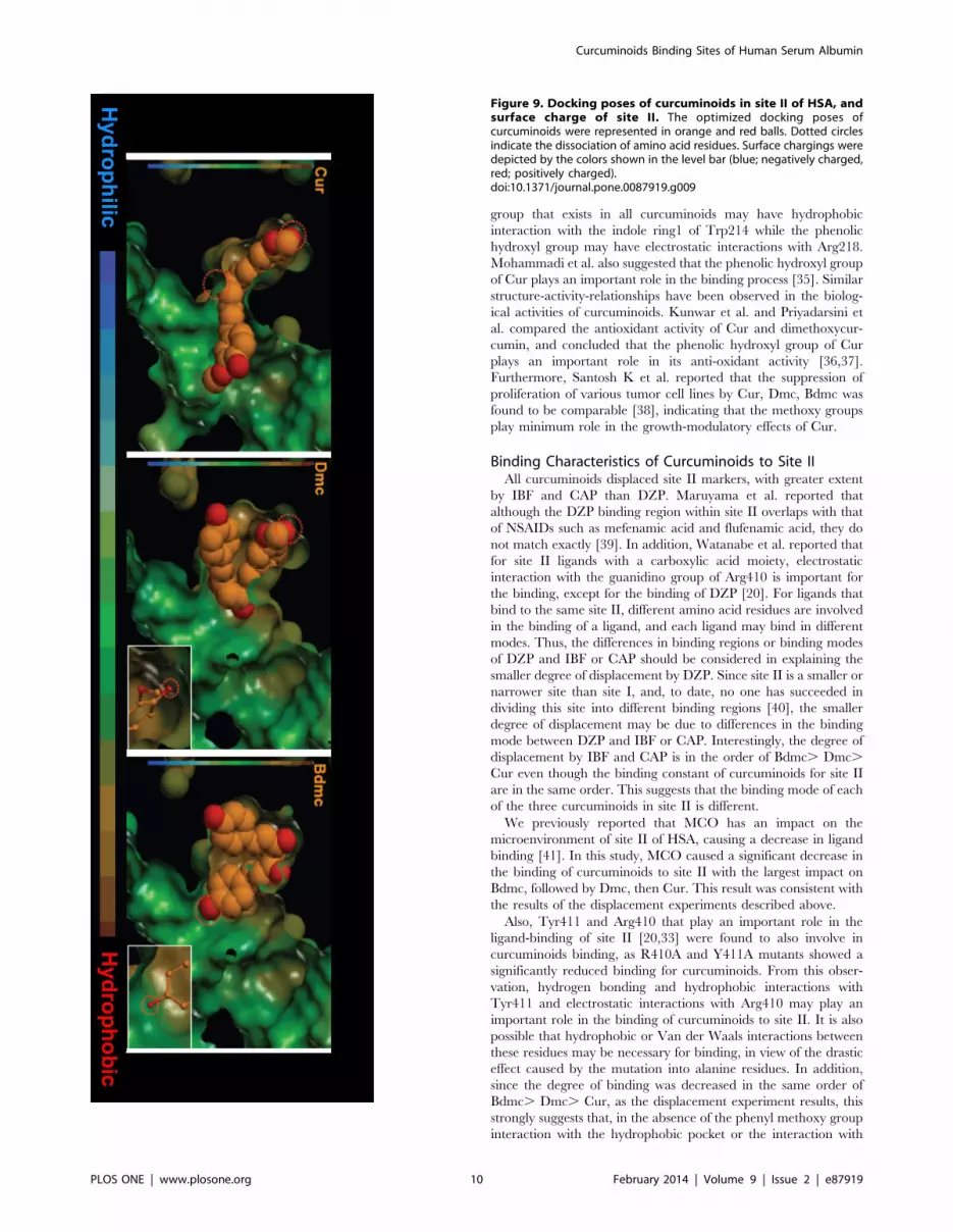
Next, the degree of surface hydrophobicity or charge state of site
II was identified, the relationship between this and the docking
pose of curcuminoids were then examined. As shown in Figure 9,
the docking poses of Dmc and Bdmc are curled, mostly present in
the hydrophobic regions. On the other hand, the docking pose of
Cur is not so extensively curled, and extends across the hydrophilic
region. These results suggest that the contribution of hydrophobic
interactions for site II is likely to be diminished in the order of
Bdmc. Dmc. Cur. Furthermore, we compared the site of
localized negatively charged dissociable group of Bdmc and Dmc,
and found that Bdmc tends to bind to an environment that is more
electrostatically positive (Figure 10).
Discussion
Curcuminoids have been found to possess anti-tumor and anti-
Alzheimer’s effects as well as a variety of biological activities
[2,3,28,29]. Before curcuminoids can be used clinically, further
studies will be needed, including gathering information for
predicting treatment effects, drug pharmacokinetics or drug
interactions when used in combination with other therapeutic
agents. Hence, it is essential to elucidate the full picture of the
interaction with serum proteins, in particular HSA. Unfortunately,
Figure 5. Effects of metal catalized oxidation on the intensity of CD induced of curcuminoids-HSA systems. The binding ofcurcuminoids (25 mM) to HSA and oxidized HSAs (50 mM) in Tris-HCl buffer (pH 9.0 and 25uC) was studied by CD measurement. Results are the means6 SD of triplicate experiments. *, **, ***P, 0.05 versus native HSA.doi:10.1371/journal.pone.0087919.g005
Figure 6. Effects of HSA mutation on the intensity of induced CD of curcuminoids-HSA systems at pH 9.0. The binding of curcuminoids(25 mM) to WT-HSA and HSA mutants (50 mM) in Tris-HCl buffer (pH 9.0 and 25uC) was studied by CD measurement. Results are the means 6 SD oftriplicate experiments. *, **, ***P, 0.05 versus native HSA.doi:10.1371/journal.pone.0087919.g006
Curcuminoids Binding Sites of Human Serum Albumin
PLOS ONE | www.plosone.org 7 February 2014 | Volume 9 | Issue 2 | e87919

although studies on the pharmacological effects of curcuminoids
have been reported, most studies on the interaction with HSA
have been limited to Cur only.
In the present study, the binding properties of the curcuminoids
were examined by the spectroscopic methods only, without
measuring directly the unbound concentration of curcuminoids.
This is because curcuminoids is readily hydrolyzed in the neutral-
alkaline pH range [30]. In fact, the free concentration of Cur
reported so far were obtained from indirect assessments, and no
direct measurement has been performed based on binding
experiments [15,31,32]. In addition, the CD measurements were
performed at pH 9.0 in this study because no induced CD
spectrum could be observed for curcuminoids at pH 7.4 [16]. Zsila
et al. previously reported a distinct feature of the induced CD of
Cur-HSA complex at different pH values and based on a detailed
analysis of this induced CD, they concluded that Cur binds to
HSA in a twisted structure (keto form) at pH 9.0 but binds to HSA
in a planar structure (enol form) at pH 7.4 [16]. As shown in
Figure 3, like Cur, Dmc and Bdmc also exhibit similar pH
dependent induced CD spectra in the presence of HSA, suggesting
that different forms of Dmc and Bdmc bind to HSA at these pHs,
as mentioned above. However, since curcuminoids bind to the
same binding site on HSA at pH 9.0 and pH 7.4, and that pH
change does not affect the binding displacement of curcuminoids,
interpretation of the binding site data obtained at pH 9.0 in
relation to the physiological conditions (pH 7.4) should not be a
problem.
Identification of Curcuminoids Binding Sites on HSAIn this study, all of the curcuminoids examined caused induced-
CD and fluorescence quenching of residue Trp214 in a
concentration-dependent manner, suggesting that Bdmc and
Dmc, like Cur, also bind to HSA. Fluorescence quenching by
the addition of curcuminoids is considered to be due to energy
transfer between the indole ring of Trp214 and the curcuminoids
molecule. Since the only Trp residue of HSA is in the vicinity of
site I, indicating that curcuminoids may bind to site I. This is
consistent with data reported by Zsila [31]. On the other hand,
from the displacement experiments, two binding sites for
curcuminoids may exist. This is a seemingly contradictory result
with the number of binding sites for curcuminoids calculated from
the fluorescence quenching experiment (n = 1). A possible reason
for this is that there is a curcuminoid molecule that binds to HSA
but cannot quench the fluorescence derived from the Trp residue
in the fluorescence quenching experiment. In fact, no intrinsic
fluorescence quenching phenomenon from the ligand binding to
site I of HSA was observed for site II-selective ligands such as IBF,
CAP and DZP (data not shown). Therefore, the findings obtained
in the fluorescence quenching experiments reflect the binding
affinity of curcuminoids for site I.
In order to identify the binding sites of curcuminoids on HSA,
fluorescence and CD spectra displacement experiments were
performed. In the fluorescent probe displacement experiments
performed at pH 7.4 and pH 9.0, all curcuminoids displaced the
site I marker, WF. Curcuminoids also caused a displacement of the
Figure 7. Molecular docking of curcuminoids in site I of HSA.The optimized docking poses of curcuminoids were represented inorange color. Hydrogen bonds were highlighted by the lines in yellow-green color, and amino acid residues which make hydrogen bondingwith curcuminoids were underlined. Dotted circles indicate dissociationof amino acid residues. Right-blue, green, pink and white lettersrepresent basic, aromatic, hydrophilic and hydrophobic amino acidresidues, respectively.doi:10.1371/journal.pone.0087919.g007
Curcuminoids Binding Sites of Human Serum Albumin
PLOS ONE | www.plosone.org 8 February 2014 | Volume 9 | Issue 2 | e87919

site II marker, DNSS, in a concentration-dependent manner. It is
interesting to note that the extent of displacement of DNSS (or the
binding constant to site II) varies among the curcuminoids, in the
decreasing order Bdmc. Dmc. Cur. Similar results were
obtained in the CD displacement experiment at pH 9.0 where
the intensity of curcuminoids induced CD spectrum was reduced
significantly in the presence of the site I and II markers. These
results strongly suggested that curcuminoids bind to both sites I
and site II.
Binding Characteristics of Curcuminoids to Site ICur was reported to bind to site I by Zsila et al. [31] and
Mandeville et al. [32]. Our results indicate that the other
curcuminoids (Dmc and Bdmc) also bind to site I. We previously
reported that site I is not a simple binding region but is rather
complex and is comprised of three subsites, namely, Ia, Ib and Ic
[22]. Of the site I markers used in this study, WF binds to subsite
Ia, PBZ binds to subsites Ia , Ib, and IDP binds to subsites Ia ,
Ic [22]. Ghuman et al. also determined the locations of the binding
regions of WF, PB and IDP in site I based on crystallographic
studies [33]. WF, PB and IDP all bind to the main cavity of site I,
which is composed of two apolar chambers in which Tyr150,
His242, Arg257 or Arg218, Arg222 are positioned. WF accesses
one of the chambers (where Tyr150, His242 and Arg257 are
located), whereas PB accesses both chambers. IDP accesses not
only both chambers in the main cavity, but a distinct sub-chamber
separate from the main cavity which is extended to residues from
subdomains IIB and IIIA. From the degree of displacement of
these markers, it can be concluded that curcuminoids bind to a
region of site I that is also bound by WF, PBZ but not IDP. This
suggests that curcuminoids potentially bind to mainly PBZ binding
region of site I, which may correspond to subsites Ia , Ib. In
contrast, IDP showed a slight potentiation of the induced Cotton
effect of curcuminoids. Taking this phenomenon into consider-
ation it is possible that curcuminoids and IDP both bind within site
I with the aromatic ring motility of IDP immobilized by
curcuminoids. Kragh-Hansen reported that site I is a large and
flexible region based on a diversity of interaction ligands and that
it is capable of accommodating more than one of them at a time
[34]. Furthermore, crystallographic data reported by Ghuman et
al. showed that significant room exists for different compounds to
occupy different parts of site I, and the co-binding of indomethacin
and azapropazone or phenylbutazone by shifting their position
with respect to each other within site I is likely to occur [33].
Although the size of molecules that co-bind this site was not
mentioned in their studies, site I may accommodate two even
larger molecules, such as iodipamide and curcuminoids.
The conclusion that curcuminoids binds to site I was also
supported by the reduced binding of curcuminoids to the W214A
and R218A mutants, because Trp214 and Arg218 are known to
be involved in the binding of site I ligands [33]. Since the degree of
binding reduction to mutants for all curcuminoids were similar,
the methoxy groups of curcuminoids do not appear to play a
significant role in site I binding. The aromatic ring of the phenolic
Figure 8. Molecular docking of curcuminoids in site II of HSA.The optimized docking poses of curcuminoids were represented inorange color. Hydrogen bonds were highlighted by the lines in yellow-green color, and amino acid residues which make hydrogen bondingwith curcuminoids were underlined. Dotted circles indicate dissociationof amino acid residues. Red, right-blue, green, pink and white lettersrepresent acidic, basic, aromatic, hydrophilic and hydrophobic aminoacid residues, respectively. Cysteine residues and disulphide bond areshown in yellow letters and stick, respectively.doi:10.1371/journal.pone.0087919.g008
Curcuminoids Binding Sites of Human Serum Albumin
PLOS ONE | www.plosone.org 9 February 2014 | Volume 9 | Issue 2 | e87919
group that exists in all curcuminoids may have hydrophobic
interaction with the indole ring1 of Trp214 while the phenolic
hydroxyl group may have electrostatic interactions with Arg218.
Mohammadi et al. also suggested that the phenolic hydroxyl group
of Cur plays an important role in the binding process [35]. Similar
structure-activity-relationships have been observed in the biolog-
ical activities of curcuminoids. Kunwar et al. and Priyadarsini et
al. compared the antioxidant activity of Cur and dimethoxycur-
cumin, and concluded that the phenolic hydroxyl group of Cur
plays an important role in its anti-oxidant activity [36,37].
Furthermore, Santosh K et al. reported that the suppression of
proliferation of various tumor cell lines by Cur, Dmc, Bdmc was
found to be comparable [38], indicating that the methoxy groups
play minimum role in the growth-modulatory effects of Cur.
Binding Characteristics of Curcuminoids to Site IIAll curcuminoids displaced site II markers, with greater extent
by IBF and CAP than DZP. Maruyama et al. reported that
although the DZP binding region within site II overlaps with that
of NSAIDs such as mefenamic acid and flufenamic acid, they do
not match exactly [39]. In addition, Watanabe et al. reported that
for site II ligands with a carboxylic acid moiety, electrostatic
interaction with the guanidino group of Arg410 is important for
the binding, except for the binding of DZP [20]. For ligands that
bind to the same site II, different amino acid residues are involved
in the binding of a ligand, and each ligand may bind in different
modes. Thus, the differences in binding regions or binding modes
of DZP and IBF or CAP should be considered in explaining the
smaller degree of displacement by DZP. Since site II is a smaller or
narrower site than site I, and, to date, no one has succeeded in
dividing this site into different binding regions [40], the smaller
degree of displacement may be due to differences in the binding
mode between DZP and IBF or CAP. Interestingly, the degree of
displacement by IBF and CAP is in the order of Bdmc. Dmc.
Cur even though the binding constant of curcuminoids for site II
are in the same order. This suggests that the binding mode of each
of the three curcuminoids in site II is different.
We previously reported that MCO has an impact on the
microenvironment of site II of HSA, causing a decrease in ligand
binding [41]. In this study, MCO caused a significant decrease in
the binding of curcuminoids to site II with the largest impact on
Bdmc, followed by Dmc, then Cur. This result was consistent with
the results of the displacement experiments described above.
Also, Tyr411 and Arg410 that play an important role in the
ligand-binding of site II [20,33] were found to also involve in
curcuminoids binding, as R410A and Y411A mutants showed a
significantly reduced binding for curcuminoids. From this obser-
vation, hydrogen bonding and hydrophobic interactions with
Tyr411 and electrostatic interactions with Arg410 may play an
important role in the binding of curcuminoids to site II. It is also
possible that hydrophobic or Van der Waals interactions between
these residues may be necessary for binding, in view of the drastic
effect caused by the mutation into alanine residues. In addition,
since the degree of binding was decreased in the same order of
Bdmc. Dmc. Cur, as the displacement experiment results, this
strongly suggests that, in the absence of the phenyl methoxy group
interaction with the hydrophobic pocket or the interaction with
Figure 9. Docking poses of curcuminoids in site II of HSA, andsurface charge of site II. The optimized docking poses ofcurcuminoids were represented in orange and red balls. Dotted circlesindicate the dissociation of amino acid residues. Surface chargings weredepicted by the colors shown in the level bar (blue; negatively charged,red; positively charged).doi:10.1371/journal.pone.0087919.g009
Curcuminoids Binding Sites of Human Serum Albumin
PLOS ONE | www.plosone.org 10 February 2014 | Volume 9 | Issue 2 | e87919

Arg410 and Tyr411, a decrease in the binding affinity to site II will
result. Structurally speaking, site II is less flexible than site I [24],
the molecular size of Bdmc makes it easier to fit into site II than
Cur. Similar relationship between curcuminoids was found by
Ahmed T et al. that curcuminoids rescue long-term potentiation
impaired by amyloid peptide in rat hippocampal slices in the order
of Bdmc . Dmc . Cur [42].
Investigation of the Binding Sites with ModelingThe results obtained from docking simulations of curcuminoids
in site I and site II of HSA were in agreement with the results of
the displacement experiments. In the docking model of site I, the
docking pose of each curcuminoid and the environment of
molecular interactions such as hydrogen bonding were similar.
These results confirmed that the binding affinity among the
curcuminoids for site I is similar.
In contrast, the docking poses of curcuminoids for site II site
were clearly different. Bdmc and Dmc took a rounded form when
binding to a region in site II that was positively charged and
hydrophobic, whereas Cur was in a more extended form binding
to a region that was relatively negatively charged and hydrophilic.
Therefore, the hydrophobic or electrostatic interaction of Cur with
site II is weaker than Dmc and Bdmc. In addition, Bdmc is more
suitable for evaluating electrostatic or hydrophobic interactions
than Dmc with the surrounding environment and with the
negatively charged dissociated oxygen atoms. Bdmc forms
stronger hydrogen bonds than Dmc. Having cross interpreted
these results, the binding affinity of curcuminoids for site II
diminished in the order of Bdmc. Dmc. Cur, which matched
and agreed very well with the results of binding and displacement
experiments. In order to further verify these findings, Bdmc was
docked in site II with a methoxy group introduced into two
locations of the molecule similar to those in Cur. The methoxy
groups were found to be in obstructive contact with the pocket
region of site II, making it difficult for Bdmc to fit in properly at
the binding region. Hence, the reason for why Cur binds in an
extended form to site II was the two methoxy groups of Cur
introduced steric hindrance that prevented it from binding to site
II in the same manner as Dmc and Bdmc. In addition, the
discrepancy in the binding affinity to site II was also due to
different amino acid residues involved in the binding interaction of
curcuminoids.
Conclusions
In this study, Dmc and Bdmc were found to also bind to both
site I and site II of HSA. No difference among curcuminoids when
binding to site I was found, but binding at site II differs depending
on the number of methoxy groups present on the phenyl group of
the curcuminoids.
Acknowledgments
We thank the Chemo-Sera-Therapeutic Research Institute for donating
HSA.
Figure 10. Docking poses of curcuminoids in site II of HSA, andsurface hydrophobisity of site II. The optimized docking poses ofcurcuminoids were represented in orange and red balls. Dotted circlesindicate the dissociation of amino acid residues. Surface hydrophobi-sities were depicted by the colors shown in the level bar (blue;hydrophilic, red; hydrophobic).doi:10.1371/journal.pone.0087919.g010
Curcuminoids Binding Sites of Human Serum Albumin
PLOS ONE | www.plosone.org 11 February 2014 | Volume 9 | Issue 2 | e87919

Author Contributions
Conceived and designed the experiments: HS KY NY. Performed the
experiments: HS. Analyzed the data: HS MA. Contributed reagents/
materials/analysis tools: KN DK SH MO. Wrote the paper: VC HW YI
MO TM.
References
1. Aggarwal BB, Sung B (2009) Pharmacological basis for the role of curcumin inchronic diseases: an age-old spice with modern targets. Trends Pharmacol Sci30: 85–94.
2. Duvoix A, Blasius R, Delhalle S, Schnekenburger M, Morceau F, et al. (2005)Chemopreventive and therapeutic effects of curcumin. Cancer Lett 223: 181–190.
3. Cui L, Miao J (2007) Cytotoxic effect of curcumin on malaria parasitePlasmodium falciparum: inhibition of histone acetylation and generation ofreactive oxygen species. Antimicrob Agents Chemother 51: 488–494.
4. Kim JE, Kim AR, Chung HY, Han SY, Kim BS, et al. (2003) In vitroperoxynitrite scavenging activity of diarylheptanoids from Curcuma longa.Phytother Res 17: 481–484.
5. Somparn P, Phisalaphong C, Nakornchai S, Unchern S, Morales NP (2007)Comparative antioxidant activities of curcumin and its demethoxy andhydrogenated derivatives. Biol Pharm Bull 30: 74–78.
6. Yodkeeree S, Chaiwangyen W, Garbisa S, Limtrakul P (2009) Curcumin,demethoxycurcumin and bisdemethoxycurcumin differentially inhibit cancercell invasion through the down-regulation of MMPs and uPA. J Nutr Biochem20: 87–95.
7. Fiala M, Liu PT, Espinosa-Jeffrey A, Rosenthal MJ, Bernard G, et al. (2007)Innate immunity and transcription of MGAT-III and Toll-like receptors inAlzheimer’s disease patients are improved by bisdemethoxycurcumin. Proc NatlAcad Sci U S A 104: 12849–12854.
8. Garcia-Alloza M, Borrelli LA, Rozkalne A, Hyman BT, Bacskai BJ, et al. (2007)Curcumin labels amyloid pathology in vivo, disrupts existing plaques, andpartially restores distorted neurites in an Alzheimer mouse model. J Neurochem102: 1095–1104.
9. Peters TJ (1996) All About Albumin: Biochemistry, Genetics, and MedicalApplications, Academic Press.
10. Fasano M, Curry S, Terreno E, Galliano M, Fanali G, et al. (2005) Theextraordinary ligand binding properties of human serum albumin. IUBMB Life57: 787–796.
11. Bertucci C, Domenici E (2002) Reversible and covalent binding of drugs tohuman serum albumin: methodological approaches and physiological relevance.Curr Med Chem 9: 1463–1481.
12. Sudlow G, Birkett DJ, Wade DN (1975) The characterization of two specificdrug binding sites on human serum albumin. Mol Pharmacol 11: 824–832.
13. He XM, Carter DC (1992) Atomic structure and chemistry of human serumalbumin. Nature 358: 209–215.
14. Sugio S, Kashima A, Mochizuki S, Noda M, Kobayashi K (1999) Crystalstructure of human serum albumin at 2.5 A resolution. Protein Eng 12: 439–446.
15. Pulla Reddy AC, Sudharshan E, Appu Rao AG, Lokesh BR (1999) Interactionof curcumin with human serum albumin-a spectroscopic study. Lipids 34: 1025–1029.
16. Zsila F, Bikadi Z, Simonyi M (2003) Unique, pH-dependent biphasic band shapeof the visible circular dichroism of curcumin-serum albumin complex. BiochemBiophys Res Commun 301: 776–782.
17. Chen RF (1967) Removal of fatty acids from serum albumin by charcoaltreatment. J Biol Chem 242: 173–181.
18. Meucci E, Mordente A, Martorana GE (1991) Metal-catalyzed oxidation ofhuman serum albumin: conformational and functional changes. Implications inprotein aging. J Biol Chem 266: 4692–4699.
19. Watanabe H, Kragh-Hansen U, Tanase S, Nakajou K, Mitarai M, et al. (2001)Conformational stability and warfarin-binding properties of human serumalbumin studied by recombinant mutants. Biochem J 357: 269–274.
20. Watanabe H, Tanase S, Nakajou K, Maruyama T, Kragh-Hansen U, et al.(2000) Role of arg-410 and tyr-411 in human serum albumin for ligand bindingand esterase-like activity. Biochem J 349: 813–819.
21. Panjehshahin MR, Bowmer CJ, Yates MS (1991) Effect of valproic acid, itsunsaturated metabolites and some structurally related fatty acids on the bindingof warfarin and dansylsarcosine to human albumin. Biochem Pharmacol 41:1227–1233.
22. Yamasaki K, Maruyama T, Kragh-Hansen U, Otagiri M (1996) Characteriza-tion of site I on human serum albumin: concept about the structure of a drugbinding site. Biochim Biophys Acta 1295: 147–157.
23. Sudlow G, Birkett DJ, Wade DN (1973) Spectroscopic techniques in the study of
protein binding: the use of 1-anilino-8-naphthalenesulphonate as a fluorescent
probe for the study of the binding of iophenoxic and iopanoic acids to human
serum albumin. Mol Pharmacol 9: 649–657.
24. Kragh-Hansen U, Chuang VT, Otagiri M (2002) Practical aspects of the ligand-
binding and enzymatic properties of human serum albumin. Biol Pharm Bull 25:
695–704.
25. Friesner RA, Banks JL, Murphy RB, Halgren TA, Klicic JJ, et al. (2004) Glide: a
new approach for rapid, accurate docking and scoring. 1. Method and
assessment of docking accuracy. J Med Chem 47: 1739–1749.
26. Person RV, David BRP, Lightner A (1994) Bilirubin conformational analysis
and circular dichroism. J Am Chem Soc 116: 42–59.
27. Iwao Y, Anraku M, Yamasaki K, Kragh-Hansen U, Kawai K, et al. (2006)
Oxidation of Arg-410 promotes the elimination of human serum albumin.
Biochim Biophys Acta 1764: 743–749.
28. Ringman JM, Frautschy SA, Cole GM, Masterman DL, Cummings JL, et al.
(2005) A potential role of the curry spice curcumin in Alzheimer’s disease. Curr
Alzheimer Res 2: 131–136.
29. Ji JL, Huang XF, Zhu HL (2012) Curcumin and its formulations: potential anti-
cancer agents. Anticancer Agents Med Chem 12: 210–218.
30. Wang YJ, Pan MH, Cheng AL, Lin LI, Ho YS, et al. (1997) Stability of
curcumin in buffer solutions and characterization of its degradation products.
J Pharm Biomed Anal 15: 1867–1876.
31. Zsila F, Bikadi Z, Simonyi M (2003) Molecular basis of the Cotton effects
induced by the binding of curcumin to human serum albumin. Tetrahedron:
Asymmetry 14: 2433–2444.
32. Mandeville JS, Froehlich E, Tajmir-Riahi HA (2009) Study of curcumin and
genistein interactions with human serum albumin. J Pharm Biomed Anal 49:
468–474.
33. Ghuman J, Zunszain PA, Petitpas I, Bhattacharya AA, Otagiri M, et al. (2005)
Structural basis of the drug-binding specificity of human serum albumin. J Mol
Biol 353: 38–52.
34. Kragh-Hansen U (1988) Evidence for a large and flexible region of human
serum albumin possessing high affinity binding sites for salicylate, warfarin, and
other ligands. Mol Pharmacol 34:160–171.
35. Mohammadi F, Bordbar AK, Divsalar A, Mohammadi K, Saboury AA (2009)
Analysis of binding interaction of curcumin and diacetylcurcumin with human
and bovine serum albumin using fluorescence and circular dichroism
spectroscopy. Protein J 28: 189–196.
36. Kunwar A, Barik A, Sandur SK, Indira Priyadarsini K (2011) Differential
antioxidant/pro-oxidant activity of dimethoxycurcumin, a synthetic analogue of
curcumin. Free Radic Res 45: 959–965.
37. Priyadarsini KI, Maity DK, Naik GH, Kumar MS, Unnikrishnan MK, et al.
(2003) Role of phenolic O-H and methylene hydrogen on the free radical
reactions and antioxidant activity of curcumin. Free Radic Biol Med 35: 475–
484.
38. Sandur SK, Pandey MK, Sung B, Ahn KS, Murakami A, et al. (2007)
Curcumin, demethoxycurcumin, bisdemethoxycurcumin, tetrahydrocurcumin
and turmerones differentially regulate anti-inflammatory and anti-proliferative
responses through a ROS-independent mechanism. Carcinogenesis 28: 1765–
1773.
39. Maruyama K., Nishigori H, Iwatsuru M (1985) Characterization of the
benzodiazepine binding site (diazepam site) on human serum albumin. Chem
Pharm Bull 33: 5002–5012.
40. Yamasaki K, Chuang VT, Maruyama T, Otagiri M (2013) Albumin-drug
interaction and its clinical implication. Biochim Biophys Acta 1830: 5435–5443.
41. Anraku M, Yamasaki K, Maruyama T, Kragh-Hansen U, Otagiri M (2001)
Effect of oxidative stress on the structure and function of human serum albumin.
Pharm Res 18: 632–639.
42. Ahmed T, Gilani AH, Hosseinmardi N, Semnanian S, Enam SA, et al. (2011)
Curcuminoids rescue long-term potentiation impaired by amyloid peptide in rat
hippocampal slices. Synapse 65: 572–582.
Curcuminoids Binding Sites of Human Serum Albumin
PLOS ONE | www.plosone.org 12 February 2014 | Volume 9 | Issue 2 | e87919
Copyright © 2022 FDOKUMEN