The distribution of ligand-binding pockets around ... - PNAS
6
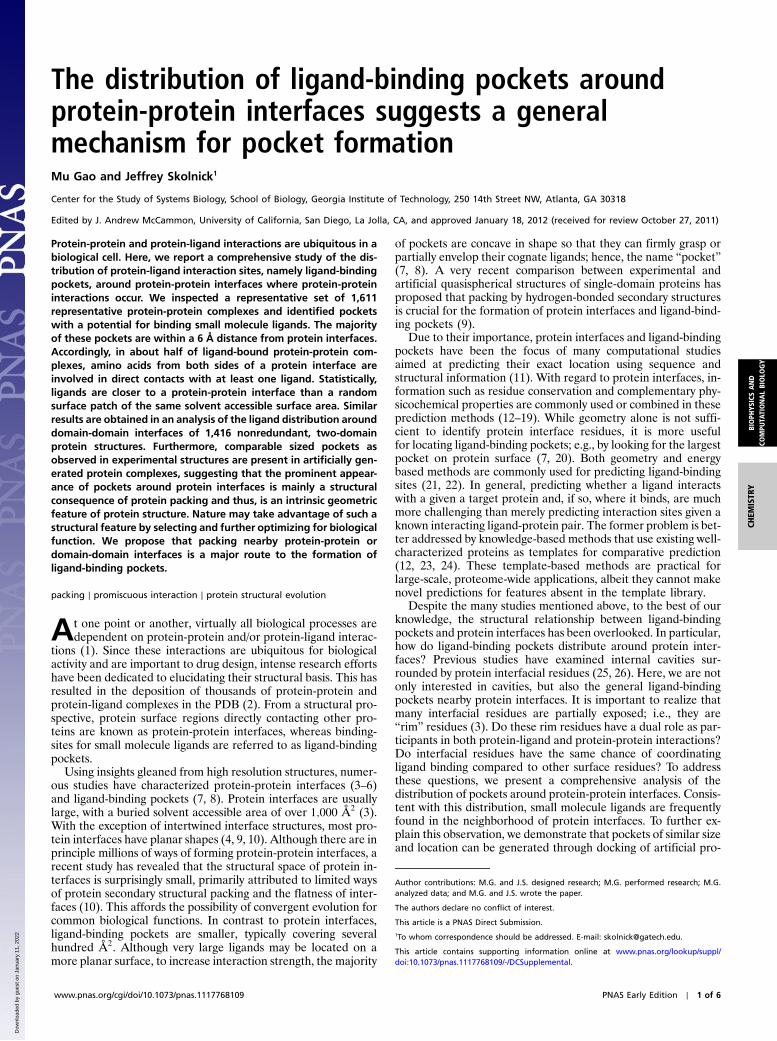
The distribution of ligand-binding pockets around protein-protein interfaces suggests a general mechanism for pocket formation Mu Gao and Jeffrey Skolnick 1 Center for the Study of Systems Biology, School of Biology, Georgia Institute of Technology, 250 14th Street NW, Atlanta, GA 30318 Edited by J. Andrew McCammon, University of California, San Diego, La Jolla, CA, and approved January 18, 2012 (received for review October 27, 2011) Protein-protein and protein-ligand interactions are ubiquitous in a biological cell. Here, we report a comprehensive study of the dis- tribution of protein-ligand interaction sites, namely ligand-binding pockets, around protein-protein interfaces where protein-protein interactions occur. We inspected a representative set of 1,611 representative protein-protein complexes and identified pockets with a potential for binding small molecule ligands. The majority of these pockets are within a 6 Å distance from protein interfaces. Accordingly, in about half of ligand-bound protein-protein com- plexes, amino acids from both sides of a protein interface are involved in direct contacts with at least one ligand. Statistically, ligands are closer to a protein-protein interface than a random surface patch of the same solvent accessible surface area. Similar results are obtained in an analysis of the ligand distribution around domain-domain interfaces of 1,416 nonredundant, two-domain protein structures. Furthermore, comparable sized pockets as observed in experimental structures are present in artificially gen- erated protein complexes, suggesting that the prominent appear- ance of pockets around protein interfaces is mainly a structural consequence of protein packing and thus, is an intrinsic geometric feature of protein structure. Nature may take advantage of such a structural feature by selecting and further optimizing for biological function. We propose that packing nearby protein-protein or domain-domain interfaces is a major route to the formation of ligand-binding pockets. packing ∣ promiscuous interaction ∣ protein structural evolution A t one point or another, virtually all biological processes are dependent on protein-protein and/or protein-ligand interac- tions (1). Since these interactions are ubiquitous for biological activity and are important to drug design, intense research efforts have been dedicated to elucidating their structural basis. This has resulted in the deposition of thousands of protein-protein and protein-ligand complexes in the PDB (2). From a structural pro- spective, protein surface regions directly contacting other pro- teins are known as protein-protein interfaces, whereas binding- sites for small molecule ligands are referred to as ligand-binding pockets. Using insights gleaned from high resolution structures, numer- ous studies have characterized protein-protein interfaces (3–6) and ligand-binding pockets (7, 8). Protein interfaces are usually large, with a buried solvent accessible area of over 1;000 Å 2 (3). With the exception of intertwined interface structures, most pro- tein interfaces have planar shapes (4, 9, 10). Although there are in principle millions of ways of forming protein-protein interfaces, a recent study has revealed that the structural space of protein in- terfaces is surprisingly small, primarily attributed to limited ways of protein secondary structural packing and the flatness of inter- faces (10). This affords the possibility of convergent evolution for common biological functions. In contrast to protein interfaces, ligand-binding pockets are smaller, typically covering several hundred Å 2 . Although very large ligands may be located on a more planar surface, to increase interaction strength, the majority of pockets are concave in shape so that they can firmly grasp or partially envelop their cognate ligands; hence, the name “pocket” (7, 8). A very recent comparison between experimental and artificial quasispherical structures of single-domain proteins has proposed that packing by hydrogen-bonded secondary structures is crucial for the formation of protein interfaces and ligand-bind- ing pockets (9). Due to their importance, protein interfaces and ligand-binding pockets have been the focus of many computational studies aimed at predicting their exact location using sequence and structural information (11). With regard to protein interfaces, in- formation such as residue conservation and complementary phy- sicochemical properties are commonly used or combined in these prediction methods (12–19). While geometry alone is not suffi- cient to identify protein interface residues, it is more useful for locating ligand-binding pockets; e.g., by looking for the largest pocket on protein surface (7, 20). Both geometry and energy based methods are commonly used for predicting ligand-binding sites (21, 22). In general, predicting whether a ligand interacts with a given a target protein and, if so, where it binds, are much more challenging than merely predicting interaction sites given a known interacting ligand-protein pair. The former problem is bet- ter addressed by knowledge-based methods that use existing well- characterized proteins as templates for comparative prediction (12, 23, 24). These template-based methods are practical for large-scale, proteome-wide applications, albeit they cannot make novel predictions for features absent in the template library. Despite the many studies mentioned above, to the best of our knowledge, the structural relationship between ligand-binding pockets and protein interfaces has been overlooked. In particular, how do ligand-binding pockets distribute around protein inter- faces? Previous studies have examined internal cavities sur- rounded by protein interfacial residues (25, 26). Here, we are not only interested in cavities, but also the general ligand-binding pockets nearby protein interfaces. It is important to realize that many interfacial residues are partially exposed; i.e., they are “rim” residues (3). Do these rim residues have a dual role as par- ticipants in both protein-ligand and protein-protein interactions? Do interfacial residues have the same chance of coordinating ligand binding compared to other surface residues? To address these questions, we present a comprehensive analysis of the distribution of pockets around protein-protein interfaces. Consis- tent with this distribution, small molecule ligands are frequently found in the neighborhood of protein interfaces. To further ex- plain this observation, we demonstrate that pockets of similar size and location can be generated through docking of artificial pro- Author contributions: M.G. and J.S. designed research; M.G. performed research; M.G. analyzed data; and M.G. and J.S. wrote the paper. The authors declare no conflict of interest. This article is a PNAS Direct Submission. 1 To whom correspondence should be addressed. E-mail: [email protected]. This article contains supporting information online at www.pnas.org/lookup/suppl/ doi:10.1073/pnas.1117768109/-/DCSupplemental. www.pnas.org/cgi/doi/10.1073/pnas.1117768109 PNAS Early Edition ∣ 1 of 6 CHEMISTRY BIOPHYSICS AND COMPUTATIONAL BIOLOGY Downloaded by guest on January 11, 2022
-
Upload
khangminh22 -
Category
Documents
-
view
1 -
download
0
Transcript of The distribution of ligand-binding pockets around ... - PNAS
The distribution of ligand-binding pockets aroundprotein-protein interfaces suggests a generalmechanism for pocket formationMu Gao and Jeffrey Skolnick1
Center for the Study of Systems Biology, School of Biology, Georgia Institute of Technology, 250 14th Street NW, Atlanta, GA 30318
Edited by J. Andrew McCammon, University of California, San Diego, La Jolla, CA, and approved January 18, 2012 (received for review October 27, 2011)
Protein-protein and protein-ligand interactions are ubiquitous in abiological cell. Here, we report a comprehensive study of the dis-tribution of protein-ligand interaction sites, namely ligand-bindingpockets, around protein-protein interfaces where protein-proteininteractions occur. We inspected a representative set of 1,611representative protein-protein complexes and identified pocketswith a potential for binding small molecule ligands. The majorityof these pockets are within a 6 Å distance from protein interfaces.Accordingly, in about half of ligand-bound protein-protein com-plexes, amino acids from both sides of a protein interface areinvolved in direct contacts with at least one ligand. Statistically,ligands are closer to a protein-protein interface than a randomsurface patch of the same solvent accessible surface area. Similarresults are obtained in an analysis of the ligand distribution arounddomain-domain interfaces of 1,416 nonredundant, two-domainprotein structures. Furthermore, comparable sized pockets asobserved in experimental structures are present in artificially gen-erated protein complexes, suggesting that the prominent appear-ance of pockets around protein interfaces is mainly a structuralconsequence of protein packing and thus, is an intrinsic geometricfeature of protein structure. Nature may take advantage of such astructural feature by selecting and further optimizing for biologicalfunction. We propose that packing nearby protein-protein ordomain-domain interfaces is a major route to the formation ofligand-binding pockets.
packing ∣ promiscuous interaction ∣ protein structural evolution
At one point or another, virtually all biological processes aredependent on protein-protein and/or protein-ligand interac-
tions (1). Since these interactions are ubiquitous for biologicalactivity and are important to drug design, intense research effortshave been dedicated to elucidating their structural basis. This hasresulted in the deposition of thousands of protein-protein andprotein-ligand complexes in the PDB (2). From a structural pro-spective, protein surface regions directly contacting other pro-teins are known as protein-protein interfaces, whereas binding-sites for small molecule ligands are referred to as ligand-bindingpockets.
Using insights gleaned from high resolution structures, numer-ous studies have characterized protein-protein interfaces (3–6)and ligand-binding pockets (7, 8). Protein interfaces are usuallylarge, with a buried solvent accessible area of over 1;000 Å2 (3).With the exception of intertwined interface structures, most pro-tein interfaces have planar shapes (4, 9, 10). Although there are inprinciple millions of ways of forming protein-protein interfaces, arecent study has revealed that the structural space of protein in-terfaces is surprisingly small, primarily attributed to limited waysof protein secondary structural packing and the flatness of inter-faces (10). This affords the possibility of convergent evolution forcommon biological functions. In contrast to protein interfaces,ligand-binding pockets are smaller, typically covering severalhundred Å2. Although very large ligands may be located on amore planar surface, to increase interaction strength, the majority
of pockets are concave in shape so that they can firmly grasp orpartially envelop their cognate ligands; hence, the name “pocket”(7, 8). A very recent comparison between experimental andartificial quasispherical structures of single-domain proteins hasproposed that packing by hydrogen-bonded secondary structuresis crucial for the formation of protein interfaces and ligand-bind-ing pockets (9).
Due to their importance, protein interfaces and ligand-bindingpockets have been the focus of many computational studiesaimed at predicting their exact location using sequence andstructural information (11). With regard to protein interfaces, in-formation such as residue conservation and complementary phy-sicochemical properties are commonly used or combined in theseprediction methods (12–19). While geometry alone is not suffi-cient to identify protein interface residues, it is more usefulfor locating ligand-binding pockets; e.g., by looking for the largestpocket on protein surface (7, 20). Both geometry and energybased methods are commonly used for predicting ligand-bindingsites (21, 22). In general, predicting whether a ligand interactswith a given a target protein and, if so, where it binds, are muchmore challenging than merely predicting interaction sites given aknown interacting ligand-protein pair. The former problem is bet-ter addressed by knowledge-based methods that use existing well-characterized proteins as templates for comparative prediction(12, 23, 24). These template-based methods are practical forlarge-scale, proteome-wide applications, albeit they cannot makenovel predictions for features absent in the template library.
Despite the many studies mentioned above, to the best of ourknowledge, the structural relationship between ligand-bindingpockets and protein interfaces has been overlooked. In particular,how do ligand-binding pockets distribute around protein inter-faces? Previous studies have examined internal cavities sur-rounded by protein interfacial residues (25, 26). Here, we are notonly interested in cavities, but also the general ligand-bindingpockets nearby protein interfaces. It is important to realize thatmany interfacial residues are partially exposed; i.e., they are“rim” residues (3). Do these rim residues have a dual role as par-ticipants in both protein-ligand and protein-protein interactions?Do interfacial residues have the same chance of coordinatingligand binding compared to other surface residues? To addressthese questions, we present a comprehensive analysis of thedistribution of pockets around protein-protein interfaces. Consis-tent with this distribution, small molecule ligands are frequentlyfound in the neighborhood of protein interfaces. To further ex-plain this observation, we demonstrate that pockets of similar sizeand location can be generated through docking of artificial pro-
Author contributions: M.G. and J.S. designed research; M.G. performed research; M.G.analyzed data; and M.G. and J.S. wrote the paper.
The authors declare no conflict of interest.
This article is a PNAS Direct Submission.1To whom correspondence should be addressed. E-mail: [email protected].
This article contains supporting information online at www.pnas.org/lookup/suppl/doi:10.1073/pnas.1117768109/-/DCSupplemental.
www.pnas.org/cgi/doi/10.1073/pnas.1117768109 PNAS Early Edition ∣ 1 of 6
CHEM
ISTR
YBIOPH
YSICSAND
COMPU
TATIONALBIOLO
GY
Dow
nloa
ded
by g
uest
on
Janu
ary
11, 2
022
tein structures. Moreover, we show that domain-domain inter-faces of multi-domain proteins provide structural pockets that in-teract with ligands. These results suggest that many pockets aregeometric in origin and arise from the intrinsic physical proper-ties of proteins without the requirement of evolution. Evolutionplays a role in optimizing their sequence properties to enable aspecific biochemical function. Finally, we propose that packing ofproteins or domains is a general mechanism for creating ligand-binding pockets.
ResultsDistribution of Pockets in Protein Complexes. We first investigatehow pockets distribute within protein complexes, using experi-mentally solved crystal structures of 1,611 representative protein-protein complexes from previous studies (13, 27). To characterizethe distance between a detected pocket and a protein interface,we introduce Rmin, the minimum of all distances between the geo-metric center of the pocket and heavy atoms of protein-proteininterfacial residues (see Methods). Each pocket is also assigned avolume in units of grid points, as reported by the grid-based pock-et detection program LIGSITECSC (20). Unless specified other-wise, we consider pockets of a volume larger than 100 grid points,each with a grid spacing of 1 Å. This cutoff is arbitrary and coversabout 80% of pockets occupied by ligands. Nevertheless, chan-ging the cutoff values does not qualitatively affect the results pre-sented below.
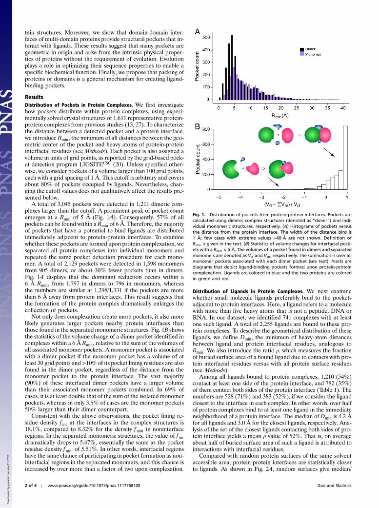
A total of 3,045 pockets were detected in 1,211 dimeric com-plexes larger than the cutoff. A prominent peak of pocket countemerges at a Rmin of 5 Å (Fig. 1A). Consequently, 57% of allpockets can be found within a Rmin of 6 Å. Therefore, the majorityof pockets that have a potential to bind ligands are distributedimmediately adjacent to protein-protein interfaces. To examinewhether these pockets are formed upon protein complexation, weseparated all protein complexes into individual monomers andrepeated the same pocket detection procedure for each mono-mer. A total of 2,129 pockets were detected in 1,598 monomersfrom 905 dimers, or about 30% fewer pockets than in dimers.Fig. 1A displays that the dominant reduction occurs within a6 Å Rmin, from 1,797 in dimers to 796 in monomers, whereasthe numbers are similar at 1,298/1,331 if the pockets are morethan 6 Å away from protein interfaces. This result suggests thatthe formation of the protein complex dramatically enlarges thecollection of pockets.
Not only does complexation create more pockets, it also morelikely generates larger pockets nearby protein interfaces thanthose found in the separated monomeric structures. Fig. 1B showsthe statistics of the volume change of a dimer pocket identified incomplexes within a 6 Å Rmin, relative to the sum of the volumes ofall associated monomer pockets. A monomer pocket is associatedwith a dimer pocket if the monomer pocket has a volume of atleast 30 grid points and >10% of its pocket lining residues are alsofound in the dimer pocket, regardless of the distance from themonomer pocket to the protein interface. The vast majority(90%) of these interfacial dimer pockets have a larger volumethan their associated monomer pockets combined. In 69% ofcases, it is at least double that of the sum of the isolated monomerpockets, whereas in only 3.5% of cases are the monomer pockets50% larger than their dimer counterpart.
Consistent with the above observations, the pocket lining re-sidue density f int at the interfaces in the complex structures is18.1%, compared to 8.32% for the density f nint in noninterfaceregions. In the separated monomeric structures, the value of f intdramatically drops to 5.47%, essentially the same as the pocketresidue density f nint of 5.51%. In other words, interfacial regionshave the same chance of participating in pocket formation as non-interfacial regions in the separated monomers, and this chance isincreased by over more than a factor of two upon complexation.
Distribution of Ligands in Protein Complexes. We next examinewhether small molecule ligands preferably bind to the pocketsadjacent to protein interfaces. Here, a ligand refers to a moleculewith more than five heavy atoms that is not a peptide, DNA orRNA. In our dataset, we identified 741 complexes with at leastone such ligand. A total of 2,255 ligands are bound to these pro-tein complexes. To describe the geometrical distribution of theseligands, we define Dmin, the minimum of heavy-atom distancesbetween ligand and protein interfacial residues, analogous toRmin. We also introduce the ratio ρ, which measures the fractionof buried surface area of a bound ligand due to contacts with pro-tein interfacial residues versus with all protein surface residues(see Methods).
Among all ligands bound to protein complexes, 1,210 (54%)contact at least one side of the protein interface, and 782 (35%)of them contact both sides of the protein interface (Table 1). Thenumbers are 528 (71%) and 383 (52%), if we consider the ligandclosest to the interface in each complex. In other words, over halfof protein complexes bind to at least one ligand in the immediateneighborhood of a protein interface. The median of Dmin is 4.2 Åfor all ligands and 3.0 Å for the closest ligands, respectively. Ana-lysis of the set of the closest ligands contacting both sides of pro-tein interface yields a mean ρ value of 52%. That is, on averageabout half of buried surface area of such a ligand is attributed tointeractions with interfacial residues.
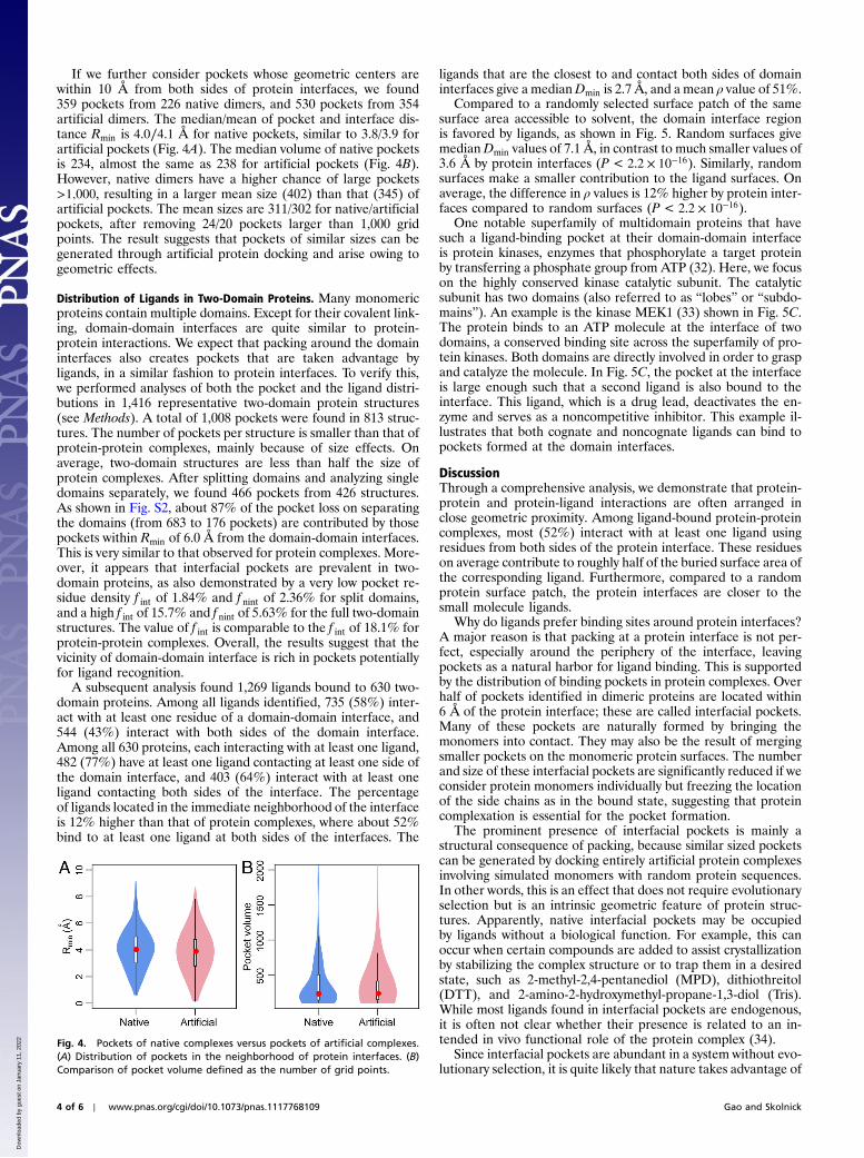
Compared with random protein surfaces of the same solventaccessible area, protein-protein interfaces are statistically closerto ligands. As shown in Fig. 2A, random surfaces give median/
Fig. 1. Distribution of pockets from protein-protein interfaces. Pockets arecalculated using dimeric complex structures (denoted as “dimer”) and indi-vidual monomeric structures, respectively. (A) Histograms of pockets versusthe distance from the protein interface. The width of the distance bins is1 Å; few cases with extreme values >40 Å are not shown. Definition ofRmin is given in the text. (B) Statistics of volume changes for interfacial pock-ets with a Rmin < 6 Å. The volumes of a pocket found in dimers and separatedmonomers are denoted as Vd and Vm, respectively. The summation is over allmonomer pockets associated with each dimer pocket (see text). Insets arediagrams that depict ligand-binding pockets formed upon protein-proteincomplexation. Ligands are colored in blue and the two proteins are coloredin green and red.
2 of 6 ∣ www.pnas.org/cgi/doi/10.1073/pnas.1117768109 Gao and Skolnick
Dow
nloa
ded
by g
uest
on
Janu
ary
11, 2
022
mean Dmin values of 7.4∕12.2 Å, in contrast to the much smallervalues of 4.2∕7.9 Å for protein interfaces (P < 2.2 × 10⁻¹⁶, Wil-coxon paired one tailed test). Similarly, random surfaces make acontribution to burying ligand surfaces, as displayed in Fig. 2B.Protein interfaces make contacts to about 23%more ligands thanrandomly selected protein surfaces. On average, the difference inρ values is 4.5% higher by protein interfaces than by random sur-faces (P ¼ 5.0 × 10⁻⁸).
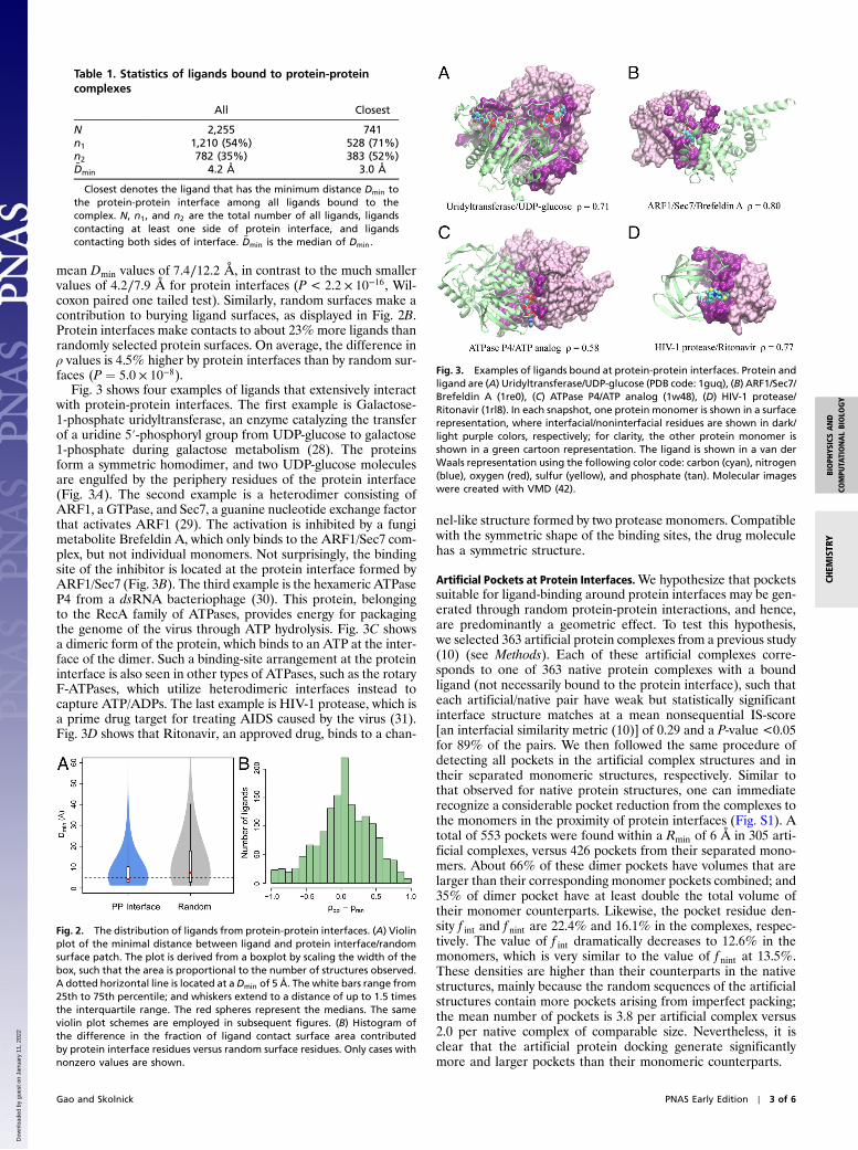
Fig. 3 shows four examples of ligands that extensively interactwith protein-protein interfaces. The first example is Galactose-1-phosphate uridyltransferase, an enzyme catalyzing the transferof a uridine 5′-phosphoryl group from UDP-glucose to galactose1-phosphate during galactose metabolism (28). The proteinsform a symmetric homodimer, and two UDP-glucose moleculesare engulfed by the periphery residues of the protein interface(Fig. 3A). The second example is a heterodimer consisting ofARF1, a GTPase, and Sec7, a guanine nucleotide exchange factorthat activates ARF1 (29). The activation is inhibited by a fungimetabolite Brefeldin A, which only binds to the ARF1/Sec7 com-plex, but not individual monomers. Not surprisingly, the bindingsite of the inhibitor is located at the protein interface formed byARF1/Sec7 (Fig. 3B). The third example is the hexameric ATPaseP4 from a dsRNA bacteriophage (30). This protein, belongingto the RecA family of ATPases, provides energy for packagingthe genome of the virus through ATP hydrolysis. Fig. 3C showsa dimeric form of the protein, which binds to an ATP at the inter-face of the dimer. Such a binding-site arrangement at the proteininterface is also seen in other types of ATPases, such as the rotaryF-ATPases, which utilize heterodimeric interfaces instead tocapture ATP/ADPs. The last example is HIV-1 protease, which isa prime drug target for treating AIDS caused by the virus (31).Fig. 3D shows that Ritonavir, an approved drug, binds to a chan-
nel-like structure formed by two protease monomers. Compatiblewith the symmetric shape of the binding sites, the drug moleculehas a symmetric structure.
Artificial Pockets at Protein Interfaces.We hypothesize that pocketssuitable for ligand-binding around protein interfaces may be gen-erated through random protein-protein interactions, and hence,are predominantly a geometric effect. To test this hypothesis,we selected 363 artificial protein complexes from a previous study(10) (see Methods). Each of these artificial complexes corre-sponds to one of 363 native protein complexes with a boundligand (not necessarily bound to the protein interface), such thateach artificial/native pair have weak but statistically significantinterface structure matches at a mean nonsequential IS-score[an interfacial similarity metric (10)] of 0.29 and a P-value <0.05for 89% of the pairs. We then followed the same procedure ofdetecting all pockets in the artificial complex structures and intheir separated monomeric structures, respectively. Similar tothat observed for native protein structures, one can immediaterecognize a considerable pocket reduction from the complexes tothe monomers in the proximity of protein interfaces (Fig. S1). Atotal of 553 pockets were found within a Rmin of 6 Å in 305 arti-ficial complexes, versus 426 pockets from their separated mono-mers. About 66% of these dimer pockets have volumes that arelarger than their corresponding monomer pockets combined; and35% of dimer pocket have at least double the total volume oftheir monomer counterparts. Likewise, the pocket residue den-sity f int and f nint are 22.4% and 16.1% in the complexes, respec-tively. The value of f int dramatically decreases to 12.6% in themonomers, which is very similar to the value of f nint at 13.5%.These densities are higher than their counterparts in the nativestructures, mainly because the random sequences of the artificialstructures contain more pockets arising from imperfect packing;the mean number of pockets is 3.8 per artificial complex versus2.0 per native complex of comparable size. Nevertheless, it isclear that the artificial protein docking generate significantlymore and larger pockets than their monomeric counterparts.
Table 1. Statistics of ligands bound to protein-proteincomplexes
All Closest
N 2,255 741n1 1,210 (54%) 528 (71%)n2 782 (35%) 383 (52%)~Dmin 4.2 Å 3.0 Å
Closest denotes the ligand that has the minimum distance Dmin tothe protein-protein interface among all ligands bound to thecomplex. N, n1, and n2 are the total number of all ligands, ligandscontacting at least one side of protein interface, and ligandscontacting both sides of interface. ~Dmin is the median of Dmin.
Fig. 2. The distribution of ligands from protein-protein interfaces. (A) Violinplot of the minimal distance between ligand and protein interface/randomsurface patch. The plot is derived from a boxplot by scaling the width of thebox, such that the area is proportional to the number of structures observed.A dotted horizontal line is located at aDmin of 5 Å. The white bars range from25th to 75th percentile; and whiskers extend to a distance of up to 1.5 timesthe interquartile range. The red spheres represent the medians. The sameviolin plot schemes are employed in subsequent figures. (B) Histogram ofthe difference in the fraction of ligand contact surface area contributedby protein interface residues versus random surface residues. Only cases withnonzero values are shown.
Fig. 3. Examples of ligands bound at protein-protein interfaces. Protein andligand are (A) Uridyltransferase/UDP-glucose (PDB code: 1guq), (B) ARF1/Sec7/Brefeldin A (1re0), (C) ATPase P4/ATP analog (1w48), (D) HIV-1 protease/Ritonavir (1rl8). In each snapshot, one protein monomer is shown in a surfacerepresentation, where interfacial/noninterfacial residues are shown in dark/light purple colors, respectively; for clarity, the other protein monomer isshown in a green cartoon representation. The ligand is shown in a van derWaals representation using the following color code: carbon (cyan), nitrogen(blue), oxygen (red), sulfur (yellow), and phosphate (tan). Molecular imageswere created with VMD (42).
Gao and Skolnick PNAS Early Edition ∣ 3 of 6
CHEM
ISTR
YBIOPH
YSICSAND
COMPU
TATIONALBIOLO
GY
Dow
nloa
ded
by g
uest
on
Janu
ary
11, 2
022
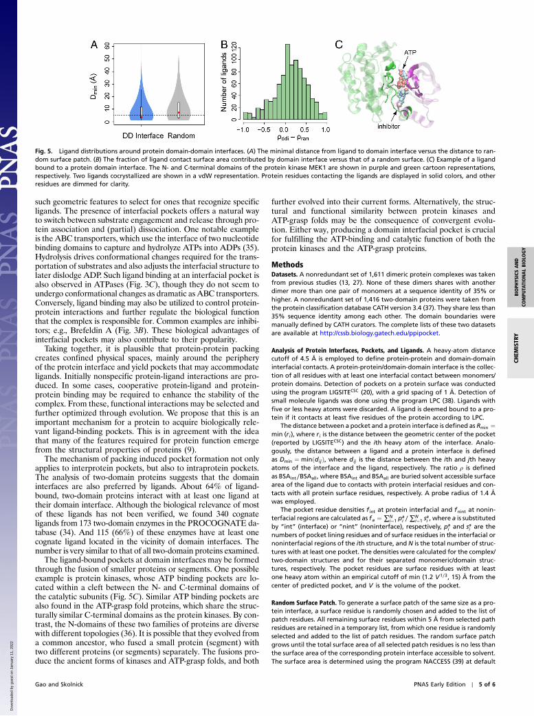
If we further consider pockets whose geometric centers arewithin 10 Å from both sides of protein interfaces, we found359 pockets from 226 native dimers, and 530 pockets from 354artificial dimers. The median/mean of pocket and interface dis-tance Rmin is 4.0∕4.1 Å for native pockets, similar to 3.8/3.9 forartificial pockets (Fig. 4A). The median volume of native pocketsis 234, almost the same as 238 for artificial pockets (Fig. 4B).However, native dimers have a higher chance of large pockets>1;000, resulting in a larger mean size (402) than that (345) ofartificial pockets. The mean sizes are 311/302 for native/artificialpockets, after removing 24/20 pockets larger than 1,000 gridpoints. The result suggests that pockets of similar sizes can begenerated through artificial protein docking and arise owing togeometric effects.
Distribution of Ligands in Two-Domain Proteins. Many monomericproteins contain multiple domains. Except for their covalent link-ing, domain-domain interfaces are quite similar to protein-protein interactions. We expect that packing around the domaininterfaces also creates pockets that are taken advantage byligands, in a similar fashion to protein interfaces. To verify this,we performed analyses of both the pocket and the ligand distri-butions in 1,416 representative two-domain protein structures(see Methods). A total of 1,008 pockets were found in 813 struc-tures. The number of pockets per structure is smaller than that ofprotein-protein complexes, mainly because of size effects. Onaverage, two-domain structures are less than half the size ofprotein complexes. After splitting domains and analyzing singledomains separately, we found 466 pockets from 426 structures.As shown in Fig. S2, about 87% of the pocket loss on separatingthe domains (from 683 to 176 pockets) are contributed by thosepockets within Rmin of 6.0 Å from the domain-domain interfaces.This is very similar to that observed for protein complexes. More-over, it appears that interfacial pockets are prevalent in two-domain proteins, as also demonstrated by a very low pocket re-sidue density f int of 1.84% and f nint of 2.36% for split domains,and a high f int of 15.7% and f nint of 5.63% for the full two-domainstructures. The value of f int is comparable to the f int of 18.1% forprotein-protein complexes. Overall, the results suggest that thevicinity of domain-domain interface is rich in pockets potentiallyfor ligand recognition.
A subsequent analysis found 1,269 ligands bound to 630 two-domain proteins. Among all ligands identified, 735 (58%) inter-act with at least one residue of a domain-domain interface, and544 (43%) interact with both sides of the domain interface.Among all 630 proteins, each interacting with at least one ligand,482 (77%) have at least one ligand contacting at least one side ofthe domain interface, and 403 (64%) interact with at least oneligand contacting both sides of the interface. The percentageof ligands located in the immediate neighborhood of the interfaceis 12% higher than that of protein complexes, where about 52%bind to at least one ligand at both sides of the interfaces. The
ligands that are the closest to and contact both sides of domaininterfaces give a medianDmin is 2.7 Å, and a mean ρ value of 51%.
Compared to a randomly selected surface patch of the samesurface area accessible to solvent, the domain interface regionis favored by ligands, as shown in Fig. 5. Random surfaces givemedianDmin values of 7.1 Å, in contrast to much smaller values of3.6 Å by protein interfaces (P < 2.2 × 10⁻¹⁶). Similarly, randomsurfaces make a smaller contribution to the ligand surfaces. Onaverage, the difference in ρ values is 12% higher by protein inter-faces compared to random surfaces (P < 2.2 × 10⁻¹⁶).
One notable superfamily of multidomain proteins that havesuch a ligand-binding pocket at their domain-domain interfaceis protein kinases, enzymes that phosphorylate a target proteinby transferring a phosphate group from ATP (32). Here, we focuson the highly conserved kinase catalytic subunit. The catalyticsubunit has two domains (also referred to as “lobes” or “subdo-mains”). An example is the kinase MEK1 (33) shown in Fig. 5C.The protein binds to an ATP molecule at the interface of twodomains, a conserved binding site across the superfamily of pro-tein kinases. Both domains are directly involved in order to graspand catalyze the molecule. In Fig. 5C, the pocket at the interfaceis large enough such that a second ligand is also bound to theinterface. This ligand, which is a drug lead, deactivates the en-zyme and serves as a noncompetitive inhibitor. This example il-lustrates that both cognate and noncognate ligands can bind topockets formed at the domain interfaces.
DiscussionThrough a comprehensive analysis, we demonstrate that protein-protein and protein-ligand interactions are often arranged inclose geometric proximity. Among ligand-bound protein-proteincomplexes, most (52%) interact with at least one ligand usingresidues from both sides of the protein interface. These residueson average contribute to roughly half of the buried surface area ofthe corresponding ligand. Furthermore, compared to a randomprotein surface patch, the protein interfaces are closer to thesmall molecule ligands.
Why do ligands prefer binding sites around protein interfaces?A major reason is that packing at a protein interface is not per-fect, especially around the periphery of the interface, leavingpockets as a natural harbor for ligand binding. This is supportedby the distribution of binding pockets in protein complexes. Overhalf of pockets identified in dimeric proteins are located within6 Å of the protein interface; these are called interfacial pockets.Many of these pockets are naturally formed by bringing themonomers into contact. They may also be the result of mergingsmaller pockets on the monomeric protein surfaces. The numberand size of these interfacial pockets are significantly reduced if weconsider protein monomers individually but freezing the locationof the side chains as in the bound state, suggesting that proteincomplexation is essential for the pocket formation.
The prominent presence of interfacial pockets is mainly astructural consequence of packing, because similar sized pocketscan be generated by docking entirely artificial protein complexesinvolving simulated monomers with random protein sequences.In other words, this is an effect that does not require evolutionaryselection but is an intrinsic geometric feature of protein struc-tures. Apparently, native interfacial pockets may be occupiedby ligands without a biological function. For example, this canoccur when certain compounds are added to assist crystallizationby stabilizing the complex structure or to trap them in a desiredstate, such as 2-methyl-2,4-pentanediol (MPD), dithiothreitol(DTT), and 2-amino-2-hydroxymethyl-propane-1,3-diol (Tris).While most ligands found in interfacial pockets are endogenous,it is often not clear whether their presence is related to an in-tended in vivo functional role of the protein complex (34).
Since interfacial pockets are abundant in a system without evo-lutionary selection, it is quite likely that nature takes advantage of
Fig. 4. Pockets of native complexes versus pockets of artificial complexes.(A) Distribution of pockets in the neighborhood of protein interfaces. (B)Comparison of pocket volume defined as the number of grid points.
4 of 6 ∣ www.pnas.org/cgi/doi/10.1073/pnas.1117768109 Gao and Skolnick
Dow
nloa
ded
by g
uest
on
Janu
ary
11, 2
022
such geometric features to select for ones that recognize specificligands. The presence of interfacial pockets offers a natural wayto switch between substrate engagement and release through pro-tein association and (partial) dissociation. One notable exampleis the ABC transporters, which use the interface of two nucleotidebinding domains to capture and hydrolyze ATPs into ADPs (35).Hydrolysis drives conformational changes required for the trans-portation of substrates and also adjusts the interfacial structure tolater dislodge ADP. Such ligand binding at an interfacial pocket isalso observed in ATPases (Fig. 3C), though they do not seem toundergo conformational changes as dramatic as ABC transporters.Conversely, ligand binding may also be utilized to control protein-protein interactions and further regulate the biological functionthat the complex is responsible for. Common examples are inhibi-tors; e.g., Brefeldin A (Fig. 3B). These biological advantages ofinterfacial pockets may also contribute to their popularity.
Taking together, it is plausible that protein-protein packingcreates confined physical spaces, mainly around the peripheryof the protein interface and yield pockets that may accommodateligands. Initially nonspecific protein-ligand interactions are pro-duced. In some cases, cooperative protein-ligand and protein-protein binding may be required to enhance the stability of thecomplex. From these, functional interactions may be selected andfurther optimized through evolution. We propose that this is animportant mechanism for a protein to acquire biologically rele-vant ligand-binding pockets. This is in agreement with the ideathat many of the features required for protein function emergefrom the structural properties of proteins (9).
The mechanism of packing induced pocket formation not onlyapplies to interprotein pockets, but also to intraprotein pockets.The analysis of two-domain proteins suggests that the domaininterfaces are also preferred by ligands. About 64% of ligand-bound, two-domain proteins interact with at least one ligand attheir domain interface. Although the biological relevance of mostof these ligands has not been verified, we found 340 cognateligands from 173 two-domain enzymes in the PROCOGNATE da-tabase (34). And 115 (66%) of these enzymes have at least onecognate ligand located in the vicinity of domain interfaces. Thenumber is very similar to that of all two-domain proteins examined.
The ligand-bound pockets at domain interfaces may be formedthrough the fusion of smaller proteins or segments. One possibleexample is protein kinases, whose ATP binding pockets are lo-cated within a cleft between the N- and C-terminal domains ofthe catalytic subunits (Fig. 5C). Similar ATP binding pockets arealso found in the ATP-grasp fold proteins, which share the struc-turally similar C-terminal domains as the protein kinases. By con-trast, the N-domains of these two families of proteins are diversewith different topologies (36). It is possible that they evolved froma common ancestor, who fused a small protein (segment) withtwo different proteins (or segments) separately. The fusions pro-duce the ancient forms of kinases and ATP-grasp folds, and both
further evolved into their current forms. Alternatively, the struc-tural and functional similarity between protein kinases andATP-grasp folds may be the consequence of convergent evolu-tion. Either way, producing a domain interfacial pocket is crucialfor fulfilling the ATP-binding and catalytic function of both theprotein kinases and the ATP-grasp proteins.
MethodsDatasets. A nonredundant set of 1,611 dimeric protein complexes was takenfrom previous studies (13, 27). None of these dimers shares with anotherdimer more than one pair of monomers at a sequence identity of 35% orhigher. A nonredundant set of 1,416 two-domain proteins were taken fromthe protein classification database CATH version 3.4 (37). They share less than35% sequence identity among each other. The domain boundaries weremanually defined by CATH curators. The complete lists of these two datasetsare available at http://cssb.biology.gatech.edu/ppipocket.
Analysis of Protein Interfaces, Pockets, and Ligands. A heavy-atom distancecutoff of 4.5 Å is employed to define protein-protein and domain-domaininterfacial contacts. A protein-protein/domain-domain interface is the collec-tion of all residues with at least one interfacial contact between monomers/protein domains. Detection of pockets on a protein surface was conductedusing the program LIGSITECSC (20), with a grid spacing of 1 Å. Detection ofsmall molecule ligands was done using the program LPC (38). Ligands withfive or less heavy atoms were discarded. A ligand is deemed bound to a pro-tein if it contacts at least five residues of the protein according to LPC.
The distance between a pocket and a protein interface is defined as Rmin ¼min (ri), where ri is the distance between the geometric center of the pocket(reported by LIGSITECSC) and the ith heavy atom of the interface. Analo-gously, the distance between a ligand and a protein interface is definedas Dmin ¼ minðdijÞ, where dij is the distance between the ith and jth heavyatoms of the interface and the ligand, respectively. The ratio ρ is definedas BSAint∕BSAall, where BSAint and BSAall are buried solvent accessible surfacearea of the ligand due to contacts with protein interfacial residues and con-tacts with all protein surface residues, respectively. A probe radius of 1.4 Åwas employed.
The pocket residue densities f int at protein interfacial and fnint at nonin-terfacial regions are calculated as fa ¼ ∑N
i¼1 pai ∕∑
Ni¼1 s
ai , where a is substituted
by “int” (interface) or “nint” (noninterface), respectively, pai and sai are the
numbers of pocket lining residues and of surface residues in the interfacial ornoninterfacial regions of the ith structure, and N is the total number of struc-tures with at least one pocket. The densities were calculated for the complex/two-domain structures and for their separated monomeric/domain struc-tures, respectively. The pocket residues are surface residues with at leastone heavy atom within an empirical cutoff of min (1.2 V1∕3, 15) Å from thecenter of predicted pocket, and V is the volume of the pocket.
Random Surface Patch. To generate a surface patch of the same size as a pro-tein interface, a surface residue is randomly chosen and added to the list ofpatch residues. All remaining surface residues within 5 Å from selected pathresidues are retained in a temporary list, from which one residue is randomlyselected and added to the list of patch residues. The random surface patchgrows until the total surface area of all selected patch residues is no less thanthe surface area of the corresponding protein interface accessible to solvent.The surface area is determined using the program NACCESS (39) at default
Fig. 5. Ligand distributions around protein domain-domain interfaces. (A) The minimal distance from ligand to domain interface versus the distance to ran-dom surface patch. (B) The fraction of ligand contact surface area contributed by domain interface versus that of a random surface. (C) Example of a ligandbound to a protein domain interface. The N- and C-terminal domains of the protein kinase MEK1 are shown in purple and green cartoon representations,respectively. Two ligands cocrystallized are shown in a vdW representation. Protein residues contacting the ligands are displayed in solid colors, and otherresidues are dimmed for clarity.
Gao and Skolnick PNAS Early Edition ∣ 5 of 6
CHEM
ISTR
YBIOPH
YSICSAND
COMPU
TATIONALBIOLO
GY
Dow
nloa
ded
by g
uest
on
Janu
ary
11, 2
022
parameters. One random surface patch was generated for each proteinstructure.
Artificial Protein-Protein Interfaces. Artificial protein-protein interfaces wereextracted from previously built artificial protein complexes (10). These arti-ficial complexes were originally taken from a library of polyvaline structuresgenerated with the protein structure prediction package TASSER (40). Theywere then converted to all-atom models using random protein sequences(10). From these structures, a total of 2,000 pairs of artificial structures wererandomly chosen and rigid-body docking was subsequently conducted withFT-dock (41). For each docking pair, the top 10 cluster representative protein-
protein complex models were retained. Since artificial complex structureshave a size limit of 600 amino acids, we chose 363 native complexes withinthis size limit that are bound to at least one ligand for the comparative pock-et analysis. For each native complex, we selected a unique artificial structurethat has the best interface similarity according nonsequential alignment bythe program iAlign (10, 27), yielding a total set of 363 artificial complexes forpocket analysis.
ACKNOWLEDGMENTS. This work was supported by the National Institutes ofHealth Grant Nos. GM-48835 and GM-37408.
1. Alberts B (2008) Molecular biology of the cell (Garland Science, New York), 5th Ed.2. Berman HM, et al. (2000) The Protein Data Bank. Nucleic Acids Res 28:235–242.3. Janin J, Bahadur RP, Chakrabarti P (2008) Protein-protein interaction and quaternary
structure. Q Rev Biophys 41:133–180.4. Jones S, Thornton JM (1996) Principles of protein-protein interactions. Proc Natl Acad
Sci USA 93:13–20.5. Keskin Z, Gursoy A, Ma B, Nussinov R (2008) Principles of protein-protein interactions:
What are the preferred ways for proteins to interact? Chem Rev 108:1225–1244.6. Lo Conte L, Chothia C, Janin J (1999) The atomic structure of protein-protein recogni-
tion sites. J Mol Biol 285:2177–2198.7. Laskowski RA, Luscombe NM, Swindells MB, Thornton JM (1996) Protein clefts in mo-
lecular recognition and function. Protein Sci 5:2438–2452.8. Liang J, Edelsbrunner H, Woodward C (1998) Anatomy of protein pockets and cavities:
Measurement of binding site geometry and implications for ligand design. Protein Sci7:1884–1897.
9. Brylinski M, Gao M, Skolnick J (2011) Why not consider a spherical protein? Implica-tions of backbone hydrogen bonding for protein structure and function. Phys ChemChem Phys 13:17044–17055.
10. Gao M, Skolnick J (2010) Structural space of protein-protein interfaces is degenerate,close to complete, and highly connected. Proc Natl Acad Sci USA 107:22517–22522.
11. Leis S, Schneider S, Zacharias M (2010) In silico prediction of binding sites on proteins.Curr Med Chem 17:1550–1562.
12. Aloy P, et al. (2004) Structure-based assembly of protein complexes in yeast. Science303:2026–2029.
13. Chen HL, Skolnick J (2008) M-TASSER: An algorithm for protein quaternary structureprediction. Biophys J 94:918–928.
14. de Vries SJ, Bonvin A (2008) How proteins get in touch: Interface prediction in thestudy of biomolecular complexes. Curr Protein Peptide Sci 9:394–406.
15. Zhou HX, Qin SB (2007) Interaction-site prediction for protein complexes: A criticalassessment. Bioinformatics 23:2203–2209.
16. Bordner AJ, Abagyan R (2005) Statistical analysis and prediction of protein-proteininterfaces. Proteins: Struct Funct Bioinform 60:353–366.
17. Bradford JR, Westhead DR (2005) Improved prediction of protein-protein binding sitesusing a support vector machines approach. Bioinformatics 21:1487–1494.
18. Glaser F, et al. (2003) ConSurf: Identification of functional regions in proteins by sur-face-mapping of phylogenetic information. Bioinformatics 19:163–164.
19. Lichtarge O, Bourne HR, Cohen FE (1996) An evolutionary trace method defines bind-ing surfaces common to protein families. J Mol Biol 257:342–358.
20. Huang BD, Schroeder M (2006) LIGSITE(csc): Predicting ligand binding sites using theConnolly surface and degree of conservation. BMC Struct Biol 6:19.
21. Vajda S, Guarnieri F (2006) Characterization of protein-ligand interaction sites usingexperimental and computational methods. Curr Opin Drug Discov Dev 9:363–369.
22. Campbell SJ, Gold ND, Jackson RM, Westhead DR (2003) Ligand binding: functionalsite location, similarity and docking. Curr Opin Struct Biol 13:389–395.
23. Lu L, Arakaki AK, Lu H, Skolnick J (2003) Multimeric threading-based prediction ofprotein-protein interactions on a genomic scale: Application to the Saccharomyces cer-evisiae proteome. Genome Res 13:1146–1154.
24. Skolnick J, Brylinski M (2009) FINDSITE: A combined evolution/structure-based ap-proach to protein function prediction. Brief Bioinform 10:378–391.
25. Hubbard SJ, Argos P (1994) Cavities and packing at protein interfaces. Protein Sci3:2194–2206.
26. Sonavane S, Chakrabarti P (2008) Cavities and atomic packing in protein structures andinterfaces. PLoS Comp Biol 4:e1000188.
27. Gao M, Skolnick J (2010) iAlign: A method for the structural comparison of protein-protein interfaces. Bioinformatics 26:2259–2265.
28. Thoden JB, Ruzicka FJ, Frey PA, Rayment I, Holden HM (1997) Structural analysis of theH166G site-directed mutant of galactose-1-phosphate uridylyltransferase complexedwith either UDP-glucose or UDP-galactose: Detailed description of the nucleotide su-gar binding site. Biochemistry 36:1212–1222.
29. Mossessova E, Corpina RA, Goldberg J (2003) Crystal structure of ARF1 center dot Sec7complexed with brefeldin A and its implications for the guanine nucleotide exchangemechanism. Mol Cell 12:1403–1411.
30. Mancini EJ, et al. (2004) Atomic snapshots of an RNA packaging motor reveal confor-mational changes linking ATP hydrolysis to RNA translocation. Cell 118:743–755.
31. Kempf DJ, et al. (1995) ABT-538 is a potent inhibitor of human-immunodeficiency-virusprotease and has high oral bioavailability in humans. Proc Natl Acad Sci USA92:2484–2488.
32. Taylor SS, Kornev AP (2011) Protein kinases: Evolution of dynamic regulatory proteins.Trends Biochem Sci 36:65–77.
33. Ohren JF, et al. (2004) Structures of human MAP kinase kinase 1 (MEK1) and MEK2describe novel noncompetitive kinase inhibition. Nat Struct Mol Biol 11:1192–1197.
34. Bashton M, Nobeli I, Thornton JM (2006) Cognate ligand domain mapping for en-zymes. J Mol Biol 364:836–852.
35. Rees DC, Johnson E, Lewinson O (2009) ABC transporters: the power to change. NatRev Mol Cell Biol 10:218–227.
36. Grishin NV (1999) Phosphatidylinositol phosphate kinase: A link between protein ki-nase and glutathione synthase folds. J Mol Biol 291:239–247.
37. Orengo CA, et al. (1997) CATH—a hierarchic classification of protein domain struc-tures. Structure 5:1093–1108.
38. Sobolev V, Sorokine A, Prilusky J, Abola EE, Edelman M (1999) Automated analysis ofinteratomic contacts in proteins. Bioinformatics 15:327–332.
39. Hubbard SJ, Thornton JM (1993) “NACCESS,” Computer Program, Department of Bio-chemistry and Molecular Biology (University College London, London).
40. Skolnick J, Arakaki AK, Lee SY, Brylinski M (2009) The continuity of protein structurespace is an intrinsic property of proteins. Proc Natl Acad Sci USA 106:15690–15695.
41. Gabb HA, Jackson RM, Sternberg MJE (1997) Modelling protein docking using shapecomplementarity, electrostatics and biochemical information. J Mol Biol 272:106–120.
42. Humphrey W, Dalke A, Schulten K (1996) VMD: visual molecular dynamics. J Mo Gra-phics 14:33–38.
6 of 6 ∣ www.pnas.org/cgi/doi/10.1073/pnas.1117768109 Gao and Skolnick
Dow
nloa
ded
by g
uest
on
Janu
ary
11, 2
022