Characterization of MADS homeotic genes in the fern ... - PNAS
6
Proc. Natl. Acad. Sci. USA Vol. 95, pp. 6222–6227, May 1998 Evolution Characterization of MADS homeotic genes in the fern Ceratopteris richardii MITSUYASU HASEBE* ² ,CHI-KUANG WEN ‡ ,MASAHIRO KATO § , AND JO ANN BANKS ‡ *National Institute for Basic Biology, Okazaki 444-8585, Japan; ‡ Department of Botany and Plant Pathology, Purdue University, West Lafayette, IN 47907-1155; and § Department of Biological Sciences, Graduate School of Science, University of Tokyo, Tokyo 113-0033, Japan Communicated by Elliot M. Meyerowitz, California Institute of Technology, Pasadena, CA, March 16, 1998 (received for review December 23, 1997) ABSTRACT The MADS genes encode a family of tran- scription factors, some of which control the identities of f loral organs in flowering plants. To understand the role of MADS genes in the evolution of floral organs, five MADS genes (CMADS1, 2, 3, 4, and 6) were cloned from the fern Cera- topteris richardii, a nonflowering plant. A gene tree of partial amino acid sequences of seed plant and fern MADS genes showed that the fern genes form three subfamilies. All mem- bers of one of the fern MADS subfamilies have additional amino-terminal amino acids, which is a synapomorphic char- acter of the AGAMOUS subfamily of the flowering plant MADS genes. Their structural similarity indicates a sister relationship between the two subfamilies. The temporal and spatial patterns of expression of the five fern MADS genes were assessed by Northern blot analyses and in situ hybrid- izations. CMADS1, 2, 3, and 4 are expressed similarly in the meristematic regions and primordia of sporophyte shoots and roots, as well as in reproductive structures, including sporo- phylls and sporangial initials, although the amount of expres- sion in each tissue is different in each gene. CMADS6 is expressed in gametophytic tissues but not in sporophytic tissues. The lack of organ-specific expression of MADS genes in the reproductive structures of the fern sporophyte may indicate that the restriction of MADS gene expression to specific reproductive organs and the specialization of MADS gene functions as homeotic selector genes in the flowering plant lineage were important in floral organ evolution. The reproductive structure of angiosperms, the flower, typi- cally bears four types of organs: sepals, petals, stamens, and carpels. Within the carpels and stamens, specialized mega- or microspore mother cells undergo meiosis to form the mega- or microspores, which divide mitotically to produce the haploid female or male gametophytes. These, in turn, form the haploid eggs or sperm. More primitive vascular plants (e.g., ferns) have simpler reproductive structures that lack all accessory floral organs. Ferns produce naked sporangia on the abaxial sides of leaves (Fig. 1); each sporangium contains spore mother cells that undergo meiosis to form the haploid reproductive spores. Ferns are usually homosporous, i.e., they produce only one kind of spore mother cell and spore. Unlike flowering plants, the haploid spores of ferns are shed, and the gametophytes derived from them are entirely independent of the spore- producing plant (the sporophyte). The fossil record supports the hypothesis that the common ancestor of flowering plants and ferns had no floral organs but, like ferns, had naked sporangia and an independent gametophyte generation (1, 2). The two groups are thought to have diverged from one another about 400 million years ago (1, 2). Studies of mutations that affect floral organ identity in Arabidopsis thaliana, Antirrhinum majus, and some other f low- ering plants have shown that the identity of each floral organ is specified by the combinatorial action of three classes of homeotic selector genes (3, 4). Most of the homeotic genes are collectively referred to as MADS genes because they contain a functional motif termed the MADS box (5, 6). The MADS genes encode transcription factors, and the MADS box is a highly conserved, ca. 60-aa sequence that functions in DNA- binding, dimerization, and accessory-factor interactions (6). Plant MADS genes have an additional conserved domain of ca. 70 aa residues named the K box, which potentially forms amphipathic helices that may be involved in protein–protein interactions (7). MADS genes have been reported from the animal, fungi, and plant kingdoms (6, 8 –10). Recently, several MADS genes containing the K box were isolated from some ferns (11). A MADS gene tree, incorporating the seed plant and fern MADS genes, revealed that the known fern MADS genes form only three divergent gene groups, which is consid- erably less than the number of MADS gene groups in seed plants (11). To address the function of the MADS genes in a plant with primitive reproductive structures lacking specialized floral organs, MADS genes were newly sought from the fern Cera- topteris richardii (Fig. 1). A study of this fern is important because of its tractable genetics, its relatively rapid life cycle, and the large body of information known about its develop- ment (12, 13). Five genes, representing all three fern MADS gene groups, were cloned from Ceratopteris, and their patterns of expression during sporophyte and gametophyte develop- ment were examined. Because four of the five genes were found to be similarly expressed in both reproductive and vegetative organs, we speculate that the ancestral MADS gene(s) was globally expressed in developing organs of the plant and that, as specialized f loral organs evolved, the number of MADS genes increased while expression patterns of some of the genes became more restricted. The evolution of genes that regulate MADS gene expression thus had an important role in the evolution of floral organs. MATERIALS AND METHODS RNA was isolated from pulverized tissue in a buffer containing 4 M guanidine thiocyanate, 1 M ammonium thiocyanate, 1% lauryl sarcosine, 0.5% PVP 360,000, and 1% 2-mercaptoetha- nol. After three chloroformyisoamyl alcohol (24:1) extrac- tions, nucleic acids were ethanol precipitated. RNA was pu- rified further by CTAB precipitation (14). The publication costs of this article were defrayed in part by page charge payment. This article must therefore be hereby marked ‘‘advertisement’’ in accordance with 18 U.S.C. §1734 solely to indicate this fact. © 1998 by The National Academy of Sciences 0027-8424y98y956222-6$2.00y0 PNAS is available online at http:yywww.pnas.org. Abbreviation: AG, AGAMOUS. Data deposition: The sequences reported in this paper have been deposited in the GenBank database (accession nos. U91415, U91416, U95608, U95609, and AF012529). ² To whom reprint requests should be addressed at: National Institute for Basic Biology, 38 Nishigonaka, Myodaiji-cho, Okazaki 444-8585, Japan. e-mail: [email protected]. 6222 Downloaded by guest on February 10, 2022
-
Upload
khangminh22 -
Category
Documents
-
view
2 -
download
0
Transcript of Characterization of MADS homeotic genes in the fern ... - PNAS
Proc. Natl. Acad. Sci. USAVol. 95, pp. 6222–6227, May 1998Evolution
Characterization of MADS homeotic genes in the fernCeratopteris richardii
MITSUYASU HASEBE*†, CHI-KUANG WEN‡, MASAHIRO KATO§, AND JO ANN BANKS‡
*National Institute for Basic Biology, Okazaki 444-8585, Japan; ‡Department of Botany and Plant Pathology, Purdue University, West Lafayette, IN 47907-1155;and §Department of Biological Sciences, Graduate School of Science, University of Tokyo, Tokyo 113-0033, Japan
Communicated by Elliot M. Meyerowitz, California Institute of Technology, Pasadena, CA, March 16, 1998 (received for review December 23, 1997)
ABSTRACT The MADS genes encode a family of tran-scription factors, some of which control the identities of f loralorgans in f lowering plants. To understand the role of MADSgenes in the evolution of f loral organs, five MADS genes(CMADS1, 2, 3, 4, and 6) were cloned from the fern Cera-topteris richardii, a nonflowering plant. A gene tree of partialamino acid sequences of seed plant and fern MADS genesshowed that the fern genes form three subfamilies. All mem-bers of one of the fern MADS subfamilies have additionalamino-terminal amino acids, which is a synapomorphic char-acter of the AGAMOUS subfamily of the f lowering plantMADS genes. Their structural similarity indicates a sisterrelationship between the two subfamilies. The temporal andspatial patterns of expression of the five fern MADS geneswere assessed by Northern blot analyses and in situ hybrid-izations. CMADS1, 2, 3, and 4 are expressed similarly in themeristematic regions and primordia of sporophyte shoots androots, as well as in reproductive structures, including sporo-phylls and sporangial initials, although the amount of expres-sion in each tissue is different in each gene. CMADS6 isexpressed in gametophytic tissues but not in sporophytictissues. The lack of organ-specific expression of MADS genesin the reproductive structures of the fern sporophyte mayindicate that the restriction of MADS gene expression tospecific reproductive organs and the specialization of MADSgene functions as homeotic selector genes in the f loweringplant lineage were important in f loral organ evolution.
The reproductive structure of angiosperms, the flower, typi-cally bears four types of organs: sepals, petals, stamens, andcarpels. Within the carpels and stamens, specialized mega- ormicrospore mother cells undergo meiosis to form the mega- ormicrospores, which divide mitotically to produce the haploidfemale or male gametophytes. These, in turn, form the haploideggs or sperm. More primitive vascular plants (e.g., ferns) havesimpler reproductive structures that lack all accessory floralorgans. Ferns produce naked sporangia on the abaxial sides ofleaves (Fig. 1); each sporangium contains spore mother cellsthat undergo meiosis to form the haploid reproductive spores.Ferns are usually homosporous, i.e., they produce only onekind of spore mother cell and spore. Unlike flowering plants,the haploid spores of ferns are shed, and the gametophytesderived from them are entirely independent of the spore-producing plant (the sporophyte). The fossil record supportsthe hypothesis that the common ancestor of flowering plantsand ferns had no floral organs but, like ferns, had nakedsporangia and an independent gametophyte generation (1, 2).The two groups are thought to have diverged from one anotherabout 400 million years ago (1, 2).
Studies of mutations that affect f loral organ identity inArabidopsis thaliana, Antirrhinum majus, and some other flow-ering plants have shown that the identity of each floral organis specified by the combinatorial action of three classes ofhomeotic selector genes (3, 4). Most of the homeotic genes arecollectively referred to as MADS genes because they containa functional motif termed the MADS box (5, 6). The MADSgenes encode transcription factors, and the MADS box is ahighly conserved, ca. 60-aa sequence that functions in DNA-binding, dimerization, and accessory-factor interactions (6).Plant MADS genes have an additional conserved domain of ca.70 aa residues named the K box, which potentially formsamphipathic helices that may be involved in protein–proteininteractions (7). MADS genes have been reported from theanimal, fungi, and plant kingdoms (6, 8–10). Recently, severalMADS genes containing the K box were isolated from someferns (11). A MADS gene tree, incorporating the seed plantand fern MADS genes, revealed that the known fern MADSgenes form only three divergent gene groups, which is consid-erably less than the number of MADS gene groups in seedplants (11).
To address the function of the MADS genes in a plant withprimitive reproductive structures lacking specialized floralorgans, MADS genes were newly sought from the fern Cera-topteris richardii (Fig. 1). A study of this fern is importantbecause of its tractable genetics, its relatively rapid life cycle,and the large body of information known about its develop-ment (12, 13). Five genes, representing all three fern MADSgene groups, were cloned from Ceratopteris, and their patternsof expression during sporophyte and gametophyte develop-ment were examined. Because four of the five genes werefound to be similarly expressed in both reproductive andvegetative organs, we speculate that the ancestral MADSgene(s) was globally expressed in developing organs of theplant and that, as specialized floral organs evolved, the numberof MADS genes increased while expression patterns of someof the genes became more restricted. The evolution of genesthat regulate MADS gene expression thus had an importantrole in the evolution of floral organs.
MATERIALS AND METHODS
RNA was isolated from pulverized tissue in a buffer containing4 M guanidine thiocyanate, 1 M ammonium thiocyanate, 1%lauryl sarcosine, 0.5% PVP 360,000, and 1% 2-mercaptoetha-nol. After three chloroformyisoamyl alcohol (24:1) extrac-tions, nucleic acids were ethanol precipitated. RNA was pu-rified further by CTAB precipitation (14).
The publication costs of this article were defrayed in part by page chargepayment. This article must therefore be hereby marked ‘‘advertisement’’ inaccordance with 18 U.S.C. §1734 solely to indicate this fact.
© 1998 by The National Academy of Sciences 0027-8424y98y956222-6$2.00y0PNAS is available online at http:yywww.pnas.org.
Abbreviation: AG, AGAMOUS.Data deposition: The sequences reported in this paper have beendeposited in the GenBank database (accession nos. U91415, U91416,U95608, U95609, and AF012529).†To whom reprint requests should be addressed at: National Institutefor Basic Biology, 38 Nishigonaka, Myodaiji-cho, Okazaki 444-8585,Japan. e-mail: [email protected].
6222
Dow
nloa
ded
by g
uest
on
Feb
ruar
y 10
, 202
2
For reverse transcription–PCR, total RNA was extractedfrom the vegetative leaves (1 to 2 cm long), the partially coiledreproductive leaves ('10 cm long) without stipules, and thereproductive shoot tips including apical meristems, leaf pri-mordia, young leaves less than 5-mm long, and root primordialess than 1 mm. Complementary DNAs from each RNAsample were synthesized by using the Superscript II reversetranscriptase (GIBCOyBRL) and the adapter primer [59-CUACUACUACUAGGCCACGCGTCGACTAGTACT16-39) supplied by GIBCOyBRL. PCR was performed by usingthe cDNA as template, the adapter primer, and the MADSdomain-specific primer encoding the amino acids Lys-Lys-Ala-Tyr-Glu-Leu-Ser-Val. The degenerate primer sequence was59-CAUCAUCAUCAUAARAARGCITAYGARCTITCN-TCNGT-39 (where R 5 A or G; I 5 inosine; Y 5 C or T; N 5A, C, G, or T). The PCR mix contained 1/10th volume of thecDNA solution, 2 mM MADS domain-specific primer, 1 mMadapter primer, 0.2 mM deoxyribonucleotides, 10 mMTriszHCl (pH 9.0), 50 mM KCl, 0.1% Triton X-100, and 1.5mM MgCl2. The mixture was heated to 80°C before adding 2.5units Taq DNA polymerase (Promega). PCR was performedas follows: 5 min at 94°C and 35–40 cycles of 1 min at 94°C, 1min at 52°C, and 2 min at 72°C. The PCR products wereseparated on 1% agarose gels. Fragments between 0.6 kb and1.5 kb were purified and cloned into the pAMP1 vector(GIBCOyBRL); 158 clones (40, 78, and 40 clones for eachcDNA sample from vegetative leaves, reproductive stem tips,and roots, respectively) were characterized. All clones weredigested with Sau3A1, sorted, and sequenced.
Northern hybridizations were performed as described inChurch and Gilbert (15). Membranes were hybridized at 68°Cfor 16 hr in a hybridization buffer (0.5 M phosphate, pH 7.2y1mM EDTAy7% SDS) and then washed at 68°C in a washbuffer (40 mM phosphate, pH 7.2y1 mM EDTAy1% SDS).
The CMADS1-specific probe was a 575-bp EcoRV-SacIfragment of the cloned cDNA, the CMADS2-specific probewas a 486-bp HindIII-SspI fragment of the cloned cDNA, andthe CMADS4-specific probe was a 465-bp SspI-BstEII frag-ment of the cloned cDNA. The CMADS3-specific probe (676bp) extended from the CMADS3 cDNA PstI site to the 39 endof the gene. The CMADS6-specific probe (470 bp) was cloned
by PCR with the CMADS6-specific internal primer (59-CGATCTGAAGCTTCTGTCTGCAG-39) and poly(T)primer. The probes did not include the conserved MADSdomain-encoding region (Fig. 2). Each probe hybridized tounique genomic fragments (data not shown) and did nothybridize to the others in Southern hybridization experiments,indicating that the probes are gene-specific. For Northern andSouthern hybridization experiments, DNA fragments werelabeled with 32P by PCR labeling (16). In situ hybridizationswith digoxigenin-labeled probes were performed as describedpreviously (17). Using the sense probes as negative controls,weak signals were detected in the protoxylem of the petiolevascular bundle, but no other sense signals could be detected(data not shown). All sections were 8 mm thick.
The amino acid sequences of plant MADS genes, obtainedfrom the GenBank DNA database, were aligned by using theCLUSTAL W program (18) and then were revised manually.(The alignment is available from M.H. on request.) The 113 aaresidues corresponding to positions 25–61, 116–131, 142–146,149–176, 184–193, and 197–213 from the initial methioninecodon of CMADS2 were used to calculate the evolutionarydistances with the PROTDIS program (19) under the Dayhoffand PAM matrix. The regions used for phylogenetic analysesincluded the MADS and K domains. The residues between thetwo domains and the residues beyond the K domain were notincluded in the analysis because these regions could not bealigned well. The tree was obtained with the neighbor-joiningmethod (20) by using the NEIGHBOR program (19).
RESULTS
Cloning of MADS Genes from Ceratopteris richardii. Can-didate MADS-like Ceratopteris cDNA clones obtained by PCRusing a MADS box-specific degenerate primer and an adapterprimer were sorted into five distinct groups depending on theirdigestion patterns with Sau3AI. Each group was representedby 104, 1, 7, 39, and 7 clones, respectively, and 17, 1, 5, 7, and4 clones of each group were sequenced, respectively. Thesequences confirmed that each group represents a singledistinct gene. The genes are named CMADS1, 2, 3, 4, and 5.The 59 regions of the CMADS genes were cloned by using the59 RACE kit (GIBCOyBRL) and then sequenced. Further
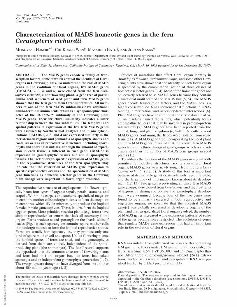
FIG. 1. The Ceratopteris richardii sporophyte. (A) A whole plantwith reproductive (arrow) and vegetative leaves. The vegetative leavesare less dissected than reproductive leaves. All postembryonic rootsare adventitious roots and originate at the base of each leaf. (Bar 51 cm.) (B) Naked sporangia on the abaxial surface of a reproductiveleaf. Part of the leaf margin was removed to show the sporangia. Mostsporangia mature at the same time, but a few sporangia develop laterthan others (arrow). (Bar 5 0.5 mm.)
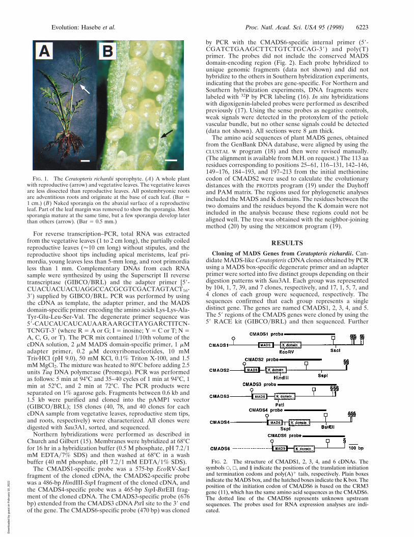
FIG. 2. The structure of CMADS1, 2, 3, 4, and 6 cDNAs. Thesymbols E, h, and § indicate the positions of the translation initiationand termination codons and poly(A)1 tails, respectively. Plain boxesindicate the MADS box, and the hatched boxes indicate the K box. Theposition of the initiation codon of CMADS6 is based on the CRM3gene (11), which has the same amino acid sequences as the CMADS6.The dotted line of the CMADS6 represents unknown upstreamsequences. The probes used for RNA expression analyses are indi-cated.
Evolution: Hasebe et al. Proc. Natl. Acad. Sci. USA 95 (1998) 6223
Dow
nloa
ded
by g
uest
on
Feb
ruar
y 10
, 202
2

FIG. 3. A gene tree of plant MADS genes based on the neighbor-joining method (20). The horizontal branch length is proportional to theestimated number of amino acids substitutions per residue. (Bar 5 0.1 aa substitution per residue.) Internal branches with more than 50% bootstrapvalues in 100 bootstrap replicates performed by using the SEQBOOT program (19) are indicated as broader lines. This is an unrooted tree. The symbolsafter the gene names indicate the dicots (open circles), monocots (hatched circles), or gymnosperms (solid circles). Three Ceratopteris MADS genegroups and the monophyletic AGAMOUS group (8–10) are indicated by brackets.
6224 Evolution: Hasebe et al. Proc. Natl. Acad. Sci. USA 95 (1998)
Dow
nloa
ded
by g
uest
on
Feb
ruar
y 10
, 202
2
screening for CMADS genes from total RNA isolated from14-day-old gametophytes produced one more clone,CMADS6. Because CMADS3 and CMADS6 are identical inamino acid sequence to the previously reported CRM1 andCRM3 genes (11), 59 RACE for CMADS6 was not performed.CMADS5 was omitted from further studies because some ofthe PCR products contained in-frame stop codons, indicatingthat CMADS5 may be an atypically truncated MADS gene ora pseudogene. Percentages of identical DNA sequences be-tween the newly cloned CMADS genes (CMADS1, 2, 4, and5) and previously reported Ceratopteris richardii genes (CRM1,3, and 7) are around 50%.
CMADS1, 2, 3, 4, and 6 all potentially encode proteins thatcontain the MADS domain as well as the K domain, anotherconserved domain common to all plant MADS genes (6, 8–10)(Fig. 2). The initiation codons of the angiosperm MADS genesare located adjacent to the 59 end of the sequences encodingthe MADS domain with the exception of most of the membersof the AGAMOUS (AG) group (9, 10). The latter encodeproteins with additional amino-terminal amino acids.CMADS1 contains the additional 59 sequence found in themembers of the AG group, whereas CMADS2, 3, 4, and 6 donot (Fig. 2).
Gene Tree of Plant MADS Genes. A gene tree based on thecomparison of the amino acid sequences of plant MADS genesshows that the CMADS genes form three divergent groups asdescribed previously (11). CMADS1, CMADS2y3y4, andCMADS6 are included in the CRM6, CRM1, and CRM3groups previously isolated from Ceratopteris (11), respectively.The three groups do not cluster with any other seed plantMADS gene group with high statistical confidence (Fig. 3).Because both CMADS1 and the AG group of MADS domain-encoded proteins have the additional amino-terminal aminoacids, it is likely that the CRM6 group, which includesCMADS1, is more closely related to the AG group than toother seed plant MADS genes. Kofuji and Yamaguchi (21)reported the presence of two other genes in the CRM6 group
(CerMADS2 and 3 in Fig. 3), which also have the additionalamino-terminal amino acids. The amino-terminal regions ofother genes in the CRM6 group have not been reported.
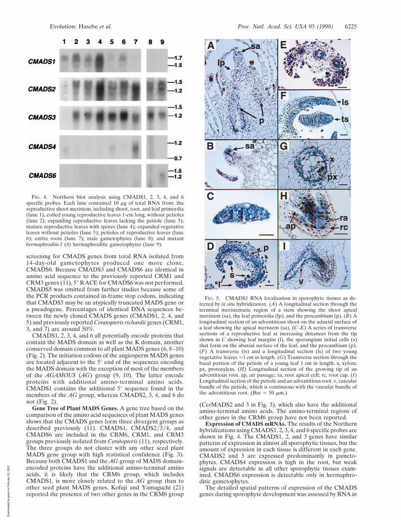
Expression of CMADS mRNAs. The results of the Northernhybridizations using CMADS1, 2, 3, 4, and 6 specific probes areshown in Fig. 4. The CMADS1, 2, and 3 genes have similarpatterns of expression in almost all sporophytic tissues, but theamount of expression in each tissue is different in each gene.CMADS2 and 3 are expressed predominantly in gameto-phytes. CMADS4 expression is high in the root, but weaksignals are detectable in all other sporophytic tissues exam-ined. CMADS6 expression is detectable only in hermaphro-ditic gametophytes.
The detailed spatial patterns of expression of the CMADSgenes during sporophyte development was assessed by RNA in
FIG. 4. Northern blot analysis using CMADS1, 2, 3, 4, and 6specific probes. Each lane contained 10 mg of total RNA from: thereproductive shoot meristem, including shoot, root, and leaf primordia(lane 1), coiled young reproductive leaves 1-cm long, without petioles(lane 2); expanding reproductive leaves lacking the petiole (lane 3);mature reproductive leaves with spores (lane 4); expanded vegetativeleaves without petioles (lane 5); petioles of reproductive leaves (lane6); entire roots (lane 7); male gametophytes (lane 8); and mutanthermaphroditic1 (4) hermaphroditic gametophytes (lane 9).
FIG. 5. CMADS1 RNA localization in sporophyte tissues as de-tected by in situ hybridization. (A) A longitudinal section through theterminal meristematic region of a stem showing the shoot apicalmeristem (sa), the leaf primordia (lp), and the procambium (p). (B) Alongitudinal section of an adventitious shoot on the adaxial surface ofa leaf showing the apical meristem (sa). (C–E) A series of transversesections of a reproductive leaf at increasing distances from the tipshown in C showing leaf margins (l), the sporangium initial cells (s)that form on the abaxial surface of the leaf, and the procambium (p).(F) A transverse (ts) and a longitudinal section (ls) of two youngvegetative leaves '1 cm in length. (G) Transverse section through thebasal portion of the petiole of a young leaf 1 cm in length. x, xylem;px, protoxylem. (H) Longitudinal section of the growing tip of anadventitious root. ap, air passage; ra, root apical cell; rc, root cap. (I)Longitudinal section of the petiole and an adventitious root. v, vascularbundle of the petiole, which is continuous with the vascular bundle ofthe adventitious root. (Bar 5 50 mm.)
Evolution: Hasebe et al. Proc. Natl. Acad. Sci. USA 95 (1998) 6225
Dow
nloa
ded
by g
uest
on
Feb
ruar
y 10
, 202
2
situ hybridization for CMADS1–5. In general, the patterns ofCMADS1, 2, and 3 expression are very similar, althoughCMADS2 and 3 signals are always weaker than CMADS1signals (data not shown). The CMADS4 expression pattern inthe root is not distinguishable from CMADS1, 2, and 3,although CMADS1 signal is weaker than other CMADSsignals (data not shown). For these reasons, the results of in situhybridizations using the CMADS1 antisense probe (Fig. 2) areshown in Fig. 5. In developing shoot systems, expression is seenin the shoot apical meristem, leaf primordia, and procambium(Fig. 5A). The expression in the apical meristem and leafprimordia tissues appears weaker than in the procambium. Thesame patterns of expression are observed in the apical mer-istems of the many adventitious shoots that develop on theadaxial surface of leaves as they mature (Fig. 5B). As the leafincreases in cell number, the CMADS signal becomes strongerand distributed uniformly in all cells at the tip of each leaf (Fig.5 C and F). The meristematic tip forms the coiled main axis ofthe leaf, or crosier (22). As tissue systems differentiate in theleaf, the CMADS signal gradually becomes restricted to threeparts of the leaf: the procambium, the sporangium initials, andthe regions of the leaf that will give rise to the lamina, or pinnae(Fig. 5 C–E). The signal observed in the developing procam-bium of the leaf continues into the differentiated vascularbundles of the petiole (Fig. 5G). Ceratopteris forms adventi-tious roots at the base of each leaf, and hybridization signalsare detected in the root apical meristems and their associatedprovascular cell files (Fig. 5H). The hybridization signals of theroot provascular cell files are continuous with the vascularbundles in the petiole of the leaf (Fig. 5I).
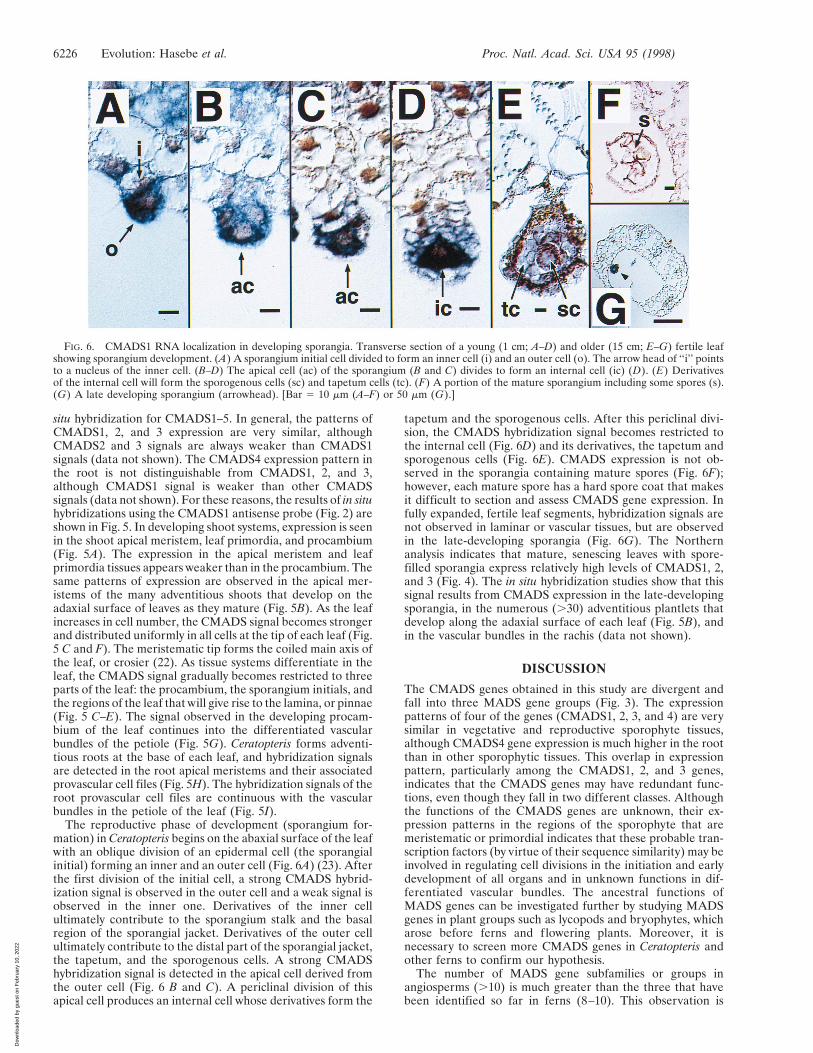
The reproductive phase of development (sporangium for-mation) in Ceratopteris begins on the abaxial surface of the leafwith an oblique division of an epidermal cell (the sporangialinitial) forming an inner and an outer cell (Fig. 6A) (23). Afterthe first division of the initial cell, a strong CMADS hybrid-ization signal is observed in the outer cell and a weak signal isobserved in the inner one. Derivatives of the inner cellultimately contribute to the sporangium stalk and the basalregion of the sporangial jacket. Derivatives of the outer cellultimately contribute to the distal part of the sporangial jacket,the tapetum, and the sporogenous cells. A strong CMADShybridization signal is detected in the apical cell derived fromthe outer cell (Fig. 6 B and C). A periclinal division of thisapical cell produces an internal cell whose derivatives form the
tapetum and the sporogenous cells. After this periclinal divi-sion, the CMADS hybridization signal becomes restricted tothe internal cell (Fig. 6D) and its derivatives, the tapetum andsporogenous cells (Fig. 6E). CMADS expression is not ob-served in the sporangia containing mature spores (Fig. 6F);however, each mature spore has a hard spore coat that makesit difficult to section and assess CMADS gene expression. Infully expanded, fertile leaf segments, hybridization signals arenot observed in laminar or vascular tissues, but are observedin the late-developing sporangia (Fig. 6G). The Northernanalysis indicates that mature, senescing leaves with spore-filled sporangia express relatively high levels of CMADS1, 2,and 3 (Fig. 4). The in situ hybridization studies show that thissignal results from CMADS expression in the late-developingsporangia, in the numerous (.30) adventitious plantlets thatdevelop along the adaxial surface of each leaf (Fig. 5B), andin the vascular bundles in the rachis (data not shown).
DISCUSSION
The CMADS genes obtained in this study are divergent andfall into three MADS gene groups (Fig. 3). The expressionpatterns of four of the genes (CMADS1, 2, 3, and 4) are verysimilar in vegetative and reproductive sporophyte tissues,although CMADS4 gene expression is much higher in the rootthan in other sporophytic tissues. This overlap in expressionpattern, particularly among the CMADS1, 2, and 3 genes,indicates that the CMADS genes may have redundant func-tions, even though they fall in two different classes. Althoughthe functions of the CMADS genes are unknown, their ex-pression patterns in the regions of the sporophyte that aremeristematic or primordial indicates that these probable tran-scription factors (by virtue of their sequence similarity) may beinvolved in regulating cell divisions in the initiation and earlydevelopment of all organs and in unknown functions in dif-ferentiated vascular bundles. The ancestral functions ofMADS genes can be investigated further by studying MADSgenes in plant groups such as lycopods and bryophytes, whicharose before ferns and flowering plants. Moreover, it isnecessary to screen more CMADS genes in Ceratopteris andother ferns to confirm our hypothesis.
The number of MADS gene subfamilies or groups inangiosperms (.10) is much greater than the three that havebeen identified so far in ferns (8–10). This observation is
FIG. 6. CMADS1 RNA localization in developing sporangia. Transverse section of a young (1 cm; A–D) and older (15 cm; E–G) fertile leafshowing sporangium development. (A) A sporangium initial cell divided to form an inner cell (i) and an outer cell (o). The arrow head of ‘‘i’’ pointsto a nucleus of the inner cell. (B–D) The apical cell (ac) of the sporangium (B and C) divides to form an internal cell (ic) (D). (E) Derivativesof the internal cell will form the sporogenous cells (sc) and tapetum cells (tc). (F) A portion of the mature sporangium including some spores (s).(G) A late developing sporangium (arrowhead). [Bar 5 10 mm (A–F) or 50 mm (G).]
6226 Evolution: Hasebe et al. Proc. Natl. Acad. Sci. USA 95 (1998)
Dow
nloa
ded
by g
uest
on
Feb
ruar
y 10
, 202
2

consistent with the hypothesis that duplication of the MADSgenes and their subsequent divergence is related to the co-option of MADS genes as a homeotic selector genes and to theincrease in the complexity of reproductive organs presentlyobserved in angiosperms. The analyses of MADS gene homo-logues in other plant groups (e.g., gymnosperms), whichbranched off from the vascular plant lineage after ferns, willbe useful in understanding the relationship between MADSgene evolution and the evolution of reproductive organs.
In flowering plants, some MADS genes are expressed inspecific f loral organ primordia as homeotic selector genes,whereas other MADS genes are expressed in both reproduc-tive and vegetative organs (8–10). As with the latter type offlowering plant MADS genes, most of the fern CMADS genesare expressed in both the reproductive and vegetative organs.The generally expressed MADS genes may be more primitivethan the reproductive organ-specific MADS genes. If so, it islikely that some of the MADS genes were co-opted as ho-meotic selector genes of specialized reproductive organs andtheir expression was restricted to specific f loral organ primor-dia, events that occurred after the divergence of ferns andangiosperms. The restriction of MADS gene expression mayhave been caused by the evolution of other genes that regulatethe MADS genes. The Arabidopsis LEAFY (24) andyorCURLY LEAF (25) genes, for example, have been shown toregulate or restrict MADS gene expression in Arabidopsis. Bycomparing the functions of these types of regulatory genes inArabidopsis and their homologues in Ceratopteris, importantinsights into the evolution of the regulatory cascade of MADSgenes and subsequent floral organ evolution will be gained.
We thank H. Sakai and E. Meyerowitz for help with the in situhybridizations; R. Sano for providing total RNA; C. Juarez, C.Chapple, G. Rothwell, T. Nishiyama, and anonymous reviewers forsuggestions on the manuscript; R. Kofuji and K. Yamaguchi for apreprint of their manuscript; and K. Iwatsuki, T. Nagata, and H.Fukuda for experimental facilities. This research was supported bygrants from The Japan Society for the Promotion of Science (M.H.);Nissan Science Foundation (M.H.); Ministry of Education, Scienceand Culture, Japan (M.H., M.K.); and the National Science Founda-tion (J.A.B.). This is journal paper 15515 of the Purdue UniversityAgricultural Experimental Station.
1. Gifford, E. M. & Foster, A. S. (1988) Morphology and Evolutionof Vascular Plants (Freeman, New York), 3rd Ed.
2. Stewart, W. N. & Rothwell, G. W. (1993) Paleobotany and theEvolution of Plants (Cambridge Univ. Press, Cambridge, U.K.),2nd Ed.
3. Coen, E. S. & Meyerowitz, E. M. (1991) Nature (London) 353,31–37.
4. Meyerowitz, E. M., Smyth, D. R. & Bowman, J. L. (1989)Development 106, 209–217.
5. Schwarz-Sommer, Z., Huijser, P., Naken, W., Saedler, H. &Sommer, H. (1990) Science 250, 931–936.
6. Shore, P. S. & Sharrocks, A. D. (1995) Eur. J. Biochem. 229, 1–13.7. Davies, B. & Schwartz-Sommer, Z. (1994) in Results and Prob-
lems in Cell Differentiation, ed. Nover, L. (Springer, Berlin), Vol.20, pp. 235–258.
8. Theissen, G. & Saedler, H. (1995) Curr. Opin. Genet. Dev. 5,628–639.
9. Theissen, G., Kim, J. T. & Saedler, H. (1996) J. Mol. Evol. 43,484–516.
10. Hasebe, M. & Banks, J. A. (1997) in Evolution and Diversificationof Land Plants, eds. Iwatsuki, K. & Raven, P. H. (Springer,Tokyo), pp. 179–197.
11. Munster, T., Pahnke, J., Rosa, A. D., Kim, J. T., Martin, W.,Saedler, H. & Theissen, G. (1997) Proc. Natl. Acad. Sci. USA 94,2415–2420.
12. Hickok, L. G., Warne, T. & Fribourg, R. S. (1995) Int. J. Plant Sci.156, 332–345.
13. Eberle, J., Nemacheck, J., Wen, C.-K., Hasebe, M. & Banks, J. A.(1995) Int. J. Plant Sci. 156, 359–366.
14. Murray, M. G. & Thompson, W. F. (1980) Nucleic Acids Res. 8,4321–4328.
15. Church, G. M. & Gilbert, W. (1984) Proc. Natl. Acad. Sci. USA81, 1991–1995.
16. Schowalter, B. D. & Sommer, S. S. (1989) Anal. Biochem. 177,90–94.
17. Coen, E. S., Romero, J. M., Doyle, S., Elliot, R., Murphy, G. &Carpenter, R. (1990) Cell 63, 1311–1322.
18. Thompson, J. D., Higgins, D. G. & Gibson, T. J. (1994) NucleicAcids Res. 22, 4673–4680.
19. Felsenstein, J. (1993) PHYLIP, Phylogenetic Inference Package(Univ. of Washington, Seattle), Version 3.572c.
20. Saitou, N. & Nei, M. (1987) Mol. Biol. Evol. 4, 406–425.21. Kofuji, R. & Yamaguchi, K. (1997) J. Phytogeography Taxon. 45,
83–91.22. Steeves, T. A. & Sussex, I. M. (1989) Patterns in Plant Develop-
ment (Cambridge Univ. Press, New York).23. Pal, N. & Pal, S. (1963) Bot. Gaz. 125, 405–412.24. Weigel, D. & Meyerowitz, E. M. (1993) Science 261, 1723–1726.25. Goodrich, J., Puangsomlee, P., Martin, M., Long, D., Meyerow-
itz, E. M. & Coupland, G. (1997) Nature (London) 386, 44–51.
Evolution: Hasebe et al. Proc. Natl. Acad. Sci. USA 95 (1998) 6227
Dow
nloa
ded
by g
uest
on
Feb
ruar
y 10
, 202
2