Systems-Based Design of Bi-Ligand Inhibitors of Oxidoreductases
10
Chemistry & Biology, Vol. 11, 185–194, February, 2004, 2004 Elsevier Science Ltd. All rights reserved. DOI 10.1016/j.chembio.2004.02.012 Systems-Based Design of Bi-Ligand Inhibitors of Oxidoreductases: Filling the Chemical Proteomic Toolbox factor binding site, coupled with the modular nature of this gene family, has led us to develop a highly parallel approach to inhibitor design. In this chemical proteomic strategy, focused chemical libraries are tailored to sub- families of large gene families to produce nM inhibitors Daniel S. Sem, 1,2, * Bonnie Bertolaet, 2 Brian Baker, 2,3 Edcon Chang, 2 Aurora D. Costache, 1 Stephen Coutts, 2 Qing Dong, 2 Mark Hansen, 2 Victor Hong, 2,4 Xuemei Huang, 2 Richard M. Jack, 2 Richard Kho, 2 Henk Lang, 2 Chen-Ting Ma, 2 David Meininger, 2,5 for multiple members of the subfamily. The parallel pro- duction of inhibitors across a gene family such as the Maurizio Pellecchia, 2,6 Fabrice Pierre, 2 Hugo Villar, 2 and Lin Yu 2 oxidoreductases will have applications in chemogeno- mic and functional genomic efforts to define protein 1 Chemical Proteomics Facility at Marquette Department of Chemistry functions, as well as in drug design. The latter is a signifi- cant point considering the large number of new proteins Marquette University P.O. Box 1881 and drug targets being identified as a result of functional genomics efforts [4]. Inhibitors for various members of Milwaukee, Wisconsin 53201 2 Triad Therapeutics, Inc. the oxidoreductase gene family could be used to gener- ate chemical knockouts as a probe of protein function 9381 Judicial Drive San Diego, California 92121 in vivo (chemogenomics) or of protein-ligand interac- tions in vitro (chemical proteomics). Such molecules, if designed for optimal ADMET (adsorption, distribution. metabolism, excretion, and toxicology) properties could Summary even serve as early-stage leads in a parallel drug discov- ery program. Genomics-driven growth in the number of enzymes of unknown function has created a need for better Such a systems-based approach to inhibitor design requires a solid understanding of the common binding strategies to characterize them. Since enzyme inhibi- tors have traditionally served this purpose, we present sites in a gene family. Since cofactor conformation is a reflection of the common binding site shape, we had here an efficient systems-based inhibitor design strat- egy, enabled by bioinformatic and NMR structural de- previously performed cluster analysis [5] on cofactors extracted from 288 oxidoreductase crystal structures. velopments. First, we parse the oxidoreductase gene family into structural subfamilies termed pharmaco- In that study [6], oxidoreductases clustered into subfam- ilies termed pharmacofamilies that were related by co- families, which share pharmacophore features in their cofactor binding sites. Then we identify a ligand for factor geometry, protein sequence, and protein fold (SCOP classification). These structural proteomic stud- this site and use NMR-based binding site mapping (NMR SOLVE) to determine where to extend a combi- ies are being extended and used herein to enable a parallel/gene family-based approach to the design of natorial library, such that diversity elements are di- rected into the adjacent substrate site. The cofactor bi-ligand inhibitors. NMR methods are used to design bi-ligand libraries off of a privileged scaffold that occu- mimic is reused in the library in a manner that parallels the reuse of cofactor domains in the oxidoreductase pies the cofactor site, which is conserved within the pharmacofamily. The strategy is finally validated by the gene family. A library designed in this manner yielded specific inhibitors for multiple oxidoreductases. identification of potent bi-ligand inhibitors for multiple members of this pharmacofamily. Introduction Results and Discussion Proteomes are inherently modular since most domains in proteins belong to superfamilies common to many Structural Proteomic Analysis of organisms [1–3], and proteins are generally thought to Oxidoreductases: pharmacofamilies be created by gene duplication and shuffling of a limited The conserved cofactor geometry for oxidoreductases repertoire of domains [1, 2]. For instance, oxidoreduc- is apparent in Figure 1, where cofactors have been over- tases frequently use the same Rossmann fold domain laid for members of the largest pharmacofamily, the to bind the NAD(P)(H) cofactor but use an additional two-domain Rossmann fold proteins. There are two sub- unique domain for the substrate that defines the function families that differ only by a 180 rotation around the for a given enzyme. The presence of the conserved co- glycosidic bond, with pharmacofamily 1 having the nico- tinamide ring anti and pharmacofamily 2 having the nico- *Correspondence: [email protected] 3 Present address: Vertex Pharmaceuticals, Inc., 11010 Torreyana tinamide ring syn [6]. Especially relevant for the current Road, San Diego, CA 92121. studies is that this conservation of cofactor geometry 4 Present address, Millennium Pharmaceuticals, Inc., 40 Lands- is paralleled by a conservation of binding site features downe Street, Cambridge, MA 02139. that describe the pharmacophore for this family. The 5 Present address, Tularik, Inc., 1120 Veteran’s Boulevard, South conserved heteroatoms that define the hydrogen bond San Francisco, CA 94080. donors and acceptors that comprise this pharmaco- 6 Present address, The Burnham Research Institute, 10901 N. Torrey Pines Road, La Jolla, CA 92037. phore are shown in Figure 1C. The major oxidoreductase
-
Upload
independent -
Category
Documents
-
view
1 -
download
0
Transcript of Systems-Based Design of Bi-Ligand Inhibitors of Oxidoreductases

Chemistry & Biology, Vol. 11, 185–194, February, 2004, 2004 Elsevier Science Ltd. All rights reserved. DOI 10.1016/j .chembio.2004.02.012
Systems-Based Designof Bi-Ligand Inhibitors of Oxidoreductases:Filling the Chemical Proteomic Toolbox
factor binding site, coupled with the modular nature ofthis gene family, has led us to develop a highly parallelapproach to inhibitor design. In this chemical proteomicstrategy, focused chemical libraries are tailored to sub-families of large gene families to produce nM inhibitors
Daniel S. Sem,1,2,* Bonnie Bertolaet,2 Brian Baker,2,3
Edcon Chang,2 Aurora D. Costache,1
Stephen Coutts,2 Qing Dong,2 Mark Hansen,2
Victor Hong,2,4 Xuemei Huang,2
Richard M. Jack,2 Richard Kho,2 Henk Lang,2
Chen-Ting Ma,2 David Meininger,2,5 for multiple members of the subfamily. The parallel pro-duction of inhibitors across a gene family such as theMaurizio Pellecchia,2,6 Fabrice Pierre,2
Hugo Villar,2 and Lin Yu2 oxidoreductases will have applications in chemogeno-mic and functional genomic efforts to define protein1Chemical Proteomics Facility at Marquette
Department of Chemistry functions, as well as in drug design. The latter is a signifi-cant point considering the large number of new proteinsMarquette University
P.O. Box 1881 and drug targets being identified as a result of functionalgenomics efforts [4]. Inhibitors for various members ofMilwaukee, Wisconsin 53201
2 Triad Therapeutics, Inc. the oxidoreductase gene family could be used to gener-ate chemical knockouts as a probe of protein function9381 Judicial Drive
San Diego, California 92121 in vivo (chemogenomics) or of protein-ligand interac-tions in vitro (chemical proteomics). Such molecules, ifdesigned for optimal ADMET (adsorption, distribution.metabolism, excretion, and toxicology) properties couldSummaryeven serve as early-stage leads in a parallel drug discov-ery program.Genomics-driven growth in the number of enzymes
of unknown function has created a need for better Such a systems-based approach to inhibitor designrequires a solid understanding of the common bindingstrategies to characterize them. Since enzyme inhibi-
tors have traditionally served this purpose, we present sites in a gene family. Since cofactor conformation is areflection of the common binding site shape, we hadhere an efficient systems-based inhibitor design strat-
egy, enabled by bioinformatic and NMR structural de- previously performed cluster analysis [5] on cofactorsextracted from 288 oxidoreductase crystal structures.velopments. First, we parse the oxidoreductase gene
family into structural subfamilies termed pharmaco- In that study [6], oxidoreductases clustered into subfam-ilies termed pharmacofamilies that were related by co-families, which share pharmacophore features in their
cofactor binding sites. Then we identify a ligand for factor geometry, protein sequence, and protein fold(SCOP classification). These structural proteomic stud-this site and use NMR-based binding site mapping
(NMR SOLVE) to determine where to extend a combi- ies are being extended and used herein to enable aparallel/gene family-based approach to the design ofnatorial library, such that diversity elements are di-
rected into the adjacent substrate site. The cofactor bi-ligand inhibitors. NMR methods are used to designbi-ligand libraries off of a privileged scaffold that occu-mimic is reused in the library in a manner that parallels
the reuse of cofactor domains in the oxidoreductase pies the cofactor site, which is conserved within thepharmacofamily. The strategy is finally validated by thegene family. A library designed in this manner yielded
specific inhibitors for multiple oxidoreductases. identification of potent bi-ligand inhibitors for multiplemembers of this pharmacofamily.
Introduction
Results and DiscussionProteomes are inherently modular since most domainsin proteins belong to superfamilies common to many
Structural Proteomic Analysis oforganisms [1–3], and proteins are generally thought toOxidoreductases: pharmacofamiliesbe created by gene duplication and shuffling of a limitedThe conserved cofactor geometry for oxidoreductasesrepertoire of domains [1, 2]. For instance, oxidoreduc-is apparent in Figure 1, where cofactors have been over-tases frequently use the same Rossmann fold domainlaid for members of the largest pharmacofamily, theto bind the NAD(P)(H) cofactor but use an additionaltwo-domain Rossmann fold proteins. There are two sub-unique domain for the substrate that defines the functionfamilies that differ only by a 180� rotation around thefor a given enzyme. The presence of the conserved co-glycosidic bond, with pharmacofamily 1 having the nico-tinamide ring anti and pharmacofamily 2 having the nico-*Correspondence: [email protected]
3 Present address: Vertex Pharmaceuticals, Inc., 11010 Torreyana tinamide ring syn [6]. Especially relevant for the currentRoad, San Diego, CA 92121. studies is that this conservation of cofactor geometry4 Present address, Millennium Pharmaceuticals, Inc., 40 Lands- is paralleled by a conservation of binding site featuresdowne Street, Cambridge, MA 02139. that describe the pharmacophore for this family. The5 Present address, Tularik, Inc., 1120 Veteran’s Boulevard, South
conserved heteroatoms that define the hydrogen bondSan Francisco, CA 94080.donors and acceptors that comprise this pharmaco-6 Present address, The Burnham Research Institute, 10901 N. Torrey
Pines Road, La Jolla, CA 92037. phore are shown in Figure 1C. The major oxidoreductase
Chemistry & Biology186
Figure 1. Pharmacofamilies 1 and 2
(A) Structure of the NADH cofactor bound by the oxidoreductases.(B) Overlays of a unique subset of NAD(P)(H) geometries obtained from 288 crystal structures of oxidoreductases, yielding pharmacofamiliesrelated by the geometry of bound cofactor. The largest families are shown here, corresponding to the two-domain Rossmann fold enzymesin pharmacofamilies 1 (anti) and 2 (syn).(C) Corresponding pharmacophores for pharmacofamilies 1 and 2, with all protein heteroatoms indicated that are within hydrogen bondingdistance of atoms in the cofactor in the binding site. Regions occupied by Thr104 and Thr80 in E. coli DHPR (dihydrodipicolinate reductase)are indicated for reference.(D) Summary of the major pharmacofamilies that were previously derived based on parsing the oxidoreductases according to geometry ofbound cofactor [6]. Geometry around the C-N glycosidic bond connecting nicotinamide and ribose rings is indicated.

Chemical Proteomic Tools: Bi-Ligand Inhibitors187
Figure 2. Range of Calculated and Predicted Physicochemical Properties of Oxidoreductase Substrates as Well as Specificity Ligands (SLs)Used in the Bi-Ligand Library
Key chemical properties of these diversity elements are compared with those for 460 known oxidoreductase substrates [7]. Calculatedproperties are: (A) molecular weight; (B) AlogP, a measure of hydrophobicity; (C) number of hydrogen bond acceptors; and (D) number ofhydrogen bond donors.
pharmacofamilies are summarized in Figure 1D, and ini- there are a number with molecular weights in the 550and 850 Da range, and some that are quite hydrophobic,tial studies reported herein focus on enzymes in phar-
macofamilies 1 and 2. The only differences between with AlogP (Ghose and Crippen water/octanol parti-tioning [9]) values in excess of five.pharmacofamilies 1 and 2 is the placement of groups
around the carboxamide substituent on the nicotin-amide ring. Noteworthy is the tendency of the carbonyl Modular Inhibitor Design Strategy to Parallel
a Modular Gene Familyof the carboxamides to point in the same direction, af-fecting the relative placement of hydrogen bond donors Proteins that are evolutionarily related and have con-
served pharmacophore features in a binding site would(to the carboxamide C � O) and acceptors (from thecarboxamide �NH2) within a pharmacofamily. be expected to have similar ligand binding preferences.
As such, our systems-based approach to the design ofThe binding site for the nicotinamide ring of the cofac-tor is always close to the substrate site since the nicotin- bi-ligand inhibitors of oxidoreductases begins within a
pharmacofamily, initially chosen to be the two-domainamide ring is involved in a hydride transfer reaction withthe substrate. Although the binding site for the cofactor Rossmann fold family (Figure 1) because it is the largest
and most well-characterized pharmacofamily. Oxido-is conserved within a pharmacofamily, the adjacent sub-strate site is quite variable. This variability is reflected in reductases, viewed in a systems-based manner, are
comprised of two adjacent binding sites: the NAD(P)Hthe diversity of substrates acted on by oxidoreductases.We analyzed 460 oxidoreductase substrates [7] in terms cofactor (common ligand) and substrate (specificity li-
gand) binding sites, exemplified in Figure 3 with theof properties of interest in the drug design process [8](Figure 2). Although most oxidoreductase substrates are enzyme dihydrodipicolinate reductase (DHPR). The in-
hibitor design strategy used herein parallels the modularin the 100–180 Da range and of modest hydrophobicity,

Chemistry & Biology188
Figure 3. Comparison of Binding Modes forComputationally Docked Cofactor Analogand the NADH Cofactor
(A) Computationally docked structure of thepropylamide derivative of CLM-1 (white) inthe E. coli DHPR binding site, overlaid on theNADH structure (yellow) and adjacent to the2,6-pyridine dicarboxylate (PDC) substrateanalog (green). Docking was done with thedocking algorithm contained within theMOE software package (Chemical Comput-ing Group), with the MMFF94 forcefield andwith the 1arz coordinates for DHPR [10].Binding site threonine residues are identifiedin brown, with methyl groups rendered asballs. Proximity of methyl groups on theCLM’s propylamide group, Thr104, andThr170 to the PDC ligand is indicated withdashed lines.(B) A solvent-accessible surface map colorcoded by partial charge (red, negative; blue,positive), with the region surrounding thenegatively charged catechol oxygen shownexpanded. In the expansion, red is the sur-face exposed para-hydroxyl group of CLM-1,and the four surrounding blue regions repre-sent guanido groups of arginines 81, 16, 39,and 19 that approach within 5.5, 5.7, 7.0, and9.0 A, respectively.
design of the oxidoreductase gene family [11] and pro- able substrate pocket. One of the inhibitors identifiedin this screening process was modified to produce aduces inhibitors across a pharmacofamily, since it starts
by identifying a small molecule that binds in the common more potent and soluble analog by replacing a phenylring with an acetic acid group, resulting in CLM-1 (Tableligand site (a common ligand mimic or CLM) for that
class of proteins. Diversity elements are then directed 1; Figure 4). The modeled structure of a propylamidederivative of CLM-1 is shown docked into the bindingfrom the CLM into the adjacent specificity site in the
construction of a bi-ligand library. site of DHPR and overlaid on NADH (Figure 3A). Thedocked structure binds in a mode that differs from thatoriginally predicted based on direct comparisons to co-Identification and Characterization of CLMs
We selected CLM candidates computationally by factor, which may be a reflection of an inherent symme-try in the NADH molecule that has a nicotinamide ringmatching the pharmacophore properties of the nicotin-
amide mononucleotide portion of NADH bound to DHPR on one end and an adenine ring on the other. Indeed,another low-energy docked structure had the propylam-[10], an oxidoreductase in pharmacofamily 1, and an
enzyme essential for cell wall synthesis in Mycobacte- ide group in the adenine site, but the orientation shownhere with the propylamide group proximal to the sub-rium tuberculosis [12]. This ligand-based search employed
the icosahedral matching algorithm [13] contained strate site is most consistent with the NMR SOLVE datadescribed below. The electrostatic surface shown inwithin the THREEDOM software package (Interprobe,
Inc.) to identify potential inhibitors, which were then Figure 3B indicates that the catechol ring is somewhatsolvent exposed and surrounded by positive chargepurchased and tested against DHPR as well as other
dehydrogenases in this pharmacofamily. The most density from adjacent arginines.All computationally selected CLM candidates weredrug-like and crossreactive of these were resynthesized
and retested. Crossreactivity is a desired property, since commercially available and tested for binding potencythrough steady-state kinetic inhibition studies witha CLM is effectively a privileged scaffold that is going
to provide baseline affinity across a pharmacofamily, DHPR, with a representative set of compounds shownin Figure 4A. CLMs that bind at the NAD(P)(H) site werewith further increases in affinity later achieved for spe-
cific targets by directing bi-ligand libraries into the vari- identified based on inhibition profiles. For example,
Chemical Proteomic Tools: Bi-Ligand Inhibitors189
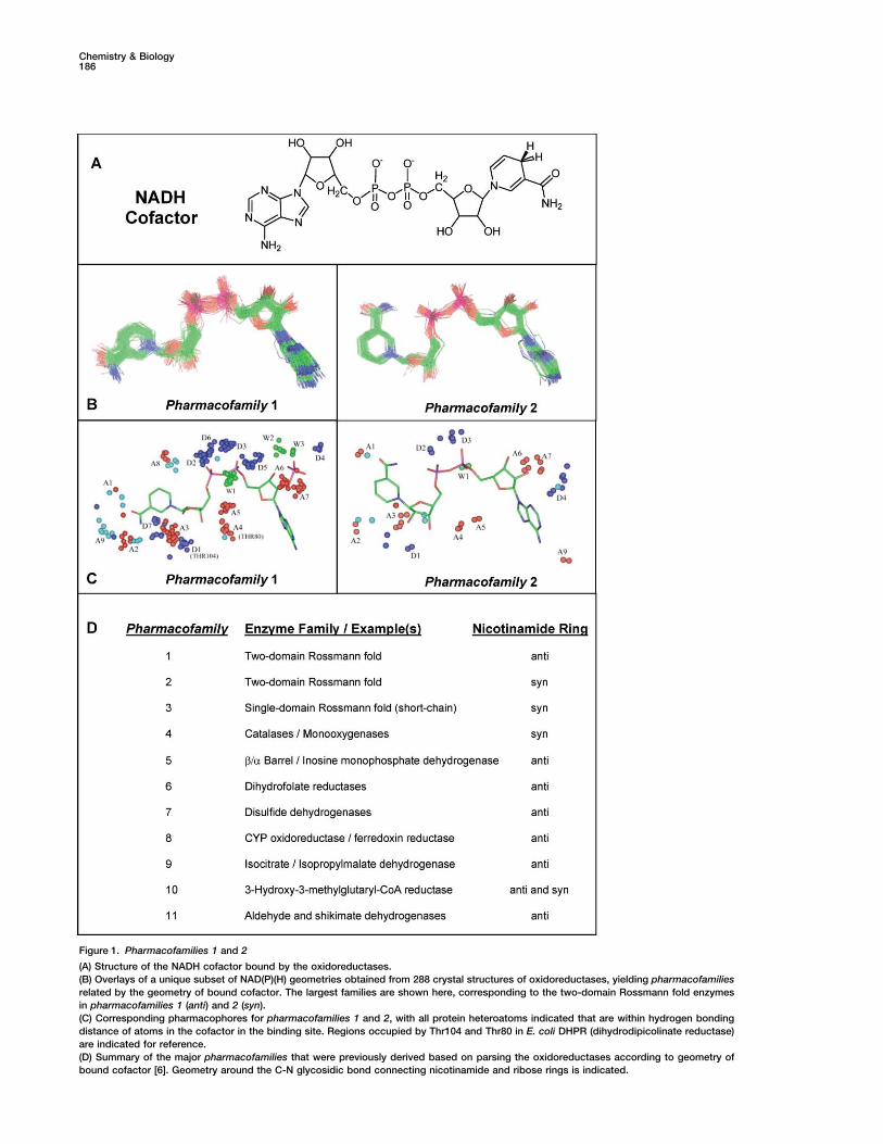
Table 1. Affinity and Specificity of CLM and Bi-Ligand Molecules for Oxidoreductases in pharmacofamilies 1 and 2
Structurea LDHb DHPRb DOXPRb
55 �M 26 �M �50 �M
42 nMc �50 �M 10 �M
12 �M �25 �M 202 nMc
620 nM 100 nMc 7.9 �M
a While the SL of the first bi-ligand was condensed with the carboxylic acid of the linker in Figure 5C, the other 2 SLs were condensed withthe acid of the shorter linker on CLM-1 [15].b Numbers are Kis values except for DOXPR, which has an IC50, which should approximate a Kis. LDH (lactate dehydrogenase) and DHPR arein pharmacofamily 1, while DOXPR (1-deoxy-D-xylulose-5-phosphate reductoisomerase) is in pharmacofamily 2 [6].c Most potent inhibition value amongst the three enzymes is indicated in bold.
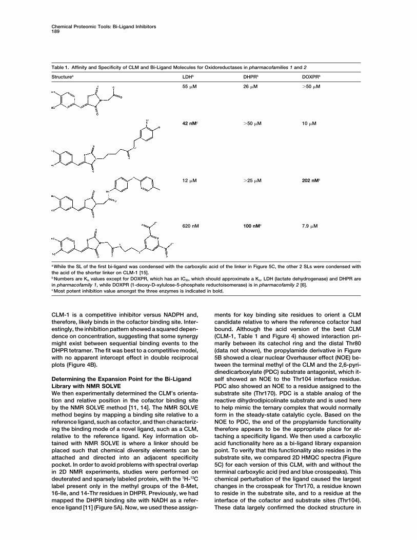
CLM-1 is a competitive inhibitor versus NADPH and, ments for key binding site residues to orient a CLMcandidate relative to where the reference cofactor hadtherefore, likely binds in the cofactor binding site. Inter-
estingly, the inhibition pattern showed a squared depen- bound. Although the acid version of the best CLM(CLM-1, Table 1 and Figure 4) showed interaction pri-dence on concentration, suggesting that some synergy
might exist between sequential binding events to the marily between its catechol ring and the distal Thr80(data not shown), the propylamide derivative in FigureDHPR tetramer. The fit was best to a competitive model,
with no apparent intercept effect in double reciprocal 5B showed a clear nuclear Overhauser effect (NOE) be-tween the terminal methyl of the CLM and the 2,6-pyri-plots (Figure 4B).dinedicarboxylate (PDC) substrate antagonist, which it-self showed an NOE to the Thr104 interface residue.Determining the Expansion Point for the Bi-Ligand
Library with NMR SOLVE PDC also showed an NOE to a residue assigned to thesubstrate site (Thr170). PDC is a stable analog of theWe then experimentally determined the CLM’s orienta-
tion and relative position in the cofactor binding site reactive dihydrodipicolinate substrate and is used hereto help mimic the ternary complex that would normallyby the NMR SOLVE method [11, 14]. The NMR SOLVE
method begins by mapping a binding site relative to a form in the steady-state catalytic cycle. Based on theNOE to PDC, the end of the propylamide functionalityreference ligand, such as cofactor, and then characteriz-
ing the binding mode of a novel ligand, such as a CLM, therefore appears to be the appropriate place for at-taching a specificity ligand. We then used a carboxylicrelative to the reference ligand. Key information ob-
tained with NMR SOLVE is where a linker should be acid functionality here as a bi-ligand library expansionpoint. To verify that this functionality also resides in theplaced such that chemical diversity elements can be
attached and directed into an adjacent specificity substrate site, we compared 2D HMQC spectra (Figure5C) for each version of this CLM, with and without thepocket. In order to avoid problems with spectral overlap
in 2D NMR experiments, studies were performed on terminal carboxylic acid (red and blue crosspeaks). Thischemical perturbation of the ligand caused the largestdeuterated and sparsely labeled protein, with the 1H-13C
label present only in the methyl groups of the 8-Met, changes in the crosspeak for Thr170, a residue knownto reside in the substrate site, and to a residue at the16-Ile, and 14-Thr residues in DHPR. Previously, we had
mapped the DHPR binding site with NADH as a refer- interface of the cofactor and substrate sites (Thr104).These data largely confirmed the docked structure inence ligand [11] (Figure 5A). Now, we used these assign-

Chemistry & Biology190
residue at the interface of the NADH and PDC bindingsites. Structural data were included here only to illustratethe method. The NMR data not only suggested the bi-ligand library expansion point, but it also confirmed thatPDC binding mode was not significantly altered in theCLM:PDC:DHPR ternary complex (compared to theNADH:PDC:DHPR ternary complex), since PDC showedthe same pattern of NOEs to threonine methyl protonsin both complexes. Finally, the selective perturbation ofactive site methyl proton chemical shifts in the complexwith CLM and the complex with the bi-ligand (see below)allow us to rule out any nonspecific mechanism for inhi-bition that could have produced competitive inhibitionprofiles, at least for these inhibitors.
Validation of the NMR-Selected LibraryExpansion PointBased on these NMR data, CLM-1 was linked to PDC,the specificity ligand analog. The corresponding bi-ligand compound had a Kis of 100 nM, which representsa 250-fold increase in affinity over the starting CLM(Table 1). The common and specificity sites were bothoccupied, based on NMR chemical shift mapping stud-ies showing perturbations of residues in both bindingsites (Figure 5D). Although the squared effect on inhibi-tion complicated steady-state analysis, analogous bi-ligands were made with the same specificity ligand butwith variants of the CLM that gave less pronouncedcooperativity effects. Steady-state kinetic profiles fortwo of these bi-ligand molecules are shown in Figure 6,showing a best fit to a competitive inhibition model ver-sus both the NADPH and dihydrodipicolinate substrates,as expected for a bi-ligand inhibitor.
Building the Focused Bi-Ligand LibraryThe last step of this systems-based bi-ligand designprocess involves the addition of diversity elements tothe CLM such that they are directed into the substrate(specificity ligand [SL]) site in the manner suggested byNMR SOLVE. Although the geometric relationship of the
Figure 4. Computationally Selected Cofactor Analogs and Steady- CLM and SL sites is conserved in oxidoreductases, theState Characterization as a Cofactor Analog
actual size and electrostatic properties of the SL binding(A) A representative set of 11 computationally selected and tested
site will vary in a way that parallels the diversity of sub-cofactor mimics, with the top structure being the only one thatstrates used by oxidoreductases, as discussed above.showed significant inhibitory activity.The diversity elements attached to the conserved CLM(B) Steady-state inhibition profile for CLM-1, which is a modified
form of the top inhibitor in (A). Profile is with inhibitor versus NADPH. scaffold were matched to the properties of substratesProfile represents the fit to the equation for a competitive inhibitor used throughout the oxidoreductase gene family in thatwith a squared dependence on inhibitor concentration. The fit gave they roughly paralleled the distribution of the chemicala Kis value of 26 � 2 �M. Curves for alcohol dehydrogenase (Kis �
properties surveyed in Figure 2.101 �M), lactate dehydrogenase (Kis � 55 �M), and DOXPR (Kis �We selected 300 diversity elements (commercially50 �M) fit best to a model for competitive inhibition.
available) and chemically joined them to the CLM-linkerconstruct. The resulting bi-ligand library was then screenedagainst three Rossmann-fold enzymes in pharmaco-Figure 3A, since the terminal methyl of the propylamide
is that part of the CLM that is closest to PDC (within families 1 and 2: DHPR, lactate dehydrogenase (LDH),and 1-deoxy-D-xylulose-5-phosphate reductoisomer-5 A). It should be noted that the NMR SOLVE experi-
ments would have suggested the same library expan- ase (DOXPR). The starting CLM bound only weakly tothe three enzymes, with Kis values in the 25–100 �Msion point in the absence of any protein structural infor-
mation, since crosspeaks for threonine residues were range (Table 1). However, after adding the diversity ele-ments in the position selected with NMR SOLVE, steady-assigned based on proximity to reference ligands (NADH
and PDC). That is, it was never necessary to assign state enzyme kinetic screening identified a specific bi-ligand inhibitor of LDH with a Kis of 42 nM and a bestThr104 to a specific residue number, as it would have
been adequate to view it only as the crosspeak for the fit to a model for competitive inhibition. This represents

Chemical Proteomic Tools: Bi-Ligand Inhibitors191
Figure 5. NMR SOLVE Data for DHPR
(A) The binding site is mapped relative to the NADH cofactor, identifying NMR probe atoms [14].(B) NOESY data for a CLM-1 analog, which places the methyl terminus of the propylamide functionality closest to the SL site.(C) Chemical alteration of the end of this alkyl chain in creating CLM-2 produces changes to crosspeaks for atoms in the SL site in the overlayof HMQC spectra for complexes of DHPR with both versions of the CLM (red and blue).(D) HMQC spectra of DHPR in the absence (red) and presence (blue) of the bi-ligand inhibitor.
an increase in affinity over the starting CLM of a thou- based bi-ligand design process described herein makesfragment assembly more likely to produce inhibitors forsand-fold, with most of the increased affinity directed
toward specificity interactions, since the bi-ligand binds multiple, related proteins, since the CLM fragment pro-vides a baseline of affinity across a pharmacofamilytwo to three orders of magnitude stronger to LDH than
to DHPR or DOXPR. We also identified a bi-ligand inhibi- (�GCLM). Furthermore, since the mere joining of two mo-lecular fragments has been proposed to provide astor that bound with an IC50 of 202 nM to DOXPR, also
with selectivity. Based on these data, we propose that much as 45 entropy units, corresponding to an increasein affinity of 108-fold [19, 20] associated with the “chelatea bi-ligand collection of sufficient size and diversity,
built with an appropriately chosen CLM and well placed effect,” the combined effect of adding a CLM to an SLcould be as large as 108/KCLM fold. This could only occurlinkers, will produce nM inhibitors for most members of
a pharmacofamily. for those SLs binding in the specificity pocket adjacentto the CLM, thus ensuring that linkage with the CLMprovides specificity for a given oxidoreductase. Al-Thermodynamic Foundation for Gene Family
Focused Bi-Ligand Libraries though the full magnitude of the chelate effect is stillbeing investigated by researchers, it is in any case quiteFragment-based assembly strategies are a very efficient
means of designing inhibitors [16–18]. The systems- large and reports of enhancements in affinity or rates

Chemistry & Biology192
Figure 6. Steady-State Inhibition Profiles for Bi-Ligand Molecules with CLMs that Are Variants of CLM-1
Bi-ligand structures are shown to the left of their respective inhibition profiles. Curves fit best to equations where inhibition was competitiveversus both cofactor (NADPH) and substrate. Enzyme was E. coli DHPR. The first bi-ligand was varied (0, 400, 750, 1100, 1500 nM) versusNADPH and gave a Kis of 370 � 90 nM (A), and versus dihydrodipicolinate (DHP) gave a Kis of 170 � 50 nM (B). The second bi-ligand wasvaried (0, 500, 1000, 1500, 2000 nM) versus NADPH and gave a Kis of 500 � 100 nM (C), and versus dihydrodipicolinate gave a Kis of 530 �
140 nM (D).
approaching 108-fold have been reported [21]. In prac- source of drug leads for the many new targets beingidentified in functional genomics efforts.tice, only a tiny fraction of this affinity increase will ever
be fully realized because linkers might be flexible, pro-Experimental Proceduresduce nonoptimal placement of the CLM and SL ligands,
or have repulsive interactions with a binding site. Still,Computational Search for CLMsthis combinatorial strategy for fragment linkage is anComputational methods were used to identify CLM candidates to
efficient way of focusing a library, since even imperfect be tested as cofactor mimics. Coordinates of the nicotinamide mo-linkage can produce large affinity boosts for multiple nonucleotide (NMN) portion of the NADH cofactor were extractedenzymes within a pharmacofamily. from the structure of cofactor in complex with DHPR (pdb code:
1arz). These coordinates were used to search against databases ofcommercially available compounds (such as from ASINEX [Moscow,Russia]), which contained molecules with precalculated three-Significancedimensional structures. The search was quite fast since it involvedonly matching the shape of the NMN portion of the cofactor to theGenomes have now been sequenced for numerousprecalculated structures in the small molecule database through anorganisms, and expectations are that this informationicosahedral matching algorithm [13, 22]. Still, an ellipsoid shape
will provide a better understanding of biology, as well prefilter was used as a first screen to identify molecules that wereas yield new and better therapeutics. This cannot be roughly the shape of NMN to prescreen and eliminate obviously
poor matches. To address heteroatom composition and hydrogenrealized until more efficient strategies are developedbonding capabilities at an approximate level, hybrid shape-match-to characterize proteins, and tools are created toing scores were used. Shapes were compared both as a function oftranslate this knowledge into inhibitors as mechanisticall atoms and as a function of heteratoms only, with scores weightedprobes and drugs. To this end, we had previously re-equally in the hybrid score. After prefiltering the circa 40,000 mole-
ported a structural proteomic analysis of oxidoreduc- cules, shape-matching scores were calculated with the THREEDOMtases, which comprise 2%–4% of most proteomes [6]. software package by comparing full structures or only the hetera-
toms. These two sets of scores were combined to create an averageHerein, we presented a systems-based strategy for“hybrid” score through Perl scripts developed in-house. Generallydesigning inhibitors of oxidoreductases. This strategyaround 5% of the computationally selected compounds were foundparallels nature’s modular approach for designing theto inhibit one of the three dehydrogenases tested. Althoughoxidoreductase gene family itself. Chemical librariessearches against the full-length NADH cofactor produced a some-
designed in this manner could be used as a source of what higher hit rate (up to 10% versus lactate dehydrogenase), thesechemogenomic probes for defining functions of mem- compounds were not used due to their higher molecular weight and
unsuitability as drug leads.bers of the oxidoreductase gene family, as well as a

Chemical Proteomic Tools: Bi-Ligand Inhibitors193
Computational Docking of the Propylamide dence on inhibitor concentration (Figure 4B), still fitting best to acompetitive model:Derivative of CLM-1
The propylamide derivative of CLM-1 was docked into the crystalstructure of the DHPR/NADH/2,6-PDC ternary complex after remov- v �
Vmax AA � Km (1 � (I/Kis)2)ing the cofactor. First, the ligand was minimized by AM1
(Gaussian98) with a net charge of –1, localized on the para-hydroxywhere v is the initial velocity, I is inhibitor concentration, Kis is thegroup of the catechol ring. Docking was then performed with theslope inhibition constant, A is cofactor (NADH) concentration, VmaxMOE software package (Chemical Computing Group) and theis the maximum velocity, and Km is the Michaelis constant.MMFF94 forcefield, with ligand flexible and protein kept rigid. Pro-
tein was also energy minimized (MMFF94) before docking, and thenNMR Spectroscopy25 docking runs were performed with random starting orientations.NMR experiments were performed on a Bruker DRX700 spectrome-Optimization was with simulated annealing, with an initial tempera-ter equipped with a triple resonance probe and triple axis gradientture of 1000 K and six cycles per run. Docking calculations includedcoil. Tetrameric DHPR concentration was �75 �M (300 �M mono-protein atoms within a 62 A 62 A 62 A box surrounding themer) in 25 mM Tris-D11 buffer (pH 7.8) at a temperature of 303 K,site previously occupied by cofactor. The choice to dock into thewith a sample volume of 150 �l in Shigemi tubes, as describedcofactor site was based on the observation of competitive inhibitionpreviously [14]. Most NMR experiments were performed in the pres-patterns (with the more soluble CLM-1) versus cofactor in theence of the PDC substrate analog along with either NADH or CLM,steady-state enzyme kinetic studies described below. One of thein order to mimic the active ternary complex that is produced in thetwo lowest energy structures is shown in Figure 3A, with the NADHsteady-state catalytic cycle. PDC was not used for studies of bi-structure overlaid back in its original orientation so that the relativeligands or for the inhibitors being compared in Figure 5C. PDC is abinding mode of CLM-1 derivative and “reference” cofactor can befairly potent substrate analog, with a Kis value of 26 �M versuscompared. Although the lowest energy structure had the propylam-the dihydrodipicolinate substrate [24]. Selective WURST adiabaticide group extending into the adenine site, only the orientation showndecoupling [26] of the from the � 13C of Thr was used in NMRin Figure 3A was consistent with the NMR SOLVE data describedexperiments to decrease overlap of the 14 Thr residues. Typical 2Dbelow. A solvent-accessible surface map color coded by partial[13C,1H] HMQC spectra were recorded in 30 min. Typical 2D [1H,1H]charge is shown in Figure 3B, with the region surrounding the nega-NOESY spectra were acquired with 256 2048 complex points andtive (red) catechol ring shown in the expansion. The four surroundingwith mixing times between 50 and 500 ms. 13C decoupling duringblue regions represent guanido groups of arginines 81, 16, 39, andacquisition was with a GARP composite decoupling sequence [27],19 that approach within 5.5, 5.7, 7.0, and 9.0 A, respectively, of thewhile 13C decoupling during the evolution period was with a 180�charged oxygen of the catechol ring.refocusing pulse. Ambiguities due to proton overlap among Thr andMet methyl proton chemical shifts were removed by recording a 3DSynthesis of Bi-Ligand Library[13C,1H] resolved [1H,1H] NOESY experiment [28]. NOEs were laterThe 3,4-dihydroxyphenylmethylene-rhodanine CLM (CLM-1) wasverified as not being due to spin diffusion through the QUIET-NOESYsynthesized by heating a solution of 13.6 g 3,4-dihydroxybenzalde-experiment [29, 30].hyde, and 19.3 g rhodanine acetic acid at 95�C in 200 ml acetic acid
for 6 hr. After cooling the solution, the precipitate was collectedAcknowledgmentsand washed with acetic acid (2 5 mL) to give 20 g of product
CLM-1. The CLM-1 acid (or amine; 1.5 eq) and PS-carbodiimideWe thank Dr. John Blanchard (Albert Einstein College of Medicine,resin (2 eq) were reacted in THF (10 mL/g) for 1 hr. The desiredNY) for the original pET11a DHPR expression construct. The workamine (or acid) to be conjugated to the CLM-1 was then addeddescribed in this article was performed at Triad Therapeutics, with(1 eq) and reacted overnight. Product was extracted twice with THFthe exception of the modeling in Figure 3, which was done by A.D.C.and solvent evaporated to give desired product.at Marquette University.
Protein ProductionReceived: September 10, 2003The E. coli DHPR protein was expressed with a pET21a vectorRevised: November 17, 2003(Novagen) and purified as described previously [14]. Briefly, DHPRAccepted: November 17, 2003was uniformly enriched in 2H and 15N and contained 1H/15N/13C-Published: February 20, 2004labeled threonine, 1H/13C(�-methyl)-labeled isoleucine, and 1H/13C
( -methyl)-labeled methionine, and was produced through a modi-fied version of supplemented M9 minimal media. Purification was Referenceson a Q Sepharose anion exchange column (Amersham). The DOXPRgene was cloned from E. coli genomic DNA by PCR utilizing the 1. Bashton, M., and Chothia, C. (2002). The geometry of domainfollowing primers: 5�-GCCACTGCATATGAAGCAACTCACCATTCTGG combination in proteins. J. Mol. Biol. 315, 927–939.and 3�-GCCACTGGGATCCTCAGCTTGCGAGACGCATC. DOXPR 2. Apic, G., Gough, J., and Teichmann, S.A. (2001). An insight intowas expressed and purified as reported [14, 23]. domain combinations. Bioinformatics 17, S83–S89.
3. Todd, A.E., Orengo, C.A., and Thornton, J.M. (2001). Evolutionof function in protein superfamilies, from a structural perspec-Steady-State Inhibition Studies
All reactions were monitored spectrophotometrically at 340 nm by tive. J. Mol. Biol. 307, 1113–1143.4. Drews, J. (1998). In Search of Tomorrow’s Medicines (New York:using initial rates from the first 5% of reaction. Absorbance changes
at 340 nm are from production or consumption of NAD(P)H cofactor. Springer-Verlag).5. Willett, P. (1987). Similarity and Clustering in Chemical Informa-LDH reaction mixtures contained 100 mM Hepes buffer (pH 7.4), 2.5
mM pyruvate, 10 �M NADH, and 5 ng/mL lactate dehydrogenase. tion Systems (Letchworth, UK: Research Studies Press).6. Kho, R., Baker, B.L., Newman, J.V., Jack, R.M., Sem, D.S., Villar,DOXPR reaction mixtures contained 100 mM Hepes buffer (pH 7.4),
1.2 mM DOXP, 8 �M NADPH, 1 mM MnCl2, and 10 �g/mL DOXPR. H.O., and Hansen, M.R. (2003). A path from primary proteinsequence to ligand recognition. Proteins 50, 589–599.DHPR reactions were as described previously [24], with either NADH
or NADPH cofactor. In all cases when screening bi-ligands, nonvar- 7. You, K.S. (1985). Stereospecificity for nicotinamide nucleotidesin enzymatic and chemical hydride transfer reactions. CRC Crit.ied substrate concentrations were kept close to their Km values.
Data were fitted to appropriate equations for competitive, noncom- Rev. Biochem. 17, 313–451.8. Lipinski, C.A., Lombardo, F., Dominy, B.W., and Feeney, P.J.petitive, and uncompetitive inhibition through nonlinear least-
squares fitting [25]. Mechanism of inhibition was established (2001). Experimental and computational approaches to estimatesolubility and permeability in drug discovery and developmentthrough analysis of the overall sigma value for the fit, as well as the
magnitude of standard deviations for inhibition constants (Kii, Kis). settings. Adv. Drug Deliv. Rev. 46, 3–26.9. Ghose, A.K., and Crippen, G.M. (1987). Atomic physicochemicalThe inhibition pattern for CLM-1 (Table 1) showed a squared depen-

Chemistry & Biology194
parameters for three-dimensional-structure-directed quantita-tive structure-activity relationships. 2. Modeling dispersive andhydrophobic interactions. J. Chem. Inf. Comput. Sci. 27, 21–35.
10. Scapin, G., Reddy, S.G., Zheng, R., and Blanchard, J.S. (1997).Three-dimensional structure of Escherichia coli dihydrodipicoli-nate reductase in complex with NADH and the inhibitor 2,6-pyridine dicarboxylate. Biochemistry 36, 15081–15088.
11. Sem, D.S., Yu, L., Coutts, S.M., and Jack, R. (2001). Object-oriented approach to drug design enabled by NMR SOLVE:First real-time structural tool for characterizing protein-ligandinteractions. J. Cell. Biochem. S37, 99–105.
12. Cirillo, J.D., Weisbrod, T.R., Banerjee, A., Bloom, B.R., and Ja-cobs, W.R., Jr. (1994). Genetic determination of the meso-diami-nopimelate biosynthetic pathway of mycobacteria. J. Bacteriol.176, 4424–4429.
13. Bladon, P. (1989). A rapid method for comparing and matchingthe spherical parameter surfaces of molecules and other irregu-lar objects. J. Mol. Graph. 7, 130–137.
14. Pellecchia, M., Meininger, D., Dong, Q., Chang, E., Jack, R.,and Sem, D.S. (2002). NMR-based structural characterizationof large protein-ligand interactions. J. Biomol. NMR 22, 165–173.
15. Parlow, J.J., Mischke, D.A., and Woodard, S.S. (1997). Utility ofcomplementary molecular reactivity and molecular recognition(CMR/R) technology and polymer-supported reagents in thesolution-phase synthesis of heterocyclic carboxamides. J. Org.Chem. 62, 5908.
16. Shuker, S.B., Hajduk, P.J., Meadows, R.P., and Fesik, S.W.(1996). Discovery of high affinity ligands for proteins: SAR byNMR. Science 274, 1531–1534.
17. Fejzo, J., Lepre, C.A., Peng, J.W., Bemis, G.W., Ajay, Murcko,M.A., and Moore, J.M. (1999). The SHAPES strategy: an NMR-based approach for lead generation in drug discovery. Chem.Biol. 6, 755–769.
18. Erlanson, D.A., Braisted, A.C., Raphael, D.R., Randal, M.,Stroud, R.M., Gordon, E.M., and Wells, J.A. (2000). Site-directedligand discovery. Proc. Natl. Acad. Sci. USA 97, 9367–9372.
19. Page, M.I., and Jencks, W.P. (1971). Entropic contributions torate accelerations in enzymic and intramolecular reactions andthe chelate effect. Proc. Natl. Acad. Sci. USA 68, 1678–1683.
20. Snider, M.J., Lazarevic, D., and Wolfenden, R. (2002). Catalysisby entropic effects: the action of cytidine deaminase on 5,6-dihydrocytidine. Biochemistry 41, 3925–3930.
21. Carlow, D., and Wolfenden, R. (1998). Substrate connectivityeffects in the transition state for cytidine deaminase. Biochemis-try 37, 11873–11878.
22. Doucet, J.-P., and Weber, J. (1996). Computer-Aided MolecularDesign: Theory and Applications (San Diego: Academic Press).
23. Meininger, D.P., Rance, M., Starovasnik, M.A., Fairbrother, W.J.,and Skelton, N.J. (2000). Characterization of the binding inter-face between the E-domain of staphylococcal protein A and anantibody Fv-fragment. Biochemistry 39, 26–36.
24. Reddy, S.G., Sacchettini, J.C., and Blanchard, J.S. (1995). Expres-sion, purification, and characterization of Escherichia coli dihy-drodipicolinate dehydrogenase. Biochemistry 34, 3492–3501.
25. Cleland, W.W. (1979). Statistical analysis of enzyme kinetic data.Methods Enzymol. 63, 103–138.
26. Kupce, E., and Wagner, G. (1995). Wideband homonuclear de-coupling in protein spectra. J. Magn. Reson. B109, 329–333.
27. Shaka, A.J., Barker, P.B., and Freeman, R. (1985). Computer-optimized decoupling scheme for wideband applications andlow-level operation. J. Magn. Reson. 64, 547–552.
28. Cavanagh, H.J., Fairbrother, W.J., Palmer, A.G., III, and Skelton,N.J. (1996). Protein NMR Spectroscopy, Principles and Practice,First Edition (New York: Academic Press).
29. Neuhaus, D., and Williamson, M.P. (2000). The Nuclear Over-hauser Effect in Structural and Conformational Analysis, FirstEdition (New York: Wiley-VCH).
30. Vincent, S.J.F., Zwahlen, C., and Bodenhausen, G. (1996). Sup-pression of spin diffusion in selected frequency bands of nuclearOverhauser spectra. J. Biomol. NMR 7, 169–172.