Interaction of Multimicrobial Synthetic Inhibitor 1,2- Bis(2-Benzimidazolyl)-1,2-Ethanediol with...
11
Interaction of Multimicrobial Synthetic Inhibitor 1,2- Bis(2-Benzimidazolyl)-1,2-Ethanediol with Serum Albumin: Spectroscopic and Computational Studies Nayana Kamtekar 1 , Anita Pandey 1 , Neeraj Agrawal 1 , Raghuvir R. S. Pissurlenkar 2 , Mohanish Borana 1 , Basir Ahmad 1 * 1 UM-DAE Centre for Excellence in Basic Sciences, University of Mumbai, Kalina Campus, Mumbai, India, 2 Molecular Simulations, Pharmaceutical Chemistry Division, Bombay College of Pharmacy, Mumbai, India Abstract The molecule, 1,2-Bis(2-benzimidazolyl)-1,2-ethanediol (BBE) is known to act as a selective inhibitor of poliovirus, rhinovirus, Candida albicans, several bacterial species, and is easily synthesized by Phillips reaction. The interaction of BBE with BSA and the effects of its binding on the conformation and unfolding/refolding pathways of the protein were investigated using multispectroscopic techniques and molecular modeling. The binding studies indicate that BSA has one high affinity BBE binding site with association constant 6.0260.05 6 10 4 M 21 at 298 K. By measuring binding at different temperatures, we determined the changes in enthalpy (DH= 215.1362.15 kJ mol 21 ), entropy (DS = 40.8767.25 J mol 21 K 21 ) and free energy (DG = 26.7861.02) of interaction, which indicate that the binding was spontaneous and both enthalpically and entropically driven. Based on molecular modeling and thermodynamic parameters, we proposed that the complex formation involved mainly hydrophilic interaction such as hydrogen bonding between hydroxyl groups of ethane-1,2-diol fragment with Tyr410 and benzimidazole sp 2 nitrogen atom with Ser488 and hydrophobic interaction between phenyl ring of one benzimidazole of the ligand and hydrophobic residues namely, Ile387, Cys391, Phe402, Val432 and Cys437. The sequential unfolding mechanism of BSA, site-specific marker displacement experiments and molecular modeling showed that the molecule preferably binds in subdomain IIIA. The BBE binding to BSA was found to cause both secondary and tertiary structural alterations in the protein as studied by intrinsic fluorescence, near-UV and far-UV circular dichroism results. The unfolding/ refolding study showed that BBE stabilized native to intermediate states (N=I) transition of the protein by ,2 kJ mol 21 without affecting the intermediate to unfolded states (I=U) transition and general mechanism of unfolding of BSA. Citation: Kamtekar N, Pandey A, Agrawal N, Pissurlenkar RRS, Borana M, et al. (2013) Interaction of Multimicrobial Synthetic Inhibitor 1,2-Bis(2-Benzimidazolyl)- 1,2-Ethanediol with Serum Albumin: Spectroscopic and Computational Studies. PLoS ONE 8(1): e53499. doi:10.1371/journal.pone.0053499 Editor: Eugene A. Permyakov, Russian Academy of Sciences, Institute for Biological Instrumentation, Russian Federation Received October 22, 2012; Accepted November 30, 2012; Published January 4, 2013 Copyright: ß 2013 Kamtekar et al. This is an open-access article distributed under the terms of the Creative Commons Attribution License, which permits unrestricted use, distribution, and reproduction in any medium, provided the original author and source are credited. Funding: Financial Support was provided by UM-DAE Centre for Excellence in Basic Sciences. The funders had no role in study design, data collection and analysis, decision to publish, or preparation of the manuscript. Competing Interests: The authors have declared that no competing interests exist. * E-mail: [email protected] Introduction Synthetic chemistry and ligand-protein interaction studies have played an important role in speeding up the recent drug discovery processes. The methods of synthetic chemistry create small molecules rapidly for screening, and ligand-protein interaction studies provide information on how a potential drug interacts with target protein [1,2]. The knowledge of ligand-protein interaction is also vital for the accurate prediction of its biological function [3]. Therefore, the characterization of a synthetic potential drug molecule interaction with proteins is mandatory and requires determination of association constant, number of binding sites, thermodynamic properties, binding induced conformational alter- ations and stability of the protein. It is now understood that the ligand residence time, the total duration of ligand-protein binding have a vital impact on the ligand’s effectiveness and selectivity [4,5]. Several studies have reported that 1,2-Bis(2-benzimidazolyl)- 1,2-ethanediol (BBE) acts as a selective inhibitor of poliovirus [6,7] and rhinovirus infections [8]. The compound and its metal ion complexes also show broad-spectrum antibacterial activity against Staphylococcus aureus, S. epidermis, Klebsiella pneumoniae, Salmonella typhi and antifungal activity against Candida albicans [9]. The molecule 1,2-Bis(2-benzimidazolyl)-1,2-ethanediol (BBE) is a chiral triden- tate ligand. It can be prepared with Tartaric acid and 1,2- diaminobenzene by a simple Phillips reaction [10] (Figure 1). Despite broad inhibitory effects against all kinds of pathogens and the ease of synthesis, its interaction with target pathogen proteins or drug transport proteins, such as serum albumin, is not known. With this perspective, we carried out detailed investigations of the interaction of BBE with bovine serum albumin (BSA), which shares ,80% sequence homology with human serum albumin and contains almost identical ligand binding sites for drugs and other endo- and exogenous ligands [11]. Previous studies on ligand- serum albumin binding showed that subdomain IIA and IIIA of the proteins contain the main binding sites for drugs [12,13]. The BBE interaction to serum albumin cannot be expected to explain the effect of the molecule directly on target pathogen protein but it may be used as a model to study the effect of the molecule on pathogen proteins. Moreover, serum albumin is the major PLOS ONE | www.plosone.org 1 January 2013 | Volume 8 | Issue 1 | e53499
Transcript of Interaction of Multimicrobial Synthetic Inhibitor 1,2- Bis(2-Benzimidazolyl)-1,2-Ethanediol with...
Interaction of Multimicrobial Synthetic Inhibitor 1,2-Bis(2-Benzimidazolyl)-1,2-Ethanediol with SerumAlbumin: Spectroscopic and Computational StudiesNayana Kamtekar1, Anita Pandey1, Neeraj Agrawal1, Raghuvir R. S. Pissurlenkar2, Mohanish Borana1,
Basir Ahmad1*
1 UM-DAE Centre for Excellence in Basic Sciences, University of Mumbai, Kalina Campus, Mumbai, India, 2 Molecular Simulations, Pharmaceutical Chemistry Division,
Bombay College of Pharmacy, Mumbai, India
Abstract
The molecule, 1,2-Bis(2-benzimidazolyl)-1,2-ethanediol (BBE) is known to act as a selective inhibitor of poliovirus, rhinovirus,Candida albicans, several bacterial species, and is easily synthesized by Phillips reaction. The interaction of BBE with BSA andthe effects of its binding on the conformation and unfolding/refolding pathways of the protein were investigated usingmultispectroscopic techniques and molecular modeling. The binding studies indicate that BSA has one high affinity BBEbinding site with association constant 6.0260.056104 M21 at 298 K. By measuring binding at different temperatures, wedetermined the changes in enthalpy (DH = 215.1362.15 kJ mol21), entropy (DS = 40.8767.25 J mol21 K21) and free energy(DG = 26.7861.02) of interaction, which indicate that the binding was spontaneous and both enthalpically and entropicallydriven. Based on molecular modeling and thermodynamic parameters, we proposed that the complex formation involvedmainly hydrophilic interaction such as hydrogen bonding between hydroxyl groups of ethane-1,2-diol fragment with Tyr410and benzimidazole sp2 nitrogen atom with Ser488 and hydrophobic interaction between phenyl ring of one benzimidazoleof the ligand and hydrophobic residues namely, Ile387, Cys391, Phe402, Val432 and Cys437. The sequential unfoldingmechanism of BSA, site-specific marker displacement experiments and molecular modeling showed that the moleculepreferably binds in subdomain IIIA. The BBE binding to BSA was found to cause both secondary and tertiary structuralalterations in the protein as studied by intrinsic fluorescence, near-UV and far-UV circular dichroism results. The unfolding/refolding study showed that BBE stabilized native to intermediate states (N=I) transition of the protein by ,2 kJ mol21
without affecting the intermediate to unfolded states (I=U) transition and general mechanism of unfolding of BSA.
Citation: Kamtekar N, Pandey A, Agrawal N, Pissurlenkar RRS, Borana M, et al. (2013) Interaction of Multimicrobial Synthetic Inhibitor 1,2-Bis(2-Benzimidazolyl)-1,2-Ethanediol with Serum Albumin: Spectroscopic and Computational Studies. PLoS ONE 8(1): e53499. doi:10.1371/journal.pone.0053499
Editor: Eugene A. Permyakov, Russian Academy of Sciences, Institute for Biological Instrumentation, Russian Federation
Received October 22, 2012; Accepted November 30, 2012; Published January 4, 2013
Copyright: � 2013 Kamtekar et al. This is an open-access article distributed under the terms of the Creative Commons Attribution License, which permitsunrestricted use, distribution, and reproduction in any medium, provided the original author and source are credited.
Funding: Financial Support was provided by UM-DAE Centre for Excellence in Basic Sciences. The funders had no role in study design, data collection andanalysis, decision to publish, or preparation of the manuscript.
Competing Interests: The authors have declared that no competing interests exist.
* E-mail: [email protected]
Introduction
Synthetic chemistry and ligand-protein interaction studies have
played an important role in speeding up the recent drug discovery
processes. The methods of synthetic chemistry create small
molecules rapidly for screening, and ligand-protein interaction
studies provide information on how a potential drug interacts with
target protein [1,2]. The knowledge of ligand-protein interaction is
also vital for the accurate prediction of its biological function [3].
Therefore, the characterization of a synthetic potential drug
molecule interaction with proteins is mandatory and requires
determination of association constant, number of binding sites,
thermodynamic properties, binding induced conformational alter-
ations and stability of the protein. It is now understood that the
ligand residence time, the total duration of ligand-protein binding
have a vital impact on the ligand’s effectiveness and selectivity
[4,5].
Several studies have reported that 1,2-Bis(2-benzimidazolyl)-
1,2-ethanediol (BBE) acts as a selective inhibitor of poliovirus [6,7]
and rhinovirus infections [8]. The compound and its metal ion
complexes also show broad-spectrum antibacterial activity against
Staphylococcus aureus, S. epidermis, Klebsiella pneumoniae, Salmonella typhi
and antifungal activity against Candida albicans [9]. The molecule
1,2-Bis(2-benzimidazolyl)-1,2-ethanediol (BBE) is a chiral triden-
tate ligand. It can be prepared with Tartaric acid and 1,2-
diaminobenzene by a simple Phillips reaction [10] (Figure 1).
Despite broad inhibitory effects against all kinds of pathogens and
the ease of synthesis, its interaction with target pathogen proteins
or drug transport proteins, such as serum albumin, is not known.
With this perspective, we carried out detailed investigations of
the interaction of BBE with bovine serum albumin (BSA), which
shares ,80% sequence homology with human serum albumin and
contains almost identical ligand binding sites for drugs and other
endo- and exogenous ligands [11]. Previous studies on ligand-
serum albumin binding showed that subdomain IIA and IIIA of
the proteins contain the main binding sites for drugs [12,13]. The
BBE interaction to serum albumin cannot be expected to explain
the effect of the molecule directly on target pathogen protein but it
may be used as a model to study the effect of the molecule on
pathogen proteins. Moreover, serum albumin is the major
PLOS ONE | www.plosone.org 1 January 2013 | Volume 8 | Issue 1 | e53499
transport and depot protein of mammalian systems. The
bioavailability, distribution and metabolism of a drug are largely
controlled by its interaction with serum albumin [14,15].
In this study, we investigate the BBE-BSA interaction mecha-
nism and characterize the binding parameters (Ka and n), binding
location, thermodynamics parameters (DH, DS and DG) and
residence lifetime (t) of the complex. We also show the effect of
interaction on conformation, unfolding/refolding pathways and
stability of the protein. We used the widely applied fluorescence
quench titration method to investigate the binding mechanism
[16,17]. The molecule binding location was determined by using
methods such as sequential unfolding mechanism of serum
albumin [18], displacement of site-specific marker [19] and
molecular modeling. The conformational alteration and unfold-
ing/refolding studies were made by using far-UV CD as a probe
for secondary structure and intrinsic fluorescence and near-UV
CD as probes for tertiary structure.
Materials and Methods
MaterialsAll Chemicals and reagents were purchased from Sigma-
Aldrich or SD fine chemical Ltd. The ligand 1,2-bis(2-benzimi-
dazolyl)-1,2-ethanediol (BBE) was synthesized according to the
reported procedure [10] and purity was checked by thin layer
chromatography, melting point and 1H NMR. Spectroscopic
studies were performed using spectrophotometer (Shimadzu UV
1800), spectrofluorimeter (Fluoromax 4, Horiba), and Circular
dichromism (Jasco J-815).
Interaction Studies of BBE with BSABovine serum albumin stock solution (150 mM) was prepared in
60 mM sodium phosphate buffer of pH 7.4 and the concentration
was determined spectrophotometrically using e1%1cm of 6.67 at
279 nm [20]. BBE stock solution (2.7 mM) was prepared by
dissolving 1 mg of the compound in 1 mL methanol. For the
binding studies, to a fixed volume (3.0 mL) of 5 mM protein
solution was added an increasing volume (0220 mL) of the ligand.
The fluorescence was measured between 300 and 400 nm after
exciting the samples at 280 nm. The effect of highest concentra-
tion of methanol (0.66% v/v) added in the samples during titration
was investigated on the far-UV CD spectrum and intrinsic
fluorescence spectrum of the protein. We found that both far-UV
CD and intrinsic fluorescence spectra of BSA were almost
identical in the absence and presence of 0.66% v/v methanol,
which indicated that the conformation of BSA did not alter in the
presence of highest concentration of methanol (0.66% v/v) used in
this study.
Site-specific marker displacement experiments were performed
by titrating diazepam-BSA complex obtained by mixing the two at
different diazepam/BSA molar ratios (0:1, 0.5:1, 1:1, 2:1) with
increasing concentrations of BBE. The intrinsic fluorescence of
BSA was recorded between 300–400 nm after excitation at
280 nm. Each spectrum was background subtracted with the
spectrum of free BBE.
Conformational StudiesIntrinsic fluorescence. Intrinsic fluorescence of BSA in the
absence and presence of different concentrations of BBE was
measured between 300 and 400 nm by exciting at 280 nm. The
excitation and emission slits were set at 4 nm and the fluorescence
spectra were taken at three different temperatures. Rayleigh
scattering measurements in the absence presence of 0.01% Triton
X-100 were performed at room temperature in a 1 cm path-length
cell. Samples were excited at 350 nm and spectra were recorded in
the range of 300–400 nm. Both excitation and emission slits were
fixed at 5 nm. Data were plotted at 350 nm.Far- and near-UV circular dichroism. Circular dichroism
studies were carried out with a Jasco J-815 spectropolarimeter
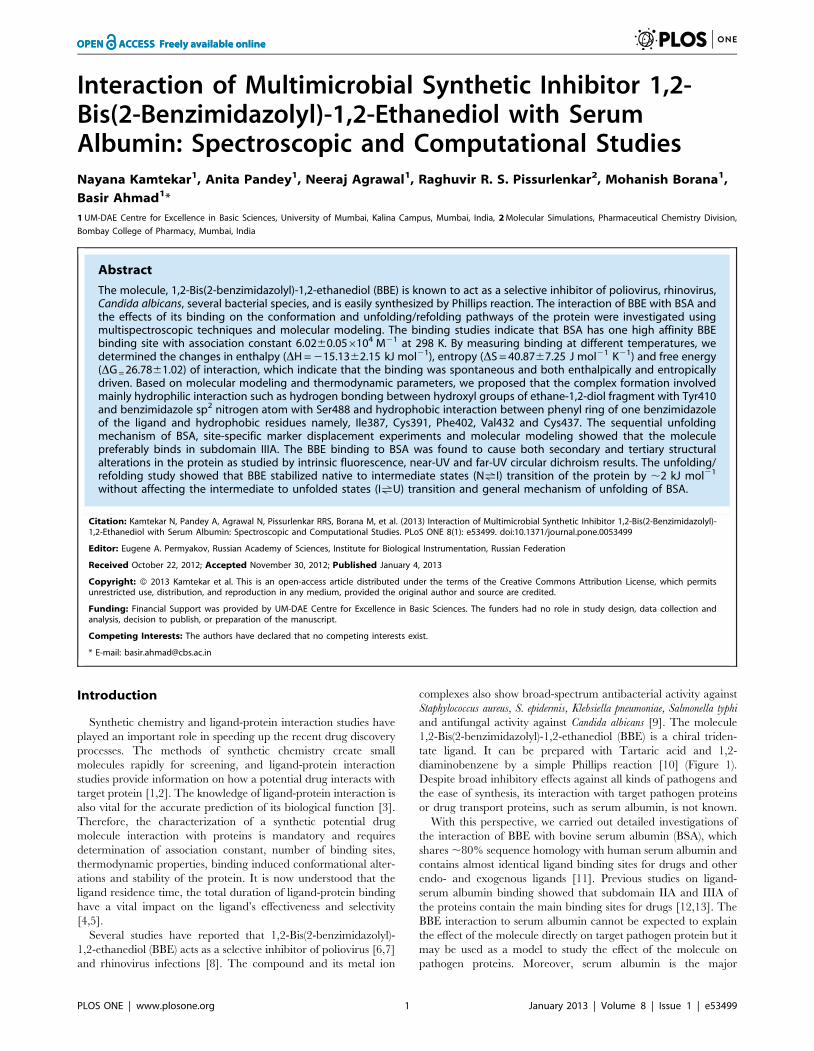
Figure 1. Preparation and structure of 1,2-Bis(2-Benzimidazo-lyl)-1,2-Ethanediol (BBE).doi:10.1371/journal.pone.0053499.g001
Figure 2. 1,2-Bis(2-Benzimidazolyl)-1,2-Ethanediol (BBE) in-duced fluorescence quenching mechanism of bovine serumalbumin. The BBE induced intrinsic fluorescence quenching (A) andStern-Volmer plots for fluorescence quenching data (B) of BSA atdifferent temperatures. The inset of Figure A shows fluorescencespectra of BSA in the absence and presence of increasing BBEconcentrations at 298 K. The concentration of BSA was 5 mM and theintrinsic fluorescence of the protein was measured in 60 mM sodiumphosphate buffer of pH 7.4 upon excitation at 280 nm.doi:10.1371/journal.pone.0053499.g002
Interaction of Multimicrobial Inhibitor with BSA
PLOS ONE | www.plosone.org 2 January 2013 | Volume 8 | Issue 1 | e53499
after calibrating it with D-10-camphorsulfonic acid under a
constant nitrogen flow. Far- and near-UV CD spectra in the
absence and presence of different molar ratios of BBE and BSA
were taken at the protein concentrations of 5 and 25 mM with 0.1-
and 1.0 cm path length cells, respectively. Each spectrum was the
average of four scans and background subtracted with the
spectrum of free BBE. The results were presented as mean residue
ellipticity [h] in deg cm2dmol21, which is defined as
h½ �~CD= 10|n|l|Cp
� �ð1Þ
where CD is in milli-degree, n is the number of amino acid
residues (583), l is the path length of the cell in cm, and Cp is molar
concentration of the protein.
The amount of regular secondary structures (a-helix and b-
strand) were determined by analysis of the CD spectra using K2D3
deconvolution software (21).
Protein Unfolding and Stability StudiesThe unfolding/refolding studies of BSA upon BBE binding
were carried out by urea denaturation of the protein. To a 100 mL
stock BSA solution at pH 7.4 were first added different volumes of
the 60 mM sodium phosphate buffer of pH 7.4, followed by the
addition of stock urea (10 M) and BBE to get a desired
concentration of denaturant and the ligand. The final solution
mixture (3.0 mL) was incubated for 8–10 hours at room
temperature and the unfolding process was monitored by intrinsic
fluorescence at 343 nm by exciting the protein at 280 nm.
Unfolding data analysis. The unfolding transitions of BSA
and ligand-protein complex followed a two-step three state
mechanism and can be represented by the equilibrium equation
NuIuU ð2Þ
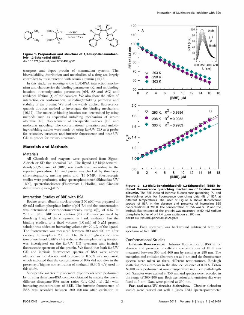
Table 1. 1,2-Bis(2-Benzimidazolyl) 1,2-Ethanediol interaction and thermodynamic parameters.
T (K) Ksv 6104(M21) kq 61012 (M21s21) Ka 6104 (M21) n 2DH (kJ mol21) DS (J mol21 K21) 2DG (kJ mol21)
293 4.5960.06 9.1860.12 6.8560.07 1.0460.03 27.1061.06
298 4.4060.07 8.8060.14 6.0260.05 1.0360.02 15.1362.15 40.8767.25 26.7861.02
303 3.7460.04 7.4860.08 5.5860.05 1.0460.04 26.0660.97
doi:10.1371/journal.pone.0053499.t001
Figure 3. The BBE binding to BSA is specific. Particle formation byBBE in the absence and presence of 0.01% Triton X-100 as monitored byRayleigh light scattering at 350 nm (A). Stern-Volmer plots of BBE-induced fluorescence quenching of BSA with increasing concentrationsof the protein (B).doi:10.1371/journal.pone.0053499.g003
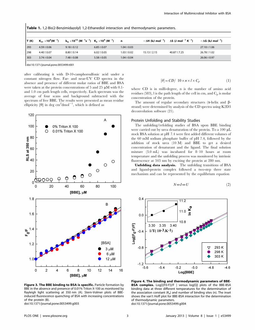
Figure 4. The binding and thermodynamic parameters of BBE-BSA complex. Log[{(F0-F)}/F ] versus log[Q] plots of the BBE-BSAbinding data at three different temperatures for the determination ofthe association constant (Ka) and number of binding sites (n). The insetshows the van’t Hoff plot for BBE-BSA interaction for the determinationof thermodynamic parameters.doi:10.1371/journal.pone.0053499.g004
Interaction of Multimicrobial Inhibitor with BSA
PLOS ONE | www.plosone.org 3 January 2013 | Volume 8 | Issue 1 | e53499

where N, I, U are native, intermediate and unfolded states of the
protein, respectively. In such type of transition, each step can be
assumed to follow a two state mechanism [22]. The data
monitored by intrinsic fluorescence at 343 nm were expressed in
term of the fraction of intermediate state (fI) for the N=I transition
and the fraction of unfolded state (fU) for the I=U transition
calculated from following equations
fI~(y1{yN)=(yI{yN) ð3Þ
fU~(y2{yI)=(yU{yI) ð4Þ
where y1 and y2 are the observed fluorescence intensity
corresponding to first and second transitions, respectively. yN, yI
and yU are the fluorescence intensities of the N, I, and U states of
BSA and were obtained by linear regression of the data
corresponding to pre-, intermediate and post-transition regions
of unfolding profile, respectively. The changes in free energy of the
two transitions at different urea concentration were calculated by
following equations.
DG~{RTln(fI=fN) ð5Þ
DG~{RTln(fU=fI) ð6Þ
The free energy change in the absence of urea DGH2O was
determined by least square analysis of DG versus [urea] plot using
equation
DG~DGH2Ozm½urea� ð7Þ
The DG values were also determined by nonlinear regression of
the data using equation.
fd~e(({(DGzm½urea�)=RT)=(1ze(({(DGzm½urea�)=RT))ð8Þ
where fd is the fraction unfolded (fI and fU), R and T are universal
gas constant (8.3145 J/mol) and absolute temperature (298 K),
respectively. m is the measure of dependence of DG on urea
concentrations.
Computational StudiesThe docking studies were carried out with the docking program
Glide v5.8 [23,24] module of Schrodinger Suite 2012 (Schrodin-
ger LLC, USA) running on an Intel Xeon processor based HPC
Cluster with Rocks 5.4 Cluster Suite as the operating environ-
ment.
Table 2. 1,2-Bis(2-Benzimidazolyl) 1,2-Ethanediol induced conformational alteration in BSA.
BBE/BSAMolar ratio K2D3% a-helices K2D3% b-strand [h]268/[h]262 ratio lmax Emission
0:1 65.6164.11 7.4461.31 0.93 343.0
1:1 63.0863.27 7.8161.44 1.16 345.5
2:1 60.3462.78 8,0161.13 1.31 348.5
doi:10.1371/journal.pone.0053499.t002
Figure 5. The location of the BBE binding site using unfoldingmechanism of BSA and site specific marker. Stern-Volmer plots ofBBE-induced fluorescence quenching of BSA with increasing concen-tration of urea (A). The inset shows the effect of increasingconcentrations of urea on the BBE-induced fluorescence quenching ofthe protein at a fixed BBE/BSA molar ratio of 2:1. Site specific markerdisplacement experiments.Stern-Volmer plots of BBE-induced fluores-cence quenching of BSA pre-incubated with different concentrations ofdiazepam, a subdomain IIIA site marker (B).doi:10.1371/journal.pone.0053499.g005
Interaction of Multimicrobial Inhibitor with BSA
PLOS ONE | www.plosone.org 4 January 2013 | Volume 8 | Issue 1 | e53499

Protein and Ligand Preparation for Computational StudyThe X-ray crystal structures of human serum albumin (PDB
code 2BXF) and bovine serum albumin (PDB code 3V03) were
retrieved from the Protein Data Bank [25,26]. Due to the absence
of bound ligand with BSA, the binding site was defined based on
the facts that (i) BSA shares high sequence homology with HSA,
and (ii) that BBE was found to interact with the diazepam binding
site (subdomain IIIA), identified from the experimental studies.
The coordinates of diazepam were extracted into the coordinate
frame of BSA after alignment with the HSA, which is bound to
diazepam in the subdomain IIIA.
The structure 3V03 was prepared for the docking studies with
the Protein Preparation Wizard (Schrodinger Suite 2012). The
protein BSA was structurally aligned to HSA after which the
ligand (diazepam) coordinates were extracted in the BSA
coordinate frame. The crystal waters were removed, hydrogen
atoms were added, and atom types and partial charges were
assigned based on the OPLS2005 force field. The formal charges
for the acidic and basic amino acids were set according to the
physiological pH 7.4. The missing side chains were added using
the Prime v3.1 (Schrodinger Suite 2012). The complex system was
relaxed using energy minimization protocol until an energy
gradient of 0.01 kcal/mol/A (1 cal = 4.184 J) was reached with
the OPLS2005 force field.
The structure of 1,2-Bis(2-benzimidazolyl)-1,2-ethanediol was
prepared using LigPrep v2.6 (Schrodinger 2012 Suite). The atom
types and partial charges were assigned based on the OPLS2005
force field, corresponding to the physiological pH 7.4.
Docking studies. A grid around the bound ligand (diaze-
pam) defined the active site of BSA. The docking grid comprised
of two grid boxes - the inner grid box set to dimensions 10 s3
while outer grid set to 20 s3, thus providing ample space for the
generation of diverse ligand conformations in the subdomain IIIA
binding site by the search and score algorithm. The vdW radius
scaling of 1.0 s was applied to soften the potential in the nonpolar
areas of the protein present within the grid extents with the partial
atomic charges set to 0.25, while no scaling was applied to receptor
atoms beyond the extent of the grid. The rotation of side-chain
hydroxyl functions was allowed for certain amino acids namely
serine, threonine, and tyrosine to increase the chances for the
formation of hydrogen bonds with the polar ligand substitution.
The settings for the docking study were validated based on the
protocol’s ability to reproduce the X-ray conformation of the
bound ligand in the subdomain IIIA of HSA and subsequently of
BSA.
The validated protocol was used to identify the docking
solutions to the binding of BBE in the subdomain IIIA of BSA.
The poses were ranked with scoring function GlideScore XP [27].
Results
1,2-Bis(2-benzimidazolyl) 1,2-ethanediol (BBE) InducedQuenching Mechanism of BSA Fluorescence
The fluorescence quenching titration method has been exten-
sively used to elucidate the ligand binding behavior of proteins
[16,18,28]. Figure 2A shows the 1,2-bis(2-benzimidazolyl)-1,2-
ethanediol (BBE) induced quenching of bovine serum albumin
(BSA) fluorescence at different temperatures. The inset of
Figure 2A shows the effect of different concentrations of BBE on
fluorescence spectra of BSA at 298 K. It is apparent from the
Figure 2 that equilibration of the protein with BBE caused
concentration dependent quenching of the intensity and a gradual
red shift (,6 nm) in emission maximum (lmax) of BSA. These
changes in the fluorescence properties of BSA suggest formation of
stable complex with BBE.
To confirm the quenching mechanism, the data at different
temperatures were analyzed according to the Stern-Volmer
Figure 6. The BBE-induced conformational alterations in BSA.The intrinsic fluorescence (A), near-UV CD (B) and far-UV CD (C) spectraof BSA in the presence of BBE/BSA molar ratios of 0:1, 1:1 and 2:1,respectively. The inset of figure A shows the changes in emissionmaximum (lmax) with increasing BBE concentrations.doi:10.1371/journal.pone.0053499.g006
Interaction of Multimicrobial Inhibitor with BSA
PLOS ONE | www.plosone.org 5 January 2013 | Volume 8 | Issue 1 | e53499

equation [29].
F0=F~1zKsv½BBE� ð9Þ
where F0 and F are the fluorescence intensity in the absence and
presence of the quencher (BBE) and Ksv is the Stern-Volmer
constant. The Ksv at different temperatures were determined by
linear regression of plots of F0/F versus [BBE] (Figure 2B). The
Ksv for BBE at different temperatures were found to be of the
order of 46104 M21 (Table 1). Linear Stern-Volmer plots
(regression coefficient R2.0.996) at all temperatures suggest that
only one kind of quenching mechanism, static or dynamic,
dominates. It also indicates that there is only one set of equivalent
BBE binding sites on BSA. The Stern-Volmer quenching constant
is given by kqt0, where kq and t0 are the bimolecular quenching
rate constant and lifetime of the protein fluorescence in the
absence of ligand, respectively. The lifetime t0 of BSA has been
accurately estimated to be 561029s [30] and using the Ksv values
in Table 1, kq values at different temperatures were calculated and
were found to be in the range of 1013 M21s21 (Table 1). These
values were markedly larger than the maximum collisional
quenching constant (1010 M21s21) [31], which indicated that
BBE-induced quenching was due to complex formation. More-
over, a decrease in Ksv was measured with increasing temperature
(Table 1), which also indicated that the BBE-induced quenching
mechanism was due to complex formation. Taken together, the
linearity of Stern-Volmer plots, decrease in Stern-Volmer
constants and very high values of the quenching rate constants
which increase with temperature support the fact that the BBE-
induced quenching of BSA fluorescence is due to complex
formation and BSA contains a single type of equivalent binding
sites for the molecule.
Many small molecules are known to form colloidal aggregates in
aqueous solution at micromolar concentrations and adsorb
proteins, which leads to artifacts in screens for ligands of proteins
[32,33]. To check that BBE is binding specifically to BSA, we
monitored the colloid aggregate formation by BBE using Rayleigh
scattering measurements at 350 nm and measurements of binding
affinity at different BSA concentration. As can be seen from the
Figure 3A, particles formation is not noticed upto 60 mM BBE
concentration. Moreover, BBE binding profile does not alter
significantly with increasing protein concentration (Figure 3B).
The Ksv values are found to be 4.596104, 4.116104 and
4.036104 M21 at 3, 6 and 12 mM BSA, respectively. Since it is
know that colloid based sequestration of proteins significantly alter
the binding affinity at higher protein concentration [33],we
conclude that the BBE binds to BSA specifically.
Interaction, Thermodynamic Parameters and ResidenceTime of BBE-BSA Complex
The BBE-BSA complex interaction parameters, association
constant (Ka) and number of binding sites (n) were determined by
the following equation [34]
log½(F0{F)=F�~logKaznlog½BBE� ð10Þ
The least-square analysis of log[(F0-F)/F] versus log[BBE] plots
gave straight lines at all temperatures whose slope were equal to n
and intercept to logKa (Figure 4). As shown in Table 1, BBE binds
strongly to BSA with an association constant (Ka) of ,66104 M
and binding affinity (n) of ,1. At the physiological temperature
range (25 to 37uC), several exogenous and endogenous ligands
bind to serum albumin with association constants between 103 and
105 M [16,34,35], which are similar to the values of Ka and n
obtained for our system (Table 1).
Using association constants (Ka) at various temperatures
(Table 1), we determined the change in enthalpy (DH) and
entropy (DS) of interaction using the van’t Hoff equation [36].
lnKa~(DH=RT){(DS=R) ð11Þ
where T is the temperature in Kelvin, and R is the gas constant
(8.3145 J mol21 ). The DH and DS were calculated from the slope
and intercept, respectively, of the van’t Hoff plot between lnKa
and 1/T (inset Figure 4). Subsequently, the free energy changes
(DG) of the reactions at different temperatures were calculated by
substituting the values of DH and DS thus obtained using the
equation, DG~DH{TDS. The values of thermodynamic pa-
rameters, DH, DS and DG are shown in Table 2. The high
negative DH and positive DS values makes the DG value more
negative for the BBE-BSA complex formation. This indicates that
BBE-BSA complex formation is a spontaneous process and both
enthalpically and entropically driven. These results suggest that
binding forces involved are both hydrophilic interaction (hydrogen
bonding) and hydrophobic interaction.
Currently, the residence time (t) of protein-drug complex is
considered as an important touchstone for both the dose response
relationship and the rate of drug elimination [37,38]. The
residence time is defined as the reciprocal of the dissociation rate
constant (t= 1/kb). The residence time of BBE-BSA complex was
determined with the kinetic model [16].
Table 3. The effect of 1,2-Bis(2-Benzimidazolyl) 1,2-Ethanediol binding on unfolding transition of BSA.
BBE/BSA Molar ratio Regression DGN=I (kJ mol21)DGI=U
(kJ mol21) DGH2O (kJ mol21) kJ mol21 (DDGH2O)
0:1 Nonlinear 17.1961.39 15.2361.45 32.4262.84
Linear 15.7261.08 9.9961.23 25.7162.31
2:1 Nonlinear 19.0961.24 15.3261.87 34.4163.11 1.9960.27
Linear 17.4761.71 10.3061.89 27.7763.60 2.0660.29
doi:10.1371/journal.pone.0053499.t003
Interaction of Multimicrobial Inhibitor with BSA
PLOS ONE | www.plosone.org 6 January 2013 | Volume 8 | Issue 1 | e53499

PzL
kf
ukb
PL ð12Þ
Assuming that the forward reaction is diffusion-limited, the rate
of the forward reaction (kf~4NArD=1000) was found to be
46109 M21s21. D = 161025 cm2s21 is the diffusion coefficient of
the ligand [39], r (0.4 nm) and NA are typical van der Waals radius
and Avogadro’s number, respectively. By using association
constant of BBE-BSA complex (Ka = kf/kb = 66104) and kf as
obtained above, the residence time of the complex (t ) was found
to ,13 ms at 298 K, which is similar to our earlier report on
interaction of curcumin with a-synuclein [16].
Location of BBE Binding Site on BSA Subdomain IIIAIt is generally accepted that the three domains of serum albumin
unfold and refold sequentially. It has been established that domain
III unfolds first followed by domain I and the domain II. Urea
induced unfolding of BSA and HSA followed by domain specific
ligand binding studies showed that domain III unfolded com-
pletely between 1 and 3 M urea concentrations without affecting
the native conformation of domain I and II [40,41]. In this study
we used this information to locate the binding site of BBE [18].
Stern-Volmer plots of BBE induced quenching of BSA
fluorescence in the absence and presence of different concentra-
tions of urea are shown in Figure 5A. As can be seen, a continuous
decrease in the Ksv is observed between 1 and 3.0 M urea
concentrations (Figure 5A and inset Figure 5A), which suggests a
continuous loss of BBE-BSA complex formation with increasing
urea concentrations. The complete unfolding of domain III
[40,41] and complete loss of BBE binding at 3.0 M urea
concentration indicate that the BBE binding site might be located
on domain III of the protein.
In order to confirm the BBE binding domain and assign an
exact binding location on BSA subdomain, we used diazepam as a
site marker of domain IIIA [25]. Figure 5B shows the results of site
marker competitive displacement experiments in the form of
Stern-Volmer plots. We find that the BBE-induced Ksv of BSA
decreases continuously as the diazepam/BSA molar ratios increase
and no BBE binding was observed at diazepam/BSA molar ratios
of 1:1 and above. This suggests that BBE and diazepam share the
same binding site in the subdomain IIIA of BSA and BBE (Ka
,66104) binds with lower affinity than diazepam (Ka ,106 M21)
[42].
The Effect of BBE Binding on the Conformation of BSAFluorescence studies. The intrinsic fluorescence properties
such as intensity and emission maxima (lmax) have been used to
monitor the extent of solvation of protein core and as a probe for
tertiary structure. Figure 6A shows the effect of BBE binding on
the intrinsic fluorescence of BSA in the presence of BBE/BSA
molar ratios of 0:1, 1:1 and 2:1. The fluorescence spectra of the
BBE-BSA complex show distinct quenching with red shifts of
,6 nm at highest concentrations of BBE studied. As can be seen
from the inset of Figure 6A, BBE induced red shifts are gradual
and concentration dependent. These data indicate that BBE
binding induces conformational changes in BSA, which bring
Trp134 or Trp212 to a more hydrophilic environment.
Near-UV circular dichroism studies. To investigate the
changes in the tertiary structure of the protein in details, CD
measurements in the near-UV region (250–300 nm) were
Figure 7. The equilibrium unfolding process and stability ofBBE-BSA complex. The urea-induced unfolding profile of BSA andBBE-BSA complex at BBE/BSA molar ratios of 0:1 and 2:1 monitored byintrinsic fluorescence of the protein (A). The fraction denatured (fd)versus [urea] plots for N=I and I=U transitions of the protein andcomplex. Lines represent the nonlinear regression fitting of the dataaccording to equation 7 (B). The dependence of free energy change as afunction of urea concentrations for the transitions shown in Figure B (C).doi:10.1371/journal.pone.0053499.g007
Interaction of Multimicrobial Inhibitor with BSA
PLOS ONE | www.plosone.org 7 January 2013 | Volume 8 | Issue 1 | e53499
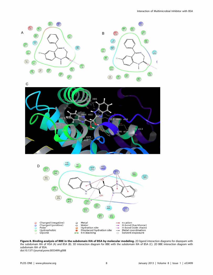
Figure 8. Binding analysis of BBE in the subdomain IIIA of BSA by molecular modeling. 2D ligand interaction diagrams for diazepam withthe subdomain IIIA of HSA (A) and BSA (B). 3D interaction diagram for BBE with the subdomain IIIA of BSA (C). 2D BBE interaction diagram withsubdomain IIIA of BSA.doi:10.1371/journal.pone.0053499.g008
Interaction of Multimicrobial Inhibitor with BSA
PLOS ONE | www.plosone.org 8 January 2013 | Volume 8 | Issue 1 | e53499

performed in the absence and presence of different BBE/BSA
molar ratios. Figure 6B shows the near-UV CD spectra of BSA in
the absence and presence of 1:1 and 2:1 BBE/BSA molar ratios.
In the absence of the ligand two visible minima around 262 and
268 nm are observed, which are similar to the previous reports
[43]. The ellipticity 268/262 ratios of the protein in the absence
and presence of the ligand are observed to be 0.93 and 1.16, 1.31,
respectively (Table 2). On addition of BBE, the increase in the
ellipticity ratio is observed which is due to the appearance of a
more prominent minimum at 268 nm and a maximum at 262 nm.
These changes in the shape of near-UV CD spectrum in the
presence of ligand indicate alteration in the tertiary structure of
BSA upon binding to the drug.
Far-UV circular dichroism studies. The changes in the
secondary structure contents of BSA on BBE binding were
monitored by far-UV CD measurements between 200 and
250 nm Figure 6C shows the far-UV CD spectra of BSA in the
absence and presence of BBE/BSA molar ratios of 1:1 and 2:1. In
the absence of the ligand, BSA shows two negative minima at 208
and 222 nm, characteristic features of helical proteins. The
decrease in the magnitude of mean residue ellipticities [h] at 222
and 208 nm are indicative of loss of helical structures of BSA in
the presence of the ligand. The secondary structural contents (a-
helices and b-strands) as calculated by analysis of the CD spectra
using K2D3 deconvolution software [21] are shown in Table 2.
The values of secondary structures obtained for free BSA are in
good agreement with the values reported in the literature [44–46].
Analysis of the CD spectra using K2D3 deconvolution software
[21] shows a decrease in the alpha-helical contents of BSA without
inducing any other regular secondary structure (b-strand) (Table 2).
The effect of BBE on Unfolding Behavior and Stability ofBSA
The effect of a molecule on the unfolding behavior of serum
albumin is crucial for the molecule to be qualified as a drug. The
effects of BBE binding on the equilibrium unfolding of BSA was
investigated by urea-induced denaturation as monitored by
measurements of fluorescence intensity at 343 nm after exciting
the protein at 280 nm. Figure 7A shows the urea-induced
denaturation transitions of BSA in the absence and presence of
BBE/BSA molar ratio of 2:1. As reported earlier [40] in the
absence of the ligand, unfolding starts ,2.4 M urea and follows a
two-step, three state transition with the accumulation of an
equilibrium intermediate state (I) around 4.8–5.2 M urea concen-
trations. The addition of the ligand at a BBE/BSA molar ratio of
2:1 shifts the native-to-intermediate state (N=I) transition towards
higher urea concentrations without significantly affecting the
intermediate- to-unfolded state (I=U) transition (Figure 7A and
7B). The free energy of stabilization (DDGH2
O) in the presence of
BBE was found to be 2.0660.29 and 1.9960.27 kJ mol21 as
determined by linear and non-linear fittings of intrinsic fluores-
cence data, respectively (Figure 7B and 7C). The stabilization
occurrs in the first transition, which corresponds to the formation
of intermediate state (I), whereas the second transition, corre-
sponding to the unfolding of I state to U state remains unaffected
(Table 3). Since N to I state transition is characterized by
unfolding of domain III, stabilization of the N = I transition also
indicates the binding of the ligand in domain III of BSA [40,41].
Computational Study on the Binding of BBE withSubdomain IIIA of BSA
Our experimental studies showed that BBE bound to the
diazepam binding site on sub domain IIIA of BSA. Here we
further study it by a computational method to strengthen our
findings. Since no ligand is bound to the X-ray crystal structure of
BSA (PDB code 3V03), the interaction site was determined based
on the X-ray crystal structure of HSA bound to diazepam (PDB
code 2BXF) [25,26]. The two proteins HSA and BSA have high
sequence similarity with 100% match in the subdomain IIIA. The
two proteins were structurally aligned to a single coordinate frame,
subsequent to which the atom coordinates of diazepam were
extracted from HSA into the coordinate frame of BSA. The 10 A3
grid was built with diazepam at the grid center for the docking.
The protocol was validated by the efficient docking of diazepam in
subdomains IIIA pocket of HSA and BSA having less than 1.0 A
root mean square deviation (RMSD) of the docking poses to that
of the bound structure. Identical interactions were observed for
diazepam with the amino acids in the IIIA subdomain of HSA and
BSA, especially the hydrogen bonding with Tyr411 and Tyr 410
with the respective proteins (Figure 8a and 8b).
Binding Analysis of BBE in the Subdomain IIIA of BSAThe ligand shows a snug fit in between the three helices of the
subdomain IIIA of BSA as depicted in Figure 8C. Being a
symmetric molecule and based on the pocket made by the three
helices, a single orientation was identified for BBE in the cavity.
The phenyl ring of one benzimidazole was surrounded by the
hydrophobic residues namely, Ile387, Cys391, Phe402, Val432
and Cys437 while the other phenyl on the second benzimidazole
was exposed to solvent. Hydrogen bond interactions were seen
between hydroxyl groups of ethane-1, 2-diol fragment with
Tyr410 and benzimidazole sp2 nitrogen atom with Ser488. The
2D ligand interaction diagram (Figure 8D) also revealed p-cation
interactions of the imidazole rings of benzimidazole with amino
acid Arg409.
The association constant (Ka) obtained from modeling
(104 M21) is very much consistent with the Ka value (104 M21)
calculated from fluorescence quenching data (Table 1, Table 4).
Moreover, the GlideScore XP for BBE shows a lower affinity
(25.01 kJ mol21) to BSA than the score computed for diazepam
(34.51 kJ mol21) (Table 4). These docking scores support the
experimental ligand binding and displacement studies wherein
BBE was found inept to displace diazepam from the subdomain
IIIA.
Discussion
Serum albumin is the main carrier of drugs in the mammalian
circulatory system including for humans. It contains many binding
sites for a large variety of substances, two of which, Sudlow site I
located in domain IIA and Sudlow site II located in domain IIIA
are the main sites for drug binding [11,12]. The drug-serum
albumin binding parameters and drug induced conformational
changes determine the distribution, efficacy, clearance and brain
penetration of the drug [11,14,15]. Therefore, the drug-protein
interaction study is an important part of the drug design.
Table 4. Docking score (GlideScore XP) as calculated for thebest poses for diazepam and BBE.
Protein Ligand GlideScore (kJ mol21
HSA (subdomain IIIA) Diazepam 233.77
BSA (subdomain IIIA) Diazepam 234.51
BSA (subdomain IIIA) BBE 225.04
doi:10.1371/journal.pone.0053499.t004
Interaction of Multimicrobial Inhibitor with BSA
PLOS ONE | www.plosone.org 9 January 2013 | Volume 8 | Issue 1 | e53499
In this study, we have investigated the binding parameters,
thermodynamic parameters, structural changes and conforma-
tional stability associated with the BBE interaction with bovine
serum albumin (BSA). The binding data with urea-unfolded BSA
and in the presence of a site-specific marker suggest one binding
site for the molecule, which is located in subdomain IIIA of BSA.
On the basis of molecular modeling, large negative values of DH,
DG and small positive values of DS for BBE-BSA complex
formation, we proposed that BBE readily bound BSA mainly
through hydrophilic interaction such as hydrogen bonding and
hydrophobic interactions in subdomain IIIA. The computational
studies indicated hydrogen bonding between hydroxyl groups of
BBE with Tyr410 and benzimidazole nitrogen atom with Ser488
and hydrophobic interaction between phenyl ring of one
benzimidazole of the ligand and hydrophobic residues namely,
Ile387, Cys391, Phe402, Val432 and Cys437.
Intrinsic fluorescence, near-UV and far-UV CD data suggest
that the interaction of BBE with BSA causes small but significant
alterations in both tertiary and secondary structure of the protein.
The study of small molecules-induced conformational changes is
crucial for structure-based rational drug design. Small molecules
binding to serum albumin are known to cause physiologically
relevant and as well as harmful conformational changes. For
example, the binding of long chain fatty acid to serum albumin
cause alteration in the microenvironments of the lone free
sulfhydryl group (Cys-34) and regulate the radical-trapping
antioxidant activity [47]. Some small-molecule-induced structural
changes modulate the b-lactamase activity of the albumin and may
increase the drug resistance through a non-microbial process [48].
The binding of small molecules to serum albumin can also alter
the conformation of binding sites for other drugs and thus
modulate the ADMET (absorption, distribution, metabolism,
excretion and toxicity), which is a major challenge in drug design.
Urea-induced unfolding of BSA-BBE complex indicated that
the native state is stabilized by ,2kJ mol21 relative to intermediate
state and the I=U transition is unaffected. Binding to the native
state is generally known to increase the stability of proteins [49].
Therefore, these results suggested that the molecule bound to the
native conformation of domain III. These findings and confor-
mational alterations indicate that binding of the BBE to domain
IIIA may be inducing conformational changes in domain I and/or
domain II, which brought the tryptophan residues to a more polar
environment. Similar ligand-induced propagation of conforma-
tional changes between domains has also been reported in other
protein [50].
This work yields information about the mechanism of interac-
tion of synthetic multi-microbial inhibitor, 1,2-bis(2-benzimidazo-
lyl)-1,2-ethanediol (BBE) with bovine serum albumin. We
observed that its binding mechanism is similar to the drugs, which
bind to domain IIIA of the serum albumin. The BBE molecule,
which is a tridentate ligand, can be prepared by simple Phillips
reaction and offers many sites for derivatization. Therefore, this
molecule or its derivatives could be a good drug candidate for the
treatment of multimicrobial infections of animals and/or human.
Acknowledgments
The authors are highly thankful to Prof. Lisa J Lapidus (Michigan State
University, USA) for her suggestions, thorough editing and proofreading of
the manuscript. UM-DAE Centre for Excellence in Basic Sciences and
University of Mumbai are gratefully acknowledged for instrumentation
facilities. Authors also acknowledge the Department of Pharmaceutical
Chemistry, Bombay College of Pharmacy for the computational studies.
Author Contributions
Conceived and designed the experiments: BA NK. Performed the
experiments: BA NK AP MB. Analyzed the data: BA NK RRSP.
Contributed reagents/materials/analysis tools: NA AP RRSP. Wrote the
paper: BA NK NA. Revised manuscript critically: NK NA AP MB RRSP.
References
1. Thomas GL, Johannes CW (2011) Natural product-like synthetic libraries. CurrOpin Chem Biol 15: 516–522.
2. de Azevedo WF Jr, Dias R (2008) Experimental approaches to evaluate the
thermodynamics of protein-drug interactions. Curr Drug Targets 9: 1071–1076.
3. Seeliger D, de Groot BL (2010) Conformational transitions upon ligand binding:holo-structure prediction from apo conformations. PLoS Comput Biol 6:
e1000634.
4. Zhang R, Monsma F (2009) The importance of drug-target residence time. CurrOpin Drug Discov Devel 12: 488–496.
5. Copeland RA, Pompliano DL, Meek TD (2006) Drug-target residence time and
its implications for lead optimization. Nat Rev Drug Discov 5: 730–739.
6. O’Sullivan DG, Wallis AK (1963) New benzimidazole derivatives with powerfulprotective action on tissue-culture cells infected with types 1, 2, and 3 polio virus.
Nature, 198: 1270.
7. Akihama S, Okude M, Sato K, Iwabuchi S (1968) Inhibitory effect of 1,2-bis(2-benzimidazolyl)-1,2-ethanediol derivatives on poliovirus. Nature, 217: 562–563.
8. Shipkowitz NL, Bower RR, Schleicher JB, Aquino F, Appell RN, et al. (1972)
Antiviral activity of a bis-benzimidazole against experimental rhinoviruinfections in chimpanzees. Appl Microbiol 23: 117–122.
9. Tavman A, Birteksoz S, Otuk G (2005) Antimicrobial activity of 1,2-bis-[2-(5-R)-
1H benzimidazolyl]- 1,2-ethanediols, 1,4-bis-[2-(5-R)-1H-benzimidazolyl]-1,2,3,4 butanetetraols and their FeIII, CuII, and AgI complexes. Folia Microbiol
(Praha) 50: 467–472.
10. Isele K, Broughton V, Matthews CJ, Williams AF, Bernardinelli G, et al. (2002)1,2-Bis(2- benzimidazolyl)-1,2-ethanediol, a chiral, tridentate, facially coordi-
nating ligand. J Chem Soc Dalton Trans 3899–3905.
11. Peters T Jr (1996) All about albumin: biochemistry, genetics and medical
application. Academic Press, Inc.: New York.
12. Sudlow G, Birkett DJ, Wade DN (1975) The Characterization of Two Specific
Drug Binding Sites on Human Serum Albumin. Mol Pharmacol 11: 824–832.
13. Sjoholm I, Ekman B, Kober A, Ljungstedt-Pahlman I, Seiving B, et al. (1979)Binding of drugs to human serum albumins, Mol Pharmacol 16: 767–777.
14. Ryan CW, Vogelzang NJ, Vokes EE, Kindler HL, Undevia SD, et al. (2004)
Dose-ranging study of the safety and pharmacokinetics of atrasentan in patients
with refractory malignancies. Clin Cancer Res 10: 4406–4411.
15. Schmidt S, Rock K, Sahre M, Burkhardt O, Brunner M, et al. (2008) Effect of
protein binding on the pharmacological activity of highly bound antibiotics.
Antimicrob Agents Chemother 52: 3994–4000.
16. Ahmad B, Lapidus LJ (2012) Curcumin prevents aggregation in a-synuclein by
increasing reconfiguration rate. J Biol Chem 287: 9193–9199.
17. Hung HC, Liu CL, Hsu JT, Horng JT, Fang MY, et al. (2012) Development of
an anti-influenza drug screening assay targeting nucleoproteins with tryptophan
fluorescence quenching. Anal Chem 84: 6391–6399.
18. Ahmad B, Parveen S, Khan RH (2006) Effect of albumin conformation on the
binding of ciprofloxacin to human serum albumin: a novel approach directly
assigning binding site. Biomacromolecules 7: 1350–1356.
19. Han XL, Tian FF, Ge YS, Jiang FL, Lai L, et al. (2012) Spectroscopic, structural
and thermodynamic properties of chlorpyrifos bound to serum albumin: A
comparative study between BSA and HSA. J Photochem Photobiol B 109: 1–11.
20. Peters T Jr (1985) Serum albumin. Adv Protein Chem 37: 161–245.
21. Louis-Jeune C, Andrade-Navarro MA, Perez-Iratxeta C (2012). Prediction of
protein secondary structure from circular dichroism using theoretically derived
spectra. Proteins Structure, Function, and Bioinformatics 80: 374–381.
22. Tanford C (1968) Protein denaturation. Adv Protein Chem 23: 122–282.
23. Friesner RA, Banks JL, Murphy RB, Halgren TA, Klicic JJ, et al. (2004) Glide: a
new approach for rapid, accurate docking and scoring. 1. Method and
assessment of docking accuracy. J med chem 47: 1739–1749.
24. Halgren TA, Murphy RB, Friesner RA, Beard HS, Frye LL, et al. (2004) Glide:
a new approach for rapid, accurate docking and scoring. 2. Enrichment factors
in database screening. J med chem 47: 1750–1759.
25. Ghuman J, Zunszain PA, Petitpas I, Bhattacharya AA, Otagiri M, et al. (2005)
Structural basis of the drug-binding specificity of human serum albumin. J Mol
Biol 353: 38–52.
26. Majorek KA, Porebski PJ, Dayal A, Zimmerman MD, Jablonska K, et al. (2012)
Structural and immunologic characterization of bovine, horse, and rabbit serum
albumins. Mol Immunol 52: 174–182.
27. Friesner RA, Murphy RB, Repasky MP, Frye LL, Greenwood JR, et al. (2006)
Extra precision glide: docking and scoring incorporating a model of hydrophobic
enclosure for protein-ligand complexes. J Med Chem 49: 6177–6196.
Interaction of Multimicrobial Inhibitor with BSA
PLOS ONE | www.plosone.org 10 January 2013 | Volume 8 | Issue 1 | e53499

28. Kume S, Lee YH, Miyamoto Y, Fukada H, Goto Y, et al. (2012) Systematic
interaction analysis of human lipocalin-type prostaglandin D synthase with smalllipophilic ligands. Biochem J 446: 279–89.
29. Eftink MR, Ghiron CA (1981) Fluorescence quenching studies with proteins.
Anal Biochem 114: 199–227.30. Lakowicz JR (2004) Principles of fluorescence spectroscopy. 2nd Edition,
Springer, New York.31. Ware WR (1962) Oxygen quenching of fluorescence in solution: An
experimental study of the diffusion process. J Phys Chem 66: 4552458.
32. Coan KE, Shoichet BK (2008) Stoichiometry and physical chemistry ofpromiscuous aggregate-based inhibitors. J Am Chem Soc 130: 9606–9612.
33. Owen SC, Doak AK, Wassam P, Shoichet MS, Shoichet BK (2012) Colloidalaggregation affects the efficacy of anticancer drugs in cell culture. ACS Chem
Biol 7: 1429–1435.34. Kang J, Liu Y, Xie MX, Li S, Jiang M, et al. (2004) Interactions of human serum
albumin with chlorogenic acid and ferulic acid. Biochim Biophys Acta. 1674:
205–214.35. Agudelo D, Bourassa P, Bruneau J, Berube G, Asselin E, et al. (2012) Probing
the Binding Sites of Antibiotic Drugs Doxorubicin and N-(trifluoroacetyl)Dox-orubicin with Human and Bovine Serum Albumins. PLoS One 7: e43814.
36. Wallevik K (1973) Reversible denaturation of human serum albumin by pH,
temperature and guanidine hydrochloride. J Biol Chem 245: 265022655.37. Tummino PJ, Copeland RA (2008) Residence time of receptor-ligand complexes
and its effect on biological function. Biochemistry 47: 5481–5492.38. Copeland RA (2010) The dynamics of drug–target interactions: drug–target
residence time and its impact of efficacy and safety. Expert Opin Drug Discov 5:305–310.
39. Creighton TE (1993) Proteins:structure and molecular properties, W.H.
Freeman and Company, New York.40. Tayyab S, Ahmad B, Kumar Y, Khan MM (2002) Salt-induced refolding in
different domains of partially folded bovine serum albumin. Int J Biol Macromol30: 17–22.
41. Ahmad B, Khan MK, Haq SK, Khan RH (2004) Intermediate formation at
lower urea concentration in ‘B’ isomer of human serum albumin: a case study
using domain specific ligands. Biochem Biophys Res Commun 314: 166–173.
42. Bagatolli LA, Kivatinitz SC, Fidelio GD (1996) Interaction of small ligands with
human serum albumin IIIA subdomain. How to determine the affinity constant
using an easy steady state fluorescent method. J Pharm Sci 85: 1131–1132.
43. Patel AB, Srivastava S, Phadke RS (1999) Interaction of 7-hydroxy-8-
(phenylazo)1,3 naphthalenedisulfonate with bovine plasma albumin. Spectro-
scopic studies. J Biol Chem 274: 21755–62.
44. Dubeau S, Bourassa P, Thomas TJ, Tajmir-Riahi HA (2010) Biogenic and
synthetic plyamines bind bovine serum albumin. Biomacromolecules 11: 1507–
1515.
45. Charbonneau DM, Tajmir-Riahi HA (2010) Study on the interaction of cationic
lipids with bovine serum albumin. J Phys Chem B 114: 1148–1155.
46. Bourassa P, Kanakis DC, Tarantilis PA, Polissiou MG, Tajmir-Riahi HA (2010)
Resveratrol, genistein and curcumin bind human serum albumin. J Phys Chem B
114: 3348–3354.
47. Narazaki R, Maruyama T, Otagiri M (1997) Probing the cysteine 34 residue in
human serum albumin using fluorescence techniques. Biochim Biophys Acta
1338: 275–281.
48. Ahmad E, Rabbani G, Zaidi N, Ahmad B, Khan RH (2012) Pollutant-induced
modulation in conformation and b-lactamase activity of human serum albumin.
PLoS One 7: e38372.
49. Waldron TT, Murphy KP (2003) Stabilization of proteins by ligand binding:
application to drug screening and determination of unfolding energetics.
Biochemistry 42: 5058–5064.
50. Strunk JJ, Gregor I, Becker Y, Li Z, Gavutis M, et al. (2008) Ligand binding
induces a conformational change in ifnar1 that is propagated to its membrane-
proximal domain. J Mol Biol 377: 725–739.
Interaction of Multimicrobial Inhibitor with BSA
PLOS ONE | www.plosone.org 11 January 2013 | Volume 8 | Issue 1 | e53499
![Voltammetric monitoring of Cd (II) by nano-TiO2 modified carbon paste electrode sensitized using 1,2-bis-[o-aminophenyl thio] ethane as a new ion receptor](https://static.fdokumen.com/doc/165x107/63328e4ef008040551048773/voltammetric-monitoring-of-cd-ii-by-nano-tio2-modified-carbon-paste-electrode.jpg)


![Bis[(1 S *,2 S *)- trans -1,2-bis(diphenylphosphinoxy)cyclohexane]chloridoruthenium(II) trifluoromethanesulfonate dichloromethane disolvate](https://static.fdokumen.com/doc/165x107/63360a7bcd4bf2402c0b5520/bis1-s-2-s-trans-12-bisdiphenylphosphinoxycyclohexanechloridorutheniumii.jpg)


![Bisindenoisoquinoline bis-1,3-{(5,6-dihydro-5,11-diketo-11H-indeno[1,2-c]isoquinoline)-6-propylamino}propane bis(trifluoroacetate) (NSC 727357), a DNA intercalator and topoisomerase](https://static.fdokumen.com/doc/165x107/63195970e9c87e0c0910145e/bisindenoisoquinoline-bis-13-56-dihydro-511-diketo-11h-indeno12-cisoquinoline-6-propylaminopropane.jpg)

![Reactions of bis[1,2-bis(dialkylphosphino)ethane]-(dihydrogen)hydridoiron(1+) with alkynes](https://static.fdokumen.com/doc/165x107/63146d10c32ab5e46f0ce1ad/reactions-of-bis12-bisdialkylphosphinoethane-dihydrogenhydridoiron1-with.jpg)


