Seasonal variation of ascorbic acid and nitrate levels in ...
Upload
khangminh22Category
view
3download
0
Citation: Miazek, K.; Beton, K.;
Sliwinska, A.; Brozek-Płuska, B. The
Effect of β-Carotene, Tocopherols
and Ascorbic Acid as Anti-Oxidant
Molecules on Human and Animal In
Vitro/In Vivo Studies: A Review of
Research Design and Analytical
Techniques Used. Biomolecules 2022,
12, 1087. https://doi.org/10.3390/
biom12081087
Academic Editor: Vito Verardo
Received: 9 June 2022
Accepted: 2 August 2022
Published: 7 August 2022
Publisher’s Note: MDPI stays neutral
with regard to jurisdictional claims in
published maps and institutional affil-
iations.
Copyright: © 2022 by the authors.
Licensee MDPI, Basel, Switzerland.
This article is an open access article
distributed under the terms and
conditions of the Creative Commons
Attribution (CC BY) license (https://
creativecommons.org/licenses/by/
4.0/).
biomolecules
Review
The Effect of β-Carotene, Tocopherols and Ascorbic Acid asAnti-Oxidant Molecules on Human and Animal In Vitro/InVivo Studies: A Review of Research Design and AnalyticalTechniques UsedKrystian Miazek 1,*, Karolina Beton 1 , Agnieszka Sliwinska 2 and Beata Brozek-Płuska 1
1 Laboratory of Laser Molecular Spectroscopy, Institute of Applied Radiation Chemistry,Lodz University of Technology, Wroblewskiego 15, 93-590 Lodz, Poland
2 Department of Nucleic Acid Biochemistry, Medical University of Lodz, 251 Pomorska Str.,92-213 Lodz, Poland
* Correspondence: [email protected]
Abstract: Prolonged elevated oxidative stress (OS) possesses negative effect on cell structure andfunctioning, and is associated with the development of numerous disorders. Naturally occurredanti-oxidant compounds reduce the oxidative stress in living organisms. In this review, antioxidantproperties of β-carotene, tocopherols and ascorbic acid are presented based on in vitro, in vivo andpopulational studies. Firstly, environmental factors contributing to the OS occurrence and intracellularsources of Reactive Oxygen Species (ROS) generation, as well as ROS-mediated cellular structuredegradation, are introduced. Secondly, enzymatic and non-enzymatic mechanism of anti-oxidantdefence against OS development, is presented. Furthermore, ROS-preventing mechanisms andeffectiveness of β-carotene, tocopherols and ascorbic acid as anti-oxidants are summarized, based onstudies where different ROS-generating (oxidizing) agents are used. Oxidative stress biomarkers,as indicators on OS level and prevention by anti-oxidant supplementation, are presented with afocus on the methods (spectrophotometric, fluorometric, chromatographic, immuno-enzymatic) oftheir detection. Finally, the application of Raman spectroscopy and imaging as a tool for monitoringthe effect of anti-oxidant (β-carotene, ascorbic acid) on cell structure and metabolism, is proposed.Literature data gathered suggest that β-carotene, tocopherols and ascorbic acid possess potential tomitigate oxidative stress in various biological systems. Moreover, Raman spectroscopy and imagingcan be a valuable technique to study the effect of oxidative stress and anti-oxidant molecules incell studies.
Keywords: oxidative stress; reactive oxygen species; antioxidants; biomarkers; raman spectroscopyand imaging
1. Introduction
Oxidative stress (OS) is defined by the production and accumulation of Reactive Oxy-gen Species (ROS) in biological systems above the capacity of cells and tissues to neutralizeROS presence to a safe level [1]. Reactive Oxygen Species (ROS) are molecular oxygen(O2)-derived characterized by high reactivity towards other molecules. ROS comprise freeradicals such as superoxide anion radical (O2
•−) and hydroxyl radical (•OH), and nonradi-cal molecules such as singlet oxygen (1O2) and hydrogen peroxide (H2O2). Singlet oxygen(1O2) is the exited state of ground state triplet molecular oxygen (3O2), and is generated viaabsorption of energy sufficient to reverse the spin of one of its unpaired electrons what leadsto the formation of singlet state where two electrons possess opposite spin [2]. Superoxideanion radical (O2
•−) is generated by one-electron reduction of molecular oxygen (O2), whatresults in the production of a charged ionic species (O2
•−) possessing a single unpaired
Biomolecules 2022, 12, 1087. https://doi.org/10.3390/biom12081087 https://www.mdpi.com/journal/biomolecules
Biomolecules 2022, 12, 1087 2 of 45
electron and a negative charge [3]. Hydrogen peroxide (H2O2), a compound possessing anoxygen-oxygen single bond [4], is generated from dismutation of O2
•− [3]. H2O2 can bedecomposed via Fenton reaction to OH− and •OH with Fe2+ oxidation, or can react withO2•− to form •OH, OH− and O2 [2]. Hydroxyl radical (•OH) possesses a single unpaired
electron and is generated by oxidation of water (H2O) or hydroxide ions (OH−), or bydecomposition of hydrogen peroxide (H2O2) via Fenton reaction [5,6].
In living organisms, ROS are produced during indigenous metabolism to serve assignalling molecules, but can be also generated at high concentrations due to exposure to en-vironmental stress factors (radiation, pollution, pathogens, etc.) [7]. Generated ROS includesinglet oxygen (1O2), superoxide anion radical (O2
•−), hydrogen peroxide (H2O2) and hy-droxyl radical (•OH), and are characterized by different reactivity. 1O2 is a highly reactiveform of oxygen possessing two electrons with opposite spins, and oxidizing molecules viareaction with double bonds [8,9]. Although O2
•− is not a strong oxidant, it can undergodismutation to H2O2 that can decompose to form a •OH, one of the strongest oxidants.O2•− can also react with nitric oxide radical to form a peroxynitrite, also considered a
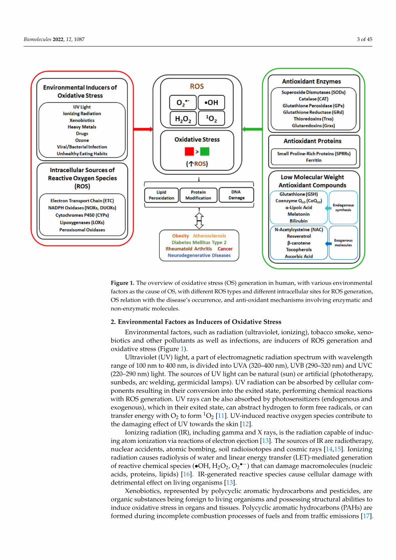
powerful oxidant [8]. The elevated production of ROS and oxidative stress cause detrimen-tal effect on organisms due to the damage of cellular macromolecules, with oxidation oflipids, proteins and nucleic acids. In human, the occurrence and development of manydiseases such as obesity, atherosclerosis, diabetes mellitus type 2 (DMT2), rheumatoidarthritis, cancer and neurodegenerative diseases are associated with oxidative stress [10].To cope with oxidative stress, organisms developed defence mechanisms (Figure 1) involv-ing anti-oxidant enzymes, as well as non-enzymatic molecules. The human enzymaticanti-oxidant mechanisms include enzymes such as superoxide dismutase (SOD), cata-lase (CAT), glutathione peroxidase (GPx), glutathione reductase (GRd) and thioredoxins(Trx), with glutathione (GSH) serving as electron donor and used in reactions catalysedby GPx. The non-enzymatic anti-oxidant system in human body is composed of anti-oxidant proteins, such as proline-rich proteins and ferritin, and low-weight anti-oxidantmolecules synthesized indigenously, such as coenzyme Q10, α-lipoic acid, melatonin andbilirubin. Moreover, addition of external low-weight anti-oxidant molecules, such as N-acetylcysteine, resveratrol, β-carotene, tocopherols and ascorbic acid, can strengthen thecapability of indigenous defence system against OS.
The mitigating effect of β-carotene, tocopherols and ascorbic acid against oxidativestress is presented in this article. β-carotene classified as provitamin A, ascorbic acidnamed as vitamin C and tocopherols possessing activity of vitamin E, are exogenousanti-oxidant molecules with ROS-scavenging properties that are considered useful in pre-venting oxidative stress in mammalian cells and organisms. In this review, anti-oxidantproperties of β-carotene, tocopherols and ascorbic acid to alleviate oxidative stress basedon in vitro, in vivo as well as populational studies, are described. Moreover, different ana-lytical techniques (spectrophotometric, fluorometric, chromatographic, immune-enzymatic,spectroscopic) used commonly to assess the OS presence and anti-oxidant properties oftested molecules, are compared and discussed.
Overall, this review provides a broad description regarding the effect of β-carotene,ascorbic acid and tocopherols as potential antioxidants to mitigate oxidative stress in humanand animal studies, in terms of research designs, experimental outcomes and analyticaltechniques applied.
Biomolecules 2022, 12, 1087 3 of 45
Figure 1. The overview of oxidative stress (OS) generation in human, with various environmentalfactors as the cause of OS, with different ROS types and different intracellular sites for ROS generation,OS relation with the disease’s occurrence, and anti-oxidant mechanisms involving enzymatic andnon-enzymatic molecules.
2. Environmental Factors as Inducers of Oxidative Stress
Environmental factors, such as radiation (ultraviolet, ionizing), tobacco smoke, xeno-biotics and other pollutants as well as infections, are inducers of ROS generation andoxidative stress (Figure 1).
Ultraviolet (UV) light, a part of electromagnetic radiation spectrum with wavelengthrange of 100 nm to 400 nm, is divided into UVA (320–400 nm), UVB (290–320 nm) and UVC(220–290 nm) light. The sources of UV light can be natural (sun) or artificial (phototherapy,sunbeds, arc welding, germicidal lamps). UV radiation can be absorbed by cellular com-ponents resulting in their conversion into the exited state, performing chemical reactionswith ROS generation. UV rays can be also absorbed by photosensitizers (endogenous andexogenous), which in their exited state, can abstract hydrogen to form free radicals, or cantransfer energy with O2 to form 1O2 [11]. UV-induced reactive oxygen species contribute tothe damaging effect of UV towards the skin [12].
Ionizing radiation (IR), including gamma and X rays, is the radiation capable of induc-ing atom ionization via reactions of electron ejection [13]. The sources of IR are radiotherapy,nuclear accidents, atomic bombing, soil radioisotopes and cosmic rays [14,15]. Ionizingradiation causes radiolysis of water and linear energy transfer (LET)-mediated generationof reactive chemical species (•OH, H2O2, O2
•−) that can damage macromolecules (nucleicacids, proteins, lipids) [16]. IR-generated reactive species cause cellular damage withdetrimental effect on living organisms [13].
Xenobiotics, represented by polycyclic aromatic hydrocarbons and pesticides, areorganic substances being foreign to living organisms and possessing structural abilities toinduce oxidative stress in organs and tissues. Polycyclic aromatic hydrocarbons (PAHs) areformed during incomplete combustion processes of fuels and from traffic emissions [17].
Biomolecules 2022, 12, 1087 4 of 45
PAHs undergo oxidation into phenolic intermediates which are converted via semiquinoneanion radicals into quinones, with generation of superoxide anion radicals and H2O2 [18,19].Pesticides (herbicides, insecticides, fungicides, etc.), used in agriculture for crop protection,can be found as contaminants in air, water and food. The mode of oxidative action ofpesticides, with herbicide paraquat as an example, is the induction of mitochondrial damageand the redox cycle involving quaternary ammonium nitrogen atoms and a bipirydyl ringin paraquat structure, what leads to production of ROS and paraquat radicals [20].
Heavy metals (HMs), such as Pb, Cd, Cr, Hg and As, are contaminants released fromindustry (effluents, waste product storage) to the surroundings (atmosphere, soil, water)and affecting human beings [21]. Accumulation of HMs ions in body induces oxidativestress within a range of mechanisms, including inhibition of antioxidant enzyme expression(by Cd), interaction with cofactors and/or disulphide bonds in antioxidant enzymes (e.g.,Pb, Hg), haemoglobin autooxidation (by Pb2+), binding to sulfhydryl groups (−SH) andreducing thiol pools (through Cd2+ or Hg2+), generating glutathione-thiyl radicals (viaCr(VI)), changing the oxidation state of HMs with formation of H2O2 and hydroxyl radicals(through Cr or As), affecting calcium homeostasis and stimulating oxidative enzymes (byHg), cytokine-mediated ROS generation (via Cd2+), and others [18,22,23]. Heavy metal-induced oxidative stress and macromolecules modification/degradation are the cause ofmany diseases, amongst which are cancer, cardiovascular disease, neurological disordersand chronic inflammation [24].
Drugs used for illness treatment are also the source of ROS generation in humanbody [25]. Anti-neoplastic agents, such as doxorubicin and cisplatin, are used for thetreatment of different types of cancer. Doxorubicin, a representative of anthracyclineantibiotics, generates ROS by undergoing mitochondrial reductase-mediated one-electronreduction to anthracycline semiquinone free radicals, that can react with O2 to form O2
•−
or H2O2. Doxorubicin can also interact with Fe3+ to form Fe2+-doxorubicin free radical,that can reduce oxygen. Oxidative stress induction is the mechanism of doxorubicincardiotoxicity [25]. Cisplatin, a platinum containing drug, was reported to increase the ROSlevel, via NAPDH oxidase or xanthine oxidase [25,26]. Oxidative stress induction in theproposed mechanism of cisplatin nephrotoxicity and ototoxicity [26,27].
Smoking, with cigarette smoke containing nicotine, ammonia, acrolein, phenols, ac-etaldehyde, polycyclic aromatic hydrocarbons, polyphenols, hydrogen cyanide, heavymetals, etc., is another source of ROS production and oxidative stress occurrence [28,29].The tobacco smoke contains gas phase and tar phase. The gas phase contains short-livedradicals, superoxide anion and nitric oxide, which react together to form highly reactive per-oxynitrite. The tar phase contains stable semiquinone radicals and iron (Fe2+). Semiquinoneradicals reduce O2 to O2
•−, that can dismutate into H2O2, which in turn can react with Fe2+
via Fenton reaction to form •OH [29,30]. Oxidative stress is considered as a crucial factorin the pathogenesis of smoking-related disorders, such as lung cancer, chronic obstructivepulmonary disease and atherosclerosis [31].
Ozone (O3) is a gaseous tropospheric pollutant, generated through reactions betweenintense solar radiation and pollutants (nitric oxides, sulphur oxides, carbon oxides, volatileorganic compounds) produced from combustion of fossil fuels [32,33]. Contact of O3 withbiological matrix results in creation of H2O2 and lipids oxidation products [34]. Ozoneexposure-induced oxidative stress is associated with neurodegenerative diseases [33,35].
The infections by viruses or bacteria can be also the cause of oxidative stress in humanbody. The body can be infected by viruses, such as DNA and RNA viruses, which enter andreplicate inside host cells [36,37]. ROS are generated during viral infection via inducingactivation of phagocytes [36] or via mediation of viral proteins expressed in host cellsto support viral life cycle [37]. Oxidative stress occurring during bacterial infection isdescribed for Helicobacter pylori, a gram negative, stomach-infecting bacterium. H. pyloriinfection results in oxidative stress via the immune and gastric epithelial cells producingROS in an attempt to kill the bacteria, and via bacterial virulence factors inducing epitheliumcellular responses and ROS generation [38].
Biomolecules 2022, 12, 1087 5 of 45
Unhealthy dietary patterns, based on overconsumption of high-carbohydrate andhigh-fat food, are associated with increased risk of overweight and obesity occurrence anddevelopment of diabetes mellitus type 2 (DMT2) and cardiovascular diseases. High-fat orhigh-carbohydrate diets results in the elevated influx of substrates into mitochondrial respi-ration and increased donation of electrons to electron transport chain, leading to the electronleakage at complex III and elevated superoxide (O2
•−) generation. [39]. NADPH oxidase(NOX), an enzyme converting molecular oxygen to its superoxide radical, is also involvedin nutrient-based ROS generation [40]. High-calorie diets may alter oxygen metabolismand are considered as one of the main factors leading to excessive ROS production [41].
3. Metabolic Pathways as Sources of ROS Generation in Cells
The main endogenous sources of ROS are enzymes of mitochondrial respiratory chainand nicotinamide adenine dinucleotide phosphate (NADPH) oxidase enzymes, whereasother sources of ROS are cytochromes P450, lipoxygenases and peroxisomal enzymes(Figure 1).
Electron transport chain (ETC), embedded within mitochondrial inner membranes, aresources of ROS production. The ETC includes transmembrane protein complexes NADH:ubiquinone oxidoreductase (Complex I), succinate dehydrogenase (Complex II), ubiquinol:cytochrome c oxidoreductase (Complex III) and cytochrome c oxidase (Complex IV), as wellas mobile electron transporters (ubiquinone, cytochrome c). ETC participates in the processof oxidative phosphorylation (OXPHOS), where O2 is converted to H2O and adenosinetriphosphate (ATP) is produced. In brief, electrons from mitochondrial matrix tricarboxylicacid (TCA) cycle are donated via NADH to Complex I and via FADH2 to Complex II,and are transferred from Complex I and II to ubiquinone (Q) that undergoes reductionto ubiquinol (QH2). Ubiquinol, carrying electrons, becomes re-oxidized via Q-cycle inComplex III, that passes electrons via cytochrome c (CytC) to Complex IV. Subsequently,complex IV transfers electrons to O2 as electron acceptor with generation of H2O. Theelectron flow (CI&CII→QH2→CIII→CytC→CIV→H2O) is coupled with the pumping ofprotons (H+) from mitochondrial matrix into the intermembrane space by complexes I, IIIand IV. The proton gradient formed across the mitochondrial inner membrane is used bycomplex V (ATP synthase) to produce (ATP). ETC can produce reactive oxygen species dueto the leakage of electrons from Complex I, II and III, resulting in one-electron reduction ofoxygen to O2
•−. O2•−, formed via Complex I and II, occur in the matrix, whereas Complex
III O2•− is released into both the matrix and intermembrane space. O2
•− is dismutated toH2O2 by SOD2 in the matrix and by SOD1 in the intermembrane space [7,42,43]. ComplexIV is not directly involved in ROS production, but its activity can affect the overall electronflow, with an impact on the electron leakage by previous complexes [44]. It is estimatedthat 0.2–2% of the electrons passing through the ETC leak out and interact with oxygen toproduce ROS [43].
The NADPH oxidases (NOX) are multi-subunit enzymes localized in membranes.NOXs transfer electrons across biological membranes to oxygen, thereby generating su-peroxide anion radical and or hydrogen peroxide. The mechanism of catalysis involvestransfer of two electrons from NADPH through FAD domain and two heme prostheticgroups in enzyme structure to O2 [45]. The NOX family comprises of seven isoforms(NOX1, NOX2, NOX3, NOX4, NOX5, DUOX1 and DUOX2) [46,47] expressed in differ-ent tissues throughout the body. NOX1 is highly expressed in the epithelial cells of thegastrointestinal tract [48], and is also present in prostate, uterus, vascular cells [49] anderythrocytes [50]. NOX2 is typically expressed in phagocytic cells, but also in endothelialcells, cardiomyocytes, hematopoietic stem cells and platelets [51]. NOX3 is expressedin the inner ear [48], and is also found in low abundance in the brain, lung and in fetaltissue [49]. NOX4 is commonly distributed in human tissues, and highly expressed inkidney, osteoclasts, fibroblasts and endothelial cells [47,52]. NOX5 is expressed in testis andlymphoid tissue, but also in placenta, uterus, stomach, skeletal muscle, hepatocytes, cellsof the cardiovascular system (cardiomyocytes, endothelial and vascular smooth muscle

Biomolecules 2022, 12, 1087 6 of 45
cells) [49] and erythrocytes [50]. DUOX1 and DUOX2 are highly expressed in thyroidgland [49]. O2
•− is generated by NOX1-3 and NOX5, while H2O2 is produced by DUOX1-2and NOX4 [53].
Cytochromes P450 (CYPs) are enzymes localized primarily in the endoplasmic reticu-lum and found mostly in liver and intestinal tissues [54]. CYPs are enzymes containinga heme prosthetic group in the form of iron protoporphyrin IX and participating in themetabolism of xenobiotics and endogenous compounds. CYPs possess monooxygenaseactivity and catalyse the incorporation of one atom of oxygen from O2 to organic substrateand reduction of the second oxygen atom to water. The catalytic mechanism of CYPs relieson redox reaction cycle of cysteine-bound iron atom involving substrate binding to ferriciron and reduction of Fe3+ to Fe2+, followed by O2 binding to iron and formation of complex(Fe2+-O2) that undergoes one-electron reduction (Fe2+-O2
−) and protonation to cleave O-Obond and release H2O, and the transfer of oxygen atom from FeO3+ complex to substrate,with the formation of mono-oxygenated substrate and regeneration of Fe3+ in CYP. DuringCYP monooxygenase cycle, reactive oxygen species can be produced either via releasing ofsuperoxide radical with its dismutation to H2O2 or direct releasing H2O2 [55,56].
Lipoxygenases (LOXs) are nonheme, iron-containing enzymes that catalyse oxygena-tion of arachidonic acid (AA) to hydroperoxyeicosatetraenoic acids (HPETEs), which canbe further converted to hydroxyeicosatetraenoic acids (HETEs), leukotrienes, lipoxins andhepoxilins [57,58]. In human, there are 5-LOX, 12-LOX, and 15-LOX, that catalyse the inser-tion of oxygen molecule at C-5, C-12 and C-15 of AA, respectively [59,60]. LOX-generatedAA metabolites can induce ROS generation in various cells via NOX upregulation [59].
Peroxisomes are single-membrane subcellular organelles, found in all eukaryoticcells, participating in the range of metabolic pathways such as α- and β-oxidation ofvery long fatty acids, the synthesis of bile acids, detoxification of glyoxylate and H2O2metabolism [61,62]. Peroxisomal enzymes, such as xanthine oxidase and acyl-CoA oxidases,are involved in ROS generation [63]. Xanthine oxidase (XO) participates in the catabolismof purine nucleic acids [64]. XO catalyses the oxidation of hypoxanthine and xanthine withgeneration of superoxide anion or hydrogen peroxide, respectively, via the monovalent anddivalent electron transfer to O2 [65]. Acyl-CoA oxidases are enzymes involved in the firststep of β-oxidation of different substances, such as CoA-esters of very long-chain fatty acids,branched-chain fatty acids and C27-bile acid intermediates [66]. In human peroxisomes,straight-chain acyl-CoA oxidase, branched-chain acyl-CoA oxidase and pristanoyl-CoAoxidase, are available. Acyl-CoA oxidases catalyse dehydrogenation of acyl-CoA esters,with the Flavin Adenine Dinucleotide (FAD)-mediated transfer of the protons from theβ-carbon bond of an acyl-CoA, resulting in generation of trans-2-enoyl-CoA esters andFADH2. Subsequently, FADH2 is regenerated to FAD and hydrogen atoms are transferredto O2 to form H2O2 [67,68].
4. Effect of ROS on Cellular Macromolecules
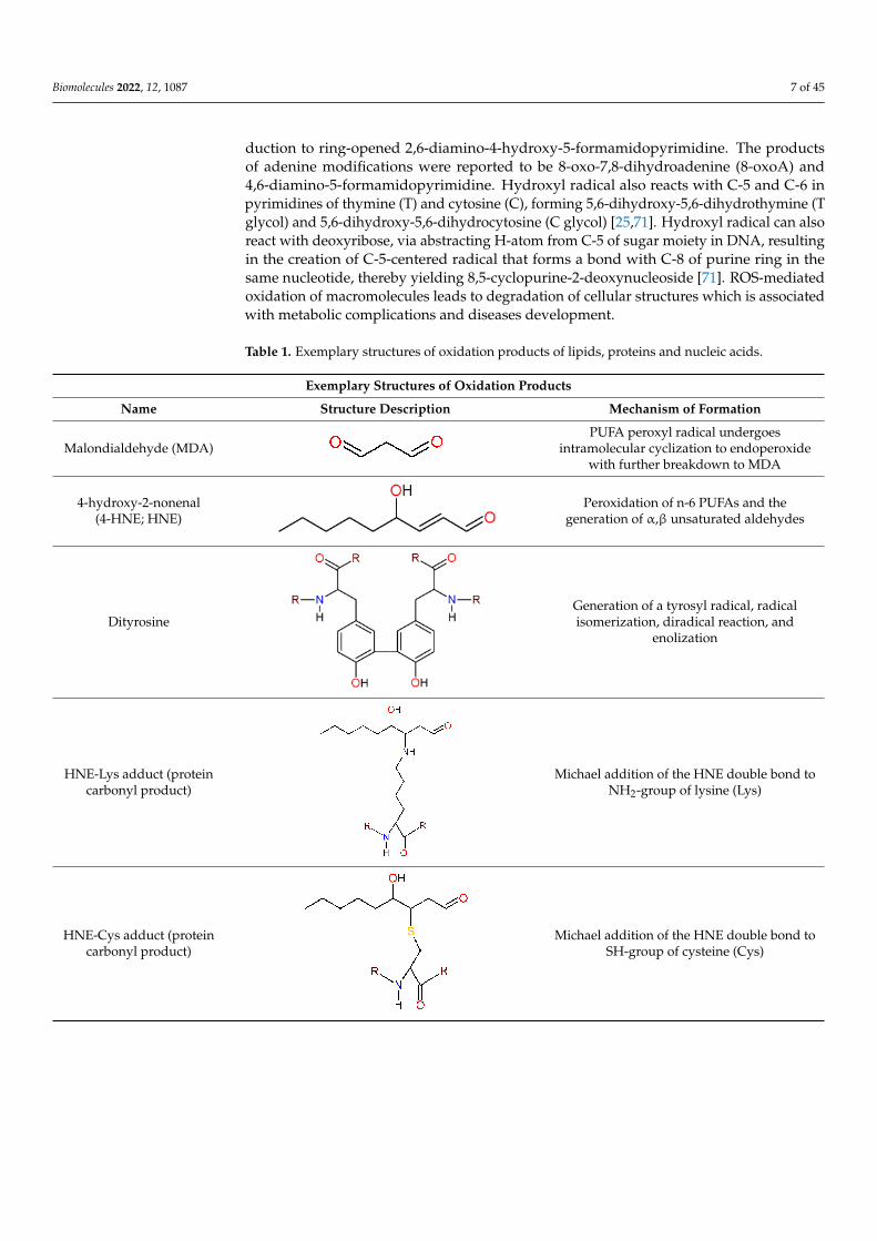
Reactive oxygen species can react with lipids, proteins and nucleic acids (Table 1),the primal macromolecules in cellular structures. ROS cause peroxidation of polyunsat-urated fatty acids (PUFAs) with the formation of lipid hydroperoxides, that undergo thecleavage to dialdehydes, such as malondialdehyde (MDA) and glyoxal, and a range ofunsaturated aldehydes including 4-hydroxynonenal (4-HNE), crotonaldehyde, 2-propenal(acrolein) and 2-hexenal [69]. ROS can react with amino-acids in protein structure, causingconversion of phenylalanine to o- or m-tyrosine, tyrosine to di-tyrosine, tryptophan toN-formylkynurenine, leucine to 4- or 5-hydroxyleucine, valine to 3- or 4-hydroxyvaline, aswell as oxidation of thiol groups in cysteine and methionine. Aldehydes (e.g., 4-HNE), gen-erated from PUFAs peroxidation, can cause the carbonylation of proteins via addition withS atom of cysteine, N in the imidizole of histidine and N in the amine of lysine [69,70]. ROS,such as hydroxyl radical, can also react with DNA bases (G, T, C, A) and sugar moieties. Hy-droxyl radical reacts with C-8 of guanine (G) to generate an 8-hydroxy-7,8-dihydroguanylradical, that can undergo the oxidation to 8-oxo-7,8-dihydroguanine (8-oxoG), or the re-
Biomolecules 2022, 12, 1087 7 of 45
duction to ring-opened 2,6-diamino-4-hydroxy-5-formamidopyrimidine. The productsof adenine modifications were reported to be 8-oxo-7,8-dihydroadenine (8-oxoA) and4,6-diamino-5-formamidopyrimidine. Hydroxyl radical also reacts with C-5 and C-6 inpyrimidines of thymine (T) and cytosine (C), forming 5,6-dihydroxy-5,6-dihydrothymine (Tglycol) and 5,6-dihydroxy-5,6-dihydrocytosine (C glycol) [25,71]. Hydroxyl radical can alsoreact with deoxyribose, via abstracting H-atom from C-5 of sugar moiety in DNA, resultingin the creation of C-5-centered radical that forms a bond with C-8 of purine ring in thesame nucleotide, thereby yielding 8,5-cyclopurine-2-deoxynucleoside [71]. ROS-mediatedoxidation of macromolecules leads to degradation of cellular structures which is associatedwith metabolic complications and diseases development.
Table 1. Exemplary structures of oxidation products of lipids, proteins and nucleic acids.
Exemplary Structures of Oxidation Products
Name Structure Description Mechanism of Formation
Malondialdehyde (MDA)PUFA peroxyl radical undergoes
intramolecular cyclization to endoperoxidewith further breakdown to MDA
4-hydroxy-2-nonenal(4-HNE; HNE)
Peroxidation of n-6 PUFAs and thegeneration of α,β unsaturated aldehydes
DityrosineGeneration of a tyrosyl radical, radicalisomerization, diradical reaction, and
enolization
HNE-Lys adduct (proteincarbonyl product)
Michael addition of the HNE double bond toNH2-group of lysine (Lys)
HNE-Cys adduct (proteincarbonyl product)
Michael addition of the HNE double bond toSH-group of cysteine (Cys)

Biomolecules 2022, 12, 1087 8 of 45
Table 1. Cont.
Exemplary Structures of Oxidation Products
Name Structure Description Mechanism of Formation
HNE-His adduct (proteincarbonyl product)
Michael addition of the HNE double bond toNH in an imidazole of histidine (His)
8-oxo-2′-deoxyguanosinenucleotide
Reaction between C-8 of guanine (G) andhydroxyl radical (•OH)
8-oxo-2′-deoxyadenosinenucleotide
Reaction between C-8 of adenine (A) andhydroxyl radical (•OH)
5,6-dihydroxy-5,6-dihydrothymidine
nucleotide
Reaction of hydroxyl radical (•OH) with C-5and C-6 of thymine (T)
5,6-dihydroxy-5,6-dihydrocytidine
nucleotide
Reaction of hydroxyl radical (•OH) with C-5and C-6 of cytosine (C)

Biomolecules 2022, 12, 1087 9 of 45
5. Oxidative Stress and Diseases Development
Oxidative stress is involved in the occurrence and physiopathology of chronic diseases,such as obesity, atherosclerosis, diabetes mellitus type 2 (DMT2), rheumatoid arthritis,cancer and neurodegenerative diseases [10].
Obesity, characterized by an increase in body weight resulting in excessive fat accumu-lation, represents a public health problem with increasing worldwide prevalence [72,73].Oxidative stress performs a role in the pathogenesis of obesity by stimulating the depositionof adipose tissue and altering food intake [74]. Oxidative stress is also induced by obesity,as the excess of free fatty acids (FFA) leads to increased FFA oxidation and mitochondrialROS overproduction. Moreover, the increased release of FA from over-accumulated fatcan result in NOX activation and progressed ROS generation [40]. In obesity, the num-ber and size of adipocytes are increased, and secretion of pro-inflammatory moleculesis promoted [73]. Oxidative stress occurring in obesity is linked with inflammation andcan contribute to the obesity-associated development of diseases, such as atherosclerosis,diabetes and cancer [40].
Atherosclerosis is a chronic inflammatory condition, characterized by gradual accu-mulation of plaques, composed of fibrous cap, lipid-rich core and calcium, within theartery wall [75]. The rupture of atherosclerotic plaques leads to thrombosis which is thereason of myocardial infarction or stroke [76]. The origin of atherosclerosis is ascribed tohypertension, hypercholesterolaemia, smoking or hyperglycemia that cause the damage ofendothelium and the entry of cholesterol-carrying low-density lipoproteins (LDLs) fromthe blood stream into the epithelium intima [77]. The endothelial cells, smooth muscle cells(SMCs) and macrophages are the source of oxidative stress, that cause the oxidation ofLDL particles [78]. Oxidized LDL (Ox-LDL) particles are taken up by macrophages, whichare converted into foam cells. The death of foam cells results in the accumulation of cellsdebris and lipids, and release of proinflammatory cytokines that cause SMCs migrationand proliferation. The plaque is progressively formed by agglomerating calcium deposits,SMCs, collagen and foam cells [75,77,79].
Diabetes mellitus type 2 (DMT2) is a disease characterized by tissue resistance to in-sulin, hyperglycemia and decreased secretion of insulin by pancreatic β-cells [80]. In DMT2,the occurrence of OS is ascribed to frequent hyperglycemia, mitochondrial dysfunctionand endoplasmic reticulum (ER) stress in β-cells [81], and OS-induced complications ofdiabetes may include macrovascular (coronary heart diseases, stroke) and microvascular(neuropathy, retinopathy, nephropathy) complications [80].
Rheumatoid arthritis (RA) is an autoimmune disease characterized by joint destruction.Oxidative stress is involved in the pathogenesis of RA, by stimulating inflammation andbeing stimulated by inflammation, what results in the establishment of synovitis, whichcauses cartilage and bone damage [82].
Cancer is a disease characterized by transformation of normal cells into malignantcells that proliferate in an uncontrolled manner and invade normal tissues and organs,eventually spreading throughout the body. Cancer is a major disease and the secondleading cause of mortality worldwide, with more than 277 different types of cancer diseasesaffecting different parts of body (colon, breast, lung, liver, prostate, brain, skin, bladder,renal, stomach etc.) [83–86]. Oxidative damage of DNA leads to disruption of genomefunction, distribution of mutation, selective clonal expansion of the mutated cell and furthercancer progression [87,88]. Oxidative stress may be an initiating factor in carcinogenesisand can be also the consequence of cancer development [89].
Neurodegenerative diseases, characterized by progressive dysfunction of neural cellsand losses of neurons, include Alzheimer’s disease, Parkinson’s disease and amyotrophiclateral sclerosis [90]. Oxidative stress is one of factors involved in the pathogenesis ofneurodegenerative disorders [91]. Neural microenvironment is susceptible to oxidativestress due to high oxygen demand, the abundant presence of redox-active metals, highlevel of cellular membrane PUFAs and low levels of GSH in the brain [92].
Biomolecules 2022, 12, 1087 10 of 45
6. Anti-Oxidant Mechanisms as a Protection against Oxidative-Stress
Cells possess antioxidant mechanisms, such as enzymes and other proteins, as wellas endogenous and exogenous low molecular weight anti-oxidant molecules, to protectcellular structures from oxidative stress and damage (Figure 1).
6.1. Superoxide Dismutase (SOD) and Catalase (CAT)
Superoxide dismutase (SOD) is a metalloenzyme that prevents intracellular O2•−
accumulation by catalysing dismutation of two molecules of O2•− to O2 and H2O2. The
mechanism of SOD catalytic action is based on the redox cycle of metal ion in the active site,involving metal reduction and oxidation of first O2
•− to O2, followed by metal oxidationand reduction of a second O2
•− to H2O2. Three isoforms of SOD: Cu,Zn-SOD (SOD1)homodimer distributed in the cytosol and the mitochondrial intermembrane, Mn-SOD(SOD2) homotetramer located in the mitochondrial matrix and inner membrane, and Cu,Zn-SOD (SOD3) homotetramer anchored to the extracellular matrix, are known to be presentin human [93,94].
Catalase (CAT) is a tetrameric protein with 4 similar subunits, each containing aferriprotoporphyrin. CAT catalyses the reduction of 2 molecules of H2O2 to 2 moleculesof H2O and one O2, within a two-step reaction mode. The first step involves reduction ofone H2O2 molecule with oxidation of heme to an oxyferryl species [porphyrin Fe(IV)-O],having a porphyrin π-cation radical. In the second step, a porphyrin radical is reducedby a two-electron transfer from the second H2O2 molecule to generate the enzyme atresting state [porphyrin Fe(III)] and produce water and oxygen. In mammalian cells, CATis primarily present in peroxisomes, and its absence in mitochondria is compensated byglutathione peroxidase [95,96].
6.2. Glutathione (GSH), Glutathione Peroxidase (GPx) and Glutathione Reductase (GRd)
Glutathione (GSH) is a tripeptide, possessing L-γ-glutamyl-L-cysteinyl-glycine struc-ture, present in most cells. GSH is synthetized de novo in the cytosol through a two-stepprocess starting from L-glutamate and cysteine to form γ-glutamylcysteine intermediatevia the enzyme glutamate cysteine ligase (γ-glutamylcysteine synthetase). Subsequently,L-glycine is added to the C-terminus of γ-glutamylcysteine via the enzyme glutathionesynthetase to form glutathione. GSH is maintained at high concentrations in cells, whereit performs a role as an antioxidant. GSH serves as electron donor to H2O2 and lipidperoxides, that are reduced to water and lipid alcohols, respectively, in reactions catalysedby glutathione peroxidase (GPx) [97–99].
Glutathione peroxidase (GPx), present in the cytosol and in mitochondria, is a tetramericenzyme containing seleno-cysteine (SeC) in the active site [99,100]. The SeC active site(Se-H) reacts with peroxide to form a selenenic acid (Se-OH), which is reduced by GSHmolecule, resulted in the formation of a glutathiolated selenol (Se-SG) intermediate. Sub-sequently, the Se-SG bond is reduced by second GSH molecule, leading to the restorationof the GPx active site and the formation of oxidized glutathione (GSSG). GSSG, formedvia creating a disulphide bridge between two glutathione molecules, is a product of GPx-catalysed reduction of peroxides [99]. GSSG can be reduced to GSH in reaction catalysedby glutathione reductase (GRd).
Glutathione reductase (GRd), a homodimer containing one flavine adenine dinu-cleotide (FAD) per subunit, catalyses the conversion of GSSG to GSH via using the reducedform of nicotinamide adenine dinucleotide phosphate (NADPH). Mechanism of GSHrestoration from GSSG involves GPx reduction by NADPH and transfer of electrons toGSSG, facilitated by several key residues in GPx active site [101]. The high availability ofGSH is important for scavenging oxidants and maintaining redox-state balance in healthycells [98]. The intracellular concentrations of GSH can be increased via exogenous sources,such as N-acetylcysteine.

Biomolecules 2022, 12, 1087 11 of 45
6.3. Thioredoxins
Thioredoxins (Trx) are proteins performing important role as endogenous antioxidantsystem against oxidative stress. The Trx antioxidant system is composed by thioredoxin(Trx), NADPH and thioredoxin reductase (TrxR). Trx, an enzyme with cys-gly-pro-cysin its active site, can be present in either oxidized disulphide form (Trx-S2) or reduceddithiol form [Trx-(SH)2]. The reduced form of Trx can act as a reductase towards disulphidebonds in oxidatively damaged proteins, via a disulphide–dithiol exchange mechanismresulting in thiol restoration in targeted protein and Trx oxidation [7,102]. The disulphidesin oxidized Trxs are converted to dithiol form in reaction involving the presence of NADPHand catalysed by thioredoxin reductase (TrxR), a selenocysteine and FAD-containing pro-tein [103,104]. The TrxR/Trx system can also catalyse reduction of dehydroascorbate [104]and reduction of GSSG to GSH [101].
Glutaredoxins (Grx) are intracellular redox enzymes belonging to the Trx proteinfamily. Grx can catalyse reduction of disulphide bonds in substrates via dithiol and/ormonothiol mechanism. In the first reaction mode, dithiols in Grx [Grx-(SH)2] reduce disul-phide in target proteins via disulphide–dithiol exchange mechanism, with the formationof oxidized Grx (Grx-S2) [102]. The active site of oxidized Grx can be reduced back todithiol form via GSH. In the monothiol reaction mode, Grx-SH catalyses the reductionof the disulphide bond (protein-S-SG) between GSH and target protein, resulting in therelease of protein-SH and formation of a new disulphide (Grx-S-SG) between Grx andGSH. Subsequently, the Grx-S-SG disulphide is reduced by another GSH molecule, yieldingGrx-SH and GSSG [105,106].
6.4. Other Antioxidant Proteins
Except for enzymes, there are other anti-oxidant proteins such as small proline-richproteins and ferritin, that participate in anti-oxidant system in human body.
Small proline-rich proteins (SPRRs) are structural components of cornified envelopein corneocytes, localized in the outermost layer of skin known as stratum corneum [107].In the structure of cornified cell envelope (CE), SPRRs serve as crosslinking proteins viatransglutaminase-mediated formation of ε-(γ-glutamyl) lysine cross-linkages with otherproteins such as loricrin, thereby increasing the rigidity of CE [107–109]. The structureof SPRRs, composed of repeating β-turns, is determined by proline content, while highnumber of cysteine (-SH) residues in SPRRs are responsible for ROS quenching, withformation of inter- and intramolecular disulphide (S–S) bonds [110]. Therefore, SPRRsare part of defence mechanism protecting epidermis from ROS damage induced by UV,xenobiotics and pollutants [111].
Ferritin is a ubiquitous protein, distributed in the serum and in the cytoplasm, nu-cleus and mitochondria of cells, with a principal function for iron storage. The ferritinmolecule consists of 24 subunits, organised within two distinct subunit types, heavy (H)and light (L), forming together a spherical structure [112–114]. Ferritin possesses antioxi-dant properties by sequestering iron, which in free form (Fe2+) can catalyse reduction ofH2O2 and production of highly reactive •OH via Fenton chemistry [115]. The H subunit offerritin possesses ferroxidase activity catalysing oxidation of Fe2+ to stable Fe3+, while theL subunit stabilizes protein structure and facilitates the uptake of iron, stored inside theferritin shell. Even 4500 iron atoms can be sequestered by ferritin, in the balance betweenferritin-bound iron (Fe3+) and Fe2+ pool in the cells, thereby preventing ROS generation viaFenton reaction [112–114]. It was suggested that increase in ferritin synthesis can serve asthe defence mechanism against oxidative stress [116,117], and overexpression of ferritin Hor L subunits diminished ROS formation in HeLa cells exposed to H2O2 [117].
6.5. Antioxidant Low Molecular Weight Molecules
Except for enzymes and other proteins, there are anti-oxidant low molecular weightmolecules, of endogenous and exogenous origin, participating in anti-oxidant system inhuman body.

Biomolecules 2022, 12, 1087 12 of 45
Coenzyme Q10 (CoQ10, ubiquinone), with a structure of 2,3-dimethoxy-5-methyl-6-decaprenyl-1,4-benzoquinone, is a lipophilic molecule synthetized in human and an-imal cells [118]. In CoQ10, a benzoquinone ring is synthetized from tyrosine, with 4-hydroxybenzoate (4HB) as a precursor of CoQ, while an isoprenoid side chain of CoQ10is obtained from farnesyl pyrophosphate, a product of the mevalonate pathway [119].CoQ10, as a component of the mitochondrial respiratory chain, accepts electrons fromcomplex I (NADH: coenzyme Q reductase) and complex II (succinate: coenzyme Q re-ductase) and transfers (as CoQ10H2) electrons to complex III (coenzyme Q: cytochromec reductase) [42]. The reduced form of CoQ10 (CoQ10H2, ubiquinol) acts as a phenolicantioxidant, protecting DNA, membrane phospholipids and mitochondrial membranephospholipids from free-radical-induced oxidative damage [120]. Coenzyme Q10 can bealso obtained exogenously via supplementation. A meta-analysis comprising multipleclinical trials concluded that CoQ10 administration dosages resulted in the decrease inMDA levels amongst participants [121].
Melatonin, also named as N-acetyl-5-methoxy-tryptamine, is an endogenous indolehormone controlling physiologic processes such as sleep and circadian rhythm [122]. Mela-tonin is secreted by the pineal gland, where it is synthetized via a pathway involvingtryptophane and serotonin, but its synthesis also occurs in brain, lens, skin, retina, lym-phocytes and bone marrow [123]. Melatonin is synthetized, taken up by, and concentratedin mitochondria [122]. Melatonin functions as an antioxidant by directly scavenging freeradicals, stimulating anti-oxidant enzymes (SOD, CAT, GPx, GRd), as well as improv-ing mitochondrial OXPHOS efficiency [124]. Melatonin is a direct ROS scavenger (1O2,O2•−,•OH) by donating electron(s) to the ROS, with the formation of products such as
cyclic 3-hydroxymelatonin or indolyl radical cation of low reactivity [123]. Melatonin canbe also supplied exogenously from diet or supplements [125] to heal jet lag, insomnia,narcolepsy and other sleep disorders, and is considered as a potential cardioprotective,anti-inflammatory and anti-cancer agent [126].
Bilirubin (BR) is a tetrapyrrole pigment, composed of two rigid dipyrroles joined by amethylene bridge at carbon 10, present in the plasma as a form bound to albumin, whilea form conjugated with glucuronic acid is excreted into the intestine with bile [127,128].Synthesis of bilirubin includes two steps, the cleavage of heme IX via heme-oxygenase(HO) into biliverdin IXα, carbon monoxide and free ferrous iron, and subsequent conver-sion of biliverdin (BV) into bilirubin IXα via biliverdin reductase [128,129]. Bilirubin isa strong endogenous anti-oxidant cytoprotectant that interacts with free oxygen radicalsresulting in the oxidation of BR to BV and immediate reduction to bilirubin via biliverdinreductase [130]. Bilirubin was reported to neutralize free radicals, providing efficientprotection against 10,000-fold higher concentration of H2O2 [128,130], and prevent peroxi-dation of lipids [131]. The anti-oxidant properties of bilirubin also stem from its ability toinhibit NADPH oxidase [132]. Bilirubin is considered as a crucial substance acting as anantioxidant substance in serum of human beings [131].
α-Lipoic acid (α-LA), also named as 1,2-dithiolane-3-pentanoic acid, is an organosulfurcompound synthetized by plants, animals and humans [133]. α-LA is synthetized inmitochondria from octanoic acid and cysteine, and play a crucial role in mitochondrialbioenergetic reactions. α-LA can be reduced by NADH/NADPH to dihydrolipoic acid(DHLA), and both forms possess anti-oxidant activity. α-LA and DHLA are capable ofscavenging hydroxyl radicals and preventing protein carbonyl formation, whereas DHLAcan also reduce the oxidized forms of vitamin C and E, and GSH [134]. α-LA is synthetizedendogenously, but is also considered as a valuable supplement with beneficial therapeuticeffects [135–137].
N-acetylcysteine (NAC), a derivative of amino acid L-cysteine, is used as a drug forthe treatment of acetaminophen overdose and as a mucolytic agent in respiratory diseases.NAC, administered orally, intravenously or by inhalation, is metabolized into cysteine,cystine, inorganic sulfate and glutathione [138]. N-acetylcysteine possesses direct andindirect antioxidant activity. In a direct mode, sulfhydryl group (-SH) in the NAC reduces

Biomolecules 2022, 12, 1087 13 of 45
radical species via electron donation. In an indirect antioxidant action, NAC undergoesdeacetylation via acylase to cysteine, the building block in glutathione synthesis. NACcan also break S-S bonds via thiol-disulphide interchange reaction, thereby restoring thiolpools involved in redox state regulation [139]. NAC can also bind to metal ions, such ascopper (Cu2+), iron (Fe3+), cadmium (Cd2+), mercury (Hg2+) and lead (Pb2+), via formingcomplexes which are excreted from the body [138]. N-acetylcysteine is considered as avaluable supplement for conventional treatment of oxidative stress-linked diseases [140].
Resveratrol (RV) is a plant-derived stilbene polyphenol (3,5,4′-trihydroxystilbene)compound, found in numerous food sources including grapes, wine, blueberry, bilberry,cranberry and peanuts [141]. RV is administered orally, absorbed via passive diffusionor in complex with membrane transporters and released into the bloodstream. In liver,RV is converted to conjugated glucuronides and sulfate metabolites possessing biologicalactivity [142]. Resveratrol possesses a wide range of biological properties, such as car-dioprotective, neuroprotective, anti-inflammatory, anticancer, antidiabetic, antimicrobialand antioxidant activities [143]. Resveratrol showed hydrogen peroxide and superoxideradical scavenging activities, as well as ferrous ion (Fe2+) chelating activity [144]. The ROS-scavenging activity of resveratrol depends on hydrogen donation via hydrogen abstractionfrom para-OH (4′-OH) group or from meta-OH (3-OH or 5-OH) group, leading to generationof resveratrol phenoxyl radicals, which structures can undergo further reorganization toform semiquinones [145]. Resveratrol can chelate the ferrous ion with its two hydroxylgroups (3-OH and 5-OH), forming a complex composed of one Fe2+ and two resveratrolmolecules [144]. Resveratrol also possesses indirect antioxidant activity as a gene regu-lator. Resveratrol was reported to increase the expression of antioxidant enzymes (SOD,GPx, CAT), down-regulate the expression and activity of NADPH oxidase, and reducemitochondrial superoxide generation via stimulating mitochondria biogenesis [146].
Vitamins A, C and E are important constituents of anti-oxidant barrier against oxida-tive stress in human body.
7. Vitamins as Antioxidants
β-Carotene (as precursor of vitamin A), tocopherols (as vitamin E forms) and ascorbate(vitamin C) are described in terms of their structures and anti-oxidant activities.
7.1. Structural and Antioxidant Characteristics of β-Carotene, Tocopherols and Ascorbate7.1.1. β-Carotene: Structure and Anti-Oxidant Property
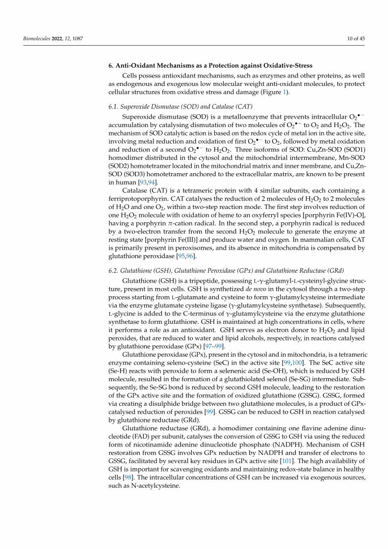
β-carotene (β-C) is a non-oxygenated carotenoid molecule, with 40 carbons, 11 con-jugated double bonds and 2 β-ionone rings (Figure 2), possessing provitamin A activ-ity [147,148]. β-carotene is an antioxidant molecule serving as a quencher of singlet oxygenand a scavenger of peroxyl radicals. The physical quenching of singlet oxygen is carriedout by transferring excitation energy from singlet oxygen to carotenoid molecule, to gen-erate excited triplet-state β-carotene and ground-state oxygen. Subsequently, the energyis dissipated between the excited β-C and the surroundings to yield normal energy statecarotenoid and thermal energy [147,148]. The chemical quenching of singlet oxygen is car-ried out by oxygenation of β-carotene with the formation of β-C endoperoxides [149,150].The scavenging of peroxyl radicals is carried out by addition of peroxyl radical to a suitabledouble bond in provitamin A/vitamin A molecules resulting in formation of carbon radicalthat can further undergo the conversion into epoxides or react with new peroxyl radicalto form bis-peroxyl products [71,151,152]. β-carotene also showed scavenging activitytowards superoxide anion radical, hydroxyl radical and hydrogen peroxide [153], andreaction of carotenoids with superoxide anion or hydroxyl radical results in formation ofcarotenoid epoxides [154,155].
Biomolecules 2022, 12, 1087 14 of 45
Figure 2. The scavenging mechanism of provitamin A and vitamin A.
7.1.2. Tocopherols: Structure and Anti-Oxidant Property
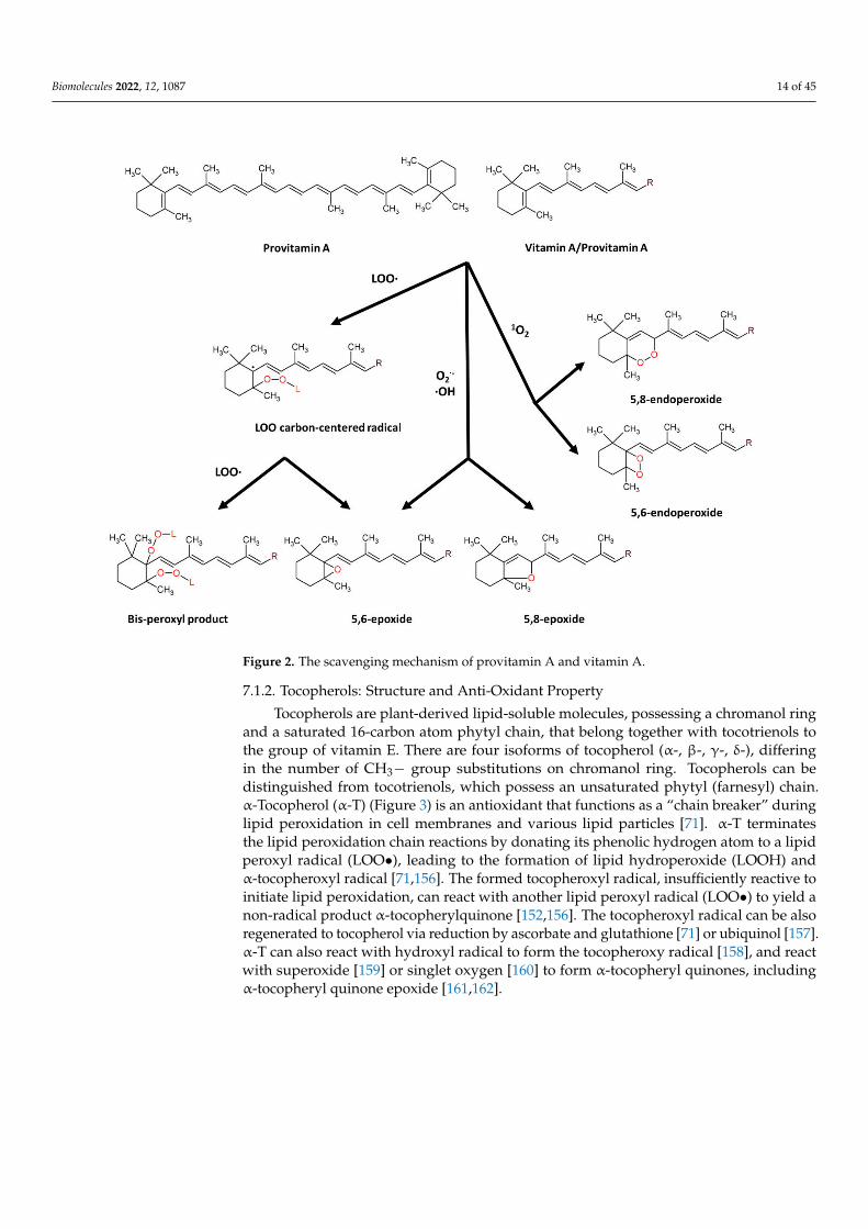
Tocopherols are plant-derived lipid-soluble molecules, possessing a chromanol ringand a saturated 16-carbon atom phytyl chain, that belong together with tocotrienols tothe group of vitamin E. There are four isoforms of tocopherol (α-, β-, γ-, δ-), differingin the number of CH3− group substitutions on chromanol ring. Tocopherols can bedistinguished from tocotrienols, which possess an unsaturated phytyl (farnesyl) chain.α-Tocopherol (α-T) (Figure 3) is an antioxidant that functions as a “chain breaker” duringlipid peroxidation in cell membranes and various lipid particles [71]. α-T terminatesthe lipid peroxidation chain reactions by donating its phenolic hydrogen atom to a lipidperoxyl radical (LOO•), leading to the formation of lipid hydroperoxide (LOOH) andα-tocopheroxyl radical [71,156]. The formed tocopheroxyl radical, insufficiently reactive toinitiate lipid peroxidation, can react with another lipid peroxyl radical (LOO•) to yield anon-radical product α-tocopherylquinone [152,156]. The tocopheroxyl radical can be alsoregenerated to tocopherol via reduction by ascorbate and glutathione [71] or ubiquinol [157].α-T can also react with hydroxyl radical to form the tocopheroxy radical [158], and reactwith superoxide [159] or singlet oxygen [160] to form α-tocopheryl quinones, includingα-tocopheryl quinone epoxide [161,162].
Biomolecules 2022, 12, 1087 15 of 45
Figure 3. The scavenging mechanism of α-tocopherol.
7.1.3. Ascorbic Acid: Structure and Anti-Oxidant Property
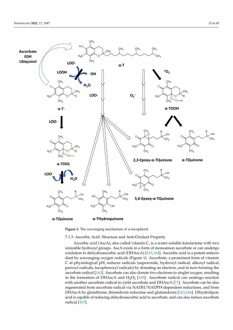
Ascorbic acid (AscA), also called vitamin C, is a water-soluble ketolactone with twoionizable hydroxyl groups. AscA exists in a form of monoanion ascorbate or can undergooxidation to dehydroascorbic acid (DHAscA) [163,164]. Ascorbic acid is a potent antioxi-dant by scavenging oxygen radicals (Figure 4). Ascorbate, a prominent form of vitaminC at physiological pH, reduces radicals (superoxide, hydroxyl radical, alkoxyl radical,peroxyl radicals, tocopheroxyl radicals) by donating an electron, and in turn forming theascorbate radical [163]. Ascorbate can also donate two electrons to singlet oxygen, resultingin the formation of DHAscA and H2O2 [165]. Ascorbate radical can undergo reactionwith another ascorbate radical to yield ascorbate and DHAscA [71]. Ascorbate can be alsoregenerated from ascorbate radical via NADH/NADPH-dependent reductases, and fromDHAscA by glutathione, thioredoxin reductase and glutaredoxin [163,166]. Dihydrolipoicacid is capable of reducing dehydroascorbic acid to ascorbate, and can also reduce ascorbateradical [167].
Biomolecules 2022, 12, 1087 16 of 45
Figure 4. The scavenging mechanism of ascorbate.
7.2. In Vivo and In Vitro Antioxidant Characteristics of β-Carotene, Tocopherols and Ascorbate7.2.1. Oxidative Stress Inducers Used during In Vitro and In Vivo Studies
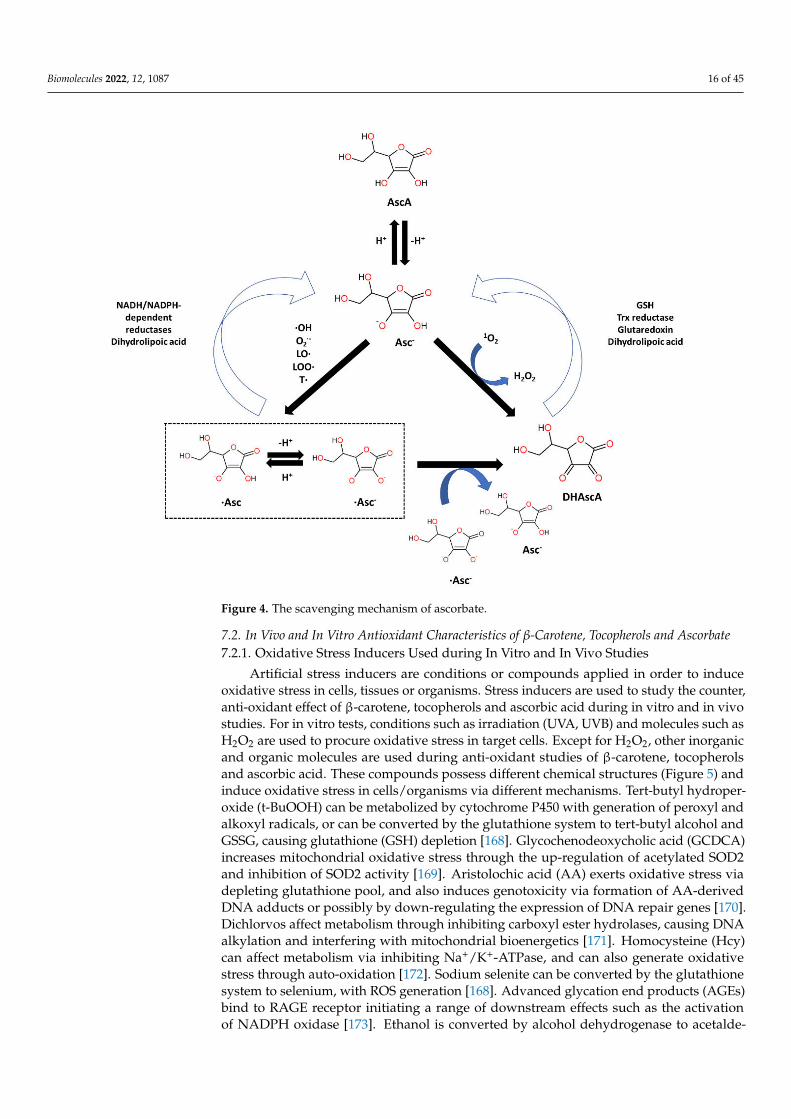
Artificial stress inducers are conditions or compounds applied in order to induceoxidative stress in cells, tissues or organisms. Stress inducers are used to study the counter,anti-oxidant effect of β-carotene, tocopherols and ascorbic acid during in vitro and in vivostudies. For in vitro tests, conditions such as irradiation (UVA, UVB) and molecules such asH2O2 are used to procure oxidative stress in target cells. Except for H2O2, other inorganicand organic molecules are used during anti-oxidant studies of β-carotene, tocopherolsand ascorbic acid. These compounds possess different chemical structures (Figure 5) andinduce oxidative stress in cells/organisms via different mechanisms. Tert-butyl hydroper-oxide (t-BuOOH) can be metabolized by cytochrome P450 with generation of peroxyl andalkoxyl radicals, or can be converted by the glutathione system to tert-butyl alcohol andGSSG, causing glutathione (GSH) depletion [168]. Glycochenodeoxycholic acid (GCDCA)increases mitochondrial oxidative stress through the up-regulation of acetylated SOD2and inhibition of SOD2 activity [169]. Aristolochic acid (AA) exerts oxidative stress viadepleting glutathione pool, and also induces genotoxicity via formation of AA-derivedDNA adducts or possibly by down-regulating the expression of DNA repair genes [170].Dichlorvos affect metabolism through inhibiting carboxyl ester hydrolases, causing DNAalkylation and interfering with mitochondrial bioenergetics [171]. Homocysteine (Hcy)can affect metabolism via inhibiting Na+/K+-ATPase, and can also generate oxidativestress through auto-oxidation [172]. Sodium selenite can be converted by the glutathionesystem to selenium, with ROS generation [168]. Advanced glycation end products (AGEs)bind to RAGE receptor initiating a range of downstream effects such as the activationof NADPH oxidase [173]. Ethanol is converted by alcohol dehydrogenase to acetalde-
Biomolecules 2022, 12, 1087 17 of 45
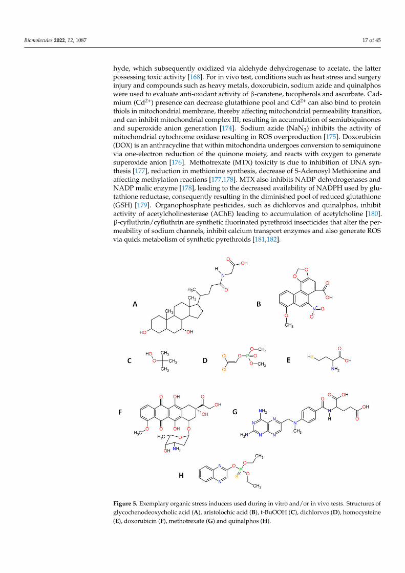
hyde, which subsequently oxidized via aldehyde dehydrogenase to acetate, the latterpossessing toxic activity [168]. For in vivo test, conditions such as heat stress and surgeryinjury and compounds such as heavy metals, doxorubicin, sodium azide and quinalphoswere used to evaluate anti-oxidant activity of β-carotene, tocopherols and ascorbate. Cad-mium (Cd2+) presence can decrease glutathione pool and Cd2+ can also bind to proteinthiols in mitochondrial membrane, thereby affecting mitochondrial permeability transition,and can inhibit mitochondrial complex III, resulting in accumulation of semiubiquinonesand superoxide anion generation [174]. Sodium azide (NaN3) inhibits the activity ofmitochondrial cytochrome oxidase resulting in ROS overproduction [175]. Doxorubicin(DOX) is an anthracycline that within mitochondria undergoes conversion to semiquinonevia one-electron reduction of the quinone moiety, and reacts with oxygen to generatesuperoxide anion [176]. Methotrexate (MTX) toxicity is due to inhibition of DNA syn-thesis [177], reduction in methionine synthesis, decrease of S-Adenosyl Methionine andaffecting methylation reactions [177,178]. MTX also inhibits NADP-dehydrogenases andNADP malic enzyme [178], leading to the decreased availability of NADPH used by glu-tathione reductase, consequently resulting in the diminished pool of reduced glutathione(GSH) [179]. Organophosphate pesticides, such as dichlorvos and quinalphos, inhibitactivity of acetylcholinesterase (AChE) leading to accumulation of acetylcholine [180].β-cyfluthrin/cyfluthrin are synthetic fluorinated pyrethroid insecticides that alter the per-meability of sodium channels, inhibit calcium transport enzymes and also generate ROSvia quick metabolism of synthetic pyrethroids [181,182].
Figure 5. Exemplary organic stress inducers used during in vitro and/or in vivo tests. Structures ofglycochenodeoxycholic acid (A), aristolochic acid (B), t-BuOOH (C), dichlorvos (D), homocysteine(E), doxorubicin (F), methotrexate (G) and quinalphos (H).
Biomolecules 2022, 12, 1087 18 of 45
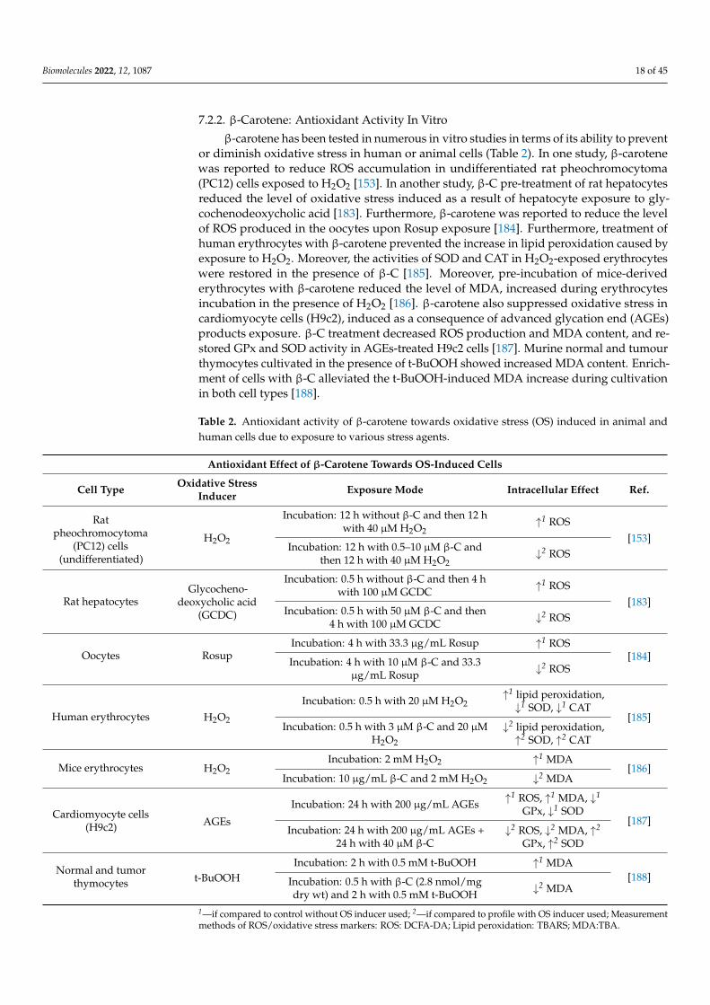
7.2.2. β-Carotene: Antioxidant Activity In Vitro
β-carotene has been tested in numerous in vitro studies in terms of its ability to preventor diminish oxidative stress in human or animal cells (Table 2). In one study, β-carotenewas reported to reduce ROS accumulation in undifferentiated rat pheochromocytoma(PC12) cells exposed to H2O2 [153]. In another study, β-C pre-treatment of rat hepatocytesreduced the level of oxidative stress induced as a result of hepatocyte exposure to gly-cochenodeoxycholic acid [183]. Furthermore, β-carotene was reported to reduce the levelof ROS produced in the oocytes upon Rosup exposure [184]. Furthermore, treatment ofhuman erythrocytes with β-carotene prevented the increase in lipid peroxidation caused byexposure to H2O2. Moreover, the activities of SOD and CAT in H2O2-exposed erythrocyteswere restored in the presence of β-C [185]. Moreover, pre-incubation of mice-derivederythrocytes with β-carotene reduced the level of MDA, increased during erythrocytesincubation in the presence of H2O2 [186]. β-carotene also suppressed oxidative stress incardiomyocyte cells (H9c2), induced as a consequence of advanced glycation end (AGEs)products exposure. β-C treatment decreased ROS production and MDA content, and re-stored GPx and SOD activity in AGEs-treated H9c2 cells [187]. Murine normal and tumourthymocytes cultivated in the presence of t-BuOOH showed increased MDA content. Enrich-ment of cells with β-C alleviated the t-BuOOH-induced MDA increase during cultivationin both cell types [188].
Table 2. Antioxidant activity of β-carotene towards oxidative stress (OS) induced in animal andhuman cells due to exposure to various stress agents.
Antioxidant Effect of β-Carotene Towards OS-Induced Cells
Cell Type Oxidative StressInducer Exposure Mode Intracellular Effect Ref.
Ratpheochromocytoma
(PC12) cells(undifferentiated)
H2O2
Incubation: 12 h without β-C and then 12 hwith 40 µM H2O2
↑1 ROS[153]
Incubation: 12 h with 0.5–10 µM β-C andthen 12 h with 40 µM H2O2
↓2 ROS
Rat hepatocytesGlycocheno-
deoxycholic acid(GCDC)
Incubation: 0.5 h without β-C and then 4 hwith 100 µM GCDC ↑1 ROS
[183]Incubation: 0.5 h with 50 µM β-C and then
4 h with 100 µM GCDC ↓2 ROS
Oocytes RosupIncubation: 4 h with 33.3 µg/mL Rosup ↑1 ROS
[184]Incubation: 4 h with 10 µM β-C and 33.3µg/mL Rosup ↓2 ROS
Human erythrocytes H2O2
Incubation: 0.5 h with 20 µM H2O2↑1 lipid peroxidation,↓1 SOD, ↓1 CAT
[185]Incubation: 0.5 h with 3 µM β-C and 20 µM
H2O2
↓2 lipid peroxidation,↑2 SOD, ↑2 CAT
Mice erythrocytes H2O2Incubation: 2 mM H2O2 ↑1 MDA
[186]Incubation: 10 µg/mL β-C and 2 mM H2O2 ↓2 MDA
Cardiomyocyte cells(H9c2) AGEs
Incubation: 24 h with 200 µg/mL AGEs ↑1 ROS, ↑1 MDA, ↓1
GPx, ↓1 SOD[187]
Incubation: 24 h with 200 µg/mL AGEs +24 h with 40 µM β-C
↓2 ROS, ↓2 MDA, ↑2
GPx, ↑2 SOD
Normal and tumorthymocytes t-BuOOH
Incubation: 2 h with 0.5 mM t-BuOOH ↑1 MDA[188]Incubation: 0.5 h with β-C (2.8 nmol/mg
dry wt) and 2 h with 0.5 mM t-BuOOH ↓2 MDA
1—if compared to control without OS inducer used; 2—if compared to profile with OS inducer used; Measurementmethods of ROS/oxidative stress markers: ROS: DCFA-DA; Lipid peroxidation: TBARS; MDA:TBA.

Biomolecules 2022, 12, 1087 19 of 45
7.2.3. Tocopherols: Antioxidant Activity In Vitro
The ability of tocopherols to decrease oxidative stress has been proved in numerousstudies (Table 3). α-T showed hydrogen peroxide and superoxide radical scavengingactivities, as well as ferrous ion (Fe2+) chelating activity [144]. Pre-treatment of human ker-atinocyte cells with α-T decreased the level of MDA and ROS, induced due to keratinocyteexposure to ultraviolet A radiation [189]. α-T suppressed formation of lipid peroxidationand protein carbonyl products in human neuroblastoma (SH-SY5Y) cells exposed to ad-vanced glycation end products (AGEs) [173]. In another study, vitamin E supplementationdecreased ROS and MDA levels promoted in human umbilical vein endothelial cells afterexposure to homocysteine (Hcy) [172]. Treatment of human erythrocytes with α-T dimin-ished the increase in MDA content exerted by pesticide dichlorvos [190]. Furthermore,treatment of human colorectal adenocarcinoma cell line (Caco-2) with vitamin E reducedthe level of MDA, increased in Caco-2 due to exposure to H2O2 [191]. Furthermore, pre-treatment of rat hepatocytes with α-T reduced the level of oxidative stress induced as aresult of hepatocyte exposure to glycochenodeoxycholic acid [183]. α-T also attenuated theH2O2 level in rat renal tubular epithelial cells (NRK-52E), increased in NRK-52E cells dueto exposure to aristolochic acid [192].
Table 3. Antioxidant activity of tocopherols/vitamin E towards oxidative stress (OS) induced inanimal and human cells due to exposure to various stress agents.
Antioxidant Effect of Vitamin E towards OS-Induced Cells
Cell Type Oxidative StressInducer Exposure Mode Exposure Effect Ref.
Human keratinocytecells
UVAIrradiation (UVA, 8 J/cm2) + incubation for 24 h ↑1 ROS, ↑1 MDA
[189]Incubation for 24 h with α-T (2.9–14.7 IU/mL),irradiation (UVA, 8 J/cm2) + incubation for 24 h ↓2 ROS, ↓2 MDA
Human neuroblastoma(SH-SY5Y) cells AGEs
Incubation for 24 h without α-T, then incubationwith 1.5 mg/mL AGEs for 72 h
↑1 lipid peroxidation, ↑1
protein carbonyls[173]
Incubation for 24 h with α-T (200 µM), thenincubation with 1.5 mg/mL AGEs for 72 h
↓2 lipid peroxidation, ↓2
protein carbonyls
Human umbilical veinendothelial cells
HcyIncubation with Hcy (1 mM) ↑1 ROS, ↑1 MDA
[172]Incubation with Hcy (1 mM) and VitE (50 µM) ↓2 ROS, ↓2 MDA
Human erythrocytes Dichlorvos (DDVP)Incubation with DDVP (10 µM) ↑1 MDA, ↓1 SOD, ↓1
CAT, ↓1 GPx[190]
Incubation with DDVP (10 µM) and Vit E (30 µM) ↓2 MDA, ↑2 SOD, ↑2
CAT, ↑2 GPx
Human colorectaladenocarcinoma cell line
(Caco-2)H2O2
Incubation for 24 h, incubation for 48 h withH2O2 (250 µM) ↑1 MDA
[191]Incubation for 24 h, incubation for 48 h with
H2O2 (250 µM) and VitE (10 µM) ↓2 MDA
Rat hepatocytesGlycocheno-deoxycholic
acid
Incubation: 0.5 h without α-T and then 4 h with100 µM GCDC ↑1 ROS
[183]Incubation: 0.5 h with 100 µM α-T and then 4 h
with 100 µM GCDC ↓2 ROS
Rat renal tubularepithelial cells
(NRK-52E)Aristolochic acid (AA)
Incubation: AA (10 µM) ↑1 H2O2[192]
Incubation: AA (10 µM) + α-T (5–100 µM) ↓2 H2O2
1—if compared to control without OS inducer used; 2—if compared to profile with OS inducer used; Measurementmethods of ROS/oxidative stress markers: ROS: DCFA-DA; Lipid peroxidation: TBARS; MDA: TBA; Proteincarbonyls: Immunoblotting; H2O2: Chemiluminescence.
Biomolecules 2022, 12, 1087 20 of 45
7.2.4. Ascorbic Acid: Antioxidant Activity In Vitro
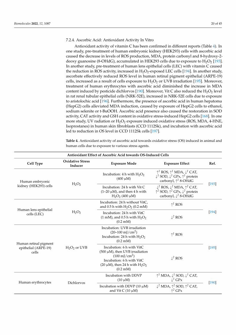
Antioxidant activity of vitamin C has been confirmed in different reports (Table 4). Inone study, pre-treatment of human embryonic kidney (HEK293) cells with ascorbic acidcaused the decrease in levels of ROS production, MDA, protein carbonyl and 8-hydroxy-2-deoxy guanosine (8-OHdG), accumulated in HEK293 cells due to exposure to H2O2 [193].In another study, pre-treatment of human lens epithelial cells (LEC) with vitamin C causedthe reduction in ROS activity, increased in H2O2-exposed LEC cells [194]. In another study,ascorbate effectively reduced ROS level in human retinal pigment epithelial (ARPE-19)cells, increased as a result of cells exposure to H2O2 or UVB irradiation [195]. Moreover,treatment of human erythrocytes with ascorbic acid diminished the increase in MDAcontent induced by pesticide dichlorvos [190]. Moreover, Vit C also reduced the H2O2 levelin rat renal tubular epithelial cells (NRK-52E), increased in NRK-52E cells due to exposureto aristolochic acid [196]. Furthermore, the presence of ascorbic acid in human hepatoma(HepG2) cells alleviated MDA induction, caused by exposure of HepG2 cells to ethanol,sodium selenite or t-BuOOH. Ascorbic acid presence also caused the restoration in SODactivity, CAT activity and GSH content in oxidative stress-induced HepG2 cells [168]. In onemore study, UV radiation or H2O2 exposure induced oxidative stress (ROS, MDA, 4-HNE,Isoprostanes) in human skin fibroblasts (CCD 1112Sk), and incubation with ascorbic acidled to reduction in OS level in CCD 1112Sk cells [197].
Table 4. Antioxidant activity of ascorbic acid towards oxidative stress (OS) induced in animal andhuman cells due to exposure to various stress agents.
Antioxidant Effect of Ascorbic Acid towards OS-Induced Cells
Cell Type Oxidative StressInducer Exposure Mode Exposure Effect Ref.
Human embryonickidney (HEK293) cells H2O2
Incubation: 4 h with H2O2(400 µM)
↑1 ROS, ↑1 MDA, ↓1 CAT,↓1 SOD, ↓1 GPx, ↑1 protein
carbonyl, ↑1 8-OHdG[193]
Incubation: 24 h with Vit C(1–20 µM), and then 4 h with
H2O2 (400 µM)
↓2 ROS, ↓2 MDA, ↑2 CAT,↑2 SOD, ↑2 GPx, ↓2 protein
carbonyl, ↓2 8-OHdG
Human lens epithelialcells (LEC)
H2O2
Incubation: 24 h without VitC,and 0.5 h with H2O2 (0.2 mM) ↑1 ROS
[194]Incubation: 24 h with VitC(1 mM), and 0.5 h with H2O2
(0.2 mM)↓2 ROS
Human retinal pigmentepithelial (ARPE-19)
cellsH2O2 or UVB
Incubation: UVB irradiation(20–100 mJ/cm2)
Incubation: 24 h with H2O2(0.2 mM)
↑1 ROS
[195]Incubation: 6 h with VitC(500 µM), then UVB irradiation
(100 mJ/cm2)Incubation: 6 h with VitC
(20 µM), then 24 h with H2O2(0.2 mM)
↓2 ROS
Human erythrocytes Dichlorvos
Incubation with DDVP(10 µM)
↑1 MDA, ↓1 SOD, ↓1 CAT,↓1 GPx
[190]Incubation with DDVP (10 µM)
and Vit C (10 µM)↓2 MDA, ↑2 SOD, ↑2 CAT,
↑2 GPx

Biomolecules 2022, 12, 1087 21 of 45
Table 4. Cont.
Antioxidant Effect of Ascorbic Acid towards OS-Induced Cells
Cell Type Oxidative StressInducer Exposure Mode Exposure Effect Ref.
Human hepatoma(HepG2) cells
Ethanol, sodiumselenite or t-BuOOH
Incubation with ethanol(10–500 µM) or sodium
selenite (1–10 µM) or t-BuOOH(20–200 µM) for 24 h
↑1 MDA ↓1 SOD, ↓1 CAT,↓1 GSH
[168]
Cotreatment with Vit C(25–100 µM) and one of OS
inducer for 24 h
↓2 MDA, ↑2 SOD, ↑2 CAT,↑2 GSH
Human skin fibroblasts(CCD 1112Sk)
UVA, UVB or H2O2
For irradiation treatment:20 J/cm2 (UVA) or 200 mJ/cm2
(UVB) + 24 h incubation; ForH2O2 treatment: incubationwith 200 µM H2O2 for 24 h
↑1 ROS, ↑1 MDA, ↑1 4-HNE,↑1 Carbonyl groups (for all
stress inducers);↑1 Isoprostanes (for
UVA, H2O2)[197]
Stress induction treatment +24 h incubation with 100 µM
ascorbic acid
↓2 ROS, ↓2 MDA, ↓2 4-HNE,↓2 Carbonyl groups (for all
stress inducers);↓2 Isoprostanes (for
UVA, H2O2)
Rat renal tubularepithelial cells
(NRK-52E)Aristolochic acid (AA)
Incubation: AA (10 µM) ↑1 H2O2[196]Incubation: AA (10 µM) + Vit
C (5 µM) ↓2 H2O2
1—if compared to control without OS inducer used; 2—if compared to profile with OS inducer used; Measurementmethods of ROS/oxidative stress markers: ROS: DCFA-DA, ESR [197]; MDA:TBA, GC-MS [197]; 4-HNE: GC-MS [197]; Isoprostanes: LC-MS [197]; Protein carbonyls: DNPH; 8-OHdG: ELISA [193], LC-MS [197]; H2O2:Chemiluminescence.
7.2.5. β-Carotene: Antioxidant Activity In Vivo
Carotenoids, such as β-carotene, are lipid soluble tetraterpenoid molecules naturallyfound in plants and microorganisms (algae, yeast and some bacteria), and available infood. In mammals, lipid-soluble β-carotene is absorbed in small intestine and deliveredto the peripheral tissues (liver, adipose tissue, kidney, skin, lungs) via various lipoproteinparticles. In one of the possible mechanisms for vitamin A synthesis (Figure 6), all trans β-carotene, the predominant form of β-carotene found in nature, is symmetrically cleaved bythe enzyme β-carotene-15,15′-oxygenase (CMOI) to yield two molecules of retinaldehyde.Ingested β-C can be cleaved via CMOI in intestine and in various tissues within the body.Retinaldehyde can be reduced via alcohol dehydrogenase or retinol dehydrogenase toretinol, a vitamin A. Retinol can be also esterified via lecithin:retinol acyltransferase (LRAT)to retinyl esters, which constitute vitamin A reserves. Retinaldehyde can be also oxidized,by enzymes (ALDH 1 or RALDH) from the aldehyde dehydrogenase 1 family, into all-transretinoic acid which constitutes the biologically active form of vitamin A, responsible fortranscriptional regulation [198,199].

Biomolecules 2022, 12, 1087 22 of 45
Figure 6. The structures of provitamin A (β-carotene) and vitamin A constituents (retinaldehyde,retinol, retinoic acid).
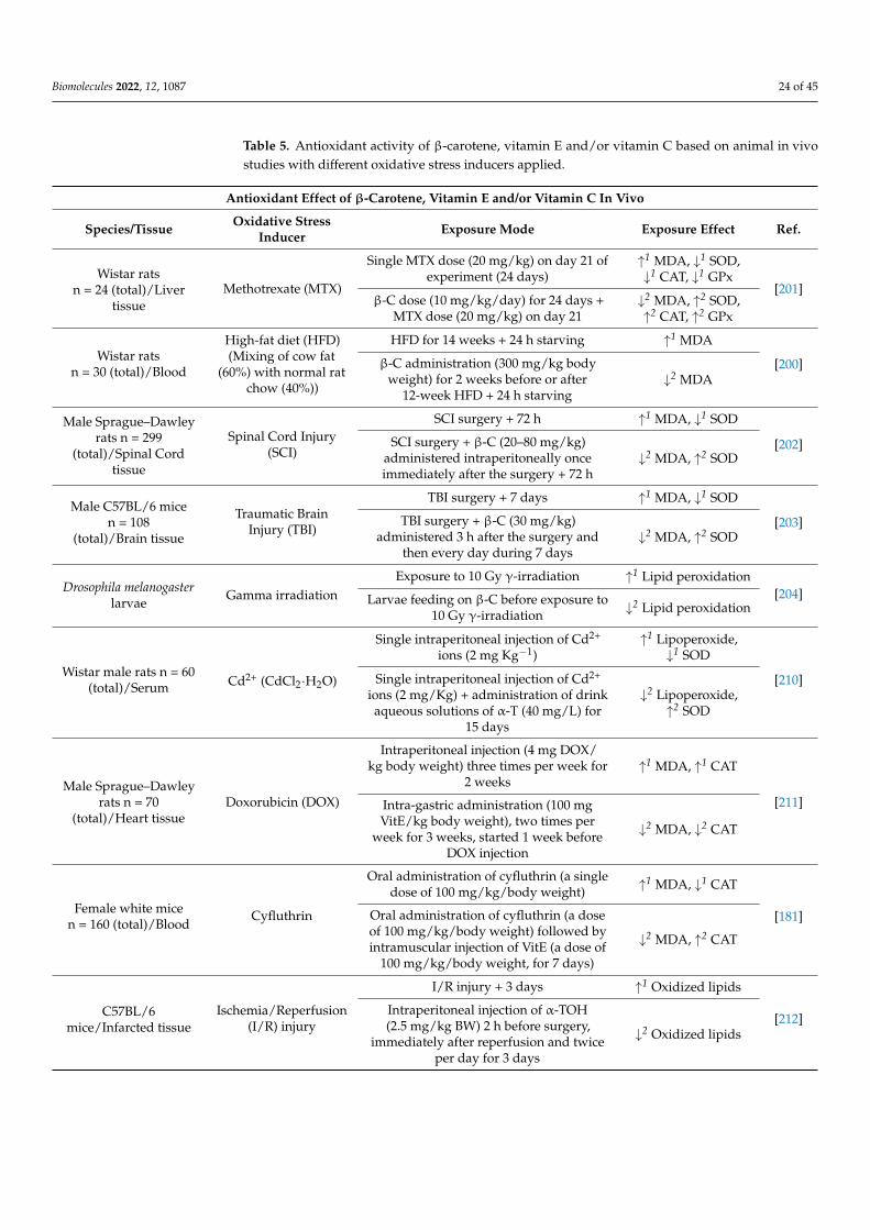
The antioxidant activity of β-carotene was evaluated in vivo with different animalspecies (Table 5). Wistar rats were characterized by increased MDA level and decreasedSOD and CAT activity, as a result of 2-week high-fat diet (HFD), if compared to controlwhere normal diet was administered. The addition of β-carotene for two weeks, before orafter 12-week HFD, resulted in reduction of MDA level and restoration in SOD and CATactivity, in investigated rats [200]. Wistar albino rats were administered methotrexate (MTX)what resulted in the increased MDA level and reduced activities of SOD, CAT and GPx inthe livers of rats exposed to MTX. Co-administration of MTX with β-carotene diminishedhepatic MDA level and restored activities of SOD, CAT and GPx in rat livers [201]. β-carotene was reported to attenuate oxidative stress in the spinal cord of rats with spinal cordinjury (SCI). Decreased ROS production and MDA level, and restored SOD activity weredetected in spinal cord tissues of SCI rats fed with β-C [202]. β-carotene also diminishedoxidative stress in mice with traumatic brain injury (TBI). Decreased MDA content andrestored SOD activity were detected in brain tissue of TBI-mice, when β-C doses wereadministered [203]. In one more study, incubation of Drosophila melanogaster larvae withβ-carotene diminished the OS level induced as a consequence of larvae exposure to γ-irradiation [204].
7.2.6. Tocopherols: Antioxidant Activity In Vivo
Vitamin E is found in vegetable oils and seeds, with α-tocopherol being commonlyavailable in wheat germ, olive, and sunflower oil, and γ-tocopherol being prominent insoybean, corn, and cottonseed oil. Lipid-containing food intake is the source of vitamin Ein human body [157,205,206]. During the digestion process, triacylglycerols and other ester-ified fat-soluble compounds are partially processed in the stomach by gastric lipase [207],and absorption of vitamin E occurs in the small intestine [157]. Vitamin E along with other
Biomolecules 2022, 12, 1087 23 of 45
lipids is incorporated into mixed micelles, the process aided by pancreatic lipases and bilesalts, in the duodenum [206,207]. In the intestine lumen, micelles solubilize hydrophobiccomponents and diffuse into the glycocalix to approach the brush border membrane of theenterocytes [157,208]. Vitamin E transport across the enterocyte membrane occurs via pas-sive diffusion and/or is also mediated by membrane proteins (NPC1L1, SR-BI, CD36) [208].In enterocytes, vitamin E is incorporated into chylomicrons, which are secreted into the in-testinal lymph system and released into the bloodstream [208,209]. Vitamin E, transportedin blood via different lipoproteins, is distributed within liver and extrahepatic tissues (adi-pose tissue, muscle, adrenal glands) [206,209]. In liver, RRR α-tocopherol selectively bindsto the α-tocopherol transfer protein (α-TTP), while other isoforms of vitamin E are excretedin the bile [206]. α-Tocopherol, incorporated via α-TTP into lipoproteins and re-secreted tothe circulation [206], is the major tocopherol found in human blood and tissues [205].
The antioxidant effect of α-tocopherol has been tested in vivo on animal species(Table 5). A 15-day intake of α-T by Wistar rats led to reduction in lipoperoxide concen-trations, increased as a consequence of intraperitoneal injection of Cd2+ ions [210]. Inanother study, vitamin E administration reduced cardiac MDA level in rats, increaseddue to doxorubicin (DOX) injection. Additionally, cardiac GSH level in DOX-injectedrats fed with vitamin E were higher, if compared to DOX-injected group without vitaminadministered [211]. Further, administration of Vit E (α-tocopheryl acetate) to female whitemice treated with cyfluthrin resulted in the reduction in plasma MDA level and elevationin erythrocytes CAT activity, if compared to group where cyfluthrin was administered, butwithout Vit E [181]. In another study, α-T decreased ischemia/reperfusion injury-inducedROS production and oxidative modification of phospholipids in mice [212]. Vitamin Ewas also tested in terms of its cytoprotective effect towards cardiovascular system in micesubjected to heat stress. Heart tissue of mice exposed to heat stress (HS) conditions showedincreased ROS levels, but administration of vitamin E possessed ameliorating effect. More-over, cardiomyocytes of mice subjected to heat stress and fed with vitamin E showeddecreased MDA levels and restored SOD and GSH levels, if compared to HS conditions butwithout vitamin administration [213]. Supplementation of VitE (α-tocopheryl acetate) to ratdiet reduced plasma lipid peroxidation and restored plasma SOD and GPx activity, affecteddue to rats feeding on high-fat diet [214]. Moreover, supplementation of VitE (α-tocopherylacetate) reduced MDA serum concentration and MDA breast muscle content in chickenbroilers, fed on linseed oil-enriched diet [215].
7.2.7. Ascorbic Acid: Antioxidant Activity In Vivo
Vitamin C is naturally present in fruits (kiwifruit, orange, lemon, black currant, rasp-berry, strawberry, grapes etc.) and vegetables (broccoli, cabbage, spinach, tomato, potato,pepper etc.) [216,217], but can be also manufactured industrially in reactions involvingvarious intermediates such as D-sorbitol, L-sorbose and 2-keto-L-gulonic acid [218]. Inanimals, L-ascorbic acid is synthetized through glucuronic acid pathway in liver (mam-mals) or kidney (reptiles, birds) [219]. Humans, apes, guinea pigs and fruit-eating batscannot synthetize ascorbic acid due to the lack of enzyme L-gulonolactone oxidase [220]. Inhumans, ascorbate and DHAscA (vitamin C equivalents) are absorbed from ingested foodby enterocytes of the small intestine. Ascorbate is absorbed via Na+-dependent vitamin Ctransporters (SVCTs) while DHAscA is absorbed via Na+-independent facilitative glucosetransporters (GLUTs) and reduced to ascorbate. Vitamin C, absorbed from the intestinallumen, is transported with the blood to various peripheral organs that differ in tissueascorbate content [163,164,221].
Biomolecules 2022, 12, 1087 24 of 45
Table 5. Antioxidant activity of β-carotene, vitamin E and/or vitamin C based on animal in vivostudies with different oxidative stress inducers applied.
Antioxidant Effect of β-Carotene, Vitamin E and/or Vitamin C In Vivo
Species/Tissue Oxidative StressInducer Exposure Mode Exposure Effect Ref.
Wistar ratsn = 24 (total)/Liver
tissueMethotrexate (MTX)
Single MTX dose (20 mg/kg) on day 21 ofexperiment (24 days)
↑1 MDA, ↓1 SOD,↓1 CAT, ↓1 GPx
[201]β-C dose (10 mg/kg/day) for 24 days +
MTX dose (20 mg/kg) on day 21↓2 MDA, ↑2 SOD,↑2 CAT, ↑2 GPx
Wistar ratsn = 30 (total)/Blood
High-fat diet (HFD)(Mixing of cow fat
(60%) with normal ratchow (40%))
HFD for 14 weeks + 24 h starving ↑1 MDA
[200]β-C administration (300 mg/kg bodyweight) for 2 weeks before or after
12-week HFD + 24 h starving↓2 MDA
Male Sprague–Dawleyrats n = 299
(total)/Spinal Cordtissue
Spinal Cord Injury(SCI)
SCI surgery + 72 h ↑1 MDA, ↓1 SOD
[202]SCI surgery + β-C (20–80 mg/kg)administered intraperitoneally onceimmediately after the surgery + 72 h
↓2 MDA, ↑2 SOD
Male C57BL/6 micen = 108
(total)/Brain tissue
Traumatic BrainInjury (TBI)
TBI surgery + 7 days ↑1 MDA, ↓1 SOD
[203]TBI surgery + β-C (30 mg/kg)administered 3 h after the surgery and
then every day during 7 days↓2 MDA, ↑2 SOD
Drosophila melanogasterlarvae
Gamma irradiationExposure to 10 Gy γ-irradiation ↑1 Lipid peroxidation
[204]Larvae feeding on β-C before exposure to10 Gy γ-irradiation ↓2 Lipid peroxidation
Wistar male rats n = 60(total)/Serum Cd2+ (CdCl2·H2O)
Single intraperitoneal injection of Cd2+
ions (2 mg Kg−1)↑1 Lipoperoxide,↓1 SOD
[210]Single intraperitoneal injection of Cd2+
ions (2 mg/Kg) + administration of drinkaqueous solutions of α-T (40 mg/L) for
15 days
↓2 Lipoperoxide,↑2 SOD
Male Sprague–Dawleyrats n = 70
(total)/Heart tissueDoxorubicin (DOX)
Intraperitoneal injection (4 mg DOX/kg body weight) three times per week for
2 weeks↑1 MDA, ↑1 CAT
[211]Intra-gastric administration (100 mgVitE/kg body weight), two times per
week for 3 weeks, started 1 week beforeDOX injection
↓2 MDA, ↓2 CAT
Female white micen = 160 (total)/Blood
Cyfluthrin
Oral administration of cyfluthrin (a singledose of 100 mg/kg/body weight) ↑1 MDA, ↓1 CAT
[181]Oral administration of cyfluthrin (a doseof 100 mg/kg/body weight) followed byintramuscular injection of VitE (a dose of
100 mg/kg/body weight, for 7 days)
↓2 MDA, ↑2 CAT
C57BL/6mice/Infarcted tissue
Ischemia/Reperfusion(I/R) injury
I/R injury + 3 days ↑1 Oxidized lipids
[212]Intraperitoneal injection of α-TOH(2.5 mg/kg BW) 2 h before surgery,
immediately after reperfusion and twiceper day for 3 days
↓2 Oxidized lipids

Biomolecules 2022, 12, 1087 25 of 45
Table 5. Cont.
Antioxidant Effect of β-Carotene, Vitamin E and/or Vitamin C In Vivo
Species/Tissue Oxidative StressInducer Exposure Mode Exposure Effect Ref.
BALB/c mice n = 40(total)/Heart tissue
Heat stress (HS)
HS conditions (temperature: 40 ◦C;humidity: 60%) for 4 h per day during a
4-week period↑1 MDA, ↓1 SOD
[213]
Oral administration of VitE (500 mg/kg)2 h before the initiation of HS ↓2 MDA, ↑2 SOD
Sprague-Dawley malerats n = 30
(total)/BloodHigh-fat diet
A 10-week feeding on high-fat diet ↑1 Lipid peroxidation,↓1 SOD, ↓1 GPx [214]
A 10-week feeding on high-fat dietsupplemented with VitE (350 mg/kg diet)
↓2 Lipid peroxidation,↑2 SOD, ↑2 GPx
Ross 308 male broilers(21-day old) n = 400(total)/Blood, Breast
muscle
High n-3 dietaryPUFAs intake
Chickens fed with commercial starter diet(1–12 days), commercial grower diet
(13–20 days), finisher diet enriched with5% cold-pressed linseed oil and
supplemented with VitE (200 IU/kg)(21–40 days)
↓3 MDA [215]
Wistar rats n = 28(total)/Stomach, Colon,
Kidney tissueSodium azide (NaN3)
Oral administration of NaN3 (20 mg/kgBW) for 9 days
↑1 MDA, ↑1 ProteinCarbonyls [222]
Oral administration of NaN3 and VitC(200 mg/kg BW) for 9 days
↓2 MDA, ↓2 ProteinCarbonyls
Wistar male ratsn = 46/Heart tissue
Doxorubicin (DOX)
Six intraperitoneal DOX injections (2.5mg/kg body wt) over 3 weeks
↑1 Superoxide anion,↑1 Lipid peroxidation,↑1 Protein Carbonyls
[223]Oral daily administration of VitC (50mg/kg) started 1 week before the start ofDOX administration and continued for 2
weeks after the last DOX injection
↓2 Superoxide anion,↓2 Lipid peroxidation,↓2 Protein Carbonyls
Sprague–Dawley malerats n = 18
(total)/Heart tissueQuinalphos (QP)
Oral dose of QP (14 mg/kg), daily for10 days
↑1 MDA, ↓1 CAT,↓1 GPx
[224]Oral administration of VitC (20 mg/kg)daily, 4 h after QP administration, for
10 days
↓2 MDA, ↑2 CAT,↑2 GPx
1—if compared to control without OS applied; 2—if compared to profile with OS applied; 3—if compared toprofile without anti-oxidant applied; Measurement methods of ROS/oxidative stress markers: Lipid peroxidation:TBARS; MDA: TBA; Protein carbonyls: DNPH, Oxidized lipids: LC-MS; Superoxide anion: Adrenaline assay.
The antioxidant effect of ascorbic acid has been tested in vivo on animal species(Table 5). In a study comprising Wistar rats, a 9-day supplementation of ascorbic acid re-sulted in the decreased levels of malondialdehyde (MDA) and protein carbonyl, increasedin the stomach, colon and kidneys of rats feeding with sodium azide (NaN3), as an oxida-tive stress inducer [222]. In another study, the antioxidant activity of ascorbate was testedin doxorubicin-administered male Wistar rats. The cardiac tissue of DOX-administeredrats fed with ascorbate, showed reduced levels of superoxide anion, lipid peroxidationand protein carbonyl products, if compared to the group subjected to DOX administrationbut without Vit C treatment. Moreover, ascorbate treatment restored SOD and GPx activ-ities, decreased in rat cardiac tissue due to DOX administration [223]. In another study,insecticide quinalphos (QP) administered to Sprague–Dawley rats caused induction ofoxidative stress in rat cardiac tissue, indicated by reduced GPx activity and CAT activity,and increased MDA content. Co-administration of Vit C to investigated rat group resulted
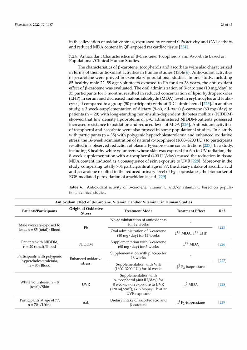
Biomolecules 2022, 12, 1087 26 of 45
in the alleviation of oxidative stress, expressed by restored GPx activity and CAT activity,and reduced MDA content in QP-exposed rat cardiac tissue [224].
7.2.8. Antioxidant Characteristics of β-Carotene, Tocopherols and Ascorbate Based onPopulational/Clinical Human Studies
The characteristics of β-carotene, tocopherols and ascorbate were also characterizedin terms of their antioxidant activities in human studies (Table 6). Antioxidant activitiesof β-carotene were proved in exemplary populational studies. In one study, including85 healthy male 22–58 age-volunteers exposed to Pb for 4 to 38 years, the anti-oxidanteffect of β-carotene was evaluated. The oral administration of β-carotene (10 mg/day) to35 participants for 3 months, resulted in reduced concentration of lipid hydroperoxides(LHP) in serum and decreased malondialdehyde (MDA) level in erythrocytes and leuko-cytes, if compared to a group (50 participant) without β-C administered [225]. In anotherstudy, a 3 week-supplementation of dietary (9-cis, all-trans) β-carotene (60 mg/day) topatients (n = 20) with long-standing non-insulin-dependent diabetes mellitus (NIDDM)showed that low density lipoproteins of β-C administered NIDDM-patients possessedincreased resistance to oxidation and reduced level of MDA [226]. Antioxidant activitiesof tocopherol and ascorbate were also proved in some populational studies. In a studywith participants (n = 35) with polygenic hypercholesterolemia and enhanced oxidativestress, the 16-week administration of natural α-tocopherol (1600–3200 I.U.) to participantsresulted in a observed reduction of plasma F2-isoprostane concentrations [227]. In a study,including 8 healthy white volunteers whose skin was exposed for 6 h to UV radiation, the8-week supplementation with α-tocopherol (400 IU/day) caused the reduction in tissueMDA content, induced as a consequence of skin exposure to UVR [228]. Moreover in thestudy, comprising totally 704 participants at age of 77, the dietary intake of ascorbic acidand β-carotene resulted in the reduced urinary level of F2-isoprostanes, the biomarker ofROS-mediated peroxidation of arachidonic acid [229].
Table 6. Antioxidant activity of β-carotene, vitamin E and/or vitamin C based on popula-tional/clinical studies.
Antioxidant Effect of β-Carotene, Vitamin E and/or Vitamin C in Human Studies
Patients/Participants Origin of OxidativeStress Treatment Mode Treatment Effect Ref.
Male workers exposed tolead, n = 85 (total)/Blood Pb
No administration of antioxidantsfor 12 weeks -
[225]Oral administration of β-carotene
(10 mg/day) for 12 weeks ↓1,2 MDA, ↓1,2 LHP
Patients with NIDDM,n = 20 (total)/Blood NIDDM Supplementation with β-carotene
(60 mg/day) for 3 weeks ↓C1 MDA [226]
Participants with polygenichypercholesterolemia,
n = 35/Blood
Enhanced oxidativestress
Supplementation with placebo for16 weeks -
[227]Supplementation with VitE
(1600–3200 I.U.) for 16 weeks ↓2 F2-isoprostane
White volunteers, n = 8(total)/Skin UVR
Supplementation withα-tocopherol (400 IU/day) for8 weeks, skin exposure to UVR
(120 mJ/cm2), skin biopsy 6 h afterUVR exposure
↓2 MDA [228]
Participants at age of 77,n = 704/Urine n.d. Dietary intake of ascorbic acid and
β-carotene ↓I F2-isoprostane [229]

Biomolecules 2022, 12, 1087 27 of 45
Table 6. Cont.
Antioxidant Effect of β-Carotene, Vitamin E and/or Vitamin C in Human Studies
Patients/Participants Origin of OxidativeStress Treatment Mode Treatment Effect Ref.
Female participants withtype 2 diabetes, n = 34
(total)/BloodType 2 diabetes
Oral administration of placebo(800 mg/day) for 6 weeks -
[230]Oral administration ofα-tocopherol (800 IU/day) for
6 weeks↓1,2 MDA
Participants with DMT2,n = 5 or 8 (total)/Blood DMT2 Supplementation with VitC
(1000 mg/day) for 6 weeks↓1 MDA
↓1 F2-isoprostane[231]
Participants with DMT2and administered with
hypoglycemic drug, n = 80(total)/Blood
DMT2
No supplementation of VitEwithin 3 months ↑1,C2 MDA
[232]Supplementation of VitE
(400 mg/day) for 3 months ↓1,2 MDA
Patients with late-stageknee osteoarthritis, n = 72
(total)/Blood,synovial fluid
Enhanced oxidativestress
Oral administration of placeboonce daily for 2 months -
[233]Oral administration of VitE(400 IU/day) for 2 months ↓2 MDA
1—if compared to the beginning of the study; 2—if compared to profile without antioxidant used; C1—if comparedto control: normoglycemic profile; C2—if compared to control: healthy subjects; I—if compared to lower intake ofantioxidants; Measurement methods of oxidative stress biomarkers: MDA: TBA; LHP: FOX assay; F2-Isoprostanes:GC-MS [227], Radioimmunoassay [229], ELISA [231].
In a study consisting of 34 female participants with type 2 diabetes, the oral sup-plementation of α-tocopherol (800 IU/day) to 13 participants for 6 weeks resulted in thedecrease in MDA erythrocyte level, if compared to a group (n = 21) without α-tocopheroladministration [230]. In another study, 5 participants with diabetes mellitus type 2 whoreceived vitamin C (1000 mg/day) for 6 weeks, showed decreased plasma levels of MDAand F2-isoprostanes, if compared to results before supplementation [231]. In a group of40 participants with clinically diagnosed DMT2, hypoglycemic drug-administered diabetic(27 macrovascular, 13 microvascular) patients supplemented for 3 months with vitaminE (400 mg/day) showed decreased MDA serum concentration and increased serum SODactivity and erythrocyte GSH content, if compared to the beginning of study, or when com-pared with 40 DMT2 patients treated for 3 months with hypoglycemic drugs but withoutVitE supplementation [232]. The antioxidant activity of VitE has been also evaluated ina study comprising 72 patients with late-stage knee osteoarthritis (OA) and concludingthat a two-month intake of VitE (400 IU) could reduce oxidative stress conditions (MDA inplasma and synovial fluid) in patients with OA [233].
7.2.9. Antioxidant Characteristics of β-Carotene, Tocopherols and Ascorbate: A Summary
The literature data gathered show that β-carotene, tocopherols and ascorbic acid actas efficient antioxidants towards human and animal cells in vitro, under stress conditions.Supplementation with β-carotene, tocopherols or ascorbic acid, as free-radical quenchers,decreased the level of OS (bio)markers (MDA, ROS, Protein carbonyls, Isoprostanes, 8-OHdG) in numerous types of cells exposed to stress conditions. Conducted in vitro studiesvaried in terms of not only tested cell lines, but also research conditions including methodsof oxidative stress induction, the form and concentration of antioxidant and the orderof culture supplementation. These differences amongst studies can create limitationsfor results comparison and interpretations. Different methods, such as physicochemicalfactors as well as inorganic and organic oxidizing agents, were used to induce oxidativestress (OA) in investigated cell cultures. These stress inducers can cause oxidative stress viadifferent mechanisms, and were tested towards different human and animal cells. Moreover,

Biomolecules 2022, 12, 1087 28 of 45
different stress inducers used for the same cell line can result in the different OS extent [186].The concentrations of in vitro tested antioxidants differ amongst studies (Tables 2–4), butthe anti-oxidant concentrations strictly influences the mitigation of oxidative stress [190].Another important barrier for interpretation of anti-oxidative efficiency is the chemicalform of anti-oxidant tested. Amongst tocopherols, α-T is commonly used during in vitrotests and its antioxidant activity was confirmed. However, the ability of α-T again tomitigate OS is not always proved and different tocopherol forms (α-T, γ-T) can possessdifferent anti-oxidant activity [234]. Furthermore, antioxidant evaluation in vitro includestheir different modes: cell pre-treatment with a selected antioxidant prior to oxidizingagent (OA)-exposure, co-treatment of cells with antioxidant and OA, or the subject of cellsto oxidizing agent before adding the selected antioxidant. The differences in oxidizingagent/anti-oxidant supplementation order can also have influence of the outcome ofresearch studies. Nevertheless, β-carotene, tocopherols and ascorbic acid proved to act asefficient antioxidants, despite differences between conducted studies.
Animal in vivo studies (Table 5) show that β-carotene, tocopherols and ascorbic acidcan be efficient compounds to mitigate oxidative stress, as indicated by decreased levelsof OS biomarkers (MDA, Lipid peroxidation, Protein carbonyls) in investigated species.As in case of in vitro research, the problems with interpretation of different experimentalconditions (OS generation method, supplementation mode) also occur, and further discrep-ancies (single cells vs. whole organism) between in vitro and in vivo appear. Nevertheless,the antioxidant properties of β-carotene, tocopherols and ascorbic acid was shown, despitedifferences between conducted in vivo studies.
Some human populational/clinical studies (Table 6) show that β-carotene, α-tocopherolor ascorbic acid can improve anti-oxidant status in a selected group of participants. How-ever, due to a scarce number of studies and a variety of conditions (different number ofparticipants and health condition status, various time span and biomarkers) influencingstudies outcome, the effectiveness of β-carotene, α-tocopherol or ascorbic acid as effectiveanti-oxidants, is not certain. Moreover, other reports do not confirm improvements in anti-oxidant level upon β-carotene [235], vitamin E [236] or ascorbic acid [237] administration.Moreover, the connection between free-radical scavenging ability of anti-oxidants and theuse of antioxidants for prevention of human diseases has not been established. Althoughα-tocopherol exhibits anti-oxidant activity and inhibits oxidation of low-density lipoproteincholesterol, α-T supplements failed to reduce atherosclerosis-related events [78]. Whatis more, beneficial effect of vitamin C in reference to human diseases (cancer, atheroscle-rosis, diabetes, neurodegenerative disease) remains equivocal [217], and a shortage ofclinical trials impedes drawing clear conclusions about therapeutic role of ascorbic acidadministration [238]. Furthermore, it was also reported that supplementation with anti-oxidant nutrients (vitamin C, vitamin E or β-carotene) offered no prevention againstcancer incidence [239], and there is no certainty that possible cancer-preventive ability ofanti-oxidant molecules, such as β-carotene, can be strictly due to free radical quenchingmechanism [240]. In addition to mentioned discrepancies, it should be also noted thatβ-carotene and vitamin C can possess pro-oxidant activity [217,240]. Therefore, furtherstudies are required to determine the effect of β-carotene, α-tocopherol or ascorbic acid onthe level of oxidative stress and the antioxidant status in broader groups within healthyand disease-diagnosed populations.
Exposure to oxidative stress causes the alteration in anti-oxidant mechanisms, withthe change in the activity of SOD, CAT and/or GPx (Tables 2–6). The exposure of cells todifferent OS inducers resulted in decrease in SOD, CAT and GPx activity. SOD, CAT andGPx are enzymes participating together to prevent oxidative damage to cells. Saturation ofSOD and CAT during ROS conversion, and decreased availability of GSH for GPx can leadto decreased activities of these enzymes [190]. Reduced GPx activity can result in decreasedSOD and CAT activities [179], and activity of SOD and CAT can be also inhibited by ROS,such as H2O2, [181] via affecting enzyme active site or via mechanism of cell regulation(de-/phosphorylation) [185]. Moreover, oxidizing agent tested can be directly involved

Biomolecules 2022, 12, 1087 29 of 45
in inhibition of anti-oxidant enzymes [181]. Overproduction of ROS is accompaniedby inactivation of detoxification systems, consumptions of antioxidants and insufficientreplenishment of antioxidants in cells/tissues [179]. However, the opposite trend withincreased CAT activity is response to oxidizing agent exposure, was observed [211], andVitE doses were reported to decrease levels of erythrocyte antioxidant enzymes in humanand animal studies [230]. Moreover, increased expression and activity of antioxidantenzymes was concluded to be a result of compensatory defence mechanisms againstoxidative stress [225]. Nevertheless, the majority of literature data show that the presenceof anti-oxidants (β-C, Vit E, Vit C) increased SOD, CAT and GPx activity in stressed cells,proving the ROS-scavenging properties of tested molecules.
7.3. Measurement of the Oxidative Stress in Cells and Tissues
The induction of oxidative stress in cells/tissues is typically measured by monitoringthe cellular level of ROS (direct techniques) or the level of specific markers of oxidativestress, such as lipid peroxidation, protein modification and DNA oxidation products(indirect techniques). These techniques are used (or can be used) to evaluate the efficacyof various anti-oxidant molecules, such as β-carotene, vitamin E and ascorbic acid, oncells/tissues exposed to oxidative stress.
7.3.1. Measurement of ROS Level: ESR Spectroscopy
Electron spin resonance (ESR) spectroscopy is a technique based on the absorptionof microwave radiation by unpaired electron-possessing molecules (radicals) situatedin the applied static magnetic field [241]. ESR spectroscopy is able to monitor the real-time generation of short-lived ROS radicals (O2
•−, •OH) when coupled with spin traps.Spin-trapping reagents (nitrone and nitroso compounds) react with ROS radicals to yieldlong-lived radicals called spin-adducts [242]. 5,5-dimethyl-1-pyrroline N-oxide (DMPO),a commonly used nitrone, reacts with O2
•− or •OH to form DMPO-OOH or DMPO-OHradicals, respectively, while DMPO-OOH further decomposes to DMPO-OH [243]. Freeradicals can be determined by analysing the distinct ESR spectrum of a spin adduct [242].Apart from spin traps, ESR exploits spin probes (such as cyclic hydroxylamines) thatundergo one-electron oxidation to form nitroxide radical detected by ESR, with highersensitivity than spin traps [244]. ESR technique was used to measure ROS generation inoxidative stress-exposed human skin fibroblasts (CCD 1112Sk). 1-hydroxy-3-methoxy-carbonyl-2,2,5,5-tetramethyl pyrrolidine (CMH), as a spin probe, was used to react withROS and to yield a stable nitroxide CM-radical, while ROS formation was measuredaccording to the rate of nitroxide accumulation [197].
7.3.2. Measurement of ROS Level: Fluorescent Method (DCFH-DA Probe Assay)
The intracellular level of ROS can be measured by means of the DCFH-DA probe assay.2′,7′-dichlorodihydrofluorescein diacetate (DCFH-DA) (Figure S1) is a non-fluorescentcompound that diffuses through the cellular membrane and undergoes the cleavage atthe two ester bonds via intracellular esterase to 2′,7′-dichlorodihydrofluorescein (DCFH)which accumulates in cells. The non-fluorescent DCFH undergoes oxidation by ROS, suchas H2O2, hydroxyl radicals and peroxyl radicals, to fluorescent 2′,7′-dichlorofluorescein(DCF). The presence of DCF can be detected fluorometrically with λexcitation = 485 nm andλemission = 525 nm [245,246]. ROS detection with DCFH-DA faces possible limitations suchas insufficient accessibility of esterases sequestered within cells, oxidation reaction withother cellular components, a lack of ability to react with O2
•− or the leakage of DCF out ofthe cells [245,246]. DCFH-DA probe assay is commonly used to evaluate the anti-oxidanteffect of vitamins towards OS-induced cells (Tables 2–4).
7.3.3. Measurement of ROS Level: Other Methods
O2•− and H2O2 can be measured by means of chemiluminescence method, where
light is emitted as a result of chemical reactions. H2O2 reacts in the presence of catalyst
Biomolecules 2022, 12, 1087 30 of 45
with luminol (3-aminophthalhydrazide) to produce the excited 3-aminophthalate anion,that emits light when relaxed to the ground state [247]. O2
•− reaction with lucigenin (N,N′-dimethyl-9,9′-biacridinium dinitrate, Luc2+) includes one-electron reduction of Luc2+ tolucigenin cation radical (Luc•+) that reacts with O2
•− to form lucigenin dioxetane (LucO2)which decomposes to N-methylacridone (NMA) with light emission [248].
O2•− can be detected via oxidation of adrenaline to adrenochrome, that can be deter-
mined spectrophotometrically at 480 nm [249].
7.3.4. Measurement of Lipid Peroxidation Products
Lipid peroxidation (LPO), the structural degradation of lipids occurring as a conse-quence of oxidative damage, is a widely used marker for OS presence. LPO is determinedby measuring the level of malondialdehyde (MDA), 4-hydroxynonenal (4-HNE) or F2-isoprostanes (F2-IsoPs).
Measurement of Lipid Peroxidation: MDA, 4-HNE
MDA, a three-carbon dialdehyde, and 4-HNE, an unsaturated aldehyde, are productsof oxidative degradation of polyunsaturated fatty acid (PUFAs), where double bonds inPUFAs structures are the target for ROS attack to form unstable fatty acid peroxides.
The MDA level is measured spectrophotometrically, and the principle of method isthe reaction (Figure S2) between MDA and 4,6-dihydroxypyrimidine-2-thiol (thiobarbituricacid; TBA) to form MDA-TBA2 adduct, that absorbs strongly at λ = 532 nm. Alterna-tively, MDA-TBA2 adduct can be measured spectrofluorometrically (λexcitation = 515 nm/λemission = 553 nm) [250]. MDA assay is the most common method used to monitor OSlevel and evaluate the anti-oxidant effect of β-carotene, vitamin E and ascorbic acid oncells/tissues subjected to oxidative stress (Tables 2–5). However, MDA determination basedon TBA assay, lacks selectivity due to dependence on reaction conditions (pH, temperature)as well as reaction of TBA with other components of biological samples such as sugars,amino acids, bilirubin, albumin and other aldehydes, known as thiobarbituric acid reactivesubstances (TBARS) [250,251].
Improvement in MDA detection is achieved by using techniques of Chromatographyand Mass Spectrometry, where molecules are analysed based on chromatogram retentiontime and mass to charge (m/z) ratio spectrum. MDA, after reaction with TBA, was reportedto be analysed by HPLC-UV-Vis [252] or HPLC coupled with fluorescence detection [253].MDA also undergoes derivatization with 2,4-dinitrophenylhydrazine (DNPH) to yieldhydrazone and pyrazole (Figure S3) derivatives (MDA-DNPH) that can be analysed byHPLC-UV [254] or LC-MS [255]. Analysis of MDA by GC-MS requires derivatizationof MDA with pentafluorobenzyl bromide (PFBB) or pentafluorobenzyl hydroxylamine(PFBHA) to form MDA-PFB2 (Figure S4) or MDA-PFB2-oxime (Figure S5) derivatives,respectively [256].
Low density lipoproteins (LDL) modified by MDA (LDL-MDA), markers of atheroscle-rosis development, can be determined by the enzyme-linked immunosorbent assay (ELISA).The principle of this assay is the reaction of the specific monoclonal antibody 3B2 withMDA-LDL in plasma, the capture of the complex by anti-IgM antibody coated to an ELISAplate, the binding of polyclonal anti-(MDA-LDL) antibody, the binding of anti-rabbit IgG(secondary antibody) conjugated with horseradish peroxidase (HRP) and the reactionof HRP with o-phenylenediamine (OPD) to yield a product detected spectrophotometri-cally [257].
4-HNE can be analysed by means of GC-MS, upon derivatization (Figure S6) withPFBHA to yield 4-HNE-PFB-oxime product, that subsequently undergoes silylation byN,O-bis(trimethylsilyl)trifluoroacetamide (BSTFA) in trimethylchlorosilane (TMCS) to form4-HNE-PFB-oxime-TMS derivative [197,256].
4-HNE protein adducts can be analysed by ELISA, involving the anti-HNE antibodyand HRP-labelled secondary antibody (indirect ELISA) or HRP-labelled anti-HNE antibody(Sandwich ELISA), and tetramethylbenzidine (TMB) as a HRP-specific substrate [258].

Biomolecules 2022, 12, 1087 31 of 45
Measurement of Lipid Peroxidation: F2-Isoprostanes (F2-IsoPs)
Isoprostanes (IsoPs) are prostaglandin (PG)-like compounds formed by non-enzymaticfree-radical mediated peroxidation of arachidonic acid. The 11-, 9-, 12- or 8-peroxidationof arachidonic acid leads to formations of F2-isoprostanes classified as 15-series, 5-series,8-series or 12-series F2-IsoPs, respectively [251].
F2-IsoPs are considered as reliable biomarkers of oxidative stress and can be mea-sured by liquid chromatography-mass spectrometry (LC-MS), where F2-IsoPs moleculesare detected based on chromatogram retention time and mass to charge (m/z) ratio spec-trum [259]. F2-IsoPs can be also measured by gas chromatography-mass spectrometry(GC-MS), and F2-isoprostanes undergo reaction (Figure S7) with pentafluorobenzyl bro-mide (PFBB) to form pentafluorobenzyl (PFB) esters, and react with N,O-bis(trimethylsilyl)trifluoroacetamide (BSTFA) to form trimethylsilyl (TMS) ether derivatives of F2-IsoPs PFBesters, prior to GC-MS analysis [260].
F2-IsoPs can be also determined by means of commercially available enzyme-linkedimmunosorbent assay (ELISA) kit [253], or by radioimmunoassay technique [229,261,262].
Detection of isoprostanes were applied during in vitro (Table 4) and populational(Table 6) OS studies.
Measurement of Lipid Peroxidation: Lipid Hydroperoxides
Lipid hydroperoxides (LOOHs) are the initial products formed as a result of theperoxidation of unsaturated fatty acids. Hydroperoxides can be measured via ferrousoxidation in xylenol orange (FOX) assay, which is based on oxidation of Fe2+ to Fe3+ byhydroperoxides under acidic conditions and complexation of Fe3+ by xylenol orange (XO)to form a blue-purple complex with an absorbance maximum at 550–600 nm. For plasmasamples, FOX assay is authenticated by triphenylphosphine (TPP) which selectively reducesthe hydroperoxides into their corresponding alcohols, whereby providing a control andeliminating the interference from plasma components (Fe3+) [263,264].
7.3.5. Measurement of Protein Oxidation: Protein Carbonyls
Protein-bound carbonyls are the most commonly used biomarker for protein oxidation.Protein carbonyls originate from oxidative cleavage of the protein structure, direct oxidationof amino acids (lysine, arginine, histidine, proline, glutamic acid, threonine), or introducingcarbonyl groups via reaction of lipid-oxidation derived-aldehydes with cysteine (Cys),histidine (His), arginine (Arg) and lysine (Lys) residues.
The principle of protein carbonyl measurement is the use of 2,4-dinitrophenylhydrazine(DNPH) that reacts with protein carbonyl groups producing a protein carbonyl-DNP hydra-zone (Figure S8), which can be detected spectrophotometrically at λ = 360–390 nm [265,266]or by HPLC-UV [252]. DNPH-derivatized protein carbonyls can be also detected by im-munoblotting technique, with antibodies specific to the DNP moiety of the proteins [70,173].Protein carbonyls were determined during in vitro (Tables 3 and 4) and in vivo (Table 5)OS studies.
7.3.6. Measurement of DNA Oxidation: 8-OHdG
Oxidation of DNA is commonly measured based on biomarkers such as 8-hydroxydeoxyguanosine (8-OHdG), the major form of oxidative deoxyribonucleic acid (DNA) dam-age [267].
The standard technique to detect 8-OHdG is the enzyme-linked immunosorbent assay(ELISA). The principle of this assay (Figure S9) comprises binding of antibody to 8-OHdG,subsequent binding of horse radish peroxidase (HRP)-conjugated secondary antibody tothe anti(8-OHdG) antibody, conversion of tetramethylbenzidine (TMB) via HRP in thepresence of H2O2 to a product and termination of reaction with the use of phosphoric acid.The product formed as a result of TMB conversion can be quantified spectrophotometricallyat λ = 450 nm [268]. Such a technique was used to monitor oxidative stress in HEK293 cellsexposed to H2O2 and ascorbic acid [193].

Biomolecules 2022, 12, 1087 32 of 45
8-OHdG can be also detected by means of HPLC with electrochemical detection(HPLC-ECD) [269], or by LS-MS techniques [270]. A technique of LC-MS was used to detectand quantify 8-OHdG in OS-exposed human skin fibroblasts (CCD 1112Sk) [197].
7.3.7. Analysis of Oxidative Stress and Vitamin Effect by Raman Spectroscopy and Imaging
The effect of oxidative stress and vitamin supplementation in cells can be also evalu-ated by means of Raman spectroscopy and imaging. Raman spectroscopy, is a techniqueexploiting the ability of a molecule to vibrate and to inelastically scatter (Raman effect)absorbed light [271]. A source of light to be absorbed by molecules are lasers emittingmonochromatic light from ultraviolet (355 nm), green (532 nm), red (633 nm) or near-infrared (785 nm, 1064 nm) regions of electromagnetic radiation [271]. The principle ofRaman spectroscopy technique is to analyse the Raman scattered light for chemical andstructural characterization of investigated samples [201]. It is based on the fact that chemicalbonds (C-H, C-C, C=C, C-N, C=O, N-H, -O-P-O-, etc.) vibrate in their specific modes and,when interacting with light, produce specific spectral bands with different Raman shift. Ra-man spectroscopy enables structural description of single molecules and polymers as wellas higher macroscopic structures, based on spectral profiles in fingerprint (500–1800 cm−1)and high wavenumber (2700–3100 cm−1) region [272]. Raman spectroscopy can be coupledwith optical confocal microscopy, enabling to conduct high-resolution chemical imag-ing. Raman spectroscopy and imaging have been used for structural investigation ofdifferent types of cells, including brain [273], breast [274] and colon [275] cells. Ramanspectroscopy and imaging were used by our team for structural characterization of humanfibroblast colon (CCD-18Co) cells (Figure 7). Raman spectra contain bands assigned tospecific chemical structures [276], based on vibrational features of molecules within an anal-ysed cell. The presence of nucleic acids is detected due to Raman bands at 716–723 cm−1
(nucleotides, DNA), 781–787 cm−1 (nucleotides, DNA, RNA), 1070–1093 cm−1 (PO2−,
PO43−). Protein presence can be concluded based on Raman bands at 748–757 cm−1 (tryp-
tophan), 852–858 cm−1 (proline, hydroxyproline, tyrosine), 992–1010 cm−1 (phenylalanine),1263–1272 cm−1 (Amide III) and 1658–1664 cm−1 (Amide I), and 2926 cm−1 (CH3). Thepresence of lipids is determined according to Raman bands at 1127–1133 cm−1 (acyl back-bone, fatty acids), 1299–1305 cm−1 (acyl chains, fatty acids), 1440–1444 cm−1 (CH2, CH3,fatty acids, cholesterol), 2854 cm−1 (CH2) and 3009 cm−1 (=CH). Particular signals inspectra are ascribed to nucleic acids, proteins and/or lipids, thereby providing structuralcharacterization of CCD-18Co cells. Furthermore, Raman images of individual cells can beconstructed with the use of Cluster Analysis (CA) method, where Raman images of all clus-ters identified by CA are assigned to: nucleus, mitochondria, lipid-rich regions, membrane,cytoplasm, and cell surroundings, within spectral wavenumber (500–3100 cm−1) region.
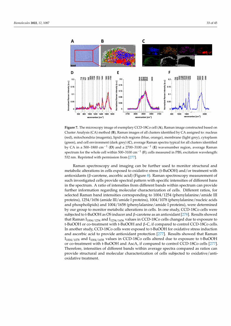
Biomolecules 2022, 12, 1087 33 of 45
Figure 7. The microscopy image of exemplary CCD-18Co cell (A), Raman image constructed based onCluster Analysis (CA) method (B), Raman images of all clusters identified by CA assigned to: nucleus(red), mitochondria (magenta), lipid-rich regions (blue, orange), membrane (light grey), cytoplasm(green), and cell environment (dark grey) (C), average Raman spectra typical for all clusters identifiedby CA in a 500–1800 cm−1 (D) and a 2700–3100 cm−1 (E) wavenumber region, average Ramanspectrum for the whole cell within 500–3100 cm−1 (F); cells measured in PBS, excitation wavelength:532 nm. Reprinted with permission from [277].
Raman spectroscopy and imaging can be further used to monitor structural andmetabolic alterations in cells exposed to oxidative stress (t-BuOOH) and/or treatment withantioxidants (β-carotene, ascorbic acid) (Figure 8). Raman spectroscopy measurement ofsuch investigated cells provide spectral pattern with specific intensities of different bansin the spectrum. A ratio of intensities from different bands within spectrum can providefurther information regarding molecular characterization of cells. Different ratios, forselected Raman band intensities corresponding to 1004/1254 (phenylalanine/amide IIIproteins), 1254/1656 (amide III/amide I proteins), 1004/1078 (phenylalanine/nucleic acidsand phospholipids) and 1004/1658 (phenylalanine/amide I proteins), were determinedby our group to monitor metabolic alterations in cells. In one study, CCD-18Co cells weresubjected to t-BuOOH as OS inducer and β-carotene as an antioxidant [278]. Results showedthat Raman I1004/1254 and I1254/1656 values in CCD-18Co cells changed due to exposure tot-BuOOH or co-treatment with t-BuOOH and β-C, if compared to control CCD-18Co cells.In another study, CCD-18Co cells were exposed to t-BuOOH for oxidative stress inductionand ascorbic acid to provide antioxidant protection [277]. Results showed that RamanI1004/1078 and I1004/1658 values in CCD-18Co cells altered due to exposure to t-BuOOHor co-treatment with t-BuOOH and AscA, if compared to control CCD-18Co cells [277].Therefore, intensities of different bands within average spectra compared as ratios canprovide structural and molecular characterization of cells subjected to oxidative/anti-oxidative treatment.

Biomolecules 2022, 12, 1087 34 of 45
Figure 8. Raman spectroscopy analysis of cells exposed to t-BuOOH, β-carotene and/or Vit C; Ramanimage of CCD-18Co exemplary cells (A), average Raman spectra of exemplary CCD-18Co cell (B),Raman I1004/1254 (C) and Raman I1254/1656 (D) graph values with bars: control, t-BuOOH, t-BuOOH+ β-C, Raman I1004/1078 (E) and Raman I1004/1658 (F) graph values with bars: control, t-BuOOH,t-BuOOH + Vit C. Reprinted and adapted with permission from [277,278].
7.3.8. Measurement of the Oxidative Stress: A Summary
OS increase during in vitro, in vivo and populational studies can be characterized byelevated level of direct and indirect specific (bio)markers, such as intracellular ROS level,fatty acids (lipids) oxidation products (MDA, 4-HNE, F2-isoprostanes), protein oxidationproducts (protein carbonyls) and DNA oxidation products (8-hydroxydeoxyguanosine).The measurements of biomarkers usually depend on the use of spectrophotometric, flu-orometric, chromatographic techniques also coupled with mass spectrometry (LC-MS,GC-MS), as well as ELISA assay. However, the use of these techniques involves a seriesof cumbersome steps including cell disruption and extract preparation, its purificationand separation into specific fractions and derivatization of target molecules with specificcompounds prior to analysis. The alternative and/or support to these techniques could beRaman spectroscopy and imaging that enable the whole-cell characterization of differentchemical structures (proteins, lipids, nucleic acids), without the necessity of cell disrup-tion, and further fractionization and derivatization. Raman spectroscopy measurementcan provide the monitoring of the alteration of cellular chemical profile, based on Ramanspectra analysis, when cells are exposed to stress inducer and/or antioxidant treatment.Indeed, results published by our group show that chemical changes in colon CCD-18Cocells exposed to stress conditions and treated with anti-oxidant (β-carotene, ascorbate) canbe monitored by Raman spectroscopy and imaging. The use of this technique can be furtherextended for characterization of other cells and studying the effect of new anti-oxidants.
8. Conclusions
In this review, antioxidant properties of β-carotene, tocopherols and ascorbic acidare presented based on in vitro, in vivo and populational/clinical studies. Literature datagathered suggests that β-carotene, tocopherols and ascorbic acid possess the potential tomitigate oxidative stress in various biological systems. However, interpretation of gatheredresults faces possible limitations due to different cells, animal species and OS-inducingagents used, discrepancies between in vitro and in vivo studies, as well as insufficientnumber of participants and various origins of oxidative stress in populational/clinical

Biomolecules 2022, 12, 1087 35 of 45
studies. Therefore, further studies are required to determine the effect of β-carotene,tocopherols or ascorbic acid on oxidative stress level and the efficacy of tested antioxidantsin different biological systems (in vitro, in vivo animals, populational/clinical). Oxidativestress (bio)markers are measured by a range of different techniques (spectrophotometric,fluorometric, chromatographic, immuno-enzymatic). Apart from commonly used analyticalmethods, Raman spectroscopy and imaging can be a useful technique to study the effect ofoxidative stress and anti-oxidant molecules in cell studies.
Supplementary Materials: The following are available online at https://www.mdpi.com/article/10.3390/biom12081087/s1, Figure S1: The mechanism of DCFH-DA conversion to DCFH and DCF;Figure S2: The mechanism of reaction between MDA and TBA to form MDA-TBA2; Figure S3:The mechanism of reaction between MDA and DNPH to form MDA-DNPH; Figure S4: The mech-anism of reaction between MDA and PFBB to form MDA-PFB2; Figure S5: The mechanism ofreaction between MDA and PFBHA to form MDA-PFB2-oxime; Figure S6: The mechanism of 4-HNE-PFB-oxime-TMS formation and analysis. PFBHA: pentafluorobenzyl hydroxylamine; BSTFA: N,O-bis(trimethylsilyl)trifluoroacetamide; TMCS: trimethylchlorosilane; TMS: trimethylsilyl; Figure S7:The mechanism of F2-isoprostane (F2-IsoPs) formation and analysis. PFBB: pentafluorobenzyl bro-mide; BSTFA: N,O-bis(trimethylsilyl)trifluoroacetamide; TMS: trimethylsilyl; Figure S8: The mecha-nism of protein carbonyl reaction with dinitrophenylhydrazine (DNPH). Figure S9. The mechanism ofdeoxyguanosine (dG) oxidation into 8-hydroxydeoxyguanosine (8-OHdG) and 8-oxodeoxyguanosine(8-OxodG), and the mechanism of 8-OHdG measurement with ELISA.
Author Contributions: Literature research, K.M.; writing—original draft preparation, K.M.; visual-ization, K.M.; writing—review and editing, K.M.; conceptualization, K.B.; validation and reviewing,A.S.; funding acquisition, B.B.-P. All authors have read and agreed to the published version ofthe manuscript.
Funding: This research received no external funding.
Conflicts of Interest: The authors declare no conflict of interest.
References1. Pizzino, G.; Irrera, N.; Cucinotta, M.; Pallio, G.; Mannino, F.; Arcoraci, V.; Squadrito, F.; Altavilla, D.; Bitto, A. Oxidative Stress:
Harms and Benefits for Human Health. Oxid. Med. Cell Longev. 2017, 2017, 8416763. [CrossRef] [PubMed]2. Sharma, P.; Jha, A.B.; Dubey, R.S.; Pessarakli, M. Reactive Oxygen Species, Oxidative Damage, and Antioxidative Defense
Mechanism in Plants under Stressful Conditions. J. Bot. 2012, 2012, 217037. [CrossRef]3. Juan, C.A.; Pérez de la Lastra, J.M.; Plou, F.J.; Pérez-Lebeña, E. The Chemistry of Reactive Oxygen Species (ROS) Revisited:
Outlining Their Role in Biological Macromolecules (DNA, Lipids and Proteins) and Induced Pathologies. Int. J. Mol. Sci.2021, 22, 4642. [CrossRef] [PubMed]
4. Pedziwiatr, P.; Mikołajczyk, F.; Zawadzki, D.; Mikołajczyk, K.; Bedka, A. Decomposition of Hydrogen Peroxide-Kinetics andReview of Chosen Catalysts. Acta Innov. 2018, 26, 45–52. [CrossRef]
5. Haider, M.S.; Jaskani, M.J.; Fang, J. Overproduction of ROS: Underlying Molecular Mechanism of Scavenging and Redox Signaling.In Biocontrol Agents and Secondary Metabolites; Elsevier: Amsterdam, The Netherlands, 2021; pp. 347–382. ISBN 9780128229194.
6. Kaur, H.; Hippargi, G.; Pophali, G.R.; Bansiwal, A.K. Treatment Methods for Removal of Pharmaceuticals and Personal CareProducts from Domestic Wastewater. In Pharmaceuticals and Personal Care Products: Waste Management and Treatment Technology;Elsevier: Amsterdam, The Netherlands, 2019; pp. 129–150. ISBN 9780128161890.
7. He, L.; He, T.; Farrar, S.; Ji, L.; Liu, T.; Ma, X. Antioxidants Maintain Cellular Redox Homeostasis by Elimination of ReactiveOxygen Species. Cell. Physiol. Biochem. 2017, 44, 532–553. [CrossRef] [PubMed]
8. Turrens, J.F. Mitochondrial Formation of Reactive Oxygen Species. J. Physiol. 2003, 552, 335–344. [CrossRef]9. Kim, S.Y.; Park, J.-W. Cellular Defense against Singlet Oxygen-Induced Oxidative Damage by Cytosolic NADP+-Dependent
Isocitrate Dehydrogenase. Free Radic. Res. 2003, 37, 309–316. [CrossRef]10. García-Sánchez, A.; Miranda-Díaz, A.G.; Cardona-Muñoz, E.G. The Role of Oxidative Stress in Physiopathology and Pharma-
cological Treatment with Pro- and Antioxidant Properties in Chronic Diseases. Oxid. Med. Cell. Longev. 2020, 2020, 2082145.[CrossRef]
11. De Jager, T.L.; Cockrell, A.E.; Du Plessis, S.S. Ultraviolet Light Induced Generation of Reactive Oxygen Species. Adv. Exp.Med. Biol. 2017, 996, 15–23. [CrossRef]
12. Brand, R.M.; Wipf, P.; Durham, A.; Epperly, M.W.; Greenberger, J.S.; Falo, L.D., Jr. Targeting Mitochondrial Oxidative Stress toMitigate UV-Induced Skin Damage. Front. Pharmacol. 2018, 9, 920. [CrossRef]

Biomolecules 2022, 12, 1087 36 of 45
13. Reisz, J.A.; Bansal, N.; Qian, J.; Zhao, W.; Furdui, C.M. Effects of Ionizing Radiation on Biological Molecules—Mechanisms ofDamage and Emerging Methods of Detection. Antioxid. Redox Signal. 2014, 21, 260–292. [CrossRef]
14. Mu, H.; Sun, J.; Li, L.; Yin, J.; Hu, N.; Zhao, W.; Ding, D.; Yi, L. Ionizing Radiation Exposure: Hazards, Prevention, and BiomarkerScreening. Environ. Sci. Pollut. Res. Int. 2018, 25, 15294–15306. [CrossRef]
15. Nuszkiewicz, J.; Wozniak, A.; Szewczyk-Golec, K. Ionizing Radiation as a Source of Oxidative Stress—The Protective Role ofMelatonin and Vitamin D. Int. J. Mol. Sci. 2020, 21, 5804. [CrossRef]
16. Azzam, E.I.; Jay-Gerin, J.-P.; Pain, D. Ionizing Radiation-Induced Metabolic Oxidative Stress and Prolonged Cell Injury. Cancer Lett.2012, 327, 48–60. [CrossRef]
17. Zhang, Y.-J.; Huang, C.; Lv, Y.-S.; Ma, S.-X.; Guo, Y.; Zeng, E.Y. Polycyclic Aromatic Hydrocarbon Exposure, Oxidative Potentialin Dust, and Their Relationships to Oxidative Stress in Human Body: A Case Study in the Indoor Environment of Guangzhou,South China. Environ. Int. 2021, 149, 106405. [CrossRef]
18. Henkler, F.; Brinkmann, J.; Luch, A. The Role of Oxidative Stress in Carcinogenesis Induced by Metals and Xenobiotics. Cancers2010, 2, 376–396. [CrossRef]
19. Klotz, L.-O.; Steinbrenner, H. Cellular Adaptation to Xenobiotics: Interplay between Xenosensors, Reactive Oxygen Species andFOXO Transcription Factors. Redox Biol. 2017, 13, 646–654. [CrossRef]
20. Jabłonska-Trypuc, A.; Wołejko, E.; Wydro, U.; Butarewicz, A. The Impact of Pesticides on Oxidative Stress Level in HumanOrganism and Their Activity as an Endocrine Disruptor. J. Environ. Sci. Health B 2017, 52, 483–494. [CrossRef]
21. Azeh Engwa, G.; Udoka Ferdinand, P.; Nweke Nwalo, F.; Unachukwu, M.N. Mechanism and Health Effects of Heavy Metal Toxic-ity in Humans. In Poisoning in the Modern World-New Tricks for an Old Dog? IntechOpen: London, UK, 2019; ISBN 9781838807856.
22. Ercal, N.; Gurer-Orhan, H.; Aykin-Burns, N. Toxic Metals and Oxidative Stress Part I: Mechanisms Involved in Metal-InducedOxidative Damage. Curr. Top. Med. Chem. 2001, 1, 529–539. [CrossRef]
23. Jomova, K.; Jenisova, Z.; Feszterova, M.; Baros, S.; Liska, J.; Hudecova, D.; Rhodes, C.J.; Valko, M. Arsenic: Toxicity, OxidativeStress and Human Disease: Toxicity of Arsenic. J. Appl. Toxicol. 2011, 31, 95–107. [CrossRef]
24. Jomova, K.; Valko, M. Advances in Metal-Induced Oxidative Stress and Human Disease. Toxicology 2011, 283, 65–87. [CrossRef][PubMed]
25. Deavall, D.G.; Martin, E.A.; Horner, J.M.; Roberts, R. Drug-Induced Oxidative Stress and Toxicity. J. Toxicol. 2012, 2012, 645460.[CrossRef] [PubMed]
26. Ramkumar, V.; Mukherjea, D.; Dhukhwa, A.; Rybak, L.P. Oxidative Stress and Inflammation Caused by Cisplatin Ototoxicity.Antioxidants 2021, 10, 1919. [CrossRef] [PubMed]
27. McSweeney, K.R.; Gadanec, L.K.; Qaradakhi, T.; Ali, B.A.; Zulli, A.; Apostolopoulos, V. Mechanisms of Cisplatin-InducedAcute Kidney Injury: Pathological Mechanisms, Pharmacological Interventions, and Genetic Mitigations. Cancers 2021, 13, 1572.[CrossRef]
28. Kamceva, G.; Arsova-Sarafinovska, Z.; Ruskovska, T.; Zdravkovska, M.; Kamceva-Panova, L.; Stikova, E. Cigarette Smoking andOxidative Stress in Patients with Coronary Artery Disease. Open Access Maced. J. Med. Sci. 2016, 4, 636–640. [CrossRef]
29. Valavanidis, A.; Vlachogianni, T.; Fiotakis, K. Tobacco Smoke: Involvement of Reactive Oxygen Species and Stable Free Radicalsin Mechanisms of Oxidative Damage, Carcinogenesis and Synergistic Effects with Other Respirable Particles. Int. J. Environ. Res.Public Health 2009, 6, 445–462. [CrossRef]
30. Faux, S.P.; Tai, T.; Thorne, D.; Xu, Y.; Breheny, D.; Gaca, M. The Role of Oxidative Stress in the Biological Responses of LungEpithelial Cells to Cigarette Smoke. Biomarkers 2009, 14 (Suppl. 1), 90–96. [CrossRef]
31. Isik, B.; Ceylan, A.; Isik, R. Oxidative Stress in Smokers and Non-Smokers. Inhal. Toxicol. 2007, 19, 767–769. [CrossRef]32. Iriti, M.; Faoro, F. Oxidative Stress, the Paradigm of Ozone Toxicity in Plants and Animals. Water Air Soil Pollut. 2007, 187, 285–301.
[CrossRef]33. Nery-Flores, S.D.; Mendoza-Magaña, M.L.; Ramírez-Herrera, M.A.; Ramírez-Vázquez, J.d.J.; Romero-Prado, M.M.d.J.; Cortez-
Álvarez, C.R.; Ramírez-Mendoza, A.A. Curcumin Exerted Neuroprotection against Ozone-Induced Oxidative Damage andDecreased NF-KB Activation in Rat Hippocampus and Serum Levels of Inflammatory Cytokines. Oxid. Med. Cell. Longev.2018, 2018, 9620684. [CrossRef]
34. Tricarico, G.; Travagli, V. The Relationship between Ozone and Human Blood in the Course of a Well-Controlled, Mild, andTransitory Oxidative Eustress. Antioxidants 2021, 10, 1946. [CrossRef]
35. Rivas-Arancibia, S.; Guevara-Guzmán, R.; López-Vidal, Y.; Rodríguez-Martínez, E.; Zanardo-Gomes, M.; Angoa-Pérez, M.;Raisman-Vozari, R. Oxidative Stress Caused by Ozone Exposure Induces Loss of Brain Repair in the Hippocampus of Adult Rats.Toxicol. Sci. 2010, 113, 187–197. [CrossRef]
36. Schwarz, K.B. Oxidative Stress during Viral Infection: A Review. Free Radic. Biol. Med. 1996, 21, 641–649. [CrossRef]37. Lee, C. Therapeutic Modulation of Virus-Induced Oxidative Stress via the Nrf2-Dependent Antioxidative Pathway. Oxid. Med.
Cell. Longev. 2018, 2018, 6208067. [CrossRef]38. Butcher, L.D.; den Hartog, G.; Ernst, P.B.; Crowe, S.E. Oxidative Stress Resulting from Helicobacter Pylori Infection Contributes to
Gastric Carcinogenesis. Cell Mol. Gastroenterol. Hepatol. 2017, 3, 316–322. [CrossRef]39. Tan, B.L.; Norhaizan, M.E.; Liew, W.-P.-P. Nutrients and Oxidative Stress: Friend or Foe? Oxid. Med. Cell. Longev.
2018, 2018, 9719584. [CrossRef]

Biomolecules 2022, 12, 1087 37 of 45
40. Matsuda, M.; Shimomura, I. Increased Oxidative Stress in Obesity: Implications for Metabolic Syndrome, Diabetes, Hypertension,Dyslipidemia, Atherosclerosis, and Cancer. Obes. Res. Clin. Pract. 2013, 7, e330–e341. [CrossRef]
41. Jiang, S.; Liu, H.; Li, C. Dietary Regulation of Oxidative Stress in Chronic Metabolic Diseases. Foods 2021, 10, 1854. [CrossRef]42. Nolfi-Donegan, D.; Braganza, A.; Shiva, S. Mitochondrial Electron Transport Chain: Oxidative Phosphorylation, Oxidant
Production, and Methods of Measurement. Redox Biol. 2020, 37, 101674. [CrossRef]43. Zhao, R.-Z.; Jiang, S.; Zhang, L.; Yu, Z.-B. Mitochondrial Electron Transport Chain, ROS Generation and Uncoupling (Review).
Int. J. Mol. Med. 2019, 44, 3–15. [CrossRef]44. Raimondi, V.; Ciccarese, F.; Ciminale, V. Oncogenic Pathways and the Electron Transport Chain: A DangeROS Liaison. Br. J. Cancer
2020, 122, 168–181. [CrossRef]45. Tarafdar, A.; Pula, G. The Role of NADPH Oxidases and Oxidative Stress in Neurodegenerative Disorders. Int. J. Mol. Sci.
2018, 19, 3824. [CrossRef]46. Paravicini, T.M.; Touyz, R.M. NADPH Oxidases, Reactive Oxygen Species, and Hypertension: Clinical Implications and
Therapeutic Possibilities. Diabetes Care 2008, 31, S170–S180. [CrossRef]47. Guo, S.; Chen, X. The Human Nox4: Gene, Structure, Physiological Function and Pathological Significance. J. Drug Target.
2015, 23, 888–896. [CrossRef] [PubMed]48. Brandes, R.P.; Weissmann, N.; Schröder, K. Nox Family NADPH Oxidases: Molecular Mechanisms of Activation. Free Radic.
Biol. Med. 2014, 76, 208–226. [CrossRef] [PubMed]49. Touyz, R.M.; Anagnostopoulou, A.; Rios, F.; Montezano, A.C.; Camargo, L.L. NOX5: Molecular Biology and Pathophysiology.
Exp. Physiol. 2019, 104, 605–616. [CrossRef] [PubMed]50. George, A.; Pushkaran, S.; Konstantinidis, D.G.; Koochaki, S.; Malik, P.; Mohandas, N.; Zheng, Y.; Joiner, C.H.; Kalfa, T.A.
Erythrocyte NADPH Oxidase Activity Modulated by Rac GTPases, PKC, and Plasma Cytokines Contributes to Oxidative Stressin Sickle Cell Disease. Blood 2013, 121, 2099–2107. [CrossRef]
51. Violi, F.; Carnevale, R.; Loffredo, L.; Pignatelli, P.; Gallin, J.I. NADPH Oxidase-2 and Atherothrombosis: Insight from ChronicGranulomatous Disease. Arterioscler. Thromb. Vasc. Biol. 2017, 37, 218–225. [CrossRef]
52. Vermot, A.; Petit-Härtlein, I.; Smith, S.M.E.; Fieschi, F. NADPH Oxidases (NOX): An Overview from Discovery, MolecularMechanisms to Physiology and Pathology. Antioxidants 2021, 10, 890. [CrossRef]
53. Magnani, F.; Mattevi, A. Structure and Mechanisms of ROS Generation by NADPH Oxidases. Curr. Opin. Struct. Biol.2019, 59, 91–97. [CrossRef]
54. Stavropoulou, E.; Pircalabioru, G.G.; Bezirtzoglou, E. The Role of Cytochromes P450 in Infection. Front. Immunol. 2018, 9, 89.[CrossRef]
55. Hrycay, E.G.; Bandiera, S.M. Involvement of Cytochrome P450 in Reactive Oxygen Species Formation and Cancer. Adv. Pharmacol.2015, 74, 35–84. [CrossRef]
56. Veith, A.; Moorthy, B. Role of Cytochrome P450s in the Generation and Metabolism of Reactive Oxygen Species. Curr. Opin. Toxicol.2018, 7, 44–51. [CrossRef]
57. Mashima, R.; Okuyama, T. The Role of Lipoxygenases in Pathophysiology; New Insights and Future Perspectives. Redox Biol.2015, 6, 297–310. [CrossRef]
58. Qian, S.; Guo, L.; Tang, Q. Lipids in the Transcriptional Regulation of Adipocyte Differentiation and Metabolism. In Lipid Signalingand Metabolism; Elsevier: Amsterdam, The Netherlands, 2020; pp. 81–98. ISBN 9780128194041.
59. Cho, K.-J.; Seo, J.-M.; Kim, J.-H. Bioactive Lipoxygenase Metabolites Stimulation of NADPH Oxidases and Reactive OxygenSpecies. Mol. Cells 2011, 32, 1–5. [CrossRef]
60. Suzuki, H.; Kayama, Y.; Sakamoto, M.; Iuchi, H.; Shimizu, I.; Yoshino, T.; Katoh, D.; Nagoshi, T.; Tojo, K.; Minamino, T.; et al.Arachidonate 12/15-Lipoxygenase-Induced Inflammation and Oxidative Stress Are Involved in the Development of DiabeticCardiomyopathy. Diabetes 2015, 64, 618–630. [CrossRef]
61. Chornyi, S.; IJlst, L.; van Roermund, C.W.T.; Wanders, R.J.A.; Waterham, H.R. Peroxisomal Metabolite and Cofactor Transport inHumans. Front. Cell Dev. Biol. 2020, 8, 613892. [CrossRef]
62. Wanders, R.J.A.; Vaz, F.M.; Ferdinandusse, S.; Kemp, S.; Ebberink, M.S.; Waterham, H.R. Laboratory Diagnosis of PeroxisomalDisorders in the -Omics Era and the Continued Importance of Biomarkers and Biochemical Studies. J. Inborn Errors Metab. Screen.2018, 6, 232640981881028. [CrossRef]
63. Schrader, M.; Fahimi, H.D. Peroxisomes and Oxidative Stress. Biochim. Biophys. Acta 2006, 1763, 1755–1766. [CrossRef]64. Aziz, N.; Jamil, R.T. Biochemistry, Xanthine Oxidase; StatPearls Publishing: Treasure Island, FL, USA, 2022.65. Bortolotti, M.; Polito, L.; Battelli, M.G.; Bolognesi, A. Xanthine Oxidoreductase: One Enzyme for Multiple Physiological Tasks.
Redox Biol. 2021, 41, 101882. [CrossRef]66. Ferdinandusse, S.; Denis, S.; van Roermund, C.W.T.; Preece, M.A.; Koster, J.; Ebberink, M.S.; Waterham, H.R.; Wanders, R.J.A. A
Novel Case of ACOX2 Deficiency Leads to Recognition of a Third Human Peroxisomal Acyl-CoA Oxidase. Biochim. Biophys. ActaMol. Basis Dis. 2018, 1864, 952–958. [CrossRef] [PubMed]
67. Dansen, T.B.; Wirtz, K.W. The Peroxisome in Oxidative Stress. IUBMB Life 2001, 51, 223–230. [CrossRef] [PubMed]68. Oaxaca-Castillo, D.; Andreoletti, P.; Vluggens, A.; Yu, S.; van Veldhoven, P.P.; Reddy, J.K.; Cherkaoui-Malki, M. Biochemical Char-
acterization of Two Functional Human Liver Acyl-CoA Oxidase Isoforms 1a and 1b Encoded by a Single Gene. Biochem. Biophys.Res. Commun. 2007, 360, 314–319. [CrossRef]

Biomolecules 2022, 12, 1087 38 of 45
69. Grimsrud, P.A.; Xie, H.; Griffin, T.J.; Bernlohr, D.A. Oxidative Stress and Covalent Modification of Protein with BioactiveAldehydes. J. Biol. Chem. 2008, 283, 21837–21841. [CrossRef]
70. Goto, S.; Radak, Z. Implications of Oxidative Damage to Proteins and DNA in Aging and Its Intervention by Caloric Restrictionand Exercise. J. Sport Health Sci. 2013, 2, 75–80. [CrossRef]
71. Nimse, S.B.; Pal, D. Free Radicals, Natural Antioxidants, and Their Reaction Mechanisms. RSC Adv. 2015, 5, 27986–28006.[CrossRef]
72. Marseglia, L.; Manti, S.; D’Angelo, G.; Nicotera, A.; Parisi, E.; Di Rosa, G.; Gitto, E.; Arrigo, T. Oxidative Stress in Obesity: ACritical Component in Human Diseases. Int. J. Mol. Sci. 2015, 16, 378–400. [CrossRef] [PubMed]
73. Pérez-Torres, I.; Castrejón-Téllez, V.; Soto, M.E.; Rubio-Ruiz, M.E.; Manzano-Pech, L.; Guarner-Lans, V. Oxidative Stress, PlantNatural Antioxidants, and Obesity. Int. J. Mol. Sci. 2021, 22, 1786. [CrossRef]
74. Manna, P.; Jain, S.K. Obesity, Oxidative Stress, Adipose Tissue Dysfunction, and the Associated Health Risks: Causes andTherapeutic Strategies. Metab. Syndr. Relat. Disord. 2015, 13, 423–444. [CrossRef]
75. Aquila, G.; Marracino, L.; Martino, V.; Calabria, D.; Campo, G.; Caliceti, C.; Rizzo, P. The Use of Nutraceuticals to CounteractAtherosclerosis: The Role of the Notch Pathway. Oxid. Med. Cell. Longev. 2019, 2019, 5470470. [CrossRef]
76. Zmysłowski, A.; Szterk, A. Current Knowledge on the Mechanism of Atherosclerosis and Pro-Atherosclerotic Properties ofOxysterols. Lipids Health Dis. 2017, 16, 188. [CrossRef]
77. Khatana, C.; Saini, N.K.; Chakrabarti, S.; Saini, V.; Sharma, A.; Saini, R.V.; Saini, A.K. Mechanistic Insights into the OxidizedLow-Density Lipoprotein-Induced Atherosclerosis. Oxid. Med. Cell Longev. 2020, 2020, 5245308. [CrossRef]
78. Mathur, P.; Ding, Z.; Saldeen, T.; Mehta, J.L. Tocopherols in the Prevention and Treatment of Atherosclerosis and RelatedCardiovascular Disease: Tocopherols in Atherosclerosis. Clin. Cardiol. 2015, 38, 570–576. [CrossRef]
79. Babaniamansour, P.; Mohammadi, M.; Babaniamansour, S.; Aliniagerdroudbari, E. The Relation between Atherosclerosis PlaqueComposition and Plaque Rupture. J. Med. Signals Sens. 2020, 10, 267–273. [CrossRef]
80. Asmat, U.; Abad, K.; Ismail, K. Diabetes Mellitus and Oxidative Stress—A Concise Review. Saudi Pharm. J. 2016, 24, 547–553.[CrossRef]
81. Burgos-Morón, E.; Abad-Jiménez, Z.; Martínez de Marañón, A.; Iannantuoni, F.; Escribano-López, I.; López-Domènech, S.; Salom,C.; Jover, A.; Mora, V.; Roldan, I.; et al. Relationship between Oxidative Stress, ER Stress, and Inflammation in Type 2 Diabetes:The Battle Continues. J. Clin. Med. 2019, 8, 1385. [CrossRef] [PubMed]
82. Quiñonez-Flores, C.M.; González-Chávez, S.A.; Del Río Nájera, D.; Pacheco-Tena, C. Oxidative Stress Relevance in the Pathogene-sis of the Rheumatoid Arthritis: A Systematic Review. BioMed. Res. Int. 2016, 2016, 6097417. [CrossRef]
83. Cooper, G.M. The Development and Causes of Cancer. In The Cell: A Molecular Approach, 2nd ed.; Sinauer Associates: Sunderland,MA, USA, 2000; ISBN 100878931066.
84. Hassanpour, S.H.; Dehghani, M. Review of Cancer from Perspective of Molecular. J. Cancer Res. Pract. 2017, 4, 127–129. [CrossRef]85. Kamal, N.; Ilowefah, M.A.; Hilles, A.R.; Anua, N.A.; Awin, T.; Alshwyeh, H.A.; Aldosary, S.K.; Jambocus, N.G.S.; Alosaimi, A.A.;
Rahman, A.; et al. Genesis and Mechanism of Some Cancer Types and an Overview on the Role of Diet and Nutrition in CancerPrevention. Molecules 2022, 27, 1794. [CrossRef]
86. Sung, H.; Ferlay, J.; Siegel, R.L.; Laversanne, M.; Soerjomataram, I.; Jemal, A.; Bray, F. Global Cancer Statistics 2020: GLOBOCANEstimates of Incidence and Mortality Worldwide for 36 Cancers in 185 Countries. CA Cancer J. Clin. 2021, 71, 209–249. [CrossRef]
87. Poetsch, A.R. The Genomics of Oxidative DNA Damage, Repair, and Resulting Mutagenesis. Comput. Struct. Biotechnol. J.2020, 18, 207–219. [CrossRef] [PubMed]
88. Klaunig, J.E.; Kamendulis, L.M.; Hocevar, B.A. Oxidative Stress and Oxidative Damage in Carcinogenesis. Toxicol. Pathol.2010, 38, 96–109. [CrossRef] [PubMed]
89. Bridge, G.; Rashid, S.; Martin, S.A. DNA Mismatch Repair and Oxidative DNA Damage: Implications for Cancer Biology andTreatment. Cancers 2014, 6, 1597–1614. [CrossRef] [PubMed]
90. Liu, Z.; Zhou, T.; Ziegler, A.C.; Dimitrion, P.; Zuo, L. Oxidative Stress in Neurodegenerative Diseases: From Molecular Mechanismsto Clinical Applications. Oxid. Med. Cell. Longev. 2017, 2017, 2525967. [CrossRef]
91. Niedzielska, E.; Smaga, I.; Gawlik, M.; Moniczewski, A.; Stankowicz, P.; Pera, J.; Filip, M. Oxidative Stress in NeurodegenerativeDiseases. Mol. Neurobiol. 2016, 53, 4094–4125. [CrossRef]
92. Kim, G.H.; Kim, J.E.; Rhie, S.J.; Yoon, S. The Role of Oxidative Stress in Neurodegenerative Diseases. Exp. Neurobiol. 2015, 24,325–340. [CrossRef]
93. Rosa, A.C.; Corsi, D.; Cavi, N.; Bruni, N.; Dosio, F. Superoxide Dismutase Administration: A Review of Proposed Human Uses.Molecules 2021, 26, 1844. [CrossRef]
94. Trist, B.G.; Hilton, J.B.; Hare, D.J.; Crouch, P.J.; Double, K.L. Superoxide Dismutase 1 in Health and Disease: How a FrontlineAntioxidant Becomes Neurotoxic. Angew. Chem. Int. Ed. Engl. 2021, 60, 9215–9246. [CrossRef]
95. Ighodaro, O.M.; Akinloye, O.A. First Line Defence Antioxidants-Superoxide Dismutase (SOD), Catalase (CAT) and GlutathionePeroxidase (GPX): Their Fundamental Role in the Entire Antioxidant Defence Grid. Alex. J. Med. 2018, 54, 287–293. [CrossRef]
96. Nandi, A.; Yan, L.-J.; Jana, C.K.; Das, N. Role of Catalase in Oxidative Stress- and Age-Associated Degenerative Diseases.Oxid. Med. Cell. Longev. 2019, 2019, 9613090. [CrossRef]
97. Sarıkaya, E.; Dogan, S. Glutathione Peroxidase in Health and Diseases. In Glutathione System and Oxidative Stress in Health andDisease; IntechOpen: London, UK, 2020; ISBN 9781838801250.
Biomolecules 2022, 12, 1087 39 of 45
98. Pizzorno, J. Glutathione! Integ. Med. 2014, 13, 8–12.99. Lubos, E.; Loscalzo, J.; Handy, D.E. Glutathione Peroxidase-1 in Health and Disease: From Molecular Mechanisms to Therapeutic
Opportunities. Antioxid. Redox Signal. 2011, 15, 1957–1997. [CrossRef]100. Marín-García, J. Oxidative Stress and Cell Death in Cardiovascular Disease. In Post-Genomic Cardiology; Elsevier: Amsterdam, The
Netherlands, 2014; pp. 471–498. ISBN 9780124045996.101. Deponte, M. Glutathione Catalysis and the Reaction Mechanisms of Glutathione-Dependent Enzymes. Biochim. Biophys. Acta
2013, 1830, 3217–3266. [CrossRef]102. Ren, X.; Zou, L.; Zhang, X.; Branco, V.; Wang, J.; Carvalho, C.; Holmgren, A.; Lu, J. Redox Signaling Mediated by Thioredoxin and
Glutathione Systems in the Central Nervous System. Antioxid. Redox Signal. 2017, 27, 989–1010. [CrossRef]103. Tabet, F.; Touyz, R.M. Reactive Oxygen Species, Oxidative Stress, and Vascular Biology in Hypertension. In Comprehensive
Hypertension; Elsevier: Amsterdam, The Netherlands, 2007; pp. 337–347. ISBN 9780323039611.104. Becker, K.; Gromer, S.; Schirmer, R.H.; Müller, S. Thioredoxin Reductase as a Pathophysiological Factor and Drug Target:
Thioredoxin Reductase in Medicine and Parasitology. Eur. J. Biochem. 2000, 267, 6118–6125. [CrossRef]105. Vlamis-Gardikas, A.; Holmgren, A. Thioredoxin and Glutaredoxin Isoforms. Methods Enzymol. 2002, 347, 286–296. [CrossRef]106. Lundberg, M.; Johansson, C.; Chandra, J.; Enoksson, M.; Jacobsson, G.; Ljung, J.; Johansson, M.; Holmgren, A. Cloning
and Expression of a Novel Human Glutaredoxin (Grx2) with Mitochondrial and Nuclear Isoforms. J. Biol. Chem. 2001, 276,26269–26275. [CrossRef]
107. Évora, A.S.; Adams, M.J.; Johnson, S.A.; Zhang, Z. Corneocytes: Relationship between Structural and Biomechanical Properties.Ski. Pharmacol. Physiol. 2021, 34, 146–161. [CrossRef]
108. Nozaki, I.; Lunz, J.G., 3rd; Specht, S.; Stolz, D.B.; Taguchi, K.; Subbotin, V.M.; Murase, N.; Demetris, A.J. Small Proline-RichProteins 2 Are Noncoordinately Upregulated by IL-6/STAT3 Signaling after Bile Duct Ligation. Lab. Investig. 2005, 85, 109–123.[CrossRef]
109. Ishitsuka, Y.; Ogawa, T.; Roop, D. The KEAP1/NRF2 Signaling Pathway in Keratinization. Antioxidants 2020, 9, 751. [CrossRef]110. Vermeij, W.P.; Alia, A.; Backendorf, C. ROS Quenching Potential of the Epidermal Cornified Cell Envelope. J. Investig. Dermatol.
2011, 131, 1435–1441. [CrossRef]111. Schäfer, M.; Werner, S. The Cornified Envelope: A First Line of Defense against Reactive Oxygen Species. J. Investig. Dermatol.
2011, 131, 1409–1411. [CrossRef]112. MacKenzie, E.L.; Iwasaki, K.; Tsuji, Y. Intracellular Iron Transport and Storage: From Molecular Mechanisms to Health Implica-
tions. Antioxid. Redox Signal. 2008, 10, 997–1030. [CrossRef]113. Katsarou, M.-S.; Papasavva, M.; Latsi, R.; Drakoulis, N. Hemochromatosis: Hereditary Hemochromatosis and HFE Gene.
Vitam. Horm. 2019, 110, 201–222. [CrossRef] [PubMed]114. Higuchi, M. Antioxidant Properties of Wheat Bran against Oxidative Stress. In Wheat and Rice in Disease Prevention and Health;
Elsevier: Amsterdam, The Netherlands, 2014; pp. 181–199. ISBN 9780124017160.115. Imam, M.U.; Zhang, S.; Ma, J.; Wang, H.; Wang, F. Antioxidants Mediate Both Iron Homeostasis and Oxidative Stress. Nutrients
2017, 9, 671. [CrossRef] [PubMed]116. Romeu, M.; Aranda, N.; Giralt, M.; Ribot, B.; Nogues, M.R.; Arija, V. Diet, Iron Biomarkers and Oxidative Stress in a Representative
Sample of Mediterranean Population. Nutr. J. 2013, 12, 102. [CrossRef] [PubMed]117. Orino, K.; Lehman, L.; Tsuji, Y.; Ayaki, H.; Torti, S.V.; Torti, F.M. Ferritin and the Response to Oxidative Stress. Biochem. J.
2001, 357, 241. [CrossRef] [PubMed]118. Marappan, G. Coenzyme Q10: Regulators of Mitochondria and Beyond. In Apolipoproteins, Triglycerides and Cholesterol; IntechOpen:
London, UK, 2020; ISBN 9781839625190.119. Acosta, M.J.; Vazquez Fonseca, L.; Desbats, M.A.; Cerqua, C.; Zordan, R.; Trevisson, E.; Salviati, L. Coenzyme Q Biosynthesis in
Health and Disease. Biochim. Biophys. Acta 2016, 1857, 1079–1085. [CrossRef] [PubMed]120. Yubero-Serrano, E.M.; Garcia-Rios, A.; Delgado-Lista, J.; Pérez-Martinez, P.; Camargo, A.; Perez-Jimenez, F.; Lopez-Miranda,
J. Coenzyme Q10 as an Antioxidant in the Elderly. In Aging; Elsevier: Amsterdam, The Netherlands, 2014; pp. 109–117.ISBN 9780124059337.
121. Sangsefidi, Z.S.; Yaghoubi, F.; Hajiahmadi, S.; Hosseinzadeh, M. The Effect of Coenzyme Q10 Supplementation on Oxidative Stress:A Systematic Review and Meta-Analysis of Randomized Controlled Clinical Trials: The Effect of Coenzyme Q10 Supplementationon Oxidative Stress: A Systematic Review and Meta-Analysis of Randomized Controlled Clinical Trials. Food Sci. Nutr. 2020, 8,1766–1776. [CrossRef]
122. Melhuish Beaupre, L.M.; Brown, G.M.; Gonçalves, V.F.; Kennedy, J.L. Melatonin’s Neuroprotective Role in Mitochondria and ItsPotential as a Biomarker in Aging, Cognition and Psychiatric Disorders. Transl. Psychiatry 2021, 11, 339. [CrossRef]
123. Kopustinskiene, D.M.; Bernatoniene, J. Molecular Mechanisms of Melatonin-Mediated Cell Protection and Signaling in Healthand Disease. Pharmaceutics 2021, 13, 129. [CrossRef]
124. Reiter, R.J.; Tan, D.-X.; Mayo, J.C.; Sainz, R.M.; Leon, J.; Czarnocki, Z. Melatonin as an Antioxidant: Biochemical Mechanisms andPathophysiological Implications in Humans. Acta Biochim. Pol. 2003, 50, 1129–1146. [CrossRef]
125. Reiter, R.J.; Sharma, R.; Rosales-Corral, S.; Manucha, W.; Chuffa, L.G.d.A.; Zuccari, D.A.P.d.C. Melatonin and Pathological CellInteractions: Mitochondrial Glucose Processing in Cancer Cells. Int. J. Mol. Sci. 2021, 22, 12494. [CrossRef]
126. Malhotra, S.; Sawhney, G.; Pandhi, P. The Therapeutic Potential of Melatonin: A Review of the Science. MedGenMed 2004, 6, 46.
Biomolecules 2022, 12, 1087 40 of 45
127. Ziberna, L.; Martelanc, M.; Franko, M.; Passamonti, S. Bilirubin Is an Endogenous Antioxidant in Human Vascular EndothelialCells. Sci. Rep. 2016, 6, 29240. [CrossRef]
128. Adin, C.A. Bilirubin as a Therapeutic Molecule: Challenges and Opportunities. Antioxidants 2021, 10, 1536. [CrossRef]129. Inoguchi, T.; Sonoda, N.; Maeda, Y. Bilirubin as an Important Physiological Modulator of Oxidative Stress and Chronic Inflamma-
tion in Metabolic Syndrome and Diabetes: A New Aspect on Old Molecule. Diabetol. Int. 2016, 7, 338–341. [CrossRef]130. Sedlak, T.W.; Saleh, M.; Higginson, D.S.; Paul, B.D.; Juluri, K.R.; Snyder, S.H. Bilirubin and Glutathione Have Complementary
Antioxidant and Cytoprotective Roles. Proc. Natl. Acad. Sci. USA 2009, 106, 5171–5176. [CrossRef]131. Otero Regino, W.; Velasco, H.; Sandoval, H. The protective role of bilirubin in human beings. Rev. Colomb. Gastroenterol. 2009, 24,
293–301.132. DiNicolantonio, J.J.; McCarty, M.F.; O’Keefe, J.H. Antioxidant Bilirubin Works in Multiple Ways to Reduce Risk for Obesity and
Its Health Complications. Open Heart 2018, 5, e000914. [CrossRef]133. Anthony, R.M.; MacLeay, J.M.; Jewell, D.E.; Brejda, J.J.; Gross, K.L. Alpha-Lipoic Acid Is an Effective Nutritive Antioxidant for
Healthy Adult Dogs. Animals 2021, 11, 274. [CrossRef]134. Golbidi, S.; Badran, M.; Laher, I. Diabetes and Alpha Lipoic Acid. Front. Pharmacol. 2011, 2, 69. [CrossRef]135. Ghibu, S.; Craciun, C.E.; Rusu, R.; Morgovan, C.; Mogosan, C.; Rochette, L.; Gal, A.F.; Dronca, M. Impact of Alpha-Lipoic
Acid Chronic Discontinuous Treatment in Cardiometabolic Disorders and Oxidative Stress Induced by Fructose Intake in Rats.Antioxidants 2019, 8, 636. [CrossRef]
136. Elshazly, S.M.; El-Moselhy, M.A.; Barakat, W. Insights in the Mechanism Underlying the Protective Effect of α-Lipoic Acid againstAcetaminophen-Hepatotoxicity. Eur. J. Pharmacol. 2014, 726, 116–123. [CrossRef]
137. Shay, K.P.; Moreau, R.F.; Smith, E.J.; Smith, A.R.; Hagen, T.M. Alpha-Lipoic Acid as a Dietary Supplement: Molecular Mechanismsand Therapeutic Potential. Biochim. Biophys. Acta Gen. Subj. 2009, 1790, 1149–1160. [CrossRef]
138. Tenório, M.C.d.S.; Graciliano, N.G.; Moura, F.A.; Oliveira, A.C.M.d.; Goulart, M.O.F. N-Acetylcysteine (NAC): Impacts on HumanHealth. Antioxidants 2021, 10, 967. [CrossRef]
139. Aldini, G.; Altomare, A.; Baron, G.; Vistoli, G.; Carini, M.; Borsani, L.; Sergio, F. N-Acetylcysteine as an Antioxidant andDisulphide Breaking Agent: The Reasons Why. Free Radic. Res. 2018, 52, 751–762. [CrossRef]
140. Magdalena Radomska-Lesniewska, D.; Skopinski, P. N-acetylcysteine as an Anti-oxidant and Anti-inflammatory Drug and itsSome Clinical Applications. Centr. Eur. J. Immunol. 2012, 37, 57–66.
141. Tian, B.; Liu, J. Resveratrol: A Review of Plant Sources, Synthesis, Stability, Modification and Food Application. J. Sci. Food Agric.2020, 100, 1392–1404. [CrossRef]
142. Ramírez-Garza, S.L.; Laveriano-Santos, E.P.; Marhuenda-Muñoz, M.; Storniolo, C.E.; Tresserra-Rimbau, A.; Vallverdú-Queralt, A.;Lamuela-Raventós, R.M. Health Effects of Resveratrol: Results from Human Intervention Trials. Nutrients 2018, 10, 1892.[CrossRef] [PubMed]
143. Salehi, B.; Mishra, A.P.; Nigam, M.; Sener, B.; Kilic, M.; Sharifi-Rad, M.; Fokou, P.V.T.; Martins, N.; Sharifi-Rad, J. Resveratrol: ADouble-Edged Sword in Health Benefits. Biomedicines 2018, 6, 91. [CrossRef] [PubMed]
144. Gülçin, I. Antioxidant Properties of Resveratrol: A Structure–Activity Insight. Innov. Food Sci. Emerg. Technol. 2010, 11, 210–218.[CrossRef]
145. Papuc, C.; Goran, G.V.; Predescu, C.N.; Nicorescu, V.; Stefan, G. Plant Polyphenols as Antioxidant and Antibacterial Agentsfor Shelf-Life Extension of Meat and Meat Products: Classification, Structures, Sources, and Action Mechanisms: PolyphenolsExtending Meat Shelf-Life . . . . Compr. Rev. Food Sci. Food Saf. 2017, 16, 1243–1268. [CrossRef] [PubMed]
146. Xia, N.; Daiber, A.; Förstermann, U.; Li, H. Antioxidant Effects of Resveratrol in the Cardiovascular System: Antioxidant Effectsof Resveratrol. Br. J. Pharmacol. 2017, 174, 1633–1646. [CrossRef]
147. Stahl, W.; Sies, H. Bioactivity and Protective Effects of Natural Carotenoids. Biochim. Biophys. Acta 2005, 1740, 101–107. [CrossRef]148. Fiedor, J.; Burda, K. Potential Role of Carotenoids as Antioxidants in Human Health and Disease. Nutrients 2014, 6, 466–488.
[CrossRef]149. Fiedor, J.; Fiedor, L.; Haessner, R.; Scheer, H. Cyclic Endoperoxides of Beta-Carotene, Potential pro-Oxidants, as Products of
Chemical Quenching of Singlet Oxygen. Biochim. Biophys. Acta 2005, 1709, 1–4. [CrossRef]150. Terao, J.; Minami, Y.; Bando, N. Singlet Molecular Oxygen-Quenching Activity of Carotenoids: Relevance to Protection of the
Skin from Photoaging. J. Clin. Biochem. Nutr. 2011, 48, 57–62. [CrossRef]151. Kiokias, S.; Gordon, M.H. Antioxidant Properties of Carotenoids in Vitro and in Vivo. Food Rev. Int. 2004, 20, 99–121. [CrossRef]152. Moussa, Z.; Judeh, Z.M.A.; Ahmed, S.A. Nonenzymatic Exogenous and Endogenous Antioxidants. In Free Radical Medicine and
Biology; IntechOpen: London, UK, 2020; ISBN 9781789851434.153. Chang, C.-S.; Chang, C.-L.; Lai, G.-H. Reactive Oxygen Species Scavenging Activities in a Chemiluminescence Model and
Neuroprotection in Rat Pheochromocytoma Cells by Astaxanthin, Beta-Carotene, and Canthaxanthin. Kaohsiung J. Med. Sci.2013, 29, 412–421. [CrossRef]
154. Maoka, T. Carotenoids as Natural Functional Pigments. J. Nat. Med. 2020, 74, 1–16. [CrossRef]155. Nishino, A.; Yasui, H.; Maoka, T. Reaction and Scavenging Mechanism of β-Carotene and Zeaxanthin with Reactive Oxygen
Species. J. Oleo Sci. 2017, 66, 77–84. [CrossRef]156. Yamauchi, R. Vitamin E: Mechanism of Its Antioxidant Activity. Food Sci. Technol. Int. Tokyo 1997, 3, 301–309. [CrossRef]
Biomolecules 2022, 12, 1087 41 of 45
157. Tucker, J.M.; Townsend, D.M. Alpha-Tocopherol: Roles in Prevention and Therapy of Human Disease. Biomed. Pharmacother.2005, 59, 380–387. [CrossRef]
158. Ebhohimen, I.E.; Okanlawon, T.S.; Osagie, A.O.; Izevbigie, O.N. Vitamin E in Human Health and Oxidative Stress RelatedDiseases. In Vitamin E in Health and Disease—Interactions, Diseases and Health Aspects; Erkekoglu, P., Santos, J.S., Eds.; IntechOpen:London, UK, 2021; Available online: https://www.intechopen.com/chapters/77766 (accessed on 7 June 2022). [CrossRef]
159. Ha, Y.L.; Csallany, A.S. Alpha-Tocopherol Oxidation Mediated by Superoxide Anion (O2-). II. Identification of the StableAlpha-Tocopherol Oxidation Products. Lipids 1992, 27, 201–205. [CrossRef]
160. Yamauchi, R.; Matsushita, S. Products Formed by Photosensitized Oxidation of Tocopherols. Agric. Biol. Chem. 1979, 43, 2151–2156.[CrossRef]
161. Niki, E.; Noguchi, N. Antioxidant Action of Vitamin E in Vivo as Assessed from Its Reaction Products with Multiple BiologicalOxidants. Free Radic. Res. 2021, 55, 352–363. [CrossRef]
162. Liebler, D.C.; Burr, J.A.; Philips, L.; Ham, A.J. Gas Chromatography-Mass Spectrometry Analysis of Vitamin E and Its OxidationProducts. Anal. Biochem. 1996, 236, 27–34. [CrossRef]
163. Du, J.; Cullen, J.J.; Buettner, G.R. Ascorbic Acid: Chemistry, Biology and the Treatment of Cancer. Biochim. Biophys. Acta2012, 1826, 443–457. [CrossRef]
164. Wilson, J.X. Regulation of Vitamin C Transport. Annu. Rev. Nutr. 2005, 25, 105–125. [CrossRef]165. Zhitkovich, A. Nuclear and Cytoplasmic Functions of Vitamin C. Chem. Res. Toxicol. 2020, 33, 2515–2526. [CrossRef]166. Carr, A.C.; Zhu, B.-Z.; Frei, B. Potential Antiatherogenic Mechanisms of Ascorbate (Vitamin C) and α-Tocopherol (Vitamin E).
Circ. Res. 2000, 87, 349–354. [CrossRef]167. Figueroa-Méndez, R.; Rivas-Arancibia, S. Vitamin C in Health and Disease: Its Role in the Metabolism of Cells and Redox State in
the Brain. Front. Physiol. 2015, 6, 397. [CrossRef]168. Sriset, Y.; Chatuphonprasert, W.; Jarukamjorn, K. Optimized Models of Xenobiotic-Induced Oxidative Stress in HepG2 Cells.
Trop. J. Pharm. Res. 2021, 18, 1001–1007. [CrossRef]169. Chen, Y.; Qing, W.; Sun, M.; Lv, L.; Guo, D.; Jiang, Y. Melatonin Protects Hepatocytes against Bile Acid-Induced Mitochondrial
Oxidative Stress via the AMPK-SIRT3-SOD2 Pathway. Free Radic. Res. 2015, 49, 1275–1284. [CrossRef]170. Yu, F.-Y.; Chu, T.-Y.; Lian, J.-D.; Wu, S.-W.; Hung, T.-W.; Chang, H.-R. Induction of Oxidative Stress and DNA Damage in Human
Renal Proximal Tubular Cells by Aristolochic Acid. J. Food Drug Anal. 2011, 19, 2. [CrossRef]171. Bist, R.; Chaudhary, B.; Bhatt, D.K. Defensive Proclivity of Bacoside A and Bromelain against Oxidative Stress and AChE Gene
Expression Induced by Dichlorvos in the Brain of Mus Musculus. Sci. Rep. 2021, 11, 3668. [CrossRef]172. Aykutoglu, G.; Tartik, M.; Darendelioglu, E.; Ayna, A.; Baydas, G. Melatonin and Vitamin E Alleviate Homocysteine-Induced
Oxidative Injury and Apoptosis in Endothelial Cells. Mol. Biol. Rep. 2020, 47, 5285–5293. [CrossRef]173. Pazdro, R.; Burgess, J.R. Differential Effects of α-Tocopherol and N-Acetyl-Cysteine on Advanced Glycation End Product-Induced
Oxidative Damage and Neurite Degeneration in SH-SY5Y Cells. Biochim. Biophys. Acta 2012, 1822, 550–556. [CrossRef] [PubMed]174. Liu, J.; Qu, W.; Kadiiska, M.B. Role of Oxidative Stress in Cadmium Toxicity and Carcinogenesis. Toxicol. Appl. Pharmacol.
2009, 238, 209–214. [CrossRef] [PubMed]175. Gao, C.; Chang, P.; Yang, L.; Wang, Y.; Zhu, S.; Shan, H.; Zhang, M.; Tao, L. Neuroprotective Effects of Hydrogen Sulfide on
Sodium Azide-Induced Oxidative Stress in PC12 Cells. Int. J. Mol. Med. 2018, 41, 242–250. [CrossRef]176. Cappetta, D.; De Angelis, A.; Sapio, L.; Prezioso, L.; Illiano, M.; Quaini, F.; Rossi, F.; Berrino, L.; Naviglio, S.; Urbanek, K. Oxidative
Stress and Cellular Response to Doxorubicin: A Common Factor in the Complex Milieu of Anthracycline Cardiotoxicity. Oxid.Med. Cell. Longev. 2017, 2017, 1521020. [CrossRef] [PubMed]
177. Wang, Y.-C.; Chiang, E.-P.I. Low-Dose Methotrexate Inhibits Methionine S-Adenosyltransferase in Vitro and in Vivo. Mol. Med.2012, 18, 423–432. [CrossRef]
178. Omolara, O.O. Induction of Oxidative Stress by Methotrexate in the Testicular Tissue of Swiss Albino Mice. Ann. Clin. Toxicol.2019, 2, 1022.
179. Oktem, F.; Yilmaz, H.R.; Ozguner, F.; Olgar, S.; Ayata, A.; Uzare, E.; Uz, E. Methotrexate-Induced Renal Oxidative Stress in Rats:The Role of a Novel Antioxidant Caffeic Acid Phenethyl Ester. Toxicol. Ind. Health 2006, 22, 241–247. [CrossRef]
180. Cortés-Iza, S.C.; Rodríguez, A.I. Oxidative Stress and Pesticide Disease: A Challenge for Toxicology. Rev. Fac. Med. Univ.Nac. Colomb. 2018, 66, 261–267. [CrossRef]
181. Eraslan, G.; Saygi, S.; Essiz, D.; Aksoy, A.; Gul, H.; Macit, E. Evaluation of Aspect of Some Oxidative Stress ParametersUsing Vitamin E, Proanthocyanidin and N-Acetylcysteine against Exposure to Cyfluthrin in Mice. Pestic. Biochem. Physiol.2007, 88, 43–49. [CrossRef]
182. Sadowska-Woda, I.; Wójcik, N.; Karowicz-Bilinska, A.; Bieszczad-Bedrejczuk, E. Effect of Selected Antioxidants in Beta-Cyfluthrin-Induced Oxidative Stress in Human Erythrocytes in Vitro. Toxicol. In Vitro 2010, 24, 879–884. [CrossRef]
183. Gumpricht, E.; Dahl, R.; Devereaux, M.W.; Sokol, R.J. B-Carotene Prevents Bile Acid-Induced Cytotoxicity in the Rat Hepatocyte:Evidence for an Antioxidant and Anti-Apoptotic Role of β-Carotene in Vitro. Pediatr. Res. 2004, 55, 814–821. [CrossRef]
184. Yu, S.; Zhao, Y.; Feng, Y.; Zhang, H.; Li, L.; Shen, W.; Zhao, M.; Min, L. B-Carotene Improves Oocyte Development and Maturationunder Oxidative Stress in Vitro. In Vitro Cell Dev. Biol. Anim. 2019, 55, 548–558. [CrossRef]

Biomolecules 2022, 12, 1087 42 of 45
185. Revin, V.V.; Gromova, N.V.; Revina, E.S.; Samonova, A.Y.; Tychkov, A.Y.; Bochkareva, S.S.; Moskovkin, A.A.; Kuzmenko, T.P.The Influence of Oxidative Stress and Natural Antioxidants on Morphometric Parameters of Red Blood Cells, the HemoglobinOxygen Binding Capacity, and the Activity of Antioxidant Enzymes. BioMed. Res. Int. 2019, 2019, 2109269. [CrossRef]
186. Luqman, S.; Kaushik, S.; Srivastava, S.; Kumar, R.; Bawankule, D.U.; Pal, A.; Darokar, M.P.; Khanuja, S.P.S. Protective Effect ofMedicinal Plant Extracts on Biomarkers of Oxidative Stress in Erythrocytes. Pharm. Biol. 2009, 47, 483–490. [CrossRef]
187. Zhao, G.; Zhang, X.; Wang, H.; Chen, Z. Beta Carotene Protects H9c2 Cardiomyocytes from Advanced Glycation EndProduct-Induced Endoplasmic Reticulum Stress, Apoptosis, and Autophagy via the PI3K/Akt/MTOR Signaling Pathway.Ann. Transl. Med. 2020, 8, 647. [CrossRef]
188. Palozza, P.; Luberto, C.; Ricci, P.; Sgarlata, E.; Calviello, G.; Bartoli, G.M. Effect of Beta-Carotene and Canthaxanthin on Tert-ButylHydroperoxide-Induced Lipid Peroxidation in Murine Normal and Tumor Thymocytes. Arch. Biochem. Biophys. 1996, 325,145–151. [CrossRef]
189. Wu, C.-M.; Cheng, Y.-L.; Dai, Y.-H.; Chen, M.-F.; Wang, C.-C. α-Tocopherol Protects Keratinocytes against Ultraviolet A Irradiationby Suppressing Glutathione Depletion, Lipid Peroxidation and Reactive Oxygen Species Generation. Biomed. Rep. 2014, 2,419–423. [CrossRef]
190. Eroglu, S.; Pandir, D.; Uzun, F.G.; Bas, H. Protective Role of Vitamins C and E in Dichlorvos-Induced Oxidative Stress in HumanErythrocytes in Vitro. Biol. Res. 2013, 46, 33–38. [CrossRef]
191. Zappe, K.; Pointner, A.; Switzeny, O.J.; Magnet, U.; Tomeva, E.; Heller, J.; Mare, G.; Wagner, K.-H.; Knasmueller, S.; Haslberger,A.G. Counteraction of Oxidative Stress by Vitamin E Affects Epigenetic Regulation by Increasing Global Methylation and GeneExpression of MLH1 and DNMT1 Dose Dependently in Caco-2 Cells. Oxid. Med. Cell. Longev. 2018, 2018, 3734250. [CrossRef][PubMed]
192. Wu, T.; Pan, Y.; Wang, H.; Wei, C.; Yu, Y. Vitamin E (A-tocopherol) Ameliorates Aristolochic Acid-induced Renal Tubular EpithelialCell Death by Attenuating Oxidative Stress and Caspase-3 Activation. Mol. Med. Rep. 2018, 17, 31–36. [CrossRef] [PubMed]
193. Tian, Y.; Zhang, X.; Du, M.; Li, F.; Xiao, M.; Zhang, W. Synergistic Antioxidant Effects of Araloside A and L-Ascorbic Acid onH2O2-Induced HEK293 Cells: Regulation of Cellular Antioxidant Status. Oxid. Med. Cell. Longev. 2021, 2021, 9996040. [CrossRef][PubMed]
194. Tram, N.K.; McLean, R.M.; Swindle-Reilly, K.E. Glutathione Improves the Antioxidant Activity of Vitamin C in Human Lens andRetinal Epithelial Cells: Implications for Vitreous Substitutes. Curr. Eye Res. 2021, 46, 470–481. [CrossRef] [PubMed]
195. Oh, S.; Kim, Y.J.; Lee, E.K.; Park, S.W.; Yu, H.G. Antioxidative Effects of Ascorbic Acid and Astaxanthin on ARPE-19 Cells in anOxidative Stress Model. Antioxidants 2020, 9, 833. [CrossRef]
196. Wu, T.-K.; Wei, C.-W.; Pan, Y.-R.; Cherng, S.-H.; Chang, W.-J.; Wang, H.-F.; Yu, Y.-L. Vitamin C Attenuates the Toxic Effect ofAristolochic Acid on Renal Tubular Cells via Decreasing Oxidative Stress-mediated Cell Death Pathways. Mol. Med. Rep. 2015, 12,6086–6092. [CrossRef]
197. Gegotek, A.; Bielawska, K.; Biernacki, M.; Zareba, I.; Surazynski, A.; Skrzydlewska, E. Comparison of Protective Effect of AscorbicAcid on Redox and Endocannabinoid Systems Interactions in in Vitro Cultured Human Skin Fibroblasts Exposed to UV Radiationand Hydrogen Peroxide. Arch. Derm. Res. 2017, 309, 285–303. [CrossRef]
198. Shete, V.; Quadro, L. Mammalian Metabolism of β-Carotene: Gaps in Knowledge. Nutrients 2013, 5, 4849–4868. [CrossRef]199. Haeseleer, F.; Huang, J.; Lebioda, L.; Saari, J.C.; Palczewski, K. Molecular Characterization of a Novel Short-Chain Dehydroge-
nase/Reductase That Reduces All-Trans-Retinal. J. Biol. Chem. 1998, 273, 21790–21799. [CrossRef]200. Getrude, N.; Albert, N.; Gabriel, G.; Finbarrs-Bello, E.; Elizabeth, C.; Austin, O. Beta-Carotene: Positive Effect on Oxidative Stress,
Lipid Peroxidation, Insulin and Leptin Resistance Induced by Dietary Fat Consumption. J. Adv. Med. Med. Res. 2018, 27, 1–7.[CrossRef]
201. Vardi, N.; Parlakpinar, H.; Cetin, A.; Erdogan, A.; Cetin Ozturk, I. Protective Effect of Beta-Carotene on Methotrexate-InducedOxidative Liver Damage. Toxicol. Pathol. 2010, 38, 592–597. [CrossRef]
202. Zhou, L.; Ouyang, L.; Lin, S.; Chen, S.; Liu, Y.; Zhou, W.; Wang, X. Protective Role of β-Carotene against Oxidative Stress andNeuroinflammation in a Rat Model of Spinal Cord Injury. Int. Immunopharmacol. 2018, 61, 92–99. [CrossRef]
203. Chen, P.; Li, L.; Gao, Y.; Xie, Z.; Zhang, Y.; Pan, Z.; Tu, Y.; Wang, H.; Han, Q.; Hu, X.; et al. β-Carotene Provides Neuro Protectionafter Experimental Traumatic Brain Injury via the Nrf2-ARE Pathway. J. Integr. Neurosci. 2019, 18, 153–161. [CrossRef]
204. Nagpal, I.; Abraham, S.K. Protective Effects of Tea Polyphenols and β-Carotene against γ-Radiation Induced Mutation andOxidative Stress in Drosophila Melanogaster. Genes Environ. 2017, 39, 24. [CrossRef]
205. Smolarek, A.K.; Suh, N. Chemopreventive Activity of Vitamin E in Breast Cancer: A Focus on γ- and δ-Tocopherol. Nutrients2011, 3, 962–986. [CrossRef] [PubMed]
206. Szewczyk, K.; Chojnacka, A.; Górnicka, M. Tocopherols and Tocotrienols—Bioactive Dietary Compounds; What Is Certain, WhatIs Doubt? Int. J. Mol. Sci. 2021, 22, 6222. [CrossRef] [PubMed]
207. Schmölz, L.; Birringer, M.; Lorkowski, S.; Wallert, M. Complexity of Vitamin E Metabolism. World J. Biol. Chem. 2016, 7, 14–43.[CrossRef] [PubMed]
208. Reboul, E. Vitamin E Intestinal Absorption: Regulation of Membrane Transport across the Enterocyte: Vitamin E MembraneTransport in the Enterocyte. IUBMB Life 2019, 71, 416–423. [CrossRef]
209. Rigotti, A. Absorption, Transport, and Tissue Delivery of Vitamin E. Mol. Asp. Med. 2007, 28, 423–436. [CrossRef]

Biomolecules 2022, 12, 1087 43 of 45
210. Novelli Filho, J.L.V.B.; Novelli, E.L.B.; Manzano, M.A.; Lopes, A.M.; Cataneo, A.C.; Barbosa, L.L.; Ribas, B.O. Effect of Alpha-Tocopherol on Superoxide Radical and Toxicity of Cadmium Exposure. Int. J. Environ. Health Res. 2000, 10, 125–134. [CrossRef]
211. Donia, T.; Eldaly, S.; Ali, E.M.M. Ameliorating Oxidative Stress and Inflammation by Hesperidin and Vitamin E in DoxorubicinInduced Cardiomyopathy. Turk. J. Biochem. 2019, 44, 207–217. [CrossRef]
212. Wallert, M.; Ziegler, M.; Wang, X.; Maluenda, A.; Xu, X.; Yap, M.L.; Witt, R.; Giles, C.; Kluge, S.; Hortmann, M.; et al.α-Tocopherol Preserves Cardiac Function by Reducing Oxidative Stress and Inflammation in Ischemia/Reperfusion Injury.Redox Biol. 2019, 26, 101292. [CrossRef]
213. Wang, X.; Dong, W.; Yuan, B.; Yang, Y.; Yang, D.; Lin, X.; Chen, C.; Zhang, W. Vitamin E Confers Cytoprotective Effects onCardiomyocytes under Conditions of Heat Stress by Increasing the Expression of Metallothionein. Int. J. Mol. Med. 2016, 37,1429–1436. [CrossRef]
214. Shen, X.; Tang, Q.; Wu, J.; Feng, Y.; Huang, J.; Cai, W. Effect of Vitamin E Supplementation on Oxidative Stress in a Rat Model ofDiet-Induced Obesity. Int. J. Vitam. Nutr. Res. 2009, 79, 255–263. [CrossRef]
215. Leskovec, J.; Levart, A.; Nemec Svete, A.; Peric, L.; Ðukic Stojcic, M.; Žikic, D.; Salobir, J.; Rezar, V. Effects of Supplementationwith α-Tocopherol, Ascorbic Acid, Selenium, or Their Combination in Linseed Oil-Enriched Diets on the Oxidative Status inBroilers. Poult. Sci. 2018, 97, 1641–1650. [CrossRef]
216. Carr, A.C.; Vissers, M.C.M. Synthetic or Food-Derived Vitamin C—Are They Equally Bioavailable? Nutrients 2013, 5, 4284–4304.[CrossRef]
217. Chambial, S.; Dwivedi, S.; Shukla, K.K.; John, P.J.; Sharma, P. Vitamin C in Disease Prevention and Cure: An Overview. Indian J.Clin. Biochem. 2013, 28, 314–328. [CrossRef]
218. Pappenberger, G.; Hohmann, H.-P. Industrial Production of L-Ascorbic Acid (Vitamin C) and D-Isoascorbic Acid. Adv. Biochem.Eng. Biotechnol. 2014, 143, 143–188. [CrossRef]
219. Ranjan, R.; Ranjan, A.; Dhaliwal, G.S.; Patra, R.C. L-Ascorbic Acid (Vitamin C) Supplementation to Optimize Health andReproduction in Cattle. Vet. Q. 2012, 32, 145–150. [CrossRef]
220. Naidu, K.A. Vitamin C in Human Health and Disease Is Still a Mystery? An Overview. Nutr. J. 2003, 2, 7. [CrossRef]221. Lindblad, M.; Tveden-Nyborg, P.; Lykkesfeldt, J. Regulation of Vitamin C Homeostasis during Deficiency. Nutrients 2013, 5,
2860–2879. [CrossRef]222. Akinrinde, A.S.; Fapuro, J.; Soetan, K.O. Zinc and Ascorbic Acid Treatment Alleviates Systemic Inflammation and Gastrointestinal
and Renal Oxidative Stress Induced by Sodium Azide in Rats. Beni-Suef Univ. J. Basic Appl. Sci. 2021, 10, 17. [CrossRef]223. Akolkar, G.; da Silva Dias, D.; Ayyappan, P.; Bagchi, A.K.; Jassal, D.S.; Salemi, V.M.C.; Irigoyen, M.C.; De Angelis, K.; Singal, P.K.
Vitamin C Mitigates Oxidative/Nitrosative Stress and Inflammation in Doxorubicin-Induced Cardiomyopathy. Am. J. Physiol.Heart Circ. Physiol. 2017, 313, H795–H809. [CrossRef]
224. Shati, A.A.; Zaki, M.S.A.; Alqahtani, Y.A.; Haidara, M.A.; Al-Shraim, M.; Dawood, A.F.; Eid, R.A. Potential Protective Effect ofVitamin C on Qunalphos-Induced Cardiac Toxicity: Histological and Tissue Biomarker Assay. Biomedicines 2022, 10, 39. [CrossRef]
225. Kasperczyk, S.; Dobrakowski, M.; Kasperczyk, J.; Ostałowska, A.; Zalejska-Fiolka, J.; Birkner, E. Beta-Carotene ReducesOxidative Stress, Improves Glutathione Metabolism and Modifies Antioxidant Defense Systems in Lead-Exposed Workers.Toxicol. Appl. Pharmacol. 2014, 280, 36–41. [CrossRef]
226. Levy, Y.; Zaltsberg, H.; Ben-Amotz, A.; Kanter, Y.; Aviram, M. Dietary Supplementation of a Natural Isomer Mixture of Beta-Carotene Inhibits Oxidation of LDL Derived from Patients with Diabetes Mellitus. Ann. Nutr. Metab. 2000, 44, 54–60. [CrossRef]
227. Roberts, L.J., 2nd; Oates, J.A.; Linton, M.F.; Fazio, S.; Meador, B.P.; Gross, M.D.; Shyr, Y.; Morrow, J.D. The Relationship betweenDose of Vitamin E and Suppression of Oxidative Stress in Humans. Free Radic. Biol. Med. 2007, 43, 1388–1393. [CrossRef]
228. McArdle, F.; Rhodes, L.E.; Parslew, R.A.G.; Close, G.L.; Jack, C.I.A.; Friedmann, P.S.; Jackson, M.J. Effects of Oral Vitamin E andBeta-Carotene Supplementation on Ultraviolet Radiation-Induced Oxidative Stress in Human Skin. Am. J. Clin. Nutr. 2004, 80,1270–1275. [CrossRef]
229. Helmersson, J.; Arnlöv, J.; Larsson, A.; Basu, S. Low Dietary Intake of Beta-Carotene, Alpha-Tocopherol and Ascorbic Acid IsAssociated with Increased Inflammatory and Oxidative Stress Status in a Swedish Cohort. Br. J. Nutr. 2009, 101, 1775–1782.[CrossRef]
230. Ble-Castillo, J.L.; Carmona-Díaz, E.; Méndez, J.D.; Larios-Medina, F.J.; Medina-Santillán, R.; Cleva-Villanueva, G.;Díaz-Zagoya, J.C. Effect of Alpha-Tocopherol on the Metabolic Control and Oxidative Stress in Female Type 2 Diabetics.Biomed. Pharmacother. 2005, 59, 290–295. [CrossRef]
231. Ruknarong, L.; Boonthongkaew, C.; Chuangchot, N.; Jumnainsong, A.; Leelayuwat, N.; Jusakul, A.; Gaudieri, S.; Leelayuwat, C.Vitamin C Supplementation Reduces Expression of Circulating MiR-451a in Subjects with Poorly Controlled Type 2 DiabetesMellitus and High Oxidative Stress. PeerJ 2021, 9, e10776. [CrossRef]
232. Shinde, S.N.; Dhadke, V.N.; Suryakar, A.N. Evaluation of Oxidative Stress in Type 2 Diabetes Mellitus and Follow-up along withVitamin E Supplementation. Indian J. Clin. Biochem. 2011, 26, 74–77. [CrossRef]
233. Tantavisut, S.; Tanavalee, A.; Honsawek, S.; Suantawee, T.; Ngarmukos, S.; Adisakwatana, S.; Callaghan, J.J. Effect of Vitamin Eon Oxidative Stress Level in Blood, Synovial Fluid, and Synovial Tissue in Severe Knee Osteoarthritis: A Randomized ControlledStudy. BMC Musculoskelet. Disord. 2017, 18, 281. [CrossRef]
234. Kuhn, M.J.; Sordillo, L.M. Vitamin E Analogs Limit in Vitro Oxidant Damage to Bovine Mammary Endothelial Cells. J. Dairy Sci.2021, 104, 7154–7167. [CrossRef]

Biomolecules 2022, 12, 1087 44 of 45
235. Bohn, T. Carotenoids and Markers of Oxidative Stress in Human Observational Studies and Intervention Trials: Implications forChronic Diseases. Antioxidants 2019, 8, 179. [CrossRef] [PubMed]
236. Huang, H.-Y.; Appel, L.J.; Croft, K.D.; Miller, E.R., 3rd; Mori, T.A.; Puddey, I.B. Effects of Vitamin C and Vitamin E on in VivoLipid Peroxidation: Results of a Randomized Controlled Trial. Am. J. Clin. Nutr. 2002, 76, 549–555. [CrossRef] [PubMed]
237. Ullegaddi, R.; Powers, H.J.; Gariballa, S.E. Antioxidant Supplementation Enhances Antioxidant Capacity and Mitigates OxidativeDamage Following Acute Ischaemic Stroke. Eur. J. Clin. Nutr. 2005, 59, 1367–1373. [CrossRef] [PubMed]
238. Berretta, M.; Quagliariello, V.; Maurea, N.; Di Francia, R.; Sharifi, S.; Facchini, G.; Rinaldi, L.; Piezzo, M.; Manuela, C.;Nunnari, G.; et al. Multiple Effects of Ascorbic Acid against Chronic Diseases: Updated Evidence from Preclinical and ClinicalStudies. Antioxidants 2020, 9, 1182. [CrossRef]
239. Lin, J.; Cook, N.R.; Albert, C.; Zaharris, E.; Gaziano, J.M.; Van Denburgh, M.; Buring, J.E.; Manson, J.E. Vitamins C and E and BetaCarotene Supplementation and Cancer Risk: A Randomized Controlled Trial. J. Natl. Cancer Inst. 2009, 101, 14–23. [CrossRef]
240. Black, H.S.; Boehm, F.; Edge, R.; Truscott, T.G. The Benefits and Risks of Certain Dietary Carotenoids that Exhibit both Anti- andPro-Oxidative Mechanisms—A Comprehensive Review. Antioxidants 2020, 9, 264. [CrossRef]
241. Lohan, S.B.; Lauer, A.-C.; Arndt, S.; Friedrich, A.; Tscherch, K.; Haag, S.F.; Darvin, M.E.; Vollert, H.; Kleemann, A.; Gersonde,I.; et al. Determination of the Antioxidant Status of the Skin by In Vivo-Electron Paramagnetic Resonance (EPR) Spectroscopy.Cosmetics 2015, 2, 286–301. [CrossRef]
242. Kohno, M. Applications of Electron Spin Resonance Spectrometry for Reactive Oxygen Species and Reactive Nitrogen SpeciesResearch. J. Clin. Biochem. Nutr. 2010, 47, 1–11. [CrossRef]
243. Togashi, H.; Shinzawa, H.; Matsuo, T.; Takeda, Y.; Takahashi, T.; Aoyama, M.; Oikawa, K.; Kamada, H. Analysis of HepaticOxidative Stress Status by Electron Spin Resonance Spectroscopy and Imaging. Free Radic. Biol. Med. 2000, 28, 846–853. [CrossRef]
244. Gotham, J.P.; Li, R.; Tipple, T.E.; Lancaster, J.R., Jr.; Liu, T.; Li, Q. Quantitation of Spin Probe-Detectable Oxidants in Cells UsingElectron Paramagnetic Resonance Spectroscopy: To Probe or to Trap? Free Radic. Biol. Med. 2020, 154, 84–94. [CrossRef]
245. Gomes, A.; Fernandes, E.; Lima, J.L.F.C. Fluorescence Probes Used for Detection of Reactive Oxygen Species. J. Biochem.Biophys. Methods 2005, 65, 45–80. [CrossRef]
246. Eruslanov, E.; Kusmartsev, S. Identification of ROS Using Oxidized DCFDA and Flow-Cytometry. Methods Mol. Biol.2010, 594, 57–72. [CrossRef]
247. Kazemi, S.Y.; Abedirad, S.M. Effect of FsDNA on Chemiluminescence Characteristics of Luminol–H2O2 System Catalyzed byMn(III)–Tetrakis (4-Sulfonatophenyl)-Porphyrin. J. Iran. Chem. Soc. 2013, 10, 251–256. [CrossRef]
248. Chi, Q.; Chen, W.; He, Z. Mechanism of Alcohol-Enhanced Lucigenin Chemiluminescence in Alkaline Solution: Alcohol-EnhancedLucigenin Chemiluminescence. Luminescence 2015, 30, 990–995. [CrossRef]
249. Misra, H.P.; Fridovich, I. The Role of Superoxide Anion in the Autoxidation of Epinephrine and a Simple Assay for SuperoxideDismutase. J. Biol. Chem. 1972, 247, 3170–3175. [CrossRef]
250. Weitner, T.; Inic, S.; Jablan, J.; Gabricevic, M.; Domijan, A.-M. Spectrophotometric Determination of Malondialdehyde in UrineSuitable for Epidemiological Studies. Croat. Chem. Acta 2016, 89, 133–139. [CrossRef]
251. Ito, F.; Sono, Y.; Ito, T. Measurement and Clinical Significance of Lipid Peroxidation as a Biomarker of Oxidative Stress: OxidativeStress in Diabetes, Atherosclerosis, and Chronic Inflammation. Antioxidants 2019, 8, 72. [CrossRef]
252. Russ, M.; Weinheimer, L.; Jauk, S.; Andrä, M.; Wintersteiger, R.; Ortner, A. Characterization of Oxidative Stress in Chicken HeartTissue by High-Performance Liquid Chromatography with Ultraviolet-visible (HPLC–UV–Vis) Detection. Anal. Lett. 2019, 52,892–901. [CrossRef]
253. Freeman, L.M.; Rush, J.E.; Milbury, P.E.; Blumberg, J.B. Antioxidant Status and Biomarkers of Oxidative Stress in Dogs withCongestive Heart Failure. J. Vet. Intern. Med. 2005, 19, 537. [CrossRef]
254. Mateos, R.; Lecumberri, E.; Ramos, S.; Goya, L.; Bravo, L. Determination of Malondialdehyde (MDA) by High-Performance LiquidChromatography in Serum and Liver as a Biomarker for Oxidative Stress. Application to a Rat Model for Hypercholesterolemiaand Evaluation of the Effect of Diets Rich in Phenolic Antioxidants from Fruits. J. Chromatogr. B Anal. Technol. Biomed. Life Sci.2005, 827, 76–82. [CrossRef]
255. Vanova, N.; Muckova, L.; Schmidt, M.; Herman, D.; Dlabkova, A.; Pejchal, J.; Jun, D. Simultaneous Determination of Malondialde-hyde and 3-Nitrotyrosine in Cultured Human Hepatoma Cells by Liquid Chromatography-Mass Spectrometry. Biomed. Chromatogr.2018, 32, e4349. [CrossRef]
256. Tsikas, D.; Rothmann, S.; Schneider, J.Y.; Gutzki, F.-M.; Beckmann, B.; Frölich, J.C. Simultaneous GC-MS/MS Measurement ofMalondialdehyde and 4-Hydroxy-2-Nonenal in Human Plasma: Effects of Long-Term L-Arginine Administration. Anal. Biochem.2017, 524, 31–44. [CrossRef]
257. Bevan, R.J.; Durand, M.F.; Hickenbotham, P.T.; Kitas, G.D.; Patel, P.R.; Podmore, I.D.; Griffiths, H.R.; Waller, H.L.; Lunec, J.Validation of a Novel ELISA for Measurement of MDA-LDL in Human Plasma. Free Radic. Biol. Med. 2003, 35, 517–527. [CrossRef]
258. Mehta, K.; Patel, V.B. Measurement of 4-Hydroxynonenal (4-HNE) Protein Adducts by ELISA. Methods Mol. Biol. 2019, 1990, 43–52.[CrossRef]
259. Coolen, S.A.J.; van Buuren, B.; Duchateau, G.; Upritchard, J.; Verhagen, H. Kinetics of Biomarkers: Biological and TechnicalValidity of Isoprostanes in Plasma. Amino Acids 2005, 29, 429–436. [CrossRef]
260. Liu, W.; Morrow, J.D.; Yin, H. Quantification of F2-Isoprostanes as a Reliable Index of Oxidative Stress in Vivo Using GasChromatography–Mass Spectrometry (GC-MS) Method. Free Radic. Biol. Med. 2009, 47, 1101–1107. [CrossRef]

Biomolecules 2022, 12, 1087 45 of 45
261. Södergren, E.; Cederberg, J.; Basu, S.; Vessby, B. Vitamin E Supplementation Decreases Basal Levels of F(2)-Isoprostanes andProstaglandin f(2alpha) in Rats. J. Nutr. 2000, 130, 10–14. [CrossRef]
262. Basu, S. ReviewIsoprostanes: Novel Bioactive Products of Lipid Peroxidation. Free Radic. Res. 2004, 38, 105–122. [CrossRef]263. Södergren, E.; Nourooz-Zadeh, J.; Berglund, L.; Vessby, B. Re-Evaluation of the Ferrous Oxidation in Xylenol Orange Assay for
the Measurement of Plasma Lipid Hydroperoxides. J. Biochem. Biophys. Methods 1998, 37, 137–146. [CrossRef]264. Nourooz-Zadeh, J. Ferrous Ion Oxidation in Presence of Xylenol Orange for Detection of Lipid Hydroperoxides in Plasma.
Methods Enzymol. 1999, 300, 58–62. [CrossRef]265. Castegna, A.; Drake, J.; Pocernich, C.; Butterfield, D.A. Protein Carbonyl Levels—An Assessment of Protein Oxidation. In Methods
in Biological Oxidative Stress; Humana Press: Totowa, NJ, USA, 2003; pp. 161–168. ISBN 9781592594245.266. Weber, D.; Davies, M.J.; Grune, T. Determination of Protein Carbonyls in Plasma, Cell Extracts, Tissue Homogenates, Isolated
Proteins: Focus on Sample Preparation and Derivatization Conditions. Redox Biol. 2015, 5, 367–380. [CrossRef] [PubMed]267. Zhang, C.; Nestorova, G.; Rissman, R.A.; Feng, J. Detection and Quantification of 8-Hydroxy-2′-Deoxyguanosine in Alzheimer’s
Transgenic Mouse Urine Using Capillary Electrophoresis: CE and CEC. Electrophoresis 2013, 34, 2268–2274. [CrossRef] [PubMed]268. Valavanidis, A.; Vlachogianni, T.; Fiotakis, C. 8-Hydroxy-2′ -Deoxyguanosine (8-OHdG): A Critical Biomarker of Oxidative Stress
and Carcinogenesis. J. Environ. Sci. Health C Environ. Carcinog. Ecotoxicol. Rev. 2009, 27, 120–139. [CrossRef] [PubMed]269. Chepelev, N.L.; Kennedy, D.A.; Gagné, R.; White, T.; Long, A.S.; Yauk, C.L.; White, P.A. HPLC Measurement of the DNA
Oxidation Biomarker, 8-Oxo-7,8-Dihydro-2′-Deoxyguanosine, in Cultured Cells and Animal Tissues. J. Vis. Exp. 2015, 102, e52697.[CrossRef]
270. Dizdaroglu, M.; Jaruga, P.; Rodriguez, H. Measurement of 8-Hydroxy-2′-Deoxyguanosine in DNA by High-PerformanceLiquid Chromatography-Mass Spectrometry: Comparison with Measurement by Gas Chromatography-Mass Spectrometry.Nucleic Acids Res. 2001, 29, E12. [CrossRef]
271. Devpura, S.; Barton, K.N.; Brown, S.L.; Palyvoda, O.; Kalkanis, S.; Naik, V.M.; Siddiqui, F.; Naik, R.; Chetty, I.J. Vision 20/20: TheRole of Raman Spectroscopy in Early Stage Cancer Detection and Feasibility for Application in Radiation Therapy ResponseAssessment: Raman Spectroscopy for Cancer Detection/Radiation Therapy Response Assessment. Med. Phys. 2014, 41, 050901.[CrossRef]
272. Pence, I.; Mahadevan-Jansen, A. Clinical Instrumentation and Applications of Raman Spectroscopy. Chem. Soc. Rev. 2016, 45,1958–1979. [CrossRef]
273. Imiela, A.; Surmacki, J.; Abramczyk, H. Novel Strategies of Raman Imaging for Monitoring the Therapeutic Benefit of Temozolo-mide in Glioblastoma. J. Mol. Struct. 2020, 1217, 128381. [CrossRef]
274. Abramczyk, H.; Surmacki, J.; Kopec, M.; Olejnik, A.K.; Kaufman-Szymczyk, A.; Fabianowska-Majewska, K. Epigenetic Changesin Cancer by Raman Imaging, Fluorescence Imaging, AFM and Scanning near-Field Optical Microscopy (SNOM). Acetylation inNormal and Human Cancer Breast Cells MCF10A, MCF7 and MDA-MB-231. Analyst 2016, 141, 5646–5658. [CrossRef]
275. Brozek-Pluska, B. Statistics Assisted Analysis of Raman Spectra and Imaging of Human Colon Cell Lines–Label Free, SpectroscopicDiagnostics of Colorectal Cancer. J. Mol. Struct. 2020, 1218, 128524. [CrossRef]
276. Movasaghi, Z.; Rehman, S.; Rehman, I.U. Raman Spectroscopy of Biological Tissues. Appl. Spectrosc. Rev. 2007, 42, 493–541.[CrossRef]
277. Beton, K.; Brozek-Pluska, B. Vitamin C—Protective Role in Oxidative Stress Conditions Induced in Human Normal Colon Cellsby Label-Free Raman Spectroscopy and Imaging. Int. J. Mol. Sci. 2021, 22, 6928. [CrossRef]
278. Brozek-Pluska, B.; Beton, K. Oxidative Stress Induced by TBHP in Human Normal Colon Cells by Label Free Raman Spectroscopyand Imaging. The Protective Role of Natural Antioxidants in the Form of β-Carotene. RSC Adv. 2021, 11, 16419–16434. [CrossRef]
Copyright © 2022 FDOKUMEN