Mesenchymal cells appearing in pancreatic tissue culture are bone marrow-derived stem cells with the...
12
TISSUE-SPECIFIC STEM CELLS Mesenchymal Cells Appearing in Pancreatic Tissue Culture Are Bone Marrow-Derived Stem Cells With the Capacity to Improve Transplanted Islet Function VALERIA SORDI, a RAFFAELLA MELZI, a ALESSIA MERCALLI, a ROBERTA FORMICOLA, a CLAUDIO DOGLIONI, b FRANCESCA TIBONI, c GIULIANA FERRARI, c,d RITA NANO, a KAROLINA CHWALEK, e ECKHARD LAMMERT, e,f EZIO BONIFACIO, g LORENZO PIEMONTI a a San Raffaele Diabetes Research Institute, San Raffaele Scientific Institute, Milan, Italy; b Vita-Salute San Raffaele University, Department of Human Pathology, Milan, Italy; c San Raffaele Telethon Institute for Gene Therapy, San Raffaele Scientific Institute, Milan, Italy; d Vita-Salute San Raffaele University, Milan, Italy; e Max Planck Institute for Molecular Cell Biology and Genetics, Dresden, Germany; f Heinrich-Heine University, Du ¨sseldorf, Germany; g Dresden University of Technology, Center for Regenerative Therapies Dresden, Dresden, Germany Key Words. Mesenchymal cells • Pancreatic tissue • Islet function ABSTRACT Adherent fibroblast-like cells have been reported to appear in cultures of human endocrine or exocrine pancreatic tis- sue during attempts to differentiate human b cells from pancreatic precursors. A thorough characterization of these mesenchymal cells has not yet been completed, and there are no conclusive data about their origin. We demonstrated that the human mesenchymal cells outgrowing from cul- tured human pancreatic endocrine or exocrine tissue are pancreatic mesenchymal stem cells (pMSC) that propagate from contaminating pMSC. The origin of pMSC is partly extrapancreatic both in humans and mice, and by using green fluorescent protein (GFP 1 ) bone marrow transplan- tation in the mouse model, we were able to demonstrate that these cells derive from the CD45 1 component of bone marrow. The pMSC express negligible levels of islet-specific genes both in basal conditions and after serum deprivation or exogenous growth factor exposure, and might not repre- sent optimal candidates for generation of physiologically competent b-cells. On the other hand, when cotransplanted with a minimal pancreatic islet mass, pMSC facilitate the restoration of normoglycemia and the neovascularization of the graft. These results suggest that pMSCs could exert an indirect role of ‘‘helper’’ cells in tissue repair processes. STEM CELLS 2010;28:140–151 Disclosure of potential conflicts of interest is found at the end of this article. INTRODUCTION In vitro and in vivo mesenchymal stem cells (MSC) have the capacity to differentiate into a variety of ectodermal, meso- dermal, and endodermal tissues [1, 2]. Because of this plastic- ity, in recent years they have been proposed for the regenera- tion of many tissues, including b cells in type 1 diabetes. In fact, it has been reported that MSC in vitro have the potential by genetic modification or defined culture conditions to differ- entiate into insulin-producing cells, and in some cases, to reverse diabetes in animal models [3–7]. Differentiation to b cells in vivo appears highly unlikely [8–10], but a wide array of experimental evidence indicates that MSC may play an indirect role in protecting remaining b cells from further destruction at onset of disease or in the stimulation of endoge- nous b cell replacement from tissue-resident stem cells or pre-existing b cells [11–15]. Adherent mesenchymal cells have been reported to appear in cultures of human endocrine or exocrine pancreatic tissue. These cells have been observed during attempts to differenti- ate human b cells from pancreatic precursors [16–22] and were initially thought to originate from from b cells having undergone an epithelial to mesenchymal transition (EMT) [16–19, 23]. Cell lineage tracing by five groups [24–28] pro- vided compelling evidence against the occurrence of b cells EMT as mechanism of mesenchymal cell differentiation. To this day, a thorough characterization of these pancreatic mes- enchymal cells has not yet been performed, and there are no conclusive data about their origin and potential role in regen- erative medicine of the pancreas. Here we demonstrate that human mesenchymal cells appearing in cultures of human Author contributions: V.S.: Conception and design, collection and assembly of data, manuscript writing; R.M., A.M., R.F., C.D., E.L.: Collection and assembly of data; G.F.: Provision of study material, collection and assembly of data; B.A., R.N.: Provision of study material; K.C., E.B.: Conception and design, manuscript writing, final approval of manuscript; L.P.: Conception and design, data analysis and interpretation, manuscript writing. Correspondence: Lorenzo Piemonti, M.D., Diabetes Research Institute, S. Raffaele Scientific Institute, Via Olgettina 60, 20132 Milan, Italy. Telephone: 39-02-26432706; Fax: 39-02-26432871; e-mail: [email protected] Received June 3, 2009; accepted for publica- tion November 6, 2009; first published online in STEM CELLS EXPRESS November 18, 2009. V C AlphaMed Press 1066-5099/2009/$30.00/0 doi: 10.1002/stem.259 STEM CELLS 2010;28:140–151 www.StemCells.com
-
Upload
independent -
Category
Documents
-
view
6 -
download
0
Transcript of Mesenchymal cells appearing in pancreatic tissue culture are bone marrow-derived stem cells with the...

TISSUE-SPECIFIC STEM CELLS
Mesenchymal Cells Appearing in Pancreatic Tissue Culture Are
Bone Marrow-Derived Stem Cells With the Capacity to
Improve Transplanted Islet Function
VALERIA SORDI,a RAFFAELLA MELZI,a ALESSIA MERCALLI,a ROBERTA FORMICOLA,a CLAUDIO DOGLIONI,b
FRANCESCA TIBONI,cGIULIANA FERRARI,
c,dRITA NANO,
aKAROLINA CHWALEK,
eECKHARD LAMMERT,
e,f
EZIO BONIFACIO,g LORENZO PIEMONTIa
aSan Raffaele Diabetes Research Institute, San Raffaele Scientific Institute, Milan, Italy; bVita-Salute San Raffaele
University, Department of Human Pathology, Milan, Italy; cSan Raffaele Telethon Institute for Gene Therapy, San
Raffaele Scientific Institute, Milan, Italy; dVita-Salute San Raffaele University, Milan, Italy; eMax Planck Institute
for Molecular Cell Biology and Genetics, Dresden, Germany; fHeinrich-Heine University, Dusseldorf, Germany;gDresden University of Technology, Center for Regenerative Therapies Dresden, Dresden, Germany
Key Words. Mesenchymal cells • Pancreatic tissue • Islet function
ABSTRACT
Adherent fibroblast-like cells have been reported to appearin cultures of human endocrine or exocrine pancreatic tis-
sue during attempts to differentiate human b cells frompancreatic precursors. A thorough characterization of thesemesenchymal cells has not yet been completed, and there
are no conclusive data about their origin. We demonstratedthat the human mesenchymal cells outgrowing from cul-
tured human pancreatic endocrine or exocrine tissue arepancreatic mesenchymal stem cells (pMSC) that propagatefrom contaminating pMSC. The origin of pMSC is partly
extrapancreatic both in humans and mice, and by usinggreen fluorescent protein (GFP1) bone marrow transplan-
tation in the mouse model, we were able to demonstratethat these cells derive from the CD451 component of bone
marrow. The pMSC express negligible levels of islet-specificgenes both in basal conditions and after serum deprivationor exogenous growth factor exposure, and might not repre-
sent optimal candidates for generation of physiologicallycompetent b-cells. On the other hand, when cotransplanted
with a minimal pancreatic islet mass, pMSC facilitate therestoration of normoglycemia and the neovascularization ofthe graft. These results suggest that pMSCs could exert an
indirect role of ‘‘helper’’ cells in tissue repair processes.STEM CELLS 2010;28:140–151
Disclosure of potential conflicts of interest is found at the end of this article.
INTRODUCTION
In vitro and in vivo mesenchymal stem cells (MSC) have thecapacity to differentiate into a variety of ectodermal, meso-dermal, and endodermal tissues [1, 2]. Because of this plastic-ity, in recent years they have been proposed for the regenera-tion of many tissues, including b cells in type 1 diabetes. Infact, it has been reported that MSC in vitro have the potentialby genetic modification or defined culture conditions to differ-entiate into insulin-producing cells, and in some cases, toreverse diabetes in animal models [3–7]. Differentiation to bcells in vivo appears highly unlikely [8–10], but a wide arrayof experimental evidence indicates that MSC may play anindirect role in protecting remaining b cells from furtherdestruction at onset of disease or in the stimulation of endoge-
nous b cell replacement from tissue-resident stem cells orpre-existing b cells [11–15].
Adherent mesenchymal cells have been reported to appearin cultures of human endocrine or exocrine pancreatic tissue.These cells have been observed during attempts to differenti-ate human b cells from pancreatic precursors [16–22] andwere initially thought to originate from from b cells havingundergone an epithelial to mesenchymal transition (EMT)[16–19, 23]. Cell lineage tracing by five groups [24–28] pro-vided compelling evidence against the occurrence of b cellsEMT as mechanism of mesenchymal cell differentiation. Tothis day, a thorough characterization of these pancreatic mes-enchymal cells has not yet been performed, and there are noconclusive data about their origin and potential role in regen-erative medicine of the pancreas. Here we demonstrate thathuman mesenchymal cells appearing in cultures of human
Author contributions: V.S.: Conception and design, collection and assembly of data, manuscript writing; R.M., A.M., R.F., C.D., E.L.:Collection and assembly of data; G.F.: Provision of study material, collection and assembly of data; B.A., R.N.: Provision of studymaterial; K.C., E.B.: Conception and design, manuscript writing, final approval of manuscript; L.P.: Conception and design, dataanalysis and interpretation, manuscript writing.
Correspondence: Lorenzo Piemonti, M.D., Diabetes Research Institute, S. Raffaele Scientific Institute, Via Olgettina 60, 20132 Milan,Italy. Telephone: 39-02-26432706; Fax: 39-02-26432871; e-mail: [email protected] Received June 3, 2009; accepted for publica-tion November 6, 2009; first published online in STEM CELLS EXPRESS November 18, 2009. VC AlphaMed Press 1066-5099/2009/$30.00/0doi: 10.1002/stem.259
STEM CELLS 2010;28:140–151 www.StemCells.com
pancreatic endocrine or exocrine tissue are mesenchymal stemcells, determine their origin as bone marrow-derived, andshow that they aid in the restoration of normoglycemia andsupport neovascularization when cotransplanted with islets ina mouse model.
MATERIALS AND METHODS
Isolation of Tissue MSCs from Mouse and HumanPancreas
Primary pancreatic tissues were obtained as previously describedfrom the digest remaining after isolation of islet cells from pan-creata of 12-week-old C57 male mice and heart-beating humanorgan donors [29]. The dense fraction recovered in the pellet,which is normally discarded, was processed to isolate MSCs. Fol-lowing two washes in phosphate buffered solution (PBS), theequivalent of 0.1 ml of packed pellet was resuspended in Eagle’sminimum essential medium, alpha modification (a-MEM) 10%FBS, plated in one T75 tissue culture treated flask (Costar, Cam-bridge, MA, www.corning.com), and grown at 37�C in a humidi-fied incubator at 5% CO2. Islets were also washed twice in PBS,seeded at a density of 100 islets/ml, and cultured under the sameconditions. After 24 hours, nonadherent material was removedand fresh medium was added to the cells. Medium was changedevery 3 days. Cells were harvested by trypsinization and replatedat a density of 2,500 cells/cm2 for up to 30 passages (200 days)under the same culture conditions.
Human bone marrow mesenchymal stem cells (BM-MSCs)were obtained from Cambrex (Cambrex, Walkersville, MD,http://www.cambrex.com) and cultured under the same condi-tions. When cells showed limited capacity to replicate, cell senes-cence was assessed with the Senescence b-galactosidase Stainingkit (Cell Signaling Technology, Beverly, MA, http://www.cell-signal.com). The pancreatic mesenchymal stem cells (pMSCs)were differentiated to adipocytes, osteoblasts, and chondroblastswith a Cambrex hMSC Differentiation BulletKit according to themanufacturer’s instructions. Adipogenic, osteogenic, and chondro-genic differentiation was detected with Oil Red O, Alizarin RedS, and Alcian Blue (Sigma-Aldrich, St. Louis, MO, http://www.sigmaaldrich.com) staining, respectively. To assess pMSCclonogenic capacity, single cells were seeded in 96-well platesand clones counted after 2 weeks in culture. To measure prolifer-ation, cells were cultured for 1, 2, 5, 9, and 12 days in thepresence of 10 lM bromodeoxyuridine (BrdU), and doublestained for incorporation and surface marker expression using aBrdU Flow Kit (BD Pharmingen, San Diego, CA, http://www.bdbiosciences.com/index_us.shtml). In some experiments,aphidicolin, an inhibitor of eukaryotic nuclear DNA replication,was added to the pMSCs every 24 hours from culture days 2through 7 at a concentration of 50 lM. Cells were photographedunder an inverted microscope (Leica DMIRB equipped with aLeica DC300Fx digital camera).
Endocrine Differentiation
Human pMSCs were grown for proliferation and differentiationexperiments with or without the addition of fibroblast growth fac-tor-basic (FGF2, 100 ng/ml), epidermal growth factor (EGF) (100ng/ml), insulin growth factor-1 (IGF1, 100 ng/ml), stromal cell-derived factor 1a (SDF-1a, 300 ng/ml) (Peprotech, Rocky Hill,NJ, http://www.peprotech.com), betacellulin (BTC, 50 ng/ml,R&D Systems Inc., Minneapolis, MN, http://www.rndsystems.com), ghrelin (30 ng/ml, Santa Cruz Biotechnology, Santa Cruz,CA, http://www.scbt.com), gastrin-1 (10 ng/ml), exendin-4 (glu-cagon-like peptide one analogue, 10 ng/ml, AnaSpec, San Jose,CA, www.anaspec.com), or activin A (20 ng/ml, Sigma), withdifferent concentrations of Fetal Calf Serum (FCS) (0%, 1%,10%) in plates treated or untreated for tissue culture. Mediumwas replaced every 3 days and cells counted after 2 weeks.
Flow Cytometry Analysis of SurfaceAntigens and Cell Sorting
Cells were directly stained using fluorochrome-conjugatedmonoclonal antibodies (mAbs) or indirectly stained using mAbs, fol-lowed by Cy5-conjugated goat antirat IgG F(ab’)2 or FITC-conju-gated goat antimouse IgG F(ab’)2 antibody (Jackson ImmunoresearchLaboratories, West Grove, PA, http://www.jacksonimmuno.com).The following mAbs were used: antimouse Sca1, CD90.2,CD117, CD45.1, CD45.2, CD34, CD44 (BD Pharmingen, SanDiego, CA, http://www.bdbiosciences.com/index_us.shtml), andCD133 (Miltenyi Biotec, Bergisch Gladbach, Germany, http://www.miltenyibiotec.com); antihuman AD2 (PE anti-CD73); G44-26 (anti-CD44); Thy1A1 (APC anti-CD90); HIB19 (anti-CD19),SR84 (PE anti-CD49a); IIA1 (PE anti-CD49e); 563 (PE anti-CD34) from BD Biosciences; 241 (anti-Ca19-9) from BiodesignInternational (Saco, ME, www.biodesign.com); 166,707 (FITCanti-CD105); 105,902 (PE anti-CD166) from R&D System, Inc.;H130 (anti-CD45); TU36 (APC anti-HLA-DR); TuK4 (anti–CD14)and MBC78.2 (anti-CD31) from Caltag Laboratories (Burlingame,CA, www.caltagmedsystems.com); AC133 (anti-CD133/1) fromMiltenyi Biotec (Auburn, CA, www.miltenyibiotec.com); LM609(anti-avb3) and PF16 (anti-avb5) from Chemicon (Temecula, CA,http://www.chemicon.com); A53-B/A2 (anti-cytokeratin19) fromSigma; sc-23788 (anti-EpCam) from Santa Cruz; and 4i112 (anti-HLA B55) from Abcam (Cambridge, U.K., http://www.abcam.com).Cell cycle analysis was performed by propidium iodide (Sigma) stain-ing of permeabilized cells, cell doublets were identified and excludedfrom analysis. Cell apoptosis analysis was performed with Annexin V(BD Biosciences) staining. Flow cytometry was carried out on aFACSCalibur flow cytometer using Cell Quest software (BD Bio-sciences, San Jose, CA, USA, www.bdbiosciences. com). Resultswere expressed as the mean percentage of positive cells and standarddeviation frommultiple experiments.
pMSC and Angiogenesis
Multiplex quantitative analysis using the suspension array system(Bio-plex from Bio-Rad, Hercules, CA, http://www.bio-rad.com)through Luminex xMAP bead technology was used to detect angio-genetic factors released by 105/ml pMSC during an overnight cul-ture. In vitro angiogenesis was assessed as formation of capillary-like structures of endothelial cells cocultured in transwell withpMSC. The experimental procedure followed the manufacturer’sprotocol provided with the in vitro angiogenesis kit (AngioKit, TCSCellworks, Buckingham, U.K., www.tcscellworks.co.uk). Briefly,pMSC were cocultured with or without suramin (a VEGF blocker)in a transwell system with endothelial cells in the very early stagesof tubule formation, and medium was replaced at days 1, 4, 7, and9. At day 11, endothelial cells were fixed and stained using an anti-CD31 antibody. To assess the formation of the capillary network,the number of tubules, of junctions between tubules and the totallength of tubules per field were quantified by image analysis at �5magnification with AngioSys software (TCS Cellworks).
BM Transplantation
BM transplantation experiments were performed using a green fluo-rescent protein (GFP) transgenic strain as a donor and a co-isogenicstrain, expressing the allelic form of CD45 antigen (CD45.1), as therecipient. Murine BM cells were harvested from 7-week-old maletransgenic C57BL/6-TgN(ACTbEGFP)1Osb mice and from C57BL/6 mice (Jackson Laboratories, Bar Harbor, ME, http://www.jax.org)by flushing femurs and tibiae. We injected 3 � 106 donor BMGFPþ cells/mouse into the tail vein of 7-week-old C57BL/6-CD45.1recipient mice (B/6.SJL-CD45a-Pep3b, Jackson Laboratories), lethallyirradiated with a dose of 975 cGy. In a separate set of experiments,CD45þ and CD45� cells were sorted from GFPþ and GFP� BM,and mice were transplanted with BM composed of CD45þGFPþ/CD45�GFP� or CD45þGFP�/CD45�GFPþ cells. Two � 106 totalcells of sorted populations (CD45þ/CD45� ratio is 1:1) were trans-planted. Sort purities were determined by reanalysis of each cell
Sordi, Melzi, Mercalli et al. 141
www.StemCells.com

population sorted. The purity for CD45 expression was >98%. Micewere killed 8 weeks after BM transplantation and GFP cell engraftmentin BM was evaluated by fluorescence-activated cell sorting (FACS)analysis. Donor chimerism was >90%, as CD45.2þ/(CD45.2þ þCD45.1þ) cells in the bone marrow. When CD45þ/GFPþ cells weretransplanted, the proportion of GFPþ cells was>20%.
We then sorted CD45bright and CD45dim cells from GFPþ andGFP� BM, and transplanted mice with cells composed ofCD45brightGFPþ/CD45dimGFP� or CD45brightGFP�/CD45dimGFPþ
subpopulations. GFP� cells were derived from the coisogenic C57BL/6-CD45.1 mice. The purity of the sorted populations was 94% and92%, respectively, for CD45dim and CD45bright. Mice were killed 2weeks after BM transplantation and GFP cell engraftment in BM andin pancreas was evaluated by fluorescence-activated cell sorting analy-sis. When CD45bright/GFPþ cells were transplanted, the proportion ofGFPþ cells ranged from 45 to 53% in BM and was 5.0% in pancreas.When CD45dim/GFPþ cells were transplanted, the proportion ofGFPþ cells ranged from 7 to 22% in BM and was 6.3% in pancreas.
Mice were killed 2 to 8 weeks after BM transplantation, he-matopoietic tissues (BM, spleen, and thymus) were collected forflow cytometry analysis, and pancreata were isolated for immuno-hystochemistry and in vitro culture. All experiments wereapproved by the Institute’s Animal Care and Use Committee.
Pancreatic Islets and pMSC Transplantation
Next, 1 � 106 human pMSCs were transplanted under the kidneycapsule of athymic mice, made diabetic by treatment with STZ(225 mg/kg). Islet function was monitored for 2 months by twice-weekly glucose measurements and once-weekly measurements ofhuman c-peptide production (RIA commercial kit, Medical Sys-
tem, Genoa, Italy, www.medicalsystem.it). In separate experi-ments, C57 Bl6 mice were made diabetic by treatment with STZ(225 mg/kg). Transplantation was performed in mice with severediabetes, defined on the basis of blood glucose levels >400 mg/dlon at least 2 consecutive days. Experiments were also performedin which 1 � 106 mouse pMSCs were transplanted retroductallyinto the pancreas or via the portal vein into the liver of diabeticmice as previously described [30, 31]. Reversal of diabetes melli-tus was defined on the basis of blood glucose levels <200 mg/dlon at least 2 consecutive days. Islet function was monitored for15 weeks by twice-weekly glucose measurements. Experimentswere performed to reach n ¼ 10 for each condition.
Additional experiments were performed using a model of mini-mal islet mass transplantation in which 200 mouse islets weretransplanted under the kidney capsule as previously described [32],or either 2.5 � 105 pMSCs or control murine mesangioblasts werecotransplanted with the islets. Mesangioblasts [33] were kindly pro-vided by Dr. Sampaolesi M (Stem Cell Research Institute, Dibit,H. San Raffaele, Milan, http://www.sanraffaele.org/EN_home/Research/Departments-Institutes_e_Research_Programs/Stem_Cells_Research_Institute_(SCRI)/index.html).
Immunofluorescence and Immunohistochemistry
Reverse Transcription Polymerase Chain Reaction
See supporting methods.
Statistical Analysis
Statistical analyses were performed using SPSS 13.0 (Chicago,IL) for Windows.
Figure 1. Pancreatic mesenchymal stem cells (pMSCs). (A): Cell surface antigen expression profile of pMSCs analyzed by flow cytometry, oneexperiment represented of 12 performed. (B): Immunohistochemistry staining for CD31 and CD73 (brown) on normal human pancreas frozen sec-tions; nuclei counterstained with hematoxylin (blue); �10 magnification. (C): CD73/CD105/CD166 triple positive cells Abbreviations: No, number.(mean þ SD, n ¼ 3) in a population of pMSCs expanded from pancreatic digest for 1 month, counted weekly, and analyzed by flow cytometry.
142 Pancreatic Mesenchymal Stem Cells

RESULTS
Mesenchymal Cells Appearing in Cultures ofHuman Pancreatic Tissue Are PancreaticMesenchymal Stem Cells
Islets and adherent cells from islet-depleted digests (<1% en-docrine tissue) of human adult pancreas were cultured in a-MEM without growth and/or differentiation factors supple-mented with 10% FCS. Under these conditions, part of thecells adhered to the plate and within 21 days formed a ho-mogenous monolayer of cells with mesenchymal morphologysimilar to that described in other culture protocols [16–21,23]. Phenotypically, these cells expressed the same markersof bone marrow mesenchymal stem cells (BM-MSCs):CD73þ (Ecto-50-nucleotidase), CD105þ (Endoglin), CD166þ
(ALCAM), CD90þ (Thy1), CD44þ (H-CAM), CD49eþ
(VLA-5), CD49aþ (VLA-1), CD45�, CD14�, CD19�, HLA-DR�, CD34� and CD31� (PECAM-1) (Fig. 1A and 1S).
We next assessed the growth properties and clonogenicpotential of pancreatic cells. Cells were grown continuouslyfor 40-50 population doublings (PD), depending on the donor,over a period of 6 months (doubling time: 84 6 16 hours)and the phenotype persisted without alteration. After 6 months
of continuous replication, we saw the onset of significant cellsenescence manifest as greatly reduced replication rate,enlarged cell size, and the expression of pH-dependent b ga-lactosidase activity (data not shown). Clonogenicity wasassessed by single cell dilution in 96-well plates: cloning effi-ciency was 43.7 6 14%. These clones maintained the pheno-type of the original culture (Fig. 2S). Overall, pMSCs exhib-ited self renewal and high clonogenic potential.
To confirm that the pancreas-derived cells were mesen-chymal stem cells, we induced their differentiation into bone,fat, and cartilage under standard differentiation conditions.Consistent with mesenchymal stem cell behavior, original pri-mary culture and clonally-derived pMSC showed the abilityto undergo adipogenic, osteogenic, and chondrogenic differen-tation (Fig. 2S). Results were reproducible with pMSCs frommultiple male and female donors aged 21-65 years (n ¼ 12).Further experiments were performed on islet depleted digest-derived pMSC.
Human pMSCs Propagate From pMSCsResiding in the Pancreas
To determine whether pMSCs in culture derive from pancre-atic cells undergoing an EMT or from a mesenchymal popula-tion resident in the pancreas, we checked for the presence of
Figure 2. CD133þduct cells are the predominant component of early pancreatic culture. (A): Left: CD133 staining (brown) by immunohisto-chemistry in pancreas before digestion and after 2 days of culture in chamber slides, �40 magnification. Right: expression of EpCAM, andCa19.9 in CD73þ and CD133þ cells by flow cytometry after 2 weeks of pancreas digest culture. (B): Left: representative morphology of mesen-chymal CD73þ/CD133� and epithelial CD73�/CD133þcells after sorting and 1 week of culture. Right: quantitative RT-PCR analysis of mRNAlevels of transcriptional factors involved in pancreas development in CD73þ/CD133� (black dots) and CD73�/CD133þcells (white dots). Abbre-viations: NE, not expressed.
Sordi, Melzi, Mercalli et al. 143
www.StemCells.com
pMSCs in normal undigested human pancreas. MesenchymalCD73þ cells with long cytoplasmic processes were detectedin the periacinar, perivascular, and periductal space of thehealthy pancreas (Fig. 1B).
When pancreatic digest fractions were cultured for 24hours, triple staining of the adhered cells identified a smallpercentage of CD73þ/CD105þ/CD166þ fibroblast-like cells(7.5 6 2.1% of the total adhered cells), whereasmost of theadherent cells were CD73� and had an epithelial morphology.The frequency of CD73þ/CD105þ/CD166þ cells in cultureincreased over time until they completely colonized the cul-ture (2nd week, 49.8 6 17.5%; 3rd week, 64.8 6 5.6%; 4thweek, 80.2 6 4,4%) (Fig. 1C). Measurement of BrdU incor-poration at different times (1, 2, 5, 9, and 12 days) showedthat CD73þ/BrdUþ cells were present at all times, whereaspractically no proliferating cells were present in the CD73�
fraction. Cell cycle evaluation by propidium iodide stainingconfirmed the data obtained by BrdU incorporation (data notshown).
Thus, detection of pMSCs in pancreatic tissue beforedigestion and at the onset of cell culture, in addition to theirextensive proliferation observed in vitro, confirms that almostall of the CD73þ pMSCs derived from the proliferation of analready present population residing originally in the pancreas.
Short-Lived CD1331 Duct Cells Account forEndoderm-Related Transcription FactorEXPRESsion and Epithelial Morphology in EarlyPrimary Cultures of Pancreatic Digests
In early primary cultures of pancreatic digests and islets,CD73þ pMSCs represented only a small fraction of adherentcells. In fact, during the first week, most of the adherent cellswere CD73� and had an epithelial morphology. Consistentwith a duct cell identity, these CD73� cells were positive forthe adult pancreatic duct cell marker CD133 [34] and epithe-lial markers, including EpCAM and Ca19.9 (Fig. 2A). ThemRNA levels of transcription factors involved in pancreas de-velopment were assessed by quantitative RT-PCR in CD73þ
pMSCs and CD133þ duct cells purified (>95%) by cell sort-ing (Fig. 2B). This revealed that all factors detected in humanpancreatic islets were absent or profoundly lower in CD73þ
pMSCs (generally three log lower), with the exception of Isl-1. The CD133þ duct cells thus heterogeneously express tran-scription factors involved in pancreas development at higherlevels than CD73þ pMSCs.
CD73þ sorted pMSCs and CD133þ sorted duct cells wereeach cultured for 1 month. The CD73þ pMSCs maintained ahomogenous monolayer of cells with mesenchymal
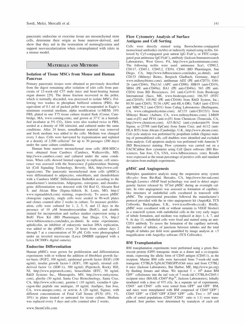
Figure 3. Pancreatic mesenchymal stem cells do not derive from CD133þduct cell EMT. (A): CD73/CD133 double staining and cell cyclestaining by propidium iodide of CD133þ sorted cells after 2, 9, and 16 days of culture. One representative experiment of four performed. (B):Flow cytometry analysis of CD73/CD133, CD73/bromodeoxyuridine and CD133/bromodeoxyuridine on a population of CD133þ sorted cells cul-tured for 7 days after isolation in the presence or absence of aphidicolin (proliferation blocker). One representative experiment of three per-formed. Abbreviations: BrdU, bromodeoxyuridine; Ctrl, control; EMT, epithelial to mesenchymal transition.
144 Pancreatic Mesenchymal Stem Cells

morphology, identical to that described above. The CD133þ
duct cells showed the expected epithelial morphology, but inthese culture conditions were never seen to proliferate andwere highly apoptotic, as demonstrated by the subG1 fractionof cell cycle (Fig. 3A) and by annexin V staining (data notshown). Strikingly, although the number of CD133þ cells inthese cultures progressively decreased, the percentage ofCD73þ mesenchymal cells increased (Fig. 3A). By the end ofthe culture period, the CD133þ sorted cells were completelyreplaced by CD73þ pMSCs. Consistent with an increase inCD73þ cells, mesenchymal marker (Snail1, nestin, vimentin)expression increased during culturing, whereas epithelialmarker (CK19, HNF1b) expression decreased (Fig. 3SA).Similarly, many pancreatic endoderm-related trascription fac-tors decreased, with the exception of Isl-1, which is alsoexpressed in pancreatic mesenchyme. Blocking proliferationby adding aphidicolin prevented appearance of the CD73þ/CD133� population in CD133þ sorted duct cell cultures, con-firming that the switch from CD133þ/CD73� to CD133�/CD73þ is exclusively dependent on cell proliferation ratherthan duct cell EMT (Fig. 3B). Additionally, single cell clon-ing of CD133þ cells never resulted in a CD73þ proliferating
clone, whereas single cell cloning of CD73þ cells gave rise toCD73þ proliferating clones with an efficiency of about 40%.We conclude that after pancreas digestion it is possible toproduce primary cultures containing two cell populations: 1) ashort-lived epithelial population originating from duct cells,and 2) a persistent mesenchymal stem cell population thatquickly expands and replaces the epithelial cells.
Bone Marrow Is a Source of pMSC
We addressed the origin of pMSCs in both human and mouse.We first verified that cells with pMSC characteristics could bederived from mouse adult pancreatic tissue, and were able toobtain a population of bona fide murine Sca1þ, CD90.2(Thy1.2)þ, CD44þ, CD34�, CD133� CD45� and CD117�
pMSCs after 6 weeks of culture (Fig. 4S). We subsequentlystudied mouse pancreas after a GFPþ bone marrow transplan-tation (BMT) to determine if pMSCs originate from bonemarrow. Total bone marrow cells from donor GFPþ trans-genic mice (C57BL/6-TgN (ACTbEGFP) 1Osb) were trans-planted into lethally irradiated coisogenic recipient C57BL/6-CD45.1 mice. After 12 weeks, all recipients had a high levelof bone marrow chimerism, scored as CD45.2þ/(CD45.2þ þ
Figure 4. Mouse pancreatic mesenchymal stem cells (pMSCs) derive from bone marrow. (A): Mean percentage þ SD of green fluorescent pro-tein (GFPþ) cells counted on 10 sections for each organ in mice 1 month after GFPþ bone marrow transplantation (BMT). (B): Cells derivedfrom endocrine and nonendocrine pancreatic tissues of mice transplanted with GFPþ bone marrow, immediately after isolation and after 7 and 14days in culture. Above: phase contrast image. Below: fluorescent image of the same field showing GFPþ scale bar: 100 lm. (C): Surface markerexpression profile by flow cytometry of GFP� (white bars) and GFPþ (green bars) pMSCs derived from pancreas of mice transplanted withGFPþBM reported as mean percentage þ SD of positive cells from three experiments. The analysis was performed 6 weeks after pancreas diges-tion. (D): Pancreatic mesenchymal stem cells cultured from explanted human pancreas stained for the expression of CD45, CD73, and recipientHLA (HLA B 55) by flow cytometry. Abbreviations: BM, human bone marrow cells; GFP, green fluorescent protein.
Sordi, Melzi, Mercalli et al. 145
www.StemCells.com
CD45.1þ)�100 (mean 89 6 10%). Analysis of histologic sec-tions after BMT showed that GFPþ bone marrow-derivedcells localized in different nonlymphatic tissues with preferen-tial homing in two organs: pancreas (GFPþ cell: 4.82 6 4%of total) and lung (GFPþ cell: 4.43 6 2.3% of total) (Fig.4A). We derived pMSCs from either isolated pancreatic isletsor the nonendocrine component. In 2 weeks, these pMSCsadhered to plastic, proliferated, exhibited a fibroblast-likemorphology, and colonized the plates (Fig. 4B). After 3 to 6weeks in culture, 18.5 6 4% of the islet-derived pMSCs wereGFPþ. These GFPþ pMSCs had the same proliferativecapacity as GFP� pMSCs, and were Sca1þ, CD90.2(Thy1.2)þ, CD44þ, CD34� (Fig. 4C). After 3 weeks understandard osteogenic culture conditions, they produced calciumthat stained positive for alizarin red S (data not shown), dem-onstrating unequivocally that the isolated GFPþ cells werepMSCs derived from BM.
In man, because of the impracticability to recover pancreasafter BM transplantation, we studied the CD73þ pMSC popula-tion in pancreas removed after allotransplantation. Eightmonths after transplantation, a 29-year-old female patient withtype 1 diabetes (HLA A: 24,26; B: 55,58; DR*03) experiencedthrombotic thrombocytopenic purpura, and consequently thetransplanted pancreas (HLA A: 1,24; B: 44,40; DR*: 16,03)was removed. We successfully isolated pMSCs (CD73þ,CD105þ, CD166þ, CD31�, CD45�, CD34�). About 6% ofpMSCs expressed recipient HLA B55, indicating that the origi-
nal donor population had been partially replaced by an extrap-ancreatic source from the recipient (Fig. 4D).
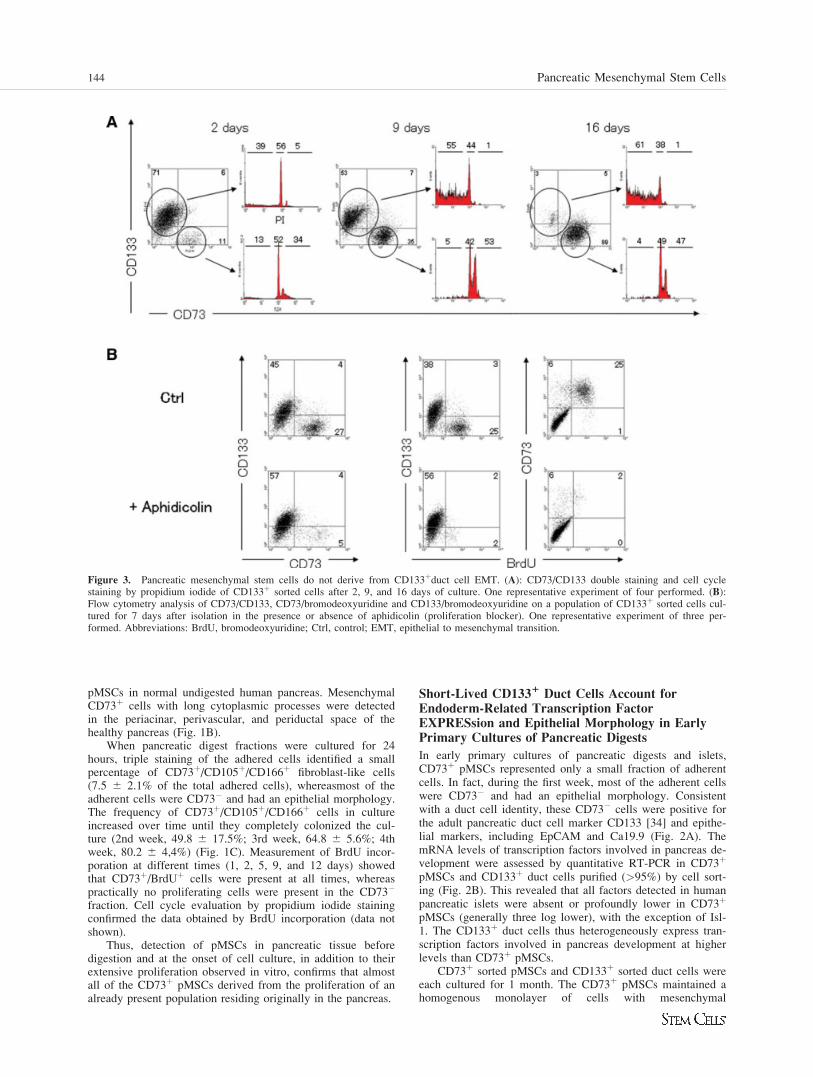
To better characterize the pMSC BM origin, we per-formed experiments in which irradiated recipient micereceived transplantation of BM with GFPþ trackable subpopu-lations segregated by CD45 expression (CD45�/GFPþ,CD45þ/GFPþ, CD45dim/GFPþ or CD45bright/GFPþ sorted sub-populations; see Materials and Methods) (Fig. 5 and 5S).
GFPþ pMSC (CD45�/Sca1þ/GFPþ) were obtained aftertransplantation of CD45þGFPþ/CD45�GFP� BM, but not af-ter transplantation of CD45þGFP�/CD45�GFPþ BM. SinceBM-MSC were reported to derive from the CD45dim subpopu-lation [35], we tested, in a second set of experiments, whetherthe CD45 expression level (CD45dim vs. CD45bright) identi-fies pMSC precursors in BM. CD45dimGFPþ/CD45brightGFP�
BM or CD45dimGFP�/CD45brightGFPþ BM were infused inirradiated C57/BL6 recipient mice (n ¼ 6). CD45 intensitydoes not discriminate pMSC precursors since GFPþ pMSCwere derived in both conditions (Fig. 5).
pMSC Expansion and Capacity to Differentiate as bCells
We cultured human pMSCs in the presence of factorsreported to influence b cell differentiation and proliferation,using either tissue or nontissue culture dishes and different se-rum concentrations (Fig. 6). Proliferation of pMSC (Fig. 6A)was stimulated 3.1-fold by fibroblast growth factor-2 (FGF-2;
Figure 5. Pancreatic mesenchymal stem cells derive from the CD45þ component of bone marrow. Cells derived from nonendocrine pancreatictissues of mice transplanted with CD45�/ green fluorescent protein (GFPþ) (upper panels), CD45dim/GFPþ (medium panels), and CD45bright/GFPþ (lower panels) bone marrow cells. Left: representative phase contrast image and fluorescent image of the same field showing GFPþcells ofnonendocrine pancreatic tissue after 1 week of culture. Right: flow cytometry analysis; reported are representative dot plots of total cells (GFPþ
versus side scatter) and of GFPþ cells (Sca1þ versus CD45þ) after 1 week of culture.
146 Pancreatic Mesenchymal Stem Cells
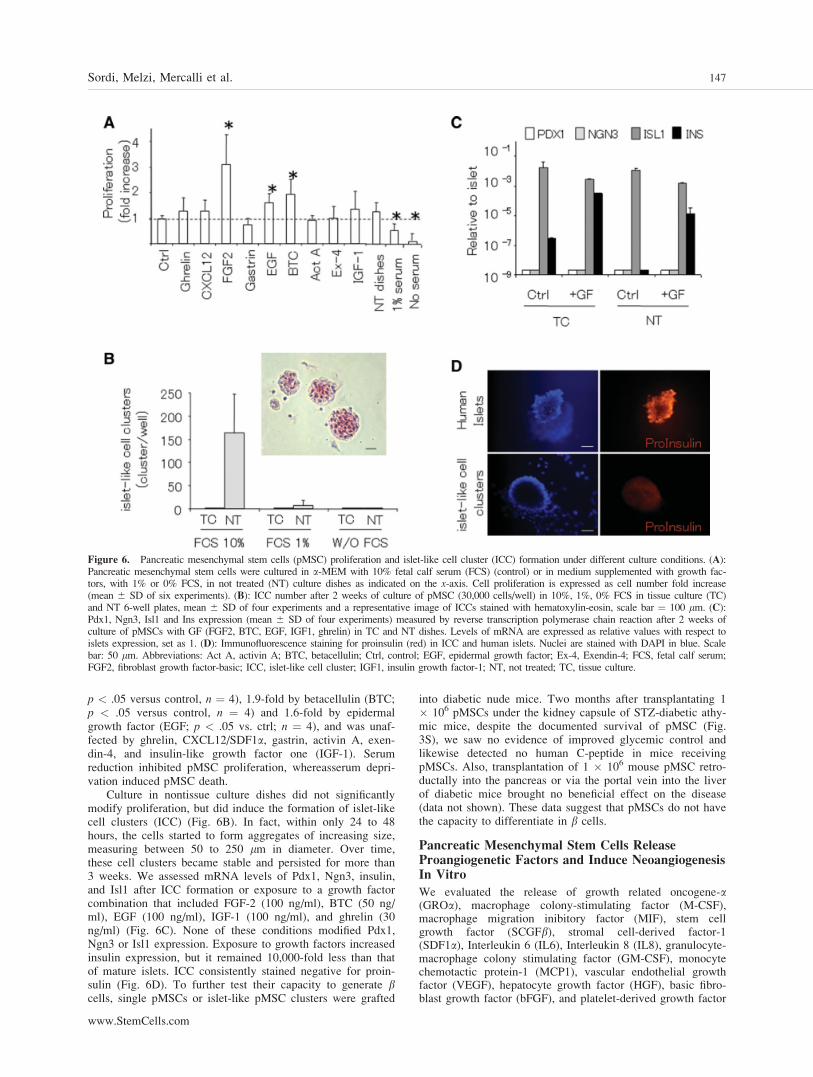
p < .05 versus control, n ¼ 4), 1.9-fold by betacellulin (BTC;p < .05 versus control, n ¼ 4) and 1.6-fold by epidermalgrowth factor (EGF; p < .05 vs. ctrl; n ¼ 4), and was unaf-fected by ghrelin, CXCL12/SDF1a, gastrin, activin A, exen-din-4, and insulin-like growth factor one (IGF-1). Serumreduction inhibited pMSC proliferation, whereasserum depri-vation induced pMSC death.
Culture in nontissue culture dishes did not significantlymodify proliferation, but did induce the formation of islet-likecell clusters (ICC) (Fig. 6B). In fact, within only 24 to 48hours, the cells started to form aggregates of increasing size,measuring between 50 to 250 lm in diameter. Over time,these cell clusters became stable and persisted for more than3 weeks. We assessed mRNA levels of Pdx1, Ngn3, insulin,and Isl1 after ICC formation or exposure to a growth factorcombination that included FGF-2 (100 ng/ml), BTC (50 ng/ml), EGF (100 ng/ml), IGF-1 (100 ng/ml), and ghrelin (30ng/ml) (Fig. 6C). None of these conditions modified Pdx1,Ngn3 or Isl1 expression. Exposure to growth factors increasedinsulin expression, but it remained 10,000-fold less than thatof mature islets. ICC consistently stained negative for proin-sulin (Fig. 6D). To further test their capacity to generate bcells, single pMSCs or islet-like pMSC clusters were grafted
into diabetic nude mice. Two months after transplantating 1� 106 pMSCs under the kidney capsule of STZ-diabetic athy-mic mice, despite the documented survival of pMSC (Fig.3S), we saw no evidence of improved glycemic control andlikewise detected no human C-peptide in mice receivingpMSCs. Also, transplantation of 1 � 106 mouse pMSC retro-ductally into the pancreas or via the portal vein into the liverof diabetic mice brought no beneficial effect on the disease(data not shown). These data suggest that pMSCs do not havethe capacity to differentiate in b cells.
Pancreatic Mesenchymal Stem Cells ReleaseProangiogenetic Factors and Induce NeoangiogenesisIn Vitro
We evaluated the release of growth related oncogene-a(GROa), macrophage colony-stimulating factor (M-CSF),macrophage migration inibitory factor (MIF), stem cellgrowth factor (SCGFb), stromal cell-derived factor-1(SDF1a), Interleukin 6 (IL6), Interleukin 8 (IL8), granulocyte-macrophage colony stimulating factor (GM-CSF), monocytechemotactic protein-1 (MCP1), vascular endothelial growthfactor (VEGF), hepatocyte growth factor (HGF), basic fibro-blast growth factor (bFGF), and platelet-derived growth factor
Figure 6. Pancreatic mesenchymal stem cells (pMSC) proliferation and islet-like cell cluster (ICC) formation under different culture conditions. (A):Pancreatic mesenchymal stem cells were cultured in a-MEM with 10% fetal calf serum (FCS) (control) or in medium supplemented with growth fac-tors, with 1% or 0% FCS, in not treated (NT) culture dishes as indicated on the x-axis. Cell proliferation is expressed as cell number fold increase(mean 6 SD of six experiments). (B): ICC number after 2 weeks of culture of pMSC (30,000 cells/well) in 10%, 1%, 0% FCS in tissue culture (TC)and NT 6-well plates, mean 6 SD of four experiments and a representative image of ICCs stained with hematoxylin-eosin, scale bar ¼ 100 lm. (C):Pdx1, Ngn3, Isl1 and Ins expression (mean 6 SD of four experiments) measured by reverse transcription polymerase chain reaction after 2 weeks ofculture of pMSCs with GF (FGF2, BTC, EGF, IGF1, ghrelin) in TC and NT dishes. Levels of mRNA are expressed as relative values with respect toislets expression, set as 1. (D): Immunofluorescence staining for proinsulin (red) in ICC and human islets. Nuclei are stained with DAPI in blue. Scalebar: 50 lm. Abbreviations: Act A, activin A; BTC, betacellulin; Ctrl, control; EGF, epidermal growth factor; Ex-4, Exendin-4; FCS, fetal calf serum;FGF2, fibroblast growth factor-basic; ICC, islet-like cell cluster; IGF1, insulin growth factor-1; NT, not treated; TC, tissue culture.
Sordi, Melzi, Mercalli et al. 147
www.StemCells.com

(PDGF) by pMSCs (105/ml) cultured for 12 hours. High con-centrations of VEGF (4.4 6 0.6 ng/ml), PDGF (1.0 6 0.8 ng/ml), GROa (3.4 6 2.3 ng/ml), M-CSF (0.7 6 0.6 ng/ml),MIF (1.9 6 0.2 ng/ml), SCGFb (39.3 6 4.2 ng/ml), SDF1a(1.0 6 0.2), IL6 (19.5 6 16.6 ng/ml), IL8 (10.8 6 10.5 ng/ml), MCP1 (71.8 6 12.1 ng/ml), but not of GM-CSF (0.1 60.2 ng/ml), HGF (0.1 6 0.1 ng/ml) and bFGF (0.1 6 0.2 ng/ml) were detected.
The angiogenetic capacity of pMSC was then analyzed inan in vitro system: pMSC stimulated tubule formation andanastomosis. This was prevented by the addition of suramin(VEGF blocker). These data demonstrate that pMSCs are ableto induce a VEGF mediated angiogenesis (Fig. 7A).
Pancreatic Mesenchymal Stem Cells Aid Islet Trans-plantation and Increase Neovascularization
Since pMSCs originate from an extrapancreatic source andthe BM-MSC have been shown to provide help to islets in a
transplant model, we asked whether pMSC recruitment is ben-eficial for islet survival or function. To address this issue, weused a syngeneic murine model of marginal islet mass trans-plantation. Implantation of a marginal b cell mass (200 islets)is characterized by achievement of normoglycemia in only aproportion of recipients or a delay in reversal of hyperglyce-mia as compared with transplantation of 500 islets. Manipula-tions that lead to a higher percentage of successful grafts orto a reduction of the lag to normoglycemia are to be inter-preted as beneficial in favoring islet survival or engraftment[36, 37]. STZ-induced severely diabetic mice (mean nonfast-ing glycemia before transplantation: 546 6 64 mg/dL) weretransplanted under the kidney capsule with 200 syngenic isletsalternatively with (n ¼ 12) or without (n ¼ 17) 2.5 � 105
murine pMSC. The probability and the median time to reacheuglycemia (<200 mg/dl) in our marginal islet mass trans-plantation were: 83% and 10 days for islet þ pMSC trans-plants (CI95%, 6.6-13) and 29% and >50 days for islet alonetransplants (p ¼ .004, Pearson chi-square test; p ¼ .007, log-
Figure 7. Pancreatic mesenchymal stem cells (pMSCs) aid islet cells in a model of minimal islet mass transplantation. (A): In vitro angiogene-sis assay: angiogenesis was evaluated as the capacity to form a netwotk of anastomosing tubules from endothelial cells. Upper panel: representa-tive images of tubules formation in the absence (Ctrl) o in the presence of pMSC. To block VEGF, suramin (1 mM) was added to the culture.Scale bar: 100 lm. Lower panel: tubule number, junction and length evaluation. (n ¼ 10). Data are presented as box plots. *<0.05, Mann-Whit-ney U test. (B): C57BL/6 recipients were transplanted with 200 autologous islets under the kidney capsule alternatively without (n ¼ 17, redline) or with 2.5 � 105 pMSC (n ¼ 12, black line) or 2.5 � 105 mesangioblasts (n ¼ 13, blue line). Kaplan-Meier analysis for reaching normo-glycemia (<200 mg/dl on consecutive days) is shown. Differences between groups were tested using the log-rank statistic (p <.001). In post hocanalysis: islets versus islets þ pMSC, p ¼ .006; islets versus islets þ mesangioblasts, p ¼ .362; islets þ pMSC versus islets þ mesangioblasts, p¼ .002. (C): Quantitative evaluation of CD31þ cell area in islet graft with islets alone (n ¼ 7) or islets plus pMSC (n ¼ 7) 3 weeks after trans-plantation. Left panel: The vessel/islet area is shown for transplanted mice. CD31þ area was determined and divided by the insulinþ area in 356 11 islet sections in islets alone and 36 6 24 islet sections in islets plus pMSC transplantation, respectively. Data are represented as box plots.Statistical analysis was performed by Mann-Whitney U test. Right panel: representative immunofluorescence images showing CD31 (green), insu-lin (red), and DAPI cell nuclei staining (blue) in islet graft with islets alone or islets plus pMSC. Abbreviations: Ctrl, control; pMSCs, pancreaticmesenchymal stem cells; VEGF, vascular endothelial growth factor.
148 Pancreatic Mesenchymal Stem Cells
rank test in Kaplan Meyer analysis). Glycemic control wasnot improved when the pMSCS were substituted by a similarnumber of other murine cells (that is, mesangioblasts) in thecotransplant (n ¼ 13), demonstrating a specific role forpMSCs (Fig. 7B). In another set of experiments, MSC derivedfrom pancreas (n ¼ 5) and from bone marrow (n ¼ 5) weretransplanted together with islets in the model of minimal isletmass transplantation and both resulted effective in normaliz-ing glycemia (Fig. 6S). Moreover, transplantation of murinepMSC alone and mesangioblast alone did not induce anychange in glycemia (data not shown). To exclude the possibil-ity that better glycemic control was caused by pMSC differen-tiation into insulin secreting cells, we cotransplanted isletsfrom wild-type mice along with pMSCs derived from GFPþ
transgenic mice. Two to 3 weeks after transplantation, thehost mice were killed and the graft analyzed. No insulin/GFPdouble positive cells were present (data not shown). In a sec-ond set of experiments with seven mice in each group, we an-alyzed CD31þ endothelial cells in the transplant 3 weeks aftertransplantation and the vessel density was significantly higherin islets plus pMSC than in islets alone (Fig. 7C), indicatingthat one of the benefits provided by pMSC is increased neo-vascularization of the graft.
DISCUSSION
In this study we show that the mesenchymal cells appearingfrom pancreatic tissue culture are not derived from an epithe-lial to mesenchymal transition, but are rather the expansion ofa pool of resident MSCs, located mainly in the periacinar,perivascular, and periductal space that have a bone marrow-derived origin and that preserve, at least in vitro, a stable phe-notype, a high proliferation rate, and multilineage differentia-tion potential. We classified cells emerging from pancreaticculture as mesenchymal stem cells on the basis of the mini-mal criteria for defining MSC established by the InternationalSociety for Cellular Therapy [38]: plastic-adherence in stand-ard culture conditions; expression of CD105, CD73 andCD90; lack of expression of CD45, CD34, CD14, CD19, andHLA-DR surface molecules; capacity to differentiate to osteo-blasts, adipocytes and chondroblasts in vitro. CD73 expressionin pMSCs proved useful to follow these cells during process-ing and culture. These pMSC seem to have an advantage interms of proliferation and survival compared with other cellsof pancreatic origin in culture, and become the predominantcell type in cultures without a requirement of epithelial tomesenchymal switch. In agreement with our data in man, fourgroups recently used direct lineage tracing via transgenic cre/loxP-based systems [25–28] to provide compelling evidenceagainst b cell EMT in the mouse. Importantly, one of thesestudies not only demonstrated that ‘‘fibroblast-like cells’’ frommouse islets are not derived from b cells, but also excludedan endoderm pancreatic origin and thus discounted the ideathat pMSCs originate from a pancreatic exocrine or endocrineEMT. In man, a recent approach of viral-mediated lineagetracing of human b cells showed that they can dedifferentiateand expand in vitro, but their lifespan and rate of division(doubling time 7 days, 16 population doublings) are muchlower than those of pMSC, so it is likely that they are twodistinct populations of pancreatic cells [24].
In man, we found that part of the CD73þ pMSC popula-tion in allogeneic pancreas removed after transplantationexpressed recipient HLA, suggesting that an extrapancreaticsource ‘‘refilled’’ the graft. In mouse recipients of a GFPþ
bone marrow transplant, we conclusively demonstrated that
pMSCs originate from BM. Even if we cannot exclude the ex-istence of other sources of pMSC, this evidence strongly sug-gests the existence of a cross talk between BM and pancreas,evidence also supported by the fact that the pancreas is apreferential site of GFPþ cells localization after GFPþ BMtransplantation in mice. Regarding the mechanisms involvedin pMSC recruitment, we previously reported that human pan-creatic islets can attract BM-MSCs in vitro, and this attractionis principally mediated by two chemokines: CX3CL1 andCXCL12 [21]. Currently, we are unable to determine whetherpMSC migration from the bone marrow occurs in the absenceof pancreatic injury, or if pMSCs constitute a renewable poolof cells that migrate early in development to create a pancre-atic stem cell niche that replaces dying cells. It is possiblethat the resident pMSCs are derived from the vessels withinthe pancreas as self-renewing ‘‘vascular’’ stem cells residentpool. On the other hand, the BM could reinforce the pancre-atic pMSCs population only in stress situations, as in the radi-ation-based bone marrow transplant setting or the organ dam-age in the inflammatory events at donor death, and do notparticipate in the pMSCs turn-over during healthy status.
Interestingly, the bone-marrow transplant model in micesuggests that the pool of MSCs in the pancreas is renewedfrom the CD45þ BM compartment. This could appear in con-trast with the established CD45 negative MSC phenotype, butthis phenomenon has already been described [35, 39–43].MSCs require prolonged culturing of dispersed cell popula-tions on plastic, and the expression of CD45 by MSC couldbe drastically downregulated during in vitro culture conditions[35, 39]. Obviously the claim that all the MSC are derivedfrom a CD45þ population goes far beyond the purposes ofour work. We limit our observation to the MSC cells obtainedby the pancreas that, although coming from the bone marrow,may have a different lineage derivation from that of BMMSC.
Since MSCs have been reported to cross the mesodermallineage and give rise to endoderm both in vitro and in vivo[1, 44–48], it is possible that pMSC could be directed tobecome b cells and thus serve as an abundant source of insu-lin-producing cells for transplantation. By adopting methodol-ogy that was previously used to differentiate b cells frommesenchymal precursors [1, 44–48], we tested the ability ofpMSCs to generate functional islets. The pMSCs formed islet-like cell clusters, but they expressed negligible levels of islet-specific genes under basal conditions, serum deprivation, orexogenous growth factor exposure. Moreover, they wereunable to significantly improve glucose metabolism in vivowhen transplanted into diabetic nude mice. In agreement withour data, it was recently reported that islet-like clustersobtained from pancreatic MSCs are closer in microarray phe-notype to mesenchymal cells than endocrine cells [49]. Thisconfirms that generation of functional islets by culture manip-ulation of expanded pMSCs is not yet achievable.
In pilot experiments, we found that transplanting pMSCstogether with a minimal islet mass enhanced islet function.We excluded that improved glycemic control was caused bypMSC differentiation into insulin-secreting cells, and pro-posed that pMSCs act as ‘‘helper’’ cells for islets. A beneficialeffect of bone marrow-derived MSCs in experimental and pre-clinical models was recently attributed to the production ofendocrine or paracrine factors, not cell differentiation. Forexample, intravenous administration of MSCs stimulated re-vascularization of ischemic tissues despite differentiation ofonly a small number of cells into cardiomyocytes and endo-thelial cells [50]. Furthermore, Prockop and colleagues [14]showed that stromal cells from human bone marrow home toand promote repair of pancreatic islets in diabetic mice.
Sordi, Melzi, Mercalli et al. 149
www.StemCells.com

Generally this happens without evidence of long-term engraft-ment, suggesting that the favorable effects of MSCs reflectthe impact of transitory paracrine effects or secreted factorsrather than engraftment, differentiation, or cell fusion [51–53]. Also in our model, the higher vessel density in isletscotransplanted with pMSC proposes that MSC have an indi-rect beneficial effect on the graft. This is supported by therecent findings that MSC enhanced endothelial cells prolifera-tion, sprout formation, and in-growth in an in vitro system ofcomposite endothelial cells-MSC-pancreatic islets [54]. Theincrease in endothelial cells in islet grafts could be consistentnot only with an increased blood flow, but also with a CD31þ
cells mediated paracrine effect in sustaining b cell survival.Further studies are ongoing to confirm how pMSCs favor neo-vasculogenesis and to explore whether the protective effect ofpMSC on transplanted tissue is mediated by other potentialmechanisms such as prevention of islet cells from apoptosis,stimulation of islet cell proliferation, or the release of growthfactors required by islets for maintenance of efficient insulinsecretion.
In conclusion, pMSCs appearing in cultures of humanpancreatic endocrine and exocrine tissue propagate from apopulation of mesenchymal stem cells, likely of bone marroworigin, resident in the pancreas. These cells express negligiblelevels of islet-specific genes, but are able to facilitate glyce-mia regulation when cotransplanted with a minimal islet massinto diabetic mice. This suggests that pMSCs are not optimal
candidates to generate physiologically competent b cells, butconsidering the large number of pMSCs obtainable fromdigested pancreas, they could be useful as islet ‘‘helper’’ cells.The better characterization of bone marrow pancreas interac-tion and the identification of the mechanisms by which thesecells aid islets will likely have implications for therapeuticcell transplantation and diabetes treatment.
ACKNOWLEDGMENTS
This work was supported by EU (DIAPREPP Project,HEALTH-F2-2008-202013), Telethon Italy and the Juvenile Di-abetes Research Foundation (JT01Y01), Italian Telethon Foun-dation (TIGET grant) by Award from Fondazione Cariplo(2007-5165) and by award from Fondazione Siemens Italia (Mi-lan, Italy). Valeria Sordi is enrolled as a Ph.D. student at the Lud-wig-Maximilians University of Munich, Germany.
DISCLOSURE OF POTENTIAL CONFLICTS
OF INTEREST
The authors indicate no potential conflicts of interest.
REFERENCES
1 Jiang Y, Jahagirdar BN, Reinhardt RL et al. Pluripotency ofmesenchymal stem cells derived from adult marrow. Nature 2002;418:41–49.
2 Krause DS, Theise ND, Collector MI et al. Multi-organ, multi-lineageengraftment by a single bone marrow-derived stem cell. Cell 2001;105:369–377.
3 Moriscot C, de Fraipont F, Richard MJ et al. Human bone marrowmesenchymal stem cells can express insulin and key transcription fac-tors of the endocrine pancreas developmental pathway upon geneticand/or microenvironmental manipulation in vitro. Stem Cells 2005;23:594–603.
4 Timper K, Seboek D, Eberhardt M et al. Human adipose tissue-derived mesenchymal stem cells differentiate into insulin, somatosta-tin, and glucagon expressing cells. Biochem Biophys Res Commun2006;341:1135–1140.
5 Denner L, Bodenburg Y, Zhao JG et al. Directed engineering of um-bilical cord blood stem cells to produce C-peptide and insulin. CellProlif 2007;40:367–380.
6 Karnieli O, Izhar-Prato Y, Bulvik S et al. Generation of insulin-pro-ducing cells from human bone marrow mesenchymal stem cells bygenetic manipulation. Stem Cells 2007;25:2837–2844.
7 Gao F, Wu DQ, Hu YH et al. In vitro cultivation of islet-like cellclusters from human umbilical cord blood-derived mesenchymal stemcells. Transl Res 2008;151:293–302.
8 Ianus A, Holz GG, Theise ND et al. In vivo derivation of glucose-competent pancreatic endocrine cells from bone marrow without evi-dence of cell fusion. J Clin Invest 2003;111:843–850.
9 Lechner A, Yang YG, Blacken RA et al. No evidence for significanttransdifferentiation of bone marrow into pancreatic beta-cells in vivo.Diabetes 2004;53:616–623.
10 Hess D, Li L, Martin M et al. Bone marrow-derived stem cells initiatepancreatic regeneration. Nat Biotechnol 2003;21:763–770.
11 Urban VS, Kiss J, Kovacs J et al. Mesenchymal stem cells cooperatewith bone marrow cells in therapy of diabetes. Stem Cells 2008;26:244–253.
12 Caplan AI, Dennis JE. Mesenchymal stem cells as trophic mediators.J Cell Biochem 2006;98:1076–1084.
13 Solari MG, Srinivasan S, Boumaza I et al. Marginal mass islet trans-plantation with autologous mesenchymal stem cells promotes long-term islet allograft survival and sustained normoglycemia. J Autoim-mun 2009;32:116–124.
14 Lee RH, Seo MJ, Reger RL et al. Multipotent stromal cells fromhuman marrow home to and promote repair of pancreatic islets and re-
nal glomeruli in diabetic NOD/scid mice. Proc Natl Acad Sci U S A2006;103:17438–17443.
15 Estrada EJ, Valacchi F, Nicora E et al. Combined treatment of intra-pancreatic autologous bone marrow stem cells and hyperbaric oxygenin type 2 diabetes mellitus. Cell Transplant 2008;17:1295–1304.
16 Ouziel-Yahalom L, Zalzman M, Anker-Kitai L et al. Expansion andredifferentiation of adult human pancreatic islet cells. Biochem Bio-phys Res Commun 2006;341:291–298.
17 Lechner A, Nolan AL, Blacken RA et al. Redifferentiation of insulin-secreting cells after in vitro expansion of adult human pancreatic islettissue. Biochem Biophys Res Commun 2005;327:581–588.
18 Gershengorn MC, Hardikar AA, Wei C et al. Epithelial-to-mesenchy-mal transition generates proliferative human islet precursor cells. Sci-ence 2004;306:2261–2264.
19 Eberhardt M, Salmon P, von Mach MA et al. Multipotential nestinand Isl-1 positive mesenchymal stem cells isolated from humanpancreatic islets. Biochem Biophys Res Commun 2006;345:1167–1176.
20 Seeberger KL, Dufour JM, Shapiro AM et al. Expansion of mesenchy-mal stem cells from human pancreatic ductal epithelium. Lab Invest2006;86:141–153.
21 Sordi V, Malosio ML, Marchesi F et al. Bone marrow mesenchymalstem cells express a restricted set of functionally active chemokinereceptors capable of promoting migration to pancreatic islets. Blood2005;106:419–427.
22 Gallo R, Gambelli F, Gava B et al. Generation and expansion of mul-tipotent mesenchymal progenitor cells from cultured human pancreaticislets. Cell Death Differ 2007;14:1860–1871.
23 Linning KD, Tai MH, Madhukar BV et al. Redox-mediated enrich-ment of self-renewing adult human pancreatic cells that possess endo-crine differentiation potential. Pancreas 2004;29:e64–76.
24 Russ HA, Bar Y, Ravassard P et al. In vitro proliferation of cellsderived from adult human beta-cells revealed by cell-lineage tracing.Diabetes 2008;57:1575–1583.
25 Chase LG, Ulloa-Montoya F, Kidder BL et al. Islet-derived fibroblast-like cells are not derived via epithelial-mesenchymal transition fromPdx-1 or insulin-positive cells. Diabetes 2007;56:3–7.
26 Atouf F, Park CH, Pechhold K et al. No evidence for mouse pancre-atic beta-cell epithelial-mesenchymal transition in vitro. Diabetes2007;56:699–702.
27 Morton RA, Geras-Raaka E, Wilson LM et al. Endocrine precursorcells from mouse islets are not generated by epithelial-to-mesenchy-mal transition of mature beta cells. Mol Cell Endocrinol 2007;270:87–93.
28 Weinberg N, Ouziel-Yahalom L, Knoller S et al. Lineage tracing evi-dence for in vitro dedifferentiation but rare proliferation of mousepancreatic beta-cells. Diabetes 2007;56:1299–1304.
150 Pancreatic Mesenchymal Stem Cells

29 Piemonti L, Leone BE, Nano R et al. Human pancreatic islets produceand secrete MCP-1/CCL2: Relevance in human islet transplantation.Diabetes 2002;51:55–65.
30 Hotz HG, Reber HA, Hotz B et al. An orthotopic nude mouse modelfor evaluating pathophysiology and therapy of pancreatic cancer. Pan-creas 2003;26:E89–98.
31 Melzi R, Sanvito F, Mercalli A et al. Intrahepatic islet transplant inthe mouse: Functional and morphological characterization. Cell Trans-plant 2008;17:1361–1370.
32 Piemonti L, Bertuzzi F, Nano R et al. Effects of cryopreservation onin vitro and in vivo long-term function of human islets. Transplanta-tion 1999;68:655–662.
33 Minasi MG, Riminucci M, De Angelis L et al. The meso-angioblast:A multipotent, self-renewing cell that originates from the dorsal aortaand differentiates into most mesodermal tissues. Development 2002;129:2773–2783.
34 Lardon J, Corbeil D, Huttner WB et al. Stem cell marker prominin-1/AC133 is expressed in duct cells of the adult human pancreas. Pan-creas 2008;36:e1–6.
35 Deschaseaux F, Gindraux F, Saadi R et al. Direct selection of humanbone marrow mesenchymal stem cells using an anti-CD49a antibodyreveals their CD45med, low phenotype. Br J Haematol 2003;122:506–517.
36 Kaufman DB, Gores PF, Field MJ et al. Effect of 15-deoxyspergualin onimmediate function and long-term survival of transplanted islets in murinerecipients of a marginal islet mass. Diabetes 1994;43:778–783.
37 Zhang YC, Molano RD, Pileggi A et al. Adeno-associated virus trans-duction of islets with interleukin-4 results in impaired metabolic func-tion in syngeneic marginal islet mass transplantation. Transplantation2002;74:1184–1186.
38 Dominici M, Le Blanc K, Mueller I et al. Minimal criteria for definingmultipotent mesenchymal stromal cells. The International Society ForCellular Therapy Position Statement. Cytotherapy 2006;8:315–317.
39 Kaiser S, Hackanson B, Follo M et al. BM cells giving rise to MSCin culture have a heterogeneous CD34 and CD45 phenotype. Cytother-apy 2007;9:439–450.
40 Koide Y, Morikawa S, Mabuchi Y et al. Two distinct stem cell line-ages in murine bone marrow. Stem Cells 2007;25:1213–1221.
41 Rogers I, Yamanaka N, Bielecki R et al. Identification and analysis ofin vitro cultured CD45-positive cells capable of multi-lineage differen-tiation. Exp Cell Res 2007;313:1839–1852.
42 Ooi YY, Ramasamy RVidyadaran S. Mouse bone marrow mesenchy-mal stem cells acquire CD45-CD106þ immunophenotype only at laterpassages. Med J Malaysia 2008;63 Suppl A:65–66.
43 Ogawa M, LaRue ACDrake CJ. Hematopoietic origin of fibroblasts/myofibroblasts: Its pathophysiologic implications. Blood 2006;108:2893–2896.
44 Chen LB, Jiang XB, Yang L. Differentiation of rat marrow mesenchy-mal stem cells into pancreatic islet beta-cells. World J Gastroenterol2004;10:3016–3020.
45 Sun J, Yang Y, Wang X et al. Expression of Pdx-1 in bone marrowmesenchymal stem cells promotes differentiation of islet-like cells invitro. Sci China C Life SCI 2006;49:480–489.
46 Battula VL, Bareiss PM, Treml S et al. Human placenta and bonemarrow derived MSC cultured in serum-free, b-FGF-containing me-dium express cell surface frizzled-9 and SSEA-4 and give rise to mul-tilineage differentiation. Differentiation 2007;75:279–291.
47 Woodbury D, Reynolds K, Black IB. Adult bone marrow stromalstem cells express germline, ectodermal, endodermal, and mesodermalgenes prior to neurogenesis. J Neurosci Res 2002;69:908–917.
48 Davani B, Ikonomou L, Raaka BM et al. Human islet-derived pre-cursor cells are mesenchymal stromal cells that differentiate andmature to hormone-expressing cells in vivo. Stem Cells 2007;25:3215–3222.
49 Kayali AG, Flores LE, Lopez AD et al. Limited capacity of humanadult islets expanded in vitro to redifferentiate into insulin-producingbeta-cells. Diabetes 2007;56:703–708.
50 Wu GD, Nolta JA, Jin YS et al. Migration of mesenchymal stem cellsto heart allografts during chronic rejection. Transplantation 2003;75:679–685.
51 Iso Y, Spees JL, Serrano C et al. Multipotent human stromal cells improvecardiac function after myocardial infarction in mice without long-termengraftment. Biochem Biophys Res Commun 2007;354:700–706.
52 Crisan M, Yap S, Casteilla L et al. A perivascular origin for mesen-chymal stem cells in multiple human organs. Cell Stem Cell 2008;3:301–313.
53 Au P, Tam J, Fukumura D et al. Bone marrow-derived mesenchymalstem cells facilitate engineering of long-lasting functional vasculature.Blood 2008;111:4551–4558.
54 Johansson U, Rasmusson I, Niclou SP et al. Formation of compositeendothelial cell-mesenchymal stem cell islets: A novel approach topromote islet revascularization. Diabetes 2008;57:2393–2401.
See www.StemCells.com for supporting information available online.
Sordi, Melzi, Mercalli et al. 151