Expression of a Distinct Set of Chemokine Receptors in Adipose Tissue-Derived Stem Cells is...
13
ORIGINAL RESEARCH—BASIC SCIENCE Expression of a Distinct Set of Chemokine Receptors in Adipose Tissue-Derived Stem Cells is Responsible for In Vitro Migration Toward Chemokines Appearing in the Major Pelvic Ganglion Following Cavernous Nerve Injury Maarten Albersen, MD, PhD,* † Joost Berkers, MD,* Philip Dekoninck, MD, † Jan Deprest, MD, PhD, † Tom F. Lue, MD, ‡ Petter Hedlund, MD, PhD, § Ching-Shwun Lin, PhD, ‡ Trinity J. Bivalacqua, MD, PhD, ¶ Hendrik Van Poppel, MD, PhD,* Dirk De Ridder, MD, PhD,* and Frank Van der Aa, MD, PhD* *Laboratory of Experimental Urology, Department of Development and Regeneration, University of Leuven, Leuven, Belgium; † Laboratory of Experimental Gynecology, Department of Development and Regeneration, University of Leuven, Leuven, Belgium; ‡ Knuppe Molecular Urology Laboratory, Department of Urology, University of California, San Francisco, CA, USA; § Department of Urology, Urological Research Institute, Vita-Salute San Raffaele University, Milan, Italy; ¶ Department of Urology, The James Buchanan Brady Urological Institute, Johns Hopkins Medical Institutions, Baltimore, MD, USA DOI: 10.1002/sm2.1 ABSTRACT Introduction. Adipose tissue-derived stem cells (ADSCs) herald tremendous promise for clinical application in a wide range of injuries and diseases. Several preclinical reports demonstrate their efficacy in the treatment of cavernous nerve (CN) injury-induced erectile dysfunction in rats. It was recently illustrated that these effects were established as a result of ADSC migration to the major pelvic ganglion (MPG) where these cells induced neuroregeneration in loco. Aims. The study aims to identify chemotactic factors in the MPG following injury and to match upregulated chemokines to their respective receptors in human ADSC on the genomic, structural, and functional levels. Methods. Quantitative real-time polymerase chain reaction, fluorescence-activated cell sorting (FACS), intracellular FACS, immunofluorescence microscopy, migration assays, and calcium imaging were used in this study. Main Outcome Measures. The main outcomes are chemokine expression in the MPG following CN injury, and the functional and structural presence of chemokine receptors in ADSC. Results. CCR4, CX3CR1, and XCR1 are functionally and structurally present in human ADSC, and are activated by the chemokines CCL2, CX3CL1, and XCL1 respectively, which are upregulated in the MPG following CN injury. CXCR4 and its ligand CXCL12 (SDF1) are likely no major homing factors for ADSC. Expression of chemokine receptor mRNA in ADSC did not necessarily translate into receptor presence at the cell surface and/or functional activation of these receptors. Most of the expressed chemokine receptors were detected in the intracellular com- partment of these cells. Conclusions. We identified the ligand/chemokine receptor pairs CCL2/CCR4, CX3CL1/CX3CR1, and XCL1/ XCR1 as potentially responsible for ADSC homing toward the MPG following CN injury. The intracellular localization of various chemokine receptors likely indicates redirecting of chemokine receptors to the cell surface under specific cellular conditions. Furthermore, modification of expression of these receptors at the genomic level may potentially lead to improved migration toward injury sites and thus enhancement of treatment efficacy. Albersen M, Berkers J, Dekoninck P, Deprest J, Lue TF, Hedlund P, Lin C-S, Bivalacqua TJ, Van Poppel H, De Ridder D, and Van der Aa F. Expression of a distinct set of chemokine receptors in adipose tissue-derived stem cells is responsible for in vitro migration toward chemokines appearing in the major pelvic ganglion following cavernous nerve injury. Sex Med 2013;1:3–15. © 2013 The Authors. Sexual Medicine published by Wiley Periodicals, Inc. on behalf of International Society for Sexual Medicine. This is an open access article under the terms of the Creative Commons Attribution-NonCommercial License, which permits use, distribution and reproduction in any medium, provided the original work is properly cited and is not used for commercial purposes. Sex Med 2013;1:3–15
Transcript of Expression of a Distinct Set of Chemokine Receptors in Adipose Tissue-Derived Stem Cells is...

ORIGINAL RESEARCH—BASIC SCIENCE
Expression of a Distinct Set of Chemokine Receptors in AdiposeTissue-Derived Stem Cells is Responsible for In Vitro MigrationToward Chemokines Appearing in the Major Pelvic GanglionFollowing Cavernous Nerve Injury
Maarten Albersen, MD, PhD,*† Joost Berkers, MD,* Philip Dekoninck, MD,† Jan Deprest, MD, PhD,†
Tom F. Lue, MD,‡ Petter Hedlund, MD, PhD,§ Ching-Shwun Lin, PhD,‡ Trinity J. Bivalacqua, MD, PhD,¶
Hendrik Van Poppel, MD, PhD,* Dirk De Ridder, MD, PhD,* and Frank Van der Aa, MD, PhD*
*Laboratory of Experimental Urology, Department of Development and Regeneration, University of Leuven, Leuven,Belgium; †Laboratory of Experimental Gynecology, Department of Development and Regeneration, University of Leuven,Leuven, Belgium; ‡Knuppe Molecular Urology Laboratory, Department of Urology, University of California, SanFrancisco, CA, USA; §Department of Urology, Urological Research Institute, Vita-Salute San Raffaele University, Milan,Italy; ¶Department of Urology, The James Buchanan Brady Urological Institute, Johns Hopkins Medical Institutions,Baltimore, MD, USA
DOI: 10.1002/sm2.1
A B S T R A C T
Introduction. Adipose tissue-derived stem cells (ADSCs) herald tremendous promise for clinical application in awide range of injuries and diseases. Several preclinical reports demonstrate their efficacy in the treatment ofcavernous nerve (CN) injury-induced erectile dysfunction in rats. It was recently illustrated that these effects wereestablished as a result of ADSC migration to the major pelvic ganglion (MPG) where these cells inducedneuroregeneration in loco.Aims. The study aims to identify chemotactic factors in the MPG following injury and to match upregulatedchemokines to their respective receptors in human ADSC on the genomic, structural, and functional levels.Methods. Quantitative real-time polymerase chain reaction, fluorescence-activated cell sorting (FACS), intracellularFACS, immunofluorescence microscopy, migration assays, and calcium imaging were used in this study.Main Outcome Measures. The main outcomes are chemokine expression in the MPG following CN injury, and thefunctional and structural presence of chemokine receptors in ADSC.Results. CCR4, CX3CR1, and XCR1 are functionally and structurally present in human ADSC, and are activated bythe chemokines CCL2, CX3CL1, and XCL1 respectively, which are upregulated in the MPG following CN injury.CXCR4 and its ligand CXCL12 (SDF1) are likely no major homing factors for ADSC. Expression of chemokinereceptor mRNA in ADSC did not necessarily translate into receptor presence at the cell surface and/or functionalactivation of these receptors. Most of the expressed chemokine receptors were detected in the intracellular com-partment of these cells.Conclusions. We identified the ligand/chemokine receptor pairs CCL2/CCR4, CX3CL1/CX3CR1, and XCL1/XCR1 as potentially responsible for ADSC homing toward the MPG following CN injury. The intracellularlocalization of various chemokine receptors likely indicates redirecting of chemokine receptors to the cell surfaceunder specific cellular conditions. Furthermore, modification of expression of these receptors at the genomic levelmay potentially lead to improved migration toward injury sites and thus enhancement of treatment efficacy.Albersen M, Berkers J, Dekoninck P, Deprest J, Lue TF, Hedlund P, Lin C-S, Bivalacqua TJ, Van Poppel H,De Ridder D, and Van der Aa F. Expression of a distinct set of chemokine receptors in adipose tissue-derivedstem cells is responsible for in vitro migration toward chemokines appearing in the major pelvic ganglionfollowing cavernous nerve injury. Sex Med 2013;1:3–15.
© 2013 The Authors. Sexual Medicine published by Wiley Periodicals, Inc.on behalf of International Society for Sexual Medicine.This is an open access article under the terms of the Creative Commons Attribution-NonCommercial License,which permits use, distribution and reproduction in any medium, provided the original work is properly cited andis not used for commercial purposes.
Sex Med 2013;1:3–15

Key Words. Adipose Tissue-Derived Stem Cells; Cavernous Nerve Injury; Cell Surface; Chemokines; ChemokineReceptors; Erectile Dysfunction; Intracellular; Migration
Introduction
D espite the development of nerve-sparingtechniques, neuropraxia of the cavernous
nerves (CNs), resulting in erectile dysfunction(ED), remains a highly frequent complication ofradical prostatectomy for prostate cancer [1].Various pharmacological “penile rehabilitation”strategies have been investigated to preventdenervation-induced smooth muscle apoptosis andfibrosis in the erectile tissue during the time framein which the injured CNs regenerate [2]. Whilepreclinical research was promising, recent clinicaltrials have not uniformly confirmed the benefit ofpenile rehabilitation in this setting. Therefore,basic researchers have in the recent years beenfocusing on stimulating CN regeneration follow-ing neuropraxia, rather than aiming at the second-arily damaged erectile tissue as a treatment target[3]. In these studies, the application of multipotentstromal cells (MSC) derived from either bonemarrow or adipose tissue has generated promisingresults [4].
Kendirci and colleagues performed bilateralCN crush injury in rats and injected bone marrow-derived MSC (BM-MSC) and BM-MSC selectedfor p75-nerve growth factor receptor expressionin the corpus cavernosum, resulting in beneficialeffects on erectile recovery [5]. These authors sug-gested paracrine effects on neuroregenerationbased on increased in vitro secretion of growthfactors by these stem cells. They noted an absenceof significant cell incorporation and differentiationinto host cell types in the penis. This finding wasin line with previous studies regarding cellulartherapy for CN injury [4,6,7]. The repeated obser-vation of erectile function improvement in theabsence of cell incorporation led to the hypothesisthat these cellular therapies may exert their effectsin a paracrine fashion [8]. In several consecutivestudies, our group has further investigated this“paracrine hypothesis.” In an initiating study,Albersen and coworkers injected either adiposetissue-derived mesenchymal stem cells (ADSCs)or ADSC-derived lysate in the corpus cavernosumof CN-injured rats and observed preservation ofneuronal nitric oxide synthase content, diminishedsmooth muscle apoptosis, and erectile tissue fibro-
sis in both treated groups [9]. The lysate treatmentexposed injured tissues to soluble factors containedin ADSC, without allowing live cells to incorpo-rate in the host tissue, and thus supportedparacrine modes of action of stem cell therapy forCN-injury induced ED [10]. This report was fol-lowed by an experiment in which the major pelvicganglion (MPG), the autonomic ganglion fromwhich the CN originates, was cultured in condi-tioned medium of either ADSC or smooth musclecells. Strongly enhanced neurite sprouting wasobserved when these ganglia were cultured inADSC-conditioned media [11]. Results from thelatter experiment indicated that ADSCs secretesubstances that induce nerve regeneration. Fandelet al. looked at the fate of EdU-labeled ADSCafter intracavernosal injection in the CN-injury ratmodel, and observed a rapid decrease of cellnumbers from the injection site in the corpuscavernosum following injection [12]. Surprisingly,ADSC were located in the MPG in the first daysfollowing injury. Furthermore, this cell recruit-ment proved essential for establishing beneficialeffects of cell therapy, including enhancedneuroregeneration and restoration of erectilefunction. Increased expression of stromal cell-derived factor 1 (SDF1; CXCL12) in the MPGwas noted and proposed as a possible homingfactor for ADSC following CN injury.
Aims
The gained results led us to conclude that recruit-ment of ADSC toward the MPG is an essentialstep in establishing the beneficial effects of cellulartherapy. In this study, we therefore aimed to iden-tify chemotactic factors in the MPG followinginjury. Furthermore, we matched upregulatedchemokines to their respective receptors in humanADSC on the genomic, structural, and functionallevels.
Methods
ADSC IsolationAfter approval from the local ethical committee,approximately 20 g of subcutaneous human
4 Albersen et al.
Sex Med 2013;1:3–15 © 2013 The Authors. Sexual Medicine published by Wiley Periodicals, Inc.on behalf of International Society for Sexual Medicine.
adipose tissue was harvested from three informedand consenting female adult patients undergoingureteral reimplantation for vesico-ureteral reflux.Adipose tissue was rinsed in sterile calcium/magnesium-free phosphate buffered saline (PBS),minced into small pieces and digested by incuba-tion in a solution containing 0.1% collagenasetype IA (Sigma-Aldrich, St. Louis, MO, USA) for1 hour at 37°C with vigorous shake for 15 secondsin 10-minute intervals. The top lipid layer wasremoved and the remaining liquid portion wascentrifuged at 1,000 × g for 5 minutes at roomtemperature. The remaining cells were suspendedin 10 mL Dulbecco’s modified Eagle medium(DMEM) supplemented with streptomycin,Amphotericin-B, penicillin, and 10% fetal bovineserum (FBS). The suspension was filtered througha 70-μm cell strainer, plated at a density of2–3 × 10⁶ cells in a 75-cm2 dish, and cultured at37°C in 5% CO2. After 24 hours, the cells wererinsed with PBS to remove floating cells such asblood cells. Cells were cultured until 90% conflu-ence and split in a 1:4 ratio. Cells were then har-vested by 3 minutes incubation in trypsin-EDTAsolution (Sigma-Aldrich) and used for furtherstudy in passage 5.
Fluorescence-Activated Cell Sorting (FACS)Human ADSC were examined for surface markerexpression by FACS. Cells were trypsinized,washed in calcium/magnesium-free PBS contain-ing 1% bovine serum albumin (FACS-buffer)twice, and pelleted. Samples were incubated at 4°C(CD markers) or room temperature (chemokinereceptors) for 30 minutes by the appropriateprimary anti-human antibody 1:25 in FACS-buffer, followed by a double wash in FACS-buffer,and resuspended in 400 μL PBS for final analysis.A minimimum of 10,000 events were recorded foreach analysis using a FACSort flow cytometer (BDBiosciences, Oxford, UK), and analyzed using cellquest software (BD Biosciences). For chemokinereceptor expression, ADSCs were first fixed in 4%paraformaldehyde, followed by washing in eitherstandard FACS-buffer (for surface chemokinereceptor detection), or by a washing step inpermeabilization buffer consisting of FACS-buffersupplemented with 0.1% saponin (for intracellularchemokine receptor detection). FACS was thenfurther conducted as described previously. CCRL1and CCRL2 expression was assessed by a two-stepincubation as no conjugated primary antibodieswere available for these human epitopes. As anegative control, cells were incubated with the
same species isotype controls as the primary anti-bodies. Antibodies used included: anti-humanCD166-FITC (Acris, Herford, Germany);CD14-PE; CD29-FITC; CD90-FITC (Biosource,Camarillo, CA, USA); HLA-DR-PE; CD35-PE;CD73-PE; HLA-ABC-FITC; CD44-PE (BDBiosciences); CD45-PE; CD105-PE; CCR1-PE;CCR3-FITC; CCR4-FITC; CCR7-FITC;CCR10-FITC; CX3CR1-PE; XCR1-FITC;CXCR4-PE; CXCR6-PE; CXCR7-PE (R&DSystems, Abingdon, Oxon, UK); CCRL1; CCRL2(1°, Abcam, Cambridge, UK); Gt anti-Rb-IgG-FITC; Gt anti-mouse-IgG-PE (2°, R&DSystems).
ADSC DifferentiationDifferentiation capacity of ADSC towardosteogenic lineage was tested by incubation inDMEM supplemented with 10% FBS, 100 nMdexamethasone, 0.2 mM ascorbic acid, and 10 mMβ-glycerol phosphate (all by Sigma-Aldrich) for2.5 weeks. To assess calcium deposit formation;cultures were stained with Alizarin Red.Adipogenic differentiation was tested by cultur-ing confluent ADSC for 2.5 weeks in DMEM,supplemented with 10% FBS, 0.5 mM isobutyl-methylxanthine, 200 μg indomethacin, 1 μMdexamethasone, 10 μg/mL insulin (all by Sigma-Aldrich). After culture, cells were stained withOil-Red O solution.
Animal StudyFourteen 12-week-old male Sprague-Dawleyrats were subjected to bilateral CN crush injuryunder isoflurane 2% as previously described [13].Twenty-four hours following injury, the bilateralMPG was harvested and the animals weresacrificed. Tissues were preserved in RNA-later(Qiagen, Valencia, CA, USA, n = 12) or fixed(n = 2). The institutional animal care and use com-mittee approved the animal studies.
ImmunohistochemistryHuman ADSCs were cultured on coverslips undernormal circumstances. Cells were washed withPBS, fixed in paraformaldehyde 4% during 10minutes, and permeabilized by incubation in 0.1%saponin in paraformaldehyde 4% for 10 minutes.Cells were washed, and then stained with CCR4-FITC (R&D Systems) 1:25 in 3% goat serum and0.1% saponin in PBS at room temperature, afterblocking with 3% goat serum and 0.1% saponinin PBS for 30 minutes. Cells were washed twicein PBS supplemented with 0.1% saponin, and
Chemokines in ADSC Migration to the MPG 5
Sex Med 2013;1:3–15© 2013 The Authors. Sexual Medicine published by Wiley Periodicals, Inc.on behalf of International Society for Sexual Medicine.

actin-filaments were stained using Alexa Fluor488-conjugated phalloidin 1:500 (Invitrogen,Carlsbad, CA, USA) in 4% paraformaldehyde.Nuclear staining was performed by 2-minute incu-bation in 4′,6-diamidino-2-phenylindole (DAPI;D-3571, Invitrogen) 1:10,000 in PBS. Coverslipswere then mounted on glass slides with fluores-cence mounting medium (Vectashield, VectorLaboratories, Burlingame, CA, USA).
Freshly dissected rat MPGs were fixed for 4hours with cold 2% formaldehyde and 0.002%picric acid in 0.1 M phosphate buffer, followed byovernight immersion in buffer solution containing30% sucrose. Tissues were frozen in optimumcutting temperature compound (Sakura Finetek,Torrance, CA, USA), and stored at −80°C untiluse. Sections were cut at 5 μm, adhered to chargedslides, air dried for 10 minutes, and rehydratedwith PBS. Goat serum 3% in PBS was applied asblocking agent for 30 minutes. Sections were incu-bated overnight at 4°C with primary antibodies,followed by 1 hour incubation in 1:500 dilution ofsecondary antibody conjugated with Alexa Fluor488 (Invitrogen) or Alexa Fluor 594 (Invitrogen).Primary antibodies were: rabbit anti-CCL2 1:400(Abcam Inc., Cambridge, MA, USA) and mouseanti-4E2 1:400 (the mouse anti-rat4E2 antibodywas obtained from the Developmental StudiesHybridoma Bank developed under the auspices ofthe NICHD and maintained by The Universityof Iowa, Department of Biology, Iowa City,IA, USA). Nuclear staining was performedby 2-minute incubation in 4′,6-diamidino-2-phenylindole (D-3571, Invitrogen).
Quantitative Real-Time Polymerase ChainReaction (qPCR)qPCR was used to identify chemokine expressionin the rat MPG after CN injury, and to test thepresence of mRNA for 21 currently known
chemokine receptors in human ADSC. MPGtissue was lysed in Trizol (Invitrogen), ADSC cellswere lysed in RLT buffer (Qiagen, Hilden,Germany) +1% β-mercaptoethanol (Sigma-Aldrich). mRNA was isolated from tissue or celllysates using the RNeasy mini kit (Qiagen S.A.,Courtaboeuf, France) and reverse transcribedusing a iScript cDNA synthesis kit (Bio-Rad, Her-cules, CA, USA). The amplification was per-formed using TaqMan™ primer sets in Tables 1and 2 and the TaqMan™ Universal Fast MasterMix (Applied Biosystems, Carlsbad, CA, USA) onan ABI7900HT System (Applied Biosystems).Normalization of the cycle threshold data wasdone to reference genes including GADPH,GUSB, 18S, and HPRT1 for ADSC, and beta-actin for the MPG.
Functional Assays on ADSCPrimers for the investigation of the MPGwere based on chemokine receptor expression(qPCR and FACS, see above) in human ADSC.Chemokines that were found to be upregulatedin the MPG following injury were acquired inhuman recombinant form and applied for furtherfunctional testing of ADSC: CCL28 (binds toCCR10), CCL19 (CCR7, included as additionalnegative control based on qPCR and FACS),CCL2 (CCR4), CXCL12 (CXCR4), CX3CL1(CX3CR1), and XCL1 (XCR1) (R&D Systems).Acquired human recombinant chemokines werethen used for functional testing as described nextunder “ADSC Migration Assay” and “ADSCCalcium Imaging Assay.”
ADSC Migration AssayPassage 5 human ADSCs were allowed to reach90% confluence in DMEM containing 10% FBSin a 96-well Oris cell migration assay plate (Platy-pus Technologies, Fitchburg, WI, USA) equipped
Table 1 Expression of chemokine and cytokine mRNA in the rat MPG in sham and cavernous nerve-injury animals.
Gene (rat) NCBI RefSeqAmpliconlength Fold exp. SHAM Fold exp. CN-injury t-Test
TGF-β1 NM_021578.2 65 1,262 ± 0,23 11,464 ± 2,83 P = 0.024TGF-β2 NM_031131.1 95 0,059 ± 0,01 0,295 ± 0,02 P < 0.001TGF-β3 NM_013174.2 61 4,120 ± 1,73 26,23 ± 11,47 P = 0.08TNF-α NM_012675.3 82 0,001 ± 0,00 0,007 ± 0,00 P = 0.042CCL2 NM_031530.1 95 0,723 ± 0,03 5,278 ± 0,38 P = 0.002CCL28 NM_053700.1 66 0,001 ± 0,00 0,017 ± 0,01 P = 0.122CXCL12 NM_001033882.1 60 10,037 ± 2,02 50,745 ± 10,2 P = 0.003CX3CL1 NM_134455.1 74 0,193 ± 0,02 1,494 ± 0,11 P < 0.001XCL1 NM_134361.1 89 0,293 ± 0,03 0,855 ± 0,16 P = 0.004
Bold font indicates statistically significant values.MPG = major pelvic ganglion.
6 Albersen et al.
Sex Med 2013;1:3–15 © 2013 The Authors. Sexual Medicine published by Wiley Periodicals, Inc.on behalf of International Society for Sexual Medicine.

with central stoppers preventing from cells toattach in a central detection area. The mediumwas then replaced with chemokine-supplementedDMEM in dosages of 1 nM, 100 nM, and 10 μM.Following incubation, the central stopper wasremoved and the cells were allowed to migratetoward the central detection zone for 24 hours. Asa negative control, DMEM lacking FBS was used,while DMEM + 10% FBS was used as a positivecontrol. Cell migration was quantified digitally byImage J software (NIH, Bethesda, MD, USA) onimages taken of the central detection area afterapplication of a detection mask.
ADSC Calcium Imaging AssayPrevious to the measurements, cells wereincubated with 2 μM Fura-2AM ester (Biotium,Hayward, CA, USA) for 30 minutes at 37°C.Intracellular Ca2+ concentration was measured ona monochromator-based imaging system consist-ing of a Polychome IV monochromator (Till Pho-tonics, Martinsried, Germany) and a CCD camera
(Roper Scientific, Tucson, AZ, USA) connectedto a Zeiss Axiovert 200 M inverted microscope(Oberkochen, Germany). Fluorescence was mea-sured during excitation at 340 and 380 nm, andafter correction for the individual backgroundfluorescence signals, the ratio of the fluorescenceat both excitation wavelengths (F340/F380) wasmonitored, as a measure for intracellular calciumconcentrations. Experiments were performedusing standard Krebs solution. All the investigatedchemokines were dissolved in Krebs solution at50 nM and cells were exposed for 2 minutes, fol-lowed by a washout period of 3–5 minutes.Ionomycin 10 μM was used as a positive control attermination of the experiment.
Results
Isolation of ADSC, Characterization, and MultilineageDifferentiation CapacityADSCs were successfully isolated from the har-vested adipose tissue of all three donors and
Table 2 Expression pattern of mRNA coding for chemokine receptors in ADSC.
Receptor NCBI RefSeq Amplicon lengthFold expression/housekeeping genes*
CCR1 NM_001295.2 77 0,021 ± 0,01CCR2 NM_001123041.2 77 NECCR3 NM_001164680.1 75 0,087 ± 0,02CCR4 NM_005508.4 87 0,095 ± 0,01CCR5 NM_000579.3 98 NECCR6 NM_004367.5 145 NECCR7 NM_001838.3 58 NECCR8 NM_005201.3 84 NECCR9 NM_006641.3 146 NECCR10 NM_016602.2 147 0,020 ± 0,00CX3CR1 NM_001171171.1 162 0,593 ± 0,14CXCR1 NM_000634.2 72 NECXCR2 NM_001168298.1 91 NECXCR3 NM_001142797.1 164 NECXCR4 NM_001008540.1 153 0,012 ± 0,01CXCR5 No RefSeq 73 NECXCR6 NM_006564.1 159 0,025 ± 0,00CXCR7 NM_020311.2 129 0,021 ± 0,00XCR1 NM_001024644.1 61 0,065 ± 0,00CCRL1 NM_016557.2 107 1,401 ± 0,12CCRL2 NM_001130910.1 103 0,031 ± 0,01
*Housekeeping genes: HPRT1, GUSB, 18S, and GADPH. Detection of chemokine receptor mRNA is expressed as fold expression over the mean expression ofthe four housekeeping genes per sample.ADSC = adipose tissue-derived stem cell; NE = not expressed (CT > 40).
Chemokines in ADSC Migration to the MPG 7
Sex Med 2013;1:3–15© 2013 The Authors. Sexual Medicine published by Wiley Periodicals, Inc.on behalf of International Society for Sexual Medicine.

formed a monolayer in approximately 1 week to 10days after initial plating (Figure 1B).
For characterization of ADSCs, expression oftheir cell surface antigens was examined by flow
cytometry (Figure 1A). Cells were strongly posi-tive for the typical MSC markers CD29, CD44,CD73, CD90, CD105, CD166, and HLA-ABC.While in the initial passages low positivity for
Figure 1 (A) Flow cytometry analysis on ADSCs isolated from female human donors shows the vast majority of cells expressan CD14−, CD34−, CD45−, HLA-DR− and CD29+, CD90+, CD166+, CD73+, CD105+, CD44+ HLA-ABC+ phenotype,consistent with the typical mesenchymal multipotent stromal cell surface marker profile (light gray area: isotype control, blackline: sample). (B) Cultured human ADSCs display a typical fibroblast-like morphology and are able to differentiate towardadipogenic and osteogenic lineages in vitro. Figure 1 is adapted from reference [14] with permission. ADSC = adiposetissue-derived stem cell.
8 Albersen et al.
Sex Med 2013;1:3–15 © 2013 The Authors. Sexual Medicine published by Wiley Periodicals, Inc.on behalf of International Society for Sexual Medicine.

CD34 was detected, cells were typically strictlynegative for CD14, CD34, CD45, and HLA-DRabove passage 3.
To investigate the differentiation potential ofADSC, these cells were directed toward theosteogenic and adipogenic lineages. The osteo-genic differentiation was confirmed by depositionof alizarin red-staining calcium in a mineralizedmatrix (Figure 1B). Adipogenic differentiation wasdemonstrated by accumulation of lipid vacuoles incytoplasm of the cells indicated by positive Oil RedO staining (Figure 1B). All ADSC samples showedboth osteogenic and adipogenic differentiationcapacities.
Posttraumatic Chemokine Expression in the MajorPelvic GanglionMajor pelvic ganglia were harvested from rats 24hours after either sham injury or bilateral CNinjury. Following injury, we observed significantlyelevated expression of various inflammatorymarkers including tumor necrosis factor alpha(TNF-α), and transforming growth factor beta(TGF-β)1 and 2, but not the anti-inflammatoryisotype TGF-β3. Furthermore, the chemokinesCCL2, CXCL12, CX3CL1, and XCL1 wereelevated after injury. CCL 28 was elevated but didnot reach significant difference compared withsham-operated animals due to large variation inthe regulation of this chemokine and low overallexpression (Table 1). We located CCL2/MCP-1expression in the MPG to the glia cells rather thanthe neurons using immunofluorescence double-staining (Figure 2).
Chemokine Receptor mRNA in ADSCsWith a detection limit of 40 transcription cycles,we detected presence of mRNA for the followingchemokines in human ADSC: CCR1, CCR3,
CCR4, CCR10, CXCR4, CXCR6, CXCR7,CX3CR1, XCR1, CCRL1, CCRL2 (Table 2).
FACS of ADSCsOther groups have noticed low surface markerexpression of chemokine receptors in MSC despitetheir ability to migrate toward a gradient of theligands of these receptors. In standard FACSanalysis, surface marker expression of anychemokine receptor was very low, with minorexpression for CCR4, CCR10, CXCR6, CXCR7,CX3CR1, and XCR1. CCR1, CCR3, CCR7,CXCR4, CCRL1, and CCRL2 were not expressedon the cell surface of human ADSC. CCR7 wasincluded in this analysis as a negative control basedon absence of mRNA expression for this receptor(Figure 3A). After cell permeabilization, however,detection of CCR4, CCR10, CX3CR1, and XCR1was strongly positive (signal : noise (S : N) > 5).CCR1, CCR3, CXCR4, CXCR6, and CXCR7were detected but displayed lower expressions(S : N < 5). CCR7, CCRL1 and CCRL2 werenot detected in the intracellular compartment(Figure 3B). By far, the strongest intracellularexpression was noted for CCR4. We confirmedthe intracellular presence of this receptor byimmunofluorescence microscopy on attached andsuspended cells (Figure 4). This investigationshowed rather low membranous and cytoplasmicexpression, and strong expression of the receptorin the perinuclear endoplasmic reticulum, indicat-ing active production of this receptor in attachedADSC in culture.
ADSC Migration AssayChemokines generally induced migration ina dosage-dependent fashion. FBS-supplementedDMEM induced migration, while in the serum-free DMEM incubated wells only few cells
Figure 2 Representative immunofluorescence image of the rat MPG 24 hours after cavernous nerve injury. Originalmagnification ×400. 4E2 (red signal) is a rat-specific peripheral glia marker, and MCP1 (monocyte chemoattractant protein)is CCL2, a chemokine. Nuclear staining was performed with DAPI (blue). Note the colocalization of MCP1 and 4E2, indicatingthis chemokine is mainly expressed in glia rather than neurons. MPG = major pelvic ganglion.
Chemokines in ADSC Migration to the MPG 9
Sex Med 2013;1:3–15© 2013 The Authors. Sexual Medicine published by Wiley Periodicals, Inc.on behalf of International Society for Sexual Medicine.

Fig
ure
3(A
)FA
CS
ofno
nper
mea
biliz
edce
llssh
ows
low
expr
essi
onof
chem
okin
ere
cept
ors
atth
ece
llsu
rfac
e.(B
)Afte
rce
llpe
rmea
biliz
atio
n,it
beco
mes
evid
entt
hatc
ells
expr
ess
mos
tof
the
rece
ptor
sth
atw
ere
dete
cted
byqP
CR
.C
CR
4,C
CR
10,
CX
3CR
1,an
dX
CR
1ar
est
rong
lyex
pres
sed,
whi
leex
pres
sion
ofth
ere
mai
ning
chem
okin
ere
cept
ors
isra
ther
low
.(Li
ghtg
ray:
isot
ype
cont
rol,
blac
klin
e:sa
mpl
e.)
FAC
S=
fluor
esce
nce-
activ
ated
cell
sort
ing;
qPC
R=
quan
titat
ive
real
-tim
epo
lym
eras
ech
ain
reac
tion.
10 Albersen et al.
Sex Med 2013;1:3–15 © 2013 The Authors. Sexual Medicine published by Wiley Periodicals, Inc.on behalf of International Society for Sexual Medicine.

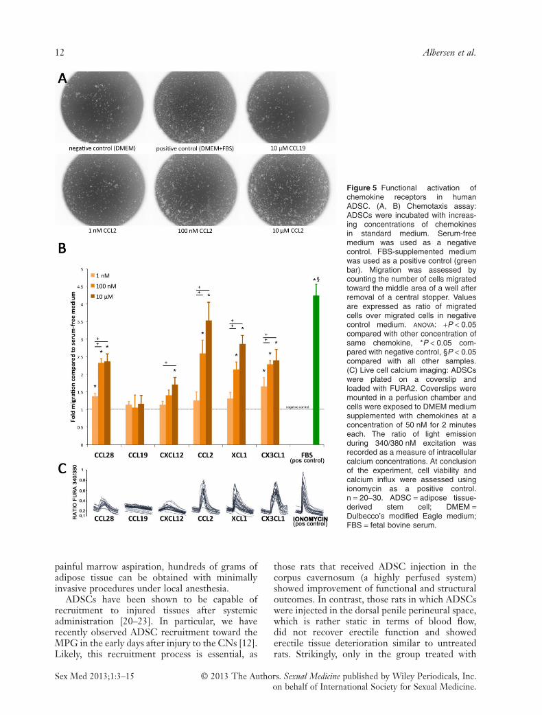
migrated into the central detection zone. In the100 nM and 10 μM concentrations, significantlymore cells were counted in the detection zonewhen cells were treated with CCL28, CCL2,XCL1, and CX3CL1 than when treated withserum-free DMEM. CCL19 did not induce anincrease in cell migration compared with serum-free medium. For CXCL12, only in the highestconcentration there was a relatively small increase(1.71 ± 0.2-fold) of number of migrated cells com-pared with serum-free medium (Figure 5A, B).
Calcium ImagingAttached ADSCs were subjected to calciumimaging to confirm the functional activation ofchemokine receptors by binding of their specificchemokines. Positive responses to administrationof 50 nM of CCL28, CCL2, XCL1, and CX3CL1were noted. CXCL12 elicited an insignificantresponse (increase in ratio fura 340/380 from base-line <0.2). Binding of CCL19 to its receptorCCR7 was not observed as indicated by a complete
absence of calcium influx upon administration ofthis chemokine (Figure 5C).
Discussion
MSCs were first described by Friedenstein andcolleagues in 1968 as fibroblast-like cells derivedfrom the bone marrow which generated colonieswhen plated at low densities [15]. Of the varioustissues in which MSC have since been isolated, thefat tissue has been of particular interest for EDresearchers investigating cellular therapy [16].ADSCs are a distinct population of MSC residingat the perivascular niche in adipose tissue [17].They share many characteristics with BM-MSCpertaining to morphology, phenotype, ex vivo dif-ferentiation ability, and they have a highly similargene expression profile [18]. However, the fre-quency of these cells is 100 to 500-fold higher inadipose tissue compared with the frequency ofMSC in bone marrow [4,19]. Furthermore, whilebone marrow is obtainable in the gram range by a
Figure 4 Immunofluorescence staining of human attached and suspended ADSC. (A, B) Cells were cultured on coverslips,permeabilized using saponin, and stained against f-actin (red), CCR4 (green), and DAPI (blue). Note that the staining patternof CCR4 is perinuclear and morphologically consistent with expression in the endoplasmic reticulum. (C) Cells in suspensionwere permeabilized using saponin and stained CCR4 (green) and DAPI (blue). Note the intracellular staining of CCR4.ADSC = adipose tissue-derived stem cell.
Chemokines in ADSC Migration to the MPG 11
Sex Med 2013;1:3–15© 2013 The Authors. Sexual Medicine published by Wiley Periodicals, Inc.on behalf of International Society for Sexual Medicine.
painful marrow aspiration, hundreds of grams ofadipose tissue can be obtained with minimallyinvasive procedures under local anesthesia.
ADSCs have been shown to be capable ofrecruitment to injured tissues after systemicadministration [20–23]. In particular, we haverecently observed ADSC recruitment toward theMPG in the early days after injury to the CNs [12].Likely, this recruitment process is essential, as
those rats that received ADSC injection in thecorpus cavernosum (a highly perfused system)showed improvement of functional and structuraloutcomes. In contrast, those rats in which ADSCswere injected in the dorsal penile perineural space,which is rather static in terms of blood flow,did not recover erectile function and showederectile tissue deterioration similar to untreatedrats. Strikingly, only in the group treated with
Figure 5 Functional activation ofchemokine receptors in humanADSC. (A, B) Chemotaxis assay:ADSCs were incubated with increas-ing concentrations of chemokinesin standard medium. Serum-freemedium was used as a negativecontrol. FBS-supplemented mediumwas used as a positive control (greenbar). Migration was assessed bycounting the number of cells migratedtoward the middle area of a well afterremoval of a central stopper. Valuesare expressed as ratio of migratedcells over migrated cells in negativecontrol medium. ANOVA: +P < 0.05compared with other concentration ofsame chemokine, *P < 0.05 com-pared with negative control, §P < 0.05compared with all other samples.(C) Live cell calcium imaging: ADSCswere plated on a coverslip andloaded with FURA2. Coverslips weremounted in a perfusion chamber andcells were exposed to DMEM mediumsupplemented with chemokines at aconcentration of 50 nM for 2 minuteseach. The ratio of light emissionduring 340/380 nM excitation wasrecorded as a measure of intracellularcalcium concentrations. At conclusionof the experiment, cell viability andcalcium influx were assessed usingionomycin as a positive control.n = 20–30. ADSC = adipose tissue-derived stem cell; DMEM =Dulbecco’s modified Eagle medium;FBS = fetal bovine serum.
12 Albersen et al.
Sex Med 2013;1:3–15 © 2013 The Authors. Sexual Medicine published by Wiley Periodicals, Inc.on behalf of International Society for Sexual Medicine.

intracavernosal injection, ADSCs were found inthe MPG early after infliction of the nerve injury.Upregulation of the chemokine CXCL12 (SDF1)was observed and it was initially assumed that thiswas an important recruitment factor for ADSC inthe MPG [12].
While CXCL12 binds to both CXCR4 andCXCR7, the affinity of CXCR4 for CXCL12 ishigh and the interaction between CXCL12 andCXCR4 has been extensively investigated and hasbeen a focus of cell migration research in variousdisease models [22–27]. However, the expressionof this receptor on the cell surface of MSC, andADSC in particular, has been debated, and hasrepeatedly been shown to be very low, and eitheror not functional [25,28]. This led us to design thecurrent study as to investigate which chemokinereceptors may be play an important role in theobserved ADSC recruitment to the MPG follow-ing injury of the CNs. CXCL12 was, as wereported before [12], indeed expressed in the ratMPG following CN injury as illustrated by anupregulation of its mRNA in the MPG 1 day afterinjury. There was a low but present intracellularexpression of CXCR4 in all three ADSC cell lines,and no expression on the surface of the cells. Thisstructural finding was reflected in the functionalexperiments in which CXCL12 did not elicit sig-nificant calcium influx at the applied concentra-tion, and induced ADSC migration in vitro only atthe highest concentration of CXCL12. It is there-fore not likely an important recruitment factor forADSC at lower concentrations. Based on theobservations made, we have been able to matchexpression of CCL28, CCL2, CX3CL1, andXCL1 in the MPG to genomic, structural, andfunctional expression and activation of theirrespective receptors in ADSC by their ligands.These data help us understand how ADSC recruit-ment takes place and may identify targets forimprovement of stem cell recruitment and thuspotentially also treatment efficacy.
Several studies have reported the structuralexpression of various chemokine receptors onhuman ADSC or MSC in general; some of theresults are inconsistent between research groups,and many studies have not looked at the full panelof chemokine receptors [28–34]. The low cellsurface expression of various chemokine receptorshas been reported before in MSC [31]. This hasbeen attributed to the use of trypsin-EDTA duringcell detachment [32], and of certain receptorsit has been shown that they are at least in partresiding in intracellular compartments. We now
observed that this holds true for most of the func-tional receptors in ADSC [31]. The intracellularlocalization does not appear to affect either migra-tory capacity or activation of these receptorsresults in calcium influx in the cell, representing animportant step in signaling of most G-proteincoupled receptors. It should however be kept inmind that in the functional experiments the cellswere attached and thus no trypsinization wasperformed. The latter issue indicates that recep-tors at the cell surface may still be the effectorsfor the observed functional activation. Ourimmunofluorescence study of attached ADSC forCCR4 localization revealed expression mainly inthe endoplasmic reticulum. Previously, a similarobservation has been made pertaining to theintracellular expression of CXCR7 in culturedneurons [35]. While the implications of thisfinding are to date not clear, it is known that theendoplasmic reticulum is part of the secretorypathway implicated in protein translocation to thecell surface, and it is commonly considered adefault pathway [35]. These data suggest thatchemokine receptors may be redirected to the cellsurface under specific cellular conditions. It mayfurther imply that these receptors are continuouslyproduced, even when cells remain in culture forseveral weeks. Some authors have proposed thatADSCs, when cultured in hypoxic conditionsor in presence of TNF-α, display a higher numberof chemokine receptors at their cell surface[25,34,36]. The findings in our study imply thatincreased cell surface expression in these condi-tions may not only be the result of elevated tran-scription and/or translation, but may be due toreceptor translocation to the surface of the cellwhen the cell is under noxious conditions. Furtherresearch pertaining to receptor cycling and trans-location is currently in progress in our laboratoriesto better understand these processes.
Three limitations inherent to the research mayrestrict the conclusions of this study. First, whilewe have studied the chemokine regulation in theneural tissue of rats, the chemokine receptor char-acterization was done in human cells. This is dueto limited availability for rat-specific chemokinereceptor primers and antibodies, as well as ratrecombinant proteins with commercial suppliers.Furthermore, for evident ethical reasons, it is notpossible to harvest human hypogastric and pelvicganglia following radical prostatectomy. On theother hand, it has been demonstrated that rodentand human chemokine receptor profiles in stemcells are highly similar, and our conclusions may
Chemokines in ADSC Migration to the MPG 13
Sex Med 2013;1:3–15© 2013 The Authors. Sexual Medicine published by Wiley Periodicals, Inc.on behalf of International Society for Sexual Medicine.

thus be extended, with caution, to the rat in vivosituation [32]. A second limitation encompassesthe fact that no in vivo recruitment experimentswith blockade or knockdown of certain ligands orreceptors were included in this study. We chosenot to perform these studies, as it is not likely thatblocking one single ligand/receptor pair wouldsignificantly impair recruitment as we have shownthat there are several receptors involved in thesemigratory processes. Third, both chemokinesand receptors are promiscuous. We chose thosechemokines that have no or very limited knowncross-reactivity with other receptors present in theADSC for functional activation assays, to limitthis bias. However, although unlikely, previouslyunknown cross-reactivity may occur.
In spite of these limitations, our study pro-vides a qPCR screening of the full panel of the 21currently known chemokine receptors, and anin-depth structural and functional characterizationof the expressed receptors. We are, to our knowl-edge, the first to assess the complete chemokinereceptor expression profile in human ADSC inthis detail. Furthermore, we performed calcium-imaging experiments with chemokine stimulationon adult stem cells to validate migration data, andto bypass the possible bias of manual cell countingin migration assays as a measure for functionalactivation of chemokine receptors. Our findingsmay further help in tailoring stem cell sources inthe function of pathophysiology of different dis-eases with different chemokine expression profiles.In addition, upregulation of chemokine receptorexpression, or translocation of known intracellularreceptors to the cell surface, may very wellimprove treatment efficacy of cellular therapy forED [34].
Conclusions
We identified the ligand/chemokine receptor pairsCCL2/CCR4, CX3CL1/CX3CR1, and XCL1/XCR1 as potentially responsible for ADSChoming toward the MPG following CN injury.While CCL28 was expressed in a low level in theMPG before and after injury, this ligand alsoinduced functional activation of ADSC and itsreceptor CCR10 was highly expressed by ADSC.Surprisingly, CXCR4-CXCL12 (SDF1) interac-tion is not likely a major homing factor for ADSC.Modification of expression of these receptors inADSC could improve migration toward injurysites and thus treatment efficacy.
Acknowledgments
M.A. is a fellow of the Research Foundation-Flanders(FWO). D.D.R. is a fundamental-clinical researcher ofthe FWO. This work was sponsored by the FedericoFoundation and the Research Foundation-Flanders(FWO). We would like to thank Lieve Coorevits,Carl Van Haute. and the Laboratory of Ion-channelResearch at the Catholic University of Leuven forexcellent technical assistance.
Corresponding Author: Maarten Albersen, MD, PhD,Laboratory of Experimental Urology, University Hos-pitals Leuven, Herestraat 49, 3000 Leuven, Belgium.Tel: +3216346930; Fax: +3216346931; E-mail: [email protected]
Conflict of Interest: The authors report no conflicts ofinterest.
References
1 Boorjian SA, Eastham JA, Graefen M, Guillonneau B, KarnesRJ, Moul JW, Schaeffer EM, Stief C, Zorn KC. A criticalanalysis of the long-term impact of radical prostatectomy oncancer control and function outcomes. Eur Urol 2012;61:664–75.
2 Albersen M, Joniau S, Claes H, Van Poppel H. Preclinicalevidence for the benefits of penile rehabilitation therapy fol-lowing nerve-sparing radical prostatectomy. Adv Urol 2008.doi:10.1155/2008/594868.
3 Bella AJ, Lin G, Lin C-S, Hickling DR, Morash C, Lue TF.Nerve growth factor modulation of the cavernous nerveresponse to injury. J Sex Med 2009;6(suppl 3):347–52.
4 Albersen M, Kendirci M, Van der Aa F, Hellstrom WJG, LueTF, Spees JL. Multipotent stromal cell therapy for cavernousnerve injury-induced erectile dysfunction. J Sex Med2011;9:385–403.
5 Kendirci M, Trost L, Bakondi B, Whitney MJ, HellstromWJG, Spees JL. Transplantation of nonhematopoietic adultbone marrow stem/progenitor cells isolated by p75 nervegrowth factor receptor into the penis rescues erectile functionin a rat model of cavernous nerve injury. J Urol2010;184:1560–6.
6 Fall PA, Izikki M, Tu L, Swieb S, Giuliano F, Bernabe J,Souktani R, Abbou C, Adnot S, Eddahibi S, Yiou R. Apoptosisand effects of intracavernous bone marrow cell injection in arat model of postprostatectomy erectile dysfunction. Eur Urol2009;56:716–25.
7 Bochinski D, Lin GT, Nunes L, Carrion R, Rahman N,Lin CS, Lue TF. The effect of neural embryonic stem celltherapy in a rat model of cavernosal nerve injury. BJU Int2004;94:904–9.
8 Meirelles LdaS, Fontes AM, Covas DT, Caplan AI. Mecha-nisms involved in the therapeutic properties of mesenchymalstem cells. Cytokine Growth Factor Rev 2009;20:419–27.
9 Albersen M, Fandel TM, Lin G, Wang G, Banie L, Lin C-S,Lue TF. Injections of adipose tissue-derived stem cells andstem cell lysate improve recovery of erectile function in a ratmodel of cavernous nerve injury. J Sex Med 2010;7:3331–40.
10 Yeghiazarians Y, Zhang Y, Prasad M, Shih H, Saini SA,Takagawa J, Sievers RE, Wong ML, Kapasi NK, Mirsky R,Koskenvuo J, Minasi P, Ye J, Viswanathan M, Angeli FS, BoyleAJ, Springer ML, Grossman W. Injection of bone marrow cell
14 Albersen et al.
Sex Med 2013;1:3–15 © 2013 The Authors. Sexual Medicine published by Wiley Periodicals, Inc.on behalf of International Society for Sexual Medicine.

extract into infarcted hearts results in functional improve-ment comparable to intact cell therapy. Mol Ther 2009;17:1250–6.
11 Zhang H, Yang R, Wang Z, Lin G, Lue TF, Lin C-S. Adiposetissue-derived stem cells secrete CXCL5 cytokine withneurotrophic effects on cavernous nerve regeneration. J SexMed 2011;8:437–46.
12 Fandel TM, Albersen M, Lin G, Qiu X, Ning H, Banie L, LueTF, Lin CS. Recruitment of intracavernously injected adipose-derived stem cells to the major pelvic ganglion improves erec-tile function in a rat model of cavernous nerve injury. Eur Urol2011;61:201–10.
13 Albersen M, Fandel TM, Zhang H, Banie L, Lin G, De RidderD, Lin CS, Lue TF. Pentoxifylline promotes recovery of erec-tile function in a rat model of postprostatectomy erectile dys-function. Eur Urol 2011;59:286–96.
14 Castiglione F, Hedlund P, Van der Aa F, Bivalacqua TJ, RigattiP, Van Poppel H, Montorsi F, De Ridder D, Albersen M.Intratunical injection of human adipose tissue-derived stemcells prevents fibrosis and is associated with improved erectilefunction in a rat model of peyronie’s disease. Eur Urol2012;63:551–60.
15 Friedenstein AJ, Petrakova KV, Kurolesova AI, Frolova GP.Heterotopic of bone marrow. Analysis of precursor cellsfor osteogenic and hematopoietic tissues. Transplantation1968;6:230–47.
16 Zhang H, Albersen M, Jin X, Lin G. Stem cells: Novel playersin the treatment of erectile dysfunction. Asian J Androl2012;14:145–55.
17 Lin C-S, Xin Z-C, Deng C-H, Ning H, Lin G, Lue TF.Defining adipose tissue-derived stem cells in tissue and inculture. Histol Histopathol 2010;25:807–15.
18 Lee RH, Kim B, Choi I, Kim H, Choi HS, Suh K, Bae YC,Jung JS. Characterization and expression analysis ofmesenchymal stem cells from human bone marrow andadipose tissue. Cell Physiol Biochem 2004;14:311–24.
19 Casteilla L, Planat-Benard V, Laharrague P, Cousin B.Adipose-derived stromal cells: Their identity and uses in clini-cal trials, an update. World J Stem Cells 2011;3:25–33.
20 Lamfers M, Idema S, van Milligen F, Schouten T, van der ValkP, Vandertop P, Dirven C, Noske D. Homing propertiesof adipose-derived stem cells to intracerebral glioma andthe effects of adenovirus infection. Cancer Lett 2009;274:78–87.
21 Kim U, Shin D-G, Park J-S, Kim Y-J, Park S-I, Moon Y-M,Jeong K-S. Homing of adipose-derived stem cells toradiofrequency catheter ablated canine atrium and differentia-tion into cardiomyocyte-like cells. Int J Cardiol 2011;146:371–8.
22 Lin G, Wang G, Banie L, Ning H, Shindel AW, Fandel TM,Lue TF, Lin CS. Treatment of stress urinary inconOtinencewith adipose tissue-derived stem cells. Cytotherapy 2010;12:88–95.
23 Bobis-Wozowicz S, Miekus K, Wybieralska E, Jarocha D,Zawisz A, Madeja Z, Majka M. Genetically modified adiposetissue-derived mesenchymal stem cells overexpressing CXCR4display increased motility, invasiveness, and homing to bone
marrow of NOD/SCID mice. Exp Hematol 2011;39:686–96.e4.
24 Cho HH, Kyoung KM, Seo MJ, Kim YJ, Bae YC, JungJS. Overexpression of CXCR4 increases migration and prolif-eration of human adipose tissue stromal cells. Stem Cells Dev2006;15:853–64.
25 Hung S-C, Pochampally RR, Hsu S-C, Sanchez C, Chen S-C,Spees J, Prockop DJ. Short-term exposure of multipotentstromal cells to low oxygen increases their expression ofCX3CR1 and CXCR4 and their engraftment in vivo. PloSONE 2007;2:e416.
26 Liu X, Duan B, Cheng Z, Jia X, Mao L, Fu H, Che Y, Ou L,Liu L, Kong D. SDF-1/CXCR4 axis modulates bone marrowmesenchymal stem cell apoptosis, migration and cytokinesecretion. Protein & Cell 2011;2:845–54.
27 Lin G, Yang R, Banie L, Wang G, Ning H, Li L-C, Lue TF,Lin CS. Effects of transplantation of adipose tissue-derivedstem cells on prostate tumor. Prostate 2010;70:1066–73.
28 Thangarajah H, Vial IN, Chang E, El-Ftesi S, Januszyk M,Chang EI, Paterno J, Neofytou E, Longaker MT, GurtnerGC. IFATS collection: Adipose stromal cells adopt aproangiogenic phenotype under the influence of hypoxia. StemCells 2009;27:266–74.
29 Baek SJ, Kang SK, Ra JC. In vitro migration capacity of humanadipose tissue-derived mesenchymal stem cells reflects theirexpression of receptors for chemokines and growth factors.Exp Mol Med 2011;43:596–603.
30 Ahmadian Kia N, Bahrami AR, Ebrahimi M, Matin MM,Neshati Z, Almohaddesin MR, Aghdami N, Bidkhori HR.Comparative analysis of chemokine receptor’s expression inmesenchymal stem cells derived from human bone marrow andadipose tissue. J Mol Neurosci 2011;44:178–85.
31 Brooke G, Tong H, Levesque J-P, Atkinson K. Moleculartrafficking mechanisms of multipotent mesenchymal stem cellsderived from human bone marrow and placenta. Stem CellsDev 2008;17:929–40.
32 Chamberlain G, Wright K, Rot A, Ashton B, Middleton J.Murine mesenchymal stem cells exhibit a restricted repertoireof functional chemokine receptors: Comparison with human.PloS ONE 2008;3:e2934.
33 Chamberlain G, Fox J, Ashton B, Middleton J. Concise review:Mesenchymal stem cells: Their phenotype, differentiationcapacity, immunological features, and potential for homing.Stem Cells 2007;25:2739–49.
34 Ponte AL, Marais E, Gallay N, Langonné A, Delorme B,Hérault O, Charbord P, Domenech J. The in vitro migrationcapacity of human bone marrow meseSnchymal stem cells:Comparison of chemokine and growth factor chemotacticactivities. Stem Cells 2007;25:1737–45.
35 Shimizu S, Brown M, Sengupta R, Penfold ME, Meucci O.CXCR7 protein expression in human adult brain and differen-tiated neurons. PloS ONE 2011;6:e20680.
36 Tang YL, Zhu W, Cheng M, Chen L, Zhang J, Sun T, KishoreR, Phillips MI, Losordo DW, Qin G. Hypoxic precondition-ing enhances the benefit of cardiac progenitor cell therapy fortreatment of myocardial infarction by inducing CXCR4expression. Circ Res 2009;104:1209–16.
Chemokines in ADSC Migration to the MPG 15
Sex Med 2013;1:3–15© 2013 The Authors. Sexual Medicine published by Wiley Periodicals, Inc.on behalf of International Society for Sexual Medicine.