Reconciling Patent Law's Doctrine of Equivalents with the FDA
Upload
independentCategory
view
1download
0
E-Cadherin Suppression Directs Cytoskeletal Rearrangement andIntraepithelial Tumor Cell Migration in 3D Human Skin Equivalents
Addy Alt-Holland1, Yulia Shamis1, Kathleen N. Riley2, Teresa M. DesRochers1, Norbert E.Fusenig3, Ira M. Herman2, and Jonathan A. Garlick11Division of Cancer Biology and Tissue Engineering, Department of Oral and MaxillofacialPathology, School of Dental Medicine Tufts University, Boston, Massachusetts, USA2Cell and Molecular Physiology, School of Medicine, Tufts University, Boston, Massachusetts, USA3German Cancer Research Center, Im Neuenheimer Feld 280, Heidelberg, Germany
AbstractThe link between loss of cell–cell adhesion, the activation of cell migration, and the behavior ofintraepithelial (IE) tumor cells during the early stages of skin cancer progression is not wellunderstood. The current study characterized the migratory behavior of a squamous cell carcinomacell line (HaCaT-II-4) upon E-cadherin suppression in both 2D, monolayer cultures and within humanskin equivalents that mimic premalignant disease. The migratory behavior of tumor cells was firstanalyzed in 3D tissue context by developing a model that mimics transepithelial tumor cell migration.We show that loss of cell adhesion enabled migration of single, IE tumor cells between normalkeratinocytes as a prerequisite for stromal invasion. To further understand this migratory behavior,E-cadherin-deficient cells were analyzed in 2D, monolayer cultures and displayed alteredcytoarchitecture and enhanced membrane protrusive activity that was associated with circumferentialactin organization and induction of the nonmuscle, β actin isoform. These features were associatedwith increased motility and random, individual cell migration in response to scrape-wounding. Thus,loss of E-cadherin-mediated adhesion led to the acquisition of phenotypic properties that augmentedcell motility and directed the transition from the precancer to cancer in skin-like tissues.
INTRODUCTIONSquamous cell carcinoma (SCC) arises as a premalignant lesion of stratified squamousepithelium that is characterized by intraepithelial (IE) expansion of dysplastic cells (Dlugoszet al., 2002). Following invasion, tumor cells migrate through the connective tissue through adynamic modulation at the tumor cell–extracellular matrix (ECM) interface (Hotary et al.,2006; Wolf et al., 2007). SCCs tend to invade as single cells or as small cell clusters that havea greater tendency to metastasize than carcinomas that invade as large tumor cell masses.Although most studies have characterized the migratory behavior of single, invading cells asthey encounter 3D ECM networks (Brabletz et al., 2005; Hotary et al., 2006; Gaggioli et al.,
© 2008 The Society for Investigative DermatologyCorrespondence: Dr Jonathan A. Garlick, Division of Cancer Biology and Tissue Engineering, School of Dental Medicine, TuftsUniversity, South Cove building, Room 116, 55 Kneeland Street, Boston, Massachusetts 02111, USA. [email protected]; DrAddy Alt-Holland, Division of Cancer Biology and Tissue Engineering, Department of Oral and Maxillofacial Pathology, School ofDental Medicine, Tufts University, Boston, Massachusetts 02111, USA. [email protected] and Dr Ira M. Herman, Departmentof Physiology, School of Medicine, Tufts University, Arnold building, 116 Harrison Avenue, Boston, Massachusetts 02111, [email protected] OF INTERESTThe authors state no conflict of interest.
NIH Public AccessAuthor ManuscriptJ Invest Dermatol. Author manuscript; available in PMC 2010 February 24.
Published in final edited form as:J Invest Dermatol. 2008 October ; 128(10): 2498–2507. doi:10.1038/jid.2008.102.
NIH
-PA Author Manuscript
NIH
-PA Author Manuscript
NIH
-PA Author Manuscript

2007), little is known about the dynamics of migration of SCC cells within a premalignanttissue before stromal invasion occurs. It is therefore important to further understand themigratory properties of IE tumor cells in precancerous lesions.
While changes in tissue architecture and loss of cell–cell adhesion are known morphologicalhallmarks of the IE stage of tumorigenesis, the role that alterations in intercellular adhesionplay in the transition from IE neoplasia to early-stage invasive carcinoma remains unclear. Ithas been shown that loss of E-cadherin function is associated with advanced stages of SCC(Birchmeier and Behrens, 1994; Behrens, 1999; Conacci-Sorrell et al., 2002) and is linked totumor cell metastasis (Bissell and Radisky, 2001; Cavallaro and Christofori, 2004) and poorclinical prognosis in a variety of epithelial cancers (Bagutti et al., 1998; Perl et al., 1998). Lossof cell–cell adhesion due to the absence of E-cadherin function is associated with invasion andmetastasis of SCC and an infiltrative pattern of single cell invasion. Although it has beenthought that loss of E-cadherin was associated only with these later stages of tumorigenesis,identification of mutations in E-cadherin in premalignant breast and stomach have indicatedthat altered E-cadherin-mediated adhesion may be involved in the early stages ofcarcinogenesis as well (Behrens, 1999). However, how loss of E-cadherin impacts on the IEstages of carcinoma progression and during the earliest transition from premalignancy toinvasive carcinoma remains unclear.
In this study, we have explored the migratory properties of individual SCC cells upon loss ofE-cadherin-mediated cell–cell adhesion. To elucidate how suppression of E-cadherin functionimpacts SCC cells motility, we first studied the migratory behavior of an SCC cell line (HaCaT-II-4) in 3D human tissue models that closely mimic the initial events in skin cancer progression.Using skin equivalent model that allowed the fate of IE tumor cells to be followed in thepresence of epidermal keratinocytes, we observed that individual, E-cadherin-deficient cellsundergo transepithelial migration as a prerequisite for their invasion into the underlying matrix.Analyzing the migratory behavior of these tumor cells in 2D cultures revealed that E-cadherinsuppression was linked to the acquisition of motility structures, reorganization of the actincytoskeleton, and induction of β actin. Collectively, these features are likely to contribute tothe activation of the invasive tumor cell phenotype seen during the transition from precancerto carcinoma in vivo.
RESULTSE-cadherin suppression is linked to IE tumor cell migration in 3D tissues
To determine whether loss of E-cadherin function was linked to a motile cell phenotype in 3Dtissue context that mimicked premalignant disease, we developed a human skin equivalentmodel to directly monitor tumor cell migration within the epithelium. β-Gal-marked II-4 tumorcells were seeded on top of a preformed epithelium that was established 2 days earlier byseeding human epidermal keratinocytes (HEK) on type I collagen gels harboring dermalfibroblasts (Figure 1a) or on an acellular, human dermal substrate (AlloDerm) (Figure 1d).This sequential seeding of tumor cells on a layer of normal keratinocytes prevented the directattachment of II-4 cells to the underlying connective tissue and allowed us to compare themigratory behavior of E-cadherin-competent and E-cadherin-deficient cells within theepithelium. Morphologic analysis of skin equivalents revealed that tissues harboring E-cadherin-competent, II-4 cells showed normal tissue architecture when grown on collagen gels(Figure 1b) or on AlloDerm (Figure 1e). Clusters of II-4 cells with altered morphology wereseen in a suprabasal position (arrows) and no cells had separated from these clusters to spreadbetween adjacent HEK. In contrast, tissues that harbored E-cadherin-deficient, H-2Kd-Ecad-II-4 cells revealed a disorganized architecture both on collagen gels (Figure 1c) and onAlloDerm (Figure 1f). Individual II-4 cells and small cell clusters appeared in the collagen gel(Figure 1c, arrows) and in the AlloDerm (Figure 1f, arrows) demonstrating that loss of E-
Alt-Holland et al. Page 2
J Invest Dermatol. Author manuscript; available in PMC 2010 February 24.
NIH
-PA Author Manuscript
NIH
-PA Author Manuscript
NIH
-PA Author Manuscript

cadherin function enabled tumor cells to migrate through the epithelium before invading intothe underlying matrix. To determine the IE fate of tumor cells, tissue sections were double-immunostained with anti-β-Gal and anti-β-catenin antibodies (Figure 1g–j). Anti-β-cateninantibody identified cells either with normal distribution of β-catenin in adhesion-competentcells or with cytoplasmic β-catenin that provided evidence of loss of cell–cell adhesion inadhesion-deficient cells (Margulis et al., 2005a). Clusters of β-Gal-marked, E-cadherin-competent cells (green) were restricted to the superficial layers of the epithelium when tissueswere grown on collagen gels (Figure 1g, arrows) or on AlloDerm (Figure 1h, arrows) andsimilarly to the HEK, showed membrane localization of β-catenin (red) at cell–cell borders(Figure 1g and h). In contrast, individual, β-Gal-marked, E-cadherin-deficient cells (green)migrated through the epithelium (Figure 1i and j, arrowheads) and invaded into the underlyingconnective tissue (Figure 1i and j, arrows). In these cells, β-catenin was limited to thecytoplasm, where it colocalized with β-Gal (Figure 1i and j, yellow), confirming that migratingtumor cells were indeed E-cadherin deficient (Alt-Holland et al., 2005). The ability to trackthe fate of single, motile IE tumor cells demonstrated that abrogation of cell–cell adhesionactivated phenotypic changes that were a prerequisite for transepithelial tumor cell migrationand invasion.
Loss of E-cadherin induces plasma membrane protrusions, cell scattering, and acceleratedcell migration in 2D, monolayer cultures
To further understand the highly motile tumor cell phenotype that enabled transepithelialmigration in 3D skin equivalents, the morphology and behavior of E-cadherin-competent andE-cadherin-deficient II-4 cells were analyzed in 2D, monolayer cultures. E-cadherin-competent cells formed well-organized colonies and showed fan-shaped lamellipodia thatextended from cells at the periphery of colonies (Figure 2a and b, long arrows and insets). Incontrast, E-cadherin-deficient cells migrated as single cells and exhibited extensive membraneruffles, filopodia and unpolarized lamellipodia (Figure 2c, short arrows and inset). Thesefindings demonstrated that E-cadherin suppression enabled cell scattering and increasedplasma membrane protrusive activity.
To determine whether these features were linked to the migratory properties of E-cadherin-deficient II-4 cells, monolayer cultures were scrape-wounded and imaged immediately uponwounding and after 24 and 48 hours (Figure 3a–i). E-cadherin-competent, pBabe- (Figure 3a–c) and H-2Kd-EcadDC25-II-4 (Figure 3d–f) cells migrated slowly into the wound gaps thatremained partly uncovered after 48 hours. In contrast, E-cadherin-deficient, H-2Kd-Ecad cells(Figure 3g–i) showed accelerated wound closure at 24 hours, and complete closure of thewound gap by 48 hours. When further quantified, E-cadherin-competent cultures showed anaverage of 40 and 70% wound closure 24 and 48 hours after wounding, respectively (Figure3j), whereas loss of E-cadherin function resulted in augmented cell migration as seen by the70% wound closure 24 hours after wounding and complete wound closure after 48 hours(Figure 3j).
The pattern of cell migration in response to scrape wounding injury was next analyzed by time-lapse video microscopy. E-cadherin-competent pBabe- (Figure 4a, and Time-lapse movie S1)and H-2Kd-EcadDC25-II-4 cultures (data not shown, and Time-lapse movie S2) developedpolarized lamellipodia at their leading edge that were oriented in the direction of the cellmovement (Figure 4a, arrows). These cells migrated into the wound gap as a coherent cellularsheet while maintaining cell–cell adhesion. In contrast, E-cadherin suppression was linked toaugmented motility of individual H-2Kd-Ecad-II-4 cells at the wound edge that demonstratedunpolarized and extensive membrane protrusions, elongated filopodia, and retraction fibers(Figure 4b arrows). These highly motile cells exhibited rapid, independent, and disorientedmigration (Figure 4b, and Time-lapse movie S3). The pattern of migration of E-cadherin-
Alt-Holland et al. Page 3
J Invest Dermatol. Author manuscript; available in PMC 2010 February 24.
NIH
-PA Author Manuscript
NIH
-PA Author Manuscript
NIH
-PA Author Manuscript
competent and E-cadherin-deficient cells (colored asterisks, Figure 4a and b, marked cells)during the time-lapse video imaging was further analyzed utilizing computer-assisted cell-tracking measurements. E-cadherin-competent pBabe cells maintained their positions inrelation to adjacent cells and showed directional cell migration (Figure 4c). In contrast, E-cadherin-deficient H-2Kd-Ecad cells showed random motility and decreased directionality asthey migrated as individual cells into the wound gap (Figure 4c). In summary, these resultsshowed that E-cadherin suppression was associated with increased plasma membraneprotrusion activity that was linked to single cell scattering and random, augmented cell motility.
E-cadherin-deficient cells demonstrate circumferential, cortical actin organization, loss ofactin stress fibers, and elevated β-actin expression
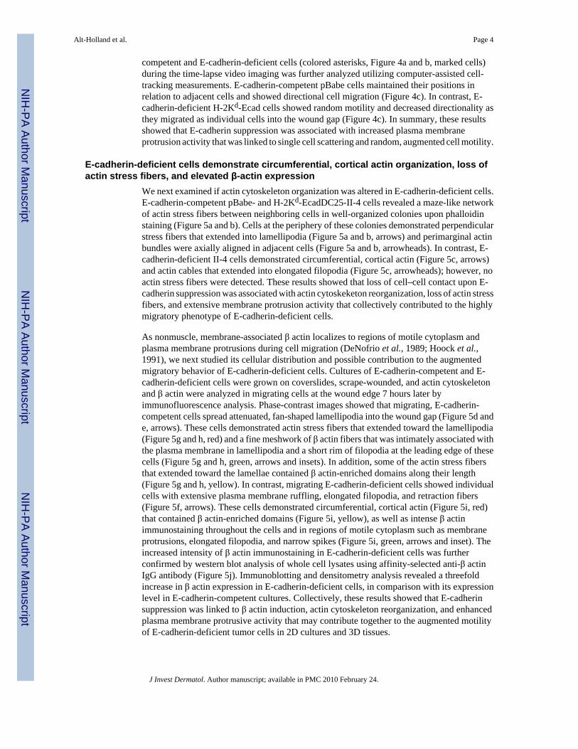
We next examined if actin cytoskeleton organization was altered in E-cadherin-deficient cells.E-cadherin-competent pBabe- and H-2Kd-EcadDC25-II-4 cells revealed a maze-like networkof actin stress fibers between neighboring cells in well-organized colonies upon phalloidinstaining (Figure 5a and b). Cells at the periphery of these colonies demonstrated perpendicularstress fibers that extended into lamellipodia (Figure 5a and b, arrows) and perimarginal actinbundles were axially aligned in adjacent cells (Figure 5a and b, arrowheads). In contrast, E-cadherin-deficient II-4 cells demonstrated circumferential, cortical actin (Figure 5c, arrows)and actin cables that extended into elongated filopodia (Figure 5c, arrowheads); however, noactin stress fibers were detected. These results showed that loss of cell–cell contact upon E-cadherin suppression was associated with actin cytoskeketon reorganization, loss of actin stressfibers, and extensive membrane protrusion activity that collectively contributed to the highlymigratory phenotype of E-cadherin-deficient cells.
As nonmuscle, membrane-associated β actin localizes to regions of motile cytoplasm andplasma membrane protrusions during cell migration (DeNofrio et al., 1989; Hoock et al.,1991), we next studied its cellular distribution and possible contribution to the augmentedmigratory behavior of E-cadherin-deficient cells. Cultures of E-cadherin-competent and E-cadherin-deficient cells were grown on coverslides, scrape-wounded, and actin cytoskeletonand β actin were analyzed in migrating cells at the wound edge 7 hours later byimmunofluorescence analysis. Phase-contrast images showed that migrating, E-cadherin-competent cells spread attenuated, fan-shaped lamellipodia into the wound gap (Figure 5d ande, arrows). These cells demonstrated actin stress fibers that extended toward the lamellipodia(Figure 5g and h, red) and a fine meshwork of β actin fibers that was intimately associated withthe plasma membrane in lamellipodia and a short rim of filopodia at the leading edge of thesecells (Figure 5g and h, green, arrows and insets). In addition, some of the actin stress fibersthat extended toward the lamellae contained β actin-enriched domains along their length(Figure 5g and h, yellow). In contrast, migrating E-cadherin-deficient cells showed individualcells with extensive plasma membrane ruffling, elongated filopodia, and retraction fibers(Figure 5f, arrows). These cells demonstrated circumferential, cortical actin (Figure 5i, red)that contained β actin-enriched domains (Figure 5i, yellow), as well as intense β actinimmunostaining throughout the cells and in regions of motile cytoplasm such as membraneprotrusions, elongated filopodia, and narrow spikes (Figure 5i, green, arrows and inset). Theincreased intensity of β actin immunostaining in E-cadherin-deficient cells was furtherconfirmed by western blot analysis of whole cell lysates using affinity-selected anti-β actinIgG antibody (Figure 5j). Immunoblotting and densitometry analysis revealed a threefoldincrease in β actin expression in E-cadherin-deficient cells, in comparison with its expressionlevel in E-cadherin-competent cultures. Collectively, these results showed that E-cadherinsuppression was linked to β actin induction, actin cytoskeleton reorganization, and enhancedplasma membrane protrusive activity that may contribute together to the augmented motilityof E-cadherin-deficient tumor cells in 2D cultures and 3D tissues.
Alt-Holland et al. Page 4
J Invest Dermatol. Author manuscript; available in PMC 2010 February 24.
NIH
-PA Author Manuscript
NIH
-PA Author Manuscript
NIH
-PA Author Manuscript

DISCUSSIONTumor cell migration, invasion, and metastasis require the integration of proteolysis andremodeling of ECM that is linked to loss of cell–cell adhesion and acquisition of cellcontractibility (Friedl and Wolf, 2003; Wolf et al., 2007). Invasive tumor cells encounternetworks of ECM proteins and activate proteases at their cell surface, localized at focaladhesion contacts and motility structures such as lamellae, pseudopodia, and invadopodia, tolocally degrade these substrates as they migrate through them. In a similar way, premalignantcells initiate their invasion upon basement membrane (BM) degradation and transmigrationthrough assembly of pseudopodia-like extensions (Hotary et al., 2003, 2006). However, itremains unclear if similar mechanisms of cell migration are active before potentially malignantcells reach the BM interface, namely as they transit within the epithelium in premalignanttissues. To address this question, we have studied the migratory properties of IE tumor cellsin 3D tissue models that mimic precancerous human skin, and we have found that E-cadherin-deficient cells acquire a profile of migratory properties that enables their transepithelialmigration and ultimate invasion into the connective tissue.
We have characterized this migratory behavior using an early stage, SCC cell line (HaCaT-II-4) in which E-cadherin function has been disrupted by using a dominant-negative retroviralvector (Margulis et al., 2005a). As a suppressor of invasiveness, loss of E-cadherin functionis correlated with tumor cell invasion and poor clinical prognosis of a variety of cancers(Gumbiner, 2000; Thiery, 2002; Sun and Herrera, 2004). We demonstrated that loss of E-cadherin activated IE transmigration of individual II-4 cells between adjacent normalkeratinocytes before their invasion into the underlying stroma. This migratory behavior waslinked to assembly of motility structures including unpolarized lamellae and filopodia, actincytoskeleton rearrangement, and β actin induction that resulted in individual, random cellscattering and augmented cell motility in 2D, monolayer cultures. These data provide a directevidence that loss of E-cadherin function plays a critical role in activating the IE migration ofSCC tumor cells during the transition from precancer to invasive SCC in a human skin-liketissue.
Stromal invasion of SCC cells occurs either as individual cell migration upon abrogation ofcell–cell adhesion or by collective cell migration as cells maintain adhesive contacts andmigrate as cohesive cellular sheets (Nabeshima et al., 1999; Friedl and Wolf, 2003; Friedl etal., 2004). As tumors spread from their primary site, they are thought to be linked to the dynamicmodulation of tumor cell–ECM interfaces that they encounter following invasion. High-gradeSCCs invade as single cells or as small clusters that have a greater tendency to metastasizethan carcinomas that invade as large tumor cell masses with well-defined, pushing borders.Loss of cell–cell adhesion due to the absence of E-cadherin function is associated with aninfiltrative pattern of single cell invasion. However, it remained unclear if loss of cell–celladhesion in IE tumor cells activated pro-migratory properties that could direct tumor cellmigration within a well-organized epithelium by spreading between adjacent epithelial cellsbefore reaching the BM. We explored whether transepithelial migration in the context of a 3D,skin equivalent tissue was a critical step during this premalignant stage of tumor progression.To address this, we developed a 3D model for transepithelial migration of tumor cells that isschematically illustrated in Figure 6. The seeding of E-cadherin-competent or E-cadherin-deficient tumor cells onto a preformed epithelium prevented tumor cells from attaching directlyto the underlying matrix and generated multilayered tissues in which tumor cells were initiallyfound in a suprabasal position. To undergo invasion, IE tumor cells would be required tomigrate as individual cells between normal surrounding keratinocytes to reach the BM. Theirability to do so indicated that the disruption of E-cadherin-mediated adhesion was associatedwith a shift from collective migration to migration of single, E-cadherin-deficient tumor cellsin both 2D, monolayer cultures and within a multilayered epithelium. Interestingly, the
Alt-Holland et al. Page 5
J Invest Dermatol. Author manuscript; available in PMC 2010 February 24.
NIH
-PA Author Manuscript
NIH
-PA Author Manuscript
NIH
-PA Author Manuscript

collective migration of E-cadherin-competent cells in 2D cultures was similar to thecompartmentalization and restricted migration of clusters of these cells in the superficial layersof the epithelium that prevented their ability to migrate between adjacent HEK.
Our results extend previous findings that matrix metalloproteinases direct degradation andmigration through the BM upon assembly of invasive pseudopodial extensions (Hotary etal., 2006), as we have previously shown that MMP-9 and MT1-MMP are elevated in E-cadherin-deficient-II-4 cells (Margulis et al., 2005a, b). These proteases may also be activeduring transepithelial migration of IE tumor cells, as it has been shown that the transepithelialmigration of dendritic cells is dependent on the MMP-9-mediated degradation of occludin intight junctions (Ichiyasu et al., 2004). Furthermore, the formation of filopodia and increasedmembrane protrusion activity seen upon loss of E-cadherin may also be linked to the abilityof IE tumor cells to migrate through adjacent epithelia. A similar enhancement of membraneprotrusions and peripheral pseudopodia has been reported upon loss of cell–cell adhesion andacquisition of migratory properties in Madin-Darby canine kidney (MDCK) cells (Behrens etal., 1989) as well as upon invasion of SCC (Hotary et al., 2006) and prostatic carcinoma cells(Partin et al., 1988). Our results further substantiate that abrogation of E-cadherin-mediatedadhesion and enhanced membrane protrusive activity act together to increase the migratoryand invasive potential of E-cadherin-deficient carcinoma cells in both 2D cultures and 3Dtissues.
Transepithelial tumor cell migration of II-4 cells appears to also be promoted by the dynamicreorganization of the actin cytoskeleton network upon loss of cell–cell adhesion. It has beenshown that whereas γ actin isoform is mainly located in stress fibers, the β actin isoform isconcentrated in regions of membrane ruffles and moving cytoplasm in nonmuscle cells(DeNofrio et al., 1989; Rubenstein, 1990; Allen et al., 1996). Herman and colleagues(DeNofrio et al., 1989; Hoock et al., 1991) have shown that β actin is found in a subset ofmembrane-associated cytoskeleton and is preferentially localized to motile regions of theplasma membrane at the leading edge of migrating microvascular pericytes, endothelial cells,and fibroblasts. In addition, highly motile MDCK cells showed both loss of actin stress fibersand increased β actin expression in multiple pseudopodia (Le et al., 1998), whereastransformation of human fibroblasts in vitro resulted in expression of a mutant form of β actinand increased tumorigenicity in vivo (Leavitt et al., 1982, 1987). We have extended thesefindings by demonstrating that loss of cell–cell adhesion can also induce β actin expression inassociation with the acquisition of migratory behavior in SCC cells. E-cadherin-deficient cellsshowed increased β actin expression and localization to areas of enhanced membrane protrusiveactivity that was orchestrated with actin cytoskeleton reorganization and loss of actin stressfibers. Our findings suggest that increased β actin expression and its association with plasmamembrane protrusions may allow it to play a functional role in cell shape regulation and actin-based motility in E-cadherin-deficient II-4 cells. As it is known that loss of E-cadherin functiontriggers epithelial–mesenchymal transition (Thiery, 2002; Grunert et al., 2003; Brabletz etal., 2005; Margulis et al., 2005b), it is intriguing to speculate that elevated levels of β actin inE-cadherin-deficient cells represent another characteristic of epithelial–mesenchymaltransition-like behavior in highly migratory, epithelial tumor cells. Our study furthersubstantiates these observations and shows that β actin induction, previously characterized ina variety of mesenchymal cell types, may also be linked to acquisition of mesenchymalproperties in epithelial tumor cells upon loss of E-cadherin function.
The ability of IE tumor cells to transmigrate through an epithelium has important clinicalimplications for the recurrence of SCC at many primary sites. It has been theorized that multipleSCCs develop throughout the aerodigestive tract (“ field cancerization”) as a result ofwidespread, IE migration of individual, tumor cells (van Oijen and Slootweg, 2000; Braakhuiset al., 2003). As a consequence, broad areas of the epithelium are at an elevated risk for the
Alt-Holland et al. Page 6
J Invest Dermatol. Author manuscript; available in PMC 2010 February 24.
NIH
-PA Author Manuscript
NIH
-PA Author Manuscript
NIH
-PA Author Manuscript

development of premalignant lesions that can progress to malignancy (van Oijen and Slootweg,2000; Braakhuis et al., 2003). Our findings that highly migratory E-cadherin-deficient cellscan spread laterally and vertically within tissues support the view that multiple SCCs seen in“field cancerization” may not develop independently of each other, but may rather be due tothe IE migration of cells beyond the margins of the site of the primary lesion. By enablingindividual cell migration within the epithelium, suppression of E-cadherin function may be animportant risk marker for IE tumor cell migration that is linked to SCC recurrence.
Taken together, our results demonstrate that abrogation of E-cadherin-mediated adhesioninduces augmented cell motility, actin cytoskeleton reorganization, and formation of activemembrane protrusions and filopodia. These features are orchestrated to establish thetransmigration of single tumor cells within epithelial tissues as a prerequisite for early SCCprogression. Studying transepithelial migration in a biologically relevant, 3D human tissuenow enables further studies to characterize mechanisms of incipient tumor cell invasion duringthe transition from precancer to malignancy. By exploring signaling pathways that regulatethese events using in vivo-like human tissues, therapeutic approaches designed to blocktransepithelial tumor cell migration may pave the way toward elimination of such lesions andprevention of cancer development.
MATERIALS AND METHODS2D, monolayer cell cultures
β-Gal-marked, HaCaT-II-4 keratinocytes (Boukamp et al., 1990) were grown in DMEMcontaining 5% fetal bovine serum. Human dermal fibroblasts used for organotypic cultureswere derived from newborn foreskins and grown in DMEM containing 10% fetal bovine serum.HEK were derived from newborn foreskins and grown on an irradiated 3T3 fibroblast feederlayer, in DMEM containing 10% fetal calf serum. Cells were grown at 37°C in 7.5%CO2. E-cadherin-competent (pBabe and H-2Kd-EcadDC25) and E-cadherin-deficient (H-2Kd-Ecad)II-4 cells were generated by retroviral infection of HaCaT-II-4 cells as described previously(Margulis et al., 2005a) (vectors provided courtesy of Dr. Fiona Watt). Studies carried out inthis investigation were approved by the Institutional Review Board of Tufts University.
IE tumor cell migration in 3D tissue constructsWe generated 3D tissues that harbored mixtures of HEK and HaCaT-II-4 cells to explore IEtumor cell migration within these tissues. First, 6 × 10⁵ HEK were seeded onto contracted typeI collagen gel or onto human de-epidermalized dermis (AlloDerm, LifeCell Corp. Branchburg,NJ) placed on contracted collagen gel (Alt-Holland et al., 2005). Cultures were maintainedsubmerged for 2 days in low calcium epidermal growth medium to allow formation of anepithelial tissue that was 1–3 cell layers in thickness. This epithelial layer was generated toprevent tumor cells from attaching directly to the connective tissue interface. At that time, 1.5× 10⁵ β-Gal-marked, E-cadherin-competent (pBabe) or E-cadherin-deficient (H-2Kd-Ecad)II-4 cells were seeded onto the preformed HEK layers and cultures were submerged foradditional 2 days in normal calcium epidermal growth medium and raised to an air–liquidinterface for 5 days. In this way, IE tumor cell migration could be determined upon localizationof β-Gal-expressing tumor cells present within the context of neighboring HEK. As II-4 cellswere prevented from attaching to the connective tissue interface, the presence of β-gal-markedcells in the basal layer or the connective tissue would provide evidence of II-4 cell migrationthrough the epithelium.
In vitro scrape-wounding and wound closure measurementsCell cultures at 80% confluence were scrape-wounded using a 2–10 µl micropipette tip.Random areas along the wound gaps were marked, photographed immediately upon wounding
Alt-Holland et al. Page 7
J Invest Dermatol. Author manuscript; available in PMC 2010 February 24.
NIH
-PA Author Manuscript
NIH
-PA Author Manuscript
NIH
-PA Author Manuscript
and 24 and 48 hours later. Wound closure measurements were based on the average change inthe width of the wound gaps over time, relative to the initial width of the scrape-wounded gaps.For each culture, measurements were performed on three captured areas, and in each woundedarea, 50–100 measurements of the wound gap were performed. In each time point, the mean± SD of the width of the open wound gaps was calculated. To quantify wound closure overtime, the averaged width of the open wound gaps at 24- and 48-hour time points were subtractedfrom the initial wound gaps measurements. This calculated measurement of wound closurewas then divided by the initial width of the wound gaps to determine percentage of woundclosure.
Time-lapse video microscopy and cell-tracking measurementsCell cultures were grown on coverslides and scrape-wounded using a 2–10 µl micropipette tip.The coverslides were immediately mounted into a growth chamber with media and placed on37°C, warmed microscope stage as reported previously (Young and Herman, 1985; DeNofrioet al., 1989). Cell migration into the wound gaps was monitored using Axiovert 10 Zeissmicroscope equipped with automated CCD camera (Hamamatsu Photonics, Japan). Imageacquisition was performed at 5 minute intervals for 5 hours, using MetaMorph software 6.0(Universal Imaging, Downingtown, PA). Cell position and migration paths were determinedby tracking the nuclei positions of individual cells at the wound edge through the planes ofeach image stack, using MetaMorph software Track Point function.
Phase-contrast and fluorescence microscopy analysisPhase-contrast images of cells were captured with PixeLINK Software 4.5 (Ottawa, ON,Canada), using inverted Axiovert 40C Zeiss microscope (Gottingen, Germany) equipped withPixeLINK Camera PL-A600 series (Ottawa, ON, Canada). For immunocyto-chemistry,cultures grown on coverslides were fixed with 4% paraformaldehyde in serum-free DMEMfor 5 minutes. Cells were permeabilized for 90 seconds in lysis buffer (0.1% Triton-X, 50mM HEPES, pH 7.1, 50 mM PIPES, pH 6.9, 1 mM MgCl2, 0.5 mM EGTA, 75 mM KCl) andimmunostained with affinity-purified rabbit anti-β actin IgG antibody (Hoock et al., 1991) for1 hour followed by rhodamine-conjugated phalloidin and Alexa 488-conjugated goat anti-rabbit secondary antibody (Molecular Probes, Eugene, OR) for 30 minutes at roomtemperature. For immunohistochemistry, tissues were frozen in embedding media in liquidnitrogen vapors and 6 µm serial sections were fixed in 4% paraformaldehyde and double-immunostained with mouse anti-β-catenin (Zymed, South San Francisco, CA) and rabbit anti-β-Gal (Cortex Biochem, San Leandro, CA) antibodies. Alexa 488-conjugated goat anti-rabbitand Alexa 594-conjugated goat anti-mouse were used as secondary antibodies. Tissue sectionswere counterstained with 4′-6-Diamidino-2-phenylindole (DAPI) in Vectashield mountingmedium (Vector, Burlingame, CA). For tissue morphology, tissues were fixed in 10% formalinand embedded in paraffin, and hematoxylin and eosin staining was performed on 6 µm tissuesections. Immunofluorescent and hematoxylin and eosin images were captured with SpotAdvanced Program 4.5, using a Nikon Eclipse 80i microscope (Nikon Instruments Inc.,Melville, NY) equipped with a SPOT RT camera (Diagnostic Instruments, Sterling Heights,MI).
Western blot analysisCell cultures were extracted on ice with Radio Immuno Precipitation Assay (RIPA) buffer (150mM NaCl, 50 mM Tris-HCl (pH 7.5), 10 mM EDTA, 1% Triton X-100, 10 mM NaF, 10 µgml−1 aprotinin, 10 µg ml−1 leupeptin, 2 µg ml−1 pepstatin, 1 mM Phenylmethylsulfonylfluoride(PMSF), 200 µM NaVO4). Protein concentrations were measured using bicinchoninic acid(BCA) Protein Assay Kit (PIERCE, Rockford, IL) and 5 µg protein samples were separatedon 7.5% SDS-PAGE gel and transferred onto nitrocellulose membrane (Bio-Rad, Hercules,
Alt-Holland et al. Page 8
J Invest Dermatol. Author manuscript; available in PMC 2010 February 24.
NIH
-PA Author Manuscript
NIH
-PA Author Manuscript
NIH
-PA Author Manuscript

CA). The blot was probed with affinity-purified rabbit anti-β actin IgG antibody (Hoock etal., 1991) followed by horseradish peroxidase-conjugated secondary antibody (Amersham,Piscataway, NJ). Membranes were stripped and reblotted with anti-GAPDH antibody (AbcamInc., Cambridge, MA) followed by horseradish peroxidase-conjugated secondary antibody.Proteins were visualized using SuperSignal West Pico Chemiluminescent Substrate (PIERCE,Rockford, IL).
Supplementary MaterialRefer to Web version on PubMed Central for supplementary material.
Abbreviations
BM basement membrane
ECM extracellular matrix
HEK human epidermal keratinocytes
IE intraepithelial
SCC squamous cell carcinoma
3D three dimensional
AcknowledgmentsWe thank Jennifer Landmann for administrative assistance, Dr Mark W. Carlson for his helpful comments, and AdamSowalsky for excellent technical assistance. We thank Dr F. Watt of the Imperial Cancer Research Center for the giftof the chimeric E-cadherin retroviral vectors. This work was supported by National Institutes of Dental andCarniofacial Research Grant no. 2RO1DE011250-06 (toJAG), and National Institutes of Health Grants EY-15125,GM-55110, and Healthpoint Inc. to IMH.
REFERENCESAllen PG, Shuster CB, Kas J, Chaponnier C, Janmey PA, Herman IM. Phalloidin binding and rheological
differences among actin isoforms. Biochemistry 1996;35:14062–14069. [PubMed: 8916891]Alt-Holland A, Zhang W, Margulis A, Garlick JA. Microenvironmental control of premalignant disease:
the role of intercellular adhesion in the progression of squamous cell carcinoma. Semin Cancer Biol2005;15:84–96. [PubMed: 15652453]
Bagutti C, Speight PM, Watt FM. Comparison of integrin, cadherin, and catenin expression in squamouscell carcinomas of the oral cavity. J Pathol 1998;186:8–16. [PubMed: 9875134]
Behrens J. Cadherins and catenins: role in signal transduction and tumor progression. Cancer MetastasisRev 1999;18:15–30. [PubMed: 10505543]
Behrens J, Mareel MM, Van Roy FM, Birchmeier W. Dissecting tumor cell invasion: epithelial cellsacquire invasive properties after the loss of uvomorulin-mediated cell–cell adhesion. J Cell Biol1989;108:2435–2447. [PubMed: 2661563]
Birchmeier W, Behrens J. Cadherin expression in carcinomas: role in the formation of cell junctions andthe prevention of invasiveness. Biochim Biophys Acta 1994;1198:11–26. [PubMed: 8199193]
Bissell MJ, Radisky D. Putting tumours in context. Nat Rev Cancer 2001;1:46–54. [PubMed: 11900251]Boukamp P, Stanbridge EJ, Foo DY, Cerutti PA, Fusenig NE. C-Ha-Ras oncogene expression in
immortalized human keratinocytes (HaCaT) alters growth potential in vivo but lacks correlation withmalignancy. Cancer Res 1990;50:2840–2847. [PubMed: 2183932]
Braakhuis BJ, Tabor MP, Kummer JA, Leemans CR, Brakenhoff RH. A genetic explanation ofslaughter’s concept of field cancerization: evidence and clinical implications. Cancer Res2003;63:1727–1730. [PubMed: 12702551]
Alt-Holland et al. Page 9
J Invest Dermatol. Author manuscript; available in PMC 2010 February 24.
NIH
-PA Author Manuscript
NIH
-PA Author Manuscript
NIH
-PA Author Manuscript

Brabletz T, Jung A, Spaderna S, Hlubek F, Kirchner T. Opinion: migrating cancer stem cells—anintegrated concept of malignant tumour progression. Nat Rev Cancer 2005;5:744–749. [PubMed:16148886]
Cavallaro U, Christofori G. Cell adhesion and signalling by cadherins and Ig-CAMs in cancer. Nat RevCancer 2004;4:118–132. [PubMed: 14964308]
Conacci-Sorrell M, Zhurinsky J, Ben Ze’ev A. The cadherin–catenin adhesion system in signaling andcancer. J Clin Invest 2002;109:987–991. [PubMed: 11956233]
DeNofrio D, Hoock TC, Herman IM. Functional sorting of actin isoforms in microvascular pericytes. JCell Biol 1989;109:191–202. [PubMed: 2745546]
Dlugosz A, Merlino G, Yuspa SH. Progress in cutaneous cancer research. J Investig Dermatol Symp Proc2002;7:17–26.
Friedl P, Hegerfeldt Y, Tusch M. Collective cell migration in morphogenesis and cancer. Int J Dev Biol2004;48:441–449. [PubMed: 15349818]
Friedl P, Wolf K. Tumour-cell invasion and migration: diversity and escape mechanisms. Nat Rev Cancer2003;3:362–374. [PubMed: 12724734]
Gaggioli C, Hooper S, Hidalgo-Carcedo C, Grosse R, Marshall JF, Harrington K, et al. Fibroblast-ledcollective invasion of carcinoma cells with differing roles for rhogtpases in leading and followingcells. Nat Cell Biol 2007;9:1392–1400. [PubMed: 18037882]
Grunert S, Jechlinger M, Beug H. Diverse cellular and molecular mechanisms contribute to epithelialplasticity and metastasis. Nat Rev Mol Cell Biol 2003;4:657–665. [PubMed: 12923528]
Gumbiner BM. Regulation of cadherin adhesive activity. J Cell Biol 2000;148:399–404. [PubMed:10662767]
Hoock TC, Newcomb PM, Herman IM. Beta actin and its MRNA are localized at the plasma membraneand the regions of moving cytoplasm during the cellular response to injury. J Cell Biol 1991;112:653–664. [PubMed: 1993736]
Hotary K, Li XY, Allen E, Stevens SL, Weiss SJ. A cancer cell metalloprotease triad regulates thebasement membrane transmigration program. Genes Dev 2006;20:2673–2686. [PubMed: 16983145]
Hotary KB, Allen ED, Brooks PC, Datta NS, Long MW, Weiss SJ. Membrane type I matrixmetalloproteinase usurps tumor growth control imposed by the three-dimensional extracellularmatrix. Cell 2003;114:33–45. [PubMed: 12859896]
Ichiyasu H, McCormack JM, McCarthy KM, Dombkowski D, Preffer FI, Schneeberger EE. Matrixmetalloproteinase-9-deficient dendritic cells have impaired migration through tracheal epithelialtight junctions. Am J Respir Cell Mol Biol 2004;30:761–770. [PubMed: 14656746]
Le PU, Nguyen TN, Drolet-Savoie P, Leclerc N, Nabi IR. Increased beta-actin expression in an invasivemoloney sarcoma virus-transformed MDCK cell variant concentrates to the tips of multiplepseudopodia. Cancer Res 1998;58:1631–1635. [PubMed: 9563473]
Leavitt J, Bushar G, Kakunaga T, Hamada H, Hirakawa T, Goldman D, et al. Variations in expressionof mutant beta actin accompanying incremental increases in human fibroblast tumorigenicity. Cell1982;28:259–268. [PubMed: 7199389]
Leavitt J, Ng SY, Varma M, Latter G, Burbeck S, Gunning P, et al. Expression of transfected mutantbeta-actin genes: transitions toward the stable tumorigenic state. Mol Cell Biol 1987;7:2467–2476.[PubMed: 3614199]
Margulis A, Zhang W, Alt-Holland A, Crawford HC, Fusenig NE, Garlick JA. E-cadherin suppressionaccelerates squamous cell carcinoma progression in three-dimensional, human tissue constructs.Cancer Res 2005a;65:1783–1791. [PubMed: 15753375]
Margulis A, Zhang W, Alt-Holland A, Pawagi S, Prabhu P, Cao J, et al. Loss of intercellular adhesionactivates a transition from low- to high-grade human squamous cell carcinoma. Int J Cancer 2005b;118:821–831. [PubMed: 16152579]
Nabeshima K, Inoue T, Shimao Y, Kataoka H, Koono M. Cohort migration of carcinoma cells:differentiated colorectal carcinoma cells move as coherent cell clusters or sheets. Histol Histopathol1999;14:1183–1197. [PubMed: 10506935]
Partin AW, Isaacs JT, Treiger B, Coffey DS. Early cell motility changes associated with an increase inmetastatic ability in rat prostatic cancer cells transfected with the V-Harvey-ras oncogene. CancerRes 1988;48:6050–6053. [PubMed: 3048653]
Alt-Holland et al. Page 10
J Invest Dermatol. Author manuscript; available in PMC 2010 February 24.
NIH
-PA Author Manuscript
NIH
-PA Author Manuscript
NIH
-PA Author Manuscript

Perl AK, Wilgenbus P, Dahl U, Semb H, Christofori G. A causal role for E-cadherin in the transitionfrom adenoma to carcinoma. Nature 1998;392:190–193. [PubMed: 9515965]
Rubenstein PA. The functional importance of multiple actin isoforms. Bioessays 1990;12:309–315.[PubMed: 2203335]
Sun W, Herrera GA. E-cadherin expression in invasive urothelial carcinoma. Ann Diagn Pathol2004;8:17–22. [PubMed: 15129905]
Thiery JP. Epithelial–mesenchymal transitions in tumour progression. Nat Rev Cancer 2002;2:442–454.[PubMed: 12189386]
van Oijen MG, Slootweg PJ. Oral field cancerization: carcinogen-induced independent events ormicrometastatic deposits? Cancer Epidemiol Biomarkers Prev 2000;9:249–256. [PubMed:10750662]
Wolf K, Wu YI, Liu Y, Geiger J, Tam E, Overall C, et al. Multi-step pericellular proteolysis controls thetransition from individual to collective cancer cell invasion. Nat Cell Biol 2007;9:893–904. [PubMed:17618273]
Young WC, Herman IM. Extracellular matrix modulation of endothelial cell shape and motility followinginjury in vitro. J Cell Sci 1985;73:19–32. [PubMed: 3894383]
Alt-Holland et al. Page 11
J Invest Dermatol. Author manuscript; available in PMC 2010 February 24.
NIH
-PA Author Manuscript
NIH
-PA Author Manuscript
NIH
-PA Author Manuscript

Figure 1. Abrogation of cell–cell adhesion results in transepithelial, individual migration of E-cadherin-deficient cells in 3D tissuesMorphology analysis of HEK following 2 days of growth showed formation of continuous celllayers on (a) contracted collagen gel or on (d) AlloDerm. Tissues generated by seeding β-Gal-positive, E-cadherin-competent pBabe-II-4 cells on top of HEK layers on (b) collagen or (e)AlloDerm showed organized architecture of basal and suprabasal layers and clusters of cellswith aberrant morphology (arrows). Tissues generated by seeding β-Gal-positive, E-cadherin-deficient H-2Kd-Ecad-II-4 cells on top of HEK layers on (c) collagen or (f) AlloDermdemonstrated cells that had migrated into (c, arrows) the collagen or (f, arrows) the AlloDerm.Dashed line indicates the interface between the epithelium and the underlying matrix. Bar, 50µm. Tissue sections were double-immunostained with anti-β-Gal (green) and anti-β-catenin(red) antibodies. (g, h) pBabe-II-4 cells were confined to superficial layers of the epithelium(arrows) and β-catenin was localized to cell–cell borders of HEK and pBabe-II-4 cells in tissuesgrown on (g) collagen or (h) AlloDerm. (i, j) H-2Kd-Ecad-II-4 cells had migrated betweenHEK layers (arrowheads) and invaded into (i, arrows) the collagen or (j, arrows) the AlloDerm.Whereas β-catenin was restricted to cell–cell borders of HEK, cytoplasmic colocalization of
Alt-Holland et al. Page 12
J Invest Dermatol. Author manuscript; available in PMC 2010 February 24.
NIH
-PA Author Manuscript
NIH
-PA Author Manuscript
NIH
-PA Author Manuscript

β-Gal and β-catenin was seen in H-2Kd-Ecad-II-4 cells (yellow). Dashed line indicates theinterface between the epithelium and the underlying matrix. Bar, 50 µm.
Alt-Holland et al. Page 13
J Invest Dermatol. Author manuscript; available in PMC 2010 February 24.
NIH
-PA Author Manuscript
NIH
-PA Author Manuscript
NIH
-PA Author Manuscript

Figure 2. Loss of E-cadherin function is associated with single cell scatteringPhase-contrast images of (a) E-cadherin-competent pBabe, (b) H-2Kd-EcadC25-II-4, and (c)E-cadherin-deficient H-2Kd-Ecad-II-4 cultures. Note the fan-shape lamellipodia of pBabe- andH-2Kd-EcadC25-II-4 cells at the periphery of well-organized colonies (long arrows and insets)and the scattered H-2Kd-Ecad-II-4 cells that displayed extensive membrane ruffles, filopodia,and retraction fibers (short arrows and inset). Representative images are shown. Bar, 50 µm.
Alt-Holland et al. Page 14
J Invest Dermatol. Author manuscript; available in PMC 2010 February 24.
NIH
-PA Author Manuscript
NIH
-PA Author Manuscript
NIH
-PA Author Manuscript
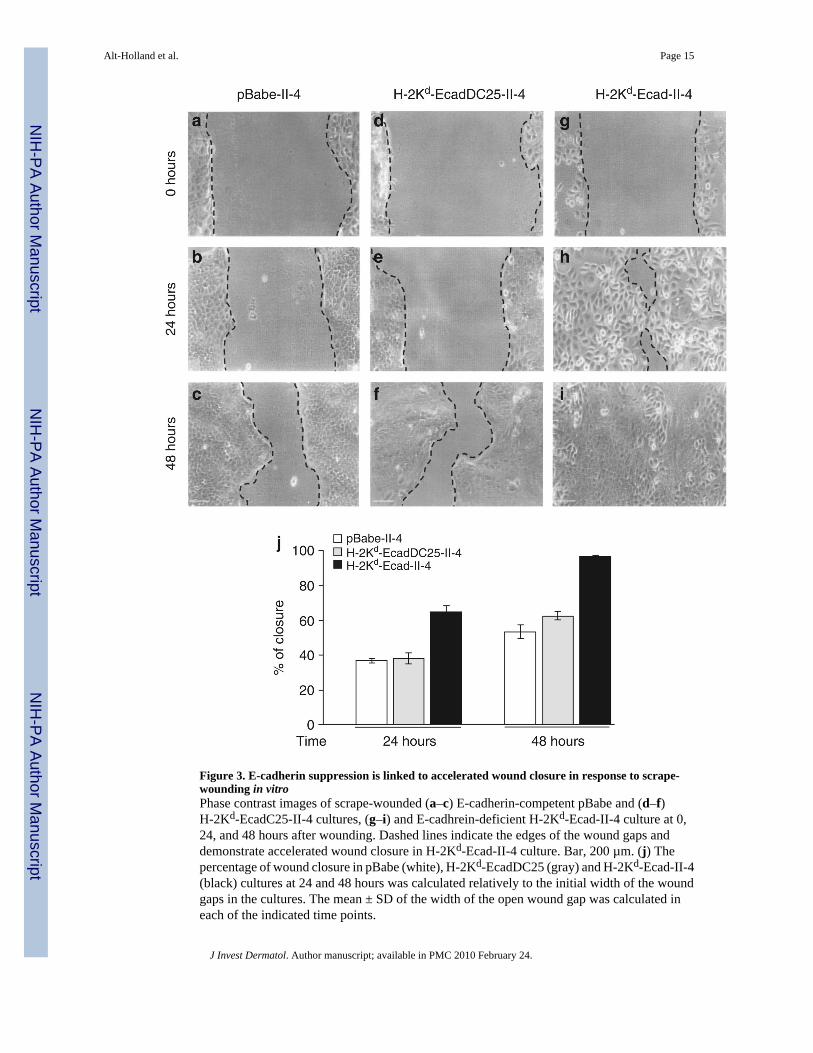
Figure 3. E-cadherin suppression is linked to accelerated wound closure in response to scrape-wounding in vitroPhase contrast images of scrape-wounded (a–c) E-cadherin-competent pBabe and (d–f)H-2Kd-EcadC25-II-4 cultures, (g–i) and E-cadhrein-deficient H-2Kd-Ecad-II-4 culture at 0,24, and 48 hours after wounding. Dashed lines indicate the edges of the wound gaps anddemonstrate accelerated wound closure in H-2Kd-Ecad-II-4 culture. Bar, 200 µm. (j) Thepercentage of wound closure in pBabe (white), H-2Kd-EcadDC25 (gray) and H-2Kd-Ecad-II-4(black) cultures at 24 and 48 hours was calculated relatively to the initial width of the woundgaps in the cultures. The mean ± SD of the width of the open wound gap was calculated ineach of the indicated time points.
Alt-Holland et al. Page 15
J Invest Dermatol. Author manuscript; available in PMC 2010 February 24.
NIH
-PA Author Manuscript
NIH
-PA Author Manuscript
NIH
-PA Author Manuscript

Figure 4. E-cadherin suppression is associated with independent, disoriented, and rapid cellmigrationE-cadherin-competent pBabe- and E-cadherin-deficient H-2Kd-Ecad-II-4 cultures were grownon coverslides, and the cellular response to scrape-wounding injury was analyzed by time-lapse video microscopy, using phase-contrast microscope-imaging workstation.Representative video frames at the indicated time points are shown. Bar, 25 µm. (a) pBabe-II-4 cells at the wound edge displayed polarized morphology, developed lamellipodia at theirleading edge (arrows), and demonstrated collective migration while maintaining their positionwithin the monolayer. (b) Highly motile H-2Kd-Ecad-II-4 cells migrated randomly as singlecells into the wound gap and displayed increased membrane protrusions and retraction fibers(arrows). (c) Graphic representation of cell tracking measurements show cell position andmigration paths of four individual cells at the wound edge, marked by colored asterisks (markedcells in panels a and b), over the experiment time course. The position of the asterisk showsdirectional migration of pBabe-II-4 cells and indicates random migration of H-2Kd-Ecad-II-4cells.
Alt-Holland et al. Page 16
J Invest Dermatol. Author manuscript; available in PMC 2010 February 24.
NIH
-PA Author Manuscript
NIH
-PA Author Manuscript
NIH
-PA Author Manuscript
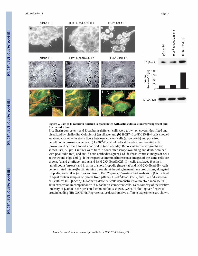
Figure 5. Loss of E-cadherin function is coordinated with actin cytoskeleton rearrangement andβ actin inductionE-cadherin-competent- and E-cadherin-deficient cells were grown on coverslides, fixed andvisualized by phalloidin. Colonies of (a) pBabe- and (b) H-2Kd-EcadDC25-II-4 cells showedan abundance of actin stress fibers between adjacent cells (arrowheads) and polarizedlamellipodia (arrows), whereas (c) H-2Kd-Ecad-II-4 cells showed circumferential actin(arrows) and actin in filopodia and spikes (arrowheads). Representative micrographs areshown. Bar, 50 µm. Cultures were fixed 7 hours after scrape-wounding and double-stainedwith phalloidin (red) and anti-β actin antibodies (green). (d–f) Phase-contrast images of cellsat the wound edge and (g–i) the respective immunofluorescence images of the same cells areshown. (d and g) pBabe- and (e and h) H-2Kd-EcadDC25-II-4 cells displayed β actin inlamellipodia (arrows) and in a rim of short filopodia (insets). (f and i) H-2Kd-Ecad-II-4 cellsdemonstrated intense β-actin staining throughout the cells, in membrane protrusions, elongatedfilopodia, and spikes (arrows and inset). Bar, 25 µm. (j) Western blot analysis of β actin levelin equal protein samples of lysates from pBabe-, H-2Kd-EcadDC25-, and H-2Kd-Ecad-II-4cell cultures (IB: β-actin). E-cadherin-deficient cells demonstrated a threefold increase in β-actin expression in comparison with E-cadherin-competent cells. Densitometry of the relativeintensity of β actin in the presented immunoblot is shown. GAPDH blotting verified equalprotein loading (IB: GAPDH). Representative data from five different experiments are shown.
Alt-Holland et al. Page 17
J Invest Dermatol. Author manuscript; available in PMC 2010 February 24.
NIH
-PA Author Manuscript
NIH
-PA Author Manuscript
NIH
-PA Author Manuscript

Figure 6. Transepithelial migration model of E-cadherin-deficient tumor cells in in vivo-like, 3Dtissues during the transition from precancer to carcinomaThe schematic model proposes the occurrence of distinct stages of transmigration of E-cadherin-deficient tumor cells in the epithelium during the transition to early cancer cellinvasion. (a) In this transepithelial migration model, E-cadherin-competent- or E-cadherin-deficient cells (designated in red) are seeded onto layers of stratified HEK. (b) When grownat an air–liquid interface, E-cadherin-competent cells are maintained in a dormant, suppressedstate within the upper layers of the epithelium. (c) E-cadherin-deficient tumor cells separatefrom adjacent normal keratinocytes and undergo transepithelial, individual cell migrationwithin the tissue, and attached to the BM, creating conditions that are permissive for invasioninto the underlying stroma.
Alt-Holland et al. Page 18
J Invest Dermatol. Author manuscript; available in PMC 2010 February 24.
NIH
-PA Author Manuscript
NIH
-PA Author Manuscript
NIH
-PA Author Manuscript
Copyright © 2022 FDOKUMEN