Dog Breeds and Body Conformations with Predisposition to ...
Upload
tuftsmedicalcenterCategory
view
1download
0
Genetic predisposition directs breast cancer phenotype bydictating progenitor cell fate
Theresa A. Proia*,1,2, Patricia J. Keller*,1,2, Piyush B. Gupta3, Ina Klebba1,2, Ainsley D.Jones1,2, Maja Sedic1,2, Hannah Gilmore4,6, Nadine Tung5,6, Stephen P. Naber7, StuartSchnitt4,6, Eric S. Lander3,8, and Charlotte Kuperwasser1,2,#1 Department of Anatomy & Cellular Biology, Sackler School of Biomedical Research, TuftsUniversity School of Medicine, 136 Harrison Ave, Boston, MA 021112 Molecular Oncology Research Institute, Tufts Medical Center, Boston, MA 021113 Department of Biology, MIT and Broad Institute of MIT and Harvard, Cambridge, MA 021394 Department of Pathology, Beth Israel Deaconess Medical Center, Harvard Medical School,Boston MA5 Department of Surgical Oncology, Beth Israel Deaconess Medical Center, Harvard MedicalSchool, Boston MA6 Department of Medicine, Harvard Medical School, Beth Israel Deaconess Medical Center,Boston MA7 Department of Pathology, Tufts Medical Center, Boston MA8 Department of Systems Biology, Harvard Medical School, Boston, MA 02115
AbstractWomen with inherited mutations in the BRCA1 gene have increased risk of developing breastcancer, but also exhibit a predisposition for the development of aggressive basal-like breasttumors. We report here that breast epithelial cells derived from patients harboring deleteriousmutations in BRCA1 (BRCA1mut/+) give rise to tumors with increased basal differentiation relativeto cells from BRCA1+/+ patients. Molecular analysis of disease-free breast tissues fromBRCA1mut/+ patients revealed defects in progenitor cell lineage commitment even before cancerincidence. Moreover, we discovered that the transcriptional repressor Slug is an importantfunctional regulator of human breast progenitor cell lineage commitment and differentiation andthat it is aberrantly expressed in BRCA1mut/+ tissues. Slug expression is necessary for increasedbasal-like phenotypes prior to and following neoplastic transformation. These findingsdemonstrate that the genetic background of patient populations, in addition to affecting incidencerates, significantly impacts progenitor cell fate commitment and, therefore, tumor phenotype.
# To whom correspondence may be addressed: Charlotte Kuperwasser, Tufts University School of Medicine, 750 Washington Street,box 5609, Boston, MA 02111, Phone: (617) 636-2364, Fax: (617) 636-6127, [email protected].*These authors contributed equally to this work.Publisher's Disclaimer: This is a PDF file of an unedited manuscript that has been accepted for publication. As a service to ourcustomers we are providing this early version of the manuscript. The manuscript will undergo copyediting, typesetting, and review ofthe resulting proof before it is published in its final citable form. Please note that during the production process errors may bediscovered which could affect the content, and all legal disclaimers that apply to the journal pertain.
NIH Public AccessAuthor ManuscriptCell Stem Cell. Author manuscript; available in PMC 2012 February 4.
Published in final edited form as:Cell Stem Cell. 2011 February 4; 8(2): 149–163. doi:10.1016/j.stem.2010.12.007.
NIH
-PA Author Manuscript
NIH
-PA Author Manuscript
NIH
-PA Author Manuscript

KeywordsBRCAl; Slug; differentiation; basal-like breast cancer
IntroductionTumor suppressor genes, such as BRCA1, repress malignant transformation by ensuring thefidelity of DNA replication and chromosomal segregation in response to potentiallydeleterious events. The increased risk of breast cancer development in individuals withinherited mutations in BRCA1 has been attributed to compromised DNA damage repairactivity (Welcsh and King, 2001). However, it has been unclear why mutations in BRCA1are also preferentially associated with an increased propensity for developing a specificsubtype of breast cancers, basal-like tumors, with a distinct molecular phenotype and a poorprognosis (Foulkes et al., 2004; Arnes et al., 2005). Recent evidence has indicated thatBRCA1 might function to regulate mammary epithelial cell morphogenesis anddifferentiation (Furuta et al., 2005; Liu et al., 2008; Kubista et al., 2002). Whether thesefunctions of BRCA1 directly relate to the increased development of basal-like breast cancer,however, is not known.
Human breast tissue contains two major specialized epithelial cell types: luminal cells withsecretory functions surrounding the inner breast duct lumen and basal/myoepithelial cellswith contractile functions that interface between luminal cells and the basement membrane.Corresponding to these cell states, human breast cancers are broadly classified into luminal-like or basal-like tumors based on their gene expression patterns (Peppercorn et al., 2008).Accordingly, it has been proposed that tumors with ‘luminal’ characteristics may result fromthe transformation of cells within the luminal lineage, while tumors exhibiting ‘basal-like’differentiation may arise from basal cells. However, there is also a wealth of evidenceindicating that breast tumors exhibiting luminal or basal-like differentiation have distinctconstellations of genetic aberrations, which may also influence the tumor phenotype. Forexample, luminal tumors frequently express elevated levels of cyclin D1 (CCND1) andsustain mutations in phosphoinositide 3-kinase (PI3K) (Gauthier et al., 2007; Loi et al.,2009; Saal et al., 2005; Campbell et al., 2004), while dysregulated expression of rasisoforms, mutations in p53, loss of PTEN expression, and loss or silencing of BRCA1 aremore commonly associated with basal-like tumors (Gluz et al., 2009; Rakha et al., 2008;Miyakis et al., 1998). Moreover, as mentioned above, inherited mutations in BRCA1(BRCA1mut/+) strongly predispose for the formation basal-like tumors (Foulkes, 2003;Foulkes et al., 2004; Arnes et al., 2005).
In principle, the predisposition for basal-like tumors in BRCA1 mutation carriers could resulteither from the differentiation state of the precursor cells that become transformed or fromthe genetic alterations acquired during tumor formation. In this study, we examined thebiology of disease-free breast tissues from patients harboring deleterious mutations inBRCA1. In doing so, we found a relationship between genetic alterations in perturbingmammary progenitor differentiation and their influence on tumor phenotype.
ResultsCreation of human breast cancers in vivo exhibiting heterogeneous differentiation
To examine the connection between the role of BRCA1 in regulating breast progenitor celldifferentiation and the susceptibility of BRCA1-mutation carriers to developing basal-likebreast cancers, we used a recently described method for creating human breast tissues invivo (Proia and Kuperwasser, 2006; Wu et al., 2009). This method involves three distinct
Proia et al. Page 2
Cell Stem Cell. Author manuscript; available in PMC 2012 February 4.
NIH
-PA Author Manuscript
NIH
-PA Author Manuscript
NIH
-PA Author Manuscript
temporal steps: (1) clearing of the murine mammary fat pad, (2) reconstitution of themammary fat pad with human stromal cells and (3) introduction of lentiviral-infectedorganoids co-mixed with activated fibroblasts into the humanized fat pad. Because thissystem does not require any cell culture, the likelihood of genetic alterations or the selectionof variant phenotypes during in vitro expansion is minimized.
In an attempt to generate tumors from patient-derived breast epithelial cells, we modifiedstep (3) above by introducing oncogenes into dissociated single cell suspensions of epithelialcells before introducing them into humanized stroma (Figure 1a). We chose a set ofoncogenes reflective of both the luminal and basal tumor classes, to reduce the potential forgenetic bias towards either tumor subtype. We infected uncultured breast epithelial cellsuspensions obtained from dissociated reduction mammoplasty tissues with lentivirusesharboring genes for a mutated form of p53 (p53R175H), wild type cyclin D1 (CCND1), aconstitutively-activated form of P13K (PI3KCA), and an oncogenic form of K-ras(RasG12V). Breast tumors developed when all four genes were introduced simultaneouslyinto the breast epithelial cells (Figure 1b,c).
Tumor formation with this procedure was observed with reduction mammoplasty tissuesobtained from multiple patient samples. Expression of the introduced genes in the generatedbreast tumors was verified by immunostaining (for p53, cyclin D1, and p-Akt) and RT-PCR(for K-ras) (Figure 1d). Hematoxylin and eosin (H&E) stains of tumor sections revealed thatthe tumors were heterogeneous invasive carcinomas with regions of mixed squamous andpapillary features, (Figure 1e, Figure S1). Immunostaining showed that cancer cells insquamous metaplastic regions expressed markers indicative of basal differentiation(cytokeratin 14 (CK14), p63, and vimentin (VIM)), and those within papillary regionsexpressed luminal markers (estrogen receptor (ER), CK8/18, and CK19) (Figure 1e).
We next applied this transformation protocol to mammary epithelial cells obtained fromprophylactic mastectomy tissues from patients harboring deleterious mutations in BRCA1(BRCA1mut/+) (Table S1, Figure S1). We observed that the identical set of oncogenes wassufficient to transform epithelial cells obtained from BRCA1mut/+ patients (Figure 2a).Although the introduced oncogenes were expressed to the same extent in wild-type andBRCA1 tumor tissues, immunostaining of tissue sections revealed strong expression of thebasal epithelial markers CK14, p63, and vimentin in BRCA1mut/+ tumor cells (Figure 2b,c).In addition, while tumors arising from BRCA1+/+ epithelium exhibited regions that wereCK8/18 and ER-positive, tumors arising from BRCA1mut/+ cells showed a statisticallysignificant reduction in both CK8/18 and ER expression and increased CK14 expression,which is typical of basal-like tumors (Figure 2c).
To evaluate more comprehensively whether the tumors generated from BRCA1mut/+
epithelium exhibited increased basal-like features, we performed global gene expressionanalyses (Table S2). Hierarchical clustering indicated that tumors arising from eitherBRCA1+/+ or BRCA1mut/+ epithelium could be segregated from one another based on globaltranscriptional profiles (Figure 2d). Gene Set Enrichment Analysis (GSEA) revealed thatBRCA1mut/+ tumors exhibited a significant upregulation of genes associated with breastepithelial basal/myoepithelial cell differentiation compared to the tumors arising fromBRCA1+/+ cells (Figure 2e: Basal Gene Set I, p<0.024; Basal Gene Set II, p<10-4, Table S3).In addition, GSEA indicated specific upregulation of genes in the human breast cancer‘basal-like” centroid, which identifies the human basal-like tumor phenotype (Hu et al.,2006) in BRCA1mut/+ tumors (Basal Centroid, Figure 2e, p<0.033; Table S3) relative toBRCA1+/+ tumors. Collectively, these results indicated that compared to BRCA1+/+ tumors,BRCA1mut/+ tumors generated with identical transforming oncogenes exhibited increasedbasal-like differentiation.
Proia et al. Page 3
Cell Stem Cell. Author manuscript; available in PMC 2012 February 4.
NIH
-PA Author Manuscript
NIH
-PA Author Manuscript
NIH
-PA Author Manuscript

Lineage differentiation defects in breast tissues from BRCA1-mutation carriersAs the BRCA1+/+ and BRCA1mut/+ tumors were generated with identical oncogenes, theseresults suggest that the predisposition of BRCA1mut/+ patients for developing basal-liketumors may result from cellular distinctions present prior to neoplastic transformation. Wetherefore purified breast epithelial cells from BRCA1+/+ and BRCA1mut/+ disease-free breasttissues and assessed the differentiation state of normal precursors in age-matched breasttissue samples. BRCA1+/+ and BRCA1mut/+ breast epithelial cells expressed similar levels ofBRCA1 transcript and protein (Figure S2). However, gene-expression profiling indicatedthat many genes were differentially expressed between BRCA1+/+ and BRCA1mut/+ epithelialcells (Figure 3a, Table S4, Figure S2). Examination of gene ontology functional processesindicated that a number of genes associated with DNA transcription (repressor andactivator), DNA binding, establishment and/or maintenance of chromatin architecture, andchromatin assembly or disassembly were differentially expressed in BRCA1mut/+ epitheliumrelative to BRCA1+/+ epithelium (Figure 3b).
Examination of genes associated with epithelial differentiation revealed that luminal genesand various hormone-related genes including progesterone and estrogen beta receptors(PGR, ESR2) (Table S4) were downregulated in BRCA1mut/ + cells, while genes associatedwith progenitor or basal cells were upregulated (Figure 3a, Table S4). We confirmed thesedifferences in breast epithelial lineage differentiation using semi-quantitativeimmunohistochemistry (Allred scoring metric, see Methods) applied to disease-freeprophylactic mastectomy tissues obtained from BRCA1mut/+ carriers and age-matchedreduction mammoplasty tissues from BRCA1+/+ non-carriers. Consistent with the microarrayresults, progesterone receptor (PGR) expression was significantly reduced in luminalepithelial cells in 88% of BRCA1mut/+ tissues compared to only 11% of BRCA1+/+ breasttissues (Allred score >5, p<0.001) (Figure 3c, Table S5). In addition, trefoil factor 3 (TFF3),which is also associated with mature luminal differentiation, was nearly absent in 88% ofBRCA1mut/+ tissues compared to only 36% of BRCA1+/+ tissues (Allred score <4, p <0.0398; Figure 3c, Table S5). In contrast, 88% BRCA1mut/+ tissue samples exhibitedmoderate-to-high expression of the basal marker vimentin compared to 16% of BRCA1+/+
tissues (Figure 3c, Table S5, (p < 0.086).
We next used flow cytometry to asses the proportion of lineage-committed and progenitorepithelial cells in breast tissues. Cells expressing CD24 or high levels of EpCAM (ESA)enrich for cells of the luminal lineage, while cells expressing high levels of CD49f enrich forcells of the myoepithelial (ME)/basal lineage (Villadsen et al., 2007b; Shipitsin et al., 2007).Analysis of reduction mammoplasty breast tissues from BRCA1+/+ patients identified fourpopulations of epithelial cells: EpCAMhi/CD49f- mature luminal cells, EpCAMhi/CD49f+
luminal progenitor cells, EpCAMlow/CD49f+ basal/myoepithelial (ME) cells, and EpCAM-/CD49f+ basal progenitor cells (Figure 3d, Figure S2, Keller et al., 2010; (Lim et al., 2009;Eirew et al., 2008).
Analysis of prophylactic mammoplasty tissues from BRCA1mut/+ (<50yrs) tissues revealed astatistically significant increase in the proportion of EpCAM-/CD49f+ basal progenitor cells(p<0.04; Figure 3d) and an appreciable but not statistically significant decrease in thenumber of EpCAMhi/CD49f+ luminal progenitor cells. These results indicate thatBRCA1mut/+ tissues exhibit luminal and basal epithelial cell differentiation defects prior toany evidence of cancer.
Characterization of progenitor cells from BRCA1-mutation carriersWe next evaluated the breast progenitor activity of mammary epithelial cells obtaineddirectly from breast tissues. We employed mammosphere (Dontu et al., 2003) and adherent
Proia et al. Page 4
Cell Stem Cell. Author manuscript; available in PMC 2012 February 4.
NIH
-PA Author Manuscript
NIH
-PA Author Manuscript
NIH
-PA Author Manuscript
colony-formation (Stingl et al., 2001) assays to assess progenitor activity, and evaluatedwhether they arose from luminal-committed, basal/ ME-committed or bipotent progenitorsby staining for the differentiation markers CK14 and CK8/18. We found no significantdifferences in the formation of primary mammospheres suggesting that the total number ofstem/progenitor cells may not differ between BRCA1+/+ and BRCA1mut/+ tissues (FigureS3). In addition, there was no significant difference in the distribution of CK8/18+ andCK14+ cells within both BRCA1+/+ and BRCA1mut/+ mammospheres (Figure S3).
Under adherent conditions, we found that human breast epithelial cells generated sphericalcolonies that grew in suspension as well as adherent colonies that grew on plastic (Figure 3e,Figures S3). While we did not observe a statistically significant difference in adherentprogenitor colonies arising from BRCA1mut/+ cells, we did observe that spherical luminalcolonies derived from BRCA1mut/+ cells expressed significantly higher levels of the basalmaker CK14 in comparison to colonies from BRCA1+/+ cells which were more uniformlyCK8/18-positive (Figure 3e).
We also assessed the in vivo outgrowth activity of progenitor cells from BRCA1mut/+ andBRCA1+/+ cells. Using the humanized cleared fat pad system, we found that BRCA1+/+ cellsgenerated bilayered ductal/acinar outgrowths that contained an inner luminal layer ofepithelial cells that stained predominantly for CK8/18 and 19, and an outer myoepitheliallayer that stained for the basal/ME marker CK14 and SMA. In contrast, BRCA1mut/+ cellsgave rise to immature ductal/acinar outgrowths that exhibited a significant increase in thenumbers of bi-potent luminal cells expressing both CK19 and CK14 and to a lesser degreeCK8/18 and CK14 (Figure3f, Figure S3). Taken together, these results reveal that luminalprogenitor cells from BRCA1mut/+ tissue exhibit defects in full maturation and differentiationand retain features of basal differentiation.
Luminal cells give rise to tumors in BRCA1-mutation carriersWe next wanted to determine whether the increased basal differentiation observed followingneoplastic transformation of BRCA1mut/+ cells was due to the increased numbers ofEpCAM- basal cells or the increased basal differentiation state of luminal progenitor cells.Accordingly, we enriched for either luminal (EpCAM+) or basal/ME (CD10+) cells (Figure4a) prior to lentiviral infection and injection into the mammary fat pad. Each subpopulationwas isolated from breast tissues to >90% purity, as gauged by immunofluorescence (Figure4b). The CD10+ subpopulation was enriched for basal/ME CK14+ cells, but CK14+ cellswere depleted from the CD10-/EpCAM+ fraction (Figure 4c). Conversely, CK8/18+ luminalcells were enriched in the CD10-/EpCAM+ fraction compared to the CD10+ and parentalunsorted populations (Figure 4c).
Basal/ME-enriched (CD10+), luminal/progenitor-enriched (CD10-/EpCAM+), and marker-depleted (CD10-/EpCAM-) cells were infected with the p53R175H, CCND1, PI3KCA, andRasG12V oncogenes and injected into humanized murine mammary glands. The luminal-enriched CD10-/EpCAM+ fraction consistently formed tumors with growth kinetics,frequencies and histopathology similar to tumors arising from unsorted cells from eitherBRCA1mut/+ or BRCA1+/+ derived tissues (Figure 4d,e). Thus, basal/ME (CD10+) ordepleted (CD10-/EpCAM-) cells from either BRCA1mut/+ or BRCA1+/+ breast epithelial cellpopulations were not preferentially transformed with this combination of oncogenes. Rather,these results indicate that the target cell for transformation likely resides within the luminalEpCAM+/CD10- population. Collectively, these results imply that the increased basalphenotype of BRCA1-associated tumors results from the pre-existing increased basaldifferentiation state of the luminal progenitor population.
Proia et al. Page 5
Cell Stem Cell. Author manuscript; available in PMC 2012 February 4.
NIH
-PA Author Manuscript
NIH
-PA Author Manuscript
NIH
-PA Author Manuscript
Slug suppresses breast progenitor cell lineage commitmentTo investigate the molecular mechanism BRCA1 effects on progenitor cell differentiation,we classified the breast epithelial gene-expression signature described above based onsignaling pathways that were differentially expressed in BRCA1mut/+ cells. Remarkably, themost significantly represented signaling pathways identified in BRCA1mut/+ breast epithelialsignature were the Wnt, Notch, and melanogenesis pathways (Figure S4).
Notably, the transcriptional repressor Slug, which is an established regulator of melanocytedevelopment, is a downstream target of both Wnt and Notch signaling (Niessen et al., 2008;DiMeo et al., 2009). This connection prompted us to examine Slug expression in breastepithelial tissues and cells harboring mutations in BRCA1. We did not find differences inSLUG mRNA expression, consistent with the microarray data, but we did observe abundantSlug protein in 87% of disease-free BRCA1mut/+ prophylactic mastectomy tissues, while itsexpression was reduced in tissues from reduction mammoplasty BRCA1+/+ tissues (Allredscore >1, p <0.01) (Figure 5a).
As Slug is a transcriptional repressor, we next investigated whether Slug expression mightbe affecting breast progenitor lineage commitment and differentiation. As serum additioncan promote cellular differentiation of immortalized human mammary epithelial cells(HMECs), which are a model for bi-potent breast progenitor cells (Keller et al,. 2010; Zhaoet al., 2010), we treated HMECs from patient-derived BRCA1+/+ and BRCA1mut/+ tissueswith serum and assessed epithelial differentiation. Treatment of BRCA1+/+ HMECs withserum resulted in luminal differentiation, as measured an increase EpCAM+/CD24+ luminalcells as well as increased CD24 expression and increased CK8/18 expression (Figure 5b,data not shown). However, addition of serum to BRCA1mut/+ HMEC cells failed to inducecomplete luminal differentiation, consistent with defects in luminal lineage commitment(Figure 5b, c). Luminal differentiation was accompanied by a reduction in Slug protein levelin both BRCA1mut/+ and BRCA1+/+ cells, although the overall reduction was somewhatreduced in BRCA1mut/+ cells (Figure 5c).
To investigate whether Slug directly inhibits breast epithelial lineage commitment anddifferentiation, we used lentiviral-mediated short hairpin-inhibition of Slug expression inprimary prophylactic mastectomy cells isolated from three different patients with deleteriousBRCA1 mutations. Slug knockdown led to a reduction in the proportion of EpCAM-/CD49f+
progenitor cells and a concomitant increase in the proportion of EpCAM+, CD44lo, andCD24+ luminal cells (Figure 5d, Figure S5). Furthermore, expression of the basal markervimentin was greatly reduced, while expression of the luminal marker CD24 was increased(Figure 5c, Figure S5). We also examined the effects of Slug inhibition on lineagecommitment of immortalized HMECs derived from BRCA1mut/+ patients. As with primarycells, inhibition of Slug expression resulted in a decrease in EpCAM-/CD49f+ basalprogenitor cells and an increase in EpCAM+ luminal cells (Figure 5e). Given these findings,we also examined whether inhibition of Slug might also be important for luminaldifferentiation in BRCA1+/+ cells. Indeed, inhibition of Slug in BRCA1+/+ cells also led to areduction in the proportion of EpCAM-/CD49f+ basal progenitor cells and an increase inluminal cells. Taken together, these findings indicate that Slug is a regulator of human breastprogenitor cell differentiation and its expression blocks luminal differentiation.
BRCA1 regulation of Slug protein stabilityTo examine whether BRCA1 regulates Slug expression, we used short interfering RNAs(siRNA) to inhibit BRCA1 expression in human breast MCF10A cells, which express wild-type BRCA1 (Elstrodt et al., 2006). Quantitative RT-PCR and western blotting confirmedknockdown of BRCA1 expression (Figure 6a). Knockdown of BRCA1 by siRNA led to a
Proia et al. Page 6
Cell Stem Cell. Author manuscript; available in PMC 2012 February 4.
NIH
-PA Author Manuscript
NIH
-PA Author Manuscript
NIH
-PA Author Manuscript

modest but highly reproducible 2-fold increase in Slug protein expression, in the absence ofincreased mRNA expression (Figure 6a). These results suggest that loss of BRCA1 may leadto increased Slug protein expression by a post-translational mechanism. We thereforeexamined the stability of Slug protein in cells following siRNA-inhibition of BRCA1 as wellas in cells with mutations in BRCA1. We confirmed that Slug protein is highly unstable inthe BRCA1+/+ MCF10A cells (Figure 6b,c). BRCA1mut cells (SUM149, SUM1315) andsiBRCA1-MCF10A cells were collected at regular time intervals subsequent tocyclohexamide (CHX) treatment and subjected to western blot analysis. While Slug proteinlevels were turned over in siControl-MCF10A cells, Slug protein was still detected up to 6hours following CHX treatment in siBRCA1-MCF10A cells and in cancer lines harboringmutations in BRCA1 (Figure 6c). Importantly, the difference in stability noted in Slugprotein in BRCA1mut SUM149 and SUM1315 cells was not due to a defect in proteasomeactivity as cyclin D1 protein was still degraded. Taken together, these results indicate thatBRCA1 regulates Slug protein stability.
To begin to understand the mechanism involved, we looked at whether the ubiquitin ligasefunction of BRCA1 might be important for regulating Slug protein stability. BRCA1associates with the BRCA1-associated RING domain-1 protein (BARD1) to form aheterodimer with ubiquitin E3 ligase activity. Therefore, we examined whether BARD1knock down might also result in increased Slug protein stability. We used siRNAs to inhibitBARD1 expression in human breast MCF10A cells and collected cell lysates at regular timeintervals after cyclohexamide (CHX) treatment. Although BARD1 protein was inhibited tonearly undetectable levels, Slug protein stability was similar to that of control cells,indicating that the ubiquitin-ligase functions of BARD1 was likely not regulating Slugprotein stability (Figure S6). We next examined direct interactions between BRCA1 andSlug proteins. However, co-immunoprecipciaton of Slug with BRCA1 failed to demonstratean interaction (Figure S6), although we did observe interaction between BRCA1 andBARD1. Further studies will be needed to determine what which BRCA1functions areinvolved in regulating Slug protein stability.
Slug regulation of basal-like breast cancer phenotypeTo study the role of Slug in basal-like breast cancer phenotype, we examined Slugexpression in sporadic and BRCA1-associated breast tumor tissues. Slug protein waspreferentially expressed in ER-negative tumors derived from BRCA1-mutation carriers aswell as ER-negative sporadic breast cancers, but its levels were higher in BRCA1-associatedbreast cancers (p<0.007) (Figure 7a). Furthermore, Slug protein was expressed in cell linesderived from basal-like breast cancers and elevated in cancer cell lines that harboredmutations in BRCA1 (Figure 7b).
To test whether Slug is necessary for regulating the basal-like tumor phenotype, we usedlentiviral-mediated short hairpin-inhibition of Slug in breast cancer cells derived fromprimary patient BRCA1mut breast cancers. We found that shSlug reduced endogenous SlugmRNA levels between 40-80% in BRCA1mut SUM149 and SUM1315 cancer cell lines andreduced protein to nearly undetectable levels (Figure 7c). Slug inhibition resulted in a ∼6-fold reduction in the proportion of CD44+/CD24- stem-like basal cells in SUM149, and a∼4-fold increase in the proportion of CD44-/CD24+ luminal cells, consistent with increaseddifferentiation and luminal lineage commitment (Figure 7c). Similarly, reducing Slugexpression in BRCA1mut SUM1315 cells increased the proportion of CD24+ luminal cells bynearly 3-fold (Figure S7). We also performed quantitative mRNA expression profiling usinga custom qRT-PCR array targeting 86 genes associated with basal/ME, luminal or stem celldifferentiation (Table S6). Consistent with the changes observed by flow cytometry,inhibition of Slug expression resulted in upregulation of luminal genes in tumor cells
Proia et al. Page 7
Cell Stem Cell. Author manuscript; available in PMC 2012 February 4.
NIH
-PA Author Manuscript
NIH
-PA Author Manuscript
NIH
-PA Author Manuscript

including CK19, CK8, E-cadherin, MUC1, CD24, and TFF3, and repression of basal,mesenchymal and stem cell genes in both SUM149 and SUM1315 lines (Figure 7d).
To further demonstrate the role of Slug in the development of basal-like breast cancers inBRCA1mut/+ cells, mammary epithelial cells from disease-free prophylactic mastectomytissues from three different BRCA1mut/+ patients were transduced with lentiviruses harboringp53R175H, CCND1, PI3KCA, and Ras oncogenes with or without targeting Slugexpression. Patient-derived BRCA1mut/+ cells expressing shSlug showed increasedexpression of genes associated with luminal tumors including CK19, CK8, MUC1, EpCAM,and TFF3 with concomitant repression of many genes associated with basal-like breastcancers including SPARC, SERPINE, CD44, CK14, CK5, CK17, and vimentin compared tocontrol patient-derived BRCA1mut/+ cells (Figure 7e).
Finally, to demonstrate the connection between Slug expression and BRCA1-mutation beforetransformation, we examined whether the genes that were induced following Slug-inhibitionwere differentially expressed based on BRCA1 status in disease-free tissues (Slug Gene Set,Table S3). We used GSEA to evaluate the expression of these Slug transcriptional targets inBRCA1mut/+ epithelium from four different patient samples. Six out of eight Slug targetgenes were repressed in BRCA1mut/+ cells relative to BRCA1+/+ cells (Figure S7), yieldingsignificant enrichment by GSEA (p<0.0207). Collectively, these results show thatupregulation of Slug blocks luminal lineage commitment and increases the propensity forbasal breast tumor formation.
DiscussionA fundamental difference between breast cancers arising in BRCA1-mutation carrierscompared to sporadic cancers is their tendency toward a basal subtype. By using an in vivomodel that minimized cell culture, we were able to create human breast cancers thatrecapitulated many features of clinically relevant tumors to validate the previously untestedidea that the predisposition for basal-like tumors in BRCA1-mutation carriers arises fromperturbations in breast epithelial differentiation caused by compromised BRCA1 function(Foulkes, 2003). Our results show that breast epithelial cells isolated from BRCA1-mutationcarriers preferentially form tumors with increased basal differentiation compared to cellsisolated from non-carrier tissues. In addition, we found that that EpCAM+/CD10- luminalcells from both BRCA1+/+ and BRCA1mut/+ tissues enriched for tumor forming ability in thismodel system, but that the latter exhibited increased features of basal differentiation prior totransformation. However, since basal progenitor cells were also expanded in disease-freebreast tissues from BRCA1mut/+ tissues, it is possible that these cells might also serve astargets of neoplastic transformation in patients. Nevertheless, our findings are consistentwith the notion that tumor phenotype can be significantly impacted by the pre-existingdifferentiation state of the normal precursor (“cell of origin”) targeted for neoplastictransformation (Gupta et al., 2005; Ince et al., 2007). However, since mutations in a singleallele of BRCA1 can alter the differentiation potential of the same cellular targets oftransformation, leading to tumors with different phenotypes, this indicates that the initiatinggenetic mutation (“mutation of origin”) is a critical factor in defining tumor subtype. Futurestudies will be needed to determine whether other combinations of cooperating oncogenesgive rise to BRCA1-associated basal-like tumors in basal/ME cells and whether mutations inother tumor suppressor genes or oncogenes also affect the differentiation potential ofprogenitor cells that drive tumor phenotypes.
While we have not excluded the possibility that LOH of the wild-type BRCA1 allele isnecessary for basal-like tumor formation, tumors in this model system were driven byectopic oncogenes suggesting that LOH was not necessarily a rate-limiting step.
Proia et al. Page 8
Cell Stem Cell. Author manuscript; available in PMC 2012 February 4.
NIH
-PA Author Manuscript
NIH
-PA Author Manuscript
NIH
-PA Author Manuscript

Furthermore, LOH is a stochastic event in BRCA1mut/+ patients, affecting the mutant orwild-type alleles at similar frequencies (Clarke et al., 2006). Since the analysis ofprophylactic mastectomy tissues showed differentiation defects in significant proportions ofthe breast tissue, this suggests that LOH was not likely responsible for the perturbations inbreast epithelial differentiation or basal tumor phenotype. Our findings, combined with thoseof others (Burga et al., 2009; Lim et al., 2009) indicate that haploinsufficiency of BRCA1affects breast epithelial differentiation and progenitor cells in patients.
The present study also provides several additional lines of evidence that breast epithelialdifferentiation is altered in the presence of BRCA1 mutations. First, genes involved inepigenetic functions including DNA transcription and chromatin modification areoverrepresented in the transcriptional signature of BRCA1-mutant cells. Interestingly, manyof the upregulated genes are involved in the establishment and/or maintenance of chromatinstructure, including demethylases, methyltransferases, histones, acetyltransferases andseveral components of the ubiquitin pathway. These observations are consistent with theidea that BRCA1 mutations affect large-scale chromatin unfolding (Ye et al., 2001),underscoring its role as an integral component of multi-protein complexes that modulategene expression (Narod and Foulkes, 2004).
Second, the distinct transcriptional profile of BRCA1mut/+ cells may reflect activation ofsignaling pathways associated with progenitor/basal cells, increased basal differentiation anddecreased luminal differentiation. Previous results suggest that reduction in BRCA1 levelsleads to a failure of luminal lineage commitment and increased expansion of anuncommitted progenitor EpCAM- population (Liu et al., 2008). Consistent with thisobservation found that BRCA1mut/+ breast tissue exhibited an increase in the proportion ofEpCAM-/CD49f+ basal progenitor cells. However, in contrast to other findings (Lim et al.,2009), we did not find an expansion of EpCAM+/CD49f+ luminal progenitor cells; althoughwe did observe defects in luminal progenitor differentiation. This difference might reflectthe genetic differences between the BRCA1 patient populations in the two studies.Nonetheless, overall these studies reinforce the idea that BRCA1 is a critical regulator ofbreast epithelial progenitor lineage commitment.
All of the BRCA1-mutation carrier samples used in this study harbored frameshift mutationsthat compromise at a minimum the C-terminal BRCT domain (Figure S2), which coulddestabilize protein-protein interactions between BRCA1 and its C-terminal binding partners.However, the overall levels of BRCA1 expression were surprisingly not affected. Inaddition, as perturbations in differentiation and Slug expression could be detected withoutchanges in BRCA1 expression level, the effects of BRCA1 mutation may be at the level ofprotein-protein interactions rather than overall expression level. Consistent with this notion,reduction of BRCA1 in MCF10A cells by RNA interference impaired differentiation andcould be rescued by expression of a wild-type or a RAD51 mutant of BRCA1 but not with aBRCA1 C-terminal BRCT domain mutant (Furuta et al., 2005). Future studies will benecessary to fully dissect the precise domains and mechanism by which BRCA1 regulatesbreast epithelial differentiation. In addition, further experiments will be needed to determinewhether certain mutations in BRCA1 affect differentiation and regulate progenitor cell fatedifferently than others and whether different mutations alter the propensity for thedevelopment of basal-like tumors.
The observation that BRCA1mut/+ epithelial cells express genes involved in melanogenesisand stem cell biology prompted us to examine Slug and its requirement for maintainingprogenitor cells and basal differentiation. We found that haploinsufficiency or knockdownof BRCA1 was associated with increased expression and stability of the transcriptionalrepressor Slug and that Slug was shown to be a regulator of the basal phenotype. This
Proia et al. Page 9
Cell Stem Cell. Author manuscript; available in PMC 2012 February 4.
NIH
-PA Author Manuscript
NIH
-PA Author Manuscript
NIH
-PA Author Manuscript

suggests that perturbations in luminal differentiation due in part to Slug expression, is likelyresponsible for the increased propensity for the development of tumors with basal-likefeatures. Although our results do not address whether Slug is sufficient to induce basaldifferentiation, Slug is expressed in breast stem/progenitor cells and has can promote abasal-like phenotype in the luminal MCF-7 breast cancer cell line and increased basaldifferentiation marker expression in the non-tumorigenic MCF10A breast line (Sarrio et al.,2008). Moreover, the fact that Slug is expressed in basal-like breast cancers not associatedwith BRCA1 mutations (Storci et al., 2008) implies that acquisition of its expression enablesbasal differentiation.
Materials and MethodsDetailed Methods are described in Supplemental Materials
BRCA1-mutation carrier tissuesAll human breast tissue procurement for these experiments was obtained in compliance withthe laws and institutional guidelines, as approved by the institutional IRB committee fromBeth Israel Deaconess Medical Center (BIDMC) and Tufts University School of Medicine.Disease-free prophylactic mastectomy (n=31; 12 fresh, 19 formalin-fixed paraffinembedded) and tumor tissues (n=19) derived from women carrying a known deleteriousBRCA1 heterozygous mutation were obtained with patient consent from the SurgicalPathology files or immediately following prophylactic mastectomy surgery at BIDMC.Tissues in which BRCA1 mutation was confirmed but not known were submitted forsequence/genotyping at Myriad Genetic Laboratories. Non-BRCA1 tumor tissues (n=20)were obtained from discarded material at Tufts Medical Center and non-cancerous breasttissue was obtained from patients undergoing elective reduction mammoplasty at TuftsMedical Center or BIDMC (n=38; 18 fresh, 24 formalin-fixed paraffin embedded). BRCA1mutation status and clinical information are listed in Table S1. The range of patient ages forfresh BRCA1+/+ tissue used in this study was 30-54 with a median age of 40; the range ofpatient ages for fresh BRCA1mut/+ tissue used in this study was 35-53 with a median age of44. All disease-free breast tissues were verified by surgical pathologists prior to use in thesestudies.
Cell Lines and Tissue CultureSUM cell lines were obtained from Dr. Stephen Ethier (Kramanos Institute, MI), while theMCF10A cell lines were purchased from ATCC. MCF10A cells were cultured in DMEMwith 10% calf serum. SUM149PT cells were cultured in Ham's F12 with 5% calf serum,insulin (5 μg/mL), and hydrocortisone (1 μg/mL) while SUM1315MO2 were in Ham's F12with 5% calf serum, insulin (5 μg/mL), and EGF (10 ng/mL). All cell lines were grown at37°C and 5% CO2. BRCA1mut/+ HMECs were immortalized with the catalytic subunit ofhuman telomerase as previously described (Elenbaas et al, 2000). BHME cells were culturedin MEGM (Lonza) supplemented with bovine pituitary extract (BPE), insulin (5 μg/mL),EGF (10 ng/mL) and hydrocortisone (1 μg/mL).
Lentiviral Constructs and virus productionLentiviral constructs used for gene transduction into human mammary epithelial cells werecreated using standard cloning techniques into the self-inactivating CS-CG (Miyoshi et al.,1998) viral vector generously provided by Inder Verma (Salk Institute, La Jolla, CA).pLENTI-KRASG12V and pLenti-CMV-PIK3CA-myr+CMV-CCND1 were obtained fromfrom Min Wu (Aveo Pharmaceuticals, Cambridge, MA). A wild-type human p53 cDNAclone was generously provided by Josh LaBaer (Harvard Institute of Proteomics, HarvardMedical School, Cambridge, MA). Site directed mutagenesis was employed to change
Proia et al. Page 10
Cell Stem Cell. Author manuscript; available in PMC 2012 February 4.
NIH
-PA Author Manuscript
NIH
-PA Author Manuscript
NIH
-PA Author Manuscript

amino acid residue 175 from R to H. The VSV-G-pseudotyped lentiviral vectors weregenerated by transient co-transfection of the vector construct with the VSV-G-expressingconstruct pCMV-VSV-G (Miyoshi et al., 1998) and the packaging construct pCMVΔR8.2Δvpr (Miyoshi et al., 1998) generously provided by Inder Verma, into 293T cells withthe FuGENE 6 transfection reagent (Roche). High-titer stocks of the virus were prepared byultracentrifugation at 100,000 × g. Lentiviral shRNA constructs targeting Slug (Addgeneplasmids 10904 and 10905) were prepared as previously described (Gupta et al., 2005).
Breast tissues were minced and enzymatically digested overnight with a mixture ofcollagenase and hyaluronidase as previously described (Kuperwasser et al., 2004; Proia andKuperwasser, 2006) and dissociated to a single cell suspension. Immediately afterdissociation, cells were resuspended with lentiviruses expressing the genes of interest andinjected into cleared and humanized mammary fat pads.
Animals and SurgeryAll animal procedures were conducted in accordance with a protocol approved by the TuftsUniversity IACUC committee. A colony of immunocompromised NOD/SCID mice wasmaintained in house under aseptic sterile conditions. Mice were administered autoclavedfood and water ad libitum. Surgeries were performed under sterile conditions, and animalsreceived antibiotics in the drinking water up to two weeks after all surgical procedures.Animals were humanized and injected as previously described (Kuperwasser et al., 2004;Proia and Kuperwasser, 2006).
ImmunohistochemistryImmunohistochemistry was performed on formalin-fixed, paraffin-embedded tissue sectionswith sodium citrate antigen retrieval, followed by visualization with the ABC Eliteperoxidase kit and NovaRed substrate (Vector labs) for detection of αSMA (1:500, clone♋sm-1), CK14 (1:500, clone LL002), CK8/18 1:500, clone DC-10), Vimentin (1:500, cloneV9), CK19 (1:500, clone b170) (all, Vector Labs), TFF3 (Abnova, clone 3D9, 1:200)) andSlug (1:200, Cell Signaling). Staining for pan-cytokeratin (Ventana Medical Systems), p53(Ventana Medical Systems), cyclin D1 (NeoMarkers), pAKT (Cell Signaling, 1:100), ER(Ventana Medical Systems), p63 (Ventana Medical Systems), and PR was performed by theHistology Special Procedures Laboratory at Tufts Medical Center. IHC results were semi-quantitatively analyzed (see Supplmental Materials for details).
Mammospheres and Adherent colony forming assaysViable cells dissociated from organoids derived from BRCA1+/+ (n=4) and BRCA1+/- (n=4)patients were plated at 35,000 cells per well in 6-well plates for adherent colony growth inMEGM media (Lonza) or at 20,000 cells per ml in 6-well ultra-low attachment plates(Corning) in MEGM media minus the addition of bovine pituitary extract for mammospheregrowth. Colonies and mammospheres were allowed to form for 8 days after which non-adherent suspension colonies from adherent culture and mammospheres from non-adherentculture were collected for analysis.
Mammospheres collected from non-adherent culture and suspension colonies collected fromadherent culture were cytospun onto glass slides and fixed in methanol forimmunofluorescence analysis. Quantification of mammosphere and suspension colonynumbers was accomplished using a Multisizer 3 COULTER COUNTER (Beckman-Coulter).
Proia et al. Page 11
Cell Stem Cell. Author manuscript; available in PMC 2012 February 4.
NIH
-PA Author Manuscript
NIH
-PA Author Manuscript
NIH
-PA Author Manuscript

Immunomagnetic Bead sortingEpithelial organoids from BRCA1+/+ and BRCA1+/mut patients were dissociated to a singlecell suspension and sorted with CELLection pan-mouse IgG magnetic beads according tothe manufacturers instructions and as described previously (Allinen et al., 2004) (Dynal,Invitrogen) conjugated to an anti-CD10 antibody (DAKO). CD10+ cells were released fromthe beads by DNase treatment per the manufacturer's instructions. Cells that did not bind tothe CD10-immunobeads were further sorted with magnetic beads conjugated to an anti-EpCAM antibody (Abcam).
Flow cytometry and FACSUncultured cells from BRCA1+/+ (n=10) or BRCA1mut/+ (n=7) organoid preparations weredissociated to a single-cell suspension as described above and filtered through a 20 μmnylon mesh (Millipore). Endothelial, lymphocytic, monocytic, and fibroblastic lineages weredepleted with antibodies to CD31, CD34 and CD45 (all Thermo/LabVision) and FibroblastSpecific Protein/IB10 (Sigma) and a cocktail of Pan-mouse IgG and IgM Dynabeads (Dynal,Invitrogen) according to the manufacturers instructions and as described previously(Villadsen et al., 2007a) and in Supplemental Methods.
Non-confluent cultures of SUM149, SUM1315, and immortalized HMECs cells weretrypsinized into single cell suspension, counted, washed with PBS, and stained withantibodies specific for human cell CD24 (PE) and CD44 (APC) (BD Biosciences).Antibody-bound cells were washed and resuspended at 1 × 10⁶ cells/ml in FB and run on aFACSCalibur flow cytometer (BD Biosciences) or sorted on a BD Influx FACS sorter (BDBiosciences). Flow cytometry data was analyzed with the Flowjo software package(TreeStar).
Microarray and RT-PCR analysisRNA was extracted from the tissues and cell lines with the RNAeasy Mini Kit (Qiagen).Standard RT-PCR to confirm expression of KRAS lentiviral construct-specific transcript,and quantitative real time PCR was used for detecting Slug, BRCA1, and Vimentintranscript in cell lines. See Supplemental Methods for all primer sequences and details.Custom qRT-PCR arrays (Table S6) were obtained from SA Biosciences.
Total RNA for microarray expression studies was isolated from fibroblast-depleted singlecell suspensions of uncultured BRCA1+/+ or BRCA1mut/+ cells or from tumors generatedfrom infected BRCA1+/+ or BRCA1mut/+ cells using the RNeasy Mini kit (Qiagen). Synthesisof cDNA from total RNA and hybridization/scanning of microarrays were performed withAffymetrix GeneChip products (HGU133A) as described in the GeneChip manual. Rawdata files (.CEL) were converted into probe set values by RMA normalization. SeeSupplemental Methods for hierarchical clustering and GSEA analysis.
Supplementary MaterialRefer to Web version on PubMed Central for supplementary material.
AcknowledgmentsWe thank Gerbug Wulf, Steve Come and Susan Troyan at Beth Israel Deaconess Medical Center for assistance withBRCA1mut/+ tissue procurement and Kai Tao and Lisa Arendt for technical assistance. We thank Myriad GeneticLaboratories for gene-specific BRACAnalysis. We thank Annette Shepard-Barry at Tufts Medical Center in theHistology-Special Procedures Lab for histological and immunohistochemical staining. We thank Stephen Kwok forassistance with cell sorting and Supriya Gupta for assistance with gene expression experiments and data collection.We thank Josh LaBaer at Harvard Medical School for generously providing us with human cyclin D1, ras, p53 and
Proia et al. Page 12
Cell Stem Cell. Author manuscript; available in PMC 2012 February 4.
NIH
-PA Author Manuscript
NIH
-PA Author Manuscript
NIH
-PA Author Manuscript

PI3K cDNAs. This work was supported by grants from the American Cancer Society-New England Division-Broadway on Beachside Postdoctoral Fellowship (P.K), the Raymond and Beverly Sackler Foundation (PK. andC.K.), the Breast Cancer Research Foundation (CK, TD), the Department of Defense Breast Cancer ResearchProgram (BC073866) (PK, TD, CK) and the NIH/NCI (CA125554, CA125554) CK, IK). C.K. is a Raymond andBeverly Sackler Foundation Scholar.
ReferencesAllinen M, Beroukhim R, Cai L, Brennan C, Lahti-Domenici J, Huang H, Porter D, Hu M, Chin L,
Richardson A, Schnitt S, Sellers WR, Polyak K. Molecular characterization of the tumormicroenvironment in breast cancer. Cancer Cell 2004;6:17–32. [PubMed: 15261139]
Arnes JB, Brunet JS, Stefansson I, Begin LR, Wong N, Chappuis PO, Akslen LA, Foulkes WD.Placental cadherin and the basal epithelial phenotype of BRCA1-related breast cancer. Clin CancerRes 2005;11:4003–4011. [PubMed: 15930334]
Burga LN, Tung NM, Troyan SL, Bostina M, Konstantinopoulos PA, Fountzilas H, Spentzos D, MironA, Yassin YA, Lee BT, Wulf GM. Altered proliferation and differentiation properties of primarymammary epithelial cells from BRCA1 mutation carriers. Cancer Res 2009;69:1273–1278.[PubMed: 19190334]
Campbell IG, Russell SE, Choong DY, Montgomery KG, Ciavarella ML, Hooi CS, Cristiano BE,Pearson RB, Phillips WA. Mutation of the PIK3CA gene in ovarian and breast cancer. Cancer Res2004;64:7678–7681. [PubMed: 15520168]
Clarke CL, Sandle J, Jones AA, Sofronis A, Patani NR, Lakhani SR. Mapping loss of heterozygosityin normal human breast cells from BRCA1/2 carriers. Br J Cancer 2006;95:515–519. [PubMed:16880780]
DiMeo TA, Anderson K, Phadke P, Feng C, Perou CM, Naber S, Kuperwasser C. A Novel LungMetastasis Signature Links Wnt Signaling with Cancer Cell Self-Renewal and Epithelial-Mesenchymal Transition in Basal-like Breast Cancer. Cancer Res. 2009
Eirew P, Stingl J, Raouf A, Turashvili G, Aparicio S, Emerman JT, Eaves CJ. A method forquantifying normal human mammary epithelial stem cells with in vivo regenerative ability. NatMed 2008;14:1384–1389. [PubMed: 19029987]
Elstrodt F, Hollestelle A, Nagel JH, Gorin M, Wasielewski M, van den OA, Merajver SD, Ethier SP,Schutte M. BRCA1 mutation analysis of 41 human breast cancer cell lines reveals three newdeleterious mutants. Cancer Res 2006;66:41–45. [PubMed: 16397213]
Foulkes WD. BRCA1 functions as a breast stem cell regulator. J Med Genet 2003:1–5. [PubMed:12525534]
Foulkes WD, Brunet JS, Stefansson IM, Straume O, Chappuis PO, Begin LR, Hamel N, Goffin JR,Wong N, Trudel M, Kapusta L, Porter P, Akslen LA. The prognostic implication of the basal-like(cyclin E high/p27 low/p53+/glomeruloid-microvascular-proliferation+) phenotype of BRCA1-related breast cancer. Cancer Res 2004;64:830–835. [PubMed: 14871808]
Furuta S, Jiang X, Gu B, Cheng E, Chen PL, Lee WH. Depletion of BRCA1 impairs differentiation butenhances proliferation of mammary epithelial cells. Proc Natl Acad Sci U S A 2005;102:9176–9181. [PubMed: 15967981]
Gauthier ML, Berman HK, Miller C, Kozakeiwicz K, Chew K, Moore D, Rabban J, Chen YY,Kerlikowske K, Tlsty TD. Abrogated response to cellular stress identifies DCIS associated withsubsequent tumor events and defines basal-like breast tumors. Cancer Cell 2007;12:479–491.[PubMed: 17996651]
Gluz O, Liedtke C, Gottschalk N, Pusztai L, Nitz U, Harbeck N. Triple-negative breast cancer--currentstatus and future directions. Ann Oncol 2009;20:1913–1927. [PubMed: 19901010]
Gupta PB, Kuperwasser C, Brunet JP, Ramaswamy S, Kuo WL, Gray JW, Naber SP, Weinberg RA.The melanocyte differentiation program predisposes to metastasis after neoplastic transformation.Nat Genet 2005;37:1047–1054. [PubMed: 16142232]
Hu Z, Fan C, Oh DS, Marron JS, He X, Qaqish BF, Livasy C, Carey LA, Reynolds E, Dressler L,Nobel A, Parker J, Ewend MG, Sawyer LR, Wu J, Liu Y, Nanda R, Tretiakova M, Ruiz OA,Dreher D, Palazzo JP, Perreard L, Nelson E, Mone M, Hansen H, Mullins M, Quackenbush JF,
Proia et al. Page 13
Cell Stem Cell. Author manuscript; available in PMC 2012 February 4.
NIH
-PA Author Manuscript
NIH
-PA Author Manuscript
NIH
-PA Author Manuscript

Ellis MJ, Olopade OI, Bernard PS, Perou CM. The molecular portraits of breast tumors areconserved across microarray platforms. BMC Genomics 2006;7:96. [PubMed: 16643655]
Ince TA, Richardson AL, Bell GW, Saitoh M, Godar S, Karnoub AE, Iglehart JD, Weinberg RA.Transformation of different human breast epithelial cell types leads to distinct tumor phenotypes.Cancer Cell 2007;12:160–170. [PubMed: 17692807]
Keller PJ, Lin AF, Arendt LM, Klebba I, Jones AD, Rudnick JA, Dimeo TA, Gilmore H, JeffersonDM, Graham RA, Naber SP, Schnitt S, Kuperwasser C. Mapping the cellular and molecularheterogeneity of normal and malignant breast tissues and cultured cell lines. Breast Cancer Res2010 Oct 21;12(5):R87. [Epub ahead of print]. [PubMed: 20964822]
Kubista M, Rosner M, Kubista E, Bernaschek G, Hengstschlager M. Brca1 regulates in vitrodifferentiation of mammary epithelial cells. Oncogene 2002;21:4747–4756. [PubMed: 12101413]
Kuperwasser C, Chavarria T, Wu M, Magrane G, Gray JW, Richardson A, Weinberg RA.Reconstruction of functionally normal and malignant human breast tissues in mice. Proc Natl AcadSci U S A 2004;101:4966–4971. [PubMed: 15051869]
Lim E, Vaillant F, Wu D, Forrest NC, Pal B, Hart AH, sselin-Labat ML, Gyorki DE, Ward T, PartanenA, Feleppa F, Huschtscha LI, Thorne HJ, Fox SB, Yan M, French JD, Brown MA, Smyth GK,Visvader JE, Lindeman GJ. Aberrant luminal progenitors as the candidate target population forbasal tumor development in BRCA1 mutation carriers. Nat Med 2009;15:907–913. [PubMed:19648928]
Liu S, Ginestier C, Charafe-Jauffret E, Foco H, Kleer CG, Merajver SD, Dontu G, Wicha MS. BRCA1regulates human mammary stem/progenitor cell fate. Proc Natl Acad Sci U S A 2008;105:1680–1685. [PubMed: 18230721]
Loi S, Haibe-Kains B, Lallemand F, Pusztai L, Bardelli A, Gillett C, Ellis P, Piccart-Gebhart MJ,Phillips WA, McArthur A, Sotiriou C. Correlation of PIK3CA mutation-associated geneexpression signature (PIK3CA-GS) with deactivation of the PI3K pathway and with prognosiswithin the luminal-B ER+ breast cancers. J Clin Oncol 2009;27:533.
Miyakis S, Sourvinos G, Spandidos DA. Differential expression and mutation of the ras family genesin human breast cancer. Biochem Biophys Res Commun 1998;251:609–612. [PubMed: 9792821]
Miyoshi H, Blomer U, Takahashi M, Gage FH, Verma IM. Development of a self-inactivatinglentivirus vector. J Virol 1998;72:8150–8157. [PubMed: 9733856]
Narod SA, Foulkes WD. BRCA1 and BRCA2: 1994 and beyond. Nat Rev Cancer 2004;4:665–676.[PubMed: 15343273]
Niessen K, Fu Y, Chang L, Hoodless PA, McFadden D, Karsan A. Slug is a direct Notch targetrequired for initiation of cardiac cushion cellularization. J Cell Biol 2008;182:315–325. [PubMed:18663143]
Peppercorn J, Perou CM, Carey LA. Molecular subtypes in breast cancer evaluation and management:divide and conquer. Cancer Invest 2008;26:1–10. [PubMed: 18181038]
Proia D, Kuperwasser C. Reconstruction of human mammary tissues in a mouse model. NatureProtocols 2006;1:206–211.
Rakha EA, Reis-Filho JS, Ellis IO. Basal-like breast cancer: a critical review. J Clin Oncol2008;26:2568–2581. [PubMed: 18487574]
Saal LH, Holm K, Maurer M, Memeo L, Su T, Wang X, Yu JS, Malmstrom PO, Mansukhani M,Enoksson J, Hibshoosh H, Borg A, Parsons R. PIK3CA mutations correlate with hormonereceptors, node metastasis, and ERBB2, and are mutually exclusive with PTEN loss in humanbreast carcinoma. Cancer Res 2005;65:2554–2559. [PubMed: 15805248]
Sarrio D, Rodriguez-Pinilla SM, Hardisson D, Cano A, Moreno-Bueno G, Palacios J. Epithelial-mesenchymal transition in breast cancer relates to the basal-like phenotype. Cancer Res2008;68:989–997. [PubMed: 18281472]
Shipitsin M, Campbell LL, Argani P, Weremowicz S, Bloushtain-Qimron N, Yao J, Nikolskaya T,Serebryiskaya T, Beroukhim R, Hu M, Halushka MK, Sukumar S, Parker LM, Anderson KS,Harris LN, Garber JE, Richardson AL, Schnitt SJ, Nikolsky Y, Gelman RS, Polyak K. Moleculardefinition of breast tumor heterogeneity. Cancer Cell 2007;11:259–273. [PubMed: 17349583]
Proia et al. Page 14
Cell Stem Cell. Author manuscript; available in PMC 2012 February 4.
NIH
-PA Author Manuscript
NIH
-PA Author Manuscript
NIH
-PA Author Manuscript

Stingl J, Eaves CJ, Zandieh I, Emerman JT. Characterization of bipotent mammary epithelialprogenitor cells in normal adult human breast tissue. Breast Cancer Res Treat 2001;67:93–109.[PubMed: 11519870]
Storci G, Sansone P, Trere D, Tavolari S, Taffurelli M, Ceccarelli C, Guarnieri T, Paterini P, ParialiM, Montanaro L, Santini D, Chieco P, Bonafe M. The basal-like breast carcinoma phenotype isregulated by SLUG gene expression. J Pathol 2008;214:25–37. [PubMed: 17973239]
Villadsen R, Fridriksdottir AJ, Ronnov-Jessen L, Gudjonsson T, Rank F, Labarge MA, Bissell MJ,Petersen OW. Evidence for a stem cell hierarchy in the adult human breast. J Cell Biol 2007b;177:87–101. [PubMed: 17420292]
Welcsh PL, King MC. BRCA1 and BRCA2 and the genetics of breast and ovarian cancer. Hum MolGenet 2001;10:705–713. [PubMed: 11257103]
Wu M, Jung L, Cooper AB, Fleet C, Chen L, Breault L, Clark K, Cai Z, Vincent S, Bottega S, Shen Q,Richardson A, Bosenburg M, Naber SP, DePinho RA, Kuperwasser C, Robinson MO. Dissectinggenetic requirements of human breast tumorigenesis in a tissue transgenic model of human breastcancer in mice. Proc Natl Acad Sci U S A 2009;106:7022–7027. [PubMed: 19369208]
Ye Q, Hu YF, Zhong H, Nye AC, Belmont AS, Li R. BRCA1-induced large-scale chromatin unfoldingand allele-specific effects of cancer-predisposing mutations. J Cell Biol 2001;155:911–921.[PubMed: 11739404]
Zhao X, Malhotra GK, Lele SM, Lele MS, West WW, Eudy JD, Band H, Band V. Telomerase-immortalized human mammary stem/progenitor cells with ability to self-renew and differentiate.Proc Natl Acad Sci U S A 2010 Aug 10;107(32):14146–51. Epub 2010 Jul 26. [PubMed:20660721]
Proia et al. Page 15
Cell Stem Cell. Author manuscript; available in PMC 2012 February 4.
NIH
-PA Author Manuscript
NIH
-PA Author Manuscript
NIH
-PA Author Manuscript

Figure 1. Generation of human breast tumors in vivo(A) Schematic depiction of the experimental strategy used to generate human breast tumorswith limited ex-vivo culturing. (B,C) Tumor incidence table and GFP wholemount ofunsorted breast epithelial cells infected with a GFP control virus or cells infected with thefour oncogenes infected with GFP-containing viruses (constructs encoding K-ras and p53)(D) Immunoperoxidase staining of tumors for p53, cyclin D1, pAKT and express K-ras byRT-PCR (scale bar = 100 μm) (E) Tumor histopathology. Tumors generated from unsortedcells have a mixed phenotype, including areas that have characteristics of basal-type tumorsincluding squamous appearance and immunoreactivity for cytokeratin 14 (CK14), vimentin(VIM) and p63, as well as areas of luminal phenotype that have a papillary growth pattern
Proia et al. Page 16
Cell Stem Cell. Author manuscript; available in PMC 2012 February 4.
NIH
-PA Author Manuscript
NIH
-PA Author Manuscript
NIH
-PA Author Manuscript

and reactivity for cytokeratins 8/18 (CK18), 19 (CK19) and estrogen receptor (ER) (scalebar = 100 μm). See also Figure S1.
Proia et al. Page 17
Cell Stem Cell. Author manuscript; available in PMC 2012 February 4.
NIH
-PA Author Manuscript
NIH
-PA Author Manuscript
NIH
-PA Author Manuscript

Figure 2. Human breast tumors derived from BRCA1mut/+ epithelial cells exhibit enhancedfeatures of basal differentiation(A) Epithelial cells derived from morphologically normal prophylactic mastectomy tissuesfrom BRCA1mut/+ carriers form tumors in mice after infection with p110/CycD1/p53R175H/KRas lentiviruses (scale bar = 2mm). (B) Similar expression levels of p53,cyclin D1, pAKT and K-ras in BRCA1mut/+ and BRCA1+/+ tumors (scale bar = 100 μm). (C)BRCA1mut/+ tumor histopathology. Immunoperoxidase staining of tumors for breastepithelial characteristics (ER and pan cytokeratin) as well as basal-like tumor features(CK14, vimentin: VIM and p63) (scale bar = 100μm). (D) Heat map of hierarchicalclustering of microarray data from tumors (n=4) arising from BRCA1+/+ epithelium andtumors (n=4) arising from BRCA1mut/+ epithelium. (scale bar = 100 μm) (E) GSEA analysisindicates the clustering is in part due to increased expression of genes associated with basaldifferentiation and with the basal-like breast cancer centroid. See also Tables S1, S2 and S3.
Proia et al. Page 18
Cell Stem Cell. Author manuscript; available in PMC 2012 February 4.
NIH
-PA Author Manuscript
NIH
-PA Author Manuscript
NIH
-PA Author Manuscript
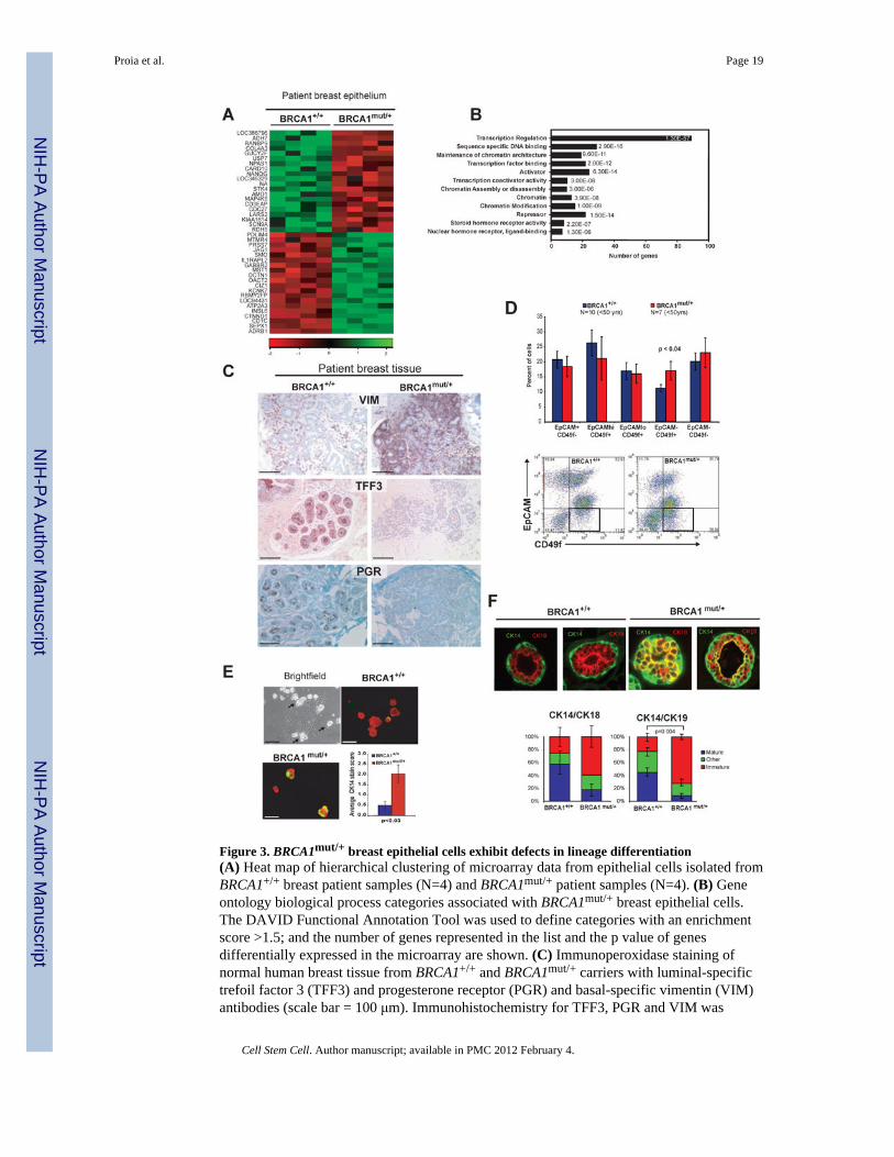
Figure 3. BRCA1mut/+ breast epithelial cells exhibit defects in lineage differentiation(A) Heat map of hierarchical clustering of microarray data from epithelial cells isolated fromBRCA1+/+ breast patient samples (N=4) and BRCA1mut/+ patient samples (N=4). (B) Geneontology biological process categories associated with BRCA1mut/+ breast epithelial cells.The DAVID Functional Annotation Tool was used to define categories with an enrichmentscore >1.5; and the number of genes represented in the list and the p value of genesdifferentially expressed in the microarray are shown. (C) Immunoperoxidase staining ofnormal human breast tissue from BRCA1+/+ and BRCA1mut/+ carriers with luminal-specifictrefoil factor 3 (TFF3) and progesterone receptor (PGR) and basal-specific vimentin (VIM)antibodies (scale bar = 100 μm). Immunohistochemistry for TFF3, PGR and VIM was
Proia et al. Page 19
Cell Stem Cell. Author manuscript; available in PMC 2012 February 4.
NIH
-PA Author Manuscript
NIH
-PA Author Manuscript
NIH
-PA Author Manuscript

performed on age matched BRCA1+/+ (N=13) and BRCA1mut/+ (N=10) disease-free breasttissues. Differences in staining were observed primarily in lobules, not ducts. (D) Freshlydissociated, uncultured epithelial cells from age matched (<50 yrs) BRCA1+/+ (N=10) andBRCA1mut/+ (N=7) patients were analyzed for EpCAM and CD49f expression by flowcytometry. Representative dot plots of a BRCA1+/+ or BRCA1mut/+ patient are shown. (E)Human breast epithelial cells produce small (∼30-50 μm) luminal suspension spheres whengrown under adherent conditions (indicated by arrows). Cytospun spheres were stained forCK 8/18 (red) and 14 (green)(scale bar = 100 μm). CK14 content in spheres was scored asdescribed in methods. At least 30 spheres were scored for each patient sample. The averagescores from 3 BRCA1+/+ and BRCA1mut/+ patient samples are shown in the graph. Error barsare +/- SEM and p-values were calculated by two-tailed t-test. (F) Acinar structures frompatient-derived BRCA1+/+ (N=4) and BRCA1mut/+ patient (N=4) cells infected with GFPlentivirus to visualize outgrowth and grown in the HIM model. Tissue outgrowths weredouble stained for CK14 and CK8/18 or CK19 (representative photos, top). The staining wascharacterized as mature (CK14+ basal/ME layer and CK8/18 and/or 19+ luminal layer),immature (CK14+ basal/ME layer and CK14 and CK8/19 and/or 19+ luminal layer) or other(CK14 only, CK8/18/19 only etc.). The average number of the 3 categories of structures areshown in the graph (n = ≥ 85 acini). Error bars are +/- SEM and p-values were calculated bytwo-tailed t-test. See also Figures S2 and S3 as well as Tables S4 and S5.
Proia et al. Page 20
Cell Stem Cell. Author manuscript; available in PMC 2012 February 4.
NIH
-PA Author Manuscript
NIH
-PA Author Manuscript
NIH
-PA Author Manuscript

Figure 4. EpCAM+ luminal cells are able to recapitulate the tumor growth(A) Flow chart describing sorting scheme. (B) Assessment of the purity of cells followingmagnetic bead sorting. Quantification of double staining for the luminal marker CK8/18 andthe basal marker CK14 following sorting indicates that the sorting strategy depletes cellspositive for these markers. Secondary antibody labeling of immunocomplexes on bead-released sorted cells indicates purity of the fractions. (C) CK14 immunofluorescence (IF)staining and quantification of sorted fractions indicates basal cell enrichment in the CD10+
fraction and depletion in the CD10-/EpCAM+ fraction. CK8/18 IF staining of sortedfractions indicates luminal cell depletion in the CD10+ fraction and enrichment in theCD10-/EpCAM+ fraction. (D, E) Sorted epithelial cell fractions infected with identical
Proia et al. Page 21
Cell Stem Cell. Author manuscript; available in PMC 2012 February 4.
NIH
-PA Author Manuscript
NIH
-PA Author Manuscript
NIH
-PA Author Manuscript

oncogenes differ in their ability to form tumors. GFP wholemount micrographs of tumoroutgrowths of sorted and infected breast epithelial cells from the four different fractions.BRCA1+/+ tumor data is compiled from three separate experiments with two different patientsamples. Unsorted (n=14), CD10+ (n=4), CD10-/EpCAM+ (n=6), Depleted (n=8).BRCA1mut/+ tumor data is compiled from two experiments with one patient sample.BRCA1mut/+ Unsorted (n=8), CD10+ (n=1), CD10-/EpCAM+ (n=4), Depleted (n=4).
Proia et al. Page 22
Cell Stem Cell. Author manuscript; available in PMC 2012 February 4.
NIH
-PA Author Manuscript
NIH
-PA Author Manuscript
NIH
-PA Author Manuscript

Figure 5. Slug regulates breast epithelial differentiation and lineage commitment(A) IHC staining of PM and RM tissues for Slug protein; staining was quantified by Allredscoring (see Supplemental Methods); two-tailed t-test used to derive p-value. (B) Flowcytometry analysis of CD24 expression in immortalized BRCA1+/+ and BRCA1mut/+
epithelial cells derived from 4 different patient tissues following serum-induceddifferentiation (C) Slug protein expression in immortalized BRCA1+/+ and BRCA1mut/+
epithelial cells derived from patient breast tissues following serum-induced differentiation.Quantification of fold reduction in Slug protein expression upon serum treatment from 3different experiments (p=0.24). (D) Flow cytometry analysis of EpCAM and CD49fexpression in patient-derived breast epithelial cells from breast tissues of 3 differentBRCA1mut/+ patients following Slug knockdown. (E) Flow cytometry analysis of EpCAMand CD49f expression in patient-derived breast epithelial cells from immortalizedBRCA1mut/+ epithelial cells derived from BRCA1mut/+ tissues following Slug knockdown.See also Figures S4 and S5.
Proia et al. Page 23
Cell Stem Cell. Author manuscript; available in PMC 2012 February 4.
NIH
-PA Author Manuscript
NIH
-PA Author Manuscript
NIH
-PA Author Manuscript

Figure 6. BRCA1-mutation promotes increased Slug protein stability(A) Loss of BRCA1 leads to increased Slug protein but not mRNA expression in MCF10Acells. QRT-PCR and Western blot analysis of BRCA1 and Slug expression in BRCA1+/+
MCF10A cells transfected with siBRCA1 or siControl. QRT-PCR data was normalized toGAPDH and to siControl. (B) BRCA1+/+ (MCF10A) and BRCA1mut (SUM1315, SUM149)cells were treated with cycloheximide (CHX) to prevent further protein synthesis atindicated time intervals. Western blot analysis demonstrates that Slug protein is highlyunstable in MCF10A cells while it had a significantly longer half-life in SUM149 andSUM1315 cells. Cyclin D1 and Actin were used as controls. (C) BRCA1+/+ (MCF10A) weretransfected with siBRCA1 or siControl and treated with cycloheximide (CHX) to prevent
Proia et al. Page 24
Cell Stem Cell. Author manuscript; available in PMC 2012 February 4.
NIH
-PA Author Manuscript
NIH
-PA Author Manuscript
NIH
-PA Author Manuscript

further protein synthesis at indicated time intervals. Western blot analysis demonstrates thatSlug protein is turned over in siControl MCF10A cells while it remained expressed insiBRCA1 MCF10A cells. See also Figure S6.
Proia et al. Page 25
Cell Stem Cell. Author manuscript; available in PMC 2012 February 4.
NIH
-PA Author Manuscript
NIH
-PA Author Manuscript
NIH
-PA Author Manuscript

Figure 7. Slug regulation of breast cancer phenotype in BRCA1-mutaion carriers(A) Immunohistochemistry of Slug protein in breast carcinomas with known BRCA1-mutation status; two-tailed t-test used to derive p-values of Allred scores. (B) Western blotanalysis of Slug and BRCA1 expression in breast cancer cell lines. SLUG mRNA levelswere normalized to GAPDH and to BRCA1 levels in the cell lines. (C) SUM149 BRCA1mut
breast cancer cells infected with lentiviruses targeting Slug (shSlug2 and shSlug3) or ascrambled sequence (shCntrl). Flow cytometry analysis of CD44 and CD24 expression inpatient-derived SUM149 cell following Slug knockdown. (D) Quantitative RT-PCR arrayagainst a panel of 86 genes expressed in breast luminal, basal and stem cells was performedon SUM1315-shSlug and SUM149-shSlug and their respective scrambled controls. Genesdifferentially expressed in both cell lines in the shSlug cells compared to the scrambledcontrols are plotted. PROM, TFF3, KRT19, KRT8, and CDH1 genes are not expressed inSUM1315 cells. (E) BRCA1mut/+ epithelial cells infected with oncogenes in the presence ofSlug knockdown leads to cells with features of luminal-like breast cancers. BRCA1mut/+
patient-derived breast epithelial cells were infected with lentiviruses encoding mutant p53,cyclin D1, and K-ras with shSlug or a scrambled sequence (shCntrl) and a quantitative RT-PCR array was performed. Genes differentially expressed >2-fold in both patient samplescompared to the scrambled controls are plotted. See also Figure S7 and Table S6.
Proia et al. Page 26
Cell Stem Cell. Author manuscript; available in PMC 2012 February 4.
NIH
-PA Author Manuscript
NIH
-PA Author Manuscript
NIH
-PA Author Manuscript
Copyright © 2022 FDOKUMEN