Structural links between the renal stem/progenitor cell niche ...
13
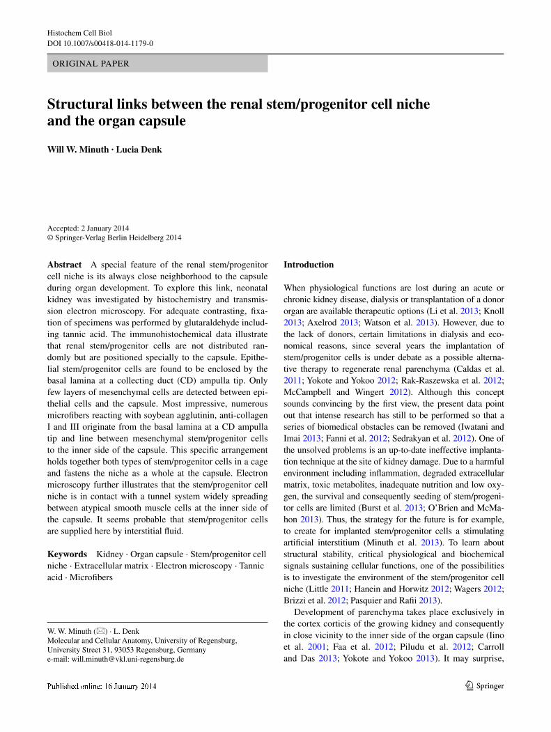
1 3 Histochem Cell Biol DOI 10.1007/s00418-014-1179-0 ORIGINAL PAPER Structural links between the renal stem/progenitor cell niche and the organ capsule Will W. Minuth · Lucia Denk Accepted: 2 January 2014 © Springer-Verlag Berlin Heidelberg 2014 Introduction When physiological functions are lost during an acute or chronic kidney disease, dialysis or transplantation of a donor organ are available therapeutic options (Li et al. 2013; Knoll 2013; Axelrod 2013; Watson et al. 2013). However, due to the lack of donors, certain limitations in dialysis and eco- nomical reasons, since several years the implantation of stem/progenitor cells is under debate as a possible alterna- tive therapy to regenerate renal parenchyma (Caldas et al. 2011; Yokote and Yokoo 2012; Rak-Raszewska et al. 2012; McCampbell and Wingert 2012). Although this concept sounds convincing by the first view, the present data point out that intense research has still to be performed so that a series of biomedical obstacles can be removed (Iwatani and Imai 2013; Fanni et al. 2012; Sedrakyan et al. 2012). One of the unsolved problems is an up-to-date ineffective implanta- tion technique at the site of kidney damage. Due to a harmful environment including inflammation, degraded extracellular matrix, toxic metabolites, inadequate nutrition and low oxy- gen, the survival and consequently seeding of stem/progeni- tor cells are limited (Burst et al. 2013; O’Brien and McMa- hon 2013). Thus, the strategy for the future is for example, to create for implanted stem/progenitor cells a stimulating artificial interstitium (Minuth et al. 2013). To learn about structural stability, critical physiological and biochemical signals sustaining cellular functions, one of the possibilities is to investigate the environment of the stem/progenitor cell niche (Little 2011; Hanein and Horwitz 2012; Wagers 2012; Brizzi et al. 2012; Pasquier and Rafii 2013). Development of parenchyma takes place exclusively in the cortex corticis of the growing kidney and consequently in close vicinity to the inner side of the organ capsule (Iino et al. 2001; Faa et al. 2012; Piludu et al. 2012; Carroll and Das 2013; Yokote and Yokoo 2013). It may surprise, Abstract A special feature of the renal stem/progenitor cell niche is its always close neighborhood to the capsule during organ development. To explore this link, neonatal kidney was investigated by histochemistry and transmis- sion electron microscopy. For adequate contrasting, fixa- tion of specimens was performed by glutaraldehyde includ- ing tannic acid. The immunohistochemical data illustrate that renal stem/progenitor cells are not distributed ran- domly but are positioned specially to the capsule. Epithe- lial stem/progenitor cells are found to be enclosed by the basal lamina at a collecting duct (CD) ampulla tip. Only few layers of mesenchymal cells are detected between epi- thelial cells and the capsule. Most impressive, numerous microfibers reacting with soybean agglutinin, anti-collagen I and III originate from the basal lamina at a CD ampulla tip and line between mesenchymal stem/progenitor cells to the inner side of the capsule. This specific arrangement holds together both types of stem/progenitor cells in a cage and fastens the niche as a whole at the capsule. Electron microscopy further illustrates that the stem/progenitor cell niche is in contact with a tunnel system widely spreading between atypical smooth muscle cells at the inner side of the capsule. It seems probable that stem/progenitor cells are supplied here by interstitial fluid. Keywords Kidney · Organ capsule · Stem/progenitor cell niche · Extracellular matrix · Electron microscopy · Tannic acid · Microfibers W. W. Minuth (*) · L. Denk Molecular and Cellular Anatomy, University of Regensburg, University Street 31, 93053 Regensburg, Germany e-mail: [email protected]
-
Upload
khangminh22 -
Category
Documents
-
view
3 -
download
0
Transcript of Structural links between the renal stem/progenitor cell niche ...
1 3
Histochem Cell BiolDOI 10.1007/s00418-014-1179-0
OrIgInal PaPer
Structural links between the renal stem/progenitor cell niche and the organ capsule
Will W. Minuth · Lucia Denk
accepted: 2 January 2014 © Springer-Verlag Berlin Heidelberg 2014
Introduction
When physiological functions are lost during an acute or chronic kidney disease, dialysis or transplantation of a donor organ are available therapeutic options (li et al. 2013; Knoll 2013; axelrod 2013; Watson et al. 2013). However, due to the lack of donors, certain limitations in dialysis and eco-nomical reasons, since several years the implantation of stem/progenitor cells is under debate as a possible alterna-tive therapy to regenerate renal parenchyma (Caldas et al. 2011; Yokote and Yokoo 2012; rak-raszewska et al. 2012; McCampbell and Wingert 2012). although this concept sounds convincing by the first view, the present data point out that intense research has still to be performed so that a series of biomedical obstacles can be removed (Iwatani and Imai 2013; Fanni et al. 2012; Sedrakyan et al. 2012). One of the unsolved problems is an up-to-date ineffective implanta-tion technique at the site of kidney damage. Due to a harmful environment including inflammation, degraded extracellular matrix, toxic metabolites, inadequate nutrition and low oxy-gen, the survival and consequently seeding of stem/progeni-tor cells are limited (Burst et al. 2013; O’Brien and McMa-hon 2013). Thus, the strategy for the future is for example, to create for implanted stem/progenitor cells a stimulating artificial interstitium (Minuth et al. 2013). To learn about structural stability, critical physiological and biochemical signals sustaining cellular functions, one of the possibilities is to investigate the environment of the stem/progenitor cell niche (little 2011; Hanein and Horwitz 2012; Wagers 2012; Brizzi et al. 2012; Pasquier and rafii 2013).
Development of parenchyma takes place exclusively in the cortex corticis of the growing kidney and consequently in close vicinity to the inner side of the organ capsule (Iino et al. 2001; Faa et al. 2012; Piludu et al. 2012; Carroll and Das 2013; Yokote and Yokoo 2013). It may surprise,
Abstract a special feature of the renal stem/progenitor cell niche is its always close neighborhood to the capsule during organ development. To explore this link, neonatal kidney was investigated by histochemistry and transmis-sion electron microscopy. For adequate contrasting, fixa-tion of specimens was performed by glutaraldehyde includ-ing tannic acid. The immunohistochemical data illustrate that renal stem/progenitor cells are not distributed ran-domly but are positioned specially to the capsule. epithe-lial stem/progenitor cells are found to be enclosed by the basal lamina at a collecting duct (CD) ampulla tip. Only few layers of mesenchymal cells are detected between epi-thelial cells and the capsule. Most impressive, numerous microfibers reacting with soybean agglutinin, anti-collagen I and III originate from the basal lamina at a CD ampulla tip and line between mesenchymal stem/progenitor cells to the inner side of the capsule. This specific arrangement holds together both types of stem/progenitor cells in a cage and fastens the niche as a whole at the capsule. electron microscopy further illustrates that the stem/progenitor cell niche is in contact with a tunnel system widely spreading between atypical smooth muscle cells at the inner side of the capsule. It seems probable that stem/progenitor cells are supplied here by interstitial fluid.
Keywords Kidney · Organ capsule · Stem/progenitor cell niche · extracellular matrix · electron microscopy · Tannic acid · Microfibers
W. W. Minuth (*) · l. Denk Molecular and Cellular anatomy, University of regensburg, University Street 31, 93053 regensburg, germanye-mail: [email protected]

Histochem Cell Biol
1 3
although at this special site capillary precursors are present, an intact microvascular system with circulating blood cells was not observed (Kloth et al. 1994, 1997).
Within the niche, epithelial and mesenchymal stem/progenitor cells are present. epithelial cells are enclosed within an ureteric bud-derived CD ampulla tip, while mes-enchymal cells are forming the surrounding cap condensate (little and McMahon 2012; Chai et al. 2013). a reciprocal exchange of morphogenetic molecules between them leads to a condensation of part of mesenchymal cells resulting in a comma-shaped body as first sign of nephron formation.
epithelial stem/progenitor cells occur within a CD ampulla tip that is covered by an individual basal lamina containing, for example, PCD amp I (Strehl et al. 1997, 1999; Strehl and Minuth 2001) and tissue transglutaminase (Tgase2) (Fig. 1a) (Schumacher et al. 2005). Between epi-thelial and mesenchymal stem/progenitor cells, an impres-sive interstitial interface is developed (Fig. 1b) (Minuth et al. 2011; Minuth and Denk 2012a). Improved contrast-ing of specimens by cupromeronic blue, ruthenium red and tannic acid demonstrates by the use of transmission electron microscopy that the extracellular matrix within the interstitial interface is not homogeneously composed but exhibits unique microheterogeneous features (Minuth and Denk 2012a, 2013). Further on, single projections from mesenchymal cells are crossing together with filigree strands of extracellular matrix the interstitial interface, penetrate the basal lamina to contact via tunneling nano-tubes epithelial stem/progenitor cells (Fig. 1c) (Minuth and Denk 2012b). In an opposite direction, microfibers labeled
by Soybean agglutinin(SBa), anti-collagen II and IV rise out of the basal lamina and cross the interstitial interface (Fig. 1d) (Schumacher et al. 2002a, b, 2003). However, the way of microfibers spanning through the interstitium and the contact between the niche and the inner side of the capsule has not previously been adequately investigated (Fig. 1e).
an often largely disregarded feature of the renal stem/progenitor cell niche is their exact orientation throughout development of parenchyma. Mesenchymal stem/progen-itor cells are persistently in contact with the inner side of the organ capsule, while epithelial stem/progenitor cells are orientated toward the medulla. They are connected in the neck of an ampulla with an elongating collecting duct.
Previous investigations further demonstrated that the capsule is not a simple envelop but an astonishingly struc-tured tissue consisting at the outer side of a tunica fibrosa and at the inner side of a tunica muscularis (Möllendorf 1930). However, cells contained in the tunica muscula-ris are different as compared to typical smooth muscle cells (Bulger 1973; Kobayashi 1978). It may surprise, but recently it was shown that within the organ capsule, numer-ous stem/progenitor cells are harbored (Park et al. 2010).
Since actual data about the structural relationship between mesenchymal stem/progenitor cells and the inner side of the organ capsule are lacking, the present morpho-logical investigation was performed. To explore microfibers spanning between the basal aspect of a CD ampulla and the organ capsule, histochemical experiments were made and
Fig. 1 Schematic illustration informs about stepwise exploration of structural links detected between the renal stem/progenitor cell niche and the organ capsule (C). a epithelial stem/progenitor cells are cov-ered at the CD ampulla (A) tip by a unique basal lamina (Strehl et al. 1997, 1999; Strehl and Minuth 2001). b epithelial and mesenchymal stem/progenitor cells are separated by a special interstitial interface containing heterogeneously composed extracellular matrix (Minuth et al. 2011; Minuth and Denk 2012a, 2013). c Individual cell projec-tions from mesenchymal cells cross the interstitial interface to pen-
etrate the basal lamina at the CD ampulla to contact epithelial stem/progenitor cells via tunneling nanotubes (Minuth and Denk 2012b). d Microfibers labeled by SBa and anti-collagens II and IV originate from the basal aspect of a CD ampulla to line through the interstitial interface and the group of mesenchymal stem/progenitor cells (Schu-macher et al. 2002a, b, 2003). e In the present investigation, structural links between the renal stem/progenitor cell niche and the capsule were analyzed

Histochem Cell Biol
1 3
their spatial distribution was analyzed by confocal laser scanning microscopy. To obtain insights in ultrastructural features, transmission electron microscopy in combination with an improved contrasting technique with tannic acid was used. The present results show structural elements of extracellular matrix fastening the renal stem/progenitor cell niche with the inner side of the organ capsule. In the complimentary space between matrix and cells, an aston-ishingly spreading tunnel system for the exchange of inter-stitial fluid is present.
Materials and methods
Tissue preparation
To investigate structural elements between the renal stem/pro-genitor cell niche and the organ capsule, 1-day-old male and female new Zealand rabbits (Seidl, Oberndorf, germany) were anesthetized with ether and killed by cervical disloca-tion. Both kidneys were immediately surgically removed to process them for light and electron microscopy.
Histochemical labeling
Isolated kidneys were surrounded by TissueTek (OCT™Compound, Sakura Finetek, Zoeterwoude, The netherlands) and frozen at −80 °C. To analyze cell bio-logical features, 20-μm-thick cryosections were made, fixed in ice-cold ethanol, washed several times with phos-phate-buffered saline (PBS) and incubated for 30 min with blocking solution (PBS, pH 7.5, 10 % horse serum from gIBCO/Invitrogen, Karlsruhe, germany; 1 % bovine serum albumin from Serva, Heidelberg, germany). For soybean agglutinin-labeling (SBa, Vector, Burlingame, USa), the samples were exposed to fluorescein isothiocy-anate (FITC)-conjugated lectin diluted 1:1,000 in blocking solution for 45 min.
For immunohistochemical label of epithelial basal lamina, anti-agrin (clone 6D4, Developmental Studies Hybridoma Bank (DSHB), Iowa, USa) was 1:20 diluted in blocking solution, while anti-laminin γ1 (clone 3e10, Santa Cruz Biotechnology, Santa Cruz, USa) was used undiluted. Monoclonal anti-collagen I (Southern Bio-tech, Birmingham, USa) and polyclonal anti-collagen III (Southern Biotech, USa) had a concentration of 1:20, whereas monoclonal anti-collagen III (clone III-53, Calbio-chem, Schwalbach, germany) was diluted 1:250 in block-ing solution. The antibodies were applied for 1 h. after washing with 1 % BSa in PBS, the specimens were incu-bated for 45 min with goat anti-rat-Igg-rhodamine, donkey anti-goat-Igg-fluorescein isothiocyanate, respectively, don-key anti-mouse-Igg-fluorescein isothiocyanate (Jackson
Immunoresearch laboratories, West grove, USa) diluted 1:50 in PBS containing 1 % BSa.
Further individual samples labeled with anti-collagen III (clone III-53) were co-incubated with 4′,6-diamino-2-phe-nylindole dihydrochloride (DaPI, Sigma-aldrich Chemie, München, germany) in a concentration of 1:10,000 in PBS.
Following several washes with PBS, the sections were embedded with Slow Fade light antifade Kit (Molecular Probes, eugene, USa) and then analyzed using either an axioskop 2 plus microscope (Zeiss, Oberkochen, ger-many) or a CM12 confocal laser scanning microscope (Zeiss). Fluorescence images were taken with a digital camera at a standard exposure time and thereafter pro-cessed with Corel DraW graphic Suite X5 (Corel Corpo-ration, Ottawa, Canada).
Transmission electron microscopy
In the present investigation, specimens were analyzed after conventional fixation in glutaraldehyde (ga) and after improved contrasting by ga solution including tannic acid as it was earlier demonstrated (Minuth et al. 2011; Minuth and Denk 2013; Hasko and richardson 1988; rothenburger et al. 2002).
For fixation of tissue, the following solutions were used:
1. Specimens for control: 5 % ga (Serva) buffered with 0.15 M sodium cacodylate, pH 7.4.
2. Specimens for improved contrast with tannic acid: 5 % ga buffered with 0.15 M sodium cacodylate, pH 7.4 + 1 % tannic acid (Sigma-aldrich Chemie).
The period of primary fixation was for 1 day at room temperature. after several washes with 0.15 M sodium cacodylate, the samples were postfixed in the same buffer but additionally containing 1 % osmium tetroxide (Sci-ence Services, München, germany). Then, the tissue was washed with sodium cacodylate buffer and dehydrated in graded series of ethanols.
Finally, the specimens were embedded in epon (Fluka, Taufkirchen, germany), which was polymerized at 60 °C for 48 h. Semithin and ultrathin sections were made with a diamond knife on an ultramicrotome eM UC6 (leica, Wetzlar, germany).
Semithin sections were labeled with richardson stain-ing and analyzed with a leica DM 750 microscope (leica). Images were taken with a digital camera and thereafter processed with Corel DraW graphic Suite X5 (Corel Corporation).
Ultrathin sections were collected onto slot grids coated with 1.5 % Pioloform (Plano, Wetzlar, germany) and con-trasted using 2 % uranyl acetate and lead citrate as earlier described (Minuth et al. 2011; Minuth and Denk 2013).

Histochem Cell Biol
1 3
analysis of them was performed at 80 kV using an eM 902 transmission electron microscope (Zeiss).
amount of analyzed specimens
a total of 54 exactly orientated renal stem cell niches were analyzed for the present study. Performed experiments are in accordance with the animal ethics Committee, Univer-sity of regensburg, regensburg, germany.
Definition of cells contained in the renal stem/progenitor cell niche
In this paper, the embryonic cortex of human fetal and neonatal rabbit kidney was described. In consequence, the nomenclature of previously published papers was used (Minuth et al. 2011; Minuth and Denk 2013).
Results
Development of renal parenchyma depends on an exact temporal–spatial formation of nephrons initiated by numer-ous reciprocal morphogenetic molecular interactions within the stem/progenitor cell niche (Faa et al. 2012; Carroll and Das 2013; Chai et al. 2013). Beginning with the organ anlage and up to the end of the neonatal phase, the niches are not randomly distributed but are always found in close neighborhood to the expanding organ capsule.
light microscopy of the renal stem/progenitor cell niche
To obtain a comparable view to both the stem/progenitor cell niche and the neighboring organ capsule, each kidney was first divided between both poles (Fig. 2a). Follow-ing this strategy, the parenchyma is orientated along the
Fig. 2 exact orientation of developing kidney is a need to obtain comparable views to the stem/progenitor cell niche. a after fixation, the kidney is first cut between both poles. b as a result, the section plane shows straightly lining collect-ing ducts ending beyond the organ capsule as an ureteric bud-derived ampulla (A). c, d When an orientated section is analyzed, the basal aspect of a dichotomously arborizing collecting duct (CD) ampulla shows a distance of 14–16 μm to the outer side of the organ capsule. In this case, mesenchy-mal stem/progenitor cells show two cell layers. e, f The same distance and the same number of mesenchymal cell layers are registered, when a CD ampulla is seen in a lateral view. g, h In contrast, when a section is not orientated along lining collecting ducts, an oblique cutting plane is obtained. In this case, the distance between a CD ampulla and the capsule is more than 20 μm so that yet multiple layers of mesenchymal cells can be seen as the cap condensate

Histochem Cell Biol
1 3
straight cortico-medullary course of lining collecting ducts (CD; Fig. 2b). now, a correct view to the renal stem/pro-genitor cell niche in relation to the covering organ capsule (C) is possible (Fig. 2c). as a consequence, all of the here demonstrated micrographs show this perspective so that comparisons between different experimental series become possible.
light microscopy shows on a semithin section that epi-thelial stem/progenitor cells are enclosed within dichoto-mously dividing tips of a ureteric bud-derived CD ampulla (a; Fig. 2d), while mesenchymal stem/progenitor cells form the surrounding cap condensate. astonishingly, both stem/progenitor cell populations do not stand closely together but are separated by a striking interstitial inter-face. In a lateral view, a single CD ampulla tip is illustrated (Fig. 2e, f). The individual distance of the basal aspect of a CD ampulla to the outer side of the organ capsule is as well in front as in lateral views between 14 and 16 μm (Fig. 2c–f).
an example illustrates that incorrect orientation of a sample is leading to an oblique cutting line and a shifting in perspective (Fig. 2g, h). Such a peripheric section through the CD ampulla tip demonstrates that yet the distance between its basal aspect and the outer side of the organ cap-sule is more than 20 μm. astonishingly, only by an oblique cutting, the extension of the multilayered cap condensate containing mesenchymal stem/progenitor cells is visible (Fig. 2h). In contrast, on an exact vertical section can be seen that the mesenchymal cap condensate consists of only two layers of cells (Fig. 2d, f).
Histochemical view to structural links between the niche and capsule
a surface view in light microscopy further illustrates that epithelial and mesenchymal stem/progenitor cells do not contact each other but are separated by an interstitial inter-face lining in parallel to the basal lamina of a CD ampulla tip (Fig. 2d, f, h). The aim of next experiments was to investigate by histochemistry and laser scanning micros-copy on orientated sections extracellular matrix molecules occurring between epithelial, mesenchymal stem/progeni-tor cells and the organ capsule.
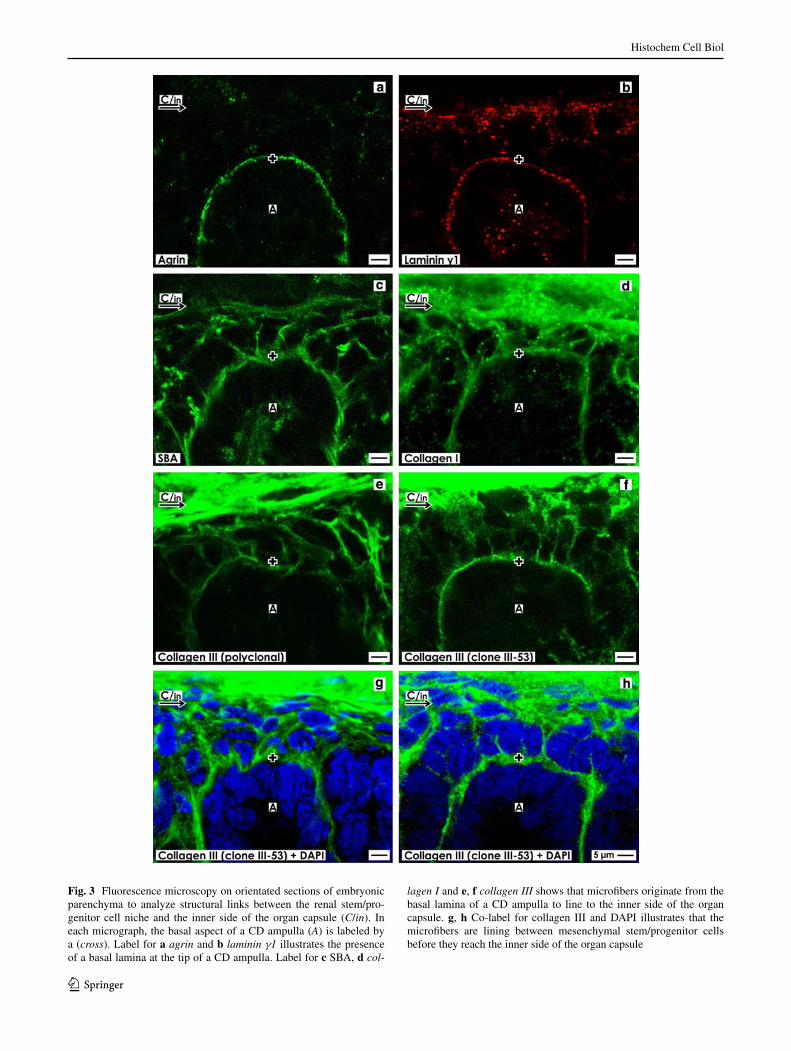
agrin label is detected in form of a punctuate pattern bordering the basal aspect at the CD ampulla tip (Fig. 3a). Only a faint punctual label of agrin is found between neighboring mesenchymal stem/progenitor cells. label for laminin γ1 is seen as a punctuate pattern at the basal aspect and within the lumen of a CD ampulla tip (Fig. 3b). Interestingly, parts of the inner side of the organ capsule show an intense but punctual label, while between mesen-chymal stem/progenitor cells only faint label for laminin γ1 is observed.
Strong label for SBa is recognized along the basal aspect of a CD ampulla (Fig. 3c). In addition, microfib-ers labeled by SBa are lining from this basal aspect to the inner side of the organ capsule. Similar to SBa, label intense reaction for collagen I was detected along the basal aspect of a CD ampulla tip (Fig. 3d). Further intensively labeled microfibers originate from the basal aspect of a CD ampulla tip and to the organ capsule. label for collagen III was investigated as well by a polyclonal (Fig. 3e) as by a monoclonal (Fig. 3f) antibody. Both antibodies exhibit intense label at the basal aspect of a CD ampulla tip and on numerous microfibers spanning toward the organ cap-sule. Thus, as well label for agrin (Fig. 3a) as laminin γ1 (Fig. 3b) illustrates that the basal aspect of a CD ampulla tip is a demarcation line between epithelial and mesen-chymal stem/progenitor cells. In contrast, label for SBa (Fig. 3c), collagen I (Fig. 3d) and collagen III (Fig. 3e, f) shows that intensively labeled microfibers originate from the basal aspect of a CD ampulla tip and line to the inner side of the organ capsule. These results demonstrate that structural elements of the renal stem/progenitor cell niche are linked with the inner side of the organ capsule.
Co-label for DaPI and collagen III illustrates that micro-fibers are not found within epithelial stem/progenitor cells of a CD ampulla tip, but line exclusively between labeled nuclei of mesenchymal stem progenitor cells to end at the inner side of the organ capsule (Fig. 3g, h). Thus, the pre-sent results clearly depict that renal stem/progenitor cells are not accidentally clustering but are contained in an unex-pectedly structured cage consisting of numerous microfib-ers. These appear as structural elements spanning between the basal aspect of a CD ampulla tip, the group of mesen-chymal stem/progenitor cells up to the inner side of the organ capsule.
Focus to the contact between the niche and capsule
In a first set of experiments, specimens were traditionally fixed with glutaraldehyde (ga) and analyzed on orientated sections by transmission electron microscopy (Fig. 4a). The outer side of the organ capsule contains flat fibroblasts but does not show high amounts of collagen fibers. Screen-ing of the space between the middle and the inner side of the organ capsule and the basal aspect of a CD ampulla tip shows that this special area is heterogenously composed and can roughly be divided into different zones. Zones I–III are belonging to the genuine organ capsule, while zone IV harbors mesenchymal stem/progenitor cells. Zone V rep-resents the interstitial interface separating epithelial from mesenchymal stem/progenitor cells.
Higher magnification of specimens fixed in ga illus-trates that the organ capsule can be split in a tunica fibrosa and a tunica muscularis (Fig. 4b). Thus, zone I shows the
Histochem Cell Biol
1 3
Fig. 3 Fluorescence microscopy on orientated sections of embryonic parenchyma to analyze structural links between the renal stem/pro-genitor cell niche and the inner side of the organ capsule (C/in). In each micrograph, the basal aspect of a CD ampulla (A) is labeled by a (cross). label for a agrin and b laminin γ1 illustrates the presence of a basal lamina at the tip of a CD ampulla. label for c SBa, d col-
lagen I and e, f collagen III shows that microfibers originate from the basal lamina of a CD ampulla to line to the inner side of the organ capsule. g, h Co-label for collagen III and DaPI illustrates that the microfibers are lining between mesenchymal stem/progenitor cells before they reach the inner side of the organ capsule

Histochem Cell Biol
1 3
outer side of the tunica fibrosa. Here, flat fibroblasts with even flat nuclei and extravagant cell projections are found. Zone II demarks the inner side of the tunica fibrosa. In this layer, the volume of cells is increased as compared to zone I. The nuclei of these cells are oval, but their endings are asy-metrically formed. Zone III depicts the tunica muscularis and belongs to the inner side of the genuine organ capsule. as compared to zones I and II, the volume of cells and nuclei is here enlarged. The nuclei show round endings on both sides. Further long cell projections are interdigitating with each other. In contrast, in zone IV, cells become poly-morph and their volume is increased as compared to zones I–III. The here contained cells represent mesenchymal stem/progenitor cells. Via numerous filigree projections, they are connected with each other. Between the cells, an extended but bright interstitial space can be observed. Zone V shows the interstitial interface, where single projections from mesenchymal stem/progenitor cells cross the intersti-tial interface to contact the basal lamina at a CD ampulla tip. The interstitial space between the organ capsule and the CD ampulla tip appears at all sites bright but more or less unremarkable (Fig. 4a, b).
Fixation of specimens by glutaraldehyde including tannic acid
In contrast, when fixation of specimens was performed by ga including tannic acid (Fig. 4c), a completely different view is obtained as compared to traditional fixation with ga (Fig. 4a, b). By a first screening, it is recognized that the impregnation by tannic acid unpacks structures on cells and extracellular matrix, which were not earlier observed. For detailed investigation, specimens were analyzed under high (Fig. 5) and very high magnification (Fig. 6).
Fig. 4 Transmission electron microscopy of the renal stem/progeni-tor cell niche and the organ capsule of specimens fixed with glutar-aldehyde (GA) or ga including tannic acid. a low magnification of specimens fixed by traditional ga depicts that epithelial stem/pro-genitor cells are enclosed by the basal lamina (cross) at a CD ampulla (A) tip. Mesenchymal stem/progenitor cells are separated from epi-thelial cells by a bright interstitial interface. b Higher magnification shows that the interstitium between mesenchymal stem/progenitor cells is bright. c In contrast, specimens fixed by ga including tan-nic acid show that the basal lamina at a CD ampulla, the intersti-tial interface (asterisk) and single structures at the inner side of the organ capsule (C/in) are darkly labeled. Further on, the overviews exhibit that the space between the outer side of the organ capsule and the basal lamina at a CD ampulla tip can be divided into five zones (I–V). Zones I represents the outer side of the tunica fibrosa, zone II shows the inner side of the tunica fibrosa, while zone III belongs to the tunica muscularis. Zone IV harbors mesenchymal stem/progeni-tor cells, while zone V shows the interstitial interface around the basal aspect of a CD ampulla
▸
Histochem Cell Biol
1 3
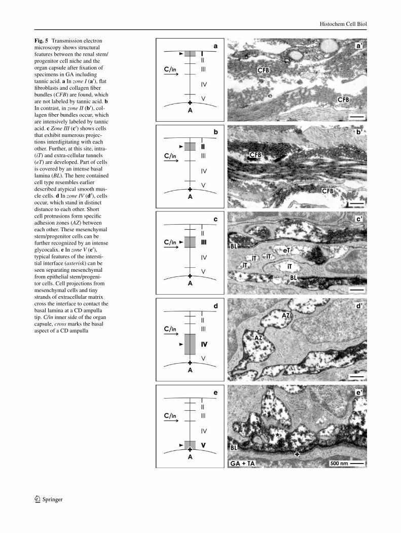
Fig. 5 Transmission electron microscopy shows structural features between the renal stem/progenitor cell niche and the organ capsule after fixation of specimens in ga including tannic acid. a In zone I (a′), flat fibroblasts and collagen fiber bundles (CFB) are found, which are not labeled by tannic acid. b In contrast, in zone II (b′), col-lagen fiber bundles occur, which are intensively labeled by tannic acid. c Zone III (c′) shows cells that exhibit numerous projec-tions interdigitating with each other. Further, at this site, intra- (iT) and extra-cellular tunnels (eT) are developed. Part of cells is covered by an intense basal lamina (BL). The here contained cell type resembles earlier described atypical smooth mus-cle cells. d In zone IV (d′), cells occur, which stand in distinct distance to each other. Short cell protrusions form specific adhesion zones (AZ) between each other. These mesenchymal stem/progenitor cells can be further recognized by an intense glycocalix. e In zone V (e′), typical features of the intersti-tial interface (asterisk) can be seen separating mesenchymal from epithelial stem/progeni-tor cells. Cell projections from mesenchymal cells and tiny strands of extracellular matrix cross the interface to contact the basal lamina at a CD ampulla tip. C/in inner side of the organ capsule, cross marks the basal aspect of a CD ampulla

Histochem Cell Biol
1 3
Fig. 6 Structural features between the renal stem/pro-genitor cell niche and the organ capsule. Specimens were fixed in ga including tannic acid and analyzed by transmission electron microscopy under high magnification. a Typical for zone I and b II are numerous collagen fiber bundles (CFB). Most impressive, on the same section can be seen that in zone I (a′) the tunica fibrosa shows bright collagen fiber bundles, while in zone II (b′) darkly labeled collagen fiber bundles are observed. c Typical for the tunica muscularis in zone III are atypical smooth muscle cells. c′ On many sites, these cells are covered by a basal lamina (BL). c″ In some cases, collagen fiber bundles are attached to the basal lamina. c′′′ Further cell projec-tions with a faint glycocalix are frequently observed contact-ing each other. d Typical for zone IV is the occurrence of mesenchymal stem/progenitor cells. d′ and d″ They exhibit short cell projections forming with neighboring cells adhe-sion zones (AZ). The cells are covered by a dense but irregular glycocalix. Between the cells, a surprisingly wide interstitium is present. e Typical for zone V is the interstitial interface (aster-isk) separating mesenchymal an epithelial stem/progenitor cells. e′ It can be recognized by an intense label on the basal lamina (BL) of a CD ampulla enclos-ing epithelial stem/progenitor cells. Within the interstitial space, single cell projections from mesenchymal cells and filigree strands of extracellular matrix are noticeable. C/in inner side of the organ capsule, cross marks the basal aspect of a CD ampulla

Histochem Cell Biol
1 3
Focus was first directed to the outer layer of the organ capsule. Zone I represents the outer side of the tunica fibrosa (Figs. 5a, 6a). Here, numerous collagen fiber bun-dles (CFB) are present (Figs. 5a′, 6a′). The micrograph can be further seen that collagen fiber bundles are bright. Cross-sections reveal that each of these fibers shows a punctuate pattern on its surface labeled by tannic acid. Between the collagen fiber bundles, single fibroblasts with long but tiny cell projections are present.
Zone II illustrates the inner side of the tunica fibrosa (Figs. 5b, 6b), where single fibroblasts with numerous and extended projections can be seen (Figs. 5b′, 6b′). In close neighborhood of them, numerous collagen fiber bundles are present. However, as compared to collagen fiber bundles in zone I (Figs. 5a′, 6a′), the here contained ones are darkly labeled by tannic acid. This distinct difference can be dem-onstrated as well on separate sections (Fig. 5a′, b′) as on the same section (Fig. 6a′, b′).
Zone III (Figs. 5c, 6c) belongs to the tunica muscularis and consequently to the inner side of the organ capsule. Here, elongated cells are found, whose numerous projec-tions interdigitate with each other. In earlier investiga-tions, they were described as atypical smooth muscle cells (Fig. 5c′) (Bulger 1973; Kobayashi 1978). The cell body of these cells is covered by a basal lamina (Bl), which is intensively label by tannic acid (Fig. 5c′). It consists of a clearly recognizable lamina rara and lamina densa (Fig. 6c′). Part of the numerous cell projections is show-ing on all sides the basal lamina, while in other cases, only one side is covered. On special sites, the basal lamina is in contact with intensively labeled collagen fiber bundles (Fig. 6c″). In some cases, contacting cell projections do not have a covering basal lamina but exhibit a tiny glyco-calyx (Fig. 6c′′′). regarding the numerous cell projections, it appears that the space between belongs to an extended intra- (iT) respectively extracellular tunnel (eT) system probably involved in the exchange of interstitial fluid (Fig. 5c′).
Zone IV (Figs. 5d, 6d) is the layer, where mesenchy-mal stem/progenitor cells are localized. These cells show an intense but not consistently developed glycocalyx (Figs. 5d′, 6d′, d″). In irregular distances, unique adhesion zones (aZ) between neighboring cells are formed. It can-not be recognized if the adhesion zones are focal or if they are extended in length by microfolds of the plasma mem-brane. It appears that between connected cell projections, a remarkable extracellular tunnel system is present (Fig. 6d′, d″).
Zone V (Figs. 5e, 6e) represents the interstitial inter-face between mesenchymal and epithelial stem/progenitor cells. This site is bordered by a concrete basal lamina at the CD ampulla tip enclosing epithelial stem/progenitor cells (Figs. 5e′, 6e′). Individual projections of mesenchymal
stem/progenitor cells cross here together with tiny strands of extracellular matrix the interstitial interface to contact the basal lamina at a CD ampulla tip as it was described earlier (Minuth and Denk 2012b).
Discussion
recent publications have shown that the renal stem/progen-itor cell niche is not an accidental accumulation of stem/progenitor cells but exhibits an unexpected complex micro-architecture (Minuth et al. 2011; Minuth and Denk 2012a, b, 2013; Carroll and Das 2013). Since microvessels are not developed at this site, provision with nutrition and oxygen is restricted. Consequently, contained cells appear both to be survival artists and performance specialists fulfilling pleiotropic tasks to maintain stemness, proliferation and differentiation (Faa et al. 2012; Carroll and Das 2013).
Coherence of the renal stem/progenitor cell niche
a special feature is that throughout kidney growth, the niche stays in the outer cortex and in close contact with the inner side of the organ capsule (Minuth and Denk 2012b). laser scanning microscopy illustrates that the basal lamina covering epithelial stem/progenitor cells at the CD ampulla tip reflects a demarcation line, which can be visualized by agrin (Fig. 3a) or laminin γ1 (Fig. 3b). at this site, micro-fibers originate, which are labeled by SBa (Fig. 3c), anti-collagen I (Fig. 3d) and anti-collagen III (Fig. 3e, f). Co-label with DaPI further illustrates that microfibers cross the interstitial interface, line through the group of mesenchy-mal cells to end at the inner side of the capsule (Fig. 3g, h). This result shows that microfibers exists forming between the basal aspect of a CD ampulla, the group of mesenchy-mal cells and the inner side of organ capsule a spatial fiber cage (Fig. 7a).
Complex histoarchitecture between the niche and capsule
To obtain more information, transmission electron micros-copy was performed (Figs. 4, 5, 6). generally, fixation is performed by traditional glutaraldehyde (ga) solution. Screening from the outer side of the capsule toward a CD ampulla elucidates that the interstitial space between cells looks inconspicuously (Fig. 4a, b).
In contrast, when fixation of specimens is performed in ga containing tannic acid, extracellular matrix is unpacked and new structural details within the interstitium and on cell surfaces become visible (Fig. 4c). according to these new data, the space between the renal stem/progenitor cell niche and the outer side of the organ capsule can be split into five different zones (Figs. 4, 5, 6, 7):

Histochem Cell Biol
1 3
• Zone I: Outer side of the tunica fibrosa.• Zone II: Inner side of the tunica fibrosa.• Zone III: Tunica muscularis with atypical smooth mus-
cle cells.• Zone IV: layer of mesenchymal stem/progenitor cells.• Zone V: Interstitial interface around the basal aspect of
a CD ampulla tip containing epithelial stem/progenitor cells.
electron microscopy further illuminates that tannic acid label is not homogeneously dispersed but very individu-ally distributed. On the same section can be seen that col-lagen fiber bundles in zone I (Fig. 6a) do not exhibit label (Fig. 6a′), while bundles in zone II (Fig. 6b) show intense label (Fig. 6b′). In between, bright areas of the interstitium and non-labeled projections from single cells can be seen. These differences in label speak for a high quality of fixa-tion by glutaraldehyde in combination with tannic acid.
Most interesting is zone III reflecting the tunica mus-cularis (Figs. 5c, 6c). In this layer, earlier described atypi-cal smooth muscle cells are contained (Figs. 5c′, 6c′–c′′′) (Bulger 1973; Kobayashi 1978). although cells lack abun-dance of contractile microfilaments, they can be recog-nized by a consistently developed basal lamina (Figs. 5c′, 6c′). On distinct sites, tannic acid-positive collagen fibers are attached to the basal lamina (Fig. 6c″). Further numer-ous cell projections are present contacting each other. In
some of the cases, cell projections are not covered by a basal lamina but exhibit a faint glycocalix (Fig. 6c′′′). The numerous interdigitating cell projections form a complex tunnel system, which may direct the flow of interstitial fluid providing cells in the niche with nutrition, respiratory gas and morphogenetic molecules.
Zone IV (Figs. 5d, 6d) can be easily recognized by an increased volume of cells and a wide but unlabeled inter-stitial space between. each of the contained mesenchymal stem/progenitor cells is covered by an irregular glycocalyx (Figs. 5d′, 6d′, d″). Further, short but broad cell projections from neighboring cells contact each other to form a clearly recognizable and structured adhesion zone. However, it cannot be recognized, if cells form here atypical tight junc-tions or even gap junctions.
Zone V (Figs. 5e, 6e) represents the interstitial interface of the renal stem/progenitor cell niche. at this site, indi-vidual projections of mesenchymal stem/progenitor cells cross together with tiny strands of extracellular matrix the interface to contact the basal lamina at a CD ampulla tip (Figs. 5e′, 6e′). Tannic acid label shows within the basal lamina a punctuate lamina rara and a dark ribbon compris-ing the lamina densa and lamina fibroreticularis (Minuth and Denk 2012a).
In conclusion, the results demonstrate that the renal stem/progenitor cell niche exhibits a special microenviron-ment and a structural link with the organ capsule (Fig. 7).
Fig. 7 Schematic illustration depicts structural features between the renal stem/progenitor cell niche and the organ capsule. a according to present findings, this area can be divided into five zones. Zone I is the outer side of the tunica fibrosa containing flat fibroblasts and bright collagen fiber bundles. Zone II represents the inner side of the tunica fibrosa. at this site, collagen fiber bundles are detected that are intensively labeled by tannic acid. In zone III, atypical smooth muscle cells are contained. This layer is the inner border of the organ capsule (C/in). In zone IV, two layers of mesenchymal stem/progenitor cells are found. Zone V represents the interstitial interface separating mes-enchymal from epithelial stem/progenitor cells enclosed within a CD
ampulla (A). The basal lamina at an ampulla tip is labeled by a cross. Histochemistry reveals that microfibers labeled by SBa, anti-collagen I and III originate at the basal lamina of a CD ampulla to cross the interstitial interface, to line through the group of mesenchymal stem/progenitor cells and atypical smooth muscle cells to end between the light and darkly labeled collagen fiber bundles of the capsule. b elec-tron microscopy further demonstrates that beside filigree extracellu-lar matrix, a widely spreading space (colored in black) for interstitial fluid exists obviously providing contained cells with nutrition and respiratory gas

Histochem Cell Biol
1 3
In this scenario, epithelial stem/progenitor cells are enclosed by the basal lamina at the CD ampulla tip, while mesenchymal cells are harbored in a cage consisting of microfibers labeled by earlier detected SBa, anti-collagen II and IV (Schumacher et al. 2003) and presently detected anti-collagen I and III (Fig. 3d–f). The fibers are span-ning from the basal aspect of a CD ampulla tip, cross the interstitial interface and the group of mesenchymal stem/progenitor cells to end at the inner side of the organ cap-sule (Fig. 7a). This specific arrangement declares the per-manent proximity of the renal stem/progenitor cell niche to the capsule throughout organ development. It appears further that the renal stem/progenitor cell niche is in con-tact with an extended tunnel system (Fig. 7b) spreading between atypical smooth muscle cells (Fig. 5c′; Zone III) and mesenchymal stem/progenitor cells (Fig. 5d′; Zone IV). These tunnels may be connected to special vessels within the organ capsule as it was earlier described for the human kidney (Hammersen and Staubesand 1961). It might be the spring providing the renal stem/progenitor cell niche at the side of mesenchymal cells with interstitial fluid distributing necessary nutrition, respiratory gas and morphogenetic molecules.
References
axelrod Da (2013) economic and financial outcomes in transplan-tation: whose dome is it anyway? Curr Opin Organ Transplant 18:222–228
Brizzi MF, Tarone g, Defilippi P (2012) extracellular matrix, inte-grins, and growth factors as tailors of the stem cell niche. Curr Opin Cell Biol 24:1–7
Bulger re (1973) rat renal capsule: presence of layers of unique squamous cells. anat rec 177:393–407
Burst V, Pütsch F, Kubacki T, Völker la, Bartram MP, Müller rU et al (2013) Survival and distribution of injected haematopoi-etic stem cells in acute kidney injury. nephrol Dial Transplant 28:1131–1139
Caldas HC, Hayashi aP, abbud-Filho M (2011) repairing the chronic damaged kidney: the role of regenerative medicine. Transplant Proc 43:3573–3576
Carroll TJ, Das a (2013) Defining the signals that constitute the nephron progenitor niche. J am Soc nephrol 24:873–876
Chai OH, Song CH, Park SK, Kim W, Cho eS (2013) Molecular reg-ulation of kidney development. anat Cell Biol 46:19–31
Faa g, gerosa C, Fanni D, Monga g, Zaffanello M, van eyken P, Fanos V (2012) Morphogenesis and molecular mecha-nisms involved in human kidney development. J Cell Physiol 227:1257–1268
Fanni D, gerosa C, nemola S, Mocci C, Pichiri g, Coni P et al (2012) “Physiological” renal regenerating medicine in VlBW preterm infants: could a dream come true? J Matern Fetal neonatal Med 25:41–48
Hammersen F, Staubesand J (1961) Über die Stromwege in der nier-enkapsel von Mensch und Hund; zugleich ein Beitrag zum Beg-riff der arterio-venösen anatomosen. Zeitschrift anatomie und entwicklungsgeschichte 122:363–381
Hanein D, Horwitz ar (2012) The structure of cell-matrix adhesions: the new frontier. Curr Opin Cell Biol 24:134–140
Hasko Ja, richardson gP (1988) The ultrastructural organization and properties of the mouse tectorial membrane matrix. Hear res 35:21–38
Iino n, gejyo F, arakawa M, Ushiki T (2001) Three-dimensional analysis of nephrogenesis in the neonatal rat kidney: light and scanning electron microscopic studies. arch Histol Cytol 64:179–190
Iwatani H, Imai e (2013) Kidney repair using stem cells: myth or reality as therapeutic option. J nephrol 23:143–146
Kloth S, aigner J, Schmidbauer a, Minuth WW (1994) Interrelation-ship of renal vascular development and nephrogenesis. Cell Tis-sue res 277:247–257
Kloth S, ebenbeck C, Monzer J, de Vries U, Minuth WW (1997) Three-dimensional organization of the developing vasculature of the kidney. Cell Tissue res 287:193–201
Knoll ga (2013) Kidney transplantation in the older adult. am J Kid-ney Dis 61:790–797
Kobayashi K (1978) Fine structure of the mammalian renal capsule: the atypical smooth muscle cell and it functional meaning. Cell Tissue res 195:381–394
li PK, Burdmann ea, Mehta rl (2013) acute kidney injury: global health alert. Transplantation 95:653–657
little MH (2011) renal organogenesis: what can it tell us about renal repair and regeneration? Organogenesis 7:229–241
little MH, McMahon a (2012) Mammalian kidney development: principles, progress, and projections. Cold Spring Harb Perspect Biol 4:a008300
McCampbell KK, Wingert ra (2012) renal stem cells: fact or sci-ence fiction? Biochem J 444:153–168
Minuth WW, Denk l (2012a) Illustration of extensive extracellular matrix at the epithelial-mesenchymal interface within the renal stem/progenitor cell niche. BMC Clin Pathol 12:16
Minuth WW, Denk l (2012b) Cell projections and extracellular matrix cross the interstitial interface within the renal stem/pro-genitor cell niche: accidental, structural or functional cues? nephron exp nephrol 122:131–140
Minuth WW, Denk l (2013) The interstitial interface within the renal stem/progenitor cell niche exhibits an unique microheterogene-ous composition. Int J Mol Sci 14:13657–13669
Minuth WW, Denk l, Miess C, glashauser a (2011) Peculiarities of the extracellular matrix in the interstitium of the renal stem/pro-genitor cell niche. Histochem Cell Biol 136:321–334
Minuth WW, Denk l, gruber M (2013) Search for chemically defined culture medium to assist initial regeneration of diseased renal parenchyma after stem/progenitor cell implantation. Int J Stem Cell res Transplant 1:202
Möllendorf W (1930) Harn- und geschlechtsapparat. In: Handbuch der Mikroskopischen anatomie des Menschen, Band VII. pp 140–143
O’Brien l, McMahon a (2013) Progenitor programming in mamma-lian nephrogenesis. nephrology 18:177–179
Park HC, Yasudo K, Kuo MC, ni J, ratliff B, Chander P, goligor-sky MS (2010) renal capsule as a stem cell niche. am J renal Physiol 298:F1254–F1262
Pasquier J, rafii a (2013) role of microenvironment in ovar-ian cancer stem cell maintenance. Biomed res Int: 630782. doi:10.1155/2013/630782
Piludu M, Fanos V, Congiu T, Piras M, gerosa C, Mocci C, Fanni D, nemolato S, Muntoni S, Iacovidou n, Faa g (2012) The pine-cone body: an intermediate structure between the cap mesen-chyme and the renal vesicle in the developing nod mouse kidney revealed by an ultrastructural study. J Mater Fetal neonatal Med 25:72–75
rak-raszewska a, Wilm B, edgar D, Kenny S, Woolf aS, Murray P (2012) Development of embryonic stem cells in recombinant kid-neys. Organogenesis 8:125–136

Histochem Cell Biol
1 3
rothenburger M, Völker W, Vischer P, glasmacher B, Scheld HH, Deiwick M (2002) Ultrastructure of proteoglycans in tissue-engi-neered cardiovascular structures. Tissue eng 8:1049–1056
Schumacher K, Strehl r, Minuth WW (2002a) Detection of glyco-sylated sites in embryonic rabbit kidney by lectin histochemistry. Histochem Cell Biol 118:79–87
Schumacher K, Strehl r, de Vries U, groene HJ, Minuth WW (2002b) SBa-positive fibers between the CD ampulla, mesen-chyme, and renal capsule. J am Soc nephrol 13:2446–2453
Schumacher K, Strehl r, Minuth WW (2003) Characterization of micro-fibers at the interface between the renal collecting duct ampulla and the cap condensate. nephron exp nephrol 95:e43–e54
Schumacher K, Klar J, Wagner C, Minuth WW (2005) Temporal-spa-tial co-localisation of tissue transglutaminase-9 (MMP-9) with SBa-positive micro-fibers in the embryonic kidney cortex. Cell Tissue res 319:491–500
Sedrakyan S, angelow S, DeFilippo re, Perin l (2012) Stem cells as a therapeutic approach to chronic kidney diseases. Curr Urol rep 13:47–54
Strehl r, Minuth WW (2001) Partial identification of the mab (CD)amp 1 antigen at the epithelial-mesenchymal interface of the developing kidney. Histochem Cell Biol 116:389–396
Strehl r, Kloth S, aigner J, Steiner P, Minuth WW (1997) PCDamp1, a new antigen at the interface of the embryonic collecting duct epithe-lium and the nephrogenic mesenchyme. Kidney Int 52:1469–1477
Strehl r, Trautner V, Kloth S, Minuth WW (1999) existence of a dense reticular meshwork surrounding the nephron inducer in neonatal rabbit kidney. Cell Tissue res 298:539–548
Wagers aJ (2012) The stem cell niche in regenerative medicine. Cell Stem Cell 10:362–369
Watson ar, Hayes Wn, Vondrak K, ariceta g, Schmitt CP, ekim M et al (2013) Factors influencing choice of renal replacement ther-apy in european Paediatrics nephrology Units. Pediatr nephrol 28:2361–2368
Yokote S, Yokoo T (2012) Stem cells in kidney regeneration. Curr Med Chem 19:6009–6017
Yokote S, Yokoo T (2013) Organogenesis for kidney regeneration. Curr Opin Organ Transplant 18:186–190