Implanted adipose progenitor cells as physicochemical regulators of breast cancer
6
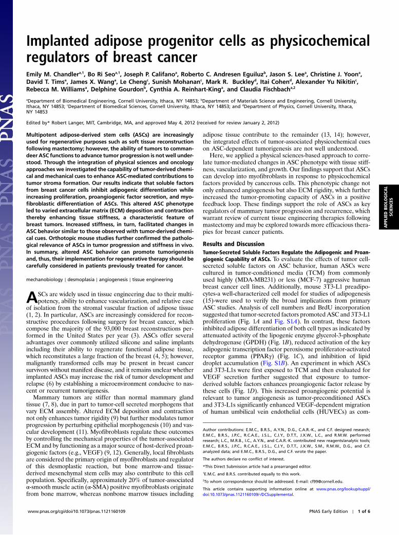
Implanted adipose progenitor cells as physicochemical regulators of breast cancer Emily M. Chandler a,1 , Bo Ri Seo a,1 , Joseph P. Califano a , Roberto C. Andresen Eguiluz b , Jason S. Lee a , Christine J. Yoon a , David T. Tims a , James X. Wang a , Le Cheng c , Sunish Mohanan c , Mark R. Buckley d , Itai Cohen d , Alexander Yu Nikitin c , Rebecca M. Williams a , Delphine Gourdon b , Cynthia A. Reinhart-King a , and Claudia Fischbach a,2 a Department of Biomedical Engineering, Cornell University, Ithaca, NY 14853; b Department of Materials Science and Engineering, Cornell University, Ithaca, NY 14853; c Department of Biomedical Sciences, Cornell University, Ithaca, NY 14853; and d Department of Physics, Cornell University, Ithaca, NY 14853 Edited by* Robert Langer, MIT, Cambridge, MA, and approved May 4, 2012 (received for review January 2, 2012) Multipotent adipose-derived stem cells (ASCs) are increasingly used for regenerative purposes such as soft tissue reconstruction following mastectomy; however, the ability of tumors to comman- deer ASC functions to advance tumor progression is not well under- stood. Through the integration of physical sciences and oncology approaches we investigated the capability of tumor-derived chemi- cal and mechanical cues to enhance ASC-mediated contributions to tumor stroma formation. Our results indicate that soluble factors from breast cancer cells inhibit adipogenic differentiation while increasing proliferation, proangiogenic factor secretion, and myo- fibroblastic differentiation of ASCs. This altered ASC phenotype led to varied extracellular matrix (ECM) deposition and contraction thereby enhancing tissue stiffness, a characteristic feature of breast tumors. Increased stiffness, in turn, facilitated changes in ASC behavior similar to those observed with tumor-derived chemi- cal cues. Orthotopic mouse studies further confirmed the patholo- gical relevance of ASCs in tumor progression and stiffness in vivo. In summary, altered ASC behavior can promote tumorigenesis and, thus, their implementation for regenerative therapy should be carefully considered in patients previously treated for cancer. mechanobiology ∣ desmoplasia ∣ angiogenesis ∣ tissue engineering A SCs are widely used in tissue engineering due to their multi- potency, ability to enhance vascularization, and relative ease of isolation from the stromal vascular fraction of adipose tissue (1, 2). In particular, ASCs are increasingly considered for recon- structive procedures following surgery for breast cancer, which compose the majority of the 93,000 breast reconstructions per- formed in the United States per year (3). ASCs offer several advantages over commonly utilized silicone and saline implants including their ability to regenerate functional adipose tissue, which reconstitutes a large fraction of the breast (4, 5); however, malignantly transformed cells may be present in breast cancer survivors without manifest disease, and it remains unclear whether implanted ASCs may increase the risk of tumor development and relapse (6) by establishing a microenvironment conducive to nas- cent or recurrent tumorigenesis. Mammary tumors are stiffer than normal mammary gland tissue (7, 8), due in part to tumor-cell secreted morphogens that vary ECM assembly. Altered ECM deposition and contraction not only enhances tumor rigidity (9) but further modulates tumor progression by perturbing epithelial morphogenesis (10) and vas- cular development (11). Myofibroblasts regulate these outcomes by controlling the mechanical properties of the tumor-associated ECM and by functioning as a major source of host-derived proan- giogenic factors (e.g., VEGF) (9, 12). Generally, local fibroblasts are considered the primary origin of myofibroblasts and regulator of this desmoplastic reaction, but bone marrow-and tissue- derived mesenchymal stem cells may also contribute to this cell population. Specifically, approximately 20% of tumor-associated α-smooth muscle actin (α-SMA) positive myofibroblasts originate from bone marrow, whereas nonbone marrow tissues including adipose tissue contribute to the remainder (13, 14); however, the integrated effects of tumor-associated physicochemical cues on ASC-dependent tumorigenesis are not well understood. Here, we applied a physical sciences-based approach to corre- late tumor-mediated changes in ASC phenotype with tissue stiff- ness, vascularization, and growth. Our findings support that ASCs can develop into myofibroblasts in response to physicochemical factors provided by cancerous cells. This phenotypic change not only enhanced angiogenesis but also ECM rigidity, which further increased the tumor-promoting capacity of ASCs in a positive feedback loop. These findings support the role of ASCs as key regulators of mammary tumor progression and recurrence, which warrant review of current tissue engineering therapies following mastectomy and may be explored towards more efficacious thera- pies for breast cancer patients. Results and Discussion Tumor-Secreted Soluble Factors Regulate the Adipogenic and Proan- giogenic Capability of ASCs. To evaluate the effects of tumor cell- secreted soluble factors on ASC behavior, human ASCs were cultured in tumor-conditioned media (TCM) from commonly used highly (MDA-MB231) or less (MCF-7) aggressive human breast cancer cell lines. Additionally, mouse 3T3-L1 preadipo- cytes-a well-characterized cell model for studies of adipogenesis (15)-were used to verify the broad implications from primary ASC studies. Analysis of cell numbers and BrdU incorporation suggested that tumor-secreted factors promoted ASC and 3T3-L1 proliferation (Fig. 1A and Fig. S1A). In contrast, these factors inhibited adipose differentiation of both cell types as indicated by attenuated activity of the lipogenic enzyme glycerol-3-phosphate dehydrogenase (GPDH) (Fig. 1B), reduced activation of the key adipogenic transcription factor peroxisome proliferator-activated receptor gamma (PPARγ) (Fig. 1C), and inhibition of lipid droplet accumulation (Fig. S1B). An experiment in which ASCs and 3T3-L1s were first exposed to TCM and then evaluated for VEGF secretion further suggested that exposure to tumor- derived soluble factors enhances proangiogenic factor release by these cells (Fig. 1D). This increased proangiogenic potential is relevant to tumor angiogenesis as tumor-preconditioned ASCs and 3T3-L1s significantly enhanced VEGF-dependent migration of human umbilical vein endothelial cells (HUVECs) as com- Author contributions: E.M.C., B.R.S., A.Y.N., D.G., C.A.R.-K., and C.F. designed research; E.M.C., B.R.S., J.P.C., R.C.A.E., J.S.L., C.J.Y., D.T.T., J.X.W., L.C., and R.M.W. performed research; L.C., M.R.B., I.C., A.Y.N., and C.A.R.-K. contributed new reagents/analytic tools; E.M.C., B.R.S., J.P.C., R.C.A.E., J.S.L., C.J.Y., D.T.T., J.X.W., S.M., R.M.W., D.G., and C.F. analyzed data; and E.M.C., B.R.S., D.G., and C.F. wrote the paper. The authors declare no conflict of interest. *This Direct Submission article had a prearranged editor. 1 E.M.C. and B.R.S. contributed equally to this work. 2 To whom correspondence should be addressed. E-mail: [email protected]. This article contains supporting information online at www.pnas.org/lookup/suppl/ doi:10.1073/pnas.1121160109/-/DCSupplemental. www.pnas.org/cgi/doi/10.1073/pnas.1121160109 PNAS Early Edition ∣ 1 of 6 APPLIED BIOLOGICAL SCIENCES
Transcript of Implanted adipose progenitor cells as physicochemical regulators of breast cancer
Implanted adipose progenitor cells as physicochemicalregulators of breast cancerEmily M. Chandlera,1, Bo Ri Seoa,1, Joseph P. Califanoa, Roberto C. Andresen Eguiluzb, Jason S. Leea, Christine J. Yoona,David T. Timsa, James X. Wanga, Le Chengc, Sunish Mohananc, Mark R. Buckleyd, Itai Cohend, Alexander Yu Nikitinc,Rebecca M. Williamsa, Delphine Gourdonb, Cynthia A. Reinhart-Kinga, and Claudia Fischbacha,2
aDepartment of Biomedical Engineering, Cornell University, Ithaca, NY 14853; bDepartment of Materials Science and Engineering, Cornell University,Ithaca, NY 14853; cDepartment of Biomedical Sciences, Cornell University, Ithaca, NY 14853; and dDepartment of Physics, Cornell University, Ithaca,NY 14853
Edited by* Robert Langer, MIT, Cambridge, MA, and approved May 4, 2012 (received for review January 2, 2012)
Multipotent adipose-derived stem cells (ASCs) are increasinglyused for regenerative purposes such as soft tissue reconstructionfollowing mastectomy; however, the ability of tumors to comman-deer ASC functions to advance tumor progression is notwell under-stood. Through the integration of physical sciences and oncologyapproaches we investigated the capability of tumor-derived chemi-cal and mechanical cues to enhance ASC-mediated contributions totumor stroma formation. Our results indicate that soluble factorsfrom breast cancer cells inhibit adipogenic differentiation whileincreasing proliferation, proangiogenic factor secretion, and myo-fibroblastic differentiation of ASCs. This altered ASC phenotypeled to varied extracellular matrix (ECM) deposition and contractionthereby enhancing tissue stiffness, a characteristic feature ofbreast tumors. Increased stiffness, in turn, facilitated changes inASC behavior similar to those observed with tumor-derived chemi-cal cues. Orthotopic mouse studies further confirmed the patholo-gical relevance of ASCs in tumor progression and stiffness in vivo.In summary, altered ASC behavior can promote tumorigenesisand, thus, their implementation for regenerative therapy should becarefully considered in patients previously treated for cancer.
mechanobiology ∣ desmoplasia ∣ angiogenesis ∣ tissue engineering
ASCs are widely used in tissue engineering due to their multi-potency, ability to enhance vascularization, and relative ease
of isolation from the stromal vascular fraction of adipose tissue(1, 2). In particular, ASCs are increasingly considered for recon-structive procedures following surgery for breast cancer, whichcompose the majority of the 93,000 breast reconstructions per-formed in the United States per year (3). ASCs offer severaladvantages over commonly utilized silicone and saline implantsincluding their ability to regenerate functional adipose tissue,which reconstitutes a large fraction of the breast (4, 5); however,malignantly transformed cells may be present in breast cancersurvivors without manifest disease, and it remains unclear whetherimplanted ASCs may increase the risk of tumor development andrelapse (6) by establishing a microenvironment conducive to nas-cent or recurrent tumorigenesis.
Mammary tumors are stiffer than normal mammary glandtissue (7, 8), due in part to tumor-cell secreted morphogens thatvary ECM assembly. Altered ECM deposition and contractionnot only enhances tumor rigidity (9) but further modulates tumorprogression by perturbing epithelial morphogenesis (10) and vas-cular development (11). Myofibroblasts regulate these outcomesby controlling the mechanical properties of the tumor-associatedECM and by functioning as a major source of host-derived proan-giogenic factors (e.g., VEGF) (9, 12). Generally, local fibroblastsare considered the primary origin of myofibroblasts and regulatorof this desmoplastic reaction, but bone marrow-and tissue-derived mesenchymal stem cells may also contribute to this cellpopulation. Specifically, approximately 20% of tumor-associatedα-smooth muscle actin (α-SMA) positive myofibroblasts originatefrom bone marrow, whereas nonbone marrow tissues including
adipose tissue contribute to the remainder (13, 14); however,the integrated effects of tumor-associated physicochemical cueson ASC-dependent tumorigenesis are not well understood.
Here, we applied a physical sciences-based approach to corre-late tumor-mediated changes in ASC phenotype with tissue stiff-ness, vascularization, and growth. Our findings support that ASCscan develop into myofibroblasts in response to physicochemicalfactors provided by cancerous cells. This phenotypic change notonly enhanced angiogenesis but also ECM rigidity, which furtherincreased the tumor-promoting capacity of ASCs in a positivefeedback loop. These findings support the role of ASCs as keyregulators of mammary tumor progression and recurrence, whichwarrant review of current tissue engineering therapies followingmastectomy and may be explored towards more efficacious thera-pies for breast cancer patients.
Results and DiscussionTumor-Secreted Soluble Factors Regulate the Adipogenic and Proan-giogenic Capability of ASCs. To evaluate the effects of tumor cell-secreted soluble factors on ASC behavior, human ASCs werecultured in tumor-conditioned media (TCM) from commonlyused highly (MDA-MB231) or less (MCF-7) aggressive humanbreast cancer cell lines. Additionally, mouse 3T3-L1 preadipo-cytes-a well-characterized cell model for studies of adipogenesis(15)-were used to verify the broad implications from primaryASC studies. Analysis of cell numbers and BrdU incorporationsuggested that tumor-secreted factors promoted ASC and 3T3-L1proliferation (Fig. 1A and Fig. S1A). In contrast, these factorsinhibited adipose differentiation of both cell types as indicated byattenuated activity of the lipogenic enzyme glycerol-3-phosphatedehydrogenase (GPDH) (Fig. 1B), reduced activation of the keyadipogenic transcription factor peroxisome proliferator-activatedreceptor gamma (PPARγ) (Fig. 1C), and inhibition of lipiddroplet accumulation (Fig. S1B). An experiment in which ASCsand 3T3-L1s were first exposed to TCM and then evaluated forVEGF secretion further suggested that exposure to tumor-derived soluble factors enhances proangiogenic factor release bythese cells (Fig. 1D). This increased proangiogenic potential isrelevant to tumor angiogenesis as tumor-preconditioned ASCsand 3T3-L1s significantly enhanced VEGF-dependent migrationof human umbilical vein endothelial cells (HUVECs) as com-
Author contributions: E.M.C., B.R.S., A.Y.N., D.G., C.A.R.-K., and C.F. designed research;E.M.C., B.R.S., J.P.C., R.C.A.E., J.S.L., C.J.Y., D.T.T., J.X.W., L.C., and R.M.W. performedresearch; L.C., M.R.B., I.C., A.Y.N., and C.A.R.-K. contributed new reagents/analytic tools;E.M.C., B.R.S., J.P.C., R.C.A.E., J.S.L., C.J.Y., D.T.T., J.X.W., S.M., R.M.W., D.G., and C.F.analyzed data; and E.M.C., B.R.S., D.G., and C.F. wrote the paper.
The authors declare no conflict of interest.
*This Direct Submission article had a prearranged editor.1E.M.C. and B.R.S. contributed equally to this work.2To whom correspondence should be addressed. E-mail: [email protected].
This article contains supporting information online at www.pnas.org/lookup/suppl/doi:10.1073/pnas.1121160109/-/DCSupplemental.
www.pnas.org/cgi/doi/10.1073/pnas.1121160109 PNAS Early Edition ∣ 1 of 6
APP
LIED
BIOLO
GICAL
SCIENCE
S
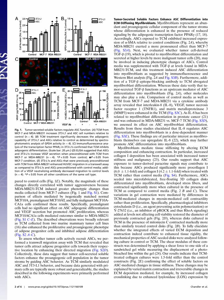
pared to control cells (Fig. 1E). Notably, the magnitude of thesechanges directly correlated with tumor aggressiveness becauseMDA-MB231-TCM induced greater phenotypic changes thanmedia collected from MCF-7 cultures (Fig. 1 and Fig. S1). Com-parison of effects mediated by isogenically matched normalMCF10A, premalignant MCF10AT, and fully malignant MCF10A-CA1a cells confirmed these results. Specifically, premalignantcells had no significant effect on ASC adipogenic differentiationand VEGF secretion but promoted ASC proliferation, whereasMCF10ACA1a cells mediated outcomes similar to MDA-MB231(Fig. S1 C–E). The described observations were broadly relevantas TCM collected from two murine mammary tumor cell lines(16) also enhanced the proliferative and proangiogenic phenotypeof adipose progenitor cells and inhibited adipose differentiation(Fig. S2 A–C).
To assess whether tumor cells actively recruit ASCs, we per-formed a transwell migration assay with TCM that revealed thattumor cells attract adipose progenitor cells towards their respec-tive location by enhancing their directed migration (Fig. S2D).Collectively, these data suggest that tumor cell-secreted solublefactors enhance the proangiogenic cell population in the tumorstroma by guiding ASC behavior. As TCM similarly modulatedASC and 3T3-L1 behavior, and because data generated with pri-mary cells are typically more robust and generalizable, the studiesdescribed in the following experiments were primarily performedwith ASCs.
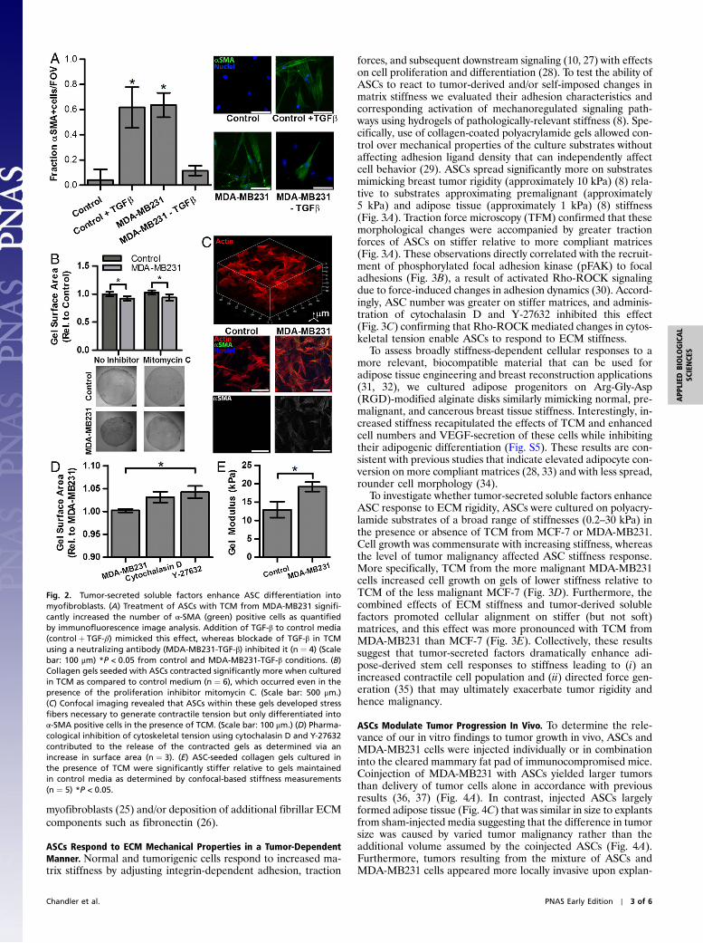
Tumor-Secreted Soluble Factors Enhance ASC Differentiation intoECM-Stiffening Myofibroblasts. Myofibroblasts represent an abun-dant and proangiogenic cellular component of the tumor stromawhose differentiation is enhanced in the presence of reducedsignaling by the adipogenic transcription factor PPARγ (17, 18).Accordingly, ASCs exposed to TCM exhibited increased expres-sion of α-SMA relative to control conditions (Fig. 2A), wherebyMDA-MB231 exerted a more pronounced effect than MCF-7(Fig. S3A). Next, we evaluated whether tumor cell-derivedTGF-β (19), which is pivotal to myofibroblast differentiation andsecreted at higher levels by more malignant tumor cells (20), maybe involved in inducing phenotypic changes of ASCs. Controlmedia was supplemented with TGF-β at levels found in MDA-MB231-TCM, and this treatment induced ASC differentiationinto myofibroblasts as suggested by immunofluorescence andWestern Blot analysis (Fig. 2A and Fig. S3B). Furthermore, addi-tion of a TGF-β epitope-blocking antibody to TCM abrogatedmyofibroblast differentiation. Whereas these data verify that tu-mor-secreted TGF-β functions as an upstream mediator of ASCdifferentiation into myofibroblasts (Fig. 2A), other moleculesmay also play a role. Comparison of control media as well asTCM from MCF-7 and MDA-MB231 via a cytokine antibodyarray revealed that interleukin-8 (IL-8), VEGF, tumor necrosisfactor receptor 1 (TNFR1), and matrix metalloproteinase 3(MMP3) were enhanced in the TCM (Fig. S3C). As IL-8 has beenrelated to myofibroblast differentiation in prostate cancer (21)and was enhanced in MDA-MB231 vs. MCF-7 TCM (Fig. S3D),we assessed its effect on ASC myofibroblast differentiation.Results from these studies elucidated that IL-8 regulates ASCdifferentiation into myofibroblasts in a dose-dependent manner(Fig. S3E). These findings are of particular interest as myofibro-blasts themselves can upregulate IL-8 (22), which may furtherpromote ASC differentiation into myofibroblasts.
Myofibroblasts mediate tissue stiffening by altering ECMcomposition and enhancing contraction. In particular, increasedcollagen I deposition has been associated with increased tumorstiffness and malignancy (23). Our results support that ASCexposure to tumor-derived paracrine signals may contribute tothis because ASCs produced significantly more procollagen I(6.0 � 1.1-fold) and collagen I (4.2� 1.1-fold) when treated withTCM rather than control media (Fig. S4). Furthermore, ASCsseeded into microfabricated, free-floating 3D collagen disksdifferentiated into myofibroblasts, developed stress fibers, andcontracted significantly more when cultured in the presence ofTCM as compared to control media (Fig. 2 B and C). Thesevariations in gel contraction were mediated by differences inTCM-mediated changes in myosin-mediated cell contractilityrather than proliferation. Specifically, pharmacological inhibitorscytochalasin D (i.e., an agent preventing actin polymerization) orY-27632 (i.e., an inhibitor of pROCK and thus RhoA signaling)added at levels not affecting cell viability restored the diameter ofpreviously contracted gels (Fig. 2D), whereas disks cultured inTCM in the presence of mitomycin C contracted markedly morethan similarly treated control disks (Fig. 2B). To fully elucidatewhether the integrated effects of varied ECM deposition andcontraction indeed contribute to enhanced tissue rigidity, themechanical properties of ASC-seeded disks were analyzed follow-ing culture in control or TCM. The shear modulus of these con-structs was determined by applying a shear force to one side of asandwiched gel while measuring the displacement of a photo-bleached line within the gel (24). Our results indicate that TCM-treated collagen cultures were 1.5-fold stiffer than the controlconstructs (Fig. 2E) confirming the effect of soluble factors onASC-mediated changes in tissue stiffness. These changes may beexplained by varied matrix contraction and irreversible changes inECM deposition mediated, for example, by increased collagencrosslinking due to enhanced lysyloxidase (LOX) expression by
Fig. 1. Tumor-secreted soluble factors regulate ASC function. (A) TCM fromMCF-7 and MDA-MB231 increases 3T3-L1 and ASC cell numbers relative tocontrol (n ¼ 4). (B) TCM treatment significantly decreases the adipogeniccapability of 3T3-L1 and ASCs relative to control as determined by spectro-photometric analysis of GPDH activity (n ¼ 4). (C) Immunofluorescence ana-lysis of the transcription factor PPARγ in 3T3-L1s confirmed that TCM inhibitsadipogenic differentiation. (Scale bar: 20 μm.) (D) ELISA suggested that ASCsand 3T3-L1s increase VEGF secretion when preconditioned with TCM fromMCF-7 or MDA-MB231 (n ¼ 4). *P < 0.05 from control; ♦P < 0.05 fromMCF-7 condition. (E) 3T3-L1s and ASCs that were previously preconditionedwith TCM fromMDA-MB231 enhanced HUVEC migration in a transwell assayas compared to 3T3-L1s and ASCs preconditioned with control media; addi-tion of a VEGF neutralizing antibody decreased migration to control levels(n ¼ 4). *P < 0.05 from all other conditions of the same cell type.
2 of 6 ∣ www.pnas.org/cgi/doi/10.1073/pnas.1121160109 Chandler et al.
myofibroblasts (25) and/or deposition of additional fibrillar ECMcomponents such as fibronectin (26).
ASCs Respond to ECM Mechanical Properties in a Tumor-DependentManner. Normal and tumorigenic cells respond to increased ma-trix stiffness by adjusting integrin-dependent adhesion, traction
forces, and subsequent downstream signaling (10, 27) with effectson cell proliferation and differentiation (28). To test the ability ofASCs to react to tumor-derived and/or self-imposed changes inmatrix stiffness we evaluated their adhesion characteristics andcorresponding activation of mechanoregulated signaling path-ways using hydrogels of pathologically-relevant stiffness (8). Spe-cifically, use of collagen-coated polyacrylamide gels allowed con-trol over mechanical properties of the culture substrates withoutaffecting adhesion ligand density that can independently affectcell behavior (29). ASCs spread significantly more on substratesmimicking breast tumor rigidity (approximately 10 kPa) (8) rela-tive to substrates approximating premalignant (approximately5 kPa) and adipose tissue (approximately 1 kPa) (8) stiffness(Fig. 3A). Traction force microscopy (TFM) confirmed that thesemorphological changes were accompanied by greater tractionforces of ASCs on stiffer relative to more compliant matrices(Fig. 3A). These observations directly correlated with the recruit-ment of phosphorylated focal adhesion kinase (pFAK) to focaladhesions (Fig. 3B), a result of activated Rho-ROCK signalingdue to force-induced changes in adhesion dynamics (30). Accord-ingly, ASC number was greater on stiffer matrices, and adminis-tration of cytochalasin D and Y-27632 inhibited this effect(Fig. 3C) confirming that Rho-ROCKmediated changes in cytos-keletal tension enable ASCs to respond to ECM stiffness.
To assess broadly stiffness-dependent cellular responses to amore relevant, biocompatible material that can be used foradipose tissue engineering and breast reconstruction applications(31, 32), we cultured adipose progenitors on Arg-Gly-Asp(RGD)-modified alginate disks similarly mimicking normal, pre-malignant, and cancerous breast tissue stiffness. Interestingly, in-creased stiffness recapitulated the effects of TCM and enhancedcell numbers and VEGF-secretion of these cells while inhibitingtheir adipogenic differentiation (Fig. S5). These results are con-sistent with previous studies that indicate elevated adipocyte con-version on more compliant matrices (28, 33) and with less spread,rounder cell morphology (34).
To investigate whether tumor-secreted soluble factors enhanceASC response to ECM rigidity, ASCs were cultured on polyacry-lamide substrates of a broad range of stiffnesses (0.2–30 kPa) inthe presence or absence of TCM from MCF-7 or MDA-MB231.Cell growth was commensurate with increasing stiffness, whereasthe level of tumor malignancy affected ASC stiffness response.More specifically, TCM from the more malignant MDA-MB231cells increased cell growth on gels of lower stiffness relative toTCM of the less malignant MCF-7 (Fig. 3D). Furthermore, thecombined effects of ECM stiffness and tumor-derived solublefactors promoted cellular alignment on stiffer (but not soft)matrices, and this effect was more pronounced with TCM fromMDA-MB231 than MCF-7 (Fig. 3E). Collectively, these resultssuggest that tumor-secreted factors dramatically enhance adi-pose-derived stem cell responses to stiffness leading to (i) anincreased contractile cell population and (ii) directed force gen-eration (35) that may ultimately exacerbate tumor rigidity andhence malignancy.
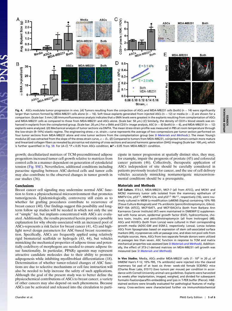
ASCs Modulate Tumor Progression In Vivo. To determine the rele-vance of our in vitro findings to tumor growth in vivo, ASCs andMDA-MB231 cells were injected individually or in combinationinto the cleared mammary fat pad of immunocompromised mice.Coinjection of MDA-MB231 with ASCs yielded larger tumorsthan delivery of tumor cells alone in accordance with previousresults (36, 37) (Fig. 4A). In contrast, injected ASCs largelyformed adipose tissue (Fig. 4C) that was similar in size to explantsfrom sham-injected media suggesting that the difference in tumorsize was caused by varied tumor malignancy rather than theadditional volume assumed by the coinjected ASCs (Fig. 4A).Furthermore, tumors resulting from the mixture of ASCs andMDA-MB231 cells appeared more locally invasive upon explan-
Fig. 2. Tumor-secreted soluble factors enhance ASC differentiation intomyofibroblasts. (A) Treatment of ASCs with TCM from MDA-MB231 signifi-cantly increased the number of α-SMA (green) positive cells as quantifiedby immunofluorescence image analysis. Addition of TGF-β to control media(controlþ TGF-β) mimicked this effect, whereas blockade of TGF-β in TCMusing a neutralizing antibody (MDA-MB231-TGF-β) inhibited it (n ¼ 4) (Scalebar: 100 μm) *P < 0.05 from control and MDA-MB231-TGF-β conditions. (B)Collagen gels seeded with ASCs contracted significantly more when culturedin TCM as compared to control medium (n ¼ 6), which occurred even in thepresence of the proliferation inhibitor mitomycin C. (Scale bar: 500 μm.)(C) Confocal imaging revealed that ASCs within these gels developed stressfibers necessary to generate contractile tension but only differentiated intoα-SMA positive cells in the presence of TCM. (Scale bar: 100 μm.) (D) Pharma-cological inhibition of cytoskeletal tension using cytochalasin D and Y-27632contributed to the release of the contracted gels as determined via anincrease in surface area (n ¼ 3). (E) ASC-seeded collagen gels cultured inthe presence of TCM were significantly stiffer relative to gels maintainedin control media as determined by confocal-based stiffness measurements(n ¼ 5) *P < 0.05.
Chandler et al. PNAS Early Edition ∣ 3 of 6
APP
LIED
BIOLO
GICAL
SCIENCE
S

tation relative to MDA-MB231-borne tumors that were easilydemarcated for dissection. Pathological evaluation of H&E-stained cross-sections confirmed this assessment and revealed anenhanced degree of local invasion and desmoplasia for the coim-planted tumors vs. the control tumors (Fig. S6 A and B). Addi-tionally, cytological features of malignancy (multinucleatedcells and rosettes) were augmented in the coimplanted groupas compared to the MDA-MB231 group (Fig. S6C). To correlatevariations in tumor growth with differences in myofibroblastdifferentiation, immunohistochemical analysis was performedthat confirmed that the density of α-SMA myofibroblastic cells(Fig. 4B) was increased in tumors originating from coinjectionof both cell types rather than tumor cells alone. Circulating ASCscan be recruited to and associate with blood vessels in the form ofα-SMA pericytes (38). In our coimplantation studies, however,immunohistochemical analysis of desmin, a pericyte marker(39), verified that the majority of α-SMA positive cells weremyofibroblasts rather than pericytes as desmin staining showedno significant difference (p ¼ 0.33) between coimplanted andcontrol tumors. Nevertheless, blood vessel density was greater inthe coimplantation group (Fig. 4C), and ELISA of tumor lysates
suggested that ASC/myofibroblast secretion of proangiogenicVEGF and IL-8 (12, 22) may have contributed to these differ-ences (Fig. S7).
To evaluate if varied myofibroblast content correlated withincreased stiffness, we determined the mechanical propertiesof the different tumors via Dynamic Mechanical Thermal Ana-lysis (DMTA) compression tests on tumor sections in the elasticregime of deformation. The mean Young’s modulus (E) was morethan 50% higher in tumors generated by coimplantation of ASCsthan in those generated in the absence of ASCs (3.1� 1.2 kPa vs.2.0� 0.8 kPa, P < 0.04) (Fig. 4D and Fig. S8). Data from the fullelastic and plastic regime of deformation (Fig. S8B) additionallyindicated that tumors from the coimplanted group exhibit higherstiffness and a smaller range of elastic (reversible) deformationthan tumors grown without ASCs. Consistent with these results,tumors resulting from coinjection contained more procollagen Iand collagen (Fig. S9 A and B), were characterized by enhancedcollagen fibril maturity and linearity (Fig. S9 C and D), and likelyalso comprised increased fibronectin (26). Such changes areindicative of enhanced desmoplasia and aggressiveness (23) andmay further contribute to ASC-mediated changes in tumor
Fig. 3. ASC response to matrix mechanical properties. (A) TFM indicated that ASCs cultured on hydrogels of increased stiffness spread significantly more andexert greater traction forces relative to cells cultured on soft matrices; representative ASC phase images with corresponding traction maps are shown(n ¼ 13–32, error bars represent standard errors) (Scale bar: 50 μm) *P < 0.05 from 1 kPa condition. (B) Immunofluorescence indicates increased recruitmentof pFAK[Y397] to ASC focal adhesions on stiffer matrices (white arrows). (Scale bar: 20 μm.) (C) Cell counting suggested that increased matrix stiffnessenhances ASC numbers and that pharmacological inhibition of cytoskeletal tension with Y-27632 or cytochalasin D inhibits this effect (n ¼ 4). (D) ASC numberson stiff matrices were further enhanced with culture in TCMwhereby TCM fromMDA-MB231 exerted a more pronounced effect than TCM fromMCF-7 (n ¼ 3)*P < 0.05 between all conditions; þP < 0.05 between MDA-MB231 and control conditions. (E) Image analysis of the deviation from the average angle ofphalloidin-stained cells revealed that ASC alignment represented an integrated effect of substrate stiffness and tumor malignancy, whereas ASCs on30 kPa gels had enhanced alignment in MCF-7 TCM. This occurred on gels of even lower stiffness in MDA-MB231 TCM *P < 0.05. (Scale bar: 50 μm.)
4 of 6 ∣ www.pnas.org/cgi/doi/10.1073/pnas.1121160109 Chandler et al.
growth; decellularized matrices of TCM-preconditioned adiposeprogenitors increased tumor cell growth relative to matrices fromcontrol cells in a manner dependent on generation of cytoskeletaltension (Fig. S9E). Nevertheless, additional conditions includingparacrine signaling between ASC-derived cells and tumor cellsmay also contribute to the observed changes in tumor growth inour studies (36).
ConclusionsBreast cancer cell signaling may undermine normal ASC func-tion to form a physicochemical microenvironment that promotestumorigenesis. Epidemiologically, controversy still exists as towhether fat grafting procedures contribute to recurrence ofbreast cancer (40). Our findings suggest this possibility and long-term follow up studies will be needed in which not only the useof “simple” fat, but implants concentrated with ASCs are evalu-ated. Additionally, the results presented herein provide a possibleexplanation for why obesity-associated with an increased pool ofASCs-represents a risk factor for breast cancer (41, 42) and high-light novel design parameters for ASC-based breast reconstruc-tion. Specifically, ASCs are frequently applied using relativelyrigid biomaterial scaffolds or hydrogels (43, 44), but vehiclesmimicking the mechanical properties of adipose tissue and poten-tially codelivery of morphogens are needed to ensure adipose tis-sue functionality. In particular, PPARγ agonists may representattractive candidate molecules due to their ability to promoteadipogenesis while inhibiting myofibroblast differentiation (18).Determination of whether tumor-related changes of ASC func-tions are due to selective mechanisms or cell fate instruction willalso be needed to help increase the safety of such applications.Although the goal of the present study was to better define thephysicochemical contributions of ASCs to breast cancer, a varietyof other cancers may also depend on such phenomena. BecauseASCs can be activated and released into the circulation to parti-
cipate in tumor progression at spatially distinct sites, they may,for example, impair the prognosis of prostate (45) and colorectalcancer patients (46). Collectively, therapeutic application ofASCs independent of site should be carefully considered inpatients previously treated for cancer, and the use of cell deliveryvehicles accurately mimicking nontumorigenic microenviron-mental conditions should be a prerequisite.
Materials and MethodsCell Culture. 3T3-L1, MDA-MB231, MCF-7 (all from ATCC), and MCN1 andMCN2 mammary tumor cells isolated from the mammary epithelium ofMMTV-Cre, p53L∕L, MMTV-Cre, and p53L∕L RbL∕L, respectively (16), were rou-tinely cultured in MEM (α-modification (αMEM) (Sigma) containing 10% FBS(Tissue Culture Biologicals) and 1% antibiotic (penicillin/streptomycin, Gibco).MCF-10A (ATCC), MCF10AT1, and MCF10ACA1a (both from Barbara AnnKarmanos Cancer Institute) (47) were maintained in DMEM/F12 supplemen-ted with horse serum, epidermal growth factor (EGF), hydrocortisone, cho-lera toxin, insulin, and penicillin/streptomycin (all from Invitrogen) (48).ASCs and HUVECs (both from Lonza) were cultured in their correspondinggrowth media (ADSC-GM and EGM-2, respectively; Lonza). Lonza isolatesASCs from lipoaspirates based on expression of stem cell-associated surfacemarkers (49), cryopreserves cells at passage one, and does not pool cells frommultiple sources. Here, ASCs from two separate female donors were utilizedat passages less than seven. ASC function in response to TCM and matrixmechanical properties was assessed (see SI Materials andMethods). Addition-ally, the effect of 3T3-L1-derived matrices on MDA-MB231 cell growth wasmeasured (see SI Materials and Methods).
In Vivo Studies. Media, ASCs and/or MDA-MB231 cells (1 · 106 in 20 μL ofDMEM/ Ham’s F-12, 10% FBS, 1% antibiotic) were injected into the clearedmammary fat pad of at least six three- week-old female SCID/NCr mice(Charles River Labs, 01S11) (two tumors per mouse) per condition in accor-dancewith Cornell University animal care guidelines. Explants were harvestedsix weeks after implantation, imaged, weighed, and divided for subsequentformalin-fixation/paraffin-embedding and lysis in T-PER buffer (Pierce). H&E-stained sections were broadly evaluated for pathological features of malig-nancy. Cross-sections were characterized further via immunohistochemical
Fig. 4. ASCs modulate tumor progression in vivo. (A) Tumors resulting from the coinjection of ASCs and MDA-MB231 cells (both) (n ¼ 14) were significantlylarger than tumors formed by MDA-MB231 cells alone (n ¼ 16). Soft tissue explants generated from injected ASCs (n ¼ 12) or media (n ¼ 2) are shown for acomparison. (Scale bar: 5 mm.) (B) Immunofluorescence analysis indicates that α-SMA levels were greatest in the explants resulting from coimplantation of ASCsand MDA-MB231 cells as compared to those from MDA-MB231 and ASCs alone. (Scale bar: 50 μm.) (C) Similarly, the density of CD31+ blood vessels was en-hanced in explants from the coimplanted group. (Scale bar: 20 μm.) For α-SMA and CD31+ image analysis, ASC (n ¼ 6) (both) (n ¼ 6), and MDA-MB231 (n ¼ 12)explants were analyzed. (D) Mechanical analysis of tumor sections via DMTA. The mean stress-strain profile was measured in PBS at room temperature throughthe low-strain (9–14%) elastic regime. The engineering stress σ vs. strain ε curve represents the average of two compressions per tumor section performed onfour tumor sections from MDA-MB231 alone and nine tumor sections from the coimplantation group (see SI Materials and Methods.). The mean Young’smodulus (E) was extracted from the slope of the stress-strain curve, σ ¼ Eε. (E) Compared to tumors fromMDA-MB231, coinjected tumors contain more matureand linearized collagen fibers as revealed by picrosirius red staining of cross-sections and second harmonic generation (SHG) imaging (Scale bar: 100 μm), whichis further quantified in Fig. S9. For (A–C) *P < 0.05 from ASCs condition; ♦P < 0.05 from MDA-MB231 condition.
Chandler et al. PNAS Early Edition ∣ 5 of 6
APP
LIED
BIOLO
GICAL
SCIENCE
S
staining for CD31, α-SMA, procollagen I, and desmin, whereas collagen wasstained via Masson’s Trichrome. For staining procedures and image analysismethods, see SI Materials and Methods. VEGF and IL-8 content in lysateswas measured via VEGF and IL-8 DuoSets (R&D Systems) and normalizedto protein content as determined via bicinchoninic acid protein assay (ThermoScientific). Tumor stiffness was determined using a DMTA as further outlinedin SI Materials and Methods.
Statistical Analysis. Statistical analysis was performed using GraphPad Prism5. Student’s t tests were used to compare two data sets, whereas ANOVAwas applied to determine the statistical significance of the differencesbetween data sets of three or more using the Tukey method to make post-hoc pairwise comparisons. A p-value less than 0.05 was considered statistically
significant. Data are represented as average� standard deviations withthe exception of the traction force measurements that are shown with stan-dard errors.
ACKNOWLEDGMENTS. Dr. David Infanger for aid in manuscript preparation,Kerstin Höger, Ana Martin-Ryals, and Christina Cossell for experimental aid,and Andrea Flesken-Nikitin for help with animal experiments. Funding byNIH/NCI RC1 CA146065 (to C.F.), R21 CA161532 (to C.F. and D.G.), R01CA96823 (to A.Y.N.), the Cornell Center on the Microenvironment andMetastasis through Award Number NCI U54 CA143876 (to C.F., C.R.K., I.C.,and R.M.W.), NSF CMMI-1031068 (to D.G.), and Graduate Research Fellowship(to E.M.C.). The content is solely the responsibility of the authors and doesnot necessarily represent the official views of NCI or NIH.
1. Cherubino M, Marra KG (2009) Adipose-derived stem cells for soft tissue reconstruc-tion. Regen Med 4:109–117.
2. Gimble JM, Katz AJ, Bunnell BA (2007) Adipose-derived stem cells for regenerativemedicine. Circ Res 100:1249–1260.
3. Surgeons ASoP (2010) Report of the 2010 Plastic Surgery Statistics: ASPS NationalClearinghouse of Plastic Surgery Procedural Statistics., Available at www.plasticsurgery.org.
4. Moioli EK, et al. (2010) Hybrid adipogenic implants from adipose stem cells for softtissue reconstruction in vivo. Tissue Eng Part A 16:3299–3307.
5. Yoshimura K, et al. (2008) Cell-assisted lipotransfer for cosmetic breast augmentation:Supportive use of adipose-derived stem/stromal cells. Aesthetic Plast Surg 32:48–55discussion 46–47.
6. Brewster AM, et al. (2008) Residual risk of breast cancer recurrence five years afteradjuvant therapy. J Natl Cancer Inst 100:1179–1183.
7. Janmey PA, Levental I, Georges PC (2007) Soft biological materials and their impact oncell function. Soft Matter 3:299–306.
8. Samani A, Bishop J, Luginbuhl C, Plewes DB (2003) Measuring the elastic modulus ofex vivo small tissue samples. Phys Med Biol 48:2183–2198.
9. Butcher DT, Alliston T, Weaver VM (2009) A tense situation: Forcing tumour progres-sion. Nat Rev Cancer 9:108–122.
10. Paszek MJ, et al. (2005) Tensional homeostasis and the malignant phenotype. CancerCell 8:241–254.
11. Mammoto A, et al. (2009) Amechanosensitive transcriptional mechanism that controlsangiogenesis. Nature 457:1103–1108.
12. Nakagawa H, et al. (2004) Role of cancer-associated stromal fibroblasts in metastaticcolon cancer to the liver and their expression profiles. Oncogene 23:7366–7377.
13. Kidd S, et al. (2012) Origins of the tumor microenvironment: Quantitative assessmentof adipose-derived and bone marrow-derived stroma. PLoS One 7:e30563.
14. Quante M, et al. (2011) Bone marrow-derived myofibroblasts contribute to themesenchymal stem cell niche and promote tumor growth. Cancer Cell 19:257–272.
15. Cornelius P, MacDougald OA, Lane MD (1994) Regulation of adipocyte development.Annu Rev Nutr 14:99–129.
16. Cheng L, et al. (2010) Rb inactivation accelerates neoplastic growth and substitutesfor recurrent amplification of cIAP1, cIAP2 and Yap1 in sporadic mammary carcinomaassociated with p53 deficiency. Oncogene 29:5700–5711.
17. Olaso E, et al. (2003) Proangiogenic role of tumor-activated hepatic stellate cells inexperimental melanoma metastasis. Hepatology 37:674–685.
18. Hong KM, Belperio JA, Keane MP, Burdick MD, Strieter RM (2007) Differentiation ofhuman circulating fibrocytes as mediated by transforming growth factor-beta andperoxisome proliferator-activated receptor gamma. J Biol Chem 282:22910–22920.
19. De Wever O, Mareel M (2003) Role of tissue stroma in cancer cell invasion. J Pathol200:429–447.
20. Guerrero J, et al. (2010) Soluble factors derived from tumor mammary cell lines inducea stromal mammary adipose reversion in human and mice adipose cells. Possible roleof TGF-beta1 and TNF-alpha. Breast Cancer Res Treat 119:497–508.
21. Schauer IG, Ressler SJ, Tuxhorn JA, Dang TD, Rowley DR (2008) Elevated epithelialexpression of interleukin-8 correlates with myofibroblast reactive stroma in benignprostatic hyperplasia. Urology 72:205–213.
22. Yagi Y, Andoh A, Inatomi O, Tsujikawa T, Fujiyama Y (2007) Inflammatory responsesinduced by interleukin-17 family members in human colonic subepithelial myofibro-blasts. J Gastroenterol 42:746–753.
23. Levental KR, et al. (2009) Matrix crosslinking forces tumor progression by enhancingintegrin signaling. Cell 139:891–906.
24. Buckley MR, Bergou AJ, Fouchard J, Bonassar LJ, Cohen I (2010) High-resolution spatialmapping of shear properties in cartilage. J Biomech 43:796–800.
25. Peyrol S, et al. (1997) Lysyl oxidase gene expression in the stromal reaction to in situand invasive ductal breast carcinoma. Am J Pathol 150:497–507.
26. Chandler EM, Saunders MP, Yoon CJ, Gourdon D, Fischbach C (2011) Adipose progeni-tor cells increase fibronectin matrix strain and unfolding in breast tumors. Phys Biol8:015008.
27. Reinhart-King CA, Dembo M, Hammer DA (2003) Endothelial cell traction forces onRGD-derivatized polyacrylamide substrata. Langmuir 19:1573–1579.
28. Engler AJ, Sen S, Sweeney HL, Discher DE (2006) Matrix elasticity directs stem cell line-age specification. Cell 126:677–689.
29. Shin H, Zygourakis K, Farach-Carson MC, Yaszemski MJ, Mikos AG (2004) Modulationof differentiation and mineralization of marrow stromal cells cultured on biomimetichydrogels modified with Arg-Gly-Asp containing peptides. J Biomed Mater Res A69:535–543.
30. McLean GW, et al. (2005) The role of focal-adhesion kinase in cancer—A new thera-peutic opportunity. Nat Rev Cancer 5:505–515.
31. Cho SW, et al. (2005) Engineering of volume-stable adipose tissues. Biomaterials26:3577–3585.
32. Davidenko N, Campbell JJ, Thian ES, Watson CJ, Cameron RE (2010) Collagen-hyaluro-nic acid scaffolds for adipose tissue engineering. Acta Biomater 6:3957–3968.
33. Chandler EM, et al. (2011) Stiffness of photocrosslinked RGD-alginate gels regulatesadipose progenitor cell behavior. Biotechnol Bioeng 108:1683–1692.
34. McBeath R, Pirone DM, Nelson CM, Bhadriraju K, Chen CS (2004) Cell shape, cytoske-letal tension, and RhoA regulate stem cell lineage commitment. Dev Cell 6:483–495.
35. Storm C, Pastore JJ, MacKintosh FC, Lubensky TC, Janmey PA (2005) Nonlinear elasti-city in biological gels. Nature 435:191–194.
36. Muehlberg FL, et al. (2009) Tissue-resident stem cells promote breast cancer growthand metastasis. Carcinogenesis 30:589–597.
37. Prantl L, et al. (2010) Adipose tissue-derived stem cells promote prostate tumorgrowth. Prostate 70:1709–1715.
38. Zhang Y, et al. (2009) White adipose tissue cells are recruited by experimental tumorsand promote cancer progression in mouse models. Cancer Res 69:5259–5266.
39. Kida Y, Duffield JS (2011) Pivotal role of pericytes in kidney fibrosis. Clin Exp PharmacolPhysiol 38:417–423.
40. Petit JY, et al. (2011) Locoregional recurrence risk after lipofilling in breast cancerpatients. Ann Oncol 23:582–588.
41. van Harmelen V, et al. (2003) Effect of BMI and age on adipose tissue cellularity anddifferentiation capacity in women. Int J Obes Relat Metab Disord 27:889–895.
42. Bianchini F, Kaaks R, Vainio H (2002) Overweight, obesity, and cancer risk. Lancet Oncol3:565–574.
43. Moutos FT, Guilak F (2010) Functional properties of cell-seeded three-dimensionallywoven poly(epsilon-caprolactone) scaffolds for cartilage tissue engineering. TissueEng Part A 16:1291–1301.
44. Ochoa I, et al. (2011) Mechanical properties of cross-linked collagen meshes afterhuman adipose derived stromal cells seeding. J Biomed Mater Res A 96:341–348.
45. Lin G, et al. (2010) Effects of transplantation of adipose tissue-derived stem cells onprostate tumor. Prostate 70:1066–1073.
46. Bellows CF, Zhang Y, Chen J, Frazier ML, Kolonin MG (2011) Circulation of progenitorcells in obese and lean colorectal cancer patients. Cancer Epidemiol Biomarkers Prev20:2461–2468.
47. Santner SJ, et al. (2001) Malignant MCF10CA1 cell lines derived from premalignanthuman breast epithelial MCF10AT cells. Breast Cancer Res Treat 65:101–110.
48. Kraning-Rush CM, Califano JP, Reinhart-King CA (2012) Cellular traction stressesincrease with increasing metastatic potential. PLoS One 7:e32572.
49. Lonza (2011) Poietics™ human adipose derived stem cells (ADSC)., Doc INST PT-5006-4,available at www.lonza.com.
6 of 6 ∣ www.pnas.org/cgi/doi/10.1073/pnas.1121160109 Chandler et al.