Paclitaxel and Mortality in PAD; The Latest Update - SummitMD
Upload
independentCategory
view
8download
0
Subscriber access provided by NORTHEASTERN UNIV LIB
Molecular Pharmaceutics is published by the American Chemical Society. 1155Sixteenth Street N.W., Washington, DC 20036
Article
Coadministration of Paclitaxel and Curcumin in NanoemulsionFormulations To Overcome Multidrug Resistance in Tumor Cells
Srinivas Ganta, and Mansoor AmijiMol. Pharmaceutics, Article ASAP • DOI: 10.1021/mp800240j • Publication Date (Web): 11 March 2009
Downloaded from http://pubs.acs.org on April 7, 2009
More About This Article
Additional resources and features associated with this article are available within the HTML version:
• Supporting Information• Access to high resolution figures• Links to articles and content related to this article• Copyright permission to reproduce figures and/or text from this article
Coadministration of Paclitaxel and Curcumin inNanoemulsion Formulations To Overcome Multidrug
Resistance in Tumor Cells
Srinivas Ganta and Mansoor Amiji*
Department of Pharmaceutical Sciences, School of Pharmacy, Northeastern UniVersity,110 Mugar Life Sciences Building, Boston, Massachusetts 02115
Received November 23, 2008; Revised Manuscript Received January 24, 2009; AcceptedMarch 11, 2009
Abstract: Development of multidrug resistance (MDR) against a variety of conventional andnovel chemotherapeutic agents is a significant challenge in effective cancer therapy. Over thelast several years, we have focused on a multimodal therapeutic strategy to overcome tumorMDR by enhancing the delivery efficiency to the tumor mass and lowering the apoptotic thresholdby modulation of the intracellular signaling mechanisms. In this study, we have examinedaugmentation of therapeutic efficacy upon coadministration of paclitaxel (PTX) and curcumin(CUR), an inhibitor of nuclear factor kappa B (NFκB) as well as a potent down-regulator of ABCtransporters, in wild-type SKOV3 and drug resistant SKOV3TR human ovarian adenocarcinomacells. PTX and CUR were encapsulated in flaxseed oil containing nanoemulsion formulations.The results showed that the encapsulated drugs were effectively delivered intracellular in bothSKOV3 and SKOV3TR cells. CUR administration was shown to inhibit NFκB activity and downregulate P-glycoprotein expression in resistant cells. Combination PTX and CUR therapy,especially when administered in the nanoemulsion formulations, was very effective in enhancingthe cytotoxicity in wild-type and resistant cells by promoting the apoptotic response. Overall,this cotherapy strategy has significant promise in the clinical management of refractory diseases,especially in ovarian cancer.
Keywords: Multidrug resistance; paclitaxel; curcumin; nanoemulsions; combination therapy;SKOV3 ovarian adenocarcinoma cells
1. IntroductionDevelopment of multidrug resistance (MDR) is one of the
most challenging aspects of cancer chemotherapy, as resis-tance develops with both conventional cytotoxic agents andthe recently developed targeted biological therapies.1 Inovarian cancer, for instance, more than 70% of initiallydiagnosed patients are resistant to taxane therapy, andeventually all of them will be resistant upon relapse.Although clinical resistance is often manifested as refractory
disease to standard chemotherapeutic regimens, there areseveral pathways by which tumor cells develop resistancedue to microenvironment selection pressures. The mostfrequently occurring causes of MDR include the overex-pression of ATP-binding cassette (ABC) super family oftransporters, which are trans-membrane proteins that acts asa drug-efflux pump by actively removing drugs from the cellsand producing intracellular drug levels below the effectiveconcentrations necessary for cytotoxicity.2 P-glycoprotein (P-gp), breast cancer resistance protein (ABCG2), and multidrugresistance associated protein (MRP-1) are the major trans-porter proteins belonging to the ABC super family that havebeen linked with MDR.1 P-gp is encoded by the MDR-1 gene
* Corresponding author. Mailing address: Northeastern University,Pharmaceutical Sciences Department, 110 Mugar Life SciencesBuilding, 360 Huntington Ave., Boston, MA 02115. Tel: 617-373-3137. Fax: 617-373-8886. E-mail: [email protected].
(1) Szakacs, G.; Paterson, J. K.; Ludwig, J. A.; Booth-Genthe, C.;Gottesman, M. M. Targeting multidrug resistance in cancer. Nat.ReV. Drug DiscoVery 2006, 5 (3), 219–234.
(2) Ejendal, K. F. K.; Hrycyna, C. A. Multidrug resistance and cancer:the role of the human ABC transporter ABCG2. Curr. ProteinPept. Sci. 2002, 3, 503–511.
articles
10.1021/mp800240j CCC: $40.75 XXXX American Chemical Society VOL. xxx, NO. xx, 000 MOLECULAR PHARMACEUTICS A
and has been shown to overexpress with the developmentof MDR in tumor biopsy samples.3 P-gp expression has beendirectly implicated in resistance to a broad spectrum ofanticancer agents such as taxanes, anthracyclines and thevinca alkaloids.4 Other causes of MDR include overexpres-sion of MRP-1, changes in topoisomerase II (Topo IIR)activity, and modification in glutathione S-transferase activity(GST).2,5-7
Over the last several years, we have suggested that arational approach to overcome MDR in clinical settingsrequires a multipronged strategy that combines enhancementin systemic drug delivery efficiency along with alterationsin the cellular phenotype that lead to more effective cell-kill response.8-12 Using poly(ethylene oxide)-modified poly-(epsilon-caprolactone) (PEO-PCL) nanoparticles encapsu-lating paclitaxel (PTX) and C6-ceramide, a lipid secondmessenger responsible for propagation of apoptotic signaling,we have shown that the cotherapy can have significantefficacy by restoring the apoptotic signaling in tumor cellsand in ViVo in SKOV3 human ovarian adenocarcinomaxenograft model established in female nu/nu mice.11 Long-circulating PEO-PCL nanoparticles can enhance the deliveryefficiency of hydrophobic molecules, such as PTX and
ceramide, by passive targeting to the tumor mass throughthe enhanced permeability and retention (EPR) effect. In amore recent study, we have also shown that tamoxifen, aninhibitor of the ceramide metabolizing enzyme glucosylce-ramide synthase, which is overexpressed in MDR cells, canalso augment the therapeutic efficacy of PTX in Vitro andin ViVo in the SKOV3 ovarian cancer model.10
In addition to intracellular ceramide modulation, either byexogenous delivery or inhibition of cellular metabolism, otherstrategies that can augment therapeutic efficacy of anticanceragents, especially by downregulating P-gp and MRP-1expression as well as enhancing cellular apoptotic response,can be significantly beneficial in overcoming MDR. Cur-cumin (CUR), a naturally occurring polyphenol extractedfrom the rhizome Curcuma longa, has a long history of useas an Asian spice as well as in traditional therapies. Excitingrecent studies, especially from Aggarwal’s group at theUniversity of Texas M. D. Anderson Cancer Center inHouston, have shown that CUR has a pleiotropic therapeuticeffect in cancer.13,14 The anticancer properties of CUR havebeen primarily attributed by its activity to block thetranscriptional factor nuclear factor kappa B (NFκB), whichis a master regulator of inflammation, cell proliferation,apoptosis, and resistance in cells.15 NFκB regulates theexpression of genes involved in suppression of apoptoticresponse and is responsible for tumor cell survival.13
Additionally, CUR is also known to downregulate theintracellular levels of three major ABC drug transporters,P-gp, MRP-1 and ABCG2, that are important in MDR.16,17
This pleiotropic effect of CUR is especially advantageouswhen administered with a delivery system that enhancesbioavailability at the tumor mass and promote intracellularavailability of the combination drugs upon systemicadministration.
Nanoemulsions are heterogeneous mixtures of oil in water,where the oil droplets are confined to nanometer size(typically less than 200 nm). Using oils rich in omega-3 andomega-6 polyunsaturated fatty acids (PUFA), we have
(3) Molinari, A.; Calcabrini, A.; Meschini, S.; Stringaro, A.; Crateri,P.; Toccacieli, L.; Marra, M.; Colone, M.; Cianfriglia, M.; Arancia,G. Subcellular detection and localization of the drug transporterP-glycoprotein in cultured tumor cells. Curr. Protein Pept. Sci.2002, 3, 653–670.
(4) Bellamy, W. T.; Dalton, W. S. Multidrug resistance in thelaboratory and clinic. AdV. Clin. Chem. 1994, 31, 1–61.
(5) Krishnamachary, N.; Center, M. S. The MRP gene associated witha non-P-glycoprotein multidrug resistance encodes a 190-kDamembrane bound glycoprotein. Cancer Res. 1993, 53 (16), 3658–3661.
(6) Deffie, A. M.; Batra, J. K.; Goldenberg, G. J. Direct correlationbetween DNA topoisomerase II activity and cytotoxicity inadriamycin-sensitive and -resistant P388 leukemia cell lines.Cancer Res. 1989, 49 (1), 58–62.
(7) Morrow, C. S.; Cowan, K. H. Glutathione S-transferases and drugresistance. Cancer Cells 1990, 2 (1), 15–22.
(8) van Vlerken, L. E.; Duan, Z.; Seiden, M. V.; Amiji, M. M.Modulation of Intracellular Ceramide Using Polymeric Nanopar-ticles to Overcome Multidrug Resistance in Cancer. Cancer Res.2007, 67 (10), 4843–4850.
(9) van Vlerken, L. E.; Duan, Z.; Little, S. R.; Seiden, M. V.; Amiji,M. M. Biodistribution and Pharmacokinetic Analysis of Paclitaxeland Ceramide Administered in Multifunctional Polymer-BlendNanoparticles in Drug Resistant Breast Cancer Model. Mol.Pharmaceutics 2008, 5 (4), 516–526.
(10) Devalapally, H.; Duan, Z.; Seiden, M. V.; Amiji, M. M.Modulation of Drug Resistance in Ovarian Adenocarcinoma byEnhancing Intracellular Ceramide Using Tamoxifen-Loaded Bio-degradable Polymeric Nanoparticles. Clin. Cancer Res. 2008, 14(10), 3193–3203.
(11) Devalapally, H.; Duan, Z.; Seiden, M. V.; Amiji, M. M. Paclitaxeland ceramide co-administration in biodegradable polymeric nano-particulate delivery system to overcome drug resistance in ovariancancer. Int. J. Cancer 2007, 121 (8), 1830–1838.
(12) Jabr-Milane, L. S.; van Vlerken, L. E.; Yadav, S.; Amiji, M. M.Multi-functional nanocarriers to overcome tumor drug resistance.Cancer Treat. ReV. 2008, 34, 592–602.
(13) Aggarwal, B. B. Nuclear factor-kB: the enemy within. CancerCell 2004, 6, 203–208.
(14) Garg, A.; Aggarwal, B. B. Nuclear transcription factor-kappa Bas a target for cancer drug development. Leukemia 2002, 16, 1053–1068.
(15) Aggarwal, B. B.; Shishodia, S.; Takada, Y.; Banerjee, S.; Newman,R. A.; Bueso-Ramos, C. E.; Price, J. E. Curcumin suppresses thepaclitaxel-induced nuclear factor-{kappa}B pathway in breastcancer cells and inhibits lung metastasis of human breast cancerin nude mice. Clin. Cancer Res. 2005, 11 (20), 7490–7498.
(16) Limtrakul, P.; Chearwae, W.; Shukla, S.; Phisalphong, C.;Ambudkar, S. V. Modulation of function of three ABC drugtransporters, P-glycoprotein (ABCB1), mitoxantrone resistanceprotein (ABCG2) and multidrug resistance protein 1 (ABCC1)by tetrahydrocurcumin, a major metabolite of curcumin. Mol. Cell.Biochem. 2007, 296 (1), 85–95.
(17) Chearwae, W.; Wu, C. P.; Chu, H. Y.; Lee, T. R.; Ambudkar,S. V.; Limtrakul, P. Curcuminoids purified from turmeric powdermodulate the function of human multidrug resistance protein 1(ABCC1). Cancer Chemother. Pharmacol. 2006, 57 (3), 376–388.
articles Ganta and Amiji
B MOLECULAR PHARMACEUTICS VOL. xxx, NO. xx
developed a number of different nanoemulsion formulationsfor oral and systemic administration.18-21 The advantagesof nanoemulsions include an opportunity to solubilizehydrophobic compounds, such as PTX and CUR, in the oilphase, surface modification of the oil droplets with poly-(ethylene glycol) (PEG) to allow for long-circulation timesand passive tumor targeting and/or active targeting ligands,and use of safe edible materials [e.g., food grade oils and“generally regarded as safe” (GRAS) grade excipients] forfabrication of the delivery system. We have also engineerednanoemulsions to carry multiple payloads (e.g., PTX andceramide) or combination of drug and image contrastenhancers (e.g., gadolinium ions for MRI).21,22 Additionally,scale up and reproducibility of nanoemulsions are facilitatedby using large scale high energy microfludic devices.19
In this study, we have examined the combination deliveryof PTX and CUR, encapsulated in flaxseed oil containingnanoemulsions, for enhancement of efficacy in wild-type(drug sensitive) SKOV3 and drug resistant SKOV3TR humanovarian adenocarcinoma cells. In addition to formulationdesign and optimization, we have examined intracellulardelivery, downregulation of P-gp and inhibition of NFκBpathway, enhancement of cell-kill efficacy, and the apoptoticresponse following treatment with single and combinationtherapy in aqueous solution and nanoemulsion formulations.
2. Materials and Methods
2.1. Materials. High omega-3 fatty acid-containing flax-seed oil was kindly provided by Jedwards International(Quincy, MA). CUR and 3-[4,5-dimethylthiazolyl]-2,5-diphenyltetrazolium bromide (MTT reagent) were purchasedfrom the Sigma Chemicals (St. Louis, MO), while PTX wasobtained from LC Laboratories (Woburn, MA). Egg phos-photidylcholine (Lipoid E80) was obtained from the LipoidGmbH (Ludwigshafen, Germany) and 1,2-distearoyl-sn-glycero-3-phosphoethanolamine-N-[methoxy-(polyethyleneglycol)-2000] (DSPE-PEG2000) was purchased from GenzymeCorporation (Cambridge, MA). The hydrophobic fluorophore,1,1′-dioctadecyl-3,3,3′,3′-tetramethylindodicarbocyanine per-
chlorate (DiD dye) was obtained from Invitrogen (Carlsbad,CA). Rhodamine-conjugated PTX was purchased fromNatural Pharmaceuticals (Beverley, MA). Propidium iodideand YO-PRO-1 were purchased from Molecular Probes(Eugene, OR). ApoONE Homogenous Caspase-3/7 AssayKit and the DeadEnd Colorimetric Apoptosis DetectionSystem (TUNEL assay) were purchased from Promega(Madison, WI). SKOV3 human ovarian adenocarcinoma cellswere purchased from American Type Culture Collections(Manassas, VA). Multidrug resistant SKOVTR cells, shownto be MDR-1 positive, were kindly provided by Dr. ZhenfengDuan at the Massachusetts General Hospital (Boston, MA).Anti-IκBR antibody and anti-NFκB p65 antibody werepurchased from Abcam (Cambridge, MA). Anti-�-actinmonoclonal antibody and horseradish peroxidase conjugatedsecondary antibody were obtained from Cell Signaling Tech(Danvers, MA). Anti-P-glycoprotein monoclonal antibodyC219 was purchased from Calbiochem (Gibbstown, NJ).BCA proteins assay kit and chemiluminescence substratewere obtained from Thermo Scientific (Rockford, IL). Allother chemicals and solvents were of reagent grade.
2.2. Preparation of Nanoemulsion Formulations. Flax-seed oil-containing nanoemulsions were prepared by coarsehomogenization followed by high energy ultrasonicationmethod as previously described.18,21 Briefly, the aqueousphase was prepared by adding egg lecithin (120 mg) to thedeionized water (4 mL), and stirred for 15 min. PTX (10mg) in chloroform and CUR (10 mg) in ethanol were addedto flaxseed oil (1 mL) taken separately in a glass vial,nitrogen was blown in the sample to evaporate the solventand to obtain the oil phase. Following this, the aqueous phaseand the oil phase were heated individually to 70-75 °C for2-4 min. The aqueous phase was added gradually to the oilphase and homogenized for 1 min at 6,000 rpm using aSilverson homogenizer to produce the coarse oil-in-wateremulsion. The coarse emulsion was then ultrasonicated at21% amplitude and 50% duty cycle using Vibra-Cell VC505 ultrasound instrument (Sonics and Materials, Inc.,Newtown) for 10 min to obtain the nanosized oil droplets.PEG-modified nanoemulsions were prepared similarly, ex-cept DSPE-PEG2000 (15 mg) was included in the aqueousphase with PTX or CUR containing poly(ethylene glycol)(PEG)-modified nanoemulsions. As a control, blank na-noemulsions without the drugs were prepared in a similarmanner.
2.3. Characterization of Nanoemulsions. 2.3.1. ParticleSize and Zeta Potential. The nanoemulsion formulations werecharacterized for particle size and size distribution using thedynamic light scattering technique with a BrookhavenInstrument’s 90Plus particle size analyzer (Holtsville, NY)at a 90° fixed angle and at 25 °C temperature. Thenanoemulsions for particle size analysis were diluted withdeionized distilled water before analysis, and the numberedaverage oil droplet hydrodynamic diameter and the polydis-persity index determined. For the zeta potential, nanoemul-
(18) Tiwari, S. B.; Amiji, M. M. Improved oral delivery of paclitaxelfollowing administration in nanoemulsion formulations. J. Nanos-ci. Nanotechnol. 2006, 6, 3215–3221.
(19) Ganta, S.; Devalapally, H.; Baguley, B. C.; Garg, S.; Amiji, M.Microfluidic preparation of chlorambucil nanoemulsion formula-tions and evaluation of cytotoxicity and pro-apoptotic activity intumor cells. J. Biomed. Nanotechnol. 2008, 4, 165–173.
(20) Vyas, T. K.; Shahiwala, A.; Amiji, M. M. Improved oralbioavailability and brain transport of Saquinavir upon administra-tion in novel nanoemulsion formulations. Int. J. Pharm. 2008,347 (1-2), 93–101.
(21) Desai, A.; Vyas, T.; Amiji, M. Cytotoxicity and apoptosisenhancement in brain tumor cells upon coadministration ofpaclitaxel and ceramide in nanoemulsion formulations. J. Pharm.Sci. 2008, 97 (7), 2745–2756.
(22) Tiwari, S.; Tan, Y.-M.; Amiji, M. Preparation and In VitroCharacterization of Multifunctional Nanoemulsions for Simulta-neous MR Imaging and Targeted Drug Delivery. J. Biomed.Nanotechnol. 2006, 2, 217–224.
PTX and CUR Coadministration in Drug Resistant Cells articles
VOL. xxx, NO. xx MOLECULAR PHARMACEUTICS C
sion samples were diluted with deionized distilled water andplaced in the electrophoretic cell of the Brookhaven Instru-ment’s ZetaPALS and the average surface charge wasdetermined.
2.3.2. Transmission Electron Microscopy (TEM). TEManalysis was used to determine the nanoemulsion morphol-ogy. For this analysis, nanoemulsions were placed onFormvar-coated copper grids (Electron Microscopy Science,Hatfield, PA) and negatively stained with 50 µL of 1% (w/v) uranyl acetate, and the staining was allowed to proceedfor 10 min at room temperature. After excess liquid wasdrained off with a Whatman filter paper, the grid containingthe nanoemulsion sample as a dry film was observed with aJEOL 100X transmission electron microscope (Peabody,MA).
2.4. Analysis of Paclitaxel and Curcumin Incorpora-tion. A high-performance liquid chromatography (HPLC)assay was used to determine the levels of PTX and CURincorporated in the nanoemulsion formulations. A WatersLC (model 2487, Waters Corporation, Milford) consistingof two pumps, an autosampler, and UV-detector was usedfor the analysis. The LC system was interfaced with theEmpower software for instrument control, data acquisition,and processing. The mobile phase was made of acetonitrileand water (55:45, v/v) and was pumped through the WatersSphersorb column (C18, particle size 5 µm, 4.6 mm × 150mm) at a 1 mL/min flow rate and PTX elution monitored ata wavelength of 230 nm. For CUR analysis, acetonitrile and1% citric acid solution (pH 3 adjusted with 1 M sodiumhydroxide solution) (50:50, v/v) at a 1 mL/min flow rate waspassed through the Agilent Zorbax column (C18, particle size5 µm, 4.6 mm × 150 mm), and CUR elution monitored at428 nm. A 10 µL aliquot was injected into the HPLC foranalysis.
The PTX and CUR encapsulation efficiency in the na-noemulsion formulation was determined by an ultrafiltrationmethod using centrifugal filter devices (molecular weight cut-off 3,000 daltons; Centricorn, Millipore, Bedford, MA).Nanoemulsion sample (1 mL) was placed in the upper donorchamber and the unit was centrifuged at 11,000 rpm for 30min. The sample along with encapsulated drug remained inthe donor chamber and aqueous phase moved into the samplerecovery chamber through the filter. The concentration ofthe PTX and CUR in the aqueous phase was estimated usingHPLC.
2.5. Stability of Nanoemulsions and the EncapsulatedDrugs. The physical stability of nanoemulsion formulationswas monitored over 3 months upon storage at 4 and 25 °C.During this storage period, the particle size and sizedistribution were determined using Brookhaven Instrument’s90Plus particle size analyzer. The chemical stability of PTXand CUR in the nanoemulsions stored at 4 and 25 °C for upto 3 months period was determined using above-describedHPLC assay.
2.6. Cellular Uptake and Intracellular Availability withNanoemulsions. 2.6.1. Cell Culture Conditions. Human ova-rian adenocarcinoma cell line, SKOV3 wild-type (drug
sensitive) and SKOV3TR (multidrug resistant, MDR-1 posi-tive) were grown in RPMI medium supplemented with 10%fetal bovine serum and 1% penicillin/streptomycin. Cellcultures were maintained in a humidified 95% O2/5% CO2
atmosphere at 37 °C. For the subculture, cells growing asmonolayer were detached from the tissue culture flasks bytreatment with 0.05% trypsin/EDTA. The viability and cellcount were monitored routinely using trypan blue dyeexclusion method.
2.6.2. Intracellular DeliVery with Nanoemulsions. Fluo-rescently labeled nanoemulsion samples were prepared byreplacing PTX with rhodamine-conjugated PTX (0.1%) andCUR with DiD dye (0.005%), respectively, for cellularuptake and distribution studies. These fluorescent markerswere added to the oil phase, and the respective nanoemul-sions prepared as previously described. Cells were platedon glass coverslips in six-well microplates and incubated with20 µL of rhodamine-paclitaxel and DiD dye containingnanoemulsions diluted with 2 mL of media per well. After6 h of incubation, cells were washed and coverslips werefixed onto glass slides with Fluoromount-G medium. Bright-field and fluorescence microscopy images were obtained at20× magnification on an Olympus fluorescence microscope.
2.7. Evaluation of P-gp and NFKB Activity by WesternBlotting. SKOV3 and SKOV3TR cells growing in T-25 flaskswere treated with 20 µM concentration of CUR in aqueoussolution, nanoemulsion and PEG-modified nanoemulsion for24 h. Following this treatment, the cells were lysed and thetotal protein was extracted and quantified by BCA proteinsassay kit. Protein was separated on a 4-15% SDS-PAGEgradient gel and transferred on to nitrocellulose membrane.Nitrocellulose membrane blots were incubated with anti-P-gp monoclonal antibody C219, anti-NFkB p65 antibody, anti-IkBR antibody, or anti-�-actin monoclonal antibody over-night at 4 °C, followed by 1 h incubation at room temperaturewith a horseradish peroxidase-conjugated secondary anti-body. To visualize the proteins of interest, the membraneswere incubated for 5 min with enhanced chemiluminescencesubstrate and the exposed film was imaged using a KodakGel-logic 100 imaging system (Carestream Health, Roch-ester, NY).
2.8. Cytotoxicity with Single and Combination Therapyin Sensitive and Resistant Cells. The cytotoxicity studieswere performed with both free drug and the nanoemulsionformulations containing graded concentrations of PTX, CURand combination of PTX and CUR. Approximately 3,000cells per well were seeded into 96-well plates and allowedto adhere overnight. The media was replaced with abovegraded concentration of drug solutions alone and in combi-nation. Controls included all of the blank nanoemulsionformulations and vehicles that did not have any drug. RPMIgrowth media was used as a negative control and treatmentwith 0.25 mg/mL poly(ethyleneimine) (molecular weight 10kDa), a cationic cytotoxic polymer, was used as positivecontrol. Eight replicates were made for each test condition,and plates were incubated for 3 days. At the end, the mediawas replaced with 100 µL of MTT (1.0 mg/mL in RPMI)
articles Ganta and Amiji
D MOLECULAR PHARMACEUTICS VOL. xxx, NO. xx
reagent and the plates were incubated for 2 h. Viable cellsreduce the tetrazolium compound into an insoluble formazandye. The supernatant medium was carefully removed, andthe formazan dye crystals were solubilized by adding 150µL of dimethylsulfoxide. The plates were read at 570 nmusing Bio-Tek Synergy HT plate reader (Winooski, VT), andthe percent cell viability values were determined relative tothe negative control.
PTX and CUR as single agents or in combinationproducing 50% inhibition of cell viability (IC50) werecalculated using GraphPad Prism 4. Additionally, PTX andCUR combination index (CI) was determined with the classicisobologram equation of Chou and Talalay.23,24 CI ) a/A+ b/B, where a is the PTX IC50 in combination with CURat concentration b, A is the PTX IC50 without CUR, and Bis the CUR IC50 in the absence of PTX. According to thisequation, when CI < 1, the interaction is synergistic; whenCI ) 1, the interaction is additive; and when CI > 1, thetwo agents are antagonistic.
2.9. Quantitative and Qualitative Apoptotic Activity inSensitive and Resistant Cells. 2.9.1. Caspase-3/7 ActiVity.For quantitative apoptotic analysis, SKOV3 and SKOV3TR
cells were seeded in 96-well microplates at a density of20,000 cells per well. Apoptosis was induced by treating thecells with PTX (10 nM for SKOV3 and 3 µM for SKOV3TR)and CUR (10 µM) either as single agents or PTX + CUR incombination (5 nM PTX + 5 µM CUR for SKOV3 and 2µM PTX + 5 µM CUR for SKOV3TR), administered inaqueous solution or in nanoemulsion formulations. After 2 hof incubation, the cells were washed with RPMI to washaway any drug that did not enter the cells and cells furtherincubated for 24 h period. After this period, 100 µL of theApo-ONE Caspase-3/7 substrate solution was added to eachwell containing 100 µL of RPMI medium. The contents ofwells were mixed using a plate shaker at 300-500 rpm for2 h at room temperature. Fluorescence of each well wasmeasured at an excitation wavelength of 490 nm andemission wavelength of 520 nm using Bio-Tek’s SynergyHT microplate reader. Caspase-3/7 activity was reported aspercent activation relative to untreated control.
2.9.2. Flow Cytometric Analysis Using YO-PRO. TheSKOV3 and SKOV3TR cells were grown in T-25 cell cultureflasks with at a seeding density of 1 × 10⁶ cells per flask.Similar to caspase-3/7 activity measurement, the cells weretreated with PTX, CUR and combination of PTX and CURfor 24 h. The cells were washed in ice-cold phosphate-buffered saline (PBS, pH 7.4) and then harvested with trypsinEDTA. After centrifugation, the cell density was adjustedto 1 × 10⁶ cells/mL in PBS. Cells that did not receive any
drug treatment served as a negative control. YO-PRO-1 stocksolution, 1 µL was added to each 1 mL of cell suspension.In addition, 1 µL of propidium iodide stock solution wasadded to the cells to identify necrotic cells. After theincubation period for 30 min on ice, stained cells wereanalyzed by flow cytometry using the BD Bioscience FACScaliber (San Jose, CA) instrument equipped with an argon488 laser. The flow cytometry results were analyzed usingCell Quest Pro software.
2.9.3. TUNEL Staining. Qualitative analysis of apoptoticactivity in SKOV3 and SKOV3TR cells following treatmentof PTX and CUR solutions and nanoemulsions was alsodetermined by terminal transferase dUTP nick end labeling(TUNEL) assay. For this study, cells growing on glasscoverslips in 6-well microplates were treated with PTX, CURand combination of PTX and CUR as described above for48 h. Cells were then fixed with 10% formaldehyde in PBSfor 25 min and permeabilized with 0.2% Triton X-100 for 5min at room temperature. After washing with PBS, thecoverslips were incubated with biotinylated nucleotidemixture with terminal deoxynucleotidyl transferase enzyme.The incorporated nucleotides were detected using diami-nobenzidine and hydrogen peroxide, which resulted in thedevelopment of brown-colored stain in the apoptotic cellnuclei.
2.10. Data Analysis. Data are reported as mean (standard deviation. Comparisons between the groups weremade using Student’s t test and with more than two groups,ANOVA was used to compare results. The p < 0.05 valueswere considered statistically significant. All statistical analy-sis was performed using SPSS, version 16.
3. Results
3.1. Nanoemulsion Formulation and Characterization.PTX and CUR containing nanoemulsion formulations wereprepared using high energy ultrasonication method. Weoptimized the processing conditions and found that a 10 minultrasonication (energy 21%, duty cycle 50%) resulted in ananoemulsion with particle size less than 200 nm. Aprolonged ultrasonication or increased energy did notimprove this result as noted previously.25 Flaxseed oil wasused as an oil phase in the nanoemulsions which is rich inPUFA such as omega-3 and omega-6 fatty acids andsolubilizes a significant amount of liphophilic anticancerdrugs. Flaxseed oil contains up to 57% by weight of linolenicacid, an example of omega-3 fatty acid, and 17% by weightlinoleic acid, an example of omega-6 fatty acid with 18carbons and 2 double bonds. These omega-3 and omega-6fatty acids have shown interesting biological propertiesincluding cancer chemopreventive effects.26 In a series ofinitial experiments, the optimal composition of nanoemulsion
(23) Chou, T. C.; Motzer, R. J.; Tong, Y.; Bosl, G. J. Computerizedquantitation of synergism and antagonism of taxol, topotecan, andcisplatin against human teratocarcinoma cell growth: a rationalapproach to clinical protocol design. J. Natl. Cancer Inst. 1994,86 (20), 1517–1524.
(24) Chou, T. C.; Talalay, P. Quantitative analysis of dose-effectrelationships: the combined effects of multiple drugs or enzymeinhibitors. AdV. Enzyme Regul. 1984, 22, 27–55.
(25) Ganta, S.; Paxton, J. W.; Baguley, B. C.; Garg, S. Pharmacoki-netics and pharmacodynamics of chlorambucil delivered inparenteral emulsion. Int. J. Pharm. 2008, 360 (1-2), 115–121.
(26) Rose, D. P.; Connolly, J. M. Omega-3 fatty acids as cancerchemopreventive agents. Pharmacol. Ther. 1999, 83, 217–244.
PTX and CUR Coadministration in Drug Resistant Cells articles
VOL. xxx, NO. xx MOLECULAR PHARMACEUTICS E
formulations was evaluated with respect to oil and emulsifierconcentration, particle size, and drug loading. The optimizednanoemulsion formulations consisted of PTX or CUR (0.2%w/v), flaxseed oil (20% w/v), egg phosphotidylcholine(lecithin) (1.2%, w/v) and deionized water. Additionally,PEG-modified nanoemulsions were prepared by incorporat-ing 0.3% w/v of DSPE-PEG2000.
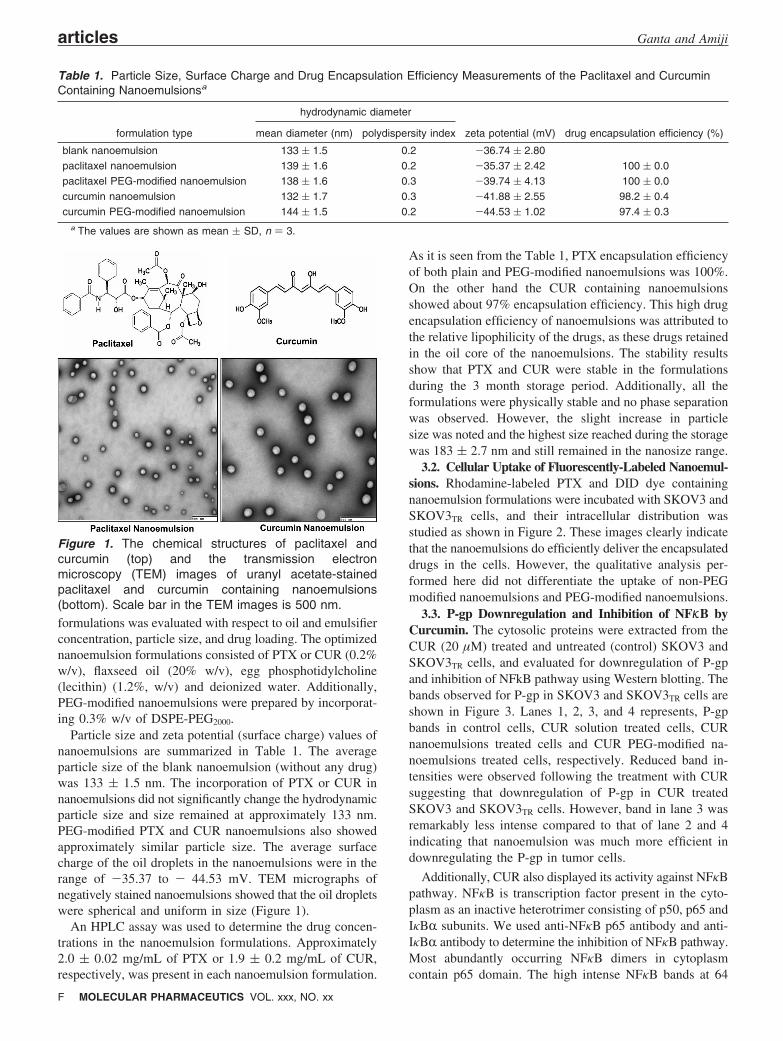
Particle size and zeta potential (surface charge) values ofnanoemulsions are summarized in Table 1. The averageparticle size of the blank nanoemulsion (without any drug)was 133 ( 1.5 nm. The incorporation of PTX or CUR innanoemulsions did not significantly change the hydrodynamicparticle size and size remained at approximately 133 nm.PEG-modified PTX and CUR nanoemulsions also showedapproximately similar particle size. The average surfacecharge of the oil droplets in the nanoemulsions were in therange of -35.37 to - 44.53 mV. TEM micrographs ofnegatively stained nanoemulsions showed that the oil dropletswere spherical and uniform in size (Figure 1).
An HPLC assay was used to determine the drug concen-trations in the nanoemulsion formulations. Approximately2.0 ( 0.02 mg/mL of PTX or 1.9 ( 0.2 mg/mL of CUR,respectively, was present in each nanoemulsion formulation.
As it is seen from the Table 1, PTX encapsulation efficiencyof both plain and PEG-modified nanoemulsions was 100%.On the other hand the CUR containing nanoemulsionsshowed about 97% encapsulation efficiency. This high drugencapsulation efficiency of nanoemulsions was attributed tothe relative lipophilicity of the drugs, as these drugs retainedin the oil core of the nanoemulsions. The stability resultsshow that PTX and CUR were stable in the formulationsduring the 3 month storage period. Additionally, all theformulations were physically stable and no phase separationwas observed. However, the slight increase in particlesize was noted and the highest size reached during the storagewas 183 ( 2.7 nm and still remained in the nanosize range.
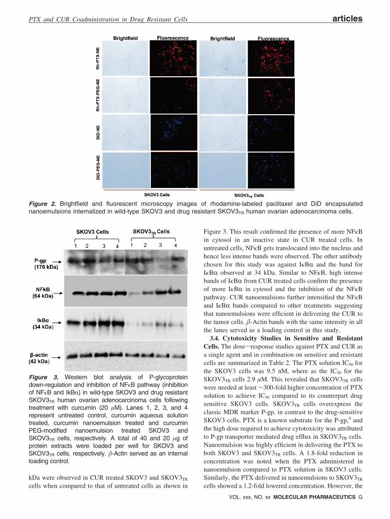
3.2. Cellular Uptake of Fluorescently-Labeled Nanoemul-sions. Rhodamine-labeled PTX and DID dye containingnanoemulsion formulations were incubated with SKOV3 andSKOV3TR cells, and their intracellular distribution wasstudied as shown in Figure 2. These images clearly indicatethat the nanoemulsions do efficiently deliver the encapsulateddrugs in the cells. However, the qualitative analysis per-formed here did not differentiate the uptake of non-PEGmodified nanoemulsions and PEG-modified nanoemulsions.
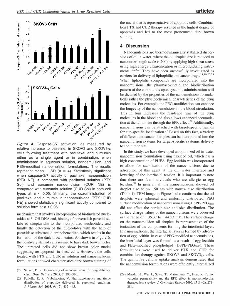
3.3. P-gp Downregulation and Inhibition of NFKB byCurcumin. The cytosolic proteins were extracted from theCUR (20 µM) treated and untreated (control) SKOV3 andSKOV3TR cells, and evaluated for downregulation of P-gpand inhibition of NFkB pathway using Western blotting. Thebands observed for P-gp in SKOV3 and SKOV3TR cells areshown in Figure 3. Lanes 1, 2, 3, and 4 represents, P-gpbands in control cells, CUR solution treated cells, CURnanoemulsions treated cells and CUR PEG-modified na-noemulsions treated cells, respectively. Reduced band in-tensities were observed following the treatment with CURsuggesting that downregulation of P-gp in CUR treatedSKOV3 and SKOV3TR cells. However, band in lane 3 wasremarkably less intense compared to that of lane 2 and 4indicating that nanoemulsion was much more efficient indownregulating the P-gp in tumor cells.
Additionally, CUR also displayed its activity against NFκBpathway. NFκB is transcription factor present in the cyto-plasm as an inactive heterotrimer consisting of p50, p65 andIκBR subunits. We used anti-NFκB p65 antibody and anti-IκBR antibody to determine the inhibition of NFκB pathway.Most abundantly occurring NFκB dimers in cytoplasmcontain p65 domain. The high intense NFκB bands at 64
Table 1. Particle Size, Surface Charge and Drug Encapsulation Efficiency Measurements of the Paclitaxel and CurcuminContaining Nanoemulsionsa
hydrodynamic diameter
formulation type mean diameter (nm) polydispersity index zeta potential (mV) drug encapsulation efficiency (%)
blank nanoemulsion 133 ( 1.5 0.2 -36.74 ( 2.80paclitaxel nanoemulsion 139 ( 1.6 0.2 -35.37 ( 2.42 100 ( 0.0paclitaxel PEG-modified nanoemulsion 138 ( 1.6 0.3 -39.74 ( 4.13 100 ( 0.0curcumin nanoemulsion 132 ( 1.7 0.3 -41.88 ( 2.55 98.2 ( 0.4curcumin PEG-modified nanoemulsion 144 ( 1.5 0.2 -44.53 ( 1.02 97.4 ( 0.3
a The values are shown as mean ( SD, n ) 3.
Figure 1. The chemical structures of paclitaxel andcurcumin (top) and the transmission electronmicroscopy (TEM) images of uranyl acetate-stainedpaclitaxel and curcumin containing nanoemulsions(bottom). Scale bar in the TEM images is 500 nm.
articles Ganta and Amiji
F MOLECULAR PHARMACEUTICS VOL. xxx, NO. xx
kDa were observed in CUR treated SKOV3 and SKOV3TR
cells when compared to that of untreated cells as shown in
Figure 3. This result confirmed the presence of more NFκBin cytosol in an inactive state in CUR treated cells. Inuntreated cells, NFκB gets translocated into the nucleus andhence less intense bands were observed. The other antibodychosen for this study was against IκBR and the band forIκBR observed at 34 kDa. Similar to NFκB, high intensebands of IκBR from CUR treated cells confirm the presenceof more IκBR in cytosol and the inhibition of the NFκBpathway. CUR nanoemulsions further intensified the NFκBand IκBR bands compared to other treatments suggestingthat nanoemulsions were efficient in delivering the CUR tothe tumor cells. �-Actin bands with the same intensity in allthe lanes served as a loading control in this study.
3.4. Cytotoxicity Studies in Sensitive and ResistantCells. The dose-response studies against PTX and CUR asa single agent and in combination on sensitive and resistantcells are summarized in Table 2. The PTX solution IC50 forthe SKOV3 cells was 9.5 nM, where as the IC50 for theSKOV3TR cells 2.9 µM. This revealed that SKOV3TR cellswere needed at least ∼300-fold higher concentration of PTXsolution to achieve IC50 compared to its counterpart drugsensitive SKOV3 cells. SKOV3TR cells overexpress theclassic MDR marker P-gp, in contrast to the drug-sensitiveSKOV3 cells. PTX is a known substrate for the P-gp,4 andthe high dose required to achieve cytotoxicity was attributedto P-gp transporter mediated drug efflux in SKOV3TR cells.Nanoemulsion was highly efficient in delivering the PTX toboth SKOV3 and SKOV3TR cells. A 1.8-fold reduction inconcentration was noted when the PTX administered innanoemulsion compared to PTX solution in SKOV3 cells.Similarly, the PTX delivered in nanoemulsions to SKOV3TR
cells showed a 1.2-fold lowered concentration. However, the
Figure 2. Brightfield and fluorescent microscopy images of rhodamine-labeled paclitaxel and DiD encapsulatednanoemulsions internalized in wild-type SKOV3 and drug resistant SKOV3TR human ovarian adenocarcinoma cells.
Figure 3. Western blot analysis of P-glycoproteindown-regulation and inhibition of NFκB pathway (inhibitionof NFκB and IkBR) in wild-type SKOV3 and drug resistantSKOV3TR human ovarian adenocarcinoma cells followingtreatment with curcumin (20 µM). Lanes 1, 2, 3, and 4represent untreated control, curcumin aqueous solutiontreated, curcumin nanoemulsion treated and curcuminPEG-modified nanoemulsion treated SKOV3 andSKOV3TR cells, respectively. A total of 40 and 20 µg ofprotein extracts were loaded per well for SKOV3 andSKOV3TR cells, respectively. �-Actin served as an internalloading control.
PTX and CUR Coadministration in Drug Resistant Cells articles
VOL. xxx, NO. xx MOLECULAR PHARMACEUTICS G

PEG-modified nanoemulsion was not as effective as plainnanoemulsion. We speculate that the hydrophilic PEG chainswere forming as a steric barrier and reducing the nanoemul-sion uptake by the tumor cells. As seen from Table 2, CURalso exhibited anticancer activity. However, the dose-response against CUR was nearly identical in both drug-sensitive and resistant cells. CUR containing nanoemulsionshowed enhanced cytotoxicity compared to CUR solution(p < 0.05) and PEG-modified nanoemulsion (p < 0.05).
To inhibit the P-gp and sensitize the SKOV3TR cells toPTX, we carried out the combination study and results arepresented in Table 3. CUR inhibits the function of threemajor ABC drug transporters (P-gp, MRP-1 and ABCG2).16,17
Therefore, the coadministration of CUR could sensitize P-gpoverexpressing cells to PTX. The results revealed that, inthe presence of CUR solution, the IC50 of PTX solution wasreduced to ∼1.8-fold in SKOV3TR cells and 2.3-fold inSKOV3 cells. This combination therapy was significantlyeffective (P < 0.05) when the PTX and CUR delivered innanoemulsion formulations compared to solution or in PEG-modified nanoemulsion. Overall, the use of PTX and CURtogether resulted in enhanced therapeutic potential. The drugscoadministered either in solution or nanoemulsion producedsynergistic cytotoxicity effect in drug-sensitive cells, whereasthe drugs coadministered in solution, nanoemulsion or PEG-modified nanoemulsions produced additive cytotoxicity effectin SKOV3TR cells.
3.5. Quantitative and Qualitative Cellular ApoptoticActivity. To further confirm that the therapeutic potentialof combination therapy was due to enhancement in cellularapoptotic activity, quantitative and qualitative apoptoticanalysis was performed. Figure 4 shows the relative caspase-3/7 activities upon treatment with single and combinationPTX and CUR in aqueous solution and in nanoemulsion
formulations. The caspase-3/7 activities were calculatedrelative to the activity in untreated cells. PTX and CURdelivered in nanoemulsions induced significantly (p < 0.05)greater caspase-3/7 activity compared to aqueous solutionand PEG-modified nanoemulsion. Additionally, as comparedwith PTX and CUR in aqueous solution, nanoemulsionmediated delivery of combination therapy significantly (p <0.05) enhanced caspase-3/7 activities in SKOV3 andSKOV3TR cells. These results collaborate with the cellviability data to confirm that nanoemulsions were effectivein delivering the payload to the cells, and combinationtherapy with PTX and CUR delivered in nanoemulsionsproduced potential therapeutic effects.
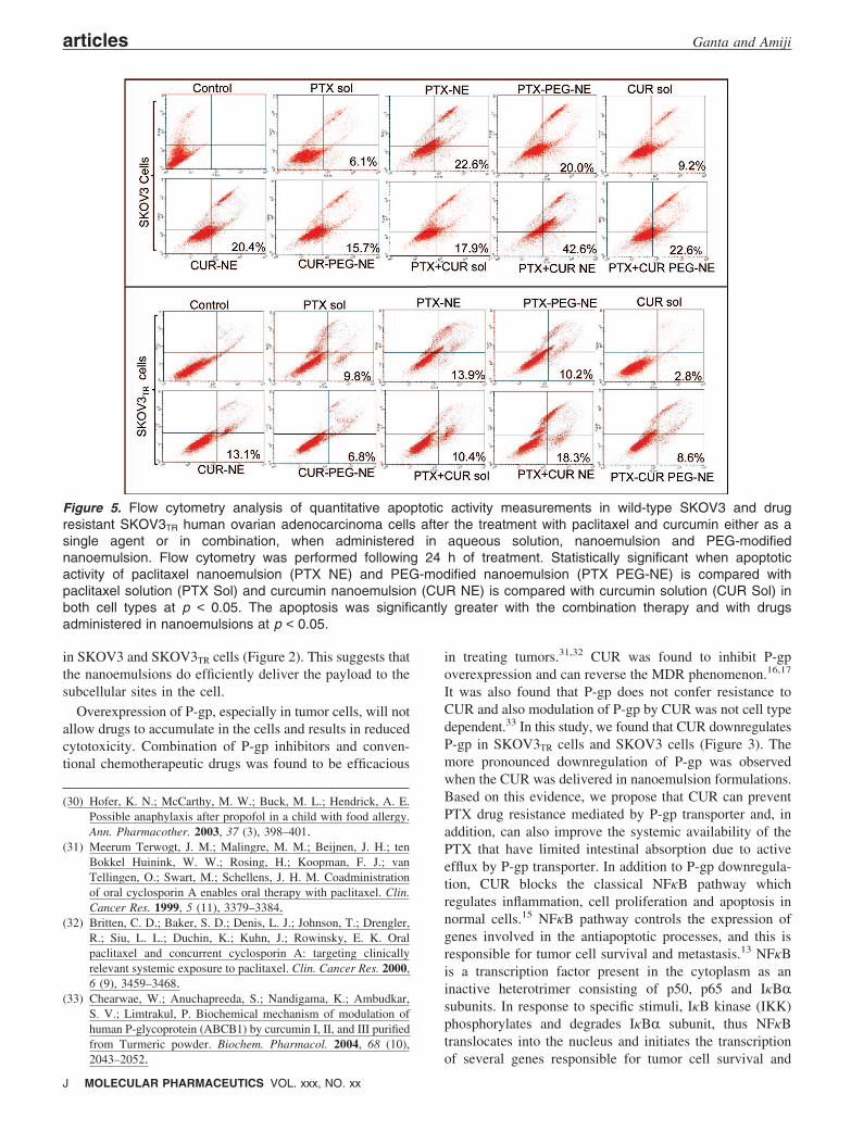
The results in Figure 5 show quantitative apoptotic activityin SKOV3 and SKOV3TR cells by Vybrant Apoptosis assayusing flow cytometry following the treatment of cells for24 h. In this assay, the YO-PRO-1 (green, apoptotic cellpermeant) dye and propidium iodide (red, apoptotic cellimpermeant) were used to label and differentiate betweenthe live cells, those undergoing apoptosis, and those that weredead. The flow cytometry plots reveal that there wasenhancement in cellular apoptosis in both SKOV3 andSKOV3TR cells when PTX and CUR were administered innanoemulsion formulations as compared to aqueous solutions(p < 0.05). In addition, when PTX and CUR were coadmin-istered in nanoemulsions, there was an increase in apoptosisat lower doses (i.e., 42.6% apoptosis in SKOV3 with 5 nMPTX and 5 µM CUR, 18.3% apoptosis in SKOV3TR cellswith 2 µM PTX and 5 µM CUR).
Additionally, qualitative analysis of cellular apoptosis aftersingle and combination therapy was evaluated using TUNELassay. It determines apoptosis as a result of DNA fragmenta-tion occurring in the apoptotic cells due to drug treatment.The apoptotic nuclei are stained brown by a three-step
Table 2. The 50% Inhibitory Concentration Values of Paclitaxel and Curcumin Alone and in Combination on SKOV3 andSKOV3TR Cells, Administered in the Form of Solution, Nanoemulsion and PEG-Modified Nanoemulsiona
IC50 values
treatment type on SKOV3 cells on SKOV3TR cells
paclitaxel solution 9.5 ( 1.2 nM 2.9 ( 0.4 µMpaclitaxel nanoemulsion 5.2 ( 1.3 nM 2.3 ( 0.1 µMpaclitaxel PEG-modified nanoemulsion 10.4 ( 0.8 nM 2.6 ( 0.3 µMcurcumin solution 9.8 ( 0.2 µM 10.8 ( 1.3 µMcurcumin nanoemulsion 6.2 ( 1.2 µM 9.6 ( 0.6 µMcurcumin PEG-modified nanoemulsion 9.4 ( 0.1 µM 10.6 ( 0.2 µMpaclitaxel + curcumin solution 4.1 nM + 5 µM 1.6 µM + 5 µMpaclitaxel + curcumin nanoemulsion 0.7 nM + 5 µM 1.1 + 5 µMpaclitaxel + curcumin PEG-modified nanoemulsion 4.5 nM + 5 µM 1.5 + 5 µM
a The values are shown as mean ( SD, n ) 8.
Table 3. Paclitaxel and Curcumin Combination Index (CI) against SKOV3 and SKOV3TR Cellsa
SKOV3 cells SKOV3TR cells
combination type CI interaction type CI interaction type
paclitaxel + curcumin solution 0.94 synergistic 1.0 additivepaclitaxel + curcumin nanoemulsion 0.93 synergistic 1.0 additivepaclitaxel + curcumin PEG-modified nanoemulsion 1.0 additive 1.0 additive
a CI < 1, synergistic; CI ) 1, additive; CI > 1, antagonistic.
articles Ganta and Amiji
H MOLECULAR PHARMACEUTICS VOL. xxx, NO. xx
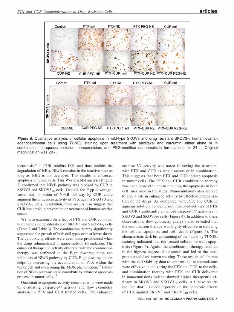
mechanism that involves incorporation of biotinylated nucle-otides at 3′-OH DNA end, binding of horseradish peroxidase-labeled streptavidin to the incorporated nucleotides, andfinally the detection of the nucleotides with the help ofperoxidase substrate, diaminobenzidine, which results in theformation of the dark brown stains. As shown in Figure 6,the positively stained cells seemed to have dark brown nuclei.The untreated cells did not show brown color nucleisuggesting no apoptosis in these cells. However, the cellstreated with PTX and CUR in solution and nanoemulsionsformulations showed characteristics dark brown staining of
the nuclei that is representative of apoptotic cells. Combina-tion PTX and CUR therapy resulted in the highest degree ofapoptosis and led to the most pronounced dark brownstaining.
4. DiscussionNanoemulsions are thermodynamically stabilized disper-
sions of oil in water, where the oil droplet size is reduced tonanometer length scale (<200) by applying high shear stressusing high energy ultrasonication or microfluidizing instru-ments.19,25,27 They have been successfully investigated ascarriers for delivery of liphophilic anticancer drugs.18,19,25,28
When liphophilic compounds are incorporated into thenanoemulsions, the pharmacokinetic and biodistributionpattern of the compounds upon systemic administration willbe dictated by the properties of the nanoemulsions formula-tions rather the physicochemical characteristics of the drugmolecules. For example, the PEG-modification can enhancethe longevity of the nanoemulsions in the blood circulation.This in turn increases the residence time of the drugmolecules in the blood and also allows enhanced accumula-tion at the tumor site through the EPR effect.29 Additionally,nanoemulsions can be attached with target-specific ligandsfor site-specific localization.27 Based on this fact, a varietyof different anticancer therapies can be incorporated into thenanoemulsion systems for target-specific systemic deliveryto the tumor site.
In this study, we have developed an optimized oil-in-waternanoemulsion formulation using flaxseed oil, which has ahigh concentration of PUFA. Egg lecithin was incorporatedto allow for stabilization of the nanoemulsions due toadsorption of this agent at the oil-water interface andlowering of the interfacial tension. It is important to notethat there are few individuals who are allergic to egglecithin.30 In general, all the nanoemulsions showed oildroplet size below 150 nm with narrow size distribution(Table 1). TEM image in Figure 1 also confirms that the oildroplets were spherical and uniformly distributed. PEGsurface modification of nanoemulsions using DSPE-PEG2000
did not affect the particle size and size distribution. Thesurface charge values of the nanoemulsions were observedin the range of -35.37 to -44.53 mV. The surface chargeon the nanoemulsion oil droplets is representative of theionization of the components forming the interfacial layer.In nanoemulsions, the interfacial layer is formed by adsorp-tion of egg lecithin. In case of PEG-modified nanoemulsions,the interfacial layer was formed as a result of egg lecithinand PEG-modified phospholipid (DSPE-PEG2000). Theseformulations were used to deliver PTX and CUR forcombination therapy against SKOV3 and SKOV3TR cells.The qualitative cellular uptake analysis demonstrated thatthe nanoemulsion formulations were efficiently internalized
(27) Sarker, D. K. Engineering of nanoemulsions for drug delivery.Curr. Drug DeliVery 2005, 2, 297–310.
(28) Patlolla, R. R.; Vobalaboina, V. Pharmacokinetics and tissuedistribution of etoposide delivered in parenteral emulsion.J. Pharm. Sci. 2005, 94 (2), 437–445.
(29) Maeda, H.; Wu, J.; Sawa, T.; Matsumura, Y.; Hori, K. Tumorvascular permeability and the EPR effect in macromoleculartherapeutics: a review. J. Controlled Release 2000, 65 (1-2), 271–284.
Figure 4. Caspase-3/7 activation, as measured byrelative increase to baseline, in SKOV3 and SKOV3TR
cells following treatment with paclitaxel and curcumineither as a single agent or in combination, whenadministered in aqueous solution, nanoemulsion, andPEG-modified nanoemulsion formulations. The resultsrepresent mean ( SD (n ) 4). Statistically significantwhen caspase-3/7 activity of paclitaxel nanoemulsion(PTX NE) is compared with paclitaxel solution (PTXSol) and curcumin nanoemulsion (CUR NE) iscompared with curcumin solution (CUR Sol) in both celltypes at p < 0.05. Similarly, the coadministration ofpaclitaxel and curcumin in nanoemulsions (PTX+CURNE) showed statistically significant activity compared tosolution form at p < 0.05.
PTX and CUR Coadministration in Drug Resistant Cells articles
VOL. xxx, NO. xx MOLECULAR PHARMACEUTICS I
in SKOV3 and SKOV3TR cells (Figure 2). This suggests thatthe nanoemulsions do efficiently deliver the payload to thesubcellular sites in the cell.
Overexpression of P-gp, especially in tumor cells, will notallow drugs to accumulate in the cells and results in reducedcytotoxicity. Combination of P-gp inhibitors and conven-tional chemotherapeutic drugs was found to be efficacious
in treating tumors.31,32 CUR was found to inhibit P-gpoverexpression and can reverse the MDR phenomenon.16,17
It was also found that P-gp does not confer resistance toCUR and also modulation of P-gp by CUR was not cell typedependent.33 In this study, we found that CUR downregulatesP-gp in SKOV3TR cells and SKOV3 cells (Figure 3). Themore pronounced downregulation of P-gp was observedwhen the CUR was delivered in nanoemulsion formulations.Based on this evidence, we propose that CUR can preventPTX drug resistance mediated by P-gp transporter and, inaddition, can also improve the systemic availability of thePTX that have limited intestinal absorption due to activeefflux by P-gp transporter. In addition to P-gp downregula-tion, CUR blocks the classical NFκB pathway whichregulates inflammation, cell proliferation and apoptosis innormal cells.15 NFκB pathway controls the expression ofgenes involved in the antiapoptotic processes, and this isresponsible for tumor cell survival and metastasis.13 NFκBis a transcription factor present in the cytoplasm as aninactive heterotrimer consisting of p50, p65 and IκBRsubunits. In response to specific stimuli, IκB kinase (IKK)phosphorylates and degrades IκBR subunit, thus NFκBtranslocates into the nucleus and initiates the transcriptionof several genes responsible for tumor cell survival and
(30) Hofer, K. N.; McCarthy, M. W.; Buck, M. L.; Hendrick, A. E.Possible anaphylaxis after propofol in a child with food allergy.Ann. Pharmacother. 2003, 37 (3), 398–401.
(31) Meerum Terwogt, J. M.; Malingre, M. M.; Beijnen, J. H.; tenBokkel Huinink, W. W.; Rosing, H.; Koopman, F. J.; vanTellingen, O.; Swart, M.; Schellens, J. H. M. Coadministrationof oral cyclosporin A enables oral therapy with paclitaxel. Clin.Cancer Res. 1999, 5 (11), 3379–3384.
(32) Britten, C. D.; Baker, S. D.; Denis, L. J.; Johnson, T.; Drengler,R.; Siu, L. L.; Duchin, K.; Kuhn, J.; Rowinsky, E. K. Oralpaclitaxel and concurrent cyclosporin A: targeting clinicallyrelevant systemic exposure to paclitaxel. Clin. Cancer Res. 2000,6 (9), 3459–3468.
(33) Chearwae, W.; Anuchapreeda, S.; Nandigama, K.; Ambudkar,S. V.; Limtrakul, P. Biochemical mechanism of modulation ofhuman P-glycoprotein (ABCB1) by curcumin I, II, and III purifiedfrom Turmeric powder. Biochem. Pharmacol. 2004, 68 (10),2043–2052.
Figure 5. Flow cytometry analysis of quantitative apoptotic activity measurements in wild-type SKOV3 and drugresistant SKOV3TR human ovarian adenocarcinoma cells after the treatment with paclitaxel and curcumin either as asingle agent or in combination, when administered in aqueous solution, nanoemulsion and PEG-modifiednanoemulsion. Flow cytometry was performed following 24 h of treatment. Statistically significant when apoptoticactivity of paclitaxel nanoemulsion (PTX NE) and PEG-modified nanoemulsion (PTX PEG-NE) is compared withpaclitaxel solution (PTX Sol) and curcumin nanoemulsion (CUR NE) is compared with curcumin solution (CUR Sol) inboth cell types at p < 0.05. The apoptosis was significantly greater with the combination therapy and with drugsadministered in nanoemulsions at p < 0.05.
articles Ganta and Amiji
J MOLECULAR PHARMACEUTICS VOL. xxx, NO. xx
metastasis.13,14 CUR inhibits IKK and thus inhibits thedegradation of IκBR. NFκB remains in the inactive state aslong as IκBR is not degraded. This results in enhancedapoptosis in tumor cells. This Western blot analysis (Figure3) confirmed that NFκB pathway was blocked by CUR inSKOV3 and SKOV3TR cells. Overall, the P-gp downregu-lation and inhibition of NFκB pathway by CUR couldaugment the anticancer activity of PTX against SKOV3 andSKOV3TR cells. In addition, these results also suggest thatCUR has a role in prevention or treatment of human ovariancancer.
We have examined the effect of PTX and CUR combina-tion therapy on proliferation of SKOV3 and SKOV3TR cells(Table 2 and Table 3). The combination therapy significantlysuppressed the growth of both cell types even at lower doses.The cytotoxicity effects were even more pronounced whenthe drugs administered in nanoemulsion formulation. Theenhanced therapeutic activity observed with the combinationtherapy was attributed to the P-gp downregulation andinhibition of NFκB pathway by CUR. P-gp downregulationhelps by increasing the accumulation of PTX within thetumor cell and overcoming the MDR phenomenon.17 Inhibi-tion of NFκB pathway could contribute to enhanced apoptosisprocess in tumor cells.15
Quantitative apoptotic activity measurements were madeby evaluating caspase-3/7 activity and flow cytometryanalysis in PTX and CUR treated cells. The enhanced
caspase-3/7 activity was noted following the treatmentwith PTX and CUR as single agents or in combination.This suggests that both PTX and CUR induce apoptosisin tumor cells. The PTX and CUR combination therapywas even more efficient in inducing the apoptosis in bothcell lines used in the study. Nanoemulsions also seemedto play a role in enhanced activity by effective internaliza-tion of the drugs. As compared with PTX and CUR inaqueous solution, nanoemulsion mediated delivery of PTXand CUR significantly enhanced caspase-3/7 activities inSKOV3 and SKOV3TR cells (Figure 4). In addition to theseobservations, flow cytometry analysis also revealed thatthe combination therapy was highly effective in inducingthe cellular apoptosis and cell death (Figure 5). Thecharacteristic dark brown staining of the nuclei by TUNELstaining indicated that the treated cells underwent apop-tosis (Figure 6). Again, the combination therapy resultedin the highest degree of apoptosis and led to the mostpronounced dark brown staining. These results collaboratewith the cell viability data to confirm that nanoemulsionswere effective in delivering the PTX and CUR to the cells,and combination therapy with PTX and CUR deliveredin nanoemulsions indeed showed higher therapeutic ef-ficacy in SKOV3 and SKOV3TR cells. All these resultsindicate that CUR could potentiate the apoptotic effectsof PTX against SKOV3 and SKOV3TR cells.
Figure 6. Qualitative analysis of cellular apoptosis in wild-type SKOV3 and drug resistant SKOV3TR human ovarianadenocarcinoma cells using TUNEL staining upon treatment with paclitaxel and curcumin, either alone or incombination in aqueous solution, nanoemulsion, and PEG-modified nanoemulsion formulations for 24 h. Originalmagnification was 20×.
PTX and CUR Coadministration in Drug Resistant Cells articles
VOL. xxx, NO. xx MOLECULAR PHARMACEUTICS K
5. ConclusionsWhen cancer cells develop a MDR phenotype either due
to intrinsic factors, such as microenvironmental selectionpressures, or after first exposure to a chemotherapeutic agent,the ensuing clinical outcomes are always very poor. Strate-gies that allow for enhancement of systemic deliveryefficiency as well as augmentation of therapeutic responseby lowering tumor apoptotic threshold can have a profoundimpact in the management of refractory diseases. In thisstudy, we have examined coadministration of PTX and CURusing nanoemulsion formulations that can aid in enhancingdelivery efficiency to the tumor mass. The oil-in-waternanoemulsion can also solubilize hydrophobic compounds,such as PTX and CUR, and allow for efficient intracellulardelivery. The pleiotropic effects of CUR are especiallybeneficial in refractory cancer due to inhibition of NFκBactivity and downregulation of important efflux transporterssuch as P-gp, and MRP-1 in tumor cells. Combination PTX
and CUR when administered in nanoemulsion formulationsshowed significant enhancement in cytotoxicity, especiallyin drug resistant SKOV3TR ovarian adenocarcinoma cells.This dual therapeutic strategy can have significant potentialin the clinical management of refractory diseases. Future inViVo studies in human tumor xenograft models will furthervalidate this hypothesis.
Acknowledgment. This study was supported by theNational Cancer Institute’s Alliance in Nanotechnology forCancer Platform Partnership grant (R01-CA119617). Trans-mission electron microscopy was performed by Mr. WilliamFowle at the Electron Microscopy Center of NortheasternUniversity. Additionally, the authors are deeply grateful toDr. Robert Campbell for assistance with the particle sizeanalysis.
MP800240J
articles Ganta and Amiji
L MOLECULAR PHARMACEUTICS VOL. xxx, NO. xx
Copyright © 2022 FDOKUMEN