Ex Vivo Uranium Decontamination Efficiency on Wounded Skin and In Vitro Skin Toxicity of a...
10
RESEARCH ARTICLE – Pharmaceutics, Drug Delivery and Pharmaceutical Technology Ex Vivo Uranium Decontamination Efficiency on Wounded Skin and In Vitro Skin Toxicity of a Calixarene-Loaded Nanoemulsion SOPHIE GRIVES, 1,2,3 GUILLAUME PHAN, 3 GUILLAUME MORAT, 3 DAVID SUHARD, 3 FRANC ¸ OIS REBIERE, 3 ELIAS FATTAL 1,2 1 Universit´ e Paris-Sud, Facult´ e de pharmacie, Institut Galien Paris-Sud, LabEx LERMIT, Chˆ atenay-Malabry Cedex 92296, France 2 CNRS, UMR 8612, Chˆ atenay-Malabry Cedex 92296, France 3 Institut de Radioprotection et de S ˆ uret´ e Nucl´ eaire (IRSN), PRP-Hom, SDI, Laboratoire de RadioChimie, Fontenay-aux-Roses 92260, France Received 13 December 2014; revised 16 February 2015; accepted 3 March 2015 Published online in Wiley Online Library (wileyonlinelibrary.com). DOI 10.1002/jps.24431 ABSTRACT: The present work aims at studying the decontamination efficacy of a calixarene-loaded nanoemulsion on two ex vivo wounded skin models mimicking superficial stings or cuts contaminated with uranium, and on a third model using excoriation. The decontaminating formulation was compared with the currently used radio-decontaminating soapy water (Trait rouge R ) treatment. Moreover, to assess skin damage potentially induced by the undiluted nanoemulsion, in vitro toxicity studies were conducted on an in vitro reconstructed human epidermis, coupled with three different toxicity tests [3-(4,5-dimethylthiazol-2-yl)-2,5 diphenyl tetrazolium bromide, lactate dehydrogenase, and interleukin-1-]. This work demonstrated not only a significant decontamination activity of the calixarene nanoemulsion on wounded skin, ranging from 92% to 94% of the applied uranium solution according to the ex vivo model used, but also the absence of side effects of this promising treatment. C 2015 Wiley Periodicals, Inc. and the American Pharmacists Association J Pharm Sci Keywords: nanotechnology; emulsion; formulation vehicle; chelation; skin; diffusion; in vitro models; toxicity INTRODUCTION Cutaneous contamination by radionuclides on intact or wounded skin is a significant source of exposure and accounted for more than half of the recorded accidents in France between 1970 and 2003. 1 The cutaneous contamination by actinides such as uranium leads to its transcutaneous passage, which is enhanced in the case of wounds, because of the disruption of the protective layer, the stratum corneum. This diffusion through the skin can vary depending on the physicochemical form of the uranium compound, as soluble compounds such as uranyl nitrate or acetate are more readily absorbed than insol- uble forms such as oxides. 2–6 The contaminant compounds are then incorporated into the body through the bloodstream and their retention in specific organs leads to potential radiochem- ical toxicity. Indeed, one part of the systemic load is quickly retained and stored in the target organs, such as the kidneys and the skeleton, whereas the other part is directly excreted in the urine over the first 24 h after contamination. 7–10 Renal uptake of uranium at the proximal tubular cells causes many dysfunctions such as membrane lesions, death of targeted cells, morphological changes in the structure of the glomerulus, and functional abnormalities including kidney reabsorption. 11,12 Moreover, this nephrotoxicity is particularly important when contamination occurs with soluble and trans- ferable uranium compounds, such as uranyl nitrate, which can cause death because of kidney damage in rats or guinea pigs. 6,13 Bone toxicity may also occur after uranium contamination be- cause of the substitution of calcium ions by uranyl ions in the skeleton, leading in particular to a decrease of radial cortical Correspondence to: Elias Fattal (Telephone: +331-46835568; Fax: +331- 46619334; E-mail: [email protected]) Journal of Pharmaceutical Sciences C 2015 Wiley Periodicals, Inc. and the American Pharmacists Association bone growth in young rats. 14 Finally, it has been shown that contamination by uranium can also have deleterious effects on the reproduction of rats and mice. 15–18 However, no specific and efficient treatment is available in case of such contamination, as only a warm shower with soap and water is generally carried out. 19 In this context, previ- ous studies by our group aimed at developing an emergency treatment for uranium decontamination. As a result, an oil-in- water (O/W) nanoemulsion loaded with calixarene molecules was designed. This nanoemulsion loaded with a specific chelat- ing agent was shown to be highly efficient in avoiding deep skin penetration of uranium on ex vivo models in Franz diffu- sion cells, on intact and excoriated pig ear skins. 20 The goal of the present study was first to confirm the de- contamination efficiency of the calixarene nanoemulsion on new superficial skin wound models, reproducing superficial cuts and stings, and mimicking the contaminations because of the mechanical injuries commonly encountered in the nuclear industry. 21 The nanoemulsion’s decontamination efficiency was also tested on a skin excoriation model, and all the data were compared with those obtained with the treatment currently used in nuclear plants: soapy water (e.g., Trait rouge R in France). The second aim of this study was to assess the poten- tial skin toxicity of the calixarene nanoemulsion after different exposure times on an in vitro reconstructed human epidermis (RHE), coupled with 3-(4,5-dimethylthiazol-2-yl)-2,5 diphenyl tetrazolium bromide (MTT) test as well as lactate dehydroge- nase (LDH) and interleukin-1-" (IL-1-") assays. MATERIALS AND METHODS Chemicals 1,3,5-OCH 3 -2,4,6-OCH 2 COOH-p-tertbutylcalix[6]arene (Fig. 1), here called calixarene, was synthesized as described. 22 The Grives et al., JOURNAL OF PHARMACEUTICAL SCIENCES 1
-
Upload
independent -
Category
Documents
-
view
1 -
download
0
Transcript of Ex Vivo Uranium Decontamination Efficiency on Wounded Skin and In Vitro Skin Toxicity of a...

RESEARCH ARTICLE – Pharmaceutics, Drug Delivery and Pharmaceutical Technology
Ex Vivo Uranium Decontamination Efficiency on Wounded Skinand In Vitro Skin Toxicity of a Calixarene-Loaded Nanoemulsion
SOPHIE GRIVES,1,2,3 GUILLAUME PHAN,3 GUILLAUME MORAT,3 DAVID SUHARD,3 FRANCOIS REBIERE,3 ELIAS FATTAL1,2
1Universite Paris-Sud, Faculte de pharmacie, Institut Galien Paris-Sud, LabEx LERMIT, Chatenay-Malabry Cedex 92296, France2CNRS, UMR 8612, Chatenay-Malabry Cedex 92296, France3Institut de Radioprotection et de Surete Nucleaire (IRSN), PRP-Hom, SDI, Laboratoire de RadioChimie, Fontenay-aux-Roses 92260,France
Received 13 December 2014; revised 16 February 2015; accepted 3 March 2015
Published online in Wiley Online Library (wileyonlinelibrary.com). DOI 10.1002/jps.24431
ABSTRACT: The present work aims at studying the decontamination efficacy of a calixarene-loaded nanoemulsion on two ex vivo woundedskin models mimicking superficial stings or cuts contaminated with uranium, and on a third model using excoriation. The decontaminatingformulation was compared with the currently used radio-decontaminating soapy water (Trait rouge R©) treatment. Moreover, to assess skindamage potentially induced by the undiluted nanoemulsion, in vitro toxicity studies were conducted on an in vitro reconstructed humanepidermis, coupled with three different toxicity tests [3-(4,5-dimethylthiazol-2-yl)-2,5 diphenyl tetrazolium bromide, lactate dehydrogenase,and interleukin-1-�]. This work demonstrated not only a significant decontamination activity of the calixarene nanoemulsion on woundedskin, ranging from 92% to 94% of the applied uranium solution according to the ex vivo model used, but also the absence of side effectsof this promising treatment. C© 2015 Wiley Periodicals, Inc. and the American Pharmacists Association J Pharm SciKeywords: nanotechnology; emulsion; formulation vehicle; chelation; skin; diffusion; in vitro models; toxicity
INTRODUCTION
Cutaneous contamination by radionuclides on intact orwounded skin is a significant source of exposure and accountedfor more than half of the recorded accidents in France between1970 and 2003.1 The cutaneous contamination by actinidessuch as uranium leads to its transcutaneous passage, whichis enhanced in the case of wounds, because of the disruptionof the protective layer, the stratum corneum. This diffusionthrough the skin can vary depending on the physicochemicalform of the uranium compound, as soluble compounds such asuranyl nitrate or acetate are more readily absorbed than insol-uble forms such as oxides.2–6 The contaminant compounds arethen incorporated into the body through the bloodstream andtheir retention in specific organs leads to potential radiochem-ical toxicity. Indeed, one part of the systemic load is quicklyretained and stored in the target organs, such as the kidneysand the skeleton, whereas the other part is directly excreted inthe urine over the first 24 h after contamination.7–10
Renal uptake of uranium at the proximal tubular cellscauses many dysfunctions such as membrane lesions, deathof targeted cells, morphological changes in the structure ofthe glomerulus, and functional abnormalities including kidneyreabsorption.11,12 Moreover, this nephrotoxicity is particularlyimportant when contamination occurs with soluble and trans-ferable uranium compounds, such as uranyl nitrate, which cancause death because of kidney damage in rats or guinea pigs.6,13
Bone toxicity may also occur after uranium contamination be-cause of the substitution of calcium ions by uranyl ions in theskeleton, leading in particular to a decrease of radial cortical
Correspondence to: Elias Fattal (Telephone: +331-46835568; Fax: +331-46619334; E-mail: [email protected])
Journal of Pharmaceutical SciencesC© 2015 Wiley Periodicals, Inc. and the American Pharmacists Association
bone growth in young rats.14 Finally, it has been shown thatcontamination by uranium can also have deleterious effects onthe reproduction of rats and mice.15–18
However, no specific and efficient treatment is available incase of such contamination, as only a warm shower with soapand water is generally carried out.19 In this context, previ-ous studies by our group aimed at developing an emergencytreatment for uranium decontamination. As a result, an oil-in-water (O/W) nanoemulsion loaded with calixarene moleculeswas designed. This nanoemulsion loaded with a specific chelat-ing agent was shown to be highly efficient in avoiding deepskin penetration of uranium on ex vivo models in Franz diffu-sion cells, on intact and excoriated pig ear skins.20
The goal of the present study was first to confirm the de-contamination efficiency of the calixarene nanoemulsion onnew superficial skin wound models, reproducing superficialcuts and stings, and mimicking the contaminations because ofthe mechanical injuries commonly encountered in the nuclearindustry.21 The nanoemulsion’s decontamination efficiency wasalso tested on a skin excoriation model, and all the data werecompared with those obtained with the treatment currentlyused in nuclear plants: soapy water (e.g., Trait rouge R© inFrance). The second aim of this study was to assess the poten-tial skin toxicity of the calixarene nanoemulsion after differentexposure times on an in vitro reconstructed human epidermis(RHE), coupled with 3-(4,5-dimethylthiazol-2-yl)-2,5 diphenyltetrazolium bromide (MTT) test as well as lactate dehydroge-nase (LDH) and interleukin-1-" (IL-1-") assays.
MATERIALS AND METHODS
Chemicals
1,3,5-OCH3-2,4,6-OCH2COOH-p-tertbutylcalix[6]arene (Fig.1), here called calixarene, was synthesized as described.22 The
Grives et al., JOURNAL OF PHARMACEUTICAL SCIENCES 1

2 RESEARCH ARTICLE – Pharmaceutics, Drug Delivery and Pharmaceutical Technology
Figure 1. Structure of 1,3,5-OCH3-2,4,6-OCH2COOH-p-tertbutyl-calix[6]arene.
other compounds used for preparing the calixarene nanoemul-sions were paraffin oil (d = 0.86; VWR, Fontenay-sous-Bois,France), nonionic surfactants sorbitan monooleate (Span R©
80), and polyoxyethylene glycol sorbitan monooleate (Tween R©
80), both purchased from Sigma–Aldrich (Saint-Quentin-Fallavier, France). Demineralized water was obtained from aMilli-Q R© Synergy 185 water purification system (Millipore,Saint-Quentin-en-Yvelines, France).
The uranium-contaminated solution was prepared from de-pleted uranium solution (1000 mg L−1 in 2% HNO3), pur-chased from SPEX Certiprep, Horiba Jobin Yvon (Longjumeau,France). The isotopic composition was 0.0013% of 234U, 0.3%of 235U, and 99.6987% of 238U. The carbonate buffer 0.025 MpH 9.6, used for the receptor medium of Franz diffusion cells,was purchased from Sigma–Aldrich. The Trait rouge R© liquidsoap used as a control was manufactured by the SORIFA Lab-oratory (Ostwald, France). The following reagents and stan-dards were used for inductively coupled plasma mass spec-trometry (ICP-MS) measurements: 67% HNO3 stock solution(Normatom; VWR), bismuth (209Bi) stock standard solution at100 ppb prepared in 2% HNO3 (from a 10–mg L−1 single el-ement standard; SPEX Certiprep, Horiba Jobin Yvon), and amultielemental standard solution containing depleted uraniumat 1 pg L−1 (from a 10-mg L−1 tuning solution; SPEX Certiprep,Horiba Jobin Yvon) prepared in 2% HNO3. For skin contami-nation, the uranium solution in acetate buffer was prepared toa final concentration of uranium at 10 mg L−1 pH 5.
Preparation and Characterization of Calixarene-LoadedNanoemulsion
The emulsion inversion point method was used to preparethe O/W calixarene nanoemulsion as previously described.23
To sum up, water was added drop by drop into a mixture ofparaffin oil (20%) and surfactants (5%) under stirring at 50°C.Calixarene was added to the oily phase at a concentration of4 mg g−1. The dispersed oil droplet size and zeta potentialwere both measured with a Nanosizer/Zetasizer R© ZS apparatus(Malvern Instruments, Worcestershire, UK) and pH measure-ments were carried out with a MeterLab/pH M240 pH-meter(Radiometer, Copenhagen, Denmark).
Ex Vivo Uranium Decontamination Efficiency
The diffusion kinetics of uranium through the skin was eval-uated using a Franz diffusion cells system (MicroettePlusTM
Apparatus; Hanson Research Corporation, Chatsworth, Cali-fornia) for 24 h. To carry out the experiments, full thicknessskin pieces were removed from pig ears that were purchasedfrom a slaughterhouse (Guy Harang Abattoir, Houdan, France),with the authorization of the French Departmental Directorateof Veterinary Services of the Hauts-de-Seine, and stored at−20°C until use. Each sample was depilated and superficiallywounded as described below.
As previously defined,24 skin samples were inserted betweenthe receptor and the donor compartments of the diffusion cells,with the dermal face in contact with the 0.025-M carbonatebuffer receptor medium, and the whole system was fixed witha clamp. The receptor medium was kept under stirring atconstant temperature (38 ± 0.1°C) during the experiments,which were conducted under occlusive conditions. Contamina-tion with 600 :L of the 10 mg L−1 uranyl nitrate solution atpH 5 was performed on intact or wounded pig ear skin. Six-hundred microliter of the calixarene nanoemulsion treatmentwas then immediately added, and the diffusion kinetics of ura-nium through the skin was evaluated for 24 h by sampling anddosage of the receptor medium at 0, 5, 15, 30 min and 2, 4,12, 18, and 24 h. At the end of the experiment, the content ofthe donor compartment in each cell was aspirated to quantifythe uranium. The diffusion of uranium was compared with thediffusion obtained after immediate application of a nonloadednanoemulsion, acetate buffer, and soapy water under the sameconditions. All skin explants were sampled and frozen until theanalysis for uranium content by ICP-MS. In order to measurethe uranium content on these explants, the digestion of skinpieces was carried out with 67% HNO3 stock solution and 30%H2O2 (VWR), using the ETHOS One R© microwave oven (Mile-stone Srl, Sorisole, Italy).
Simulations of Superficial Wounds
Three superficial wound models were simulated: (1) by mi-croneedles of 0.7 mm of length (Dermaroller R©, Wolfenbuttel,Germany), to mimic a superficial sting; (2) by incision inducedby Surgicutt R© system (Surgicutt R© Junior; CML, Nemours,France), to mimic a superficial cut by a 1-mm blade; and (3)by excoriation to remove the stratum corneum, to mimic a su-perficial scratch. In order not to interfere with the applicationof products and the diffusion of uranium, all skins sampleswere previously depilated using a depilating cream (Veet R© sen-sitive skin). The characteristics of the three types of modelsare summed up in Table 1. Samples were fixed with a solutioncontaining 2.5% glutaraldehyde for 1 h at room temperature,then dehydrated in an ethanol bath, and permeabilized with apropylene oxide/Epon mixture. Finally, samples were embeddedin pure Epon-type resin. Several thin sections (1 :m) embeddedin resin were cut.
Table 1. Summary of the Different Superficial Wounds ModelsApplied on the Pig Ear Skin for the Study of the DecontaminationEfficiency of the Calixarene Nanoemulsion
Type Wounds Means Simulated Wounds
Incision Superficial sting Microneedles Superficial stingIncision Cut Surgicutt R© Superficial cutExcision Excoriation Tape stripping Superficial scratch
Grives et al., JOURNAL OF PHARMACEUTICAL SCIENCES DOI 10.1002/jps.24431

RESEARCH ARTICLE – Pharmaceutics, Drug Delivery and Pharmaceutical Technology 3
Thickness and Transepidermal Water Loss of Skin Pieces
After wounding, the thickness of the skin pieces was measuredusing a thickness gage (Mitutoyo Corporation, Roissy, France).The mean thickness of intact or wounded skin was 1.11 ±0.1 mm. Transepidermal water loss (TEWL) was measuredusing a TewameterTM 300 R© (Monaderm R©; Monaco, France)equipped with an adapted probe. According to the apparatusspecifications, TEWL values below 15–25 g m−2 h−1 can reflectthe skin barrier integrity, whereas TEWL values exceeding 30g m−2 h−1 can indicate a disruption of the skin barrier.
Sample Analysis
After an 100-fold dilution in 2% nitric acid solution of eachsample taken from the Franz cells diffusion, 238U was directlymeasured by ICP-MS (Agilent 7700x) using the optimized pro-tocols originally designed for human urine samples.25–27 In eachsample or calibration solution, 100 :L of 209Bi was added as aninternal standard, at 100 ppb. Skin samples were previouslymineralized by microwaves as described above and then an-alyzed for uranium content as the other samples by ICP-MSmeasurement after a 200th dilution in 2% nitric acid solution.The cumulative amounts of uranium permeating through theskin were expressed as a percentage of the amount of uraniumdeposited on each skin biopsy, and plotted against time. Theamount of uranium recovered at the end of the 24-h experi-ment in skin explants and the one remaining in the donor com-partments were also expressed as a percentage of the amountof uranium initially deposited. Lag time values were calculatedby extrapolation to the time axis of the linear parts of the curvesrepresenting the amount of uranium diffused through the skinover time. The slope of the linear part of the curves yielded thepseudo-steady-state flux Jss (ng cm−2 h−1), and the permeabil-ity coefficients Kp (cm h−1) were calculated as Kp = Jss/C, with Ccorresponding to the concentration in the donor compartment.
Assessment of In Vitro Skin Toxicity of Calixarene-LoadedNanoemulsion
Tissues, Culture Conditions, and Treatments of Skin Units
The skin toxicity potentially induced by the calixarene na-noemulsion was measured using a three-dimensional RHEmodel (SkinEthic Laboratories, Lyon, France). For this pur-pose, 16 :L of each test substance were applied on tissues fordifferent exposure times and different times of postincubationtreatment. In accordance with OECD Test Guideline No. 439,28
the skin irritation Test−42bis was performed. This test was de-signed for the prediction of acute skin irritation of chemicals bymeasurements of its cytotoxic effect, as reflected in the methyltetrazolium (MTT) assay,29 purchased from Sigma ChemicalCompany (St. Louis, Missouri). First of all, each product testedwas left in contact with the tissues for 42 min under sterile con-ditions. Then, the tissues were rinsed with 25 mL of Dulbecco’ssterile phosphate buffer saline (PBS), mechanically dried, and apostincubation was carried out on 2 mL growth culture mediumfor 42 h at 37°C, 5% CO2, and 95% humidified atmosphere (allculture medium used were purchased from SkinEthic Laborato-ries). The second assay consisted in increasing the contact time,and leaving the different treatments for 2 h, with a postincu-bation of 42 h. Then, the last test consisted in a 24-h exposurewithout postincubation. PBS was used as negative control anda 20% sodium dodecyl sulfate (SDS) solution, purchased from
VWR, was used as a positive control after a fourfold dilutionin water. Nonloaded nanoemulsion was also used as a controltreatment. Each test was performed on three tissue replicates.At the end of each postincubation, a MTT test was carried outdirectly on each tissue, and the culture medium was sampledto measure the release of IL-1-" by a quantitative sandwich im-munoassay technique using the human IL-1 alpha ELISA kit.The LDH release was measured by using a LDH assay kit. Bothkits were purchased from Sigma–Aldrich (St. Louis, Missouri).
Irritation Measurement by MTT Test
The tissue viability was first assessed by MTT reduction. Theblue formazan crystals formed, qualitatively proportional to thecell viability, were extracted using 1.5 mL isopropanol for 2 hat room temperature, and three replicates per tissue were sam-pled to be quantified by spectrophotometry at a wavelength of570 nm. For each treated tissue, the cell viability was expressedas the percentage of the mean negative control tissues. For theTest−42bis, values under or equal to 50% are used to classify thetest substance as an irritant.29
Measurement of IL-1-� Release
The levels of extracellular IL-1-" released in the supernatantsby the RHE were determined by an in vitro quantitativesandwich immunoassay technique with a detection limit of0.5 pg mL−1. one-hundred microliter of each standard andnondiluted sample was added in duplicate into the appropriateprecoated wells and incubated for 2.5 h at room temperaturewith gentle shaking, allowing the molecules of IL-1-" presentin the samples to bind to the wells by the immobilized antibody.Then, the wells were washed four times with 300 :L of washsolution 1× included in the kit, and 100 :L of biotinylated de-tection antibody were added in each well, which were incubatedfor 1 h at room temperature with gentle shaking. The incuba-tion solution was removed and the washing step was repeated.one-hundred microliter of prepared HRP-Streptavidin solutionwas then added to each well, followed by incubation for 45 minat room temperature with gentle shaking. After removing thesolution and repeating the washing steps once, 100 :L of col-orimetric TMB reagent were added to each well. Incubationfor 30 min at room temperature in the dark with gentle shak-ing was performed. Fifty microliter of stop solution was thenadded in each well and the microplate was read immediately at450 nm.
After calculating the mean absorbance obtained for each du-plicate of samples and standards, and subtracting the averagezero standard optical density, the concentration of IL-1-" ineach sample was calculated on the basis of the standard curve.
Measurement of LDH Release
Lactate dehydrogenase is an oxidoreductase enzyme that cat-alyzes the interconversion of pyruvate to lactate, and its releaseis used as a marker of tissue damage by measuring its activity.The LDH assay measures the reduction of NAD to NADH bythe LDH present in the different samples through colorimetricdetection (450 nm). After preparing samples and NADH stan-dard preparations in the LDH assay buffer provided in the kit,50 :L of these samples and standards were brought into con-tact with 50 :L of the Master Mix Reaction (included in thekit) in a 96-well plate, protected from light. After 3 min of gen-tle shaking, the initial absorbance at 450 nm [(A450)initial] was
DOI 10.1002/jps.24431 Grives et al., JOURNAL OF PHARMACEUTICAL SCIENCES

4 RESEARCH ARTICLE – Pharmaceutics, Drug Delivery and Pharmaceutical Technology
measured. The plate was then incubated at 37°C in the dark,after the final measurement [(A450)final] for calculating the en-zyme activity. The incubation time was previously defined sothat the final measurement is within the linear range of thestandard curve. All samples and standards were run in dupli-cate.
After subtracting the final measurement [(A450)final] obtainedfor the blank of the other readings, the standard curve set up foreach incubation time was plotted. The change from the initialmeasurement to the final measurement [(A450)initial]–[(A450)final]for the samples was compared with the standard curve to de-termine the amount of NADH generated by the kinase assay.The LDH activity of each sample was then calculated by the fol-lowing equation, with B corresponding to the amount of NADHgenerated over the contact time (nmole), and V to the samplevolume (mL) added to the well:
LDH activity = B × Sample dilution factorV × Reaction time (min)
Statistical Analysis
In all the experiments, data were expressed as mean ± stan-dard deviation and were analyzed by Student’s t test. Differ-ences were considered to be significant if p < 0.05.
RESULTS
Superficial Wounds
The different wound models applied on pig ear skin were pre-viously validated by microscopic observation of skin sections,with several transverse skin cuts and skin top views, as shownin Figure 2. These histological images show that the excoria-tion thoroughly removes the stratum corneum. The sharp cutsinduced by the Surgicutt R© with their impacts on the skin andthe remaining presence of the stratum corneum are visible.It is also possible to observe the microlesions caused by themicroneedles. The number of tape strips required to removethe stratum corneum was determined after a histological study,and showed that 30 tape strips using a D-Squame R© 150 g cm−2
applicator (Monaderm R©; Monaco) were necessary to allow re-moval of the whole stratum corneum. After wounding the skinwith 50 superficial cuts, or 12 applications of the micronee-dle arrays containing 16 microneedles, the main TEWL valuesmeasured according to the skin condition (different superficialwounds) expressed in g m−2 h−1 were 54 ± 6 for Surgicutt R©
wounds and 48 ± 5 for microneedle wounds. The main TEWLvalues were 57 ± 4 after excoriation and 11 ± 3 for intact skin.These TEWL values indicate a disruption of the skin barrierwhatever the superficial wounds caused on the skin.
Ex Vivo Decontamination Efficiency
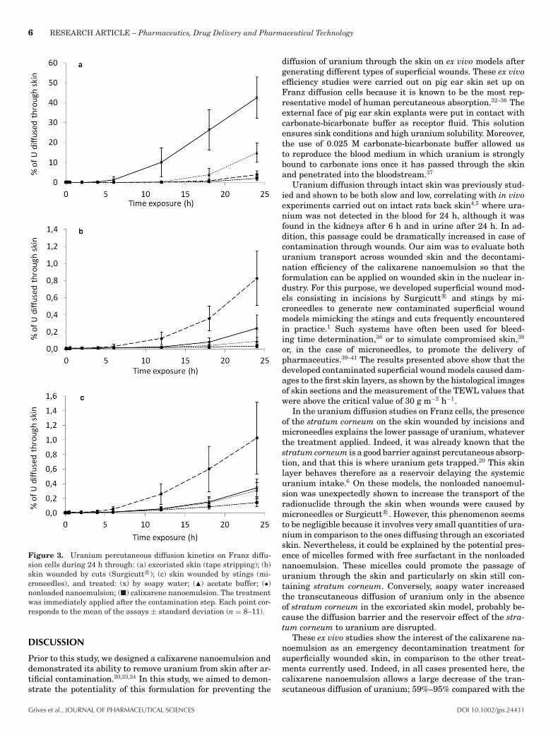
The kinetics of the percutaneous diffusion of uranium throughpig ear skin was followed for 24 h using the Franz diffusioncells system. In this study, the three different wound modelsdescribed above were used to assess the efficiency of our cal-ixarene nanoemulsion, in comparison with the nonloaded na-noemulsion, on the one hand, and with soapy water treatments,currently used in case of cutaneous contamination, or with asimple dilution with acetate buffer, on the other hand. As shownin Figure 3, the kinetics profiles were similar for the three types
of wounds generated on the skin. Uranium started to appearin the receptor medium at the earliest between 5 and 6 h af-ter contamination whatever the treatment or the nature of thewound. Diffusion across excoriated skin was higher (Fig. 3a)compared with skin wounded with Surgicutt R© (Fig. 3b) or mi-croneedles (Fig. 3c). In all cases, the diffusion was significantlyprevented when using calixarene nanoemulsion compared withthe nonloaded nanoemulsion or acetate buffer and soapy water(Fig. 3). On the excoriated skin, the diffusion of uranium washigher with acetate buffer and soapy water. On the woundscaused by Surgicutt R© or microneedles, the diffusion was higherwhen the skin was treated with the nonloaded nanoemulsioncompared with the other treatments (Figs. 3b and 3c).
On the excoriated skin, the absence of migration of uraniumthrough skin layers was observed particularly with the cal-ixarene nanoemulsion as 84.96 ± 1.89% remained in the donorphase (Fig. 4a), whereas this number was 64.96 ± 15.25% forthe nonloaded nanoemulsion, 39.49 ± 10.81% for the acetatebuffer and 40.97 ± 4.48% for soapy water. In the case of theabsence of stratum corneum, a small amount of uranium wasretained in the skin explant after a treatment with soapy waterand calixarene nanoemulsion, with 16.59 ± 7.78% and 13.02 ±2.25%, respectively, in comparison to the treatment with ac-etate buffer (45.66 ± 13.63%) and the application of nonloadednanoemulsion (31.14 ± 15.30%) (Fig. 4a).
On wounds caused by superficial incisions (Surgicutt R©), thesame trend as on the excoriated skin was observed regardingthe amount of uranium remaining in the donor compartment(Fig. 4b), the percentage of uranium not diffusing being 93.32± 2.17% after the calixarene nanoemulsion treatment, 77.47 ±11.21% after the application of the nonloaded nanoemulsion,and 61.65 ± 9.10% and 63.93 ± 7.17%, respectively, after theacetate buffer and soapy water control treatments. However,very little uranium was retained in the skin in the case ofcalixarene nanoemulsion (6.65 ± 2.18%) despite the presence ofthe stratum corneum, compared with the other groups: 21.70 ±11.51% for nonloaded nanoemulsion, 38.26 ± 9.11% for acetatebuffer, and 35.82 ± 7.20% for soapy water.
On wounds caused by microneedles, the calixarene formu-lation treatment still prevented the diffusion of uranium with94.47 ± 1.91% of nondiffused uranium and only 5.38 ± 1.91%of uranium retained in the skin. However, the distribution ofthe quantity of nondiffused uranium and uranium found in theskin was slightly different than previously observed (Fig. 4c).This time, after application of the acetate buffer, less uraniumwas trapped in the skin (13.99 ± 4.30%) and more was stillin the donor compartment (85.69 ± 4.26%) at the end of theexperiment. For the treatment with soapy water, we observed65.40 ± 8.41% and 34.25 ± 8.48%, respectively, of nondiffuseduranium and uranium in the skin, and in the same order 72.03± 9.12% and 26.94 ± 9.20% for the treatment with nonloadednanoemulsion.
Whatever the wounds caused on the skin, the permeabil-ity coefficient (Kp) and the pseudo-steady-state flux (Jss) cal-culated (Table 2) were lower after a treatment with the cal-ixarene nanoemulsion compared with the other treatments.These kinetics data confirmed the efficiency of the calixarenenanoemulsion, allowing a decrease of the transcutaneous pas-sage of uranium. Moreover, in case of excoriated skin, althoughthe theoretical lag times were overestimated, they were higherafter a nanoemulsion treatment than after application of soapywater or acetate buffer, allowing a delay of the percutaneous
Grives et al., JOURNAL OF PHARMACEUTICAL SCIENCES DOI 10.1002/jps.24431

RESEARCH ARTICLE – Pharmaceutics, Drug Delivery and Pharmaceutical Technology 5
Figure 2. Histological views of skin sections performed with an optical microscope: (a) transverse skin cuts, (b) skin top views, wounds impactsare pointed by arrows.
diffusion of uranium. These values were relevant with Kp andJss values. In contrast, the theoretical lag times in case of skinwounded with Surgicutt R© or microneedles were not represen-tative because of the low slopes of the diffusion curves.
In Vitro Toxicity of Calixarene-Loaded Nanoemulsion
To obtain a clear observation of the potential irritation inducedby our formulation, we used RHE models in which we applieddifferent tests assuming that the formulation or its componentsare able to penetrate the stratum corneum and are cytotoxic tothe cells in the underlying layers.30 The toxicity end points thatwere determined were the cell viability by MTT, as well as therelease of the cytokine IL-1-" as a potential proinflammatorycytokine31 and of the LDH release, which reflect membranedamages.
3-(4,5-Dimethylthiazol-2-yl)-2,5 Diphenyl Tetrazolium BromideTest
The studies were conducted under three different conditions,varying the exposure time between 42 min, 2, and 24 h, withor without postincubation (Fig. 5). Under these three condi-tions, the mean tissue viability expressed by the mitochondrialactivity evaluated by the MTT after SDS 5% solution expo-sure (positive control) was between 0.44 ± 0.002% and 1.44 ±0.001%. By contrast, whatever the considered conditions, themitochondrial activity was between 97.43 ± 0.024% and 107.41± 0.092% after exposure to the nonloaded nanoemulsion or tothe calixarene nanoemulsion, compared with the positive con-trol.
Interleukin-1-�
The cytotoxicity induced after the different exposure times oftreatment on the RHE tissues was also evaluated on the ba-sis of the cell release of the proinflammatory mediator IL-1-"(Fig. 6a). The concentrations of IL-1-" recovered on super-natants after exposure of the tissues to the SDS 5% solutionwere 69.72 ± 0.13, 103.58 ± 0.15, and 162.25 ± 0.04 pg mL−1,respectively, after an exposure of 42 min and 2 h (both with 42 hof postincubation treatment), and after 24 h of exposure with-out postincubation. For all the other treatments, the releaseof IL-1-" decreased when the contact time increased, and wasin any case very low, with a maximum concentration of IL-1-"of 9.57 ± 0.03 pg mL−1. In some cases, this concentration wastoo low to be detected, meaning the concentration was below0.5 pg mL−1, which corresponds to the limit of detection.
LDH Activity
The cytotoxicity induced after the different exposure times oftreatment on the RHE tissues was then evaluated on the ba-sis of the cell release of LDH, reflecting the cell membraneintegrity. The LDH activity is expressed as milliunit mL−1
(Fig. 6b), defined as the amount of enzyme that catalyzes theconversion of lactate into pyruvate to generate 1.0 mole ofNADH per minute at 37°C. The LDH activity measured afterexposure of the tissues to the SDS 5% solution were 5.00 ± 0.38,16.65 ± 2.49, and 18.59 ± 6.87 milliunit mL−1, respectively (42min and 2 h, respectively, both with 42 h of postincubationtreatment, and after 24 h of exposure without postincubation),compared with 0.36 ± 0.12 and 1.67 ± 0.35 milliunit mL−1 forall the other treatments and all conditions combined.
DOI 10.1002/jps.24431 Grives et al., JOURNAL OF PHARMACEUTICAL SCIENCES
6 RESEARCH ARTICLE – Pharmaceutics, Drug Delivery and Pharmaceutical Technology
Figure 3. Uranium percutaneous diffusion kinetics on Franz diffu-sion cells during 24 h through: (a) excoriated skin (tape stripping); (b)skin wounded by cuts (Surgicutt R©); (c) skin wounded by stings (mi-croneedles), and treated: (x) by soapy water; (�) acetate buffer; (•)nonloaded nanoemulsion; (�) calixarene nanoemulsion. The treatmentwas immediately applied after the contamination step. Each point cor-responds to the mean of the assays ± standard deviation (n = 8–11).
DISCUSSION
Prior to this study, we designed a calixarene nanoemulsion anddemonstrated its ability to remove uranium from skin after ar-tificial contamination.20,23,24 In this study, we aimed to demon-strate the potentiality of this formulation for preventing the
diffusion of uranium through the skin on ex vivo models aftergenerating different types of superficial wounds. These ex vivoefficiency studies were carried out on pig ear skin set up onFranz diffusion cells because it is known to be the most rep-resentative model of human percutaneous absorption.32–36 Theexternal face of pig ear skin explants were put in contact withcarbonate-bicarbonate buffer as receptor fluid. This solutionensures sink conditions and high uranium solubility. Moreover,the use of 0.025 M carbonate-bicarbonate buffer allowed usto reproduce the blood medium in which uranium is stronglybound to carbonate ions once it has passed through the skinand penetrated into the bloodstream.37
Uranium diffusion through intact skin was previously stud-ied and shown to be both slow and low, correlating with in vivoexperiments carried out on intact rats back skin4,5 where ura-nium was not detected in the blood for 24 h, although it wasfound in the kidneys after 6 h and in urine after 24 h. In ad-dition, this passage could be dramatically increased in case ofcontamination through wounds. Our aim was to evaluate bothuranium transport across wounded skin and the decontami-nation efficiency of the calixarene nanoemulsion so that theformulation can be applied on wounded skin in the nuclear in-dustry. For this purpose, we developed superficial wound mod-els consisting in incisions by Surgicutt R© and stings by mi-croneedles to generate new contaminated superficial woundmodels mimicking the stings and cuts frequently encounteredin practice.1 Such systems have often been used for bleed-ing time determination,38 or to simulate compromised skin,39
or, in the case of microneedles, to promote the delivery ofpharmaceutics.39–41 The results presented above show that thedeveloped contaminated superficial wound models caused dam-ages to the first skin layers, as shown by the histological imagesof skin sections and the measurement of the TEWL values thatwere above the critical value of 30 g m−2 h−1.
In the uranium diffusion studies on Franz cells, the presenceof the stratum corneum on the skin wounded by incisions andmicroneedles explains the lower passage of uranium, whateverthe treatment applied. Indeed, it was already known that thestratum corneum is a good barrier against percutaneous absorp-tion, and that this is where uranium gets trapped.20 This skinlayer behaves therefore as a reservoir delaying the systemicuranium intake.6 On these models, the nonloaded nanoemul-sion was unexpectedly shown to increase the transport of theradionuclide through the skin when wounds were caused bymicroneedles or Surgicutt R©. However, this phenomenon seemsto be negligible because it involves very small quantities of ura-nium in comparison to the ones diffusing through an excoriatedskin. Nevertheless, it could be explained by the potential pres-ence of micelles formed with free surfactant in the nonloadednanoemulsion. These micelles could promote the passage ofuranium through the skin and particularly on skin still con-taining stratum corneum. Conversely, soapy water increasedthe transcutaneous diffusion of uranium only in the absenceof stratum corneum in the excoriated skin model, probably be-cause the diffusion barrier and the reservoir effect of the stra-tum corneum to uranium are disrupted.
These ex vivo studies show the interest of the calixarene na-noemulsion as an emergency decontamination treatment forsuperficially wounded skin, in comparison to the other treat-ments currently used. Indeed, in all cases presented here, thecalixarene nanoemulsion allows a large decrease of the tran-scutaneous diffusion of uranium; 59%–95% compared with the
Grives et al., JOURNAL OF PHARMACEUTICAL SCIENCES DOI 10.1002/jps.24431

RESEARCH ARTICLE – Pharmaceutics, Drug Delivery and Pharmaceutical Technology 7
Figure 4. Distribution in the different compartments of the quantity of uranium at the end of the 24 h of percutaneous diffusion kinetics onFranz diffusion cells: uranium (i) not diffused in donor compartment; (ii) retained by the skin; (III) and diffused in receptor compartment (alreadypresented in Fig. 3). Various treatments were applied: , soapy water; �, acetate buffer; , nonloaded nanoemulsion; �, calixarene nanoemulsion;on different skin models: (a) studies on excoriated skin (tape stripping); (b) using skin wounded by cuts (Surgicutt R©); and (c) results obtained onskin wounded by sting (microneedles). Results are presented as the mean of 8–11 values ± standard deviation; *significantly different with p <
0.05 (Student’s t test); **significantly different with p < 0.01 (Student’s t test); ***significantly different with p < 0.001 (Student’s t test).
Table 2. Kinetics Data Based on the Diffusion of Uranium Through Superficial Wounded Skins After Prompt Application of DifferentTreatments (Mean ± SD of n = 8–11)
Lag Time (h) Flux, Jss (ng cm−2 h−1) Permeability Coefficient, Kp (cm h−1)
ExcoriationSoapy water 8.3 ± 2.9 1.7 × 10⁻¹ ± 3.5 × 10⁻² 1.7 × 10⁻⁵ ± 3.5 × 10⁻⁶
Acetate buffer 13.7 ± 2.8 8.9 × 10⁻² ± 2.6 × 10⁻² 8.9 × 10⁻⁶ ± 2.6 × 10⁻⁶
Unloaded nanoemulsion 13.1 ± 0.8 2.5 × 10⁻² ± 1.1 × 10⁻² 2.5 × 10⁻⁶ ± 1.1 × 10⁻⁶
Calixarene nanoemulsion 13.3 ± 0.4 1.4 × 10⁻² ± 8.0 × 10⁻³ 1.4 × 10⁻⁶ ± 8.0 × 10⁻⁷
CutSoapy water 10.1 ± 3.7 1.2 × 10⁻³ ± 8.3 × 10⁻⁴ 1.2 × 10⁻⁷ ± 8.3 × 10⁻⁸
Acetate buffer 10.8 ± 8.1 3.3 × 10⁻⁴ ± 1.1 × 10⁻⁴ 3.3 × 10⁻⁸ ± 1.1 × 10⁻⁸
Unloaded nanoemulsion 10.4 ± 1.4 3.8 × 10⁻³ ± 1.1 × 10⁻³ 3.8 × 10⁻⁷ ± 1.1 × 10⁻⁷
Calixarene nanoemulsion 5.4 ± 4.3 1.1 × 10⁻⁴ ± 6.5 × 10⁻⁵ 1.1 × 10⁻⁸ ± 6.5 × 10⁻⁹
StingSoapy water 10.3 ± 0.9 1.7 × 10⁻³ ± 6.9 × 10⁻⁴ 1.7 × 10⁻⁷ ± 6.9 × 10⁻⁸
Acetate buffer 10.3 ± 1.5 1.6 × 10⁻³ ± 5.0 × 10⁻⁴ 1.6 × 10⁻⁷ ± 5.0 × 10⁻⁸
Nonloaded nanoemulsion 7.6 ± 2.4 4.1 × 10⁻³ ± 1.8 × 10⁻³ 4.1 × 10⁻⁷ ± 1.8 × 10⁻⁷
Calixarene nanoemulsion 6.8 ± 2.3 6.4 × 10⁻⁴ ± 2.5 × 10⁻⁴ 6.4 × 10⁻⁸ ± 2.5 × 10⁻⁸
soapy water treatment, and 55%–89% compared with a sim-ple dilution effect achieved with acetate buffer. Moreover, thespecific chelation effect of the calixarene in the nanoemulsionwas clearly demonstrated in these studies, in comparison to thenonloaded nanoemulsion.
Studies were conducted to investigate the effects of cal-ixarene nanoemulsion on skin in order to ascertain the ac-ceptability of this formulation as a cutaneous treatment. Forthis purpose, RHE models (RHE R©) were used. Toxicity re-sults showed an obvious innocuousness of the nanoemulsion ascarrier and of the calixarene-loaded nanoemulsion. Cell viabil-
DOI 10.1002/jps.24431 Grives et al., JOURNAL OF PHARMACEUTICAL SCIENCES

8 RESEARCH ARTICLE – Pharmaceutics, Drug Delivery and Pharmaceutical Technology
Figure 5. Percent tissue cell viability after application of test chem-icals depending on topical exposure time and time of post-treatmentincubation: 42 min/42 h = 42 min of topical application and 42 h ofpost-treatment incubation; 2 h/42 h = 2 h of topical application and 42h of post-treatment incubation; 24 h/– = 24 h of topical application andno post-treatment incubation; , positive control: 5% SDS solution; �,negative control: PBS; , nonloaded nanoemulsion; �, calixarene na-noemulsion. Results are presented as the mean of nine values ± stan-dard deviation; ***significantly different from the positive control 5%SDS solution for a same condition of exposure time and post-incubationwith p < 0.001 (Student’s t test).
ity was evaluated by the MTT assay to predict the irritancypotential by the calculation of the mean tissue viability of tis-sue exposed to negative and positive controls as well as to theother products. SDS 5% solution was used as a positive controlbecause it is known to affect tissue integrity.42 The acceptancecriterion of a positive control is if the data of the mean viabil-ity, expressed as percentage of the negative control values, isunder 40%. Accordingly, the test substance is considered to bea skin irritant (category 2) if the mean viability after the expo-sure time and the postincubation is less or equal to 50% of thenegative control.29 Moreover, proinflammatory cytokines, suchas IL-1-", are involved in the initiation of the inflammatoryprocess. In the absence of exogenous stimuli, IL-1-" concentra-tions are low in cells, and they are re-synthesized when cellsare activated.43 This is why the quantification of IL-1-" releasewas followed as an earlier marker of tissue inflammation and
toxicity. Finally, cell membrane damage was also evaluated byquantification of LDH release.
Whatever biochemical parameters of cytotoxicity followed inthese studies (MTT activity, IL-1-", and LDH release), the re-sults revealed that the nanoemulsions used undiluted couldbe considered to be nonirritant to skin, inducing no cutaneousdamages or inflammatory effect. As regard a topical applica-tion, they were as safe as the negative control (PBS). More-over, the toxicity results on RHE R© show that the calixarenenanoemulsion was safe after 42 min and 2 h of exposure time,both followed by 42 h of postincubation after rinsing. Moreover,no damage was induced by the nanoemulsions even after 24 hof exposure time, corresponding to the contact time used duringex vivo experiments.
CONCLUSION
Cutaneous uranium contamination is currently a major prob-lem in nuclear workplaces, because of the lack of specific andefficient emergency treatment. Ex vivo results obtained in thepresent study using Franz diffusion cells demonstrate that thenanoemulsion loaded with specific uranium chelating agentssuch as calixarene can be a good alternative for treating con-taminated intact or superficially wounded skin. Moreover, thiscalixarene nanoemulsion is particularly promising as it did notinduce skin irritation as demonstrated by in vitro results fromexperiments carried out on RHE. The main perspective of thisstudy consisted in studying the cutaneous decontamination ef-ficiency of the calixarene nanoemulsion in vivo.
ACKNOWLEDGMENTS
The authors would like to thank Dr. H. Hillaireau (CNRS UMR8612, Institut Galien Paris-Sud) and Dr. J.M Bertho (IRSN,Laboratoire de RadioToxicologie) for useful scientific discus-sions. Research funds were granted by the Direction Generalepour l’Armement (DGA) and the Institut de Radioprotection etde Surete Nucleaire (IRSN).
Figure 6. Toxicity of treatments on RHE tissues by measuring (a) the release of the cytokine IL-1-" in the supernatants, and (b) the releaseof the cellular enzyme LDH in the supernatants, both reflecting the membrane damages. These releases were correlated to the exposure timebetween (i) tissues and treatment: SDS = 5% SDS solution; PBS, phosphate buffer saline; nano–, nonloaded nanoemulsion; nano+, calixarenenanoemulsion; (ii) and the time of postexposure: �, 42 min of exposure followed by 42 h of postincubation treatment; , 2 h of exposure followedby 42 h of postincubation treatment; �, 24 h of exposure without postincubation. Results are presented as the mean of nine values ± standarddeviation; ***significantly different from the positive control 5% SDS solution for a same condition of exposure time and postincubation with p< 0.001 (Student’s t test).
Grives et al., JOURNAL OF PHARMACEUTICAL SCIENCES DOI 10.1002/jps.24431
RESEARCH ARTICLE – Pharmaceutics, Drug Delivery and Pharmaceutical Technology 9
REFERENCES
1. Grappin L, Berard P, Menetrier F, Carbone L, Courtay C,Castagnet X, Le Goff JP, Neron MO, Beau P, Piechnowski J. 2007.Exposure to actinides: Report on Ca-DTPA injections in CEA-AREVAcentres. Radioprotection 42:163–196.2. Griffiths NM, Wilk JC, Abram MC, Renaud D, Chau Q, HelferN, Guichet C, Van der Meeren A. 2012. Internal contamination byactinides after wounding: A robust rodent model for assessmentof local and distant actinide retention. Health Phys 103(2):187–194.3. Petitot F, Moreels AM, Paquet F. 2004. In vitro evaluation of percu-taneous diffusion of uranyl nitrate through intact or excoriated skin ofrat and pig. Can J Physiol Pharmacol 82(2):133–139.4. Petitot F, Frelon S, Moreels AM, Claraz M, Delissen O, Tourlonias E,Dhieux B, Maubert C, Paquet F. 2007. Incorporation and distributionof uranium in rats after a contamination on intact or wounded skin.Health Phys 92(5):464–474.5. Petitot F, Gautier C, Moreels AM, Frelon S, Paquet F. 2007.Percutaneous penetration of uranium in rats after a contaminationon intact or wounded skin. Radiat Prot Dosimetry 127(1–4):125–130.6. De Rey BM, Lanfranchi HE, Cabrini RL. 1983. Percutaneous absorp-tion of uranium compounds. Environ Res 30(2):480–491.7. Henge-Napoli MH, Ansoborlo E, Houpert P, Mirto H, Paquet F, R.Burgada R, Hodgsonand S, Stradling GN. 1998. Progress and trendsin in vivo chelation of uranium. Radiat Prot Dosimetry 79(1):449–452.8. Leggett RW, Pellmar TC. 2003. The biokinetics of uranium migrat-ing from embedded DU fragments. J Environ Radioact 64(2–3):205–225.9. Bleise A, Danesi PR, Burkart W. 2003. Properties, use and health ef-fects of depleted uranium (DU): A general overview. J Environ Radioact64(2–3):93–112.10. Souidi M, Tissandie E, Racine R, Ben Soussan H, Rouas C, GrignardE, Dublineau I, Gourmelon P, Lestaevel P, Gueguen Y. 2009. Uranium:Properties and biological effects after internal contamination. Ann BiolClin (Paris) 67(1):23–38.11. Diamond G. 1989. Reversible uranyl fluoride nephrotoxicity in theLong Evans rat. Fundam Appl Toxicol 13(1):65–78.12. Ozmen M, Yurekli M. 1998. Subacute toxicity of uranyl ac-etate in Swiss-Albino mice. Environ Toxicol Pharmacol 6(2):111–115.13. Kathren RL, Burklin RK. 2008. Acute chemical toxicity of uranium.Health Phys 94(2):170–179.14. Wade-Gueye NM, Delissen O, Gourmelon P, Aigueperse J,Dublineau I, Souidi M. 2012. Chronic exposure to natural uraniumvia drinking water affects bone in growing rats. Biochim Biophys Acta1820(7):1121–1127.15. Arfsten DP, Schaeffer DJ, Johnson EW, Robert Cunningham J, StillKR, Wilfong ER. 2006. Evaluation of the effect of implanted depleteduranium on male reproductive success, sperm concentration, and spermvelocity. Environ Res 100(2):205–215.16. Domingo JL. 2001. Reproductive and developmental toxicity ofnatural and depleted uranium: A review. Reprod Toxicol 15(6):603–609.17. Ortega A, Domingo J. 1989. Treatment of experimental acuteuranium poisoning by chelating agents. Pharmacol Toxicol 64:247–251.18. Sanchez DJ, Belles M, Albina ML, Gomez M, Linares V, DomingoJL. 2006. Exposure of pregnant rats to uranium and restraint stress:Effects on postnatal development and behavior of the offspring. Toxi-cology 228(2–3):323–332.19. French Nuclear Safety Authority (ASN). 2008. National guide:Medical intervention in case of nuclear or radiological event. Ac-cessed on 03/30/2015, at: http://www.sante.gouv.fr/IMG/pdf/Guide dintervention medicale en cas d evenement nucleaire ou radiologique.pdf.
20. Spagnul A, Bouvier-Capely C, Phan G, et al. 2011. Ex vivo de-crease in uranium diffusion through intact and excoriated pig earskin by a calixarene nanoemulsion. Eur J Pharm Biopharm 79(2):258–267.21. Bailey BR, Eckerman KF, Townsend LW, Eckerman KF, TownsendLW. 2003. An analysis of a puncture wound case with medical inter-vention. Radiat Prot Dosimetry 105(1–4):509–512.22. Duval R, Cossonnet C, Bouvier-Capely C, Le Strat C, Boulet B.2006. Para-tertio-butylcalix[6]arenes portant des fonctions triacides enpositions 2, 4 et 6, membranes liquides supportees et materiaux sup-ports les comportant et leurs utilisations. Patent WO2006123051 A3.23. Spagnul A, Bouvier-Capely C, Phan G, Rebiere F,Fattal E. 2010. Calixarene-entrapped nanoemulsion for uraniumextraction from contaminated solutions. J Pharm Sci 99(3):1375–1383.24. Spagnul A, Bouvier-Capely C, Phan G, Rebiere F, Fattal E. 2010.A new formulation containing calixarene molecules as an emergencytreatment of uranium skin contamination. Health Phys 99(3):430–434.25. Baglan N, Cossonnet C, Trompier F, Ritt J, Berard P. 1999. Imple-mentation of ICP-MS protocols for uranium urinary measurements inworker monitoring. Health Phys 77(4):455–461.26. Bouvier-Capely C, Baglan N, Montegue A, Ritt J, Cossonnet C.2003. Validation of uranium determination in urine by ICP-MS. HealthPhys 85(2):216–219.27. Bouvier-Capely C, Ritt J, Baglan N, Cossonnet C. 2004. Poten-tialities of mass spectrometry (ICP-MS) for actinides determination inurine. Appl Radiat Isot 60(5):629–633.28. OECD. 2013. Test no. 439: In vitro skin irritation: Reconstructedhuman epidermis test method. In OECD guidelines for the testing ofchemicals, section 4. OECD Publishing, Paris, pp 1–21.29. EpiSkinTM SOP. 2012. Invittox protocol SkinethicTM skin irri-tation test-42bis. Accessed on 03/30/2015, at: https://eurl-ecvam.jrc.ec.europa.eu/validation-regulatory-acceptance/docs-skin-irritation-1/DOC14-SOP%20SkinEthic%20MTT.pdf.30. Eskes C. 2010. Application of alternative methods in the reg-ulatory assessment of chemical safety related to human skin cor-rosion and irritation. Accessed on 03/30/2015 at http://www.orange-house.eu/assets/r%20rt%282009%29%203%201%20final.pdf.31. Tornier C, Rosdy M, Maibach HI. 2006. In vitro skin irrita-tion testing on reconstituted human epidermis: Reproducibility for50 chemicals tested with two protocols. Toxicol In Vitro 20(4):401–416.32. Bartek MJM, Labudde JJA, Maibach HIH. 1972. Skin permeabilityin vivo: Comparison in rat, rabbit, pig and man. J Invest Dermatol58(3):114–123.33. Hadgraft J. 2004. Skin deep. Eur J Pharm Biopharm 58(2):291–299.34. Jacobi U, Kaiser M, Toll R, Mangelsdorf S, Audring H, Otberg N,Sterry W, Lademann J. 2007. Porcine ear skin: An in vitro model forhuman skin. Ski Res Technol 13(1):19–24.35. Meyer W, Kacza J, Zschemisch N-H, Godynicki S, Seeger J.2007. Observations on the actual structural conditions in the stra-tum superficiale dermidis of porcine ear skin, with special refer-ence to its use as model for human skin. Ann Anat 189(2):143–156.36. Scientific Committee on Consumer Products. 2010. Basic cri-teria for the in vitro assessment of dermal absorption of cos-metic ingredients. Accessed on, 03/30/2014 at: http://ec.europa.eu/health/scientific committees/consumer safety/docs/sccs s 002.pdf.37. Chevari S, Likhner D. 1968. Complex formation of natural uraniumin blood. Med Radiol (Mosk) 13(8):53–57.38. Kort JJ, Aslanyan S, Scherer J, Sabo JP, Kohlbrenner V, RobinsonP. 2011. Effects of tipranavir, darunavir, and ritonavir on platelet func-tion, coagulation, and fibrinolysis in healthy volunteers. Curr HIV Res9(4):237–246.39. Gujjar M, Banga AK. 2014. Vehicle influence on permeationthrough intact and compromised skin. Int J Pharm 472(1–2):362–368.
DOI 10.1002/jps.24431 Grives et al., JOURNAL OF PHARMACEUTICAL SCIENCES

10 RESEARCH ARTICLE – Pharmaceutics, Drug Delivery and Pharmaceutical Technology
40. Mohammed YH, Yamada M, Lin LL, Grice JE, Roberts MS, RaphaelAP, Benson HA, Prow TW. 2014. Microneedle enhanced delivery of cos-meceutically relevant peptides in human skin. PLoS One 9(7):e101956.41. Paleco R, Vucen SR, Crean AM, Moore A, Scalia S. 2014. Enhance-ment of the in vitro penetration of quercetin through pig skin by com-bined microneedles and lipid microparticles. Int J Pharm 472(1–2):206–213.
42. Singer M, Tjeerdema R. 1993. Fate and effects of the surfactantsodium dodecyl sulfate. Rev Environ Contam Toxicol 133:95–149.43. Lonnemann G, Endres S, Van der Meer JW, Cannon JG, KochKM, Dinarello CA. 1989. Differences in the synthesis and kinetics ofrelease of interleukin 1 alpha, interleukin 1 beta and tumor necro-sis factor from human mononuclear cells. Eur J Immunol 19(9):1531–1536.
Grives et al., JOURNAL OF PHARMACEUTICAL SCIENCES DOI 10.1002/jps.24431