Identification and characterization of glucocorticoid receptors in liver of nude mice
Upload
independentCategory
view
0download
0
Hindawi Publishing CorporationJournal of Biomedicine and BiotechnologyVolume 2010, Article ID 489759, 11 pagesdoi:10.1155/2010/489759
Research Article
Regression of Human Prostate Tumors and Metastasesin Nude Mice following Treatment with the RecombinantOncolytic Vaccinia Virus GLV-1h68
Ivaylo Gentschev,1, 2 Ulrike Donat,2 Elisabeth Hofmann,2 Stephanie Weibel,2
Marion Adelfinger,2 Viktoria Raab,2 Martin Heisig,3 Nanhai Chen,1 Yong A. Yu,1
Jochen Stritzker,1, 2 and Aladar A. Szalay1, 2, 4, 5, 6
1 Genelux Corporation, San Diego Science Center, San Diego, CA 92109, USA2 Department of Biochemistry, University of Wuerzburg, D-97074 Wuerzburg, Germany3 Institute for Medical Radiation and Cell Research, University of Wuerzburg, D-97078 Wuerzburg, Germany4 Rudolf Virchow Center for Experimental Biomedicine, University of Wuerzburg, D-97078 Wuerzburg, Germany5 Institute for Molecular Infection Biology, University of Wuerzburg, D-97078 Wuerzburg, Germany6 Department of Radiation Oncology, Moores Cancer Center, University of California, San Diego, 3855 Health Sciences Drive 0843,La Jolla, CA 92093-0843, USA
Correspondence should be addressed to Aladar A. Szalay, [email protected]
Received 17 July 2009; Revised 14 October 2009; Accepted 13 January 2010
Academic Editor: Colin Cooper
Copyright © 2010 Ivaylo Gentschev et al. This is an open access article distributed under the Creative Commons AttributionLicense, which permits unrestricted use, distribution, and reproduction in any medium, provided the original work is properlycited.
Virotherapy using oncolytic vaccinia virus strains is one of the most promising new strategies for cancer therapy. In the currentstudy, we analyzed the therapeutic efficacy of the oncolytic vaccinia virus GLV-1h68 against two human prostate cancer celllines DU-145 and PC-3 in cell culture and in tumor xenograft models. By viral proliferation assays and cell survival tests, wedemonstrated that GLV-1h68 was able to infect, replicate in, and lyse these prostate cancer cells in culture. In DU-145 and PC-3tumor xenograft models, a single intravenous injection with GLV-1h68 resulted in a significant reduction of primary tumor size.In addition, the GLV-1h68-infection led to strong inflammatory and oncolytic effects resulting in drastic reduction of regionallymph nodes with PC-3 metastases. Our data documented that the GLV-1h68 virus has a great potential for treatment of humanprostate carcinoma.
1. Introduction
Despite some progress in the diagnosis and treatment ofprostate cancer (PCa), this disease remains the secondleading cause of cancer-related death in men. The diseaseaccounts for an estimated 27,050 deaths in 2007 in the USAalone [1]. Death of prostate cancer patients is normallycaused by the formation of metastases [2, 3]. At the presenttime, there is no effective treatment for the inhibition ofPCa metastases. Therefore, the development of new therapiesand diagnostics for PCa metastases is a high priority. Oneof the most promising novel cancer therapies for humansis oncolytic virotherapy. The concept that viruses may beuseful for eradication of cancer has already been confirmed
by the use of several viruses, for example, Newcastle diseasevirus, reovirus, lentivirus, herpes simplex virus, enterovirus,Sindbis virus, Semliki Forest virus, Seneca Valley virus, andvaccinia virus [4–7].
In our study, we have investigated the therapeutic poten-tial of the oncolytic vaccinia virus GLV-1h68 in xenograftmodels of human prostate cancer. The GLV-1h68 viruswas engineered by inserting expression cassettes encodinga Renilla luciferase-green fluorescent protein (Ruc-GFP)fusion protein, β-galactosidase, and β-glucuronidase intothe genome of the LIVP strain, which is highly attenuatedcompared to the wild-type strain [8]. We have alreadydemonstrated that the injection of GLV-1h68 leads toregression and elimination of different tumor xenografts in

2 Journal of Biomedicine and Biotechnology
nude mice [8–13]. In addition, Kelly et al. reported that GLV-1h68 virus could be used as a tool for detection of melanomalymph node metastases in an immunocompetent animalmodel [14]. More recently, a GLV-1h68 derivative (GLV-1h99) that expresses the human norepinephrine transporterwas shown to be useful for both therapy and deep-tissueimaging of tumors [15, 16]. In the meantime, more and moredata reported from clinical trials using oncolytic vacciniaviruses in cancer patients have shown good safety andpromising responses [7, 17].
Here we describe that GLV-1h68 was able to infect,replicate in, and lyse human prostate DU-145 and PC-3 cellsin culture. In addition, a single injection of GLV-1h68 intomice with prostate tumor xenografts could efficiently preventtumor growth and formation of PC3-metastases in thelocal lymph nodes. Finally, the oncolytic and immunologicaleffects of GLV-1h68 in primary tumors and PC-3 metastaseswere analyzed by fluorescence imaging, immunohistochem-istry, and an Immune-Related Protein Antigen Profiling.
2. Materials and Methods
2.1. Cell Culture. African green monkey kidney fibroblasts(CV-1) and human prostate cancer cell lines (DU-145 andPC-3) were obtained from the American Type Culture Col-lection (ATCC). Cells were cultured in DMEM supplementedwith antibiotic-solution (100 units/mL penicillin G, 100units/mL streptomycin) and 10% fetal bovine serum (FBS;Invitrogen GmbH, Karlsruhe, Germany) at 37◦C under 5%CO2.
2.2. Virus Strain. GLV-1h68 is a genetically stable oncolyticvirus strain designed to locate, enter, colonize, and destroycancer cells without harming healthy tissues or organs [8].
2.3. Cell Viability Assay with GLV-1h68. DU-145 and PC-3 cells were seeded onto 24-well plates (Nunc, Wiesbaden,Germany). After 24 hours in culture, cells were infectedwith GLV-1h68 using multiplicities of infection (MOI) of0.1 and 1. Cells were incubated at 37◦C for 1 hour, thenthe infection medium was removed and the cells wereincubated in fresh growth medium. The amount of viablecells after infection with GLV-1h68 was measured using 3-(4,5-dimethylthiazol-2-yl)-2, 5-diphenyltetrazolium bromide(MTT) (Sigma, Taufkirchen, Germany). At 24, 48, 72, or96 hours after infection of cells, medium was replaced by0.5 mL MTT solution at a concentration of 2.5 mg/mL MTTdissolved in RPMI 1640 without phenol red and incubatedfor 2 hours at 37◦C in a 5% CO2 atmosphere. After removalof the MTT solution, the color reaction was stopped byadding 1 N HCl diluted in isopropanol. The optical densitywas then measured at a wavelength of 570 nm. Uninfectedcells were used as reference and were considered as 100%viable. The amount of viable cells after infection with GLV-1h68 was measured in triplicate.
2.4. Viral Replication. DU-145 and PC-3 cells grown in 24-well plates were infected with GLV-1h68 at an MOI of 0.1.
After one hour of incubation at 37◦C with gentle agitationevery 20 minutes, the infection medium was removedand replaced by fresh growth medium. Supernatants werecollected from virally treated cells at 1, 6, 12, 24, 48, 72, or96 hours postinfection. Serial dilutions of supernatants weretitrated by standard plaque assays on CV-1 cells. All sampleswere measured in triplicate.
2.5. GLV-1h68 Mediated Therapy of DU-145 and PC-3Xenografts. Tumors were generated by implanting DU-145(1 × 10⁷ cells in 100 μL of PBS) or PC-3 cells (2.5 × 10⁶
in 100 μL PBS) subcutaneously on the right flank abovethe hind leg of 6- to 8-week-old male or female nudemice (NCI/Hsd/Athymic Nude-Foxn1nu, Harlan Winkel-mann GmbH, Borchen, Germany). Tumor growth wasrecorded weekly in two dimensions using a digital caliper.Tumor volume was calculated as [(length × width2)/2]. Onday 18 for DU-145 and on the day15 for PC-3, a single doseof the GLV-1h68 virus (5× 10⁶ plaque forming units (pfu) in100 μL PBS) was injected into the tail vein (i.v.). The animalsof the control groups were injected i.v. with PBS only.
The significance of the results was calculated by two-wayanalysis of variance (ANOVA) with Bonferroni comparisonposttest (GraphPad Prism software, San Diego, USA). Theposttest was only performed when ANOVA revealed signif-icance. Results are displayed as means ± s.d. P-values of< 0.05 were considered significant.
All animal experiments were approved by the govern-ment of Unterfranken, Germany, and conducted accordingto the German animal protection guidelines.
2.6. Histology of Tumors and Lymph Nodes. Preparation oftissue sectioning and fluorescence microscopy was carriedout as described previously [10, 18]. GLV-1h68 was labeledusing polyclonal rabbit antivaccinia virus (anti-VACV) anti-body (Abcam, Cambridge, UK), which was stained usingCy3-conjugated donkey antirabbit secondary antibodiesobtained from Jackson ImmunoResearch (West Grove, PA,USA). Phalloidin-TRITC (Sigma, Taufkirchen, Germany)was used to label actin.
MHCII-positive cells were labeled using monoclonalrat anti-MHCII antibodies (NatuTec, Frankfurt, Germany)and Cy3-conjugated donkey antirat secondary antibodies(Jackson ImmunoResearch, PA, USA).
The fluorescence-labeled preparations were examinedusing a Leica MZ 16 FA stereofluorescence microscopeequipped with Leica DC500 Digital Camera. Digital imageswere processed with Photoshop 7.0 (Adobe Systems, Moun-tain View, CA, USA) and merged to yield pseudocoloredimages.
2.7. Reverse Transcription (RT)-PCR. Tumor tissue sampleswere analyzed for the presence of PC-3 cells by RT-PCR. Forthis purpose, regional lymph nodes with a diameter of morethan 2.5 mm or PC-3 primary tumors were homogenizedin TRIzol Reagent (Invitrogen Life Technologies, Karlsruhe,Germany) to isolate total RNA. Genomic DNA was removedfrom the samples using DNA-free (Ambion, Austin, TX,

Journal of Biomedicine and Biotechnology 3
(a)
0
20
40
60
80
100
120
140
Rel
.su
rviv
al(%
)
0 24 48 72(hpi)
PC-3 MOI 0.1 PC-3 MOI 1
0
20
40
60
80
100
120
140
Rel
.su
rviv
al(%
)
0 24 48 72(hpi)
DU-145 MOI 0.1 DU-145 MOI 1
(b)
1E+00
1E+01
1E+02
1E+03
1E+04
1E+05
1E+06
1E+07 DU-145
(pfu
/wel
l)
0 24 48 72 96(hpi)
1E+00
1E+01
1E+02
1E+03
1E+04
1E+05
1E+06
1E+07 PC-3
(pfu
/wel
l)
0 24 48 72 96(hpi)
Figure 1: Cytotoxicity (a) and replication (b) of GLV-1h68 virus in human prostate DU-145 and PC-3 cells. (a) Viability of DU-145 and PC-3 cells after GLV-1h68 infection at MOIs of 0.1 and 1, respectively, monitored over 72 hours. Values are shown as percentages of respectiveuninfected controls. (b) Viral titer analysis in DU-145 and PC-3 cell cultures after infection with GLV-1h68 at an MOI of 0.1. Supernatantswere collected from virally treated cells at various time points postinfection (hpi). Viral titers were determined as pfu per well in triplicateby plaque assay in CV-1 cell monolayers. Average plus standard deviation are plotted.
USA). RNA samples were converted to cDNA by RevertAidFirst Strand cDNA Synthesis Kit (Fermentas, St. Leon-Rot,Germany) and analyzed by RT-PCR, using DNA Polymerase(Phusion, Finnzymes, Espoo, Finland) and the primers forhuman β-actin (for the identification of PC-3 cells, forward:5′-CCT CTC CCA AGT CCA CAC AG-3′and reverse: 5′-CTG CCT CCA CCC ACT C-3′); for murine β-actin (aspositive control, forward: 5′-CGT CCA TGC CCT GAG TC-3′ and reverse: 5′-GCT GCC TCA ACA CCT CAA C-3′).PC-3 positive samples were also analyzed for the presenceof GLV-1h68 using vaccinia virus A21L specific primers(forward: 5′-CGT AAA CTA CAA ACG TCT AAA CAAGAA-3′and reverse: 5′-CCT GGT ATA TCG TCT CTA TCTTTA TCA C-3′).
The PCR reaction was run in a T-Gradient ThermoblockPCR machine (Biometra, Gottingen, Germany) under thefollowing cycling conditions: 30 cycles of 95◦C/30 sec,58◦C/30 sec, and 72◦C/5 sec.
2.8. Preparation of Tumor Lysates for a Mouse Immune-Related Protein Antigen Profiling. For preparation of tumorlysates, at 42 days after virus treatment, two mice from each
group were sacrificed. Tumors were removed, resuspendedin 9 volumes (W/V) lysis buffer [50 mM Tris-HCl (pH7.4), 2 mM EDTA (pH 7.4), 2 mM PMSF and CompleteMini protease inhibitors (Roche, Mannheim, Germany)],and lysed using FastPrep FP120 Cell Disruptor (BIO 101,Qbiogene, Germany) at a speed of 6 for 20 seconds (threetimes). Samples were then centrifuged at 20,000 g at 4◦C for 5minutes and the supernatants were then analyzed for mouseimmune-related protein antigen profiling by MultianalyteProfiles (mouse MAPs; Rules Based Medicine, Austin, USA)using antibody linked beads. Results were normalized basedon total protein concentration.
3. Results
3.1. Cytotoxicity and Replication Efficacy of GLV-1h68 Virusin Human Prostate Cancer DU-145 and PC-3 Cells. In orderto test the ability of the GLV-1h68 virus to infect and lyseDU-145 and PC-3 cells, we performed a cell viability assayas described in Materials and Methods. Seventy-two hoursafter GLV-1h68 infection at MOIs of 0.1 and 1.0, only 1.7%and 2.1% of the DU-145 cells and 23% and 3% of the PC-3cells survived the treatment (Figure 1(a)).

4 Journal of Biomedicine and Biotechnology
0 7 14 21 28 35 42Days post GLV-1h68 injection
GLV-1h68PBS
0
1
2
3
44×103
Tum
orsi
ze(m
m3)
∗ ∗∗∗
(a)
15 25 35 45 55 65
Time after DU-145 tumor cell implantation (days)
GLV-1h68PBS
0
2
4
6
8
10
12
Net
body
wei
ght
chan
ge(%
)
(b)
Figure 2: Effect of systemic virus injection on DU-145 xenografttumors. (a) DU-145 tumor development in mice after GLV-1h68-treatment versus PBS treatment. Two-way analysis of variance(ANOVA) with Bonferroni posttest was used to compare thetwo corresponding data points of the two groups. P < .05 wasconsidered as statistically significant ∗P < .05; ∗∗P < .01; ∗∗∗P <.001. (b) Body weights of DU-145 cell xenografted mice after virustreatment.
In addition, the replication efficacy of GLV-1h68 in DU-145 and PC-3 cells was analyzed in cell culture (Figure 1(b)).The data demonstrated that GLV-1h68 can efficiently repli-cate in both prostate cancer cell lines and that the highestvirus titer was identified in supernatants at 96 hours afterinfection (1.4× 10⁶ pfu/well DU-145 and 2.94× 10⁶ pfu/wellPC-3, resp.). This result correlated very well with cell viability
0 7 14 21 28 35Days post GLV-1h68 injection
0
100200
300400
500600700800900
1000
1100
12001300
1400
∗∗ ∗∗∗ ∗∗∗ ∗∗∗ ∗∗∗
∗∗∗
Tum
orsi
ze(m
m3)
GLV-1h68-femalePBS-female
GLV-1h68-malePBS-male
Figure 3: Effect of systemic virus injection on tumor growth in PC-3 xenografted mice of different genders. Tumor-bearing mice werei.v. injected with either a single dose of GLV-1h68 virus (5× 10⁶ pfu,groups no. 1 and no. 3) or with 100 μL PBS (groups no. 2 and no. 4).Tumor volume was monitored weekly. Two-way analysis of variance(ANOVA) with Bonferroni posttest was used to compare the twocorresponding data points of the two groups. P <.05 was consideredas statistically significant ∗P < .05; ∗∗P < .01; ∗∗∗P < .001. Malemice: groups no. 1 and no. 2, female mice groups no. 3 and no. 4.
assay data (Figure 1(a)). Generally, the GLV-1h68 viruswas able to infect, replicate in, and lyse human prostatecarcinoma DU-145 and PC-3 cells in cell culture.
3.2. Significant Regression of DU-145 and PC-3 DerivedTumors after a Single Systemic Administration of GLV-1h68.To test the therapeutic capacity of GLV-1h68 against humanprostate cancer cells in vivo, nude mice at the age of 6–8weeks were implanted with either DU-145 or PC-3 cells.
In the case of DU-145 xenografts, at day 18 postinfection, ten tumor-bearing male mice were injected with5 × 106 pfu of GLV-1h68 (Figure 2, group 1, n = 6) or withPBS only (Figure 2(a), group 2, n = 4). The data revealed thatan intravenous injection of GLV-1h68 significantly inhibitedthe growth of DU-145 cell xenografts in vivo, while noreduction of net body weight of the animals was observed(Figure 2(b)).
In the case of PC-3 xenografts, two weeks postimplan-tation, both male and female nude mice developed tumorsranging from 150 to 250 mm3 in size (Figure 3). At day 15,five female or male tumor-bearing mice (groups 1 and 3,n = 5) were injected with 5 × 10⁶ pfu of GLV-1h68 into thetail vein. The tumor-bearing mice of control groups 2 (PBS-female, n = 5) and 4 (PBS-male, n = 5) were injected withPBS only. Tumor size was measured weekly. The data showedthat a single GLV-1h68 infection caused highly significantPC-3-tumor regression in both female and male tumor-bearing animals (Figure 3, groups 1 and 3), compared to theuninfected control groups 2 and 4.
Journal of Biomedicine and Biotechnology 5
Transmission Actin Anti-VACV Overlay
(a)
(b)
(c)
(d)
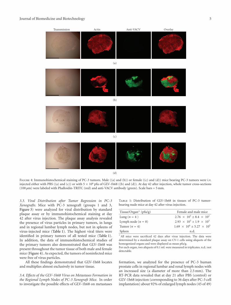
Figure 4: Immunohistochemical staining of PC-3 tumors. Male ((a) and (b)) or female ((c) and (d)) mice bearing PC-3 tumors were i.v.injected either with PBS ((a) and (c)) or with 5 × 10⁶ pfu of GLV-1h68 ((b) and (d)). At day 42 after injection, whole tumor cross-sections(100 μm) were labeled with Phalloidin-TRITC (red) and anti-VACV antibody (green). Scale bars = 5 mm.
3.3. Viral Distribution after Tumor Regression in PC-3Xenografts. Mice with PC-3 xenograft (groups 1 and 3,Figure 3) were analyzed for viral distribution by standardplaque assay or by immunohistochemical staining at day42 after virus injection. The plaque assay analysis revealedthe presence of virus particles in primary tumors, in lungsand in regional lumbar lymph nodes, but not in spleens ofvirus-injected mice (Table 1). The highest viral titers wereidentified in primary tumors of all tested mice (Table 1).In addition, the data of immunohistochemical studies ofthe primary tumors also demonstrated that GLV-1h68 waspresent throughout the tumor tissue of both male and femalemice (Figure 4). As expected, the tumors of noninfected micewere free of virus particles.
All these findings demonstrated that GLV-1h68 locatesand multiplies almost exclusively in tumor tissue.
3.4. Effects of the GLV-1h68 Virus on Metastases Formation inthe Regional Lymph Nodes of PC-3 Xenograft Mice. In orderto investigate the possible effects of GLV-1h68 on metastases
Table 1: Distribution of GLV-1h68 in tissues of PC-3 tumor-bearing nude mice at day 42 after virus injection.
Tissue/Organ∗ (pfu/g) Female and male mice
Lung (n = 4 ) 2.76 × 10³ ± 8.4 × 10²
Lymph node (n = 8) 2.93 × 10³ ± 1.9 × 10³
Tumor (n = 4) 1.69 × 10⁶ ± 5.27 × 10⁵
Spleen n.d.∗
All mice were sacrificed 42 days after virus injection. The data weredetermined by a standard plaque assay on CV-1 cells using aliquots of thehomogenized organs and were displayed as mean pfu/g.For each organ, two aliquots of 0.1 mL were measured in triplicates. n.d.: notdetectable.
formation, we analyzed for the presence of PC-3 humanprostate cells in regional lumbar and renal lymph nodes withan increased size (a diameter of more than 2.5 mm). TheRT-PCR data revealed that at day 21 after PBS (control) orGLV-1h68 injection (corresponding to 36 days after PC-3 cellimplantation) about 92% of enlarged lymph nodes (45 of 49)

6 Journal of Biomedicine and Biotechnology
1h68PBS0
20
40
60
80
100
92% (45/49)
21% (6/28)
P < .001
Lym
phn
odes
pos
itiv
efo
r
hum
anβ
-act
in(%
)
(a)
Tumor
1 2 3
ln 1
1 2 3
ln 2
1 2 3bp
300
150
300
150
PBS
1h68
(b)
Figure 5: Effects of the GLV-1h68 virus on metastases formation inthe regional lymph nodes. (a) Percentage of PC-3 positive regionallymph nodes in tumor-bearing mice at day 21 after PBS or virusinjection. Fisher’s exact test (GraphPad Prism software, San Diego,USA) was used for statistical analysis. The value of P < .001 wasconsidered significant. GLV-1h68 : 1h68. (b) RT-PCR analysis oftumors and regional lymph nodes of PC-3 xenograft mice injectedeither with PBS or with 5 × 10⁶ pfu of GLV-1h68 (1h68) usingprimers for human beta-actin (lines 1; indicating PC-3 cancer cells,PCR-fragment 205 bp), mouse beta-actin (lines 2; positive control,PCR-fragment 216 bp), and vaccinia virus (lines 3; PCR-fragment204 bp). ln: lymph node of two different mice.
in control mice (n = 11) and 21% of enlarged lymph nodes(6 of 28) in GLV-1h68-treated mice (n = 11) were positivefor human beta-actin (Figure 5(a)). The presence of humanbeta-actin was used as a marker for the presence of intacthuman PC-3 cells; therefore, the systemic treatment by GLV-1h68 appears to affect the PC-3 metastases formation in theregional lymph nodes. All lymph nodes that tested positivelyfor the presence of human beta-actin were also positive forvaccinia virus in GLV-1h68-injected mice but not in controlmice (Figure 5(b)), indicating that i.v. injected GLV-1h68 cantarget lymph node metastases. Interestingly, the GLV-1h68virus was detected in about 68% (19 of 28) of the lymphnodes in the absence of PC-3 tumor cells.
3.5. Colonization of Established Metastases in the RegionalLymph Nodes of PC-3 Xenograft Mice. In order to clarifywhether the increased inhibition of PC-3 metastases for-mation in the regional lymph nodes was a result of thepreventative or oncolytic effect of the GLV-1h68 virus, weinjected tumor-bearing nude mice (n = 4) with a single dose
of GLV-1h68 (5 × 10⁶ pfu) at day 57 after implantation ofPC-3 cells. This time point allows the analysis of establishedmetastases in the regional lymph nodes of PC-3 xenograftmice. Eleven days later, virus distribution in lymph nodemetastases was analyzed either by fluorescence imaging(Figure 6) or by immunohistochemical staining (Figure 7).The data demonstrated that GLV-1h68 can efficiently targetlocal lymph node metastases (Figure 6) and destruct tumortissue within these lymph nodes (Figure 7).
3.6. Mouse Immune-Related Protein Antigen Profiling of GLV-1h68-Infected and Noninfected PC-3 Derived Tumors. Inorder to study the immunological effect of virus infectionin vivo, we analyzed the antigen profiling of GLV-1h68-infected and noninfected PC-3 tumors 42 days after virusinjection. For this purpose, lysates of these tumors were usedfor examination of the expression levels of immune-relatedprotein antigens of mouse origin, as described in Materialsand Methods.
The data revealed that GLV-1h68 injection led toincreased production of most of the tested proinflammatorycytokines and chemokines (Table 2). Many of the testedcytokines and chemokines, such as IP-10, IL-18, IL-6, MCP-1, MCP-3, MCP-5, M-CSF, and TNF-alpha, are known toactivate macrophages, monocytes, neutrophils, eosinophils,and, so forth, and to trigger proinflammatory responses intarget tissues. In contrast, only one cytokine (MIP-1-gamma)was significantly downregulated upon virus infection in bothmale and female PC-3 tumor-bearing mice.
3.7. Active Peri- and Intra-Tumoral Infiltration of MHC ClassII-Expressing Host Cells in GLV-1h68-Infected Tumors. Theimmune-related protein antigen profiling of regressing PC-3-tumors revealed protein expression signatures consistentwith innate immune defense activation (Table 2). There-fore, we analyzed the presence of innate immune systemcomponents into the PC-3-derivated tumors by immuno-histochemistry. In this context, we labelled MHC class II-expressing host cells (like, e.g., macrophages and dendriticcells) in the tumor tissue. Indeed, the data demonstratedspecific peri- and intra-tumoral infiltration of MHC class II-expressing host cells surrounding virus-infected cancer cells(Figure 8). These host cells were absent in the noninfectedcontrol samples.
4. Discussion
Prostate cancer is the cause of more than 1% of all deathsin men and its incidence is increasing by 2-3% per year[19]. The available treatment options for advanced prostatecancer are very limited and the prognosis for patients withadvanced-stage disease is poor. Therefore, there is an urgentneed to identify novel agents for therapy and diagnosis ofadvanced prostate cancer.
In this study, we investigated the therapeutic efficacyof the oncolytic vaccinia virus strain GLV-1h68 againstadvanced human prostate carcinoma. We examined the anti-tumor efficacy of GLV-1h68 in DU-145 and PC-3 prostate

Journal of Biomedicine and Biotechnology 7
a1
a2
(a)
b1
b2
(b)
Figure 6: Detection of lymph node metastases by GLV-1h68-mediated fluorescence. (a) Bright-field, and (b) overlay fluorescence imagesof renal and lumbar lymph nodes in GLV-1h68-infected mice with PC-3 tumors. a1, b1: mouse no. 1 and a2, b2: mouse no. 2. Scale bars =2 mm.
tumor xenograft models. The androgen-independent DU-145 and PC-3 models were preferred over several androgen-dependent xenograft models, like LAPC-4 or -9 [20, 21],since the PC-3 cells are more aggressive [22] and more drug-resistant than that used in androgen-dependent models [23].It is known that the mortality of prostate cancer results fromthe progression of androgen-dependent disease to androgen-independent prostatic growth and metastases to the bonesand lymph nodes [24, 25]. Therefore, we also analyzed thepresence of PC3-metastases in the regional lymph nodes.The tumor status of these lymph nodes provides importantinformation in both the diagnosis and treatment of prostatecancer. Our PC-3 xenografts data demonstrated that GLV-1h68 can efficiently target both the primary tumor and theregional lymph nodes with metastases. These interactionsled to significant size reduction of the primary tumor andinhibition of metastases development in our PC-3 xenograftmodels.
In order to test the effect of GLV-1h68 on PC-3metastases formation, we analyzed the presence of PC-3 cellsin the regional lymph nodes at day 21 after PBS or GLV-1h68 injection. We chose this time point because, at 21days after the virus treatment, the growth of the primarytumors was efficiently inhibited (Figure 3). We found asignificant reduction in the number of PC-3 positive lymphnodes in virus-treated tumor-bearing mice compared tonon-virus-treated controls (Figure 5(a)). Moreover, lymphnodes bearing PC-3 cells in GLV-1h68-injected animalsshowed also the presence of vaccinia virus (Figure 5(b)). Inaddition, histological analysis revealed virus patches over thesurface of metastases, which was also associated with massive
destruction of tumor tissue when GLV-1h68 was injectedinto mice with well-established PC-3 lymph node metastases(Figure 7). Surprisingly, the GLV-1h68 virus was detected inabout 68% lymph nodes in the absence of PC-3 actin data.In these cases, it could be that, the PC-3 cells were alreadydestroyed, but GLV-1h68 persisted longer in the tumor tissueor the vaccinia virus was present in lymph nodes that werenot infiltrated by malignant cells. Therefore, the significantlyreduced number of lymph node metastases in GLV-1h68-injected PC-3 mice could be due to both the oncolytic effectsof vaccinia virus colonizing lymph node metastases and thevirusprevention of metastases formation.
For a better understanding of a possible immunologicaleffect of the GLV-1h68 virus on PC-3 xenografts, we investi-gated the mouse immune-related protein antigen profilingin the primary tumors with and without virus infection.The data revealed that the protein expression levels of mostof the tested proinflammatory cytokines and chemokineswere significantly upregulated after viral tumor colonization(Table 2). The resulting cytokine profile after GLV-1h68infection was similar to the cytokine profile in immunocom-petent Balb/c mice [14, 26] and in an immunocompromisednude mouse model [11]. Therefore, the cytokine profile aftervaccinia virus infection seems to be relatively independentof several factors, including: tumor origin [[11] and thisstudy], the number of replication-competent virus particlesreaching the tumor after injection, the route of injection[[10], Gentschev unpublished data)], and the sex of themice (this study). These observations support the hypothesisthat the oncolytic effect of vaccinia virus is at least in partmediated through host defense mechanisms, most likely

8 Journal of Biomedicine and Biotechnology
∗
(a)
∗
(b)
(c) (d)
Figure 7: Immunohistochemical staining of a lymph node with PC-3 metastases. At day 57 after PC-3 cells implantation, tumor-bearingmice were injected with 5 × 10⁶ pfu of GLV-1h68. At day 11 after virus injection, whole lumbar lymph node cross-sections were analyzedby bright-field imaging (a) or by fluorescence microscopy techniques. (b) Phalloidin-TRITC (red) and (c) anti-VACV antibody (green), and(d) (overlay). Scale bars = 1 mm. ∗indicates the dead tumor tissue destructed by GLV-1h68.
(a)
MHCII GLV-1h68 Transmission MHCII GLV-1h68 Transmission
(b)
Overlay Overlay
Figure 8: Immunohistochemistry staining of MHC class II positive cells in PC-3 tumors. (a) Forty-two days after GLV-1h68 administrationPC-3 tumors were excised, sectioned, and labeled for MHCII. In addition, GFP signals from GLV-1h68-infected cells and transmissionimages are shown. (b) Overlay of MHCII and GFP signals in uninfected (left) and virus-infected PC-3 tumors (right). Scale bars = 5 mm.
by the innate immune system [8, 27]. Indeed, many ofthe upregulated proteins in tumor tissue (Table 2), such asMCP-1, MCP-3, MCP-5, M-CSF, and IL-18, are involvedin activation of some components of the innate immune
system, like macrophages and neutrophils. IP-10, the mostupregulated protein in our study, has been shown to displayantitumor and antimetastatic properties [28]. This cytokineis also known to enhance the cytolytic activity of NK cells, a

Journal of Biomedicine and Biotechnology 9
Table 2: Mouse immune-related protein antigen profiling in PC-3-derived tumors of male or female nude mice with or without GLV-1h68treatment at the day 42 after virus infection (n = 2). Folds of enhancement (a) or suppression (b) of mouse protein expression after virusinjection are shown. Each value was determined as the mean of triplicate determination.
(a) Upregulated protein expression levels (day 42 after virus infection).
Antigen names GLV-1h68/untreated ratio (female) GLV-1h68/untreated ratio (male) Classification
Apo A1 2.83 2.78 Anti-inflammatory protein
GM-CSF 2.2 2.77 Granulocyte-macrophagecolony-stimulating factor
IL-6 8.44 6.8 Proinflammatory cytokine
IL-11 3.81 3.63 Pleiotropic cytokine
IL-18 30 29.72 Proinflammatory cytokine
IP-10 (CXCL10) 33.91 8.18 Interferon-gamma-inducedprotein
MCP-1 (CCL2) 11.8 7.05 Proinflammatory cytokine
MCP-3 (CCL7) 6.09 3.23 Proinflammatory cytokine
MCP-5 (CCL12) 30.24 10.64 Proinflammatory cytokine
M-CSF (KC/GROα) 2.18 2.48 Proinflammatory cytokine
MDC (CCL22) 4.5 3.23 Proinflammatorychemokine
MIP-1beta 9.09 3.31 Proinflammatory cytokine
MIP-2 (CXCL2) 2.9 3.33 Proinflammatorychemokine
RANTES (CCL5) 7.1 4.58 Proinflammatorychemokine
TIMP-1 2.6 2.43 Tissue inhibitor ofmetalloproteinase type-1
TNF-alpha 2.56 2.27 Proinflammatory cytokine
(b) Downregulated protein expression level (day 42 after virus infection).
Antigen names Untreated GLV-1h68/ratio (female) untreated GLV-1h68/ratio (male) Classification
MIP-1gamma (CCL9) 8 7.5 Cytokine
component of the innate immune system, and to contributeto vaccinia virus clearance in vivo [29].
The only cytokine in our study that was significantlydownregulated was the protein MIP-1 gamma (CCL9). MIP-1 gamma, a small cytokine belonging to the CC chemokinefamily, is secreted by the follicle-associated epitheliumand recruits CD11b+ dendritic cells [30]. Interestingly, insome adenocarcinomas, the expression of MIP-1 gammais increased in the tumor epithelium [31]. However, theeffect of the down-regulation of MIP-1 gamma in our PC-3 prostate tumor xenograft models is currently unknown.
Taken together, the immune-related protein antigen pro-filing of regressing PC-3 tumors revealed protein expressionsignatures consistent with innate immune defense activationinclusive of cytokines and chemokines (Table 2). Therefore,we analyzed the presence of some components of the innateimmune system such as macrophages, dendritic cells, andleukocytes into the PC-3-derivated tumors by immuno-histochemistry. The data demonstrated specific peri- andintratumoral infiltration of MHC class II-expressing host
cells surrounding virus-infected cancer cells (Figure 8). Thepresence of activated macrophages or dendritic cells in virus-infected PC-3 xenografts only could serve as an evidence forthe association between xenograft eradication and immuneactivation of the innate immune system. These findingssuggest that immune activation may combine with viraloncolysis to induce tumor eradication, providing a novelperspective for the design of oncolytic viral therapies forhuman cancers. However, in our experimental settings, it wasnot possible to distinguish between components of the innateimmune system directly involved in the killing of tumor cellsor in the elimination of vaccinia virus particles.
As described previously, we have used male and femalenude mice for analysis of the effect of the GLV-1h68 viruson PC-3 xenografts. The data demonstrated that in malemice, GLV-1h68 injection led to a faster and more efficienttumor regression than in female mice bearing PC-3 tumors.However, significant tumor regression was found in bothmale and female PC-3 xenografts at day 28 after virus injec-tion. In addition, we did not find any significant differences

10 Journal of Biomedicine and Biotechnology
in the resulting cytokine profiles between male and femalePC-3 xenografts after virus injection (Table 2). This meansthat female nude mice could also be successfully used inexperiments with PC-3 xenografts. This is an advantage,since the experimental handling of female animals is easierthan the handling of male animals.
In summary, the GLV-1h68 strain showed an outstandingantitumor effect and a safety profile in DU-145 and PC-3-xenografts. This makes it a promising candidate for thetreatment of advanced metastatic prostate cancer in human.
5. Conclusion
Our data demonstrate that the oncolytic virus GLV-1h68 canefficiently infect and destroy the human prostate DU-145 andPC-3 cells in cell culture and in vivo. In addition, a singlesystemic administration of GLV-1h68 causes a significantinhibition of the growth of primary tumor and metastases inPC-3 xenografts. Our findings suggest that the vaccinia virusGLV-1h68 has the potential to treat prostate cancer in humanpatients.
Acknowledgments
The authors declare that they have competing interests.I. Gentschev, Y. A. Yu, N. Chen, J. Stritzker, and A. A.Szalay have financial interests in Genelux Corporation. E.Hofmann, U. Donat, M. Adelfinger, V. Raab, and S. Weibelwere supported by grants of Genelux Corporation. The costsof publication of this paper were defrayed in part by thepayment of page charges. This paper must therefore behereby marked advertisement in accordance with 18 U.S.C.Section 1734 solely to indicate this fact. They thank Ms.J. Langbein, Mr. Jason Aguilar, and Mr. Terry Trevino forexcellent technical support, Ms. A. Feathers and Ms. Z.Sokolovic for critical reading of the paper. Ivaylo Gentschevand Ulrike Donat contributed equally to this work.
References
[1] A. Jemal, R. Siegel, E. Ward, T. Murray, J. Xu, and M. J. Thun,“Cancer statistics, 2007,” CA: A Cancer Journal for Clinicians,vol. 57, no. 1, pp. 43–66, 2007.
[2] L. Cheng, H. Zincke, M. L. Blute, E. J. Bergstralh, B. Scherer,and D. G. Bostwick, “Risk of prostate carcinoma death inpatients with lymph node metastasis,” Cancer, vol. 91, no. 1,pp. 66–73, 2001.
[3] P. R. Carroll, K. L. Lee, Z. Y. Fuks, and P. W. Kantoff, “Cancer ofthe prostate,” in Cancer: “Principals and Practices of Oncology”,V. T. DeVita, S. Hellman, and S. A. Rosenberg, Eds., pp. 1418–1479, Lippincott-Raven, New York, NY, USA, 2001.
[4] R. Cattaneo, T. Miest, E. V. Shashkova, and M. A. Barry,“Reprogrammed viruses as cancer therapeutics: targeted,armed and shielded,” Nature Reviews Microbiology, vol. 6, no.7, pp. 529–540, 2008.
[5] M. J. V. Vaha-Koskela, J. E. Heikkila, and A. E. Hinkkanen,“Oncolytic viruses in cancer therapy,” Cancer Letters, vol. 254,no. 2, pp. 178–216, 2007.
[6] A. M. Crompton and D. H. Kirn, “From ONYX-015 to armedvaccinia viruses: the education and evolution of oncolytic
virus development,” Current Cancer Drug Targets, vol. 7, no.2, pp. 133–139, 2007.
[7] T.-C. Liu, E. Galanis, and D. Kirn, “Clinical trial results withoncolytic virotherapy: a century of promise, a decade ofprogress,” Nature Clinical Practice Oncology, vol. 4, no. 2, pp.101–117, 2007.
[8] Q. Zhang, Y. A. Yu, E. Wang, et al., “Eradication of solidhuman breast tumors in nude mice with an intravenouslyinjected light-emitting oncolytic vaccinia virus,” CancerResearch, vol. 67, no. 20, pp. 10038–10046, 2007.
[9] K. J. Kelly, Y. Woo, P. Brader, et al., “Novel oncolytic agentGLV-1h68 is effective against malignant pleural mesothe-lioma,” Human Gene Therapy, vol. 19, no. 8, pp. 774–782,2008.
[10] I. Gentschev, J. Stritzker, E. Hofmann, et al., “Use of anoncolytic vaccinia virus for the treatment of canine breastcancer in nude mice: preclinical development of a therapeuticagent,” Cancer Gene Therapy, vol. 16, no. 4, pp. 320–328, 2009.
[11] Y. A. Yu, C. Galanis, Y. Woo, et al., “Regression of humanpancreatic tumor xenografts in mice after a single systemicinjection of recombinant vaccinia virus GLV-1h68,” MolecularCancer Therapeutics, vol. 8, no. 1, pp. 141–151, 2009.
[12] S.-F. Lin, D. L. Price, C.-H. Chen, et al., “Oncolytic vacciniavirotherapy of anaplastic thyroid cancer in vivo,” Journal ofClinical Endocrinology and Metabolism, vol. 93, no. 11, pp.4403–4407, 2008.
[13] S.-F. Lin, Z. Yu, C. Riedl, et al., “Treatment of anaplasticthyroid carcinoma in vitro with a mutant vaccinia virus,”Surgery, vol. 142, no. 6, pp. 976–983, 2007.
[14] K. J. Kelly, P. Brader, Y. Woo, et al., “Real-time intraoperativedetection of melanoma lymph node metastases using recom-binant vaccinia virus GLV-lh68 in an immunocompetentanimal model,” International Journal of Cancer, vol. 124, no.4, pp. 911–918, 2009.
[15] N. Chen, Q. Zhang, Y. A. Yu, et al., “A novel recombinant vac-cinia virus expressing the human norepinephrine transporterretains oncolytic potential and facilitates deep-tissue imaging,”Molecular Medicine, vol. 15, no. 5-6, pp. 144–151, 2009.
[16] P. Brader, K. J. Kelly, N. Chen, et al., “Imaging a geneticallyengineered oncolytic vaccinia virus (GLV-1h99) using ahuman norepinephrine transporter reporter gene,” ClinicalCancer Research, vol. 15, no. 11, pp. 3791–3801, 2009.
[17] T.-C. Liu and D. Kirn, “Systemic efficacy with oncolytic virustherapeutics: clinical proof-of-concept and future directions,”Cancer Research, vol. 67, no. 2, pp. 429–432, 2007.
[18] S. Weibel, J. Stritzker, M. Eck, W. Goebel, and A. A. Szalay,“Colonization of experimental murine breast tumours byEscherichia coli K-12 significantly alters the tumour microen-vironment,” Cellular Microbiology, vol. 10, no. 6, pp. 1235–1248, 2008.
[19] N. Salesi, P. Carlini, E. M. Ruggeri, G. Ferretti, E. Bria, andF. Cognetti, “Prostate cancer: the role of hormonal therapy,”Journal of Experimental and Clinical Cancer Research, vol. 24,no. 2, pp. 175–180, 2005.
[20] N. Craft, Y. Shostak, M. Carey, and C. L. Sawyers, “A mech-anism for hormone-independent prostate cancer throughmodulation of androgen receptor signaling by the HER-2/neutyrosine kinase,” Nature Medicine, vol. 5, no. 3, pp. 280–285,1999.
[21] N. Craft, C. Chhor, C. Tran, et al., “Evidence for clonal out-growth of androgen-independent prostate cancer cells fromandrogen-dependent tumors through a two-step process,”Cancer Research, vol. 59, no. 19, pp. 5030–5036, 1999.

Journal of Biomedicine and Biotechnology 11
[22] E. Brakenhielm, J. B. Burton, M. Johnson, et al., “Modulatingmetastasis by a lymphangiogenic switch in prostate cancer,”International Journal of Cancer, vol. 121, no. 10, pp. 2153–2161, 2007.
[23] S. Mohapatra, B. Chu, X. Zhao, and W. J. Pledger, “Accumu-lation of p53 and reductions in XIAP abundance promote theapoptosis of prostate cancer cells,” Cancer Research, vol. 65, no.17, pp. 7717–7723, 2005.
[24] B. J. Feldman and D. Feldman, “The development ofandrogen-independent prostate cancer,” Nature Reviews Can-cer, vol. 1, no. 1, pp. 34–45, 2001.
[25] E. M. Bruckheimer and N. Kyprianou, “Apoptosis in prostatecarcinogenesis: a growth regulator and a therapeutic target,”Cell and Tissue Research, vol. 301, no. 1, pp. 153–162, 2000.
[26] C. W. Knorr, S. D. Allen, A. R. Torres, and D. F. Smee, “Effectsof cidofovir treatment on cytokine induction in murinemodels of cowpox and vaccinia virus infection,” AntiviralResearch, vol. 72, no. 2, pp. 125–133, 2006.
[27] A. Worschech, D. Haddad, D. F. Stroncek, E. Wang,F. M. Marincola, and A. A. Szalay, “The immunologicaspects of poxvirus oncolytic therapy,” Cancer Immunology,Immunotherapy, vol. 58, no. 9, pp. 1355–1362, 2009.
[28] A. D. Luster and P. Leder, “IP-10, a -C-X-C- chemokine, elicitsa potent thymus-dependent antitumor response in vivo,”Journal of Experimental Medicine, vol. 178, no. 3, pp. 1057–1065, 1993.
[29] S. Mahalingam, J. M. Farber, and G. Karupiah, “Theinterferon-inducible chemokines MuMig and Crg-2 exhibitantiviral activity in vivo,” Journal of Virology, vol. 73, no. 2,pp. 1479–1491, 1999.
[30] X. Zhao, A. Sato, C. S. Dela Cruz, et al., “CCL9 is secretedby the follicle-associated epithelium and recruits dome regionPeyer’s patch CD11b+ dendritic cells,” Journal of Immunology,vol. 171, no. 6, pp. 2797–2803, 2003.
[31] T. Kitamura, K. Kometani, H. Hashida, et al., “SMAD4-deficient intestinal tumors recruit CCR1+ myeloid cells thatpromote invasion,” Nature Genetics, vol. 39, no. 4, pp. 467–475, 2007.
Copyright © 2022 FDOKUMEN