Expression of Interleukin 8 Correlates with the Metastatic Potential of Human Melanoma Cells in Nude...
7
1994;54:3242-3247. Cancer Res Rakesh K. Singh, Mordechai Gutman, Robert Radinsky, et al. Potential of Human Melanoma Cells in Nude Mice Expression of Interleukin 8 Correlates with the Metastatic Updated version http://cancerres.aacrjournals.org/content/54/12/3242 Access the most recent version of this article at: E-mail alerts related to this article or journal. Sign up to receive free email-alerts Subscriptions Reprints and . [email protected] Department at To order reprints of this article or to subscribe to the journal, contact the AACR Publications Permissions . [email protected] Department at To request permission to re-use all or part of this article, contact the AACR Publications Research. on October 27, 2013. © 1994 American Association for Cancer cancerres.aacrjournals.org Downloaded from Research. on October 27, 2013. © 1994 American Association for Cancer cancerres.aacrjournals.org Downloaded from Research. on October 27, 2013. © 1994 American Association for Cancer cancerres.aacrjournals.org Downloaded from Research. on October 27, 2013. © 1994 American Association for Cancer cancerres.aacrjournals.org Downloaded from Research. on October 27, 2013. © 1994 American Association for Cancer cancerres.aacrjournals.org Downloaded from Research. on October 27, 2013. © 1994 American Association for Cancer cancerres.aacrjournals.org Downloaded from Research. on October 27, 2013. © 1994 American Association for Cancer cancerres.aacrjournals.org Downloaded from
Transcript of Expression of Interleukin 8 Correlates with the Metastatic Potential of Human Melanoma Cells in Nude...
1994;54:3242-3247. Cancer Res Rakesh K. Singh, Mordechai Gutman, Robert Radinsky, et al. Potential of Human Melanoma Cells in Nude MiceExpression of Interleukin 8 Correlates with the Metastatic
Updated version
http://cancerres.aacrjournals.org/content/54/12/3242
Access the most recent version of this article at:
E-mail alerts related to this article or journal.Sign up to receive free email-alerts
Subscriptions
Reprints and
To order reprints of this article or to subscribe to the journal, contact the AACR Publications
Permissions
To request permission to re-use all or part of this article, contact the AACR Publications
Research. on October 27, 2013. © 1994 American Association for Cancercancerres.aacrjournals.org Downloaded from
Research. on October 27, 2013. © 1994 American Association for Cancercancerres.aacrjournals.org Downloaded from
Research. on October 27, 2013. © 1994 American Association for Cancercancerres.aacrjournals.org Downloaded from
Research. on October 27, 2013. © 1994 American Association for Cancercancerres.aacrjournals.org Downloaded from
Research. on October 27, 2013. © 1994 American Association for Cancercancerres.aacrjournals.org Downloaded from
Research. on October 27, 2013. © 1994 American Association for Cancercancerres.aacrjournals.org Downloaded from
Research. on October 27, 2013. © 1994 American Association for Cancercancerres.aacrjournals.org Downloaded from

ICAN@ERRE5EARa:I54,3242—3247,June15,1994J
ABSTRACT
We correlated the steady state transcription and protein secretion ofInterleukin S (IL-8) hi 13 dlffere.t human melanoma cell lines with theirability to grow and produce metastasis In nude mlce Highly metastatlccells expressed higher steady state kvels of IL-S mRNA transcripts thandid low metastatic cells. In situ mRNA hybridization analyses confirmed
the pattern of mRNA expression on a cellular leveL Increased mRNAexpression directly correlated with secretlo. of IL-S protein as determined by enzyme-linked immunosorbent assay Recombinant IL-Sstimulated the proliferation of low metastatic A375P cells In a dosedependent manner, a stimulation that was abrogated by the use of apolydonal aatlbody against IL-S. The data suggest that IL-S can be an
autocrine growth factor for human melanoma cells and that IL-S is
involved in melanoma metastasIs@
INTRODUCTION
To produce metastasis, melanoma cells must detach from the primary tumor, invade through host stroma to gain entrance into thecirculation, where they disseminate via the blood stream (1); andsurvive to reach distant capillary beds, where they must attach, cxtravasate into the organ parenchyma, and proliferate into secondarygrowths (1, 2). The growth of cells in distant sites occurs when thetumor cells produce autocrine growth factors (3) or when the tumorcells respond to paracrine growth factors produced by host cells.
Melanoma cells secrete a variety of growth factors includingTGF-a3 (4—6),TGF-f3 (7), platelet-derived growth factor A and Bchains (8), basic flbroblast growth factor (9), IL-6 (3, 10), IL-i (11,12), granulocyte macrophage colony-stimulating factor (13), and amolecule named MGSA (6). These growth factors, expressed eitherconstitutivelyor subsequentto inductionwithvariouscytokines,maycontribute to the development of the transformed melanoma phenotype either by acting as autocrine growth factors or by modulatinghost responses to the tumor cells (13—18).
The treatment of melanocytes and melanoma cells with IL-i andtumor necrosis factor results in expression of IL-S (19, 20). Similar toother cytokines, IL-8 is multifunctional. In some melanoma cells, ithas been shown to stimulate growth (19). In the presence of glucocorticoids and epidermal growth factor, it can induce proliferation ofkeratinocytes (21). IL-8 has been shown to induce angiogenesis (22,23) and to induce haptotactic migration in melanoma cells (24). Sinceangiogenesis, migration, and cell proliferation are all important cornponents of the metastatic process (2), the data suggest that IL-8expression by tumor cells could influence their metastatic capabilities.
Received 213)94;accepted 4/14/94.The costs of publication of this article were defrayed in part by the payment of page
cliarges@This aiticle must therefore be hereby marked advertisement in accordance with18 U.S.C. Section 1734 solely to indicate this fact.
1@ work was supported in part by Cancer Center Support Core Grant CA 166fl and
NatiOnalCancer Institute Grant R35-CA 42107 (L J. F.).2 To whom requests for reprints should be addressed, at the Department of Cell
Biology, Box 173, University of Texas M. D. Anderson Cancer Center, 1515 HolcombeBoulevard, Houston, TX 77030.
3 The abbreviations used are: TOF, transforming growth factor IL-8, interleukin 8;
MGSA, melanoma growth-admulating activity; cDNA, complementary DNA ELISA,enzyme-linked iinmunosorbent assay; MU, 3-(4,5-dimcthylthiazol-2-yl)-2,5-diphenyltetrazolium bromide; ISH, in situ mRNA hybridization.
The purpose of this study was to examine whether the expressionlevel of IL-8 by human melanoma cells correlated with their metastatic potential in nude mice. We describe mRNA analyses using bothNorthern blot and in situ hybridization techniques, as well as mensurements of IL-8 protein. We orthotopically implanted 13 differenthuman melanoma cell lines into nude mice and demonstrated acorrelation between IL-S mRNA and protein production and themetastatic potential of human melanoma cells.
MATERIAI@S AND MI@Th'HODS
Tumor Cell LInes. The human melanoma cell lines were originally isolated from different human patients as described previously (25-28). Briefly,TXM-1 was isolated from a lymph node metastasis, whereas TXM-13 and
TXM-18 were isolated from the brain metastases of different patients. TheA375 cell line was originally established in culture from a lymph nodemetastasis@The parental A375 cells were injected i.v. into nude mice andvariant lines designated A375M, A375 Met #1, A375 Met #2, and A375 Met#3 were establishedfrom isolatedlung metastases. A375 LN#2 was established from pooled lymph node metastases of one nude mouse. TheA37SSM line was established from a pool of lung metastases produced by
the A375 parentalcells growing s.c. in nude mice (25). The parentalA375line was also cloned in vitro by a double dilution method, A375 clone 5 andA375 clone 28 with low and high metastatic potential, respectively (26),
were used in this study. The SBC-2 line, established in culture from aprimary cutaneous melanoma and given to us by Dr. Beppino Giovandila
(Stehlin Institute, Houston, TX), is a poorly tumorigenic and nonmetastaticline in nude mice (29).
All tumor cell lines were maintained in culture as adherent monolayers inEagle's minimal essential medium supplemented with 10% fetal bovine serum,sodium pynivate, nonessential amino acids, L-glutamine,2-fold vitamin solulion, and penicillin-streptomycin (Flow Laboratories, Rockville, MD), andincubated in 5% C°295% a@rat 37°C.All cultures were free of Mycoplasmaand pathogenicmurineviruses (assayed by MicrobiologicalAssociates, Bethesda, MD). cultures were maintained for no longer than 6 weeks afterrecovery from frozen stocks.
Animals. Male athymic BALB/c nude mice were purchased from theAnimal Production Area of the National Cancer InstitUte, Frederick CancerResearch Facility (Frederick, MD). The mice were housed in laminar flowcabinets under specific pathogen-free conditions and used when 8 weeks ofage. Animals were maintained in facilities approved by the American Maociation for Accreditation of Laboratory Animal Care and in accordance withcurrent regulations and standards of the United States Department of
Agriculture, Department of Health and Human Services, and NIH.
Experimental Metastasis. Experimental metastases are tumor coloniesproduced in the lungs of nude mice after i.v. injection of tumor ceUs. AlthOUgh
these tumor cells bypass the initial steps ofmetastasis (separation from primary
neoplasm and invasion and release into blood vessels or lymphatics), all the
subsequent steps in the metastatic process must occur for metastases to beformed.In this study,we equateexperimentalmetastasiswith the blood-bornespread of tumor cells.
To preparetumorcells for inoculation,cells in exponentialgrowth phasewere harvested by a brief exposure to 0.25% trypsin-0.02% EDTA solution(w/v). The flask was tapped sharply to dislodge the cells, supplementedmedium was added, and the cell suspension was pipeted again to produce asingle cell suspension. The cells were washed and resuspended in Ca2'@-andMg@-free Hanks' balanced salt solution to the desired cell concentration. Cellviability was determined by trypan blue exclusion, and only single cell sitspensions of >90% viability were used. We injected 0.2 ml of tumor cellsuspensions into the lateral tail veins of unanesthetized nude mice. Six to 8
3242
Expression of Interleukin 8 Correlates with the Metastatic Potential of HumanMelanoma Cells in Nude Mice1
Rakesh K. Singh, Mordechai Gutman, Robed Radinsky, Corazon D. Bucana, and Isaiah J. FidIer@
Department ofCeil Biology, University ofTer.as M. D. Anderson Cancer Center, Hoierton, Texas 77030

Table 1 Levels ofIL-il mRNA and protein and production of experimeby human melanoma cellsnial
metastasisCell
lineExperimental
lung metastasis―lL-8mRNA
expressionindex―IL-8
(ng/ml/106cells)cMedianIncidence(range)SBC-20/10
00.050.17 ±0.01A375P9/98 (1—33)0.41.40 ±0.05A375M9/9
135 (6—150)0.93.45 ±0.20A375SM10/10200 (150—250)2.25.30 ±0.24A375
Met #110/10 130(90—155)0.93.19 ±0.06A375Met #210/10 160 (50—200)1.74.0 ±0.3A375Met #39/9 190(100—250)1.85.1 ±0.4A375LN #210/10 70(15—150)0.72.0 ±0.1A375C-S3/10 0 (0.2)0. 11 . 1 ±0.05A375C-2810/10 160 (70—250)0.95.07 ±0.06TXM-l10/10
100(70—200)5.25.11 ±0.40TXM-137/839 (0—200)0.72.40 ±0.10TXM-183/55(0—20)0.81.60 ±0.10
IL-s EXPRESSION AND HUMAN MELANOMA CELL METASTASIS
weeks after injection, the mice were killed and the lungs were removed,washed, and fixed in Bouin's solution to differentiate the neoplastic lesionsfrom the organ parenchyma. The lung nodules were counted with the aid of adissecting microscope.
mRNA Analysis. Polyadenylated mRNA was extracted from i0@ tumorcells growing subconfluently in culture using the FastTrack mRNA isolationkit (Invitrogen, San Diego, CA). mRNA was electrophoresed on a 1% denaturing formaldehyde/agarose gel, electrotransferred at 0.6 A to GeneScreennylon membrane (DuPont New England Nuclear, Boston, MA), and UVcross-linked with 120,000 @iJ/cm2using a UV Stratalinker 1800 (Stratagene,La Jolla, CA). Hybridizations were performed as described previously (30).Nylon filters were washed 3 times at 55—60°Cwith 30 mMNaCl-3 mMsodiumcitrate, pH 7.2-0.1% sodium dodecyl sulfate (w/v).
The cDNA probes used in these analyses were a 1.3-kilobase PstI cDNAfragment corresponding to rat glyceraldehyde 3-phosphate dehydrogenase (31)and a 0.5-kilobase EcoRI cDNA fragment corresponding to human IL-8(kindly provided by Dr. K. Matsushima, Kanazawa, Japan) (32). Each cDNAfragment was purified by agarose gel electrophoresis, recovered using GeneClean (BIO 101, Inc., La Jolla, CA), and radiolabeled using the random primertechnique with a-32P-labeled deoxyribonucleotide triphosphates (33).
Densitometric Quantitation of mRNA Transcripts. IL-8 mRNA expression was quantitated in the linear range of the film on a personal densitometerby using the ImageQuant software program (Molecular Dynamics, Sunnyvale,CA). Eachsamplemeasurementwas calculatedas the ratiobetweenthe areasof 1.8-kilobase IL-8-specific mRNA transcript and the 1.3-kilobase glyceraldehyde-3-phosphate dehydrogenase transcript.
ELISA for Human IL-8. IL-8 levels in cell-free culture supematants fromdifferent melanoma cells were determined using an ELISA kit (Quantikine,R&D Systems, Inc., Minneapolis,MN). This assay uses quantitativeimmunometric “sandwich―enzyme immunoassay. A curve of the absorbance versusthe concentration of IL-8 in the standard wells was prepared. With a comparison of the absorbance of the samples to the standard curve, the concentrationof IL-8 in the unknownsampleswas determined.
In Vitro Growth Assay. Cells (5 X 10@)were plated into multiple 38-mm2wells of 96-well plates (Falcon Laboratories, McLean, VA) in minimal essential medium containing different serum concentrations in the absence or
presence of recombinant human IL-8 (Promega, Madison, WI). The cells werecultured for 3 days and their proliferation was determined by a MTT assay (34,35): 10 @.ilof MT@I'(40p@g/ml)wereaddedto eachwell, incubatedfor 2 h,aspirated, and dissolved in dimethyl sulfoxide. The intensity of color adductformation was measured using an ELISA plate reader. The percentage of
increase in cell growth was calculated as:
. . B-A
%ofgrowthstimulation= —@@---X 100
where A is the A@ of the control cultures and B is the A@ of test cultures.In several control experiments, we counted the number of viable cells byhemocytometer. The conversion of MTT to formazan directly correlated withthe number of viable cells.
ISH. In situ hybridizationfor mRNA was performedas describedpreviously (36, 37). Briefly, an IL-8-specific oligonucleotide probe was designedcomplementary to the 5' end of human IL-8 mRNA transcript (32). The DNAoligonucleotide sequence 5'-CTC-CAC-AAC-CCT-CTG-CAC-CC-3' was ofthe antisense orientation and hence complementary to IL-8 mRNA. Thesequence corresponding to a control sense oligonucleotide was 5'-GG-GTGCAG-AGG-GIT-GTG-GAG-3'. To verify the integrity and lack of degradation of mRNA in each sample, we used a d(T)@0oligonucleotide. All DNAprobes were synthesized with 6 biotin molecules (hyperbiotinylated) at the3' end via direct coupling using standard phosphoramidite chemistry (38)
(Research Genetics, Huntsville, AL).Dry sterilized ProbeOn slides (Fisher Scientific, Pittsburgh, PA) were
seeded with cells to 50% confluence, washed, and fixed as described (36, 37).
ISHwas carriedout usingthe MicroprobeSystem(FisherScientific).Controlsfor endogenous alkaline phosphatase included treatment of samples in theabsence of the biotinylated probe and the use of chromogen alone.
Statistical Analysis. The in vitro data were analyzed for significance bythe Student t test (two-tailed), and the in vivo data were analyzed by theMann-Whitney test.
RESULTS
Production of Metastasis in Nude Mice. The ability of the humanmelanoma cells to produce experimental lung metastasis in athymicnude mice is summarized in Table 1. The SBC-2 cells were neithertumorigenic nor metastatic. The A375P cells produced a low numberof lung metastasis in all mice that were given injections. The variantcell lines established from different lung metastases (produced by theA375P cells) produced significantly higher numbers of lung metastases as did A375 LN #2 cells (isolated from lymph node metastases innude mice that were given injections of A375P cells). A375 clones 5and 28 produced low and high numbers of lung metastases, respectively (Table 1).
TXM-1 cells (isolated from a lymph node metastasis in a patient)were highly metastatic in nude mice. TXM-13 (isolated from a brainmetastasis) produced an intermediate number of experimental metastases, and TXM-18 cells (isolated from a brain metastasis) produceda low number of experimental metastases (Table 1).
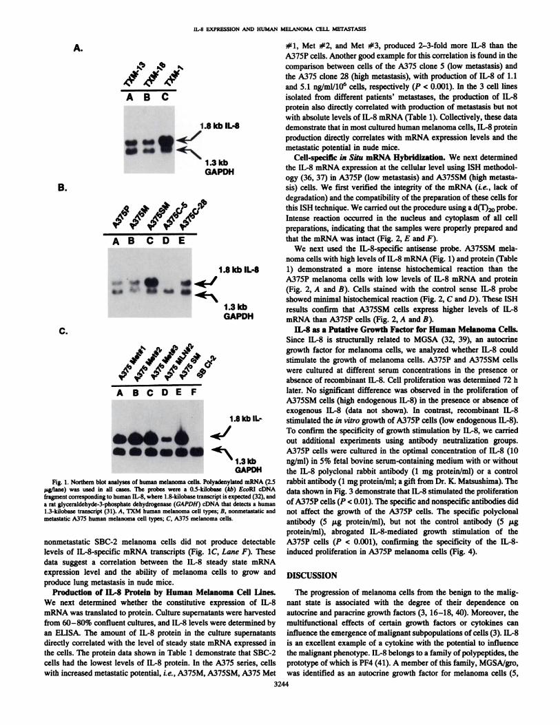
Expression of IL-S-specific mRNA Transcripts by Human Melanoma Cells. We next analyzedthe expressionof IL-8 mRNA in thedifferent human melanoma cell lines by Northern blot analysis. All themetastatic melanoma cell lines expressed the 1.8-kilobase IL-8-specific mRNA transcript (Fig. 1, A—C).The expression level of steadystate mRNA transcript for IL-8 directly correlated with the metastaticpotential of the A375 cell series. The highly metastatic A375SM andA375M cells expressed higher levels of IL-8 mRNA (5.6- and 2.2-fold) than the low metastatic A375P cells (Table 1; Fig. lB. compareLanes C and B with Lane A). The highly metastatic A375 C-28 cellsexpressed 5.0-fold higher levels of IL-8 mRNA than the low metastatic A375 C-S cells (Fig. lB, compare Lanes E and D; Table 1).A375 Met # 1, #2, #3, and LN#2 cells also expressed steady statemRNA transcripts for IL-8. The levels directly correlated with theirmetastatic potential (Table 1; Fig. 1C, LanesA, B, C, and D). Similarresults were obtained when we studied 3 cell lines established frommelanoma metastases of different patients; the highly metastaticTXM-l cells expressed the highest levels of mRNA for IL-8 ascompared with TXM-13 and TXM-18 cells (Fig. IA, compare Lane Cwith Lanes A and B; Table 1). Finally, the poorly tumorigenic and
a Cells (5 X 10@) in 0.2 ml Hanks' balanced salt solution were injected iv. into nude
mice. The mice were killed when moribund or after 8 weeks. The lungs were resected,washed in water, and placed in Bouin's fixative. The number of tumor colonies wasdetermined with the aid of a dissecting microscope.
b Densitometric quantitation of IL-8 mRNA expression. The ratio of areas between the1.8-kilobase IL-8 mRNA transcript and 1.3-kilobase glyceraldehyde-3-phosphate dehydrogenase mRNA transcript is given in each case (see “Materialsand Methods―).
C Melanoma cells were incubated in supplemented medium containing 10% fetal
bovine serum. Culture supernatants were collected after 72 h and assayed for the presenceof lL-8 by ELISA as described in “Materialsand Methods.―Values are the mean ±SDof triplicate samples. This is one representative experiment of 3.
3243
IL-8 EXPRESSION AND HUMAN MELANOMA CELL METASTASIS
#1, Met #2, and Met #3, produced 2—3-foldmore IL-8 than theA375P cells. Another good example for this correlation is found in thecomparison between cells of the A375 clone 5 (low metastasis) andthe A375 clone 28 (high metastasis), with production of IL-8 of 1.1and 5.1 ng/ml/106 cells, respectively (P < 0.001). In the 3 cell linesisolated from different patients' metastases, the production of IL-8protein also directly correlated with production of metastasis but notwith absolute levels of IL-8 mRNA (Table 1). Collectively, these datademonstrate that in most cultured human melanoma cells, IL-8 proteinproduction directly correlates with mRNA expression levels and themetastatic potential in nude mice.
I 3 kb Cell-specificin Situ mRNA Hybridization. We nextdeterminedGAPDH theIL-8mRNAexpressionatthecellularlevelusingISHmethodol
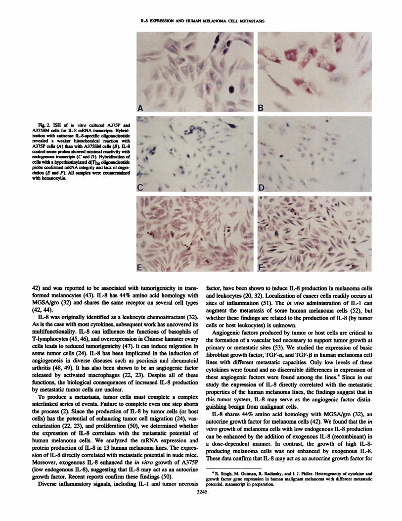
ogy (36, 37) in A375P (low metastasis) and A3755M (high metastasis) cells. We first verified the integrity of the mRNA (i.e., lack ofdegradation) and the compatibility of the preparation of these cells forthis ISH technique. We carried out the procedure using a d('F)@ probe.Intense reaction occurred in the nucleus and cytoplasm of all cellpreparations, indicating that the samples were properly prepared andthat the mRNA was intact (Fig. 2, E and F).
We next used the IL-8-specific antisense probe. A375SM melanoma cells with high levels of IL-8 mRNA (Fig. 1) and protein (Table
I .8kbIL-8 1) demonstrateda moreintensehistochemicalreactionthantheA375P melanoma cells with low levels of IL-8 mRNA and protein(Fig. 2, A and B). Cells stained with the control sense IL-8 probeshowed minimal histochemical reaction (Fig. 2, C and D). These ISH
I 3 kb resultsconfirmthat A375SM cells expresshigherlevelsof IL-8GAPDH mRNAthanA375Pcells(Fig.2,AandB).
IL-8 as a Putative Growth Factor for Human Melanoma Cells.Since IL-8 is structurally related to MGSA (32, 39), an autocrinegrowth factor for melanoma cells, we analyzed whether IL-8 couldstimulate the growth of melanoma cells. A375P and A375SM cellswere cultured at different serum concentrations in the presence orabsence of recombinant IL-8. Cell proliferation was determined 72 hlater. No signfficant difference was observed in the proliferation ofA375SM cells (high endogenous IL-8) in the presence or absence ofexogenous IL-8 (data not shown). In contrast, recombinant IL-8
1.8kb IL- stimulatedthe in vitrogrowthof A375Pcells (lowendogenousIL-8).“‘IE-' To confirm the specificity of growth stimulationby IL-8, we carried
out additional experiments using antibody neutralization groups.a*\ A375P cells were cultured in the optimal concentration of IL-8 (10
1.3 kb ng/ml) in 5% fetal bovine serum-containing medium with or withoutGAPDH the IL-8 polyclonal rabbit antibody (1 mg protein/mi) or a control
rabbit antibody (1 mg protein/mi; a gift from Dr. K. Matsushima). Thedata shown in Fig. 3 demonstrate that IL-8 stimulated the proliferationofA375P cells (P < 0.01). The specific and nonspecific antibodies didnot affect the growth of the A375P cells. The specific polyclonalantibody (5 @tgprotein/mi), but not the control antibody (5 @tgprotein/mi), abrogated IL-8-mediated growth stimulation of theA375P cells (P < 0.001), confirming the specificity of the IL-8-induced proliferation in A375P melanoma cells (Fig. 4).
A.
1,,ABC
1.8 kb IL-8
. ——
#@ I ‘a
AB CDE
@ @:@(
ABCDEF
S....
B.
C.
Fig. 1. Northern blot analyses of human melanoma cells. Polyadenylated mRNA (2.5@&aJlane)was used in all cases. The probes were a 0.5-kilobase (kb) EcoRI cDNA
fragmentcorrespondingtohumanIL-8,where1.8-kilobasetranscriptisexpected(32),anda rat glyceraldehy&-3-phosphatedehydrogenase(GAPDH)cDNAthatdetectsa human1.3-kilobase transcript (31). A, TXM human melanoma cell types; B, nonmetastatic andmetastatic A375 human melanoma cell types; C, A375 melanoma cells.
nonmetastatic SBC-2 melanoma cells did not produce detectablelevels of IL-8-specific mRNA transcripts (Fig. 1C, Lane F). Thesedata suggest a correlation between the IL-8 steady state mRNAexpression level and the ability of melanoma cells to grow andproduce lung metastasis in nude mice.
Production of IL-S PrOtein by Human Melanoma Cell Lines.We next determined whether the constitutive expression of IL-8mRNA was translated to protein. Culture supernatants were harvestedfrom 60—80%confluent cultures, and IL-8 levels were determined byan ELISA. The amount of IL-8 protein in the culture supernatantsdirectly correlated with the level of steady state mRNA expressed inthe cells. The protein data shown in Table 1 demonstrate that SBC-2cells had the lowest levels of IL-8 protein. In the A375 series, cellswith increased metastatic potential, i.e., A375M, A375SM, A375 Met
DISCUSSION
The progression of melanoma cells from the benign to the malignant state is associated with the degree of their dependence onautocrine and paracrine growth factors (3, 16—18,40). Moreover, themultifunctional effects of certain growth factors or cytokines caninfluence the emergence ofmalignant subpopulations ofcells (3). IL-8is an excellent example of a cytokine with the potential to influencethe malignant phenotype. IL-8 belongs to a family ofpolypeptides, theprototype of which is PF4 (41). A member of this family, MGSA/gro,was identified as an autocrine growth factor for melanoma cells (5,
3244
I
u@8 ExPRESmON AND HUMAN MElANOMA cIIL MFrASrASIS
ft.,
“.@ BA,i,,,
Fig. 2. ISH of in vitro cultured A375P andA375SM cells for IL-S mRNA transcripts. Hybridfrafi@ with anthcme ll@8 @gonu&@&revealed a weaker histochemical reaction withA375P cells (A) than with A375SM cells (B). IL-Scontrol sean@ showed minimal reactivity withendogenous transcripts (C and D). Hybridizarion ofcelliwith a hyperbiotinylated d(I),@oligonucIcotidc
@econfirmed mRNA integrity and lack of degradation (E and F). All samples were cetmterstainedwith bematoxylin.
;d@4
•Ii
C.@
5•' @@@‘e:.@ ,%
L@@
.-
‘@t‘ a
@ , ,S
I@ I
t@
@‘ ‘,@a
-@@‘ .@ 1'a. ‘@ e • •
.4
I@@@@ 4
S
•‘c@ ‘I',.ThJ@ F ‘@
@ :@ @“
@%“@Z@'@ z.:'@ •1@
‘aI
‘I
@ .‘@\@@ @. ,
4; .@@ :.@
..v
42) and was reported to be associated with tumorigenicity in transformed melanocytes (43). IL-8 has 44% amino acid homology withMGSA/gro (32) and shares the same receptor on several cell types(42, 44).
IL-8 was originally identified as a leukocyte chemoattractant (32).As is the case with most cytokines, subsequent work has uncovered itsmultifunctionality. IL-8 can influence the functions of basophils ofT-lymphocytes (45, 46), and overexpression in Chinese hamster ovarycells leads to reduced tumorigenicity (47). It can induce migration insome tumor cells (24). IL-8 has been implicated in the induction ofangiogenesis in diverse diseases such as psoriasis and rheumatoidarthritis (48, 49). It has also been shown to be an angiogenic factorreleased by activated macrophages (22, 23). Despite all of thesefunctions, the biological consequences of increased IL-8 productionby metastatic tumor cells are unclear.
To produce a metastasis, tumor cells must complete a complexinterlinked series of events. Failure to complete even one step abortsthe process (2). Since the production of IL-8 by tumor cells (or hostcells) has the potential of enhancing tumor cell migration (24), vascula.rization (22, 23), and proliferation (50), we determined whetherthe expression of IL-8 correlates with the metastatic potential ofhuman melanoma cells. We analyzed the mRNA expression andprotein production of IL-8 in 13 human melanoma lines. The expression of IL-8 directly correlated with metastatic potential in nude mice.Moreover, exogenous IL-8 enhanced the in vitro growth of A375P(low endogenous IL-8), suggesting that IL-8 may act as an autocrinegrowth factor. Recent reports confirm these findings (50).
Diverse inflammatory signals, including IL-i and tumor necrosis
factor, have been shown to induce IL-8 production in melanoma cellsand leukocytes (20, 32). Localization of cancer cells readily occurs atsites of inflammation (51). The in vivo administration of IL-i canaugment the metastasis of some human melanoma cells (52), butwhether these findings are related to the production of IL-8 (by tumorcells or host leukocytes) is unknown.
Angiogenic factors produced by tumor or host cells are critical to
the formation of a vascular bed necessary to support tumor growth atprimary or metastatic sites (53). We studied the expression of basic
fibroblast growth factor, TGF-a, and TGF-fi in human melanoma celllines with different metastatic capacities. Only low levels of thesecytokines were found and no discernible differences in expression ofthese angiogenic factors were found among the lines.4 Since in ourstudy the expression of IL-8 directly correlated with the metastaticproperties of the human melanoma lines, the findings suggest that inthis tumor system, IL-8 may serve as the angiogenic factor distinguishing benign from malignant cells.
IL-8 shares 44% amino acid homology with MGSA/gro (32), anautocrine growth factor for melanoma cells (42). We found that the invitro growth of melanoma cells with low endogenous IL-8 productioncan be enhanced by the addition of exogenous IL-8 (recombinant) ina dose-dependent manner. In contrast, the growth of high IL-8-producing melanoma cells was not enhanced by exogenous LL-8.These data confirm that IL-8 may act as an autocrine growth factor for
4 It Singh, M. Gutman, R. Radinsky, and L J. Fidler. Heterogeneity of cytokinc and
growth factor gene expression in human malignant melanoma with different mctastaticpotential, manuscript in preparation.
3245
15'
‘\;&ift.@.@
-

IL-S EXPRESSION AND HUMAN MELANOMA CELL METASTASIS
technical assistance, and Dahlia Garza for help in the preparation of themanuscript.
REFERENCES
1. Clark, W. H. Tumor progression and the nature ofcancer. Br. J. Cancer, 64: 631—644,1991.
2. Fidler, I. J. Critical factors in the biology of human cancer metastasis: twenty-eighth0. H. A. Clowes Memorial Award Lecture. Cancer Res., 50: 6130-6138, 1990.
3. Kerbel, R. S. Commentary: expression of multi-cytokine resistance and multi-growthfactor independence in advanced stage metastatic cancer. Am. J. Pathol., 141:519—524,1992.
4. Richmond, A., Thomas, H. G., and Roy, R. 0. B. Separation of melanoma growthstimulatory activity and human type-a transforming growth factor. MethodsEnzymol., 146: 112—126,1987.
5. Richmond, A., Lawson, D. H., Nixon, D. W., and Chawla, R. K. Characterization ofautostimulatory and transforming growth factors from human melanoma cells. CancerRca.,45:6390—6394,1985.
6. Richmond, A., Lawson, D. H., Nixon, D. W., Stedman, N. J., Stevens, S., andChawla, R. K. Extraction of a melanoma-growth stimulatory activity from culturemedium conditioned by the HS0294 human melanoma cell line. Cancer Res., 43:2106—2112,1983.
7. DeLarco, J. E., Pigott, D. A., and Lazarus, J. A. Ectopic peptides released by a humanmelanoma cell line that modulate the transformed phenotype. Proc. NatI. Aced. Sci.USA, 82: 5015—5019,1985.
8. Westermark, B., Johnsson, A., Paulsson, Y., Betsholtz, C., Heldin, C. H., Herlyn, M.,Rodeck, U., and Koprowski, H. Human melanoma cell lines of primary and metastaticorigin express the genes encoding the chains of platelet-derived growth factor andproduce a PDGF-like growth factor. Proc. NatI. Acad. Sci. USA, 83: 7197—7200,1986.
9. Halaban, R., Kwon, B. S., Ghosh, S., Delli Bovi, P., and Baird, A. bFGF as anautocrine growth factor for human melanoma. Oncogene Res., 3: 177—186,1988.
10. Lu, C., Vickers, M. F., and Kerbel, R. S. Interleukin-6: a fibroblast- derived growthinhibitor of human melanoma cells from early but not advanced stage of tumorprogression. Proc. NatI. Acad. Sci. USA, 89: 9215—9219,1992.
11. Kock, A., Schwarz, T., Urbanski, A., Peng, Z., Vetterlein, M., Miksche, M., Ansel,C.,Kung,H.F.,andLuger,T. A.Expressionandreleaseof interleukin-1bydifferenthuman melanoma cell lines. J. Natl. Cancer Inst., 81: 36—42,1989.
12. Bennicelli, J. L., Elias, J., Kem, J., and Guerry, D., Production of interleukin-1activity by cultured human melanoma cells. Cancer Res., 49: 930—935,1989.
13. Armstrong, C. A., Tare, D. C., Hart, C. E., Kock, A., Luger, T. A., and Ansel, J. C.Heterogeneity of cytokine production by human malignant melanoma cells. Exp.Dermatol., 1: 37—45,1992.
14. Rodeck, U., Melber, K., Kath, R., Menssen, H-D., Varello, M., Atkinson, B., andHerlyn, M. Constitutive expression of multiple growth factor genes by melanomacells but not normal melanocytes. J. Invest. Dermatol., 97: 20—26,1991.
15. Aaronson, S. A. Growth factors and cancer. Science (Washington DC), 254: 1146—1153, 1991.
16. Herlyn, M., Herlyn, D., Elder, D. E., Bondi, E., LaRossa, D., Hamilton, R., Scars, H.,Balaban, 0., Guerry, D., Clark, W. H., and Koprowski, H. Phenotypic characteristicsof cells derived from precursors of human melanoma. Cancer Res., 43: 5502—5508,1983.
17. Herlyn, M., Rodeck, U., Mancianti, M. L, Cardillo, F., Lang, A., Ross., A., Jambrosic, J., and Koprowski, H. Expression of melanoma-associated antigens in rapidlydividing human melanocytes. Cancer Res., 47: 3057—3061,1987.
18. Herlyn, M. Human melanoma: development and progression. Cancer Metastasis Rev.,9: 101—112,1990.
19. Forster, E., Kirnbauer, R., Urbanski, A., Kock, A., and Luger, T. A. Human melanoma cells produce interleukin-8 which functions as an autocrine growth factor.J. Invest. Dermatol., 96: 608, 1991.
20. Zachariae, C. 0. C., Thestug-Pedersen, K., and Matsushima, K. Expression andsecretion of leukocyte chemotactic cytokines by normal human melanocytes andmelanoma cells. J. Invest. Dermatol., 97: 593—599,1991.
21. Krueger, G., Jorgensen, C., Miller, C., Schroeder, J., Stiecherling, M., and Christopher, E. Effect of IL-8 on epidermal proliferation. J. Invest. Dermatol., 94: 545,1990.
22. Koch, A. E., Polverini, P. J., Kunkel, S. L., Harlow, L. A., DiPietro, L A., Elner,V. M., Elner, S. G., and Stricter, R. M. lnterleukin-8 as a macrophage-derivedmediator of angiogenesis. Science (Washington DC), 258: 1798—1801,1992.
23. Stricter, R. M., Kunkel, S. L., Elner, V. M., Martonyi, C. L., Koch, A. E., Polverini,P. 1., and Elner, S. G. Interleukin-8: a comeal factor that induces neovascularization.Am. 3. Pathol., 141: 1279—1284,1992.
24. Wang, J. M., Taraboletti, G., Matsushima, K., Damme, J. V., and Mantovani, A.Induction of hapatotactic migration of melanoma cells by neutrophil activatingprotein/IL-8. Biochem. Biophys. Rex. Commun., 169: 165—170,1990.
25. Kozlowski, J. M., Hart, I. R., Fidler, I. J., and Hanna, N. A human melanoma lineheterogeneous with respect to metastatic capacity in athymic nude mice. J. Nail.Cancer Inst., 72: 913—917,1984.
26. Li, L, Price, J. E., Fan, D., Zhang, R. D., Bucana, C. D., and Fidler, I. J. Correlationof growth capacity of human tumor cells in hard agarose with their in vivo proliferation capacity at specific metastatic sites. J. Nail. Cancer Inst., 81: 1406—1412,1989.
27. Schackert, G., Price, 1. E., Zhang, R. D., Bucana, C. D., Itoh, K., and Fidler, I. J.Regional growth of different human melanomas as metastases in the brain of nudemice. Am. J. Pathol., 136: 95—102,1990.
28. Zhang, R. D., Price, J. E., Schackert, G., Itoh, K., and Fidler, I. J. Malignant potential
0 IL-8(0.0001ng/ml)0 IL-B(0.001nglml)
@ IL-B(0.01ng/ml)@ 11-8(0.1nglml)
60@ • IL-8(1.OngIml)
U IL-B(10.0nglml)
Serum Concentrations (%)Fig. 3. Effect of IL-8 on in vitro growth analyses of A375P. Cells (5 X 1&@cells/well)
were incubated with medium alone or medium containing different doses of recombinanthuman IL-8 at different serum levels. After 72 h, growth stimulation was determined bythe MiT assay as described (35). Values are mean ±SD (bars) of triplicate culture. Thisis 1 representative of 3 experiments.
Medium
Fig. 4. Specificity of IL-8-induced proliferation of A375P cells. Cells were incubatedfor 72 h in medium alone or medium containing IL-8 (10 ng/ml) in the presence orabsence of anti-IL-8 antibody or control. MiT assay was performed as described in“Materialsand Methods.―The values are mean ±SD (bars) of triplicate cultures. This is1 representative of 2 experiments.
melanoma cells (50). Moreover, the production of IL-8 by metastaticcells populating a heterogeneous neoplasm may enhance the proliferation of nonmetastatic (and non-IL-8-producing) cells. In this role,IL-8 would act as an intratumoral paracrine growth factor (3, 54).
In summary, we have found that the expression level of IL-8correlates with the metastatic potential of human melanoma cellsimplanted into nude mice. Regardless of the exact functions of IL-8 inthe pathogenesis of metastasis, the data suggest that down-regulationof this cytokine may reduce the metastatic potential of some melanoma cells. This possibility is now under active investigation.
0a-.
C0C.)
0
C0
BE
Cl)
0a-.
50
40
30
20
70
1.25 2.5 5
0.4I o MediumC@ AntiIL-8Ab.
@. 0.3 .@ Control Ab.
@ 0.2C
.0a-0$.0
0.0
3246
IL-B (lOng/mi)
ACKNOWLEDGMENTS
We thankDr. K. Matsushima(Kanazawa,Japan)for providingcDNA forIL-8 and the antibody against lL-8, Rachel Tsan and Kenneth Dunner, Jr. for

lL-8 EXPRESSION AND HUMAN MELANOMA CELL METASTASIS
of cells isolated from lymph node or brain melastases of melanoma patients andimplication for prognosis. Cancer Rca., 51: 2029—2035,1991.
29. Verschraegen, C. R., Giovanella, B. C., Mendoza, J. T., Kozielski, A. J., and Stehlin,J. S.,Jr. Specificorganmetastasisof humanmelanomacellsinjectedintothearterialcirculation of nude mice. Anticancer Res., 11: 529—536,1991.
30. Radinsky, R., Kraemer, P. M., Raines, M. A., Kung, H. J., and ChIp, L A. Amplification and rearrangement of Kirsten ras oncogene in virus transformed BALB/3T3cells during malignant tumor progression. Proc. Nail. Acad. Sci. USA, 84: 5143-5147,1987.
31. Fort, P., Marty, L, Piechaczyk, M., Sabrouty, S. E., Darn, C., Jeanteur, P., andBlanchard, J. M. Various rat adult tissues express only one major mRNA species fromthe glyceraldehyde 3-phosphate-dehydrogenase multigenic family. Nucleic AcidsRca.,13:1431—1442,1985.
32. Matsushima, K., Morishita, K.. Yoshimura, T., Lavu, S., Kobayashi, Y., Lew, W.,Appella, E., Kung, H. F., Leonard, E. J., and Oppenheim, J. J. Molecular cloning ofa human monocyte-derived neutrophil chemotactic factor (MDNCF) and the induction of MDNCF mRNA by interleukin-1 and tumor necrosis factor. J. Exp. Med., 167:1883—1893,1988.
33. Feinberg, A. P., and Vogelstein, B. A technique for radiolabeling DNA restrictionendonuclease fragments to high specific activity. Anal. Biochem., 132: 6—13,1983.
34. Alley, M. C., Scudieno, D. A., Monks, A., Hursey, M. L, Czerwinski, M. J., Fine,D. L, AbbOtt,B. J., Mayo, J. G., Shoemaker, R. H., and Boyd, M. R. Feasibility ofdrugscreeningwithpanelsofhumantumorcelllinesusinga microculturetetrazoliumassay. Cancer Res., 48: 589-602, 1988.
35. Fan, D., Bucana, C. D., O'Brian, C. A., Zwelling, L A., Said, C., and Fidler, I. J.Enhancement of murine tumor cell sensitivity to Adriamycin by presentation of drugin phosphatidylcholine-phosphatidylserine liposomes. Cancer Res., 50: 3619—3626,1990.
36. Radinsky, R., Bucana, C. D., Ellis, L M., Sanchez, R., ae@, K. R., Brigati, D. J.,and Fidler, I. 1. A rapid colorimetric in situ messenger RNA hybridization techniquefor analysis of epidermal growth factor receptor in paraffin-embedded surgicalspecimensof humancoloncarcinomas.CancerRes.,53: 937—943,1993.
37. Bucana, C. D., Radinsky, R., Dong, Z., Sanchez, R., Brigati, D. 3., Fidler, I. J. A rapidcolorimetric in situ mRNA hybridization technique using hyperbiotinylated oligonucleotide probes for analysis of mdrl in mouse colon carcinoma cells. J. Histochem.Cytochem., 41: 499—506,1993.
38. Caruthers, M. H., Beaucage, S. L, Efcavitch, J. W., Fisher, E. F., Goldman, R. A., DcHaseth, P., Mandecki, W., Matteucci, M. D., Rosendahi, M. S., and Stabinsky, Y.Chemical synthesis and biological studies on mutated gene-control regions. ColdSpring Harbor Symp. Quant. Biol., 47: 411—418,1982.
39. Matsushima, K., Baldwin, E T., and Makaida, N. Interleukin-8 and MCAF: novelleukocyte recruitment and activating cytokines. Chem. Immunol., 51: 236—265,1989.
40. Nicolson, 0. L Gene expression, cellular diversification and tumor progression to themetastatic phenotype. Bioessays, 13: 337—342,1991.
41. Barone, A. D., Chrayeb, J., Hammerling, U., Zucher, M. B., and Thorbecke, G. J. The
expression in Escherichia coil of recombinant human platelet factor 4, a protein withimmunoregulatory activity. J. Biol. Chem., 263: 8710—8715,1988.
42. Anisowitch, A., Zojchowski, D., Stedman, 0., and Sager, R. Functional diversity ofgro gene expression in human fibroblasts and mammary epithelial cells. Proc. Nail.Aced.Sci.USA,85:9645-9649,1988.
43. Balentien, E., Mufson, B. E., Shattuck, R. L., Derynck, R., Richmond, A. Effects ofMGSA/GROa on melanocyte transformation. Oncogene, 6: 1115—1124, 1991.
44. Moser, B., Schumacher, C., Tscharne, U., Lewis, I. C., and Baggiolini, M. Neutrophil-activating peptide 2 and gro/melanoma growth-stimulatory activity interact withneutrophil-activating peptide 1/interleukin 8 receptors on human neutrophils. J. Biol.Chem., 266: 10666—10671,1991.
45. Dahinden, C. A., Kushiyuki, Y., DeWeck, A. L., Lindley, I., Dewald, B., andBaggiolini, M. The neutrophil-activating peptide NAF/NAP-1 induces histamine andleukotriene release by interleukin 3-primed basophils. J. Exp. Med., 170: 1787—1792,1989.
46. Larsen, G. G., Anderson, A. 0., Appella, E., Oppenheim, J. J., and Matsushima, K.The neutrophil-activating protein (NAP-i) is also chemotactic for T-lymphocytes.Science (Washington DC), 243: 1464—1466,1989.
47. Hirose, K., Hakozaki, M., Nyunoya, Y., Inokuchi, E., Saito, K., Matsunaga, K.,Kobayashi, Y., Matsushita, K., Takenouchi, T., Mikata, A., Mukaida, N., and Matsushima, K. Chemokine gene transfer into tumor cells reduced tumorigenicity in vivo.J. Leukocyte Biol., (Suppl.) 109, 1993.
48. Schroeder, J-M., and Christopher, E. Identification of C5a.,.@,,@and an anionicneutrophil-activating peptide (ANAP) in psoriatic scales. J. Invest. Dermatol., 87:53—58,1986.
49. deMarco, D., Kunkel, S. L., Stricter, R. M., Basha, M., and Zurier, R. B. lnterleukin-1induced gene expression of neutrophil activating protein (interleukin-8) and monocytechemotactic peptide in human synovial cells. Biophys. Biochem. Rca. Commun., 174:411—416,1991.
50. Schadendorf, D., Moller, A., Algermissen, B., Worm, M., Sticherling, M., andCzametzki, B. M. IL-8 produced by human malignant melanoma cells in vitro is anessential autocrine growth factor. J. Immunol., 151: 2667—2675,1993.
51. Murphy, P., Alexander, P., Senior, P. V., Fleming, V., Kirkham, N., and Taylor, LMechanisms of organ selective tumour growth by blood-bome cancer cells. Br. J.Cancer, 57: 19—31,1988.
52. Giavazzi, R., Garofalo, A., Bani, M. R., Abbate, M., Ghezzi, P., Boraschi, D.,Mantovani, A., and Dejana, E. Interleukin 1-induced augmentation of experimentalmetastases from a human melanoma in nude mice. Cancer Res., 50: 4771—4775,1990.
53. Folkman, J., and Klagsbrun, M. Angiogenic factors. Science (Washington DC), 235:444—447, 1987.
54. Abruzzo, L. V., Thornton, A. J., Liebert, M., Grossman, B., Evanott, H., Stricter,R. M., and Kunkel, S. Cytokine induced expression of IL-8 in human transitional cellcarcinomas and renal cell carcinomas. Am. J. Pathol., 140: 365—373,1992.
3247