Immunogenicity without Immunoselection: A Mutant but Functional Antioxidant Enzyme Retained in a...
10
2005;65:632-640. Cancer Res Marialuisa Sensi, Gabriella Nicolini, Marina Zanon, et al. Memory Phenotype T Cells with a + Metastatic Melanoma and Targeted by CD8 Functional Antioxidant Enzyme Retained in a Human Immunogenicity without Immunoselection: A Mutant but Updated version http://cancerres.aacrjournals.org/content/65/2/632 Access the most recent version of this article at: Cited Articles http://cancerres.aacrjournals.org/content/65/2/632.full.html#ref-list-1 This article cites by 35 articles, 21 of which you can access for free at: Citing articles http://cancerres.aacrjournals.org/content/65/2/632.full.html#related-urls This article has been cited by 4 HighWire-hosted articles. Access the articles at: E-mail alerts related to this article or journal. Sign up to receive free email-alerts Subscriptions Reprints and . [email protected] Department at To order reprints of this article or to subscribe to the journal, contact the AACR Publications Permissions . [email protected] Department at To request permission to re-use all or part of this article, contact the AACR Publications Research. on November 14, 2013. © 2005 American Association for Cancer cancerres.aacrjournals.org Downloaded from Research. on November 14, 2013. © 2005 American Association for Cancer cancerres.aacrjournals.org Downloaded from
-
Upload
savannahstate -
Category
Documents
-
view
1 -
download
0
Transcript of Immunogenicity without Immunoselection: A Mutant but Functional Antioxidant Enzyme Retained in a...
2005;65:632-640. Cancer Res Marialuisa Sensi, Gabriella Nicolini, Marina Zanon, et al. Memory Phenotype
T Cells with a+Metastatic Melanoma and Targeted by CD8Functional Antioxidant Enzyme Retained in a Human Immunogenicity without Immunoselection: A Mutant but
Updated version
http://cancerres.aacrjournals.org/content/65/2/632
Access the most recent version of this article at:
Cited Articles
http://cancerres.aacrjournals.org/content/65/2/632.full.html#ref-list-1
This article cites by 35 articles, 21 of which you can access for free at:
Citing articles
http://cancerres.aacrjournals.org/content/65/2/632.full.html#related-urls
This article has been cited by 4 HighWire-hosted articles. Access the articles at:
E-mail alerts related to this article or journal.Sign up to receive free email-alerts
Subscriptions
Reprints and
To order reprints of this article or to subscribe to the journal, contact the AACR Publications
Permissions
To request permission to re-use all or part of this article, contact the AACR Publications
Research. on November 14, 2013. © 2005 American Association for Cancercancerres.aacrjournals.org Downloaded from
Research. on November 14, 2013. © 2005 American Association for Cancercancerres.aacrjournals.org Downloaded from
Immunogenicity without Immunoselection: A Mutant but Functional
Antioxidant Enzyme Retained in a Human Metastatic Melanoma
and Targeted by CD8+ T Cells with a Memory Phenotype
Marialuisa Sensi,1Gabriella Nicolini,
1Marina Zanon,
1Chiara Colombo,
1Alessandra Molla,
1
Ilaria Bersani,1Raffaella Lupetti,
1Giorgio Parmiani,
2and Andrea Anichini
1
Units of 1Immunobiology of Human Tumors and 2Immunotherapy of Human Tumors, Department of Experimental Oncology, IstitutoNazionale per lo Studio e la Cura dei Tumori, Via Venezian 1, Milan, Italy
Abstract
Human melanomas can express unique tumor antigens,resulting from mutated proteins, and shared epitopesencoded for by normal genes, but these two classes ofantigens have not been previously compared for immunoge-nicity and retention in metastatic cells. Here, we identified anew unique antigen generated by a point mutation in theperoxiredoxin 5 (Prdx5) gene in an HLA-A*0201+ humanmetastatic melanoma lacking the wild-type allele. An antiox-idant assay, with recombinant Prdx5 proteins, and evaluationof peroxide accumulation in transiently transfected cells,indicated that the mutant protein retained its enzymaticactivity. The mutation in the Prdx5 protein did not generate anew HLA agretope but yielded an HLA-A*0201–restricted Tcell epitope (Prdx5110-119). By HLA-tetramer analysis, in atumor-invaded lymph node, >50% of mutant Prdx5-specificCD8+ T cells ( frequency 0.37%/CD8+) showed a CCR7+/�
CD45RA� ‘‘TCM’’ or ‘‘TEM’’ phenotype, as found in Melan-A/MART-1–specific T cells ( frequency 0.68%/CD8+) in the sametissue. In agreement with their memory phenotype, the Prdx5-specific T cells readily expanded in vitro in mixed lymphocyte-tumor culture, as did the Melan-/MART-1–specific T cells. Byimmunohistochemistry of the invaded lymph node, themutant Prdx5 protein was expressed in all neoplastic cells,in contrast with the heterogeneous expression of sharedantigens as Melan-A/MART-1, gp100 and tyrosinase. Thus, aunique tumor antigen can be as immunogenic as themelanoma differentiation antigens but, in contrast to thelatter, may be retained in all metastatic cells possibly as resultof the relevant cellular function exerted by the mutatedprotein. (Cancer Res 2005; 65(2): 632-40)
Introduction
A number of tumor antigens recognized by CD4+ and CD8+
T cells and resulting from mutated proteins have been identifiedin murine and human tumors (1, 2). These mutations, whenpresent only in the tumor in which they have been firstidentified, are truly tumor-specific and may yield unique tumorantigens (1, 2), which provide evidence for immune surveillanceof genome integrity. Although unique tumor antigens may be
difficult to exploit in therapeutic settings, nevertheless theyrepresent attractive immunologic targets for studying thedynamics of the host-tumor interaction. On one hand, identifi-cation of a number of unique tumor antigens in long-termsurvivors (3–8), has suggested that this class of antigens isimmunogenic and relevant to tumor rejection. On the otherhand, some of the mutant proteins that generate unique tumorantigens can be critical for tumor cell survival, due to theirinvolvement in basic metabolic pathways, or in regulation ofcell cycle and apoptosis (3–12). In these instances, immunoge-nicity and function of the mutant protein will likely act asopposing selective forces for the maintenance of expression ofthe antigen along tumor progression. In fact, immunogenicitymay promote the T cell–mediated response leading to emer-gence of antigen loss variants, where the mutant protein is nolonger expressed. However, neoplastic cells that will survive inthe host, during tumor progression, will attempt to maintainexpression of the mutant protein, due to its relevant cellularfunction. Experimental evidence in murine models has indi-cated that the balance between immunogenicity and cellularfunction of mutant proteins may be tilted in favor of the latterattribute, thus preventing the emergence of antigen loss variants(13, 14).The identification of new unique tumor antigens, expressed in
a human tumor, may thus offer the opportunity to evaluate thefunction of the mutant protein, its immunogenicity comparedwith other shared antigens expressed by the same tumor, and toassess its maintenance or loss of expression in metastatic cells.Here, we identify a new unique tumor antigen expressed in a
human metastatic melanoma. The mutation that led to a mis-sense amino acid substitution, occurred in the most recentlyidentified member of the peroxiredoxin (Prdx) gene family namedperoxiedoxin 5 (Prdx5 ; refs. 15–17). Prdxs are ubiquitous enzymesthat control intracellular H2O2 levels by catalyzing its reductioninto water (18, 19). Despite their biochemical similarities, Prdxproteins seem not to be functionally redundant (18, 19), since lossof expression of distinct Prdx family members has been shownto affect normal physiology and initiation/progression of patho-logic conditions in a cell type–dependent fashion (18, 19). Therelevance of these enzymes for normal cellular physiology is notonly linked to their protective activity against oxidative stress, butit is even revealed by their role as modulators of H2O2-dependentsignaling pathways activated by cytokine and growth factorreceptors (18, 19).Here we show that the missense substitution in one allele of
Prdx5 , occurring in a tumor lacking the wild-type (wt) allele, didnot impair the antioxidant function of the mutant protein andyielded a 10-mer HLA-A*0201–restricted unique T-cell epitope
Requests for reprints: Marialuisa Sensi, Unit of Immunobiology of HumanTumors, Department of Experimental Oncology, Istituto Nazionale per lo Studio e laCura dei Tumori, Via Venezian 1, 20133 Milan, Italy. Phone: 39-02-23902633; Fax: 39-02-23902630; E-mail: [email protected].
D2005 American Association for Cancer Research.
Cancer Res 2005; 65: (2). January 15, 2005 632 www.aacrjournals.org
Research Article
Research. on November 14, 2013. © 2005 American Association for Cancercancerres.aacrjournals.org Downloaded from
targeted by CD8+ T cells. Analysis of frequency, maturation stageand potential for in vitro expansion of Prdx5-specific T cells attumor site, in comparison with T cells directed to sharedmelanosomal antigens, provided evidence for immunogenicity ofthe novel unique antigen. Moreover, expression of the mutantprotein, along with that of the HLA-A2–restricting element, wasmaintained in all metastatic cells, in contrast to normal antigensof the melanocyte lineage.
Materials and Methods
Patients and Cell Lines. The clinical course of patient 8959 has been
already reported (20). Briefly, this patient (HLA typing: A*0201, A*1101/02,
BW55, CW3, DRw11, DQw7) developed a lymph node (LN) metastasis
(Me8959) 14 months after surgical removal of the primary tumor, and1 month later, died from brain metastases. All primary and metastatic
melanomas used in this study were established in culture in our laboratory
from specimens of patients admitted for surgery to our Institute. Culture
medium for melanoma, transformed embryonal kidney 293 (Invitrogen,Carlsbad, CA), and 174.CEMT2 (T2, American Type Culture Collection,
Rockville, MD) human lines was RPMI 1640 (Cambrex Bio Science,
Walkersville, MD), whereas for the mouse fibrosarcoma WEHI-164 clone
13 and the transformed African green monkey kidney COS-7 cells (kindlyprovided by Prof. T. Boon, Ludwig Institute for Cancer Research, Brussels,
Belgium) was DMEM (Cambrex Bio Science). Culture media were
supplemented with 10% FCS, 1 mmol/L sodium pyruvate, 2 mmol/Lglutamine, and 50 Ag/mL penicillin/streptomycin. Normal human epidermal
melanocyte lines (PromoCell, Heidelberg, Germany) were kept in melanocyte
growth medium M2 (PromoCell). Isolation of lymphocytes from PBMC and
LN, establishment of mixed lymphocyte-tumor cultures (MLTC), mainte-nance of PBMC-derived CTL clones, all CD3+ CD4� CD8+, T-cell receptor
(TCR) ah+, was done as described (21).
cDNA Library Construction and Screening. A cDNA library was
constructed as described (22) in the mammalian expression vectorpcDNA3.1 (Invitrogen) starting from Me8959 polyadenylated RNA.
Recombinant plasmids were electroporated into DH5-a Escherichia coli.
DNA was extracted from pooled bacteria containing 100 cDNA clones inaverage and cotransfected using the FuGENE 6 transfection reagent (Roche
Applied Science, Indianapolis, IN) with pcDNA3/HLA-A*0201 (22) into
COS-7 cells grown at 80% confluence into 96 flat-bottomed well plates. CTL
181 was added 30 hours later and, after additional 24 hours, tumornecrosis factor-a released in the supernatant was measured on the
sensitive WEHI-164 clone 13 cells with a colorimetric assay as described
(22). The entire procedure was repeated to isolate individual plasmids
encoding the antigen recognized by CTL clones. The cDNA insert wasautomatically sequenced using the ABI PRISM Big Dye Terminator Cycle
Sequencing Ready Reaction (Applied Biosystems, Foster City, CA) with T7
forward and pcDNA3.1BGH reverse sequencing primers (Invitrogen).Computer search for sequence homology was done with FASTA EMBL-
Heidelberg.
Constructs and Peptides. Full-length coding sequences and truncated
variants including translated regions corresponding to amino acids 159-214 (Prdx5159-214) or 183-214 (Prdx5183-214) were amplified by 30-cycle
stepdown reverse transcription-PCR (22) from cDNA obtained from
Me8959 and PBMC. The forward primers that contained an ATG start
codon with an appropriate Kozak consensus sequence (italicized) were:
F-1100: 5V-GCCACCATGGGACTAGCTGGCG (Prdx5)
F-1098: 5V-GCCACCATGGTGTCCATCTTTGGGAATCG (Prdx5159-214 )
F-1103: 5V-GCCACCATGAAGGAGACAGAC (Prdx5183-214)
The reverse primers which contained the stop codon (italicized) wasR-1102: 5V-GCCTCAGAGCTGTGAGATGAT. Amplification products were
subcloned into pcDNA3.1/V5/His plasmid using the Eukaryotic TOPO TA
cloning Kit (Invitrogen). Expression vectors were sequenced andtransiently transfected with or without pcDNA3/HLA-A*0201 (22) into
COS-7 or 293 cells for functional or antioxidant assays. As a control,pcDNA3.1/-gal encoding -galactosidase was used (Invitrogen). Candidate
HLA-A*0201–binding peptides were purchased from Primm (Milan, Italy).51Cr-labeled target cells were preincubated at room temperature for
1 hour with various or fixed concentrations of peptides. CTL clones werethan added and tested for cytotoxicity by standard 51Cr-release assay
(21). Means and SD (that never exceeded 10%) were calculated from
triplicates within each experiment and each experiment was repeated at
least twice.Genomic DNA. Genomic DNA, extracted according to standard
protocols, was amplified by a 30-cycle stepdown PCR using primers
F1127: 5V-TGAGCTTCCTAGTGGCCAAG and R1126: 5V-TCTCCCCACA-
GACGACTCTC. A specific band of 720 bp was gel-purified and directlysequenced using the forward primer F-1125: 5V-CAGGGAGTCAGGAC-
CAGGTA.
Bacterial Expression and Purification of Prdx5. Prdx5, without the52 amino acid–long mitochondrial targeting sequence cleaved in the
mature protein, was amplified by stepdown PCR, from plasmids contain-
ing the wt or mutant sequences using the primers described in ref. 15:
5V-CTGCACATATGGCCCCAATCAAGGTG-3V (NdeI site italicized) and 3V-GTTATAGTAGTTATAGTAGAGTGTCGAGACGCCTTCTCGCGGTCT-5V (SapI site itali-
cized). The PCR products were digested with NdeI and SapI and ligated
into vector pTYB1 (IMPACT System, New England BioLabs, Beverly, MA).
Plasmid inserts were sequenced using primers provided by the kit.The resulting constructs pTYB1-Prdx5wt (wt) or pTYB1-Prdx5Leu (mutant)
were then used to transform E. coli ER2566 strain. Recombinant proteins
were expressed after addition of 0.5 mM isopropyl -D-thiogalactopyranosideand purified onto chitin beads columns provided in the IMPACT System
according to the manufacturer’s instructions.
Western Blotting. Recombinant Prdx5 proteins and cellular proteins,
obtained by lysing cells in ice-cold lysis buffer [50 mmol/L Tris-HCl (pH
7.4), 150 mmol/L NaCl, 0.1% SDS, 1% NP40, 1 mmol/L EDTA, 1 mmol/L
EGTA, 1mmol/L NaVO3, and 1mmol/L NaF] containing protease inhibitor
cocktail (Roche Applied Science) were quantified by the Bio-Rad Assay
(Bio-Rad Laboratories, Hercules, CA). To achieve higher accuracy, re-
combinant Prdx5 proteins and escalating known amounts of a recom-
binant Prdx1 (Sigma-Aldrich, St Louis, MO) as reference standard, were
stained, after SDS-PAGE with SYPRO Red (Amersham Biosciences,
Buckinghamshire, United Kingdom). Fluorescent signal intensities were
scanned by Typhoon 8600 Imaging System and were quantified by
ImageQuant (both from Amersham Biosciences). Data were exported to
Microsoft Excel to generate a standard Prdx1 curve and, by regression
analysis, the protein concentration of both Prdx5 isoforms was cal-
culated. Cellular proteins (15 Ag) and recombinant Prdx5Leu, Prdx5wt and
thioredoxin (Calbiochem, Darmstadt, Germany), each at 10 ng, were
electrophoresed and transferred to a polyvinylidene difluoride membrane
(Amersham Biosciences). Immunoblotting was carried out with anti-
Prdx5 (clone 44, 1:5,000, BD Biosciences, San Diego, CA) and anti-
thioredoxin (1:4,000, Serotec, Oxford, United Kingdom) monoclonal
antibody (mAb) or, on stripped membranes, with polyclonal rabbit anti-
actin (Sigma-Aldrich, 1:2,000). Bound antibodies were visualized using
SuperSignal West Dura Extended Duration Substrate enhanced chemilu-
minescence (Pierce, Rockford, IL) followed by autoradiography.
Antioxidant Assay. The ability of recombinant Prdx proteins to protect
glutamine synthetase (GS, Sigma-Aldrich) from DTT/Fe3+/O2–mediated
oxidative inactivation was monitored as described (23). The assay was
conducted by adding Prdx5 proteins (1.5 Ag) into a 100 Al inactivation
mixture [10 mmol/L DTT, 3 Amol/L FeCl3, and 50 mmol/L HEPES/HCl
(pH 7)] containing 2.5 Ag of GS. After incubation at 37jC for different
time, 25 AL of the solutions were added to a g-glutamyltransferase assay
mixture [0.4 mmol/L ADP, 150 mmol/L glutamine, 10 mmol/L K-ASO4,
20 mmol/L NH2OH, 0.4 mmol/L MnCl2, and 50 mmol/L HEPES/HCl
(pH 7)], and incubated at 37jC for 15 minutes before the addition of 1 mL
of stop mixture (55 g of FeCl3�6H2O, 20 g of trichloroacetic acid, and
21 mL of concentrated HCl/liter). The absorbance resulting from the
g-glutamylhydroxamate-Fe3+ complex was read at 540 nm.
Immunogenicity and Function of a Unique Melanoma Antigen
www.aacrjournals.org 633 Cancer Res 2005; 65: (2). January 15, 2005
Research. on November 14, 2013. © 2005 American Association for Cancercancerres.aacrjournals.org Downloaded from
H2O2 Levels. Cellular H2O2 levels were determined using 5-(and 6)-chloromethyl-2V,7V-dichlorodihydrofluorescein diacetate (CM-H2-DCFDA,Molecular Probes, Eugene, OR). This cell-permeable compound becomesfluorescent and remains trapped inside the cell upon oxidation (24). COS-7and 293 cells were transfected with the appropriate expression vectors andincubated for 24 hours. The medium was then removed and replaced withHBSS without phenol red containing 5 Amol/L of CM-H2-DCFDA. Afterincubation in the dark at room temperature for 15 minutes, cells weretrypsinized, washed, and resuspended in medium containing a 100 Amol/Ltert-butylhydroperoxide solution (tBHP, Sigma-Aldrich). After 5 minutes, thecells were washed thrice and their fluorescence immediately measured by adual laser FACScalibur (BD Biosciences) using the CellQuest software.
HLA Stabilization Assay. T2 cells were incubated overnight at 37jCwith different dilutions of each peptide in serum-free RPMI 1640
supplemented with 1 mg/mL of h2 microglobulin (Sigma-Aldrich). Cellswere then washed to remove free peptides and stained with mAb CR11-351
followed by FITC-conjugated goat anti-mouse immunoglobulin G. Values
are indicated as mean fluorescence intensities (MFI) of peptide-inducedHLA-A*0201 expression after subtraction of the MFI of cells similarly
treated but without added peptide.
TCR Usage. Nomenclature for TCR gene segments is according to the
recommendations of the WHO-IUS Subcommittee on TCR Designation (25).cDNA obtained from CTL clones was amplified by stepdown PCR using
panels of TCRAV- or BV-specific forward primers and one reverse AC or BC
primer (26). Amplified fragments were gel purified. Their complementarity-
determining region 3 (CDR3) was directly sequenced as described (26).Tetramer Staining and Flow Cytometry. HLA-A*0201 PE-labeled
tetramers containing peptides from Melan-A/MART-126-35 (modifiedsequence carrying Ala at position 2), gp100154-162, gp100209-217, tyrosi-nase368-377, and Prdx5115leu were purchased from ProImmune Ltd. (Oxford,United Kingdom) and used as described (27). All other reagents, unlessotherwise specified, were purchased from BD Biosciences. Briefly, T cellswere stained for 15 minutes with tetramers (1:200 of the stock solution) at37jC and then stained for 30 minutes on ice with allophycocyanin coupledanti-CD8 mAb. To detect CCR7, staining was done with IgM anti-CCR7,followed by biotin-conjugated rat anti-mouse IgM and then by Cy-Chrome–conjugated streptavidin. After this incubation period, the cells were washedtwice and stained for 20 minutes in ice with CD8-APC and CD45RA-FITCand immediately analyzed (27). To detect TCRBV expression, T cells wereincubated for 30 minutes in ice with mAbs directed to BV8S1-S2 (Serotec,Raleigh, NC), washed once, stained with FITC-conjugated goat anti mousesecondary immunoglobulin G (1:30, Jackson ImmunoResearch, West Grove,PA) for 30 minutes in ice, incubated with PE-labeled tetramers in ice,washed twice and finally fixed in PBS with 1% formalin. Intracellularstaining of melanoma cells was done on fixed (ethanol and acetone v/v) andpermeabilized (HBSS plus 0.1% Triton X-100) cells with anti-Prdx5 (1:200) oranti-vimentin (VIM 3B4, 1:50, Chemicon, Hampshire, United Kingdom)mAbs followed by the FITC-conjugated secondary Ab. All data wereacquired by a dual laser FACScalibur (BD Biosciences) using the CellQuestsoftware.
Immunohistochemical Analysis. Immunohistochemical analysis of
tissue sections and cytospins was done as described (20, 28). Primary
mAbs were anti-Melan-A/MART-1 (A103, 1:50, Novocastra, Newcastle upon
Tyne, United Kingdom), anti-gp100 (HMB45, 1:25, Dako, Copenhagen,Denmark), anti-tyrosinase (T311, 1:50, NeoMarkers, Fremont, CA) and anti-
Prdx5 (1:200). After primary antibodies incubation overnight at 4j followed
by biotinylated goat anti-mouse immunoglobulin G (Dako, 1:100) addition,the slides were covered with streptavidin-horseradish peroxidase (1:300;
Dako) and visualized with the use of Sigma Fast 3,3V-diaminobenzidine
tablet set (Sigma-Aldrich).
Results
A Mutant Antioxidant Enzyme, Expressed in a MelanomaLacking the wt Allele, Is Recognized by Different AutologousCD8+ HLA-A2–Restricted CTL Clones. Among CTL clonesderived from PBMC of a patient with metastatic melanoma
(patient 8959), several recognized HLA-A*0201–restricted sharedmelanoma-associated antigens (29). Others, like CTL 22, 121, and181 lysed in HLA-A*0201–restricted fashion the autologous Me8959cells even if tested after a few in vitro passages, but neither of10 HLA-A*0201–matched primary or metastatic melanoma lines,nor normal HLA-A*0201+ melanocytes (data not shown). TCRrepertoire analysis followed by sequencing of the CDR3 indicatedthat the three CTL clones, although all of independent origin,shared usage of the same variable (BV8) and constant gene (BC2)segments in their h chain (data not shown). CTL 22 and 121, butnot 181 that expressed AV8 and was not sequenced, also displayedan identical AV region (AV3), identical J regions in both the a and hchains (BJ2S3 and AJ55) and quite similar CDR3 rearrangements(data not shown). Screening of a cDNA library, constructed withMe8959 poly(A)+ mRNA allowed the identification of an 800-bp-long cDNA clone (cDNA 78) able to transfer the expression of anantigen recognized by all three CTL clones, when cotransfectedwith pcDNA3.1/HLA-A*0201 into COS-7 cells (data not shown).cDNA 78 contained a poly(A) tail, a polyadenylation signal and itssequence matched the coding sequence of Prdx5 (Genbank/EMBLaccession no. AF110731; ref. 15) except for one C-T nucleotidechange located in exon 5. This change resulted in a Ser to Leusubstitution at position 115 of the translated polypeptide (Fig. 1A ;numbering according to Declercq et al. 17). We next compared thefull-length Prdx5 coding sequences derived from cDNA of Me8959and autologous PBMC. In all sequences analyzed, 10 from PBMCand 10 from Me8959, PBMC expressed always the wt TCG codon,whereas the tumor only harbored the TTG codon (data not shown).Thus, a mutation took place in the neoplastic cells that apparentlydid not harbor, or did not express the wt Prdx5 allele. To dissectthe latter mechanism, a genomic 720-bp fragment, containing exon5 sequences of Prdx5 was amplified from a frozen suspension offresh Me8959 cells and from autologous PBMC and sequenced.The electropherograms shown in Fig. 1B indicated that only Prdx5allele present in Me8959 was the mutant isoform, whereas asexpected, DNA derived from PBMC displayed the wt sequence. Thecrystallographic structure of Prdx5 has been recently published andfile coordinates deposited at the RCSB Protein Data Bank underaccession no. 1hd2 (17). Figure 1C displays a closer view of thePrdx5 structure around the wt Ser115 residue. This residue liesclose to a stretch of hydrophobic amino acids whose side chainscome in proximity of the aromatic ring of a benzoate molecule. Inthe Prdx5 molecule expressed by Me8959 cells, the presence ofLeu115 instead of Ser115, could confer further hydrophobicity tothis region. Additional HLA-A*0201 melanoma lines expressingPrdx5 were analyzed for the presence of Leu115 but none of themwas recognized by CTL clone 181 in a 51Cr release assay and alldisplayed the wt sequence (data not shown). An anti-Prdx5 mAbrecognizing both mutant and wt isoforms was then used to esti-mate, by flow cytometry, the Prdx5 levels in this panel of mela-nomas, in Me8959 and in normal cultured melanocytes. Figure 1Dshows that Me8959 has constitutive expression, in all cells, ofmutant Prdx5, the only isoform present. All additional lines werealso positive for Prdx5 expression (Fig. 1E , o). Prdx5 levels inmelanomas, as assessed by MFI values (Fig. 1E , 5), were hetero-geneous but in general lower than that displayed by normalmelanocytes. MFI value of Me8959 (Fig. 1E , n), did not differ fromthat displayed by a subset of melanomas with wt Prdx5.Mutant Prdx5 Retains Antioxidant Activity. To directly
evaluate the function of mutant (Prdx5Leu) versus wt (Prdx5wt)protein, they were expressed in E. coli , purified, and quantified
Cancer Research
Cancer Res 2005; 65: (2). January 15, 2005 634 www.aacrjournals.org
Research. on November 14, 2013. © 2005 American Association for Cancercancerres.aacrjournals.org Downloaded from
to identical concentration. By Western blot analysis, anti-Prdx5mAb recognized both purified recombinant Prdx5 proteins, butnot a control 15-kDa thioredoxin protein, whereas an anti-thioredoxin mAb recognized thioredoxin, but neither form ofPrdx5 (Fig. 2A). These results established the identity of bothrecombinant Prdx5 that appeared as a single band of an esti-mated size of 17 kDa (data not shown) and excluded that themutant residue could interfere with antibody recognition.Prdx5Leu and Prdx5wt were then compared for their ability toprevent GS inactivation induced by a low concentration of H2O2
produced by a cell-free oxidation system, a method used toevaluate antioxidant activity of the Prdx family members (23). Tothis end, GS was incubated for different times in the inactivationmixture with Prdx5Leu, Pdx5wt (1.5 Ag/each), or with EDTA(1 mmol/L) as control for oxidative inactivation prevention (23).As shown in Fig. 2B , both Prdx5 exerted a protective effectsince, after 20 minutes, 63% and 79% of GS activity could berecovered in presence of Prdx5wt and Prdx5Leu, respectively. Toprovide further support for the retained function of the mutantPrdx5 protein, we transiently transfected COS-7 or 293 cells with
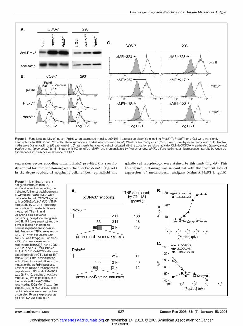
expression vectors encoding either Prdx5Leu or Prdx5wt or a -galprotein as negative control. Compared to endogenous levels,Prdx5 was over expressed in Prdx5 transfected cells as verifiedby Western blot and fluorescence-activated cell sorting analysis(Fig. 3A and B). Transfected cells were allowed to incorporateCM-H2-DCFDA indicator dye (24) and exposed to a peroxide (100mol/L tBHP) for 5 minutes. This treatment resulted in a rapidincrease of CM-H2-DCFDA fluorescence in h-gal-expressing COS-7(MFI = 323) and 293 cells (MFI = 328), reflecting the increasedoxidative status of the cells promoted by tBHP (Fig. 3C). Such CM-H2-DCFDA fluorescence increase was markedly reduced byexpression not only of the wt but even of the mutant Prdx5protein (compare MFI values in h-gal– versus Prdx5-transfectedcells in Fig. 3C). Interestingly, in both cell types, expression ofPrdx5Leu resulted in an increased protection from oxidative stress,compared with Prdx5wt (compare MFI values in wt and mutantPrdx5 transfected cells in Fig. 3C). Since transfection was notassociated with higher levels of mutant proteins (as shown inFig. 3A and B), these results suggest that the mutant protein maybe even more active than the wt one in its antioxidant activity.
Figure 1. Identification of mutant Prdx5 as a melanoma antigen. A, amino acid sequence of cDNA 78 is identical to that corresponding to Prdx5 (Genbank/EMBLaccession no. AF110731) except for a Ser (TCG) to Leu (TTG ) substitution (arrow). Alternate bold and regular fonts mark the six exons. The catalytic Cys residue,conserved in all Prdx, is boxed. Features of Prdx5 are the mitochondrial (underlined ) and peroxisomal (*) targeting sequences. B, electropherograms of a partialexon 5 sequence of Prdx5 gene amplified from genomic DNA of fresh Me8959 and of autologous PBMC. The nucleotide that differs among the two sequences isevidenced. C, ribbon modeling of Prdx5, built with DS Viewer Pro from the crystallographic file of Prdx5 as deposited at the RCSP Protein Data Bank (accession no.1HD2; ref. 17). Inset, wt Ser115 residue (pink , modified in Leu115 in the mutant protein) and adjacent hydrophobic residues (yellow ). Cys (orange ) involvedin intramolecular disulfide intermediate formation following oxidation; benzoate molecule (red). D, intracellular flow cytometry of mutant Prdx5 in Me8959. Cells stainedby FITC goat anti-mouse or anti-vimentin mAb are, respectively, the negative (grey histogram ) and positive control (dotted histogram ). E, intracellular levels of Prdx5in melanoma and melanocytes. For each cell line, analyzed by flow cytometry, data are plotted on two different Y axes as either % positive cells (o, left y axis) andas mean fluorescence intensity (5, right y axis ). n, MFI of Prdx5 expressed in Me8959.
Immunogenicity and Function of a Unique Melanoma Antigen
www.aacrjournals.org 635 Cancer Res 2005; 65: (2). January 15, 2005
Research. on November 14, 2013. © 2005 American Association for Cancercancerres.aacrjournals.org Downloaded from
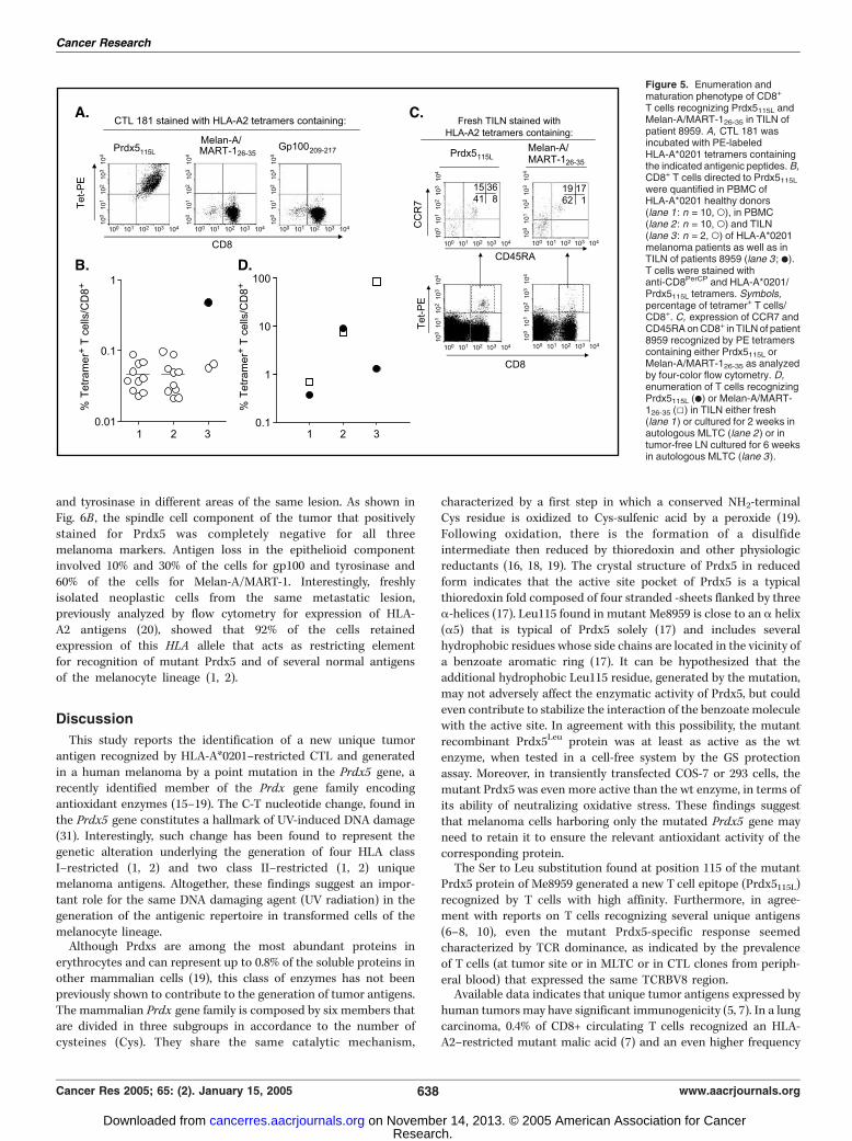
Identification of the Antigenic Peptide. To evaluate whetherthe mutation in Prdx5 was relevant to the generation of the CTLepitope, COS-7 cells were transfected with expression vectorscontaining full-length or fragmented Prdx5 cDNA amplified fromeither Me8959 (Prdx5Leu) or PBMC (Prdx5wt) along withpcDNA3/HLA-A*0201 and tested for recognition by CTL 181.Only constructs containing the mutant sequence retained theability to stimulate tumor necrosis factor-a release by CTL 181,and the immunogenic peptide mapped in a 24-amino-acidsequence that included the mutant Leu residue (Fig. 4A). Threepotential HLA-A*0201–binding nonamers or decamers ligandswere identified in this region using available algorithms (30). Thewt and the mutant peptides including positions 109 to 117, 110to 119, and 111 to 119 of Prdx5 were then pulsed, at 1 Amol/Lon an HLA-A*0201–positive melanoma that does not express themutant Prdx5 (Me18732). The decamer peptide LLLDDLLVSI110-119 (thereafter named Prdx5115L), with the sixth residue beingLeu115, was recognized in both tumor necrosis factor-a releaseand cytotoxicity assays, whereas all other peptides were not(data not shown). Dose-response curves, similar for all CTLclones recognizing mutant Prdx5, indicated that the mutantdecamer was recognized at concentration lower than 100 pmol/L, whereas no reactivity was observed against the normalpeptide (Fig. 4B , for the pattern of lysis of CTL 181) even atconcentrations up to 10 Amol/L (data not shown). The mutantas well as the corresponding wt peptides displayed similarprofiles in the HLA-A2 stabilization assay, indicating, inaccordance with their predicted identical binding scores, thatthey could bind with similar affinities to this allele (Fig. 4C).Taken together these results indicate that the Ser to Leumutation in Prdx5 of Me8959 generates a new T-cell epitope andnot a new agretope.Analysis of T-Cell Response to the Novel Unique and to
Shared Melanoma Antigens in the Same Patient. T-cellfrequency to differentiation antigens of the melanocyte lineageand to the unique Prdx5Leu antigen were compared inlymphocytes from a tumor-invaded LN (TILN) of patient 8959by HLA-A*0201 tetramer analysis. Soluble HLA-A*0201 tetramerscontaining the Prdx5115L peptide stained specifically CTL clone181 (Fig. 5A). CD8+ T cells from PBMC of HLA-A*0201+ healthydonors or melanoma patients contained <0.1% of T cells stainingwith the Prdx5-specific tetramer (Fig. 5B : lanes 1 and 2). Infresh, uncultured TILN of patient 8959, 0.37% of CD8+ T cellswere specific for Prdx5115L (Fig. 5: B , lane 3 , .; C , bottom left ;and D , lane 1 , .) in comparison to <0.1% in TILN from twoadditional HLA-A*0201 melanoma patients (Fig. 5B , lane 3 , o);57.8% of Prdx5115L-specific T cells also expressed the BV8 regionshared by anti-Prdx5115L CTL clones isolated from PBMC (datanot shown). These data indicate that both local and a systemicimmunity developed in patient 8959 to this HLA-A*0201–restricted epitope and that this response was dominated bythe expansion of Prdx5115L-specific T-cell populations expressingan identical TCRBV. In the same fresh TILN, the frequency ofCD8+ T cells directed to Melan-A/MART-126-35 was 0.68% (Fig. 5:C , bottom right ; and D , lane 1 , 5), whereas frequencies togp100154-162, gp100209-217, and tyrosinase368-377 were <0.2% (datanot shown). The maturation stage of CD8+ T cells present inTILN and specific for Prdx5115L and Melan-A/MART-126-35 wasevaluated through their expression of CCR7 and of CD45RA. Thedata shown in Fig. 5C indicate that 56% and 81% of T cellsspecific for Prdx5115L and Melan-A/MART-126-35, respectively,
showed a differentiated CCR7+/�CD45RA� (i.e., TCM or TEM)phenotype. Moreover, culture in vitro for 2 weeks in autologousMLTC induced marked outgrowth of Prdx5115L- and Melan-A/MART-126-35–specific CD8+ T cells at comparable levels (7.5% forMelan-A/MART-126-35 and 8.9% for Prdx5115L; Fig. 5D , lane 2). Apredominant expression (84%) of TCRBV8-expressing T cells wasfound among Prdx5115L tetramer–positive cells (data not shown).In contrast, repeated MLTC stimulation, for up to 6 weeks, oflymphocytes from an autologous tumor-free LN led to remark-able expansion of T cells specific for Melan-A/MART-126-35,whereas those specific for Prdx5115L constituted only 1.3% ofCD8+ T cells (Fig. 5D , lane 3). Taken together, these dataindicate that Prdx5115L-specific T cells with a differentiatedphenotype are present in metastatic deposits at frequencysimilar or higher than T cells recognizing shared melanosomalantigen and are responsive to antigen stimulation in vitro to asimilar extent as T cells directed to the differentiation antigenMelan-A/MART-1.Immunohistochemical Analysis of Metastatic Lesions. Intra-
cellular staining of Me8959 cells with anti-Prdx5 mAb (as shownin Fig. 1D) had indicated expression of the mutant protein in allneoplastic cells. Staining on the same cell line with mAbsdirected to gp100, tyrosinase, and Melan-A/MART-1 melanosomalantigens instead indicated that different proportion of cells werenegative for the latter antigens (data not shown). To assess theantigenic profile of Me8959 in deeper detail, serial sections fromthe LN metastasis were analyzed by immunohistochemistry.COS-7 cells either untransfected or transiently transfected with
Figure 2. Isolation and functional activity in a cell-free assay of recombinantPrdx5 proteins. A, Western blot analysis of Prdx5 recombinant proteins (mutantand wt) as well as unrelated control protein (thioredoxin) with anti-Prdx5 andanti-thioredoxin mAbs. B, time-dependent GS protection activities of wt andmutant Prdx5 proteins as described in Materials and Methods.
Cancer Research
Cancer Res 2005; 65: (2). January 15, 2005 636 www.aacrjournals.org
Research. on November 14, 2013. © 2005 American Association for Cancercancerres.aacrjournals.org Downloaded from
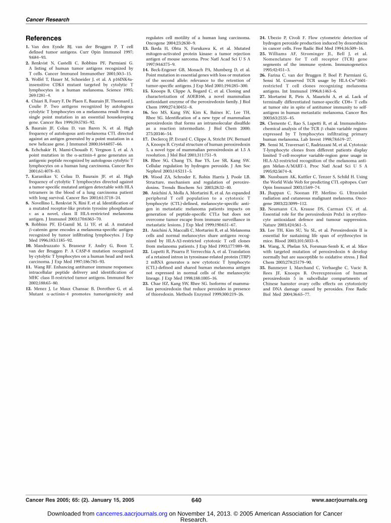
expression vector encoding mutant Prdx5 provided the specific-ity control for immunostaining with the anti-Prdx5 mAb (Fig. 6A).In the tissue section, all neoplastic cells, of both epithelioid and
spindle cell morphology, were stained by this mAb (Fig. 6B). Thishomogeneous staining was in contrast with the frequent loss ofexpression of melanosomal antigens Melan-A/MART-1, gp100,
Figure 3. Functional activity of mutant Prdx5 when expressed in cells. pcDNA3.1 expression plasmids encoding Prdx5Leu, Prdx5wt, or h-Gal were transientlytransfected into COS-7 and 293 cells. Overexpression of Prdx5 was assessed by (A) Western blot analysis or (B ) by flow cytometry in permeabilized cells. ControlmAbs were (A) anti-actin or (B) anti-vimentin. C, transiently transfected cells, incubated with the oxidation sensitive indicator CM-H2-DCFDA, were treated (empty peaks )peaks ) or not (grey peaks ) for 5 minutes with 100 Amol/L of tBHP, and then analyzed by flow cytometry. DMFI , difference in mean fluorescence intensity between cellfluorescence in presence or absence of tBHP.
Figure 4. Identification of theantigenic Prdx5 epitope. A,expression vectors encoding theindicated full-length/subfragmentsof wt/mutant Prdx5 cDNA werecotransfected into COS-7 togetherwith pcDNA3/HLA -A*0201 . TNF-a released by CTL 181 followingrecognition of transfectants wasmeasured. The minimal24-amino-acid sequencecontaining the epitope recognizedby CTL 181 (grey shading ) and thecorresponding nonantigenicnormal sequence are shown onleft . Amount of TNF-a released byCTL 181 when cocultured withMe8959 was 125 pg/mL, whereas<15 pg/mL were released inresponse to both COS-7 and COS-7/A*0201 cells. B, 51Cr-labeledHLA-A*0201+ Me18732 cells weretested for lysis by CTL 181 (at E/Tratio of 10:1) after preincubationwith different concentrations of themutant or the wt Prdx5 peptides.Lysisof Me18732 in the absence ofpeptide was 4.5% and of Me8959was 28.7%. C, binding of wt (4) ormutant (E) Prdx5 peptides, or ofthe unrelated HLA-A*6801–restricted gp100/pMel17182-191 (.)peptide (1, 2) to HLA-A*0201 alleleon T2 cells was assessed by flowcytometry. Results expressed asMFI for HLA-A2 expression.
Immunogenicity and Function of a Unique Melanoma Antigen
www.aacrjournals.org 637 Cancer Res 2005; 65: (2). January 15, 2005
Research. on November 14, 2013. © 2005 American Association for Cancercancerres.aacrjournals.org Downloaded from
and tyrosinase in different areas of the same lesion. As shown inFig. 6B , the spindle cell component of the tumor that positivelystained for Prdx5 was completely negative for all threemelanoma markers. Antigen loss in the epithelioid componentinvolved 10% and 30% of the cells for gp100 and tyrosinase and60% of the cells for Melan-A/MART-1. Interestingly, freshlyisolated neoplastic cells from the same metastatic lesion,previously analyzed by flow cytometry for expression of HLA-A2 antigens (20), showed that 92% of the cells retainedexpression of this HLA allele that acts as restricting elementfor recognition of mutant Prdx5 and of several normal antigensof the melanocyte lineage (1, 2).
Discussion
This study reports the identification of a new unique tumorantigen recognized by HLA-A*0201–restricted CTL and generatedin a human melanoma by a point mutation in the Prdx5 gene, arecently identified member of the Prdx gene family encodingantioxidant enzymes (15–19). The C-T nucleotide change, found inthe Prdx5 gene constitutes a hallmark of UV-induced DNA damage(31). Interestingly, such change has been found to represent thegenetic alteration underlying the generation of four HLA classI–restricted (1, 2) and two class II–restricted (1, 2) uniquemelanoma antigens. Altogether, these findings suggest an impor-tant role for the same DNA damaging agent (UV radiation) in thegeneration of the antigenic repertoire in transformed cells of themelanocyte lineage.Although Prdxs are among the most abundant proteins in
erythrocytes and can represent up to 0.8% of the soluble proteins inother mammalian cells (19), this class of enzymes has not beenpreviously shown to contribute to the generation of tumor antigens.The mammalian Prdx gene family is composed by six members thatare divided in three subgroups in accordance to the number ofcysteines (Cys). They share the same catalytic mechanism,
characterized by a first step in which a conserved NH2-terminalCys residue is oxidized to Cys-sulfenic acid by a peroxide (19).Following oxidation, there is the formation of a disulfideintermediate then reduced by thioredoxin and other physiologicreductants (16, 18, 19). The crystal structure of Prdx5 in reducedform indicates that the active site pocket of Prdx5 is a typicalthioredoxin fold composed of four stranded -sheets flanked by threea-helices (17). Leu115 found in mutant Me8959 is close to an a helix(a5) that is typical of Prdx5 solely (17) and includes severalhydrophobic residues whose side chains are located in the vicinity ofa benzoate aromatic ring (17). It can be hypothesized that theadditional hydrophobic Leu115 residue, generated by the mutation,may not adversely affect the enzymatic activity of Prdx5, but couldeven contribute to stabilize the interaction of the benzoate moleculewith the active site. In agreement with this possibility, the mutantrecombinant Prdx5Leu protein was at least as active as the wtenzyme, when tested in a cell-free system by the GS protectionassay. Moreover, in transiently transfected COS-7 or 293 cells, themutant Prdx5 was even more active than the wt enzyme, in terms ofits ability of neutralizing oxidative stress. These findings suggestthat melanoma cells harboring only the mutated Prdx5 gene mayneed to retain it to ensure the relevant antioxidant activity of thecorresponding protein.The Ser to Leu substitution found at position 115 of the mutant
Prdx5 protein of Me8959 generated a new T cell epitope (Prdx5115L)recognized by T cells with high affinity. Furthermore, in agree-ment with reports on T cells recognizing several unique antigens(6–8, 10), even the mutant Prdx5-specific response seemedcharacterized by TCR dominance, as indicated by the prevalenceof T cells (at tumor site or in MLTC or in CTL clones from periph-eral blood) that expressed the same TCRBV8 region.Available data indicates that unique tumor antigens expressed by
human tumorsmay have significant immunogenicity (5, 7). In a lungcarcinoma, 0.4% of CD8+ circulating T cells recognized an HLA-A2–restricted mutant malic acid (7) and an even higher frequency
Figure 5. Enumeration andmaturation phenotype of CD8+
T cells recognizing Prdx5115L andMelan-A/MART-126-35 in TILN ofpatient 8959. A, CTL 181 wasincubated with PE-labeledHLA-A*0201 tetramers containingthe indicated antigenic peptides. B,CD8+ T cells directed to Prdx5115L
were quantified in PBMC ofHLA-A*0201 healthy donors(lane 1 : n = 10, o), in PBMC(lane 2 : n = 10, o) and TILN(lane 3 : n = 2, o) of HLA-A*0201melanoma patients as well as inTILN of patients 8959 (lane 3 ; .).T cells were stained withanti-CD8PerCP and HLA-A*0201/Prdx5115L tetramers. Symbols,percentage of tetramer+ T cells/CD8+. C, expression of CCR7 andCD45RA on CD8+ in TILN of patient8959 recognized by PE tetramerscontaining either Prdx5115L orMelan-A/MART-126-35 as analyzedby four-color flow cytometry. D,enumeration of T cells recognizingPrdx5115L (.) or Melan-A/MART-126-35 (5) in TILN either fresh(lane 1) or cultured for 2 weeks inautologous MLTC (lane 2) or intumor-free LN cultured for 6 weeksin autologous MLTC (lane 3).
Cancer Research
Cancer Res 2005; 65: (2). January 15, 2005 638 www.aacrjournals.org
Research. on November 14, 2013. © 2005 American Association for Cancercancerres.aacrjournals.org Downloaded from
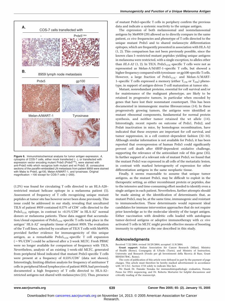
(1.2%) was found for circulating T cells directed to an HLA-A28–restricted mutant helicase epitope in a melanoma patient (5).Assessment of frequency of T cells recognizing unique mutantpeptides at tumor site has however never been done previously. Thisissue could be addressed in our study, revealing that unculturedTILN of patient 8959 contained 0.37% of CD8+ cells directed to thePrdx5115L epitope, in contrast to <0.1%/CD8+ in HLA-A2+ normaldonors or melanoma patients. These data suggest that accumula-tion/clonal expansion of Prdx5115L-specific T cells took place in theantigen+ HLA-A2+ neoplastic tissue of patient 8959. The evaluationof the T cell lines, selected by coculture of TILN Tcells with Me8959,provided further evidence for immunogenicity of this uniqueantigen, as a remarkable Prdx5115L-specific T cell expansion(f9%/CD8+) could be achieved after a 2-week MLTC. Fresh PBMCwere no longer available for comparison of frequency with TILN.Nevertheless, analysis of an existing 1-week-old MLTC, generatedfrom peripheral blood indicated that mutant Prdx5-specific T cellswere present at a frequency of 0.55%/CD8+ (data not shown).Interestingly, limiting dilution analysis for frequency of antitumor Tcells in peripheral blood lymphocytes of patient 8959, had previouslydocumented a high frequency of T cells directed to HLA-A2–retricted antigens not shared with melanocytes (21). Thus, presence
of mutant Prdx5-specific T cells in periphery confirm the previousdata and indicate a systemic reactivity to the unique antigen.The expression of both melanosomal and nonmelanosomal
antigens by Me8959 (29) allowed us to directly compare in the samepatient, ex vivo frequencies and phenotype of T cells directed to theunique mutant Prdx5 and to shared melanocyte differentiationepitopes, which are frequently presented in association with HLA-A2(1, 2). This comparison has not been previously possible, since theknown class I–restricted mutant peptides yielding unique antigensin melanomawere restricted, with a single exception, to alleles otherthan HLA-A2 (1, 2). In TILN, Prdx5115L-specific T cells were not asrepresented as Melan-A/MART-1–specific T cells, but showed ahigher frequency comparedwith tyrosinase- or gp100-specific Tcells.However, a large fraction of Prdx5115L- and Melan-A/MART-1–specific T cells expressed a memory (either TCM or TEM) pheno-type, in support of antigen-driven T-cell maturation at tumor site.Mutant, nonredundant proteins, essential for cell survival and/or
for maintenance of the malignant phenotype, are likely to beretained in progressive tumors, in particular when encoded bygenes that have lost their nonmutant counterpart. This has beendocumented in immunogenic murine fibrosarcomas (14). In theseprogressively growing tumors, the antigens were identified asmutant ribosomal components, fundamental for normal proteinsynthesis, and neither tumor retained the wt allele (14).Interestingly, recent reports on outcome of Prdx1, Prdx2, andPrdx6 inactivation in mice, by homologous recombination, haveindicated that these enzymes are important for cell survival, andtumor suppression, in a cell context–dependent fashion (32–34).Although similar information is not available for Prdx5, it has beenreported that overexpression of human Prdx5 could significantlyprevent cell death after tBHP-dependent oxidative challenge,supporting the relevance of the antioxidant role of this gene (35).In further support of a relevant role of mutant Prdx5, we found thatthe mutant Prdx5 was expressed in all cells of the metastatic lesion,in contrast with marked heterogeneity of expression for otherdifferentiation antigens in the same neoplastic tissue.Finally, it seems reasonable to assume that unique tumor
antigens, as the mutant Prdx5, may be difficult to exploit in thetherapeutic setting, as either recombinant protein or peptides, dueto the intensive and time-consuming effort needed to identify even asingle antigen in each patient. Nevertheless, further attempts shouldbe made aiming at the identification of determinants that, asmutant Prdx5, may be, at the same time, immunogenic and resistantto immunoselection. These determinants would represent idealcandidates for immune intervention approaches that do not requireprior knowledge as to the molecular identity of the target antigen.Either vaccination with dendritic cells loaded with autologoustumor-derived antigens or adoptive immunotherapy with ex vivoactivated Tcells in MLTC might provide effective means of boostingimmunity to epitopes as the one described in this study.
AcknowledgmentsReceived 7/22/2004; revised 10/29/2004; accepted 11/8/2004.
Grant support: Italian Association for Cancer Research (Milan), Ministryof Health (Rome), Compagnia di S.Paolo (Turin), and Ministry of Instruction,University and Research (Fondo per gli Investimenti della Ricerca di Base, GrantRBNE017B4C, Rome).
The costs of publication of this article were defrayed in part by the payment of pagecharges. This article must therefore be hereby marked advertisement in accordancewith 18 U.S.C. Section 1734 solely to indicate this fact.
We thank Dr. Daisuke Nonaka for immunohistopathologic evaluation, DonataPenso for DNA sequencing, and Dr. Roberta Mortarini for helpful discussions andcritically reading of the manuscript.
Figure 6. Immunohistochemical analysis for tumor antigen expression. A,cytospins of COS-7 cells, either mock transfected (�), or transfected withexpression vector encoding mutant Prdx5 (Prdx5Leu), were stained withanti-Prdx5 mAb which recognize both mutant and wt Prdx5. B, consecutivesections of the paraffin-embedded LN metastasis from patient 8959 were stainedwith Mabs to Prdx5, gp100, Melan-A/MART-1, and tyrosinase. Originalmagnification �100 except for COS-7 cells (�200).
Immunogenicity and Function of a Unique Melanoma Antigen
www.aacrjournals.org 639 Cancer Res 2005; 65: (2). January 15, 2005
Research. on November 14, 2013. © 2005 American Association for Cancercancerres.aacrjournals.org Downloaded from
References1. Van den Eynde BJ, van der Bruggen P. T celldefined tumor antigens. Curr Opin Immunol 1997;9:684–93.
2. Renkvist N, Castelli C, Robbins PF, Parmiani G.A listing of human tumor antigens recognized byT cells. Cancer Immunol Immunother 2001;50:3–15.
3. Wolfel T, Hauer M, Schneider J, et al. A p16INK4a-insensitive CDK4 mutant targeted by cytolytic Tlymphocytes in a human melanoma. Science 1995;269:1281–4.
4. Chiari R, Foury F, De Plaen E, Baurain JF, Thonnard J,Coulie P. Two antigens recognized by autologouscytolytic T lymphocytes on a melanoma result from asingle point mutation in an essential housekeepinggene. Cancer Res 1999;59:5785–92.
5. Baurain JF, Colau D, van Baren N, et al. Highfrequency of autologous anti-melanoma CTL directedagainst an antigen generated by a point mutation in anew helicase gene. J Immunol 2000;164:6057–66.
6. Echchakir H, Mami-Chouaib F, Vergnon I, et al. Apoint mutation in the a-actinin-4 gene generates anantigenic peptide recognized by autologous cytolytic Tlymphocytes on a human lung carcinoma. Cancer Res2001;61:4078–83.
7. Karanikas V, Colau D, Baurain JF, et al. Highfrequency of cytolytic T lymphocytes directed againsta tumor-specific mutated antigen detectable with HLAtetramers in the blood of a lung carcinoma patientwith long survival. Cancer Res 2001;61:3718–24.
8. Novellino L, Renkvist N, Rini F, et al. Identification ofa mutated receptor-like protein tyrosine phosphatasen as a novel, class II HLA-restricted melanomaantigen. J Immunol 2003;170:6363–70.
9. Robbins PF, El-Gamil M, Li YF, et al. A mutatedh-catenin gene encodes a melanoma-specific antigenrecognized by tumor infiltrating lymphocytes. J ExpMed 1996;183:1185–92.
10. Mandruzzato S, Brasseur F, Andry G, Boon T,van der Bruggen P. A CASP-8 mutation recognizedby cytolytic T lymphocytes on a human head and neckcarcinoma. J Exp Med 1997;186:785–93.
11. Wang RF. Enhancing antitumor immune responses:intracellular peptide delivery and identification ofMHC class II-restricted tumor antigens. Immunol Rev2002;188:65–80.
12. Menez J, Le Maux Chansac B, Dorothee G, et al.Mutant a-actinin-4 promotes tumorigenicity and
regulates cell motility of a human lung carcinoma.Oncogene 2004;23:2630–9.
13. Ikeda H, Ohta N, Furukawa K, et al. Mutatedmitogen-activated protein kinase: a tumor rejectionantigen of mouse sarcoma. Proc Natl Acad Sci U S A1997;94:6375–9.
14. Beck-Engeser GB, Monach PA, Mumberg D, et al.Point mutation in essential genes with loss or mutationof the second allele: relevance to the retention oftumor-specific antigens. J Exp Med 2001;194:285–300.
15. Knoops B, Clippe A, Bogard C, et al. Cloning andcharacterization of AOEB166, a novel mammalianantioxidant enzyme of the peroxiredoxin family. J BiolChem 1999;274:30451–8.
16. Seo MS, Kang SW, Kim K, Baines IC, Lee TH,Rhee SG. Identification of a new type of mammalianperoxiredoxin that forms an intramolecular disulfideas a reaction intermediate. J Biol Chem 2000;275:20346–54.
17. Declercq JP, Evrard C, Clippe A, Stricht DV, BernardA, Knoops B. Crystal structure of human peroxiredoxin5, a novel type of mammalian peroxiredoxin at 1.5 Aresolution. J Mol Biol 2001;311:751–9.
18. Rhee SG, Chang TS, Bae YS, Lee SR, Kang SW.Cellular regulation by hydrogen peroxide. J Am SocNephrol 2003;14:S211–5.
19. Wood ZA, Schroder E, Robin Harris J, Poole LB.Structure, mechanism and regulation of peroxire-doxins. Trends Biochem Sci 2003;28:32–40.
20. Anichini A, Molla A, Mortarini R, et al. An expandedperipheral T cell population to a cytotoxic Tlymphocyte (CTL)-defined, melanocyte-specific anti-gen in metastatic melanoma patients impacts ongeneration of peptide-specific CTLs but does notovercome tumor escape from immune surveillance inmetastatic lesions. J Exp Med 1999;190:651–67.
21. Anichini A, Maccalli C, Mortarini R, et al. Melanomacells and normal melanocytes share antigens recog-nized by HLA-A2-restricted cytotoxic T cell clonesfrom melanoma patients. J Exp Med 1993;177:989–98.
22. Lupetti R, Pisarra P, Verrecchia A, et al. Translationof a retained intron in tyrosinase-related protein (TRP)2 mRNA generates a new cytotoxic T lymphocyte(CTL)-defined and shared human melanoma antigennot expressed in normal cells of the melanocyticlineage. J Exp Med 1998;188:1005–16.
23. Chae HZ, Kang SW, Rhee SG. Isoforms of mamma-lian peroxiredoxin that reduce peroxides in presenceof thioredoxin. Methods Enzymol 1999;300:219–26.
24. Ubezio P, Civoli F. Flow cytometric detection ofhydrogen peroxide production induced by doxorubicinin cancer cells. Free Radic Biol Med 1994;16:509–16.
25. Williams AF, Strominger JL, Bell J, et al.Nomenclature for T cell receptor (TCR) genesegments of the immune system. Immunogenetics1995;42:451–3.
26. Farina C, van der Bruggen P, Boel P, Parmiani G,Sensi M. Conserved TCR usage by HLA-Cw*1601-restricted T cell clones recognizing melanomaantigens. Int Immunol 1996;8:1463–6.
27. Mortarini R, Piris A, Maurichi A, et al. Lack ofterminally differentiated tumor-specific CD8+ T cellsat tumor site in spite of antitumor immunity to self-antigens in human metastatic melanoma. Cancer Res2003;63:2535–45.
28. Clemente C, Rao S, Lupetti R, et al. Immunohisto-chemical analysis of the TCR h chain variable regionsexpressed by T lymphocytes infiltrating primaryhuman melanoma. Lab Invest 1998;78:619–27.
29. Sensi M, Traversari C, Radrizzani M, et al. CytotoxicT-lymphocyte clones from different patients displaylimited T-cell-receptor variable-region gene usage inHLA-A2-restricted recognition of the melanoma anti-gen Melan-A/MART-1. Proc Natl Acad Sci U S A1995;92:5674–8.
30. Nussbaum AK, Kuttler C, Tenzer S, Schild H. Usingthe World Wide Web for predicting CTL epitopes. CurrOpin Immunol 2003;15:69–74.
31. Jhappan C, Noonan FP, Merlino G. Ultravioletradiation and cutaneous malignant melanoma. Onco-gene 2003;22:3099–112.
32. Neumann CA, Krause DS, Carman CV, et al.Essential role for the peroxiredoxin Prdx1 in erythro-cyte antioxidant defence and tumour suppression.Nature 2003;424:561–5.
33. Lee TH, Kim SU, Yu SL, et al. Peroxiredoxin II isessential for sustaining life span of erythrocytes inmice. Blood 2003;101:5033–8.
34. Wang, X, Phelan SA, Forsman-Semb K, et al. Micewith targeted mutation of peroxiredoxin 6 developnormally but are susceptible to oxidative stress. J BiolChem 2003;278:25179–90.
35. Banmeyer I, Marchand C, Verhaeghe C, Vucic B,Rees JF, Knoops B. Overexpression of humanperoxiredoxin 5 in subcellular compartments ofChinese hamster ovary cells: effects on cytotoxicityand DNA damage caused by peroxides. Free RadicBiol Med 2004;36:65–77.
Cancer Research
Cancer Res 2005; 65: (2). January 15, 2005 640 www.aacrjournals.org
Research. on November 14, 2013. © 2005 American Association for Cancercancerres.aacrjournals.org Downloaded from