The Role, Function, and Generation of Eomeshi CD8+ T Cells ...
156
I The Role, Function, and Generation of Eomes hi CD8 + T Cells in OX40 and CTLA-4 Targeted Cancer Immunotherapy Dana Emerson Thesis Presented to the Department of Molecular Microbiology and Immunology and the Oregon Health & Science University School of Medicine In Partial fulfillment of the requirements for the degree of Doctor of Philosophy March 2021
-
Upload
khangminh22 -
Category
Documents
-
view
0 -
download
0
Transcript of The Role, Function, and Generation of Eomeshi CD8+ T Cells ...
I
The Role, Function, and Generation of Eomeshi CD8+ T Cells in OX40 and CTLA-4 Targeted Cancer Immunotherapy
Dana Emerson
Thesis
Presented to the Department of Molecular Microbiology and Immunology
and the Oregon Health & Science University
School of Medicine
In Partial fulfillment of
the requirements for the degree of
Doctor of Philosophy
March 2021

II
III
Table of Contents Acknowledgments .................................................................................................................. V
Abstract ...................................................................................................................................1
Chapter 1: Overview of T cells in the Immune System ...............................................................2
1.1. Antigen recognition in immunology and presentation .....................................................2
1.2. The T cell receptor and T cell subtypes .........................................................................5 1.3. T cell activation .............................................................................................................9
1.3.1. TCR activation........................................................................................................9 1.3.2. Co-stimulation ...................................................................................................... 11 1.3.3. Cytokine stimulation ............................................................................................. 13
1.4. T cell differentiation and function ................................................................................ 15 1.5. T cell trafficking and circulation .................................................................................... 18 1.6. Introduction to Cancer ................................................................................................. 20
Chapter 2: Immunological Complexity of the Prostate Cancer Microenvironment Influences the Response to Immunotherapy ................................................................................................. 23
2.1. Introduction ................................................................................................................. 24 2.2. CD8+ T cells in Cancer ................................................................................................ 24 2.3. Immune Response to Viruses ...................................................................................... 25 2.4. The Immune Response to Cancer ............................................................................... 28 2.5. CD8+ T cells in Cancer ................................................................................................ 36 2.6. Prostate Cancer Tumor-Associated Antigens ............................................................... 41 2.7. Models of Murine Prostate Cancer............................................................................... 50 2.8. Prostate Cancer Immunotherapy: Vaccines, Checkpoint Blockade, and Combination
Therapy......................................................................................................................... 54 2.9. Conclusions ................................................................................................................ 60
Chapter 3: Immuno-Oncology Drugs: From relieving inhibition to providing co-stimulation with T cell agonists ........................................................................................................................... 61
3.1. Introduction ............................................................................................................. 62 3.2. Checkpoint inhibitors (anti-CTLA-4, anti-PD-1) and the first bispecific antibody ......... 63 3.3. Targeting the TNFRSF, ICOS, and combination immunotherapy............................... 66
3.3.1. OX40 ................................................................................................................... 67 3.3.2. CD40 ................................................................................................................... 69 3.3.3. 4-1BB................................................................................................................... 71 3.3.4. GITR .................................................................................................................... 73 3.3.5. ICOS.................................................................................................................... 74
3.4. Conclusions............................................................................................................. 75
IV
Chapter 4: Enhancing the generation of Eomeshi CD8+ T cells augment the efficacy of OX40- and CTLA-4–targeted immunotherapy .................................................................................... 77
4.1. Introduction ............................................................................................................. 78 4.2. Materials and Methods............................................................................................. 81 4.3. Results .................................................................................................................... 87
4.3.1. Combining anti-OX40 and anti-CTLA-4 increases CD8+ T cell–specific Eomes expression............................................................................................................ 87
4.3.2. Effective anti-OX40/anti-CTLA-4 therapy requires CD8+ T-cell Eomes expression.. 89 4.3.3. Eomeshi CD8+ T cells are induced in the lymph node prior to trafficking to the tumor
............................................................................................................................ 91 4.3.4. Eomeshi CD8+ T cells exhibit a distinct transcriptional profile.................................. 92 4.3.5. Use of an ITK inhibitor does not impair Eomes expression in CD8+ T cells. ............ 96 4.3.6. ITK inhibition synergizes with anti-OX40/anti-CTLA-4-therapy ............................... 97
4.4. Discussion ............................................................................................................. 106
Chapter 5: Future Directions ................................................................................................. 111
5.1. Summary of my research and overview of questions raised .................................... 111 5.2. Future directions and questions ............................................................................. 115
5.2.1. αOX40 and αCTLA-4 signaling pathways elicit CD8+ T cell Eomes expression in the lymph node ........................................................................................................ 115
5.2.2. CD8+ T cell Eomes expression drives a beneficial immune cancer response........ 120 5.2.3. Multifaceted roles of ibrutinib in αOX40/αCTLA-4 therapy .................................. 124
5.3. Conclusions........................................................................................................... 129
6. References ..................................................................................................................... 132
V
Acknowledgements
I thank members of the EACRI Cancer Research Animal Division, Miranda Gilchrist in the
EACRI Flow Cytometry Core, and Melissa Kasiewicz in the Redmond lab for their excellent
technical assistance. I thank Annah Rolig, and Elizabeth Sturgill for their daily scientific
guidance and assistance. I thank Haydn Kissick and Nataliya Prokhneveska for their
collaboration. I thank William Redmond, Michael Gough, Evan Lind, and Mark Slifka for their
guidance, support, and encouragement.
1
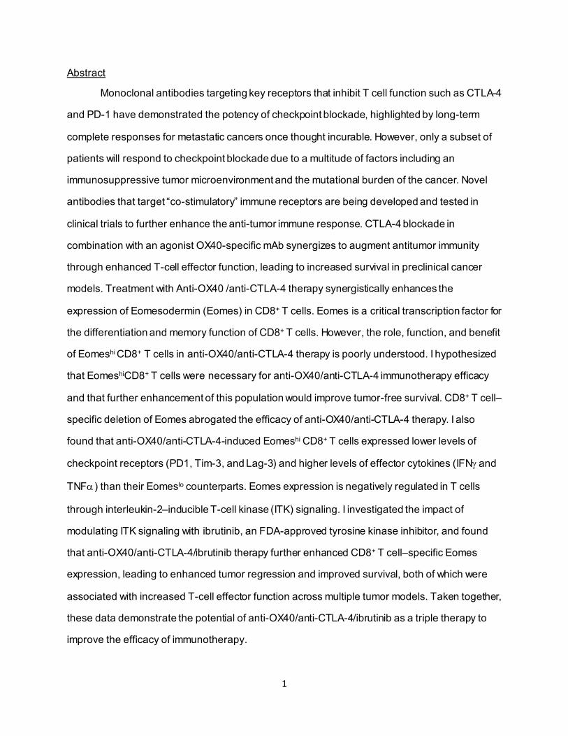
Abstract
Monoclonal antibodies targeting key receptors that inhibit T cell function such as CTLA-4
and PD-1 have demonstrated the potency of checkpoint blockade, highlighted by long-term
complete responses for metastatic cancers once thought incurable. However, only a subset of
patients will respond to checkpoint blockade due to a multitude of factors including an
immunosuppressive tumor microenvironment and the mutational burden of the cancer. Novel
antibodies that target “co-stimulatory” immune receptors are being developed and tested in
clinical trials to further enhance the anti-tumor immune response. CTLA-4 blockade in
combination with an agonist OX40-specific mAb synergizes to augment antitumor immunity
through enhanced T-cell effector function, leading to increased survival in preclinical cancer
models. Treatment with Αnti-OX40 /anti-CTLA-4 therapy synergistically enhances the
expression of Eomesodermin (Eomes) in CD8+ T cells. Eomes is a critical transcription factor for
the differentiation and memory function of CD8+ T cells. However, the role, function, and benefit
of Eomeshi CD8+ T cells in anti-OX40/anti-CTLA-4 therapy is poorly understood. I hypothesized
that EomeshiCD8+ T cells were necessary for anti-OX40/anti-CTLA-4 immunotherapy efficacy
and that further enhancement of this population would improve tumor-free survival. CD8+ T cell–
specific deletion of Eomes abrogated the efficacy of anti-OX40/anti-CTLA-4 therapy. I also
found that anti-OX40/anti-CTLA-4-induced Eomeshi CD8+ T cells expressed lower levels of
checkpoint receptors (PD1, Tim-3, and Lag-3) and higher levels of effector cytokines (IFNγ and
TNFα) than their Eomeslo counterparts. Eomes expression is negatively regulated in T cells
through interleukin-2–inducible T-cell kinase (ITK) signaling. I investigated the impact of
modulating ITK signaling with ibrutinib, an FDA-approved tyrosine kinase inhibitor, and found
that anti-OX40/anti-CTLA-4/ibrutinib therapy further enhanced CD8+ T cell–specific Eomes
expression, leading to enhanced tumor regression and improved survival, both of which were
associated with increased T-cell effector function across multiple tumor models. Taken together,
these data demonstrate the potential of anti-OX40/anti-CTLA-4/ibrutinib as a triple therapy to
improve the efficacy of immunotherapy.
2
Chapter I: Overview of T cells in the immune system
1.1. Antigen recognition in immunology and presentation
The immune system protects the organism from actual and perceived pathogens ranging
from viruses and bacteria to parasites and transformed cancerous tissue. In defending against
these threats, the immune system evolved a dynamic network of cells to handle both broad and
specific tasks involved in this response. Recognizing these threats as foreign or dangerous
material different from normal host tissue, is a key first step in this process. While the features
that differentiate between virus and self are clearly drawn, the features that discriminate
between self and transformed cancer tissue are more difficult for the immune system to
distinguish.
Recognizing, finding, and eliminating cancerous and transformed cells relies on
recognition of molecular patterns associated with damaged tissue similar to how viruses,
bacteria, and parasites are recognized. At the core of this response is the recognition of proteins
or genetic material that are different from healthy tissues. In the case of infectious disease,
foreign antigens are typically distinct from self-tissue. Common motifs found in bacteria, viruses,
and parasites are identified by pattern recognition receptors (PRRs), which can trigger an
immune response (1). However, the hallmarks of cancer are more nuanced compared to the
explicit motifs expressed by infectious organisms. For example, the immune system recognizes
damage-associated molecular patterns (DAMPs) through PRRs. This recognition can lead to
chronic inflammation and possible carcinogenesis (2). Many DAMPs, such as heat shock
proteins, activate members 2 and 4 of the toll-like receptor (TLR) family (3). Activation of TLRs
and other DAMP PRRs triggers nuanced immune responses to investigate and target the
damaged tissue.
3
The initiation and formation of an adaptive immune response against cancer starts with
the presentation of antigens derived from the mutated proteins of transformed cells. There are
two main pathways of antigen presentation differentiated by the cell’s ability to process protein-
based antigens and present them on major histocompatibility complex (MHC) proteins on the
cell surface. The two main types of MHC molecules are known as MHC class I and MHC class
II. All nucleated cells, including most non-immune cells, present antigens on MHC class I (4);
however, MHC class II expression is more specialized, and is limited to dendritic cells,
monocytes, macrophages, B cells, and thymic epithelial cells (5). These two main classes of
MHC molecules differ in the size and source of antigen they present, and what type of immune
response they elicit.
MHC class I molecules are made from three alpha chain domains covalently bound to a
β-2 microglobulin molecule anchored by a single transmembrane segment. The structure of
MHC class I produces an antigen binding cleft that is of sufficient size to bind a peptide antigen
of 8 to 10 amino acids in length. The peptide-loaded MHC I molecule is presented on the cell
surface to CD8+ T cells, which have T cell receptors (TCR) complimentary to MHC I containing
the bound peptide. Alternatively, MHC class II has a structure composed of two alpha and two
β-chain domains which are associated through noncovalent interactions and anchored by two
transmembrane segments. The structure of MHC class II produces an antigen binding cleft that
acts as an open-ended groove, sufficient to bind longer 13 to 18 amino acid peptides, which are
presented to CD4+ T helper cells. The two types of MHC molecule are also differentiated based
on the source of the antigen they present.
The two main classes of MHC molecules differ in the pathways by which antigen is taken
up by the cell, processed into small peptides, and loaded onto the molecule for presentation to T
cells. This distinction originates in whether the antigen is found endogenously within the cell or
taken up exogenously by the cell. Antigens found endogenously, indicating that foreign material
4
has made its way into the cell, are processed through the cytosolic pathway to indicate a
possible threat within the cell. Normal cellular components are also presented as a means of
sampling for damaged or mutated proteins. In this pathway, proteins within the cell are marked
for degradation by ubiquitin tagging. Ubiquitin tagging labels proteins to be processed and
broken down into peptide segments by the 26S proteasome complex, or the specialized
immunoproteasome (6). Trimming of the peptides of optimal length for presentation is achieved
through aminopeptidases such as endoplasmic reticulum aminopeptidase 1 (ERAP). The
resulting peptides are then transported across the endoplasmic reticulum (ER) membrane by
the transporter associated with antigen processing (TAP) (7). Once transported into the rough
ER lumen, peptides assemble into class I MHC with the help of chaperone proteins. Peptide
binding to MHC I results in a stabilized complex which can be presented on the cell surface to
CD8+ T cells. Without sufficiently strong peptide binding, the complex is not stable enough for
cell surface presentation. This pathway addresses antigens found within the cell, indicating a
possible cellular infection, cellular damage, or cellular transformation.
Antigens that are taken up exogenously by the cell through a variety of mechanisms are
processed through the endocytic pathway. Some cells constantly sample their environment
looking for signs of infection or cellular damage. Cells such as dendritic cells and macrophages
can sample their environment through pinocytosis and phagocytosis, which internalize potential
antigens into intracellular vesicles (8). Internalized antigens in vesicles transit though
increasingly acidic compartments in the cell, traveling through early endosomes, late
endosomes, and eventually lysosomes. These acid compartments break down the proteins into
small peptides with the help of cathepsin proteases for loading onto MHC class II. MHC class II
is assembled in the rough ER and stabilized by a trimeric protein called the invariant chain
which temporarily holds the place of the antigen peptide (9). The lysosome containing broken
down peptides fuses with the vesicle containing the MHC class II complex in the cytosol. Once
5
fused, the remains of the invariant chain are exchanged for a peptide between 13 and 18 amino
acids in length. The exchange is facilitated by HLA-DM, a nonclassical class II MHC molecule.
As with MHC class I, class II is stabilized and maintained by bound peptide. After peptide has
been bound, MHC class II is presented on the cell surface to CD4+ T helper cells. MHC class II
expression and presentation of antigen to CD4+ T helper cells is restricted to a subset of
immune cells known as professional antigen-presenting cells, which includes dendritic cells,
mononuclear phagocytes, thymic epithelial cells, and B cells. However, these two pathways do
not account for exogenous antigen presentation on MHC class I to activate CD8+ T cells.
How do naïve CD8+ T cells become activated by antigen presenting cells such as
dendritic cells if the dendritic cell does not have endogenous antigen and therefore can’t present
antigen on MHC I? Some dendritic cells specialize in presenting antigens taken up by
phagocytosis and presenting them on MHC I in a process called cross-presentation (10). For
example, some antigens are only partially degraded in the endosome and are transferred to the
endogenous pathway for MHC I presentation. This subset of cross-presenting dendritic cells is
characterized by CD8a expression in mice and BDCA-3 expression in humans and are
important for the anti-tumor immune response and in immunotherapy (11). Licensing of dendritic
cells for presentation to CD8+ T cells will be discussed in chapter 3.
1.2. The T cell receptor and T cell subtypes
T cells are activated through their T cell receptor (TCR), a highly variable receptor in the
immunoglobulin superfamily composed of either alpha-beta or gamma-delta pairs expressed on
the cell surface (12). The portion of the TCR that comes into contact with the peptide being
presented by the MHC molecule is composed of three hypervariable loops called the
complimentary determining region (CDR) (13). To produce a high level of binding variation, the
6
TCR and hypervariable CDR are generated through DNA recombination and somatic
recombination, which are facilitated by recombination activation genes (RAG) 1 and 2 (14). The
genome contains variations of the sections that recombine to form the alpha, beta, gamma, and
delta components of the TCR which produce unique alpha-beta, and gamma-delta TCR pairs.
There are a certain number of each component segments in the genome that recombine to
make the TCR consisting of V, D, J, and C segments. For example, the genes used to make the
beta chain in humans are composed 67 V segments, 2 D segments, 14 J segments, and 2 C
segments (14) The alpha chain is similarly generated, but does not incorporate a D segment.
Combination joining of one of each of these gene segments together allows for a large diversity
of possible combinations to be formed. For example, 4600 beta chains alone and 1.4 million
alpha-beta pairs combinations are possible. Junctional flexibility, and the addition of P-region
and N-region nucleotides are mechanisms that even more diversity to the TCR pool. These
mechanisms allow for TCRs with a wide variety of CDRs and potential binding partners to be
generated. Subsequently, the large diversity of T cells bearing unique TCRs must go through a
selection process to ensure two key properties: 1) The TCR is capable of binding to MHC-
antigen complexes; and 2) The TCR does not bind to self-antigens, which could result in
autoimmunity and tolerance. These processes are known as positive and negative selection,
respectively (15). Around 98% of developing thymocytes do not reach maturity, largely due to a
high positive selection fail rate. Culling of 98% of the pre-developed T cell population is
achieved through activation of the apoptosis (programmed cell death) pathway in these cells.
During thymic development, T cells express the TCR co-receptors CD4+ and CD8, the
expression of which will define what type of T cell they will mature into (16). During
development, thymocytes express both CD4+ and CD8 simultaneously. At this stage they are
referred to as double positive (DP). Double positive cells that interact with MHC class I will
mature into CD8+ T cells and lose CD4+ expression, and double positive cells that interact with
7
MHC class II will mature into CD4+ T cells and lose CD8 expression. CD8+ T cells, also known
as cytotoxic killer cells, are responsible for killing cancer cells and cells that have been infected
by virus. They are named CD8+ T cells for their CD8 co-receptor molecule, which binds to MHC
I and helps stabilize the TCR:MHC I interaction. CD4+ T cells express the CD4 co-receptor,
which serves a similar function in stabilizing the TCR:MHC II interaction. The CD8 co-receptor
takes the form of either an alpha-alpha homodimer or alpha-beta heterodimer of single
immunoglobulin-fold (Ig-fold) domains, and the monomeric CD4 co-receptor is made of four Ig-
fold domains.
CD8+ T cells specialize in cytotoxic killing of targeted cells. Once activated and engaged
with a target cell, CD8+ T cells release perforin, a pore forming toxin, that forms pores in the
target cell’s membrane (17). Granzyme serine proteases released into the target cell through
the perforin pores activates caspase 3, which triggers cell death via apoptosis (18). Additional
effector functions of CD8+ T cells include production and release of cytokines such as interferon
gamma (IFN-gamma; IFN-γ) and tumor necrosis factor alpha (TNF-alpha; TNF-α), which have
anti-tumor and anti-viral effects.
In contrast to the cytotoxic effects of CD8+ T cells, CD4+ T helper cells can have varying
functions in aiding the immune response (19). The most critical CD4+ T cells involved in the anti-
cancer immune response are Th1, Th2, and regulatory T cells. Th1 and Th2 cells differ mainly
in the cytokines they produce to direct and control the direction of the immune response, and
specifically the anti-cancer response. Th1 T helper cells aid in the anti-tumor response through
the cytokines they release (20). Th1 cytokines include many pro-inflammatory molecules that
lead to tumor elimination and control, such as IFN-γ, TNF-α, and interleukin-2 (IL-2). IFN-γ is a
class II interferon released by Th1 CD4+ T cells, CD8+ T cells, and natural killer (NK) cells. Upon
binding to the interferon receptor, IFN-γ exerts several anti-tumor properties, such as increasing
MHC class I and class II expression and inducing the immunoproteasome, which specializes in
8
producing hydrophobic c-termini peptides for better antigen presentation (21). IFN-γ receptor
activation also induces production of IgG2a by B cells, which play an important role in antibody-
dependent cellular cytotoxicity (ADCC). Additional functions of IFN-γ include producing a
positive feedback loop on Th1 CD4+ T cells which in turn produce more IFN-γ, and inhibition of
Th2 differentiation by naïve CD4+ T cells. TNF-α is a homotrimeric cytokine that signals through
tumor necrosis factor receptors to activate several intracellular signaling pathways that lead to
activation of NF-kB and AP-1 pathways in T cells. TNF signaling promotes the inflammatory
response, proliferation, survival, and generation of anti-apoptotic factors in immune cells that
express receptors for TNF-α (22). TNF signaling can also induce cell death through activation of
caspase-8 (23). IL-2 binding to its receptor induces signaling of Janus Kinases (JAK) 1 and 3.
JAK1 and JAK3 signaling leads to activation of Signal Transducer and Activator of Transcription
(STAT) 5. Activated STAT5 phosphorylates and activates NF-kB, NFAT, and AP-1, which all
promote T cell proliferation, differentiation, and support T cell survival. All of these cytokines
play an important role in the anti-tumor response and reinforce the Th1 T cell phenotype.
Th2 T helper cells, on the other hand, are generally thought to promote tumor growth
and inhibit the anti-tumor immune response, in part due to their cytokine profile (24). Cytokines
released by Th2 T helper cells include IL-4, IL-5, and IL-10. IL-4 induces B cell class switching
to production of IgE, an antibody isotype that triggers eosinophil and mast cell degranulation for
allergic diseases. IL-4 also promotes MHC class II expression and inhibits Th1 cell activity and
Th1 cytokine production. IL-5 also plays a role in allergic reactions through eosinophil activation
and IgA class switching. IL-10 inhibits the production of Th1 cytokines, downregulates MHC
expression, blocks NF-kB signaling, and aids in primary tumor growth (25).
Regulatory CD4+ T helper cells play an inhibitory role in the anti-tumor response through
the cytokines they release, and through direct suppression of effector cell function (26).
Regulatory CD4+ T helper cells are typically defined by expression of the IL-2 receptor alpha
9
chain and the transcription factor FoxP3, which programs their behavior. In terms of cytokines,
regulatory CD4+ T helper cells produce transforming growth factor beta (TGF-beta; TGF-β), IL-
10, and IL-35, all of which can contribute to tumor growth and inhibit the anti-tumor immune
response. TGF-β, in particular, plays a wide and varying role in immune cell and tumor function.
One consequence of TGF-β is supporting Treg expansion, inhibiting effector T cell function,
inhibits antigen presentation, and reduces NK cell cytotoxicity (27). Additionally, TGF-β also
plays a role in producing an immune suppressive tumor microenvironment. IL-10 has immune
suppressive effects by inhibiting Th1 responses and blocking the NF-kB pathway (28). IL-35
suppresses conventional T cell function and promotes further Treg development (29).
1.3. T cell activation
Three signals are required for full T cell activation and determine the fate of T cell
differentiation: 1) TCR stimulation (Signal 1); 2) co-stimulation through CD28 and other
corresponding receptors (Signal 2); and 3) cytokine signaling (Signal 3).
1.3.1. TCR activation
Upon interacting with peptide-loaded MHC, the TCR changes conformation to activate a
signal transduction pathway once binding has crossed past a sufficient threshold (30). The
interaction between TCR and peptide-loaded MHC requires the association of several surface
membrane proteins to start the signal transduction pathway (30). The cell will be activated
through expression of immediate, early, and late genes. Immediate genes consist primarily of
transcription factors such as NF-κB and NFAT, which are expressed in the first 30 minutes
following activation. Early genes, such as genes that for interleukin receptors and cytokines, are
expressed within 2 hours of activation. Late genes encode for adhesion molecule expression
10
and are expressed beyond 2 days after activation. Upon TCR engagement to the MHC-peptide
complex, the tyrosine kinase LCK is recruited from lipid rafts to phosphorylate the
immunoreceptor tyrosine-based activation domains (ITAMs) of the associated CD3 zeta chain
domains (31). Once phosphorylated, the CD3 zeta chains provide docking sites for another
tyrosine kinase, ZAP-70, to bind and become active. Activated ZAP-70 phosphorylates a
number of membrane-associated adaptor molecules such as LAT and SLP-76. Activation of
these and other associated molecules results in the activation of three main transcription
factors: NF-kB, NFAT, and AP-1. AP-1 activation begins through a kinase signaling cascade
that results in phosphorylated Fos and Jun transcription factors in the nucleus. In contrast, the
activation of NF-kB and NFAT are both facilitated by phospholipase C gamma (PLC-gamma;
PLC-γ).
PLC-γ is activated through phosphorylation by the associated kinases LAT and inducible
T cell kinase (ITK). Once activated, PLC-γ hydrolyzes the membrane phospholipid PIP2 to form
IP3 and diacylglycerol (DAG). IP3, in turn, activates calcium channels in the ER, and DAG
activates protein kinase C (PKC). Calcium activates calcineurin-calmodulin-Ca2+, which de-
phosphorylates NFAT, allowing it to transit to the nucleus and activate gene transcription,
primarily for cytokines that promote T cell growth. DAG activates PKC which in turn leads to the
activation and transit of NF-kB into the nucleus.
The AP-1 transcription factor promotes cell growth, proliferation, and apoptosis (32). NF-
kB plays a role in T cell development, activation, differentiation, and survival (33). NFAT plays a
role in growth through induction of IL-2 and IL-4 cytokine expression (34). These three main
transcription factors, NF-kB, AP-1, and NFAT, which are activated by TCR stimulation, signal
the cell to activate and prepare to respond to stimulation from signal 2 (co-stimulation) and
signal 3 (cytokines).
11
The strength of TCR stimulation, meaning the strength to which the TCR binds to the
MHC presented peptide, partially dictates the extent to which these and other transcription
factors will be expressed. Details of these pathways will be discussed in Chapters 4 and 5. The
strength of binding plays a role in NF-kB, AP-1, NFAT, and T cell differentiation transcription
factor expression, which determines what functional role the T cell will play. Other factors
contribute to expression of these transcription factors such as signal 2 and signal 3.
1.3.2. Co-stimulation
Once a naive T cell has been activated through TCR:peptide:MHC interactions, it must
then receive further co-stimulatory signals from antigen presenting cells to ensure that the
immune response is warranted. The primary co-stimulatory signal involves two related forms of
the B7 immunoglobulin superfamily protein: B7-1 and B7-2. These surface proteins are
expressed by antigen presenting cells such as dendritic cells, macrophages, and activated B
cells. The ligands for both B7-1 and B7-2 (also known as CD80 and CD86, respectively) are
CD28 and CTLA-4 (CD152), which are expressed on T cells (35). However, CD28 and CTLA-4
provide opposing signals to the T cell (36). CD28 is expressed by both naïve and activated T
cells and binding to B7 ligands provides a co-stimulatory signal that enhances IL-2 production
and NF-kB activation. CTLA-4, on the other hand, is an inhibitory checkpoint receptor induced
by TCR engagement that downregulates the activation of the T cell. CD28 and CTLA-4 compete
for the available B7 ligands, however CTLA-4 has 20 times higher affinity for B7 ligands
compared to CD28 and increases expression with continued CD28 co-stimulation. This provides
a regulatory brake for the T cell depending upon how much CD28 and CTLA-4 are expressed
and whether they are binding to their B7 ligands.
12
In addition to CD28-B7 signaling, CD40 and CD40 ligand (CD40L) play an important role
in the cross-talk between CD4+ and CD8+ T cells to generate CD8+ T cell memory (37). Mice
that lack CD4+ T cells can still generate effector CD8+ T cell responses but are unable to
generate CD8+ T cell memory (38). This is likely due to a requirement for both IL-2 and CD40
signaling in CD8+ T cells to generate a memory population through IL-2 autocrine signaling, and
CD4 licensing of antigen presenting cells though CD40-CD40L interactions (39). Memory T cells
are derived from both naïve T cells following activation and from differentiating effector T cells.
Memory T cells are generally long-lived and possess a heightened response to the same
antigen to generate a secondary response. The function and role of CD40 in memory T cell
generation will be discussed in greater detail in Chapter 3.
T cells that receive sufficient activation signals following TCR:MHC binding but do not
receive a sufficient signal 2 in the form of CD28 stimulation enter a state of clonal anergy in
which they fail to proliferate and have markedly reduced IL-2 production (40). An example of this
is self-reactive T cells that have made it out of thymic development and recognize their reactive
antigen in the periphery. These cells will often become anergic because non-inflamed peripheral
tissues do not express signal 2 ligands. Anergic cells may be transitioned into regulatory T cells
or may be clonally deleted. High levels of CTLA-4 signaling can also induce anergy (41).
Similarly, tolerance can form in T cells responding to self-antigens when they don’t receive any
co-stimulation or inflammatory activation either.
Several other types of co-stimulatory receptors exist that can provide signal 2. These are
typically members of the tumor necrosis family of receptors and signal through NF-κB in a
TRAF-dependent manner. Examples of other co-stimulatory receptors are OX40 (CD134), 4-
1BB (CD137), GITR (CD357), and others that will be discussed in detail in Chapter 3, as will
their relevance to cancer immunotherapy. The effects of co-stimulation between a T cell and an
APC are often bi-directional with both cell types receiving an activating signal.
13
In addition to several types of co-stimulatory receptors, there are a variety of checkpoint
inhibitory receptors that serve the opposing function of shutting down and blocking an immune
response. Inhibitory receptors include CTLA-4 and Programmed Death Receptor-1 (PD-1),
which will be discussed in Chapters 2 and 3.
1.3.3. Cytokine stimulation
Cytokines influence the differentiation and function of activated T cells depending upon
what concentrations of cytokines are present as well as which of their cognate receptors are
expressed on the T cell surface. One of the most critical cytokines for primary T cell responses
and survival is IL-2. TCR stimulation and additional co-stimulation triggers proliferation, as well
as transcription of IL-2 and the high-affinity IL-2 receptor (CD25). The IL-2 receptor is composed
of IL-2Rα, IL-2Rβ, and the common gamma chain subunits. The common gamma chain is a
component in several cytokine receptors including IL-2, IL-4, IL-7, IL-9, IL-15, and IL-21 (42). IL-
2 autocrine signaling maintains T cell proliferation and survival for several generations resulting
in subpopulations of effector and memory cells (43). Memory T cells are antigen experienced
and have high reactivity to the same antigen to form the secondary response. Memory cells are
long-lived and have a lower threshold of activation compared to naïve non-antigen experienced
T cells (44). High dose IL-2 therapy is FDA-approved for the treatment of melanoma and renal
cell cancer. However, IL-2 therapy has many severe adverse effects (45). IL-15 therapy may
give the same benefits of IL-2 therapy while mitigating the adverse effects.
IL-15 is an important cytokine for the development and survival of memory CD8+ T cells
and NK cells. IL-15 binds to CD122, which is composed of the common gamma chain, IL-2Rβ,
and IL-15Rα. One of the mechanisms by which IL-15 enhances T and NK cell survival is by
inducing expression of Bcl-2, an anti-apoptotic molecule, in addition to sharing signaling
14
pathways with IL-2. Memory CD8+ T cells also express high levels of the IL-2Rβ to respond to
IL-15 stimuli. IL-15 has gained interest as a possible immunotherapy for cancer following the
success of IL-2 therapy in an interest to limit the toxicity associated with IL-2 therapy (46, 47).
Other critical cytokines for optimal T cell activation and survival are IL-12 and type I interferon-β
(48). Lack of these cytokines, or impaired signaling in response to them, results in reduced
expansion and activity in response to stimulation.
Many of these cytokines signal though the JAK-STAT pathway, which translates the
initial cytokine signal into transcriptional changes and, ultimately, changes in T cell function (49).
Cytokine receptors are typically composed of a heterodimer or homodimer that is brought
together by the binding of the cytokine. The cytoplasmic domains of the cytokine receptors
contain JAKs which cross-phosphorylate each other when cytokine is bound. The
phosphorylated JAKs have SH2 binding domains which bind inactive STATs in the cytoplasm.
JAKs phosphorylate STAT proteins, which in turn causes them to dimerize, disassociate from
JAKs, and translocate into the nucleus. Once in the nucleus, STAT dimers act as transcription
factors to induce the expression of specific genes. There are four members of the JAK family,
and six members of the STAT family with varying binding partners and specificities. Which JAKs
become activated by specific cytokines helps determine STAT activation and subsequent
changes in gene expression.
Signal 3 cytokines are immensely critical for CD4+ T cell differentiation. For example,
TGF-β induces CD4+ T cells to express the FoxP3 transcription factor, which drives the
suppressive function of Treg cells (50). IL-12 and IFN-γ induce expression of the transcription
factor T-bet, which produces Th1 T cells. IL-4 induces GATA3 expression, which produces Th2
T cells. IL-6 plays dual roles in inducing Bcl-6 expressing T follicular helper cells, or when paired
with TGF-β, IL-6 induces RORγt-expressing Th17 cells (51). As described previously, each of
15
these cell populations have their own distinct effector functions and roles in response to
pathogens and in the regulation of anti-tumor immunity.
1.4. T cell differentiation and function
Activated T cells follow differing paths of functions, epigenetic profiles, and genetic
programs that determine how long they live and how they function. Understanding these
subsets and what dictates them is critical to understanding the anti-cancer immune response.
Determining which populations are critical to the immune response will help direct therapies to
target and expand those populations.
Following activation, CD4+ effector T cells are defined by having high CD25 expression, low
CD127 expression (IL-7 receptor), and as well as high levels of IFN-γ (52).These effector cells
can then shift into memory cells, which are defined by low CD25 expression and high CD127
expression (54).
Effector CD8+ T cells are defined by the cytotoxic molecules they release. Effector CD8+ T
cells first bind their target cell through non-specific cell adhesion molecules, including LFA-1,
ICAM, and CD2 (55). If the CD8+ T cell does not recognize its cognate antigen on the target cell
then the cells separate. However, if the cognate antigen is recognized on the target cell, then an
immunological synapse forms to direct the release of effector molecules. Changes to the
cytoskeletal protein talin and tighter binding of LFA-1 to ICAM-1 adhesion molecules form an
outer seal surrounding TCR:MHC complexes. Cytoskeletal rearrangement polarizes the T cell
towards the target cell. Perforin and granzyme are two of the main cytotoxic molecules released
in the immunological synapse (56). Perforin is a type of pore forming molecule that binds to the
target cell’s membrane to form a passive diffusion pore (57). This allows granzyme to pass
across the cell membrane and into the target cell. Perforin has structure similarities to
16
complement component 9 (C9), which is another pore forming protein that forms dimerized
structures. Granzymes are a class of serine proteases that induce apoptosis in their targeted
cell. Most granzymes induce cell death through activation of caspase-3, which leads to the
release of cytochrome c and formation of the apoptosome (58). Granzyme A and B are the most
studied granzymes for their cytotoxic properties, however over 10 distinct granzyme exist with
lesser-known roles and functions in inflammation and infection (18).
Memory CD8+ T cells are currently divided into three subgroups consisting of effector
memory (Tem), central memory (Tcm), and tissue resident memory (Trm) cells (59). All of these
populations share common defining qualities, such as being long-lived, antigen-experienced,
having a quick and heightened response to antigen encounter, and persisting in the absence of
antigen. Tem and Tcm populations are distinguished by several cell surface markers and by
where the T cells reside (60). Tcm cells express CCR7 and CD62L, which facilitates their
trafficking to secondary lymphoid organs. Alternatively, Tem cells express more integrins and
chemokine receptors to traffic to peripheral inflamed tissues such as tumors and sites of tissue
damage. The third subset, Trm cells, reside permanently in a specific tissue without recirculation
and are further defined by expression of CD69 and the integrin CD103. These subsets of
memory each have their own implications in cancer progression and cancer immunotherapy,
which will be discussed further in Chapters 4 and 5.
Beyond the subsets of CD4+ and CD8+ T cell already discussed, senescent, exhausted, and
tolerant T cells can also be generated, which can limit the efficacy of cancer therapies.
Exhausted and senescent T cells share several qualities defined by an overall defect in cellular
function. Both have impaired proliferation and proliferative capacity, exhibit cell cycle arrest, and
have impaired cytotoxic functions. Exhausted T cells are further defined by expression of
inhibitory markers such as PD-1, CTLA-4, LAG-3, and TIM-3, among others (61). Progression
towards T cell exhaustion follows a pattern of reduced IL-2 production, and then increased PD-1
17
expression, followed by LAG-3 expression. Exhausted T cells also have impaired effector
cytokine production, as defined by low expression of IL-2, IFN-γ, and TNF-α (62). Specific
transcription factors such as Eomesodermin (Eomes), TOX, and GATA3 play important roles in
T cell exhaustion in tumor-infiltrating CD8+ T cells. Eomes also plays an important role in CD8+
T cell memory and cytotoxicity. The role and function of Eomes expression in CD8+ T cells will
be discussed in great detail in Chapters 4 and 5. Certain epigenetic modifications such as
changes to the pdcd1 locus resulting in high, long-tern PD-1 expression are also observed in
exhausted T cells (63, 64). Senescent T cells are found in elderly individuals but can also be
found in younger people with chronic viral infections and certain cancers (65). Senescent T cells
have shortened telomeres, high levels of cellular stress, possible chemotherapy damage, and
DNA damage. Additionally, senescent cells lack key co-stimulatory molecule expression such
as CD27 and CD28. However, unlike exhausted T cells, senescent T cells can still express
inflammatory cytokines and suppressive cytokines, such as IL-10 and TGF-β (66, 67).
Tolerant T cells are defined by a lack of response against their target antigen. In the case of
cancer cells peripheral tolerance is often induced by the tumor microenvironment (68).
Tolerance can be induced by several factors and ligands expressed in the TME, such as
indoleamine 2,3-dioxygenase (IDO) and arginase, both of which suppress T cell activation and
proliferation. High expression of FasL, PD-L1, and B7 ligands in the TME also can induce T cell
tolerance by shutting down the T cell response. Reversing induced T cell tolerance in the TME
is a research area of great interest for cancer therapy. Treatment with checkpoint blocking
antibodies (e.g., anti-PD-1, anti-PD-L1, anti-CTLA-4, etc.) and agonist co-stimulatory antibodies
(e.g., anti-OX40, anti-4-1BB, etc.) may help reverse cancer-induced tolerance and are
discussed in more detail in Chapter 3.
18
1.5. T cell trafficking and circulation
After lymphocytes have developed in central lymphoid tissues such as the thymus for T
cells, and the bone marrow for B cells, they traffic in the blood to peripheral lymphoid tissues
such as lymph nodes (69). Lymph nodes have highly specialized lymphoid structures that divide
cell types into specific regions. The organization of these lymphoid structures is governed by
chemokine gradients, which influence different cell types depending on what receptors are
expressed. Lymph nodes are divided into two main regions consisting of an outer region called
the cortex, and an inner region called the medulla. The cortex is composed of B cells, and a
special lymph node cell called a follicular dendritic cell (FDC). The deeper region of the cortex
known as the paracortex consists of T cells and dendritic cells. The medulla is made up of
plasma cells and macrophages. Afferent lymph vessels bring lymph into the lymph node where
circulating cells encounter the cortex. Alternatively, lymphocytes can directly enter lymph nodes
through high endothelial venules (HEVs), which come from post-capillary veins. HEVs enter into
the paracortex of the lymph node. These structures provide the opportunity for B cells and T
cells to encounter antigen that has been sampled from the tissues and brought into lymph
nodes through afferent lymphatic vesicles and HEVs as part of the lymphatic system. Cells exit
the lymph node through efferent lymph vessels back into circulation. Naïve T cells are in a
constant state of trafficking in and out of lymph nodes and reentering the circulatory system until
they encounter their specific antigen or until they die.
Several cell types play key roles in the lymph node for the purpose of lymphocyte
activation by antigen. B cells are attracted to FDCs through CXCR5 chemoattraction to CXCL13
produced by FDCs and other stromal cells (70). B cells that encounter antigen in the lymph
node form structures called germinal centers (GCs). In GCs, B cells undergo rapid cell growth,
proliferation, and somatic hypermutation to produce a large pool of antibody producing cells
known as plasma cells, as well as memory B cells. T cells localize into the T cell zone through
19
expression of CCR7, which responds to both CCL19 and CCL21, predominately produced by
stromal cells (71).
T cells typically spend between 6 and 12 hours in a lymph node before exiting through
the efferent lymph vessel back into circulation (69). This process is regulated by sphingosine-1-
phosphate (S1P), produced by lymphatic endothelial cells, and its receptor (S1PR), which is
expressed by T cells (72). When in circulation, T cells express low levels of S1PR, but upon
entry into a lymph node, T cells upregulate S1PR expression. S1PR is internalized and
degraded upon S1P ligand binding. Once the amount of S1PR expressed on T cells falls below
a certain level due to binding S1P in the LN, the T cells can egress out the efferent lymph
vessel. Additionally, CCR7 regulates the duration of time spent in the LN by T cells. T cells that
lack CCR7 expression egress out of the lymph much more rapidly than WT T cells (73).
The process by which T cell enter into inflamed tissues, in this instance inflamed cancer
tissue, is regulated by both cellular adhesion molecules (CAMs) and chemokines (74). To reach
inflamed tissues, cell must past through endothelial cells of blood vessels in a process known as
extravasation. There are several families of CAMs including mucins, selectins, integrins, and Ig-
superfamily CAMs that aid in extravasation. Inflamed tissues express specific CAMs, which bind
to their complementary CAM expressed on T cells. Expression of CAMs is induced and
regulated by chemokines released from the inflamed tissue. Leukocyte extravasation follows
several steps. First, leukocytes roll along the wall of blood vessels mediated by selective binding
to mucin-like CAMs at low affinity. Next, chemokine reception triggers a conformational change
in integrin molecules to a higher affinity conformation, allowing for binding and arresting to the
endothelium. Cells adhere more firmly though interactions between Ig-superfamily CAMs and
mucin-like CAMs. Finally, leukocytes follow a chemokine gradient to transverse across tight
endothelial junctions into the underlying inflamed tissue. Critical chemokines for homing to
inflamed tissues such as cancer tissues, are CXCL9, CXCL10, and CXCL11, which all bind to
20
CXCR3 expressed on activated T cells (75). The role of CXCR3 will be discussed in greater
detail in Chapters 4 and 5.
Activation changes the circulation patterns of T cells. T cells that have become activated
enter a shutdown phase in which they spend several days in secondary lymphoid organs, such
as the lymph node, instead of the usual 6-12 hours (76).
1.6. Introduction to Cancer
Cancers, and specifically prostate cancer, will be discussed in detail in Chapter 2,
however this section will serve as a brief introduction. Dysfunction in cell growth and division as
a result of dysregulated control mechanisms has the potential to result in cancer. Cancers that
grow beyond their own tissue border and expand aggressively are malignant. When a portion of
the malignant tumor can break away from the primary tumor and travel to different tissues
through the circulatory and lymph systems to seed new tumors it is metastatic. Tumors that
arise from ectodermal or endodermal tissue are known as carcinomas, which makes up the
majority of tumors. Tumors that arise from mesodermal tissue are known as sarcomas and only
make up a small frequency of cancers. Leukemias and lymphomas are of hematopoietic origin
and make up the rest of cancer incidence.
Cancers can arise due to either environmental factors such UV light, ionizing radiation,
and carcinogens, or by genetic factors, such as gene mutations in oncogenes (77). Cancers can
also arise from viral genetic material because certain viruses carry oncogenes in their viral
genome. Oncogenes, also known as cancer genes, can cause cancer when mutated or
expressed at high levels. Before oncogenes are mutated, they are known as proto-oncogenes
and have the potential to cause cancer if mutated or over expressed. Most proto-oncogenes
play roles in regulation of cell growth and division. Cancer genes can be divided into three
21
categories; genes that induce cellular proliferation, tumor suppressor genes that suppress
cellular proliferation, and genes that regulate apoptosis. Commonly mutated or overexpressed
genes that induce cellular proliferation are neu (HER2), src, K-ras, N-ras, jun, fos, and myc (78).
Commonly mutated, but not over-expressed, tumor suppressor genes are Rb, and p53. BCL2 is
an example of a commonly mutated gene that regulates apoptosis.
The development of cancer is usually a multistep process of acquired mutations as a
result of previous mutations dysregulating cell division (79). Several cancers have well defined
states of progressive mutations that have a snowball effect on cellular proliferation
dysregulation. For example, the progression of colon cancer is well defined through loss of
function of several key tumor suppressors such as p53 as the normal epithelial tissue
progresses towards metastatic carcinoma (80). This progression is also found in mouse models
of cancer as demonstrated by mice with mutations in tumor suppressor genes and oncogenes
progressing towards metastatic cancer much more quickly compared to mice with a single
mutation in either gene.
Tumors utilize a variety of mechanisms to downregulate immune function and enhance
tumor growth. Tumor immune suppression and the tumor microenvironment will be discussed in
detail in Chapter 2. Chapter 2 will also place an emphasis on prostate cancer, and will cover
tumor specific antigens, models of prostate cancer, the immune intersection with prostate
cancer, and immunotherapy treatments of prostate cancer.
This chapter has been a brief introduction to the immune system with an emphasis on
the function of T cells and cancer. Cancer antigens are processed and presented by antigen
presenting cells, primarily through the endocytic and cytosolic pathways, to be recognized by T
cells with TCRs capable of binding to the MHC:peptide complex. The TCR undergoes complex
rearrangement during T cell development to generate a highly diverse pool of T cells capable of
responding to a variety of antigens. T cells are activated by TCR binding to peptide bound MHC,
22
co-stimulation, and cytokines to develop into effector, memory, or dysfunctional T cells
depending on their environment and stimulatory conditions. The following chapters will examine
these topics in greater depth and will focus on immune cancer therapy. They will describe my
understanding of the role and functions of T cells in cancer and cancer therapy, as well as my
contributions to the field of cancer therapy.
23
Chapter 2: Immunological complexity of the prostate cancer microenvironment
influences the response to immunotherapy
Abstract
Prostate cancer is one of the most common cancers in men and a leading cause of
cancer-related death. Recent advances in the treatment of advanced prostate cancer, including
the use of more potent and selective inhibitors of the androgen signaling pathway, have
provided significant clinical benefit for men with metastatic castration-resistant prostate cancer
(mCRPC). However, most patients develop progressive lethal disease, highlighting the need for
more effective treatments. One such approach is immunotherapy, which harness the power of
the patient’s immune system to identify and destroy cancer cells through the activation of
cytotoxic CD8+ T cells specific for tumor antigens. Although immunotherapy, particularly
checkpoint blockade, can induce significant clinical responses in patients with solid tumors or
hematological malignancies, minimal efficacy has been observed in men with mCRPC. In the
current review, we discuss our current understanding of the immunological complexity of the
immunosuppressive prostate cancer microenvironment, preclinical models of prostate cancer,
and recent advances in immunotherapy clinical trials to improve outcomes for men with
mCRPC.
24
2.1. Introduction
Current advances in cancer therapeutics have led to the development of
immunotherapies that target the body’s own immune system to combat cancer. Advances in
immunotherapy have led to the development of checkpoint blockade, with anti-programmed cell
death 1 (PD-1) and anti-cytotoxic T lymphocyte antigen-4 (CTLA-4) successfully treating many
solid tumors. Checkpoint blockade can increase both the proliferative capacity and cytotoxicity
of exhausted CD8+ T cells, leading to disease regression (81, 82). Even though these are very
effective treatments, not all cancers have a high response rate to immunotherapy. These
treatments depend on the presence of tumor-specific CD8+ T cells, which can respond to
checkpoint blockade. Therefore, understanding basic tumor immunology may help predict
patient survival and response to checkpoint blockade.
2.2. CD8+ T cells in cancer
CD8+ T cells are a crucial part of the immune response against tumors. Many studies
have shown that CD8+ T cells found within tumors acquire an exhausted phenotype (83). These
exhausted cells lose their ability to proliferate, have increased expression of inhibitory receptors,
and have lower effector function, including reduced interferon gamma (IFN-γ) production,
granzyme B expression, and/or cytolytic activity (84). Even though these cells have varying
degrees of functionality, CD8+ T cell infiltration into tumors predicts disease progression in
melanoma, breast cancer, head and neck cancer, ovarian cancer, non-small cell lung cancer,
esophageal cancer, small cell lung cancer, hepatocellular carcinoma, and renal cell carcinoma
(85-87). Recently, a group developed an “immunoscore” for tumors based on T cell infiltration
by looking at CD8+ and CD45RO+ T cells in both the tumor core and invasive margin. Higher
immunoscores indicate more CD8+ T cell infiltration, where patients with higher scores have
better disease-free survival and overall survival compared to patients with low immunoscores
(88, 89). The number of CD8+ T cells in the tumor can also predict response to PD-1 blockade,
25
making it an important biomarker for both survival and response to current immunotherapies
(90). Understanding the mechanism behind the diversity of CD8+ T cell infiltration in different
cancers is crucial to improving current immunotherapies. Additionally, recent studies have
changed our understanding of CD8+ T cell differentiation and exhaustion in the context of
chronic infections and cancer. Here, we will present evidence about the factors controlling the
magnitude of an immune response in viral infections, discuss how this response differs in
cancer, and why these responses are so variable.
2.3. Immune response to viruses
The primary role of the immune system is to protect against infections. To achieve this,
the immune system possesses incredible intricacy and organization of many cell types that co-
ordinate responses against perceived threats. In cancer, the immune system fails to function
and organize a response in the same way. By understanding the successful immune response
to a virus and applying that knowledge to tumor immunology we can better understand how the
system fails to eradicate tumors.
Danger sensing
A prototypical viral infection begins when a virus enters the body and infects permissive
cells. Infected cells sense viral infections through pattern recognition receptors (PRRs) such as
Toll-like receptors (TLR), Nod-like receptors, retinoic acid-inducible gene I (RIG-I)-like receptors,
and the stimulator of interferon genes (STING) pathway, which trigger the release of pro-
inflammatory cytokines such as interleukin-1 (IL-1), IL-6, IL-8 and tumor necrosis factor (TNF)-α
(Fig. 1A) (91, 92). This pro-inflammatory reaction recruits immune cells including antigen
presenting cells (APCs) that aid in clearing the infection, phagocytosis of antigen, and migration
through the lymphatics to activate T and B cells (Fig. 1B). This is a crucial step that connects
26
the innate immune system to the adaptive immune system, thus allowing immune-mediated
clearance of the infection.
Antigen presentation
Dendritic cells (DCs) are a critical part of the immune response. When DCs are activated
by pathogen-associated molecular patterns (PAMPs) and take up antigen, they process it into
peptides that are 8-10 amino acids in length and load them onto the major histocompatibility
complex (MHC) for presentation to T cells (93). DCs traffic to secondary and tertiary lymphoid
organs through the lymphatics via expression of chemokine receptors (e.g., CC chemokine
receptor-7; CCR7), wherein they interact with cognate antigen-specific T cells to facilitate T cell
activation. In addition to T cell receptor (TCR) recognition of the MHC-peptide complex, T cells
also need co-stimulation through the co-stimulatory receptor, CD28, which binds its ligands
CD80/86 expressed by APCs (94). Signaling through TCR and CD28 activates T cells and the
presence of pro-inflammatory cytokines produced by DCs, such as IL-12 and type I IFN, further
promote T cell activation and proliferation (95). Once a T cell is activated, it upregulates effector
molecules, chemokine receptors, and proliferates to produce a population of more antigen-
specific CD8+ T cells capable of combating the infection.
Lymphoid organization in viral infections
Only naive antigen-specific CD8+ T cells are activated by DCs presenting cognate MHC-
peptides, and for this to happen there needs to be an organization of CD8+ T cells and DCs,
which typically occurs in draining lymph nodes (dLN) or within tertiary lymphoid structures. The
organization of immune cells is crucial to the anti-tumor response since conventional DCs
(cDC1s, which are CD8α+XCR1+) and plasmacytoid DCs (pDCs) help orchestrate CD8+ T cell
activation and response to viral antigens. The expression of XCL1 by CD8+ T cells recruits
cDC1s, while the production of type I IFN by pDCs acts on DCs and CD8+ T cells to further
27
activation (96). Thus, for optimal antigen-specific CD8+ T cell activation, there must be
collaboration of different DC subsets with each other and CD8+ T cells. This level of
organization of DCs and CD8+ T cells is crucial for the immune response to viral infections.
CD8+ T cell effector function
Once activated CD8+ T cells upregulate chemokine receptors such as CXC chemokine
receptor-3 (CXCR3), they can leave lymphoid organs and migrate to the site of infection and
proliferate to produce many antigen-specific CD8+ T cells (Fig. 1C). Activated CD8+ T cells also
upregulate effector molecules such as granzyme B, perforin, and death ligands such as Fas and
TNF-related apoptosis inducing ligand (TRAIL). CD8+ T cells selectively kill cells that express
the MHC-peptide complex for which they are specific, leaving uninfected cells intact while
targeting only infected cells. When activated CD8+ T cells encounter an infected and antigen-
specific cell, the engagement of its TCR leads to the release of cytotoxic granules. Perforin
polymerizes to form a pore on the target cell, allowing different granzymes to enter the target
cell. Granzyme B is a protease that can activate caspase-3 and cleave Bid. This induces an
intrinsic apoptotic pathway wherein the activation of caspase-3 leads to DNA fragmentation and
eventually apoptosis. The cleaved truncated Bid also interacts with Bax and Bad, leading to the
release of cytochrome C from the mitochondria, which also leads to an intrinsic apoptosis.
Overall, the selective killing of infected cells by cytotoxic CD8+ T cells prevents unnecessary
inflammation and damage as the apoptotic cells are phagocytosed and cleared.
Another pathway by which CD8+ T cells can induce apoptosis is through the Fas
pathway. Activated CD8+ T cells can express Fas ligand (FasL), which binds to Fas on the
target cell, leading to the trimerization of Fas and caspase-9 activation. Similar to apoptosis
through caspase 3, the activation of caspase-9 leads to DNA degradation and apoptosis (97).
The primary function of activated CD8+ T cells is to induce apoptosis of antigen-specific infected
cells or transformed tumor cells that express tumor-specific antigens. In a viral infection when
28
the antigen is cleared, some activated CD8+ T cells survive and become memory CD8+ T cells.
This is an important function of CD8+ T cells, since having long-lived memory to a pathogen
leads to fast recall responses upon reinfection. In the context of progressively growing tumors,
CD8+ T cells do not become memory cells and instead gain an exhausted phenotype,
upregulate expression of numerous inhibitory receptors, and lose their ability to proliferate.
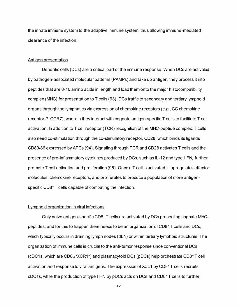
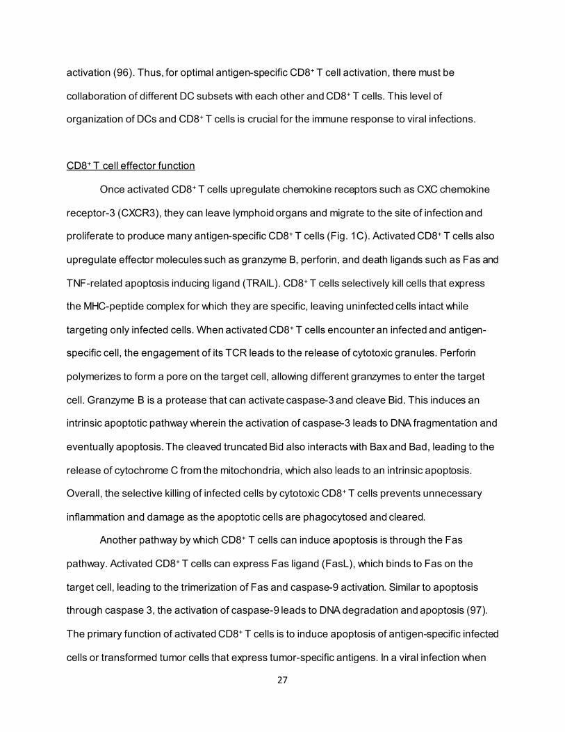
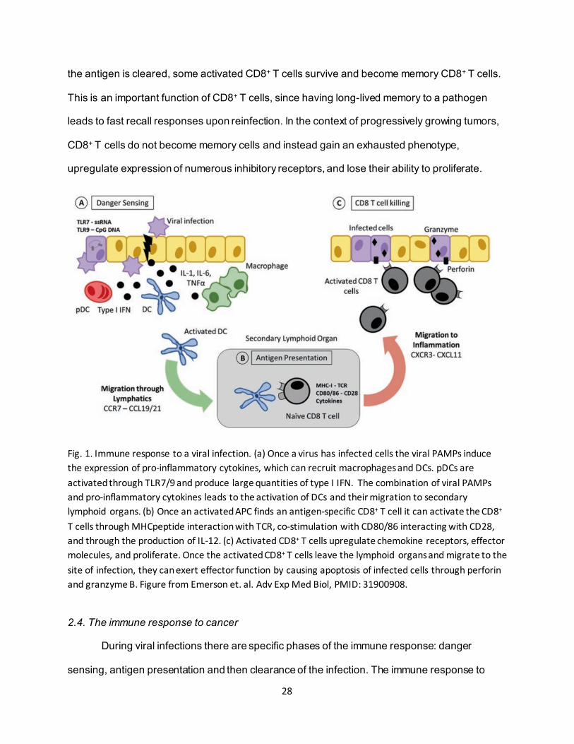
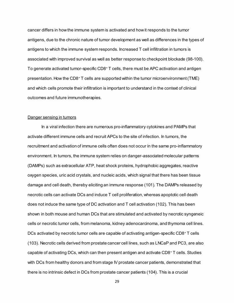
Fig. 1. Immune response to a viral infection. (a) Once a virus has infected cells the viral PAMPs induce the expression of pro-inflammatory cytokines, which can recruit macrophages and DCs. pDCs are activated through TLR7/9 and produce large quantities of type I IFN. The combination of viral PAMPs and pro-inflammatory cytokines leads to the activation of DCs and their migration to secondary lymphoid organs. (b) Once an activated APC finds an antigen-specific CD8+ T cell it can activate the CD8+ T cells through MHCpeptide interaction with TCR, co-stimulation with CD80/86 interacting with CD28, and through the production of IL-12. (c) Activated CD8+ T cells upregulate chemokine receptors, effector molecules, and proliferate. Once the activated CD8+ T cells leave the lymphoid organs and migrate to the site of infection, they can exert effector function by causing apoptosis of infected cells through perforin and granzyme B. Figure from Emerson et. al. Adv Exp Med Biol, PMID: 31900908.
2.4. The immune response to cancer
During viral infections there are specific phases of the immune response: danger
sensing, antigen presentation and then clearance of the infection. The immune response to
29
cancer differs in how the immune system is activated and how it responds to the tumor
antigens, due to the chronic nature of tumor development as well as differences in the types of
antigens to which the immune system responds. Increased T cell infiltration in tumors is
associated with improved survival as well as better response to checkpoint blockade (98-100).
To generate activated tumor-specific CD8+ T cells, there must be APC activation and antigen
presentation. How the CD8+ T cells are supported within the tumor microenvironment (TME)
and which cells promote their infiltration is important to understand in the context of clinical
outcomes and future immunotherapies.
Danger sensing in tumors
In a viral infection there are numerous pro-inflammatory cytokines and PAMPs that
activate different immune cells and recruit APCs to the site of infection. In tumors, the
recruitment and activation of immune cells often does not occur in the same pro-inflammatory
environment. In tumors, the immune system relies on danger-associated molecular patterns
(DAMPs) such as extracellular ATP, heat shock proteins, hydrophobic aggregates, reactive
oxygen species, uric acid crystals, and nucleic acids, which signal that there has been tissue
damage and cell death, thereby eliciting an immune response (101). The DAMPs released by
necrotic cells can activate DCs and induce T cell proliferation, whereas apoptotic cell death
does not induce the same type of DC activation and T cell activation (102). This has been
shown in both mouse and human DCs that are stimulated and activated by necrotic syngeneic
cells or necrotic tumor cells, from melanoma, kidney adenocarcinoma, and thymoma cell lines.
DCs activated by necrotic tumor cells are capable of activating antigen-specific CD8+ T cells
(103). Necrotic cells derived from prostate cancer cell lines, such as LNCaP and PC3, are also
capable of activating DCs, which can then present antigen and activate CD8+ T cells. Studies
with DCs from healthy donors and from stage IV prostate cancer patients, demonstrated that
there is no intrinsic defect in DCs from prostate cancer patients (104). This is a crucial
30
component of generating a productive immune response against tumors, as is the composition
of APCs within the TME capable of presenting tumor antigens to tumor-specific CD8+ T cells.
2.5. Antigen presentation of tumor antigens
APCs are the interface between antigen and T cell activation. Understanding how this
population of cells operates in cancer is key to understanding the initial generation of the anti-
tumor T cell response.
DCs in cancer
Several DC subsets have been classified based upon their phenotype and function in mice and
humans. DCs can be broadly classified by the high expression of CD11c and MHC class II
(MHC II). One crucial subset of DCs for CD8+ T cell activation is the cross-presenting DC, which
refers to the processing and presentation of exogenous antigens on MHC class I molecules
(105). These cells are of interest in the context of cancer immunology since most tumor
antigens are exogenous proteins and must be presented on MHC I in order to activate tumor-
specific CD8+ T cells. The cross-presenting DC (cDC1) subset has been thoroughly
characterized in mice. These cells are defined by CD8α and XCR1 expression and show
increased antigen uptake, processing and presentation on MHC I (106). This DC subset has
also been characterized in human tissue, distinguished by the expression of CD141, and has
been seen in the lung, liver, skin and blood compartments (107). This DC subset is
indispensable in the activation of CD8+ T cells in infection and tumor progression as cDC1-
deficient mice do not control influenza infection or immunogenic tumors in a T cell-dependent
manner (108, 109). Overall, this is a key DC subset in the immune response that specializes in
the activation of viral and tumor-specific CD8+ T cells. Another major DC subset (cDC2) in mice
is characterized by the expression of CD11b on CD11c+ MHC II+ DCs. CD11b+ DC (mice) and
CD1c (humans) are less efficient at cross- presentation of exogenous antigens and therefore
31
thought to mainly activate CD4+ T cells through MHC II (110). Even though there are many
similarities between DC subsets in mice and humans the phenotype of the cells is not always
translatable, which is an important consideration when analyzing and comparing DCs from
mouse and human tissues.
The last major subset of DCs are pDCs, which are a crucial part of the antiviral immune
response. In mouse and humans, this highly specialized subset of cells produces the largest
quantities of type I IFN early after viral infection. These cells circulate in the periphery and
express high levels of TLR7 and TLR9, which activate the pDCs and induce the expression of
pro-inflammatory cytokines. pDCs are a crucial bridge between the innate and adaptive immune
system during viral infections as they promote the activation of DCs and T cells. Although some
studies have shown pDC infiltration in breast cancer predicts poorer overall survival, more
studies are needed to determine how pDCs influence the CD8+ T cell response to tumors (111).
The activation of pDCs is a rapid response to viral infection that may not occur during tumor
growth, and this lack of pDC activation may limit the generation of potent anti-tumor CD8+ T
cells.
There have been numerous studies showing the prognostic power of DC infiltration in
tumors. For example, one study used data from the cancer genome atlas (TCGA) and analyzed
CD103/141-associated genes to determine cross-presenting DC infiltration (cDC1). The ratio of
CD103/141+ signature genes to genes not associated with CD103/141 DCs acts as a prognostic
marker that predicts overall survival in numerous cancer types including breast cancer, head-
neck squamous cell carcinoma, and lung adenocarcinoma (110, 112). This shows how the
extent of DC infiltration alone can have prognostic power over a wide variety of cancers. Other
studies using TCGA data have also determined that the CD141 gene signature correlated with
CD8+ transcript levels (108).This demonstrates the importance of DCs in the TME to promote
and support CD8+ T cell infiltration and how DC and CD8+ T cell infiltration can be used to
predict survival in patients with cancer.
32
Studies have also shown in human melanoma tumors the crucial role of CD141+ DCs
expressing CCR7, which allows for the migration into lymph nodes to present tumor antigens to
tumor-specific CD8+ T cells. The tumors containing higher levels of CCR7 transcripts correlated
with more CD3+ T cell infiltration and better survival (112). DCs need to be able to bring tumor
antigens into the lymphoid organs to activate tumor-specific CD8+ T cells more efficiently due to
the higher concentration of CD8+ T cells and DCs in lymphoid tissues. In prostate cancer after
androgen ablative therapy, there is an increase in DC and macrophage infiltration (113), but
their phenotype before and after treatment remains poorly understood. Other studies, which
also looked at DCs in the blood of prostate cancer patients before and after vaccination (GVAX)
and checkpoint blockade (αCTLA-4; ipilimumab) treatment, revealed that increased CD1c+ DCs
and CD11c+CD14lo DCs predicted better survival with the treatment (114). Collectively, these
studies show that the presence of DCs, especially CD141+ DCs in tumors, correlates with more
T cell infiltration and better prognosis and survival in many tumor types. Understanding why
DCs infiltrate certain tumors may help us understand the differences in CD8+ T cell infiltration.
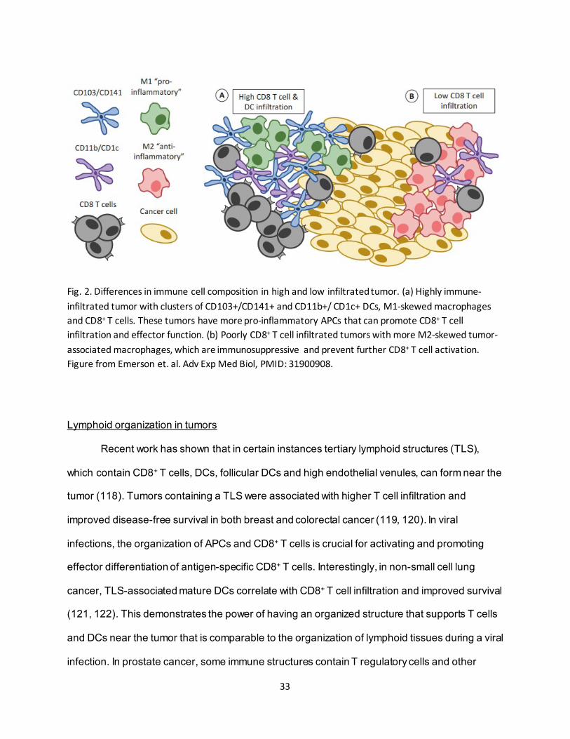
Recent studies have also shown that murine DCs present within the TME are less
efficient at presenting antigen to and activating CD8+ T cells. These DCs induce lower CD8+ T
cell proliferation, express lower levels of co-stimulatory molecules and produce less IL-12 (115).
Additional studies showed an inhibitory effect of IL-10-secreting macrophages in the TME,
which suppresses DC activation and IL-12 production (116, 117). Together, these studies show
that it is important to not only have good DC infiltration in the tumor, but also to have functional
DCs capable of activating T cells and promoting a pro-inflammatory immune response.
33
Fig. 2. Differences in immune cell composition in high and low infiltrated tumor. (a) Highly immune-infiltrated tumor with clusters of CD103+/CD141+ and CD11b+/ CD1c+ DCs, M1-skewed macrophages and CD8+ T cells. These tumors have more pro-inflammatory APCs that can promote CD8+ T cell infiltration and effector function. (b) Poorly CD8+ T cell infiltrated tumors with more M2-skewed tumor-associated macrophages, which are immunosuppressive and prevent further CD8+ T cell activation. Figure from Emerson et. al. Adv Exp Med Biol, PMID: 31900908.
Lymphoid organization in tumors
Recent work has shown that in certain instances tertiary lymphoid structures (TLS),
which contain CD8+ T cells, DCs, follicular DCs and high endothelial venules, can form near the
tumor (118). Tumors containing a TLS were associated with higher T cell infiltration and
improved disease-free survival in both breast and colorectal cancer (119, 120). In viral
infections, the organization of APCs and CD8+ T cells is crucial for activating and promoting
effector differentiation of antigen-specific CD8+ T cells. Interestingly, in non-small cell lung
cancer, TLS-associated mature DCs correlate with CD8+ T cell infiltration and improved survival
(121, 122). This demonstrates the power of having an organized structure that supports T cells
and DCs near the tumor that is comparable to the organization of lymphoid tissues during a viral
infection. In prostate cancer, some immune structures contain T regulatory cells and other
34
immunosuppressive cells (123). In prostate cancer, TLS comprised of more pro-inflammatory
Type 1 helper (Th1) and CD8+ T cells are associated with improved tumor regression (124).
Understanding how these TLS form and how the composition and organization can affect
patient outcomes is an important step towards developing novel therapeutics for patients that
are refractory to immunotherapies and may have immunologically “cold” tumors, which are
poorly infiltrated with CD8+ T cells and/or DCs.
Macrophages in cancer
Macrophages, which are APCs that are a critical component of the TME, are capable of
presenting tumor antigens to T cells, phagocytosing apoptotic cells and secreting various
cytokines (125). Due to the plasticity of these cells, they can acquire different phenotypes based
on the immune environment that influences them. A classically activated macrophage acquires
an M1 pro-inflammatory phenotype, capable of secreting TNF, IL-1, IL-6, IL-8 to promote the
activation of T cells (126). M1-skewed macrophages can also secrete reactive oxygen species
(ROS) and chemokines to attract more pro-inflammatory immune cells to mediate the
destruction of pathogens and tumor cells. The other phenotype of macrophages, that is more
common in the TME, is a “wound repair” alternatively-activated M2 macrophage. These are a
necessary part of the immune response for tissue homeostasis as well as wound repair but are
thought to promote tumorigenesis within the TME. M2 macrophages can secrete anti-
inflammatory cytokines such as IL-10 and TGF-β, and act as poor APCs. M2-skewed
macrophages can also significantly hinder the CD8+ T cell anti-tumor response through
arginase-1 and ROS secretion, which limits CD8+ T cell activation. They can also secrete
growth factors to promote tumor growth and metastasis, such as epidermal growth factor (EGF),
as well as suppress T cell anti-tumor activity (116, 127). Macrophages can also express
programmed cell death ligands 1/2 (PD-L1/2) and further hinder CD8+ T cell anti-tumor activity.
Tumor-infiltrating macrophages with an M2 phenotype can express PD-1 and respond to PD-1-
35
blockade leading to reduced tumor burden in mouse models (128). The location of
macrophages in the TME can alter CD8+ T cell responses, for example when macrophages are
in the stroma of lung squamous-cell carcinomas they prevent the interaction of CD8+ T cells with
DCs that are present in the TME (86). Overall, macrophages are a crucial component of the
TME and can directly impact the tumor-specific CD8+ T cell response.
When macrophages are incubated with conditioned media from prostate cancer cells,
they are skewed towards an M2 phenotype and produce IL-10 (127). Conditioned media from
prostate cancer cells can also re-program M1 macrophages into an M2-like phenotype,
demonstrating how the TME can alter the phenotype of immune cells. Especially in tumors that
are not highly infiltrated by CD8+ T cells and pro-inflammatory APCs, it is important to
understand how the TME can skew the immune environment to promote, rather than inhibit,
tumor growth. A higher macrophage density leads to poor prognosis in lung, hepatocellular
carcinoma, and renal cell carcinomas (129, 130). Macrophages are very versatile APCs that
can promote immune responses as well as the growth and vascularization of tumors.
Understanding what role macrophages play in the TME and how they can directly impact
CD8+ T cells as well as how they can be used to predict clinical outcomes is crucial for
understanding the highly complex TME.
The TME also contains cells broadly classified as myeloid-derived suppressor cells
(MDSCs), which can promote tumor growth and down-regulate CD8+ T cell activity. Currently
there are two subsets that have been classified, monocytic MDSCs (M-MDSCs, which originate
from monocytes) and polymorphonuclear MDSCs (PMN-MDSCs, arising from granulocytic PMN
precursors) (131). In prostate cancer, mostly PMN-MDSCs have been identified and suggested
to promote castrate-resistant prostate cancer (CRPC). M-MDSCs share many markers with
36
monocytes and macrophages, making it difficult to distinguish between these cells. Similarly,
PMN-MDSCs share many markers with other granulocytes and PMNs.
Understanding how the various APC populations influence CD8+ T cell responses is
crucial to determine how to improve immunotherapy in cancers that have not responded to
current therapies. In prostate tumors there is a large population of M2 macrophages, which
could be the reason for less CD8+ T cell infiltration and reduced activation. Historically, prostate
cancer has not responded to T cell-focused therapies. However, when DCs pulsed with tumor
antigen are given as a therapeutic intervention, such as with Sipuleucel-T treatment, a form of
prostate cancer therapy, CD8+ T cell infiltration increased and clinical outcomes were modestly
improved (132, 133). Therapies that focus on enhancing immune infiltration into prostate tumors
are a current area of interest and could potentially be combined with T cell-focused therapies in
order to achieve better clinical outcomes.
2.5. CD8+ T cells in cancer
The extent of DC infiltration correlates with improved CD8+ T cell infiltration and better
overall survival. Even though DCs can predict survival, CD8+ T cells are the effector cells
capable of destroying tumors. The connection between DCs and CD8+ T cells is crucial in
understanding the immune response to cancer. CD8+ T cells found within tumors typically have
an exhausted phenotype, with increased inhibitory receptor expression, like PD-1 or T cell
immunoglobulin and mucin domain-containing protein-3 (Tim-3), along with decreased
proliferation and effector function (83). This does not explain the necessity of DCs within the
TME, but the new stem-like model of CD8+ T cell exhaustion demonstrates the need for a DC-
rich nice to support stem-like CD8+ T cells.
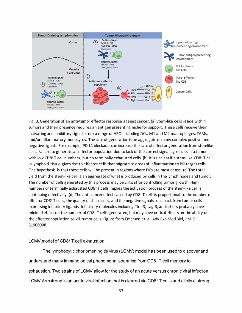
37
Fig. 3. Generation of an anti-tumor effector response against cancer. (a) Stem-like cells reside within tumors and their presence requires an antigen presenting niche for support: These cells receive their activating and inhibitory signals from a range of APCs including DCs, M1 and M2 macrophages, TAMs, and/or inflammatory monocytes. The rate of generation is an aggregate of many complex positive and negative signals. For example, PD-L1 blockade can increase the rate of effector generation from stemlike cells. Failure to generate an effector population due to lack of the correct signaling results in a tumor with low CD8+ T cell numbers, but no terminally exhausted cells. (b) It is unclear if a stem-like CD8+ T cell in lymphoid tissue gives rise to effector cells that migrate to areas of inflammation to kill target cells. One hypothesis is that these cells will be present in regions where DCs are most dense. (c) The total yield from the stem-like cell is an aggregate of what is produced by cells in the lymph nodes and tumor. The number of cells generated by this process may be critical for controlling tumor growth. High numbers of terminally exhausted CD8+ T cells implies the activation process of the stem-like cell is continuing effectively. (d) The anti-cancer effect caused by CD8+ T cells is proportional to the number of effector CD8+ T cells, the quality of these cells, and the negative signals sent back from tumor cells expressing inhibitory ligands. Inhibitory molecules including Tim-3, Lag-3, and others probably have minimal effect on the number of CD8+ T cells generated, but may have critical effects on the ability of the effector population to kill tumor cells. Figure from Emerson et. al. Adv Exp Med Biol, PMID: 31900908.
LCMV model of CD8+ T cell exhaustion
The lymphocytic choriomeningitis virus (LCMV) model has been used to discover and
understand many immunological phenomena, spanning from CD8+ T cell memory to
exhaustion. Two strains of LCMV allow for the study of an acute versus chronic viral infection.
LCMV Armstrong is an acute viral infection that is cleared via CD8+ T cells and elicits a strong
38
memory CD8+ T cell response. The LCMV clone 13 strain models a chronic infection; by
depleting CD4+ T cells and then infecting the mice with clone 13, the infection becomes truly
chronic and is not cleared by the immune system (134, 135). The early model of LCMV-induced
T cell exhaustion described a gradual increase in inhibitory receptor expression and loss of
effector function. CD8+ T cells first lose their ability to produce IL-2 and their cytotoxic function,
followed by the loss of proliferation and ability to produce IFN-γ and TNF-α (84, 136). PD-L1
blockade was shown to rescue antigen-specific exhausted CD8+ T cells in chronic LCMV clone
13 infected mice, thus restoring their ability to proliferate and produce IFN-γ (82). This was the
first example of PD-1 blockade restoring CD8+ T cell functionality in a chronic antigen setting.
Chronically stimulated CD8+ T cells can have the canonical exhausted phenotype. They
have up-regulated numerous inhibitory receptors, such as PD-1, CTLA-4, lymphocyte activation
gene 3 (Lag-3), and Tim-3. They also have a diminished ability to proliferate and produce
effector cytokines (137, 138). This shows that tumor-infiltrating CD8+ T cells have a comparable
phenotype to the antigen-specific CD8+ T cells found in the chronic viral model of LCMV clone
13 infected mice. While this model of T cell exhaustion offers an understanding of why T cells in
tumors have lost functionality, it does not explain why some tumors have very low T cell
numbers, or more importantly why some patients do not respond to checkpoint therapy.
Stem-like model of CD8+ T cell exhaustion
Recent work in the field of CD8+ T cell exhaustion has resulted in a revised model of
how T cell exhaustion occurs (139). The new working model of CD8+ T cell exhaustion has
implications in how we think about CD8+ T cell exhaustion as well as modern immunotherapy
approaches. This work described two populations of CD8+ T cells in a chronic viral infection that
express PD-1. One is a stem-like CD8+ T cell that expresses CXCR5, T cell factor 1 (TCF1),
39
and has higher expression of CD28, while the other is a terminally-differentiated Tim-3+ CD8+ T
cell. The stem-like CD8+ T cells reside in a DC-rich location of the spleen (Fig. 3B), while the
Tim-3+ subset localizes to sites of infection and is not restricted to lymphoid tissues and acts as
effectors, expressing granzyme B and IFN-γ. The CXCR5+ TCF1+ stem-like CD8+ T cell subset
can self-renew and give rise to Tim-3+ effector-like cells (Fig. 3C). During viral infection, PD-1
blockade promotes CXCR5+TCF1+ stem-like cell proliferation and differentiation into a large
population of antigen-specific effector-like CD8+ T cells capable of killing infected cells. PD-1
blockade also affected Tim-3+ CD8+ T cells by blocking negative signaling and increasing their
effector functions at the site of infection (139). This new model of CD8+ T cell exhaustion can
help us understand how the immune system responds to chronic antigen as well as how PD-1
blockade works within this model.
Since this model was described, several groups have observed a stem-like model of T
cell exhaustion in numerous cancers and murine tumor models. Populations of TCF1+ and
terminally-differentiated Tim-3+ CD8+ T cells have been described in MC38 sarcoma, B16
melanoma, Transgenic adenocarcinoma of the mouse prostate (TRAMP)-C1 prostate cancer,
Lewis Lung Carcinoma, and CT26 colon tumors (140, 141). These cells closely resemble the
originally identified CD8+ T cells from chronic viral infection. TCF1+ CD8+ T cells have higher
expression of IL-7R, CCR7 and CD62L, like CXCR5+ cells. The effector TCF1-Tim-3+ CD8+ T
cells were also found to express higher levels of effector molecules such as IFN-γ and
granzyme B, which parallels their original description. An important part of the stem-like model
of CD8+ T cell exhaustion is the ability of TCF1+ CD8+ T cells to proliferate and give rise to Tim-
3+ effector cells. While Tim-3+ CD8+ T cells lack the ability to proliferate, they do express more
effector molecules, such as IFNγ (139). These studies demonstrate that the stem-like model of
CD8+ T cell exhaustion can be applied to numerous mouse tumor models.
40
The stem-like model of CD8+ T cell exhaustion is also translatable to human cancer.
Many recent studies have shown that in different human cancers there are similar stem-like and
effector CD8+ T cell populations. In lung cancer patients, a CXCR5+ CD8+ T cell population, and
a CXCR5-Tim-3+ population was found by using high dimensional mass cytometry (CyTOF)
analysis. These two subsets closely resemble what has been described in mouse models,
wherein CXCR5+ CD8+ T cells retain proliferative capacity and give rise to CXCR5-Tim-3+
effector CD8+ T cells (142). In a single-cell RNA sequencing study of melanoma, a transitional
and dysfunctional CD8+ T cell subset was found. Transitional CD8+ T cells present within tumors
retain TCF1 expression, while dysfunctional CD8+ T cells have higher expression of inhibitory
receptors such as PD-1 and Lag-3 (143). There needs to be both a functional TCF1+ stem-like
CD8+ T cell and it must produce Tim-3+ effector-like CD8+ T cells in order to have a productive
CD8+ T cell response to the tumor.
The environment in which these CD8+ T cell stem-cells are maintained is a crucial
aspect of the stem-model of exhaustion, especially when considering that originally the stem-
like TCF1+ CD8+ T cells were discovered in lymphoid tissues surrounded by DCs in a chronic
viral infection. The organization of DCs and CD8+ T cells is crucial for the activation of CD8+ T
cells in viral infections. These discoveries have led to an increasing interest in the immune
environment that best supports stem-like CD8+ T cells and induces their proliferation and
differentiation, especially within the TME. Since the amount of CD8+ T cells within tumors has
clear positive prognostic power, it is important to understand the immune niche supporting CD8+
T cell infiltration into tumors.
Overall, the immune system is crucial in the response to cancer as CD8+ T cell
infiltration into tumors can predict patient outcomes as well as response to immunotherapy.
Understanding the stem-like model of CD8+ T cell exhaustion has led to a better understanding
of the CD8+ T cell response to PD-1 blockade, which increases the differentiation of Tim-3+
CD8+ T cells as well as their effector function. This model of CD8+ T cell exhaustion has led to
41
an interest in other immune cells within the TME that can support stem-like CD8+ T cells, such
as DCs. CD141+ DC (cDC1) infiltration has been shown to predict better outcomes in patients
as well as correlate with CD8+ T cell infiltration (144). Tumors that cannot support an
environment with DCs and T cells do not have high CD8+ T cell infiltration and therefore have
worse outcomes and respond poorly to checkpoint blockade. The future of immunotherapy likely
needs to focus on rebuilding the tumor immune microenvironment to support DCs and stem-like
CD8+ T cells and facilitate their proliferation and differentiation into effector-like CD8+ T cells
capable of destroying tumor cells. The exhausted T cell phenotype and the transcription factor
TOX will be further discussed in Chapter 5.
2.6. Prostate Cancer Tumor-Associated Antigens
Inducing an immune response against cancer is critical for producing an effective and
long-lasting response. Integral to the anti-tumor immune response is the presentation of tumor-
associated antigens (TAA) on APCs to CD8+ T cells for priming of tumor-specific cytotoxic T
cells. Identifying tumor antigens present in prostate cancer and understanding their role in
inducing an adaptive immune response is essential for developing effective vaccine strategies
that enhance the generation of tumor-specific CD8+ T cells. Several of the most common
prostate cancer tumor antigens being utilized for immune therapy in preclinical models and in
ongoing clinical trials are described below.
Prostate-Specific Antigen
Epithelial cells lining the acini and ducts of the prostate gland produce the serine
protease prostate-specific antigen (PSA), and secrete it into the prostate lumen. PSA aids in
liquefying semen coagulum and is excreted in seminal fluid. In healthy individuals, serum PSA
levels are common at low concentrations (0-2.5 ng/ml), with serum concentrations above 2.5
ng/ml indicating a cause for biopsy (145). Serum concentrations of PSA correlate with prostate
42
cancer disease progression, which makes PSA a useful prognostic marker that aids in the
grading and staging of prostate cancer, as well as an indicator of disease recurrence and
progression (146, 147). However, increased PSA levels are also associated with benign
inflammatory conditions such as benign prostate hyperplasia (BPH) and prostatitis, which may
result in a false positive screen that is not indicative of cancer progression (148).
In addition to its value as a prognostic marker, PSA is an immunogenic antigen that can
drive immune responses (149). PSA-specific CD8+ T cells are found in both healthy individuals
and prostate cancer patients (150). For this reason, PSA can serve as a target for prostate
cancer vaccination to elicit an anti-tumor immune response from CD8+ T cells. For example,
PROSTVAC is a poxvirus-based (Vaccinia and fowlpox) vaccine that expresses the PSA
antigen as well as molecules to aid in T cell stimulation including CD80, intercellular adhesion
molecule 1 (ICAM-1), and lymphocyte function-associated antigen 3 (LFA-3) (151, 152). A
phase II trial for the treatment of metastatic castration-resistant prostate cancer (mCRPC)
showed that patients receiving PROSTVAC had an increase in their median overall survival of
8.5 months compared to control (25.1 vs 16.6 months) (153). However, a recent phase III trial
failed to demonstrate improved overall survival compared to controls (NCT01322490). Failures
to respond to a PSA-expressing vaccine may be the result of a high frequency of PSA-specific
CD8+ T cells expressing Tim-3, a marker of T cell exhaustion (154). This suggests that while
patients possess tumor-specific T cells needed to mount an anti-tumor response, those T cells
may be unable to respond in a productive and effective manner. Thus, current ongoing studies
are assessing the potential benefit of combination immunotherapy, specifically PD-1 checkpoint
blockade, to improve vaccine efficacy.
Prostate Acid Phosphatase
The prostatic epithelium synthesizes prostate acid phosphatase (PAP), a prostate-
specific, secreted glycoprotein enzyme that is involved in the liquification of semen (155).
43
Expression of PAP and its serum concentration correlates with testosterone levels, disease
progression and the amount of bone metastases (156). Due to these prognostic correlations,
PAP serum levels were originally used as a prostate tumor biomarker dating back to the 1940’s,
prior to the adoption of PSA screening in the 80’s. Despite the widespread use of PSA for
prostate cancer screening, PAP still has prognostic value as a biomarker for determining and
differentiating intermediate- and high-risk patients, and for predicting clinical recurrence and
likelihood of developing distant metastases (157).
Like PSA, PAP also has properties as an immunogenic antigen recognized by T cells in
humans and mice (158). Evidence for this is provided by one study showing that intratumoral
injection of a PAP-expressing vector resulted in decreased tumor growth in a xenograft model
(159). Additional studies have shown that PAP expressed by a DNA vaccine successfully
induced PAP-specific CD8+ T cells, while also increasing PSA doubling time, suggesting that
prostate cancer patients may benefit from targeting PAP using cancer vaccines (160, 161).
Sipuleucel-T, the first therapeutic cancer vaccine to receive Food and Drug Administration
(FDA) approval works as an immunostimulant to trigger an anti-PAP immune response for the
treatment of mCRPC (133, 162). Delivery of Sipuleucel-T is achieved using leukapheresis of
patient blood to remove and isolate primary DCs. The patient’s DCs are loaded with PAP
peptide in an incubation step and are additionally stimulated with granulocyte-macrophage
colony stimulating factor (GM-CSF) to promote cell growth and survival in culture. These PAP
peptide-loaded DCs are then re-infused back into the patient to promote a CD8+ T cell-mediated
anti-tumor response. The IMPACT clinical trial assessing the efficacy of Sipuleucel-T
(NCT00065442) showed that patients receiving Sipuleucel-T experienced a statistically
significant increase in median survival time of 4.1 months compared to the control arm.
However, despite an increase in survival time, patients receiving Sipuleucel-T did not
experience a significant decrease in tumor size (133). The following D9901 trial (NCT00005947)
44
confirmed these findings (163). Sipuleucel-T was FDA-approved for mCRPC in 2010 and is in
ongoing clinical trials to evaluate its efficacy when used in combination with other therapies.
Prostate-Specific Membrane Antigen
Prostate-specific membrane antigen (PSMA) is a membrane-bound zinc metalloenzyme
expressed primarily on prostate epithelial cells, although low expression can be found in other
tissues such as the kidney (164). While PSMA is expressed in the healthy prostate epithelium
compared to other tissues, it is also one of the most commonly and highly upregulated genes
found in prostate cancers, including high staining on epithelial cells of prostatic intraepithelial
neoplasia (PIN) and on malignant carcinomas (164). Interestingly, PSMA expression appears to
be linked to androgen deprivation therapy (ADT), with increased expression in response to
therapy. (165). Like PSA and PAP, PSMA expression may be a useful prognostic biomarker. It
has been shown that expression correlates with disease progression and time to recurrence
(166), which could make PSMA useful as a prognostic marker accounting disease progression
and tumor cell disease potential; yet, attempts to utilize PSMA serum levels alone as a
prognostic marker have not been successful. However, other methods involving PSMA
expression have had success, including the ProstaScint scan that combines CT and MRI scans.
The ProstaScint technique utilizes PSMA-specific antibodies bound to radioactive indium-111 to
bind and identify prostate cancer metastases and has proven to be a valuable tool for identifying
distant, remaining, and recurrent disease (167).
PSMA can also be targeted using various forms of monoclonal antibody (mAb) therapy.
For example, unconjugated, radiolabeled, and drug-conjugated humanized mAbs against PSMA
have all been utilized to induce antibody-dependent cellular cytotoxicity (ADCC) and cell death
(168). PSMA contains a cytoplasmic tail internalization signal that induces protein internalization
to the endosome upon ligand binding, which leads to trafficking back to the cell surface or
targeting to lysosomes for protein degradation (169). This key property of PSMA internalization
45
is exploited by treatments to introduce toxins specifically into prostate cancer cells. In
treatments such as D7-PE40, a targeted immunotoxin consisting of an antibody fragment
specific for PSMA is linked to exotoxin A, a dimer protein that blocks protein synthesis through
elongation factor-2 inhibition. This conjugated therapy targeted against PSMA has shown
efficacy in blocking tumor growth in pre-clinical models (170). Other therapeutic strategies
targeting PSMA have been developed, such as chimeric antigen receptor T cells (CAR-T)
specific for PSMA. In a pre-clinical xenograft model of human prostate cancer, anti-PSMA CAR-
T cells were highly effective at eradicating tumors and inducing tumor-specific lysis (171, 172).
Prostate Stem Cell Antigen
Prostate Stem Cell Antigen (PSCA) is a GPI-anchored surface protein involved in stem
cell survival (173). PSCA is highly expressed in prostate cancer, pancreatic cancer, and bladder
cancer epithelial cells (174). Expression of PSCA is found in 90% of primary prostate cancers
and a high proportion of metastatic sites contain amplification of the PSCA gene (175). Further,
PSCA expression is associated with a higher Gleason score, high staging, and cancer
progression to bone metastases (176). In addition to these associations, PSCA detection also
functions as a marker of response to therapy, as decreased PSCA mRNA levels correlate with
response to radiation therapy (177). The PSCA gene is located about 3kb downstream of an
androgen-responsive enhancer, resulting in decreased PSCA expression in response to ADT
(178). However, despite these correlations, not much is known about the role and function of
PSCA in prostate cancer or in normal prostate tissue.
Due to its high expression in primary prostate cancers and metastases, PSCA is being
evaluated as a therapeutic target. Preclinical murine studies have examined the efficacy of a
PSCA-based DNA vaccine and the use of engineered CAR-T cells specific for PSCA (179).
Clinical trials have included examinations of vaccine and mAb therapies targeting PSCA as well.
A phase I/II clinical trial assessed the efficacy and safety of a PSCA peptide-loaded DCs
46
vaccine, which induced an immune response against PSCA and showed promise for patients
with mCRPC (180). A six-patient phase I trial for the treatment of mCRPC utilized another DC-
based vaccine that employed multiple binding epitopes of PSCA. This trial resulted in a
beneficial increase in PSA doubling time and the generation of memory T cell responses against
the peptides administered (181). Finally, a phase I clinical trial for the treatment of mCRPC
tested AGS-1C4D4, a mAb targeting PSCA (182). In this trial, the therapy was well tolerated by
the 13 patients treated, with six patients having stable disease for 24 months. Targeting PSCA
through vaccine and antibody-based therapeutics has so far shown promise, which warrants
further exploration in pre-clinical and clinical studies based upon its induction of memory T cells.
It will be interesting to determine how PSCA-targeted therapeutics will combine and potentially
synergize with established prostate cancer therapies and immunotherapies, such as PD-1
blockade.
Mucin-1
Mucin-1 (MUC1), a highly glycosylated protein, is expressed across a wide variety of
epithelial cancers including prostate adenocarcinoma, but is not detected in healthy prostate
tissue (183). MUC1 is expressed on the apical borders of epithelial cells where it plays a role in
cell adhesion and mucosal barrier protection. MUC1 exists in secreted and membrane-bound
forms; however, in prostate adenocarcinomas and neoplasms, MUC1 is also expressed in the
cytoplasm. Expression of MUC1 correlates with disease progression, tumor volume, and lymph
node metastases (184). However, MUC1 is suppressive of androgen receptor (AR) expression,
leading to decreased androgen sensitivity and response to ADT (185).
Due to its high expression in adenocarcinomas, MUC1 is a target for antibody-based
targeted therapy as well as vaccine therapy. Therapeutic targeting of MUC1 for prostate cancer
is currently being tested in clinical trials. Researchers have developed an adenovirus-based
vaccine that expresses MUC1 along with two other TAAs and contains specific gene
47
modifications that decrease viral gene expression and prevent a host immune response against
the viral protein components (186). This vaccine, ETBX-061, is currently being tested for clinical
trial use. This trial (NCT03481816) is testing the efficacy and safety of the MUC1 viral vaccine
alongside other adenovirus-based vaccines expressing PSA and brachyury proteins for patients
with mCRPC.
NY-ESO-1
NY-ESO-1 is a well-characterized cancer testis antigen of unknown function expressed
in bladder, esophageal, liver, and breast cancer (187). In transitional cell carcinoma, expression
of NY-ESO-1 correlates with staging and seropositivity in high grade patients (188). NY-ESO-1
is a MHC class I and class II antigen that induces cellular and humoral immune responses that
are associated with anti-tumor immune activity (189) and the presence of these NY-ESO-1-
specific T cells correlates with good prognosis. In one study, 10% of prostate cancer patients
had autoantibodies against NY-ESO-1 in their serum, while healthy patients had none (190).
This supports evidence that NY-ESO-1 is expressed highly in cancerous tissue and lowly in
healthy tissue, a property that makes NY-ESO-1 attractive as a potential cancer vaccine
antigen. Over 30 clinical trials have been conducted with NY-ESO-1 vaccines or NY-ESO-1-
targeted adoptive T cell therapy, several of which focus specifically on prostate cancer. Clinical
trial NCT02692976 utilized a DC-based vaccine loaded with NY-ESO-1 and MUC1 peptides,
with separate groups assessing the efficacy of plasmacytoid, myeloid, or the combination of
both subtypes of DCs. The therapy was well-tolerated, with only 33% of patients experiencing
low grade toxicity, and efficacious, with 67% of patients achieving six months of stable disease.
To increase response to this vaccine, combinations of NY-ESO-1 vaccine therapy with
immunotherapies are being considered. Preliminary work with CTLA-4 blockade/NY-ESO-1
vaccine combination therapy demonstrated an increase in the numbers of NY-ESO-1 specific T
cells in metastatic melanoma, which correlated with clinical responses (191).
48
T-cell receptor alternate reading frame protein
T-cell receptor alternate reading frame protein (TARP) is an MHC class II-restricted
protein expressed on prostate and breast cancer epithelial cells (192). TARP expression
correlates with cancer progression and is found in primary and metastatic sites (193-195).
TARP is a 58-residue sequence product of an alternate reading frame of the TCR locus (196).
This alternate reading frame sequence contains two known CD8+ T-cell binding epitopes that
are presented on APCs and prostate cancer cells (197). Testosterone upregulates TARP
expression, which suggests a role for androgens in promoting its expression (198). However,
TARP expression has been observed in androgen-sensitive and androgen-insensitive prostate
cancer (199). Its high expression in prostate cancer makes it an ideal target antigen for cancer
vaccine therapy.
Due to its immunogenicity, TARP-specific vaccination was explored in a phase I clinical
trial. This trial consisted of 41 patients with asymptomatic hormone-sensitive prostate cancer
receiving either emulsified TARP peptide with GM-CSF or DCs pulsed with TARP peptide (200).
The five-dose vaccination schedule was well tolerated and had an acceptable safety profile in
both treatment groups. Clinical outcomes were assessed based upon PSA doubling time, which
is a measure of the time it takes for serum concentrations of PSA to double. Both treatment
groups showed a decrease in PSA doubling time, with 74.2% of total patients displaying a
reduction in PSA doubling time 48 weeks post-treatment. Additionally, the TARP vaccine
increased TARP peptide-specific IFN-γ production, with responses recorded for 80% of patients.
These data suggest that TARP vaccine therapy may help in controlling micro-metastases and
slowing disease progression. A trial with a second generation of this vaccine (NCT02362451) is
underway with modifications that include the addition of MHC class II binding sites to the
peptide sequence (200).
49
GRB2-Like Endophilin B2
The GRB2-Like Endophilin B2 (SH3GLB2) peptide was discovered in the TRAMP
murine prostate model as a stimulator of prostatic adenocarcinoma specific T cells (SPAS1).
TRAMP mice receiving vaccination with the TRAMP-C2 cell line, CTLA-4 blockade, and GM-
CSF experienced a reduction in tumor growth and a reduction in spontaneously forming tumors.
This treatment method was employed to identify unknown prostate cancer antigens, as had
been done previously for melanoma (201, 202). By testing T cell activity from the spleens of
treated mice a 395 amino acid sequence was identified that induced T cell activation and shared
96% homology with human SH3GLB2, a protein of unknown function (203). SH3GLB2 is highly
expressed in prostate cancer metastases and is orthologous to SPAS1, the immunodominant
model antigen in the TRAMP-C1 murine model. Analysis of SH3GLB2 expression in human
prostate cancer has shown that it is highly expressed in lymph node metastases in patients with
aggressive cancer (204). Because T cell responses can be induced in a mouse vaccination
model, it is thought that SH3GLB2 is a viable vaccination target antigen in humans. However, to
our knowledge, there have been no attempts at this therapy as of this time.
Six transmembrane epithelial antigen of the prostate 1
Six transmembrane epithelial antigen of the prostate 1 (STEAP1) is a metalloreductase
enzyme expressed at cell-cell junctions and is overexpressed in the cell membrane and
cytoplasm of prostate cancer cells, with lower expression in healthy tissue (205, 206). STEAP1
is highly expressed in PIN lesions, which suggests that it plays a role in early prostate cancer
development and thus might be an attractive biomarker for early disease (207). STEAP1 has
been a target antigen for antibody-based therapy as well as vaccine therapy due to its high
expression in PIN and more advanced forms of prostate cancer. The CV9103 vaccine utilized
STEAP1 mRNA in a phase I/II trial of 44 patients (NCT00831467) (208). This strategy
effectively induces cytotoxic CD8+ T lymphocytes with reactivity against STEAP1. Additionally, a
50
modified vaccinia virus Ankara (MVA) vector delivery system containing STEAP1 has shown
promise as a vaccine therapy in murine models (209). Developed mAbs against STEAP1 have
also shown promise in murine models by inhibiting xenograft tumor growth. This success has
led to the development of an antibody-drug conjugate (ADC). DSTP3086S is an ADC linked to
monomethyl auristatin E, which inhibits cell growth by blocking tubulin polymerization.
DSTP3086S has shown promise in a phase I clinical trial (NCT01283373) for mCRPC and has
shown greater effectiveness at higher concentrations (210).
2.7. Models of Murine Prostate Cancer
TRAMP
The TRAMP murine model was originally developed in 1995, which was followed by
further characterization and development of cell lines and modified models of TRAMP (211).
The TRAMP model of spontaneous prostate cancer was developed using the simian
vacuolating virus 40 (SV40), a virus with oncogenic proteins, to induce cancer. Expression of
the early region of the virus which is composed of the large and small T antigen was driven by a
modified probasin-ARR2 promoter specific to the prostate. The large-T and small-t antigens of
SV40 protein bind to and inhibit the activity of p53 and Rb tumor suppressors. Additionally, the
small-t antigen binds to phosphatase PP2A along with several other oncogenic intracellular
proteins. Inhibiting p53 and Rb tumor suppressors results in a predictable progression from PIN
lesions to highly penetrant metastatic disease by week 28. All of these mice develop lymph
node metastases and 67% go on to develop pulmonary metastases. However, these mice
exhibit a variable response to ADT, although mice that are resistant to ADT are more likely to
develop metastases (212). Additionally, these mice develop phyllodes-like lesions that have a
leaf-like structure, similar to that found in human breast cancer. It should also be noted that
some studies have demonstrated that TRAMP may not actually be a form of adenocarcinoma
51
and is instead an atypical epithelial hyperplasia that develops into neuroendocrine carcinoma
(213).
Three prostate cancer cell lines were derived from tumor-bearing TRAMP mice (214).
Two of these cell lines (TRAMP-C1 and TRAMP-C2) readily form tumors when implanted
subcutaneously into syngeneic wild-type (C57/BL6) mice. However, the third cell line (TRAMP-
C3) only grows in vitro. These tumorigenic cell lines retained AR expression, but lost expression
of the T antigen found in the transgenic mouse. TRAMP-C1 and TRAMP-C2 are amenable for
studies investigating therapeutic efficacy of various immunotherapies, although it should be
noted that TRAMP-C1 is considered poorly immunogenic with a low level of basal immune
infiltration, while TRAMP-C2 is moderately immunogenic. TRAMP-C1 is responsive to CTLA-4
blockade in a T and natural killer (NK) cell-dependent manner. TRAMP-C2 cells are also
responsive to checkpoint blockade, but can form metastases in the draining lymph nodes and
lungs, thus making it a useful model for surgical resection and metastasis. For example, while
95% of mice with surgically resected primary TRAMP-C2 tumors develop metastases, this was
reduced to 50% following CTLA-4 blockade (215).
LADY
The LADY model of prostate cancer is designed similarly to the TRAMP model;
however, it has modifications in the probasin (PB) promoter region and a mutation to the small-t
antigen, which renders it unable to bind phosphatase PP2A and other oncogenic intracellular
proteins. Many different lines of the LADY model have been generated with variable tag
transgene expression. These lines are named 12t-1 through 11. The 12 in the name indicates
the approximate 12kb promoter length, t for the small-t antigen transgene, and 1 – 11 for the 11
lines generated. The mouse line that develops cancer at the fastest rate is 12t-7, which
progresses to locally invasive adenocarcinoma at 15-22 weeks, but does not become metastatic
52
(216). In contrast, Line 12t-10 grows slowly, progressing to invasive neuroendocrine carcinoma
at 33 weeks, and develops lymph node and lung metastases at 50 weeks-of-age. Considering
the range in variation in metastatic potential seen across the model, these various lines may be
helpful for comparing the genetics involved in metastatic progression.
The TRAMP and LADY transgenic mice each carry advantages and disadvantages as
models of human prostate cancer. For example, both were generated by driving expression of
SV40 T antigens off of a prostate-specific promoter to produce highly penetrant and well
characterized disease, which is a mechanism of oncogenic transformation that is not mirrored in
humans. Not all models of TRAMP and LADY are capable of progressing to metastatic disease,
but both the TRAMP and the 12t-10 LADY models have metastatic potential. Unfortunately,
these models do not reciprocate human prostate cancer progression completely, due to being
driven by an exogenous oncogene that does not exist in humans. This leads to accumulation of
different mutations and subsequently altered disease progression than is found in patients with
prostate cancer. This SV40 oncogene driver produces mostly neuroendocrine cancers and
similar carcinoma subtypes in mice, which likely represents only 25-30% of cases of men with
advanced prostate cancer (217). However, the mice that do develop neuroendocrine prostate
cancer may be useful for studying human neuroendocrine prostate cancer.
c-Myc models
c-Myc is a proto-oncogene commonly overexpressed or mutated in human prostate
cancer. Furthermore, c-Myc overexpression is also found in PIN, which suggests a role in early
cancer progression. Thus, to recapitulate the onset and progression of human prostate cancer,
several mouse models utilize mutations and overexpression of c-Myc along with other
commonly associated mutations of tumor suppressors, such as p53 and phosphatase and
tensin homolog (PTEN). Two models were developed in which c-Myc expression is driven from
either the Pb promoter or the ARR2PB promoter, similar to the TRAMP and LADY models.
53
These two distinct models of c-Myc-driven prostate cancer are categorized as Lo-Myc and Hi-
Myc. They are distinct in their responsiveness to androgens and ADT making them good
models for studying human prostate cancer disease progression and responsiveness. The Lo-
Myc model results in prostate cancer progression that is unresponsive to ADT, which makes this
model castration-resistant. In contrast, the Hi-Myc model exhibits androgen sensitivity, which
reflects the regulation of cMyc transgene expression by the androgen-regulated Pb promoter.
However, in human prostate cancer, castration does not inhibit c-Myc expression (218). Hi-Myc
mice progress to adenocarcinoma around 26 weeks-of-age, while the Lo-Myc model progresses
more slowly with adenocarcinoma developing at 56 weeks (219). However, neither Hi-Myc nor
Lo-Myc models develop spontaneous metastases (219). Similar to what is seen in human
prostate cancer, expression of the prostate-specific tumor suppressor NKX3.1 is reduced in Hi-
Myc PIN and adenocarcinoma (220). Further studies of this model have coupled it to genetic
knockouts that are common in human prostate cancer.
PTEN knockout
PTEN is a major tumor suppressor that functions to dephosphorylate activated AKT and
3-phosphoinositide-dependent protein kinase-1 (PDK1) (221, 222). PTEN is commonly mutated
or lost in many cancers, including a high percentage of human prostate cancers (223, 224).
Original models of PTEN heterozygous mice were not ideal for studying prostate cancer
because many of the mice that survived embryonic development developed other types of
cancers due to the non-specific knockout of PTEN. To overcome these limitations, prostate
tissue-specific PTEN knockout mice were generated using the Cre recombinase system, driving
Cre expression off of a prostate-specific promoter to flox out a section of PTEN flanked by loxP
sites. These mice develop progressive prostate cancer from PIN to metastatic adenocarcinoma
in a manner similar to human prostate cancer progression.
54
Two groups have developed PTEN knockout mice using this system. One group deleted
exon 5 of PTEN and these mice develop PIN at six weeks and adenocarcinoma by 9 to 29
weeks-of-age with lung and lymph node metastases forming at 12 to 29 weeks in 45% of the
mice (225). Additionally, this model may be useful for studying androgen dependency as tumors
initially regress in response to androgen ablation, but then become resistant mirroring what is
observed in humans. Another group developed a PTEN model by deleting both exon 4 and 5.
These mice developed prostate cancer more slowly compared to the exon 5 deleted mice, with
observed lesions by 42 weeks (226). An additional model utilizes a tamoxifen-inducible Cre
transgene knocked in to the NKX3.1 locus, which simultaneously knocks out one allele of
NKX3.1 and brings Cre expression under control of the NKX3.1 promoter (227). This adds
temporal control over PTEN deletion. Mice that receive tamoxifen at two months-of-age develop
high grade PIN and micro-invasive adenocarcinoma (228). PTEN knockout models have also
been combined with targeted deletion of p53 as well. This combination of tumor suppressor
knockouts results in an aggressive prostate cancer by week 11 and eventual death by week 29
(229).
2.8. Prostate cancer immunotherapy: vaccines, checkpoint blockade, and combination therapy
Vaccines
As described previously, Sipuleucel-T was the first therapeutic cancer vaccine to garner
FDA approval and uses PAP-loaded DCs to direct anti-tumor (PAP) responses in patients (133,
162). In contrast, PROSTVAC is a poxvirus-based vaccine expressing PSA along with the co-
stimulatory molecules CD80, ICAM-1, and LFA-3. In addition to these agents, GVAX is another
prostate cancer vaccine platform, and instead of focusing on a single target, such as PSA or
PAP, to induce an anti-tumor immune response, GVAX is a cellular vaccine that contains
irradiated prostate cells from two different human prostate cancer cells lines, LNCaP and PC3
(230). These cell lines have been additionally modified to produce GM-CSF in order to stimulate
55
DCs for antigen presentation. This vaccine could potentially induce immune responses to
multiple prostate cancer antigens at once, which is beneficial to eliminate a heterogeneous
population of prostate cancer cells from the body. This vaccination method would bypass the
need to human leukocyte antigen (HLA) match patients because it relies on the patient’s own
DCs to present antigen in vivo (231). However, despite this potential for an effective and easier
cancer vaccine design, GVAX has not seen success in clinical trials when compared to
docetaxel in two separate phase III trials (NCT00089856, NCT00133224).
CTLA-4 blockade
T cell costimulation by CD28 is critical for their activation and induction of cytotoxic
activity. This pathway is negatively regulated by CTLA-4, which competes for the CD28 ligand.
Ipilimumab is a CTLA-4 blocking mAb that prevents its activity and to increase CD28 signaling
on T cells (232). Initial studies of Ipilimumab in prostate cancer patients led to unacceptable
adverse effects, including death among several patients being treated for CRPC, and
unfavorable outcomes, with no increase in overall survival (233). However, several patients did
have a complete remission in response to this therapy (234). Improvement in patient and
biomarker selection, as well as new and improved CTLA-4 blocking antibodies may limit severe
adverse effects in the future. Ipilimumab is being evaluated for its ability to elicit T cell
responses against tumor-specific neoantigens as part of a phase II trial for CRPC
(NCT02113657) and as neoadjuvant therapy prior to radical prostatectomy (NCT01194271).
While initial trials testing the efficacy of ipilimumab have not resulted in increased overall
survival, ipilimumab may still be beneficial when applied in combination with other therapies. A
phase I clinical trial of GVAX in combination with ipilimumab for patients with CRPC showed that
the treatment had an acceptable safety profile. Additionally, the therapy led to favorable tumor
responses and prolonged survival, especially among patients with higher peripheral blood
56
expression of CTLA-4 and PD-1 on their CD4+ T cells and lower frequencies of regulatory T
cells prior to therapy. Overall, the best predictor of favorable outcome to this therapy was CTLA-
4 expression by CD4+ T cells, which might suggest CTLA-4 as a biomarker for selection of
patients that would benefit from this therapy. A phase II trial is ongoing to determine the efficacy
of the PROSTVAC vaccine in combination with ipilimumab (NCT02506114) as a neoadjuvant
therapy for patients with localized prostatic neoplasia.
Ipilimumab is also being tested in an ongoing phase I trial in combination with GM-CSF
for the treatment of recurrent prostate carcinomas and stage IV prostate cancer
(NCT00064129). It was found that patients with immune-related adverse events (IRAEs) had an
increase in T cell clonality two weeks after initial ipilimumab treatment, and an increase in PSA-
specific T cells was associated with increased T cell diversity (235). Additional trials of
ipilimumab include its use in combination with ADT for CRPC (NCT01377389, NCT02703623),
stage IV and recurrent prostate cancer (NCT01498978, NCT00170157), for chemotherapy-
naïve CRPC (NCT01688492), and for use prior to radical prostatectomy (NCT02020070).
PD-1/PD-L1 blockade
PD-1 is a glycoprotein expressed on T cells that interacts with PD-L1 expressed in
cancer cells and a range of other immune cells, to inhibit T cell activation. Targeting the PD-
1/PD-L1 axis with blocking mAbs is an area of great focus in cancer therapy, having received
FDA approval for several cancer types (236, 237). However, PD-1-targeted therapy in prostate
cancer may have a minimal impact due to lower PD-L1 expression in prostate cancer compared
to other cancers (238). Low PD-L1 expression may explain the minimal response seen in early
clinical trials of nivolumab, which is a PD-1 blocking antibody, for mCRPC. Other studies looking
at pembrolizumab (anti-PD-1 mAb) therapy in combination with enzalutamide for mCRPC have
demonstrated meaningful clinical benefit for a subset of patients (239). This may be a possible
therapeutic avenue to follow after patients’ progress towards androgen insensitivity because
57
during the progression to androgen insensitivity, DCs increase expression of PD-L1 and CD8+ T
cells increase expression of PD-1. A phase III trial is underway to determine the efficacy of
atezolizumab (a PD-L1 blocking antibody) in combination with enzalutamide compared to
enzalutamide alone (NCT03016312). Additionally, the use of ipilimumab is being investigated in
combination with nivolumab in mCRPC (NCT03061539, NCT02985957, NCT02601014).
A phase I/II trial is ongoing to investigate the efficacy of ADXS31-142, a PSA based
vaccine, as a monotherapy or in combination with pembrolizumab for mCRPC (NCT02325557).
The pTVG-HP PAP antigen-based DNA vaccine is being investigated in combination with
pembrolizumab (NCT02499835). An ongoing phase II trial is looking at the efficacy of
pembrolizumab in combination with Radium-223, which targets bone metastases
(NCT03093428) and has shown efficacy in pre-clinical models (240). Another phase II trial is
investigating the efficacy of combination immunotherapy using durvalumab, an anti-PD-L1
antibody, in combination with tremelimumab, and anti-CTLA-4 antibody for the treatment of
mCRPC (NCT03204812), as well as in combination with a TLR3 agonist (NCT02643303).
Nivolumab is being looked at in combination with a poxvirus-based PSA expressing cancer
vaccine (NCT02933255). An ongoing phase II trial is investigating the safety and efficacy of
nivolumab in combination with multiple ADTs and poly-ADP ribose polymerase (PARP)
inhibitors (NCT03338790, NCT02484404). Nivolumab is being investigated as part of a phase I
trial looking at the safety of AM0010, a pegylated IL-10 immune stimulating agent
(NCT02009449). Atezolizumab is being investigated as part of a phase I trial looking at the
safety of CPI-444, an adenosine A2A receptor agonist for the treatment of mCRPC
(NCT02655822).
Other therapies
58
An alternative immunotherapy strategy is using bi-specific antibodies to link T cells to
their cancer cell target (241). A therapeutic method utilizing this strategy targeted PSMA and
CD3 simultaneously to bring T cells in contact with their target cell to induce cytotoxicity. This
therapy has shown promise in xenograft models with a reduction in PSA levels, and reduced
tumor growth (242). These promising findings have led to two phase I clinical trials
(NCT01723475, NCT02262910). CAR-T cells have also been developed to target prostate
cancer antigens. These CAR-T cells are modified by lentiviral- or retroviral-induced expression
of an engineered TCR specific for a tumor surface antigen (243). CAR-T targeting PSMA has
shown promise in pre-clinical murine studies, as well as in a phase I clinical trial
(NCT01140373). CAR-T therapies targeting other prostate tumor antigens such as MUC-1 and
TARP have also shown promise in pre-clinical models (244).
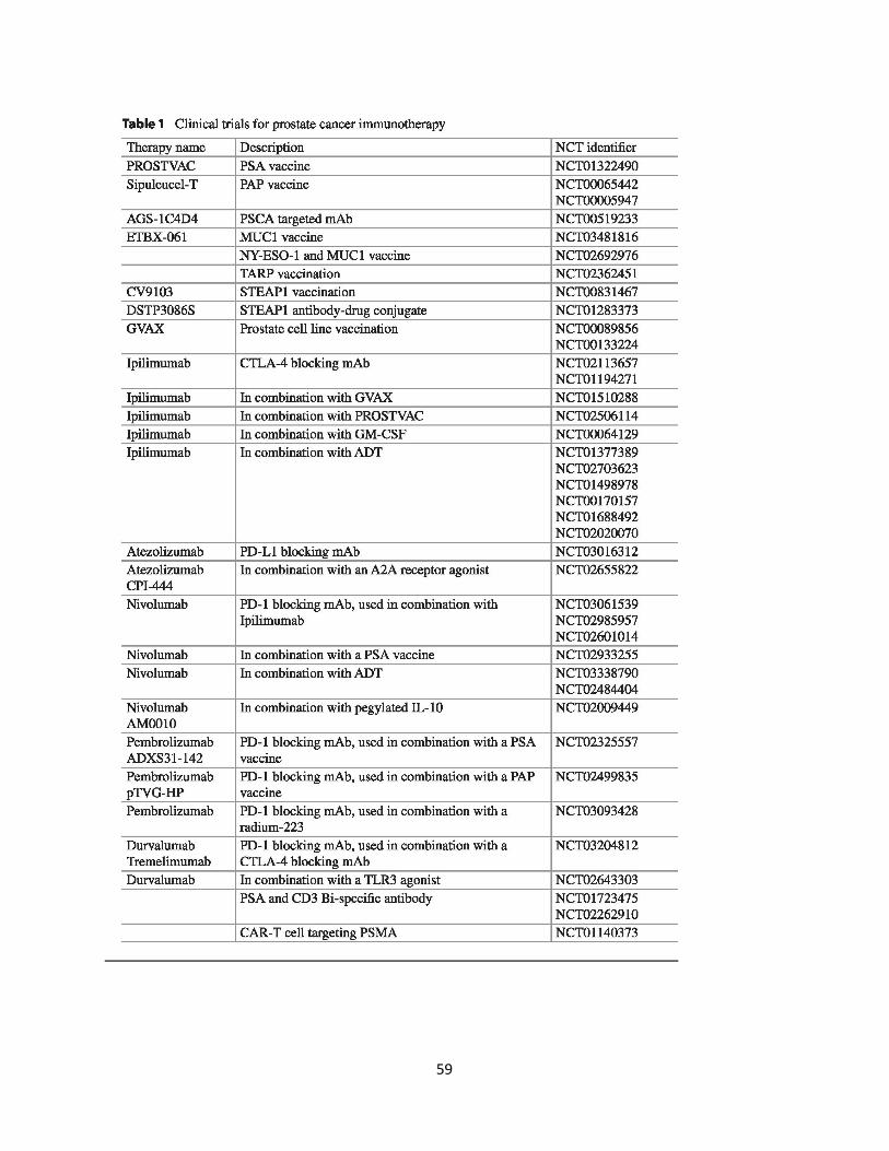
59
60
2.9. Conclusions
Inducing immune responses against TAAs is a critical step in producing an effective and
long-lasting anti-cancer response. Multiple prostate cancer antigens have been identified with
varying degrees of expression in prostate cancer as well as other cancers and healthy tissues.
Many of these associated antigens are capable of being identified by CD8+ T cells and inducing
an effective immune response. However, translating these findings into effective vaccine
therapies has been challenging. The FDA approval of Sipuleucel-T has shown that vaccine
therapy has efficacy in prostate cancer patients. The development of immune checkpoint
blocking antibodies such as ipilimumab, nivolumab, and pembrolizumab have also shown
promise as effective therapeutic strategies in treating prostate cancer. Ongoing and future
approaches are looking at combinations of tumor antigen-specific vaccines along with
checkpoint blockade to activate tumor-specific CD8+ T cells and thereby enhance ant-tumor
immunity.
61
Chapter 3: Immuno-Oncology Drugs: From relieving inhibition to providing co-
stimulation with T cell agonists
Abstract
Recent advancements in T cell biology and antibody engineering have opened the door
to significant improvements in cancer immunotherapy. Initial success with monoclonal
antibodies targeting key receptors that inhibit T cell function such as CTLA-4 and PD-1 have
demonstrated the potency of this new class of therapy, highlighted by long-term complete
responses for metastatic cancers once thought incurable. However, only a subset of patients
will respond to checkpoint blockade due to a multitude of factors including an
immunosuppressive tumor microenvironment and the mutational burden of the cancer. Novel
antibodies as well as ligand-immunoglobulin fusion proteins that target co-stimulatory immune
receptors are being developed and tested in clinical trials to further enhance the anti-tumor
immune response. Many of these co-stimulatory receptors are in the tumor necrosis factor
receptor superfamily (TNFRSF) and are expressed on multiple immune cell types, including
inhibitory cells. While TNFRSFs signal through common pathways, the outcome of targeting
different receptors is dependent on the functional status of the cell types expressing the relevant
receptors. In this review, we will discuss the current state of targeted co-stimulatory
immunotherapy.
62
3.1. Introduction
The generation of potent T cell-mediated anti-tumor immunity relies on the provision of
several critical signals including T cell receptor (TCR)-mediated recognition of peptide-MHC
molecules on antigen-presenting cells (APCs) along with appropriate co-stimulatory signals
(245, 246). Even in the presence of these optimal conditions, tumors utilize a myriad of
mechanisms to evade and suppress the immune response. For example, tumors suppress
recognition and visibility to the immune system by changing their microenvironment (TME) and
reducing MHC class I presentation of tumor-associated antigens (TAAs) (247). Changes to the
TME can occur through secretion of immunosuppressive cytokines such as IL-10 and TGF-β,
which inhibit effector T cell responses and promote regulatory FoxP3+ CD4+ T cell (Treg)
function (248) or through up-regulation of programmed death-ligand 1 (PD-L1), which inhibits
the TCR signaling pathway. PD-L1 binds to programed cell death protein 1 (PD-1) expressed by
activated or exhausted T cells, resulting in phosphatase activity that inhibits T cell receptor
(TCR) kinase signaling (249, 250). Importantly, tumors that are immunologically “silent”, which
refers to low MHC I and high PD-L1 expression, often have little or no response to
immunotherapy (251-254).
Thus, there is an urgent need to develop therapies that enhance tumor antigen
presentation along with T cell priming, activation, and differentiation in order to counteract
tumor-induced immune evasion and suppression. One such approach is to enhance T cell
recognition of TAAs presented on MHC molecules through the provision of exogenous tumor-
specific vaccines, known as therapeutic vaccination (255, 256). However, therapeutic vaccines
have achieved limited clinical success thus far. Another approach is the provision of monoclonal
antibodies (mAb) that block inhibitory checkpoint molecules such as CTLA-4, PD-1, and PD-L1.
In contrast to therapeutic vaccines, checkpoint blockade immunotherapy has demonstrated
significant therapeutic benefit for patients with metastatic cancer, which led to their FDA-
63
approval for a variety of tumor types (251, 252). A third approach is the use of agonist mAbs
that boost T cell function by engaging co-stimulatory molecules such as OX40, 4-1BB, and
CD40. In this Chapter, we review checkpoint inhibitors and their impact on the current
landscape of cancer immunotherapy, followed by a discussion of the current state of therapies
that target T cell co-stimulatory receptors with a focus on OX40, 4-1BB, GITR, CD40, and ICOS
and their use in combination with checkpoint inhibitors.
3.2. Checkpoint inhibitors (anti-CTLA-4, anti-PD-1) and the first bispecific antibody
The first T cell-targeted immunotherapies came after the discovery of immune
checkpoints that regulate activation and inhibition following TCR stimulation. One of the first
inhibitory regulators of T cell function to be cloned was Cytotoxic T Lymphocyte Antigen 4
(CTLA-4) in 1987 (257). CTLA-4 and the homologous receptor CD28 both bind to B7-1 and B7-
2 ligands expressed on APCs (258-260). When CD28 engages B7 ligands it enhances the TCR
signaling pathway through enhanced PI3K activity. However, activated T cells downregulate
expression of CD28 and upregulate CTLA-4, which inhibits T cell signaling by engaging B7
ligands with 20-fold higher affinity than CD28, thereby blocking CD28-mediated costimulation.
Additionally, CTLA-4 is constitutively expressed on Tregs. In mouse models of cancer, CTLA-4
blockade was shown to boost anti-tumor immunity by inhibiting Tregs and enhancing T cell
effector function (261, 262).
These data led to the clinical development of two blocking antibodies targeting human
CTLA-4 (αCTLA-4): ipilimumab (263), and tremelimumab (264). Clinical trials of ipilimumab for
the treatment of metastatic melanoma showed promising results, which lead to a successful
phase III trial in 2009 for patients with high grade unresectable melanoma showed a 10.1 month
overall median survival when receiving ipilimumab alone, compared to 6.4 months survival for
64
patients receiving a gp100 vaccine alone. The addition of the gp100 vaccine to ipilimumab
therapy had no additional impact on overall survival in these patients (265). Tremelimumab,
however, was not as successful perhaps due to its staggered treatment schedule of infusions
every 3 months compared to every 3 weeks for ipilimumab. Ipilimumab was subsequently
approved for metastatic melanoma in 2011. Tremelimumab is still being investigated for several
other indications, but has not seen clinical success for melanoma, small cell lung cancer, and
mesothelioma (266).
The success of immunotherapies such as ipilimumab in 2011 and the vaccine-based
therapy Sipuleucel-T for hormone refractory prostate cancer in 2010 led to the development of
multiple T cell-targeted immunotherapy agents. Leading candidates to be targeted by the next
wave of immunotherapies were T cell checkpoints PD-1 and PD-L1, molecules that inhibit T cell
activity and function when engaged (267). The PD-1/PD-L1 pathway is upregulated in many
cancers and is an important mechanism of inhibiting activated tumor-reactive T cells (268). Two
PD-1 blocking antibodies (αPD-1), pembrolizumab and nivolumab, have demonstrated clinical
success and received FDA approval. Pembrolizumab was approved in 2014 following a
successful phase III trial looking at the use of pembrolizumab following failed ipilimumab therapy
for advanced metastatic melanoma (269). Pembrolizumab was approved for metastatic non-
small cell lung cancer in 2015 (270), metastatic head and neck squamous cell carcinoma in
2016 (271), and DNA mismatch repair deficient solid tumors in 2017. Nivolumab is currently
FDA approved for metastatic melanoma, renal cell carcinoma, lung cancer, bladder cancer, and
Hodgkin’s lymphoma (272). Additionally, three antibodies targeting PD-L1 (αPD-L1) have also
seen success in clinical trials including FDA approval of atezolizumab for the treatment of
metastatic non-small cell lung cancer (273), durvalumab for the treatment of metastatic
urothelial cancer (274), and avelumab for the treatment of non-small cell lung cancer (275).
65
Among this next wave of agents was an alternate class of immunotherapeutic antibodies
that simultaneously target two different receptors, known as Bispecific T cell Engagers (BiTE)
(276). BiTEs are engineered antibodies that contain Fab (Fragment, antigen-binding) regions
against different targets to link two different surface proteins together. If the targeted surface
proteins are expressed on different cells, then the BiTE will link the two cells together. This
rationale was used to develop one of the first BiTEs, blinatumomab (277). This BiTE targets
CD19 overexpressed on B cell malignancies and CD3 expressed by T cells in order to bring the
cancer cell and the T cell together with the goal of simultaneously enhancing T cell activity
through CD3 binding (277). Blinatumomab was FDA approved for acute lymphoblastic leukemia
in 2014.
While these various immunotherapeutic agents have demonstrated marked success,
particularly in a subset of cancers (melanoma, lung), several important issues remain. One of
the primary challenges is that immunotherapy typically only benefits a subset of patients. Other
issues include the onset of potentially severe adverse events (SAE) associated with therapy
such as colitis, diarrhea, and increase alanine aminotransferase, that can be enhanced when
these agents are given in combination, such as in the case of αCTLA-4/αPD-1 therapy (278).
Additional questions remain about treatment timing, dosage, route of administration, and
identification of patients that are most likely to respond to therapy. While ipilimumab,
pembrolizumab, and nivolumab have proven that immune checkpoint blockade can lead to long-
term responses in metastatic disease, immunotherapy agents in development are aimed at
engaging T cell costimulatory receptors to activate and enhance the T cell-mediated anti-tumor
response.
66
3.3. Targeting the TNFRSF, ICOS, and combination immunotherapy
Recent studies have increasingly focused on therapies targeting costimulatory receptors
expressed on activated T cells and antigen presenting cells. Tumor necrosis factor receptor
superfamily (TNFRSF) members are a promising class of immune-modulating molecules that
are under rapid development for cancer immunotherapy. TNFRSF members are typically
homotrimeric transmembrane proteins with a cysteine rich extracellular domain (279).
Intracellular signaling domains of TNFRSFs can induce pro-apoptotic or pro-survival and pro-
inflammatory programs depending on the cell type and signaling domains expressed. TNFRSFs
signal through tumor necrosis factor receptor (TNFR) associated factor (TRAF) adaptor
proteins. Activating TRAF proteins (TRAF1, TRAF2, and TRAF5) signal to activate the
canonical NF-κΒ and JNK pathway and activate the inhibitor of apoptosis (IAP) (280). These
signaling pathways enhance T cell function and survival following TNFRSF engagement.
However, some TNFRSFs, such as TNF-related apoptosis-inducing ligand (TRAIL) receptor and
apoptosis antigen 1 (FAS) receptor, signal through death domains to initiate a caspase-
mediated apoptosis program (281). Multiple pro-survival and pro-inflammatory TNFRSFs are
expressed on T cells following TCR activation, making them promising targets as both
monotherapies and as complimentary targets to the established checkpoint inhibitor
immunotherapy agents. Thus, targeting TNFRSFs with agonistic antibodies and ligand-Fc fusion
proteins has the potential to potently activate and enhance the anti-tumor T cell response.
Antibodies that target receptors can be either inhibitory (antagonist) or activating
(agonist) and must be designed and engineered specifically to accomplish those signals. The
most commonly used antibody isotype for immunotherapy is IgG, however there are several
subtypes of IgG that influence its immune function depending on how they engage Fc𝛾𝛾
receptors or activate the complement system cascade (282). IgG1 strongly induces antibody-
dependent cellular cytotoxicity (ADCC) by engaging Fc𝛾𝛾 receptors which lends itself well to
67
targeting tumor specific antigens (e.g., Herceptin which targets the Her2 receptor on breast
cancer), or potentially depleting inhibitory immune cells, such as Tregs. Nivolumab and
pembrolizumab, however, are IgG4 isotype antibodies, which only weakly engage Fc𝛾𝛾 receptors
and makes them amenable for receptor blockade without inducing ADCC.
Antibodies used for clinical applications are typically produced in 3 forms: murine,
humanized, and fully human. Murine antibodies produced by mouse hybridomas are seen as
foreign, thus they can only be dosed for a short time before the human immune system mounts
an adaptive immune response to clear the foreign (murine) antibody. Humanized antibodies are
modified murine antibodies engineered such that the Fc regions of the Fab domain are
comprised of the human antibody sequence (283). Humanized antibodies can be dosed without
inducing an adaptive immune response (284). An alternative to antibody targeting is to generate
ligand-Fc fusion proteins, which express the natural ligand bound to the Fc region of antibody
rather than engineering a human antibody with different binding kinetics and regions of
interaction compared to the natural ligand (285). This may provide an advantage over
conventional antibodies for targeting TNFRSFs which are homotrimeric and bind homotrimeric
ligands. The natural homotrimeric TNFRSF ligand binds the TNFRSF molecules in a way that
may be different than the complimentary binding region of an antibody to produce superior
engagement.
3.3.1. OX40
OX40 (CD134; TNFRSF4) is expressed by CD4+ and CD8+ T cells following TCR ligation
(286, 287). Murine regulatory T cells (Tregs) also express OX40, although high OX40
expression on human Tregs is only seen following activation (288-290). OX40 is transiently
expressed 24 to 72 hours after T cell activation, which creates a critical window for engagement
68
with its ligand, OX40 ligand (OX40L; CD252). OX40L is also transiently expressed on APCs,
with particularly high expression on CD40-licensed dendritic cells, which are important for
priming CD8+ T cell responses (291). The costimulatory activity of OX40 was initially discovered
in 1987, when it was shown that agonistic antibody targeting of OX40 enhanced CD4+ T cell
proliferation in vitro (292). Following this, the ligand for OX40 was discovered to be a previously
known glycoprotein expressed in human T cell lymphoma/leukemia virus-1 infected cells,
formerly known as gp34 (293).
Early studies on the role of OX40-OX40L interaction in experimental autoimmune
encephalomyelitis (EAE) and murine models of arthritis demonstrated that blocking antibodies
against either OX40 or OX40L effectively decreased autoimmunity (294). Building upon these
data, it was shown that T cells isolated from the TME expressed OX40 and subsequently
hypothesized that these were tumor-reactive T cells. Additional studies revealed that treatment
with an agonist αOX40 mAb or OX40L-IgG fusion protein significantly enhanced tumor-free
survival across four different murine tumor models (295). Depletion of CD4+ or CD8+ T cells
demonstrated that the efficacy of αOX40 therapy was T cell-dependent (296). To investigate if
OX40 expression on CD8+ T cells was necessary for treatment efficacy, OT-I TCR transgenic
mice, which express a T cell receptor specific for the SIINFEKL peptide of ovalbumin protein,
were crossed with OX40-deficient mice, to create OX40-/- OT-I’s. In this antigen-specific model,
OX40-/- OT-I T cells exhibited reduced CD8+ T cell expansion and survival as compared to wild-
type OT-I T cells, highlighting the critical role that OX40 signaling plays in regulating CD8+ T cell
activation and survival (297). Moreover, direct ligation of an OX40 agonist to antigen-specific
CD8+ T cells significantly enhanced expression of the effector molecule, granzyme B, leading to
enhanced tumor-regression and long-term survival of tumor-bearing mice (298).
These preclinical studies showed the efficacy of agonist αOX40 therapy and formed a
rationale for further evaluation in clinical trials. An initial phase I clinical trial used a murine IgG
69
anti-human OX40 mAb for the treatment of patients with metastatic carcinoma, lymphoma, or
sarcoma (NCT01644968). After three doses of αOX40, 12 out of 30 patients saw a reduction in
at least one metastatic lesion (299). However, the study was limited to three sequential doses
given within 5 days due to the use of a murine antibody. Despite the use of a murine antibody,
the trial still demonstrated that αOX40 therapy led to immunological effects (T cell proliferation)
and supported further clinical development. Currently, several phase I and phase II clinical trials
are ongoing to evaluate αOX40 therapy across multiple cancer types including head and neck
(MEDI6469; NCT02274155), colorectal neoplasia (MEDI6469; NCT02559024), metastatic
prostate (MEDI6469; NCT01303705), renal cell carcinoma (PF-04518600; NCT03092856), and
solid tumors (INCAGN01949; NCT02923349). Additionally, there are multiple phase I clinical
trials exploring αOX40 in combination with other immunotherapies including tremelimumab
(αCTLA-4) and durvalumab (αPD-L1) for the treatment of advanced solid tumors (MEDI0562;
NCT02705482, and NCT02205333), in combination with pembrolizumab alone (GSK3174998;
NCT02528357), or the combination of pembrolizumab and nivolumab for advanced cancers
(BMS-986178; NCT02737475), and in combination with a TLR9 agonist for lymphomas (BMS-
986178; NCT03410901). Results from these clinical trials are still pending, however the results
from a phase 1 dose escalation trial for the treatment of advanced solid malignancies using a
combination of αOX40 and αPD-L1 (MOXR0916; NCT02410512) showed no highgrade adverse
effects at the higher dosage, indicating that this combination was well tolerated. Agonist αOX40
therapy is a promising and exciting treatment that may lend itself well to combinations with
checkpoint inhibitors such as αPD-1 and αCTLA-4.
3.3.2. CD40
CD40L (CD154; TNFSF5) is expressed primarily on activated CD4+ T cells, along with B
cells, monocytes, NK cells, basophils, and mast cells (300). This ligand plays an important role
70
in binding CD40 (TNFRSF5) expressed on antigen presenting cells and B cells and acting as an
important signal to induce activation (301). The interaction between CD40 and CD40L is critical
for developing an adaptive immune response that is highly context dependent on the cell types
and cytokines involved. Activated CD4+ T cell CD40L engagement of CD40 on B cells results in
TRAF adaptor protein induction of NF-κΒ, MAPK, PI3K, and PLC𝛾𝛾 pathways that activate B
cells (302, 303). Activated B cells form germinal centers, undergo antibody isotype switching,
and differentiate into plasma cells to produce antibodies (304). Signaling through CD40 on
dendritic cells is a critical step for dendritic cell “licensing” which enables dendritic cells to prime
CD8+ T cell responses effectively through cross-presentation (305-308). Enhancing APC
activation and DC licensing is the main rationale behind agonist aCD40 therapy and the
rationale behind its use in combination therapy (309). Studies of agonistic aCD40 antibodies in
murine models of cancer have shown significant therapeutic efficacy. In a mesothelioma model,
aCD40 was effective in inhibiting tumor growth in a dose-dependent manner (310). Interestingly,
one group discovered that aCD40 therapy was more effective at clearing tumors in a lymphoma
model when treatment was delayed (311). It was speculated that the larger tumor burden of
delayed treatment may increase the available tumor antigens that can be taken up and cross-
presented by dendritic cells.
An initial clinical trial using recombinant human CD40L for non-Hodgkin’s lymphoma
showed some promise when one patient out of 32 had a complete response and another had a
partial response in the absence of major toxicity (312). Several CD40 agonists have moved
forward into clinical trials including the humanized aCD40 CP-870,893, which led to a partial
response in 14% of melanoma patients (NCT01103635) (313). Studies investigating SGN-40
have not induced any clinical responses to date, however SGN-40 is being used in a clinical trial
for lymphomas (NCT00435916) and in combination with rituximab (NCT00655837). Other
agents in clinical trials include HCD122 (lucatumumab) for lymphomas (NCT01275209,
71
NCT00670592) (314), CDX-1140 for solid cancers (NCT03329950), APX005M for solid tumors
(NCT02482168), central nervous system tumors (NCT03389802), and esophageal cancer
(NCT03165994). Additional monotherapy trials include ADC-1013 for solid tumors and
pancreatic cancer (NCT02379741, NCT03214250), and Chi Lob 4/7 for advanced malignancies
(NCT01561911). Combination therapy trials include APX005M in combination with
pembrolizumab for metastatic melanoma (NCT02706353) and in combination with nivolumab for
metastatic lung cancer, melanoma, and pancreatic cancer (NCT03123783, NCT03214250).
While initial clinical trial data suggests that agonistic CD40 agents may not be effective
as monotherapies, CD40 agonists may be effective agents in combination with other
immunotherapies. Agonistic CD40 agents may also synergize well with chemotherapies and
radiation, which release tumor antigens to aid in the antigen presentation process. Different
antibodies have seen varying levels of SAEs, which suggests that a clear understanding of the
optimal timing and dosage for these drugs will be critical to their development and future
success (315).
3.3.3. 4-1BB
4-1BB (CD137; TNFRSF9) is a glycosylated costimulatory molecule expressed
transiently on activated T cells, NK cells, dendritic cells, as well as expressed constitutively on
Tregs (316). Engagement of 4-1BB by its ligand 4-1BBL (CD137L; TNFSF9) induces strong T
cell activation and survival, particularly in activated CD8+ T cells (317, 318). The role of 4-1BB in
cancer immunology has been evaluated in 4-1BBL-deficient mice, which develop spontaneous
B cell lymphomas, and in 4-1BB-/- mice, which develop systemic lupus erythematosus likely due
to tumor suppressive activity of 4-1BBL for B cells (319, 320). Initial studies of 4-1BB agonists in
cancer models of sarcoma and mastocytoma showed enhanced numbers of tumor-specific
72
CD8+ T cells and improved T cell memory against tumor re-challenge (321). 4-1BB agonists
have since been shown to have efficacy across multiple tumor models by inducing a population
of tumor-specific cytotoxic CD8+ T cells that produce potent pro-inflammatory cytokines and
effector molecules, such as granzymes (322-325).
4-1BB also plays an important role in activating and enhancing NK cell function through
Fc𝛾𝛾 receptors (326). Based on this finding, it was hypothesized that 4-1BB stimulation of NK
cells aids in NK mediated antibody-dependent cell-mediated cytotoxicity (ADCC). This would
make agonistic 4-1BB therapy a candidate for use in combination with tumor antigen-targeted
antibodies such as rituximab, which targets CD20 for the treatment of non-Hodgkin’s lymphoma
and chronic lymphocytic leukemia. In murine models of lymphoma, an agonist a4-1BB showed
strong efficacy when given after rituximab treatment (327).
A phase I clinical trial of utomilumab (α4-1BB) in combination with rituximab for patients
with B cell lymphomas resulted in two complete responses in follicular lymphoma that lasted
beyond 2 years (NCT01307267). Additionally, an increase in memory T cells and activated NK
cells was observed. No significant severe adverse effects were reported with no patients
stopping treatment due to toxicity in the utomilumab study (328). Utomilumab is also being
tested in multiple clinical trials with other agents such as avelumab (αPD-L1; NCT02554812),
αOX40 (NCT02315066), mogamulizumab (an aCCR4 antibody; NCT02444793), and
pembrolizumab (NCT02179918). Another phase I/II study of an a4-1BB agonist (urelumab;
NCT02253992) resulted in dose-dependent adverse effects and a 50% objective response rate
in advanced metastatic melanoma patients when given with αPD-1 (nivolumab), and is also
being tested in combination with nivolumab for bladder cancer and malignant tumors
(NCT02845323, NCT02534506). Clearly, 4-1BB agonists are an agent of great interest as both
a monotherapy and in combination with other immunotherapies.
73
3.3.4. GITR
Glucocorticoid-induced TNFR-related protein (GITR; CD357; TNFRSF18) is expressed
on activated T cells, constitutively expressed on Tregs, and moderately expressed on memory T
cells (329, 330). Once activated, T cells transiently express GITR 24 hours after stimulation.
GITR expression is regulated by the FoxP3 transcription factor in Tregs and by canonical NF-κΒ
signaling in activated T cells (331). GITR signaling in activated T cells lowers the threshold for
CD28 co-stimulation and results in NF-κΒ, MAPK, and JNK signal pathway activation through
TRAF adaptor proteins (332, 333). However, the rationale for targeting GITR with monoclonal
antibodies relies on its high constitutive expression on Tregs and costimulatory signaling in
CD4+ and CD8+ T cells. GITR was originally thought to be a unique marker of Tregs before later
being found on other cell types (334). Anti-GITR antibodies seem to be well tolerated without
inducing significant autoimmunity. Targeting GITR with IgG1 antibodies has been effective at
depleting tumor-infiltrating Tregs from the tumor microenvironment, but not in the periphery in a
B16 mouse model of melanoma (335). Using a FoxP3-GFP transgenic mouse, it was
discovered that αGITR was effective in depleting Tregs in B16 tumor-bearing mice, resulting in
tumor clearance (336). On activated CD4+ and CD8+ T cells, GITR is upregulated following TCR
stimulation and signals through activating TRAF molecules to enhance proliferation, pro-
inflammatory cytokine production, and resistance to Treg-mediated suppression (337-339).
Based on the high expression of GITR on tumor infiltrating Tregs, αGITR therapy is
expected to be more effective in cancers with high levels of infiltrating Treg such as cervical,
renal cell carcinoma, hepatocellular carcinoma, lung, and melanoma (340, 341). Therefore, it is
hypothesized that αGITR therapy would synergize well with other immunotherapies, however
the potential for more severe SAEs resulting from Treg depletion will need to be taken into
consideration. Multiple clinical trials are underway testing both αGITR mAbs and GITRL-Fc
fusions including trials as monotherapy (INCAGN0187; NCT02697591), (GWN323,
74
NCT02740270), (TRX518; NCT01239134), and (OMP-336B11; NCT03295942). Anti-GITR
mAbs are also being tested in combination with pembrolizumab (MK-4166; NCT02132754,
NCT02553499), (INCAGN0187; NCT03277352, NCT03126110) and nivolumab (BMS-986156;
NCT02598960, NCT03335540). MEDI1873 is a novel hexameric GITRL-Fc fusion protein aimed
at enhancing activated T cell function, rather than depleting Tregs, and has shown superior
activity compared to αGITR mAbs in vivo (NCT02583165) (342).
3.3.5. ICOS
Inducible T-cell COStimulator (ICOS; CD278) is an immunoglobulin superfamily receptor
expressed on activated T cells (343). Its ligand, ICOSL (CD275; B7-H2) is expressed on both B
cells and dendritic cells (344). ICOS, CD28, and CTLA-4 all share a homologous proline-rich
motif that facilitates their binding to B7 ligands. While CD28 and CTLA-4 can bind B7-1, B7-2,
and B7-H2, ICOS is only known to bind B7-H2, although in a different position from CD28 and
CTLA-4 and at much higher affinity (345, 346). ICOS stimulation by ICOSL induces PI3K and
AKT pathway signaling, which aides in CD4+ T cell differentiation into follicular T helper cells
(TFH), Th1, and Th2 T cells (347). The effect of ICOS signaling on activated CD4+ T cells
appears to be context dependent, although signaling likely drives IL-4 and IL-10 production for
Th2 differentiation in the absence of additional stimulation (348).
Interestingly, ICOS is upregulated on CD4+ T cells following αCTLA-4 therapy,
suggesting that ICOS expression is linked to CTLA-4 and may play a compensatory role to
CTLA-4 when CTLA-4 is blocked therapeutically (349). Additionally, therapeutic synergy was
observed when both pathways were targeted (349-351). In the B16 melanoma model, CTLA-4
blockade and an agonist αICOS mAb synergized to provide protection that the monotherapies
did not (349). Clinical trials for αICOS include MEDI-570 for various lymphomas
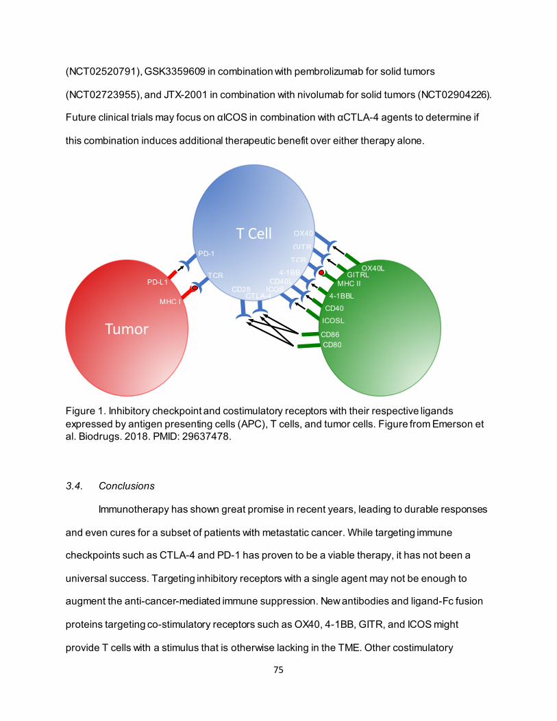
75
(NCT02520791), GSK3359609 in combination with pembrolizumab for solid tumors
(NCT02723955), and JTX-2001 in combination with nivolumab for solid tumors (NCT02904226).
Future clinical trials may focus on αICOS in combination with αCTLA-4 agents to determine if
this combination induces additional therapeutic benefit over either therapy alone.
Figure 1. Inhibitory checkpoint and costimulatory receptors with their respective ligands expressed by antigen presenting cells (APC), T cells, and tumor cells. Figure from Emerson et al. Biodrugs. 2018. PMID: 29637478.
3.4. Conclusions
Immunotherapy has shown great promise in recent years, leading to durable responses
and even cures for a subset of patients with metastatic cancer. While targeting immune
checkpoints such as CTLA-4 and PD-1 has proven to be a viable therapy, it has not been a
universal success. Targeting inhibitory receptors with a single agent may not be enough to
augment the anti-cancer-mediated immune suppression. New antibodies and ligand-Fc fusion
proteins targeting co-stimulatory receptors such as OX40, 4-1BB, GITR, and ICOS might
provide T cells with a stimulus that is otherwise lacking in the TME. Other costimulatory
76
therapies are targeting alternative (non-T cell) cell types such as dendritic cells (aCD40 therapy)
and NK cells (a4-1BB). Moving forward, there is a critical need to understand how to rationally
combine these agents, including balancing increased efficacy with the potential for increased
toxicity. As the field moves into combination therapies of inhibitory and activating antibodies,
sequencing of these agents will also be critical for eliciting potent anti-cancer responses.
However, synergistic combination therapy may have the added benefit of working effectively at
lower doses, leading to less severe adverse events. A great deal of effort is also focusing on
identifying predictive biomarkers of response. For example, tumor-specific PD-L1 expression
has already proven to be a predictive marker for αPD-1 therapy in some instances, however
additional immunological analysis may be critical for deciding which immunotherapies to use in
combination. In conclusion, immunotherapy targeting of co-stimulatory receptors, particularly of
the tumor necrosis factor receptor superfamily, are a promising addition to the growing list of
immunotherapy agents being tested in clinical trials.
77
Chapter 4: Enhancing the generation of Eomeshi CD8+ T cells augment the efficacy of
OX40- and CTLA-4–targeted immunotherapy
Abstract
CTLA-4 blockade in combination with an agonist OX40-specific mAb synergizes to
augment antitumor immunity through enhanced T-cell effector function, leading to increased
survival in preclinical cancer models. We have shown previously that anti-OX40/anti-CTLA-4
combination therapy synergistically enhances the expression of Eomesodermin (Eomes) in
CD8+ T cells. Eomes is a critical transcription factor for the differentiation and memory function
of CD8+ T cells. We hypothesized that EomeshiCD8+ T cells were necessary for anti-OX40/anti-
CTLA-4 immunotherapy efficacy and that further enhancement of this population would improve
tumor-free survival. Indeed, CD8+ T cell–specific deletion of Eomes abrogated the efficacy of
anti-OX40/anti-CTLA-4 therapy. We also found that anti-OX40/anti-CTLA-4-induced Eomeshi
CD8+ T cells expressed lower levels of checkpoint receptors (PD1, Tim-3, and Lag-3) and
higher levels of effector cytokines (IFNγ and TNFα) than their Eomeslo counterparts. Eomes
expression is negatively regulated in T cells through interleukin-2–inducible T-cell kinase (ITK)
signaling. We investigated the impact of modulating ITK signaling with ibrutinib, an FDA-
approved tyrosine kinase inhibitor designed to inhibit BTK, and found that anti-OX40/anti-CTLA-
4/ibrutinib therapy further enhanced CD8+ T cell–specific Eomes expression, leading to
enhanced tumor regression and improved survival, both of which were associated with
increased T-cell effector function across multiple tumor models. Taken together, these data
demonstrate the potential of anti-OX40/anti-CTLA-4/ibrutinib as a triple therapy to improve the
efficacy of immunotherapy.
78
4.1. Introduction
Generating effective CD8+ T-cell responses is critical to support the efficacy of cancer
immunotherapies, such as immune checkpoint blockade (ICB) (352). ICB can generate robust
tumor-specific immunity in patients, leading to improved long-term survival (265). However, the
therapeutic efficacy of ICB has been limited to a subset of patients, highlighting the need to
understand the underlying mechanisms by which they function to inform the design of rational
combinations. Blockade of the inhibitory checkpoint receptor cytotoxic T-lymphocyte-associated
protein 4 (CTLA-4) effectively releases the brakes on T cells through enhanced priming and
inhibition of regulatory FoxP3+ CD4+ T cells (Treg) (353). A CTLA-4-specific mAb, ipilimumab,
was the first checkpoint inhibitor to garner FDA approval and markedly improves 5-year survival
in patients with metastatic melanoma (265, 354, 355). Immune checkpoint inhibitors targeting
the negative regulatory molecule programmed cell death protein 1 (PD1, also known as CD279)
and its ligand PD-L1 (also known as B7-H1 and CD274) are approved for the treatment of a
variety of cancer types including melanoma, non-small cell lung cancer, Hodgkin’s Lymphoma,
renal cell carcinoma, and others (356, 357). Alternatively, agonist mAbs targeting co-stimulatory
members of the tumor necrosis factor receptor (TNFR) family, including OX40 (also known as
CD134), 4-1BB (also known as CD137) and CD27, can boost T-cell responses to augment
antitumor immunity (358). Specifically, agonist OX40-specific mAb therapy enhances T-cell
proliferation, survival, cytokine production, and the generation of long-lived memory T cells (359,
360).
Combination therapy with anti-CTLA-4 and anti-PD1 elicited robust tumor regression in
preclinical and clinical studies leading to its FDA approval for treating metastatic melanoma and
renal cell carcinoma (361), highlighting the potential of combination immunotherapy.
Alternatively, combining ICB with co-stimulatory receptor stimulation enhances antitumor
immunity in numerous preclinical models. For example, we found that anti-OX40/anti-CTLA-4
combination therapy significantly enhances overall treatment efficacy compared to either
79
monotherapy through the generation of effector T cells with increased proliferation and
expression of effector cytokines (granzyme B/IFNγ) and the T-box transcription factor
Eomesodermin (Eomes) (296, 362). Eomes and T-bet both drive the generation of effector
cytokines and cytotoxic molecules, but T-bet preferentially drives the formation of short-lived
effector cells (SLEC) and is sufficient for the effector function of T cells, whereas Eomes is
required for the generation of longer-lived memory precursor effector cells (MPEC) through
increased expression of anti-apoptotic molecules and survival (363-365). Increased Eomes
expression in CD8+ T cells is also associated with better clinical responses to immunotherapy
(366, 367), suggesting that inducing Eomes may further improve outcomes.
Eomes expression in CD8+ T cells is regulated by multiple signaling pathways triggered
by T-cell activation and differentiation, including TCR signaling, NF-κB signaling, and interleukin
receptor signaling (368, 369). TCR signaling through Zap70 and Lck phosphorylates interleukin-
2–inducible T-cell kinase (ITK) to induce phospho-IRF4 expression, which in turn represses
Eomes (370). ITK/IRF4-mediated repression of Eomes is dictated by the strength of TCR
signaling since high affinity TCR binding leads to greater Eomes repression (371). We
hypothesized that Eomeshi CD8+ T cells were a critical component of anti-OX40/anti-CTLA-4
therapy and that the efficacy of anti-OX40/anti-CTLA-4 therapy could be enhanced by
pharmacological blockade of ITK, which would enhance CD8+ T cell–specific Eomes
expression.
In the current study, we demonstrated that combined anti-OX40/anti-CTLA-4 therapy
induced a unique population of Eomeshi CD8+ T cells, which resembled central memory cells
with high levels of proliferation and effector function, as defined by RNA and protein expression.
Furthermore, ITK inhibition with the FDA-approved drug ibrutinib further enhanced Eomes
expression in the presence of anti-OX40/anti-CTLA-4 therapy, resulting in significantly improved
effector T-cell function, tumor regression, and survival in comparison to anti-OX40/anti-CTLA-4
80
alone. These data suggest that ITK inhibition enhances the efficacy of combination
immunotherapy for the treatment of cancer.
81
4.2. Materials and Methods
Mice
Wild-type BALB/c, C57BL/6, Eomesf l/fl, CD8cre, and Nur77GFP reporter mice were purchased from
Jackson Laboratory. Eomes-GFP mice (C57BL/6 background) were kindly provided by Dr. John
Wherry, University of Pennsylvania and Dr. Joseph Sun, Memorial Sloan Kettering Cancer
Center. Eomes conditional knockout mice were bred in our facility by crossing Eomesf l/fl mice
with CD8cre mice (all on the C57BL/6 background). All mice were housed under specific
pathogen-free conditions in the Providence Portland Medical Center (Portland, OR) vivarium
animal facility. All experimental procedures were approved by the Providence Portland Medical
Center Institutional Animal Care and Use Committee (IACUC) and were performed under the
NIH Guide for the Care and Use of Laboratory Animals.
Cell lines and Cell-culture Conditions and Reagents
TRAMP-C1 prostate adenocarcinoma, MCA-205 fibrosarcoma, and 4T1 mammary carcinoma
cell lines were obtained between 2009 and 2011 from Dr. Andrew Weinberg (TRAMP-C1, MCA-
205) and Dr. Emmanuel Akporiaye (4T1). Cell lines have not been re-authenticated within the
past year. All cell lines were maintained in 10% cRPMI and verified Mycoplasma-free
(MycoAlert Mycoplasma Testing Kit; Lonza) within 6 months of use.
Tumor Challenge and Treatments
TRAMP-C1 prostate adenocarcinoma cells (1 x 10⁶) were injected subcutaneously into the right
flank of naïve male C57BL/6 mice. MCA-205 fibrosarcoma cells (1 x 10⁶) were injected
subcutaneously into the right flank of naïve female C57BL/6 mice. 4T1 mammary carcinoma
cells (5 x 10⁴) were injected into the mammary fat pad of naïve BALB/c mice. Tumor-bearing
mice were measured for tumor growth every 2 to 3 days by tumor area using microcalipers and
were sacrificed when tumor area exceeded 175 mm2. Tumor-bearing animals were treated with
82
200 µg rat IgG (Sigma; I4131), and/or 250 µg anti-OX40 (clone OX86; BioXCell; BE0031),
and/or 200 µg anti-CTLA-4 (clone 9D9; BioXCell; BE0164), and/or 200 µg anti-PD-1 (clone
RMP1-14, BioXCell; BE0146) (all ip). All mAbs were verified to be endotoxin-free. Ibrutinib (Cat
S2680 SelleckChem) treatments were administered at a dose of 6 mg/kg (ip) as previously
described (372). For tumor growth and survival experiments, tumor-bearing mice were treated
with anti-OX40 on days 8, and 12; and anti-CTLA-4 was given on days 8, 10, and 12. Control
mice were treated with rat IgG on days 8, and 12. Ibrutinib was dosed on days 8, 10, 12, 15, 17,
and 19 for tumor growth and survival experiments. For tissue harvest experiments, treatments
were initiated when average tumor size exceeded 60mm2, following the same schedule as the
survival experiments. FTY720 (Sigma; SML0700) was dosed at 1 mg/kg (ip) every three days
starting one week prior to immunotherapy.
Tissue Isolation
Tumors and lymph nodes (inguinal, axillary, and brachial) were harvested from tumor-bearing
mice one week after the initiation of treatment. Lymph nodes were mechanically fragmented
between two frosted slides and filtered through nylon mesh for flow cytometry staining. Tumors
were mechanically fragmented into small pieces and digested with 5 mg/ml DNase (Sigma;
4536282001) and 1 mg/ml collagenase (Sigma; C8051) in serum-free RPMI (Lonza; 12-702Q).
Digested tumor suspension was filtered through nylon mesh and stained for flow cytometry as
described below.
Flow Cytometry/FACS Analysis
Single-cell suspensions of blood, lymph node, and tumor-infiltrating lymphocytes (TIL) were
stained with surface antibodies in flow cytometry wash buffer for 30 minutes at 4°C with surface
antibodies: CD45 BV421(Biolegend; Clone 30-F11; 103134), CD4 BV605 (Biolegend; Clone
83
RM4-5; 100548), CD8 BV785 (Biolegend; Clone 53-6.3; 100750), CXCR3 AF488 (Biolegend;
Clone CXCR3-173; 126542, PD-1 PE-Cy7 (Biolegend; Clone 29F.1A12; 135216), LAG-3
PerCP-Cy5.5 (Biolegend; Clone C9B7W; 125212), TIM-3 PE (R&D Systems; Clone NA;
FAB1529P), CD11b BV785 (Biolegend; Clone M1/70; 101243), Ly6C PerCP-Cy5.5 (Biolegend;
Clone HK1.4; 128012), Ly6G FITC (BD Biosciences; Clone 1A8; 551460), MHC II eF450
(eBioscience; Clone M5/114.15.2; 48-5321-82), PD-L1 BV711 (Biolegend; Clone 10F.9G2;
124319), CD11c APC (eBioscience; Clone N418; 17-0114-82), CD86 PE-Cy7 (Biolegend; Clone
PO3; 105116), CD44 APC (eBioscience; Clone IM7; 17-0441-83), CD62L AF700 (Biolegend;
Clone MEL-14; 104426). Cells were fixed and permeabilized using the Thermofisher FoxP3
intracellular staining buffer kit (00-5523-00) for intracellular staining as per the manufacturer’s
instructions. Cells were stained intracellularly for 30 minutes at 4°C in accordance with the
manufacturer’s protocol. For intracellular cytokine staining, cells were incubated in 96-well U-
bottom plates in 10% complete RPMIand 2.5 µl/ml Golgi Block/Brefeldin A (BD Biosciences; 51-
2301KZ). 10% complete RPMI consists of RMPI (Lonza; 12-702Q), 10% Fetal Bovine Serum
(Lonza;14-501F), 1% HEPES Bummer from 1M stock (Lonza; 17-737E), 1% Sodium Pyruvate
Solution from 100mM stock (Lonza; 13-115E), 1% Non-essential amino acids (Lonza; 13-114E),
1% Pen/Strep/Glutamine (Invitrogen; 10378016), 0.005% 2-Mercaptoethanol (Sigma Aldrich;
M3148-100ml). Cells were stimulated by 2 µg/ml plate bound anti-CD3 (BioXcell; BE0001-1-
5MG) and 5 ug/ml anti-CD28 (BioXcell; BE000150105MG) in suspension. Following a 4hr
stimulation at 37 °C in a 5% CO2 incubator, cells were stained at 4 °C for 30 minutes with:
Eomes eF660 (eBioscience; Clone Dan11mag; 50-4875-82), Granzyme A PE-Cy7
(eBioscience; Clone GzA-3G8.5; 25-5831-82), IFN-γ PE (eBioscience; Clone XMG1.2; 12-7311-
82), TNF-α PE-Cy7 (eBioscience; Clone TN3-19.12; 25-7423-82), Ki-67 FITC (BD Biosciences;
Clone B56; 556026), T-bet eF660 (eBioscience; Clone eBio4B10; 50-5825-82) , pITK PE
(Biolegend; Clone A16064A; 646904), IRF4 PE-Cy7 (Biolegend; Clone IRF4.3E4; 646414),
84
CXCL9 AF647 (Biolegend; Clone MIG-2F5.5; 515606), CCL5 PE (Biolegned; Clone 2E9/CCL5;
149104). Stained cells were analyzed using an LSR II flow cytometer running Diva (BD
Biosciences) software and the data was processed using FlowJo 10 software (BD Biosciences).
For lymph node and TIL, cells were counted and quantified for viability using the Guava cell
counter as per the manufacturer protocol.
Tetramer Staining
PE-conjugated H-2Db tetramer to SPAS-1 (STHVNHLHC) peptide and APC-conjugated H-2Ld
tetramer to AH1-A5 (SPSYAYHQF) peptide was provided by the NIH Tetramer Core Facility.
Cells were surface stained with tetramer (1µl per 106 cells) in flow cytometry wash buffer for 15
minutes at room temperature prior to surface antibody staining, as described above.
Flowcytometry wash buffer consists of PBS, 0.5% FBS (Peak Serum; PS-FB4), 0.4% EDTA
(Alfa Aesar; 60-00-4, and 0.05% NaN3 Sigma Aldrich; 26628-22-8).
CTL Assay
MCA-205 tumor-bearing mice were treated with anti-OX40/anti-CTLA-4 therapy starting on day
12 and then tumors were harvested 7 days later. Tumors were pooled and processed for
surface cell staining and sorting of TIL. Eomes-GFP+ and Eomes-GFP- CD8+ T cells were sorted
from the LN and co-cultured with MCA-205 tumor cells in a 96-well plate at an effector:target
ratio of 30:1 along with Caspase-3/7 green dye (Essen Bioscience; 4440) following the
manufacturer’s protocol and then incubated in an Essen Biosciences Incucyte imager within a
CO2 incubator. Images were acquired 6 hours later and used to determine the percent cell lysis
as a calculation of Cas3/7+ MCA-205 cells/total MCA-205 cells (*100).
In vitro Chemokine Assay
85
TRAMP-C1 and 4T1 cells were incubated for 24 hours +/- 0.3 µM ibrutinib (SelleckChem;
S2680) and stained intracellularly forCXCL9 AF647 (BioLegend; Clone MIG-2F5.5; 515606),
and CCL5 PE (BioLegend; Clone 2E9/CCL5; 149104). As a control, WT C57BL/6 splenocytes
were incubated for 24 hours +/- 5 µg/ml anti-CD3 (BioXcell; BE0001-1-5MG) and stained
intracellularly for CXCL9 and CCL5.
RNA Expression Analysis
MCA-205 tumor-bearing mice were treated with anti-OX40/anti-CTLA-4 therapy starting day 12
and tumors were harvested on day 19 post-implant and then pooled and processed for surface
cell staining. Eomes-GFP+ and Eomes-GFP- CD8+ T cells were sorted from the tumor using a
FACS Aria II flow cytometry cell sorter into RNA preserving TRIzol buffer (Qiagen). RNA was
isolated from the sorted cells using a Qiagen RNeasy mini kit (Qiagen; 74104). Three replicate
samples were hybridized to the Affymetrix Mouse 430 2.0 GeneChip array. Expression analysis
was performed using Affymetrix Transcriptome Analysis Console (TAC) software
(ThermoFisher). To identify cell types with similar transcriptional profiles as Eomes+ cells in an
unbiased manner, we performed gene set enrichment analysis (GSEA) using GSEA v3.0 (Broad
Institute). We compared our gene sets from Eomes- or Eomes+ cells against all immunologic
signature genes sets from the molecular signatures database, the C7 collection (MSigDB v6.2).
We found significant associations with genes down in effector vs. memory CD8+ T cells
(GSE9650_EFFECTOR_VS_MEMORY_CD8_TCELL_DN).
TEC Kinase Inhibitor Assay
CD8+ T cells from the spleens of Nur77-GFP mice were isolated using a CD8 bead isolation kit
(ThermoFisher; 11417D) from a single-cell suspension of splenocytes. Isolated CD8+ T cells
were stimulated with plate-bound anti-CD3 in 96-well U-bottom plates for 24 hrs. 10% complete
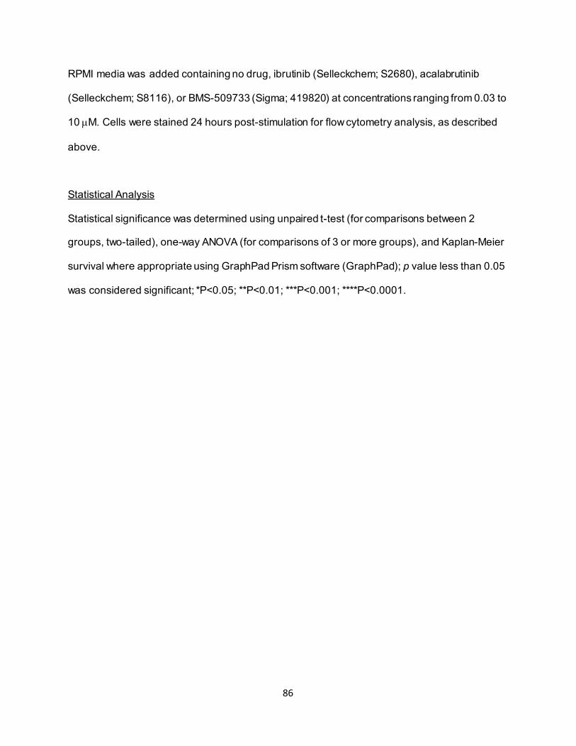
86
RPMI media was added containing no drug, ibrutinib (Selleckchem; S2680), acalabrutinib
(Selleckchem; S8116), or BMS-509733 (Sigma; 419820) at concentrations ranging from 0.03 to
10 µM. Cells were stained 24 hours post-stimulation for flow cytometry analysis, as described
above.
Statistical Analysis
Statistical significance was determined using unpaired t-test (for comparisons between 2
groups, two-tailed), one-way ANOVA (for comparisons of 3 or more groups), and Kaplan-Meier
survival where appropriate using GraphPad Prism software (GraphPad); p value less than 0.05
was considered significant; *P<0.05; **P<0.01; ***P<0.001; ****P<0.0001.
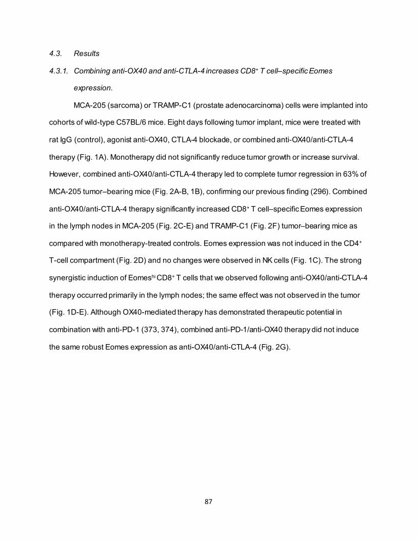
87
4.3. Results
4.3.1. Combining anti-OX40 and anti-CTLA-4 increases CD8+ T cell–specific Eomes
expression.
MCA-205 (sarcoma) or TRAMP-C1 (prostate adenocarcinoma) cells were implanted into
cohorts of wild-type C57BL/6 mice. Eight days following tumor implant, mice were treated with
rat IgG (control), agonist anti-OX40, CTLA-4 blockade, or combined anti-OX40/anti-CTLA-4
therapy (Fig. 1A). Monotherapy did not significantly reduce tumor growth or increase survival.
However, combined anti-OX40/anti-CTLA-4 therapy led to complete tumor regression in 63% of
MCA-205 tumor–bearing mice (Fig. 2A-B, 1B), confirming our previous finding (296). Combined
anti-OX40/anti-CTLA-4 therapy significantly increased CD8+ T cell–specific Eomes expression
in the lymph nodes in MCA-205 (Fig. 2C-E) and TRAMP-C1 (Fig. 2F) tumor–bearing mice as
compared with monotherapy-treated controls. Eomes expression was not induced in the CD4+
T-cell compartment (Fig. 2D) and no changes were observed in NK cells (Fig. 1C). The strong
synergistic induction of Eomeshi CD8+ T cells that we observed following anti-OX40/anti-CTLA-4
therapy occurred primarily in the lymph nodes; the same effect was not observed in the tumor
(Fig. 1D-E). Although OX40-mediated therapy has demonstrated therapeutic potential in
combination with anti-PD-1 (373, 374), combined anti-PD-1/anti-OX40 therapy did not induce
the same robust Eomes expression as anti-OX40/anti-CTLA-4 (Fig. 2G).
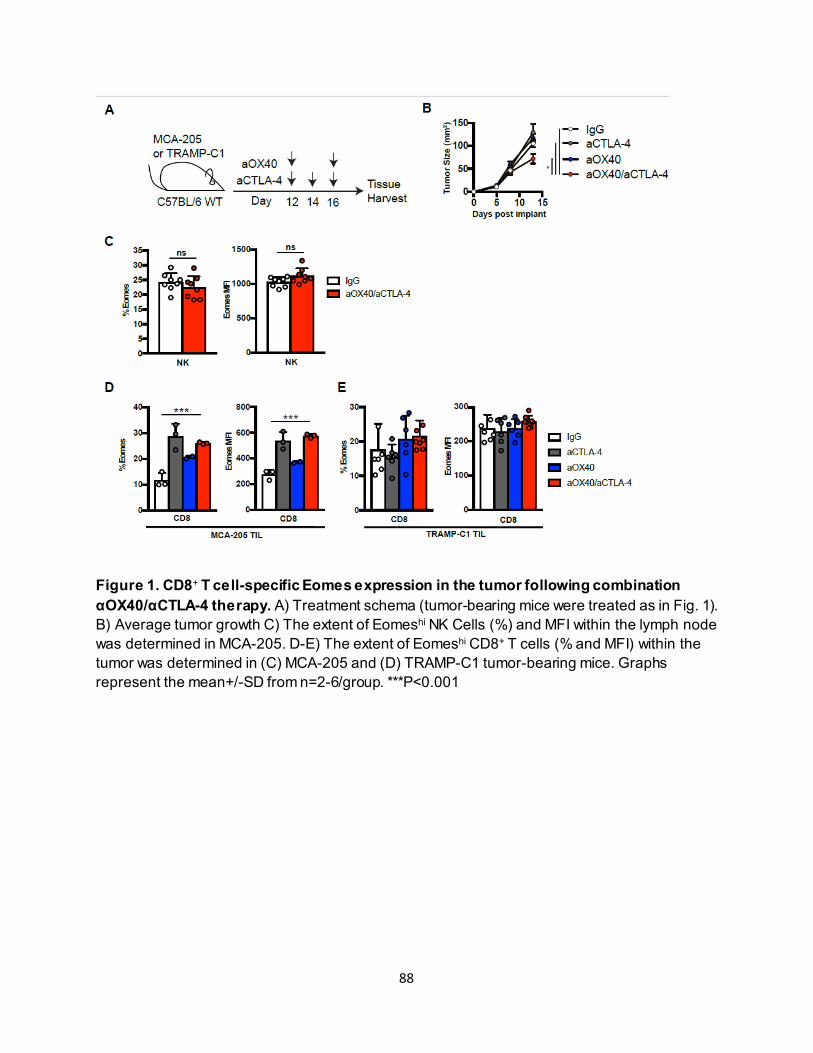
88
Figure 1. CD8+ T cell-specific Eomes expression in the tumor following combination αOX40/αCTLA-4 therapy. A) Treatment schema (tumor-bearing mice were treated as in Fig. 1). B) Average tumor growth C) The extent of Eomeshi NK Cells (%) and MFI within the lymph node was determined in MCA-205. D-E) The extent of Eomeshi CD8+ T cells (% and MFI) within the tumor was determined in (C) MCA-205 and (D) TRAMP-C1 tumor-bearing mice. Graphs represent the mean+/-SD from n=2-6/group. ***P<0.001
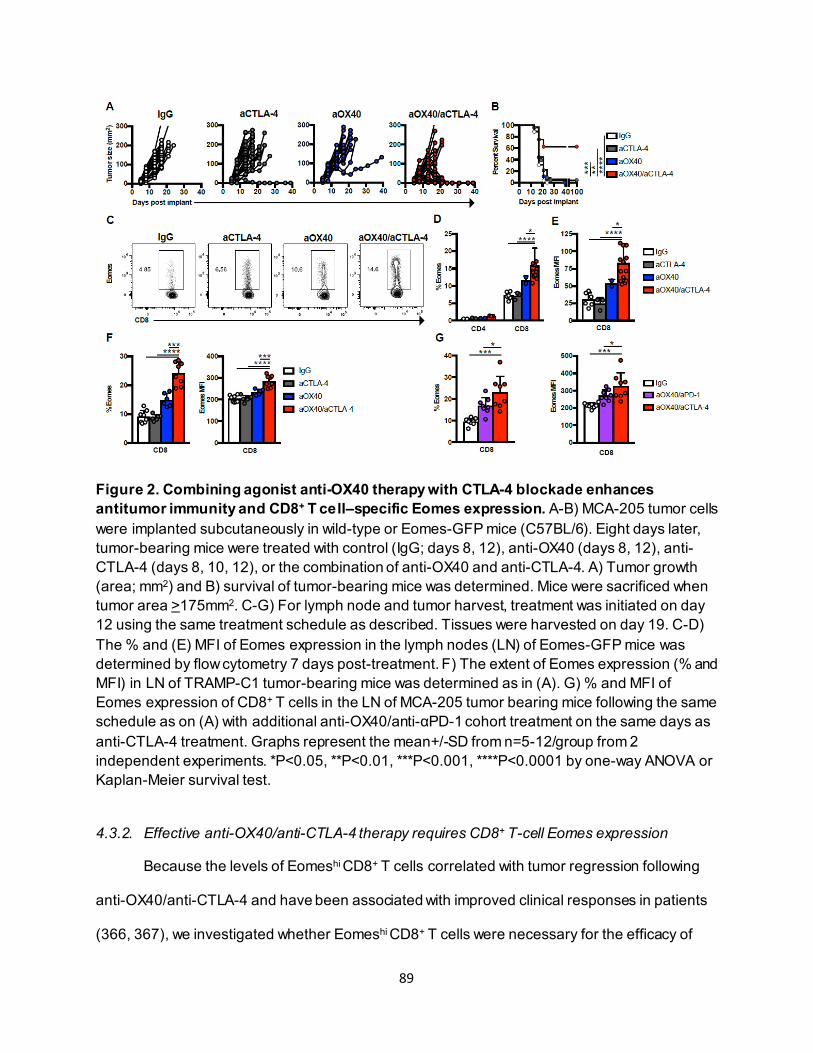
89
Figure 2. Combining agonist anti-OX40 therapy with CTLA-4 blockade enhances antitumor immunity and CD8+ T cell–specific Eomes expression. A-B) MCA-205 tumor cells were implanted subcutaneously in wild-type or Eomes-GFP mice (C57BL/6). Eight days later, tumor-bearing mice were treated with control (IgG; days 8, 12), anti-OX40 (days 8, 12), anti-CTLA-4 (days 8, 10, 12), or the combination of anti-OX40 and anti-CTLA-4. A) Tumor growth (area; mm2) and B) survival of tumor-bearing mice was determined. Mice were sacrificed when tumor area >175mm2. C-G) For lymph node and tumor harvest, treatment was initiated on day 12 using the same treatment schedule as described. Tissues were harvested on day 19. C-D) The % and (E) MFI of Eomes expression in the lymph nodes (LN) of Eomes-GFP mice was determined by flow cytometry 7 days post-treatment. F) The extent of Eomes expression (% and MFI) in LN of TRAMP-C1 tumor-bearing mice was determined as in (A). G) % and MFI of Eomes expression of CD8+ T cells in the LN of MCA-205 tumor bearing mice following the same schedule as on (A) with additional anti-OX40/anti-αPD-1 cohort treatment on the same days as anti-CTLA-4 treatment. Graphs represent the mean+/-SD from n=5-12/group from 2 independent experiments. *P<0.05, **P<0.01, ***P<0.001, ****P<0.0001 by one-way ANOVA or Kaplan-Meier survival test.
4.3.2. Effective anti-OX40/anti-CTLA-4 therapy requires CD8+ T-cell Eomes expression
Because the levels of Eomeshi CD8+ T cells correlated with tumor regression following
anti-OX40/anti-CTLA-4 and have been associated with improved clinical responses in patients
(366, 367), we investigated whether Eomeshi CD8+ T cells were necessary for the efficacy of
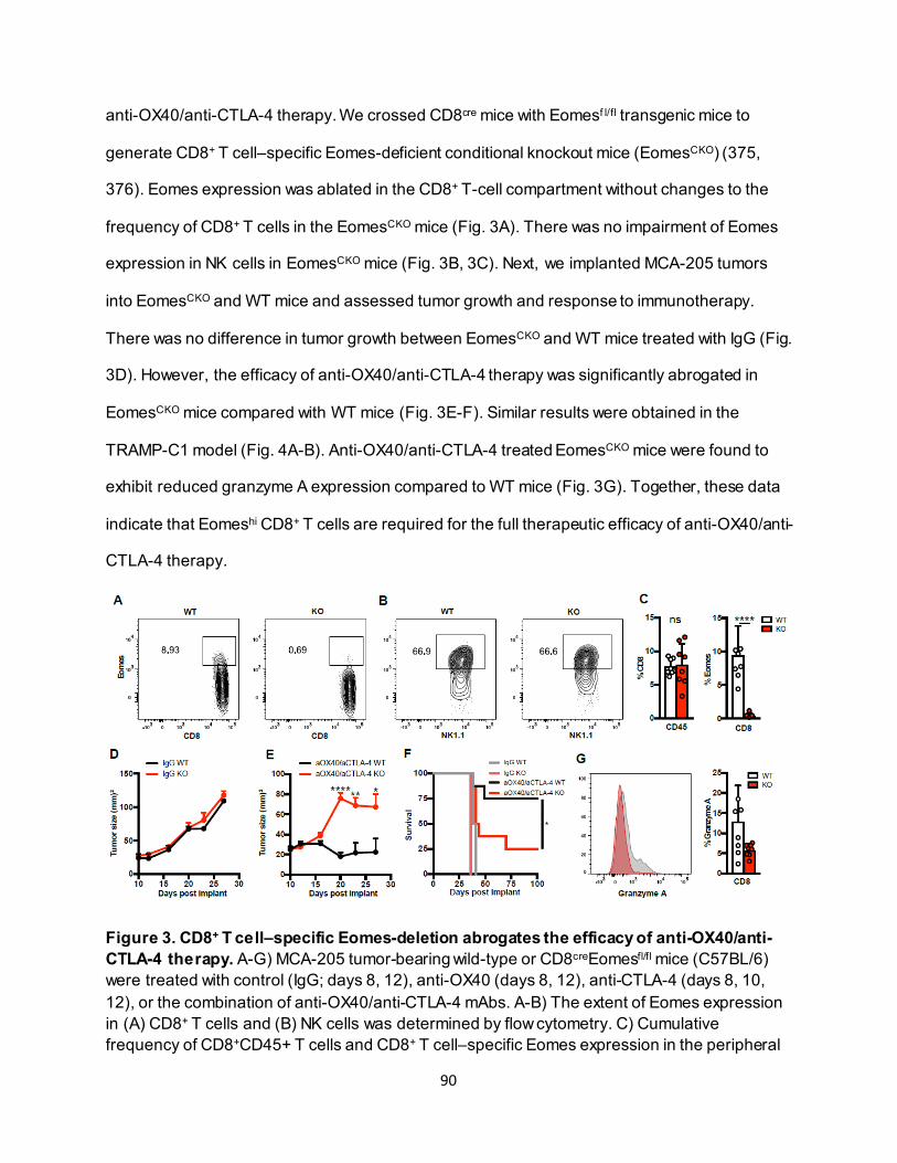
90
anti-OX40/anti-CTLA-4 therapy. We crossed CD8cre mice with Eomesf l/fl transgenic mice to
generate CD8+ T cell–specific Eomes-deficient conditional knockout mice (EomesCKO) (375,
376). Eomes expression was ablated in the CD8+ T-cell compartment without changes to the
frequency of CD8+ T cells in the EomesCKO mice (Fig. 3A). There was no impairment of Eomes
expression in NK cells in EomesCKO mice (Fig. 3B, 3C). Next, we implanted MCA-205 tumors
into EomesCKO and WT mice and assessed tumor growth and response to immunotherapy.
There was no difference in tumor growth between EomesCKO and WT mice treated with IgG (Fig.
3D). However, the efficacy of anti-OX40/anti-CTLA-4 therapy was significantly abrogated in
EomesCKO mice compared with WT mice (Fig. 3E-F). Similar results were obtained in the
TRAMP-C1 model (Fig. 4A-B). Anti-OX40/anti-CTLA-4 treated EomesCKO mice were found to
exhibit reduced granzyme A expression compared to WT mice (Fig. 3G). Together, these data
indicate that Eomeshi CD8+ T cells are required for the full therapeutic efficacy of anti-OX40/anti-
CTLA-4 therapy.
Figure 3. CD8+ T cell–specific Eomes-deletion abrogates the efficacy of anti-OX40/anti-CTLA-4 therapy. A-G) MCA-205 tumor-bearing wild-type or CD8creEomesfl/fl mice (C57BL/6) were treated with control (IgG; days 8, 12), anti-OX40 (days 8, 12), anti-CTLA-4 (days 8, 10, 12), or the combination of anti-OX40/anti-CTLA-4 mAbs. A-B) The extent of Eomes expression in (A) CD8+ T cells and (B) NK cells was determined by flow cytometry. C) Cumulative frequency of CD8+CD45+ T cells and CD8+ T cell–specific Eomes expression in the peripheral
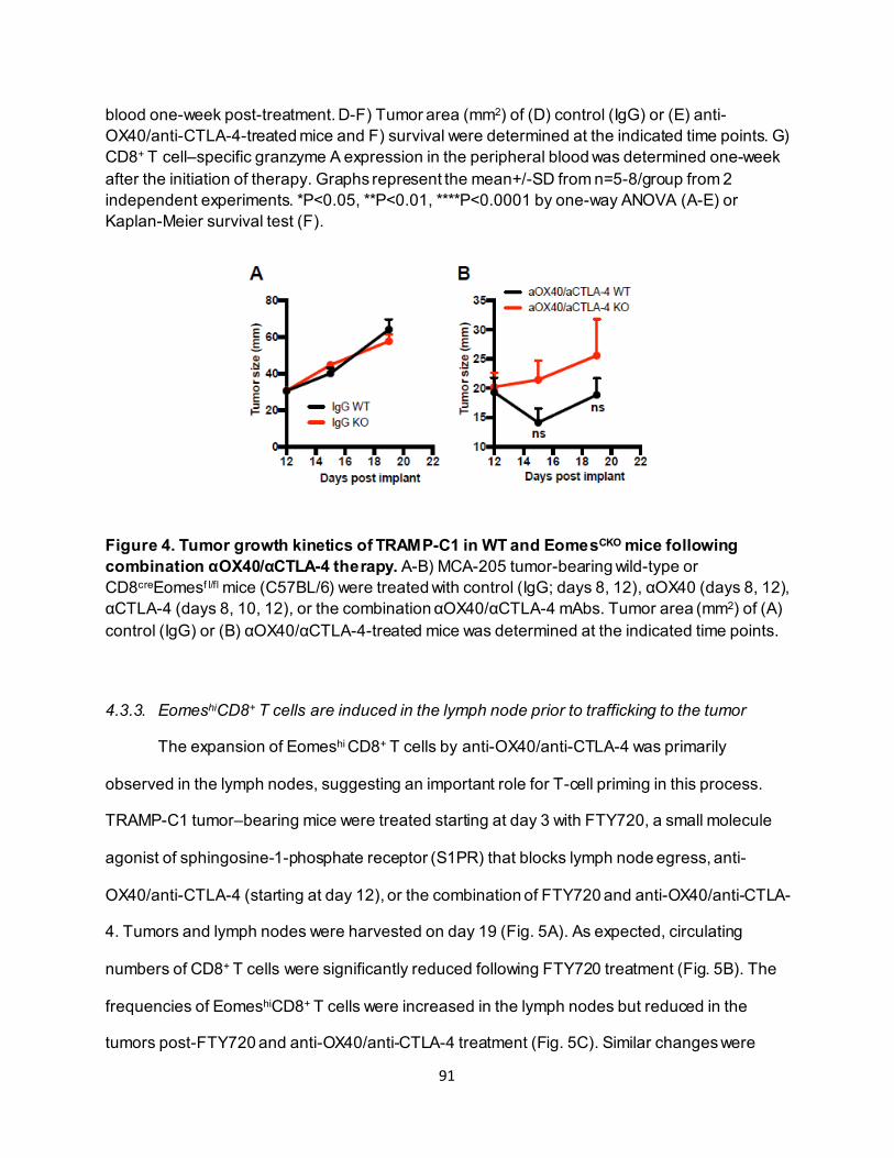
91
blood one-week post-treatment. D-F) Tumor area (mm2) of (D) control (IgG) or (E) anti-OX40/anti-CTLA-4-treated mice and F) survival were determined at the indicated time points. G) CD8+ T cell–specific granzyme A expression in the peripheral blood was determined one-week after the initiation of therapy. Graphs represent the mean+/-SD from n=5-8/group from 2 independent experiments. *P<0.05, **P<0.01, ****P<0.0001 by one-way ANOVA (A-E) or Kaplan-Meier survival test (F).
Figure 4. Tumor growth kinetics of TRAMP-C1 in WT and EomesCKO mice following combination αOX40/αCTLA-4 therapy. A-B) MCA-205 tumor-bearing wild-type or CD8creEomesf l/fl mice (C57BL/6) were treated with control (IgG; days 8, 12), αOX40 (days 8, 12), αCTLA-4 (days 8, 10, 12), or the combination αOX40/αCTLA-4 mAbs. Tumor area (mm2) of (A) control (IgG) or (B) αOX40/αCTLA-4-treated mice was determined at the indicated time points.
4.3.3. EomeshiCD8+ T cells are induced in the lymph node prior to trafficking to the tumor
The expansion of Eomeshi CD8+ T cells by anti-OX40/anti-CTLA-4 was primarily
observed in the lymph nodes, suggesting an important role for T-cell priming in this process.
TRAMP-C1 tumor–bearing mice were treated starting at day 3 with FTY720, a small molecule
agonist of sphingosine-1-phosphate receptor (S1PR) that blocks lymph node egress, anti-
OX40/anti-CTLA-4 (starting at day 12), or the combination of FTY720 and anti-OX40/anti-CTLA-
4. Tumors and lymph nodes were harvested on day 19 (Fig. 5A). As expected, circulating
numbers of CD8+ T cells were significantly reduced following FTY720 treatment (Fig. 5B). The
frequencies of EomeshiCD8+ T cells were increased in the lymph nodes but reduced in the
tumors post-FTY720 and anti-OX40/anti-CTLA-4 treatment (Fig. 5C). Similar changes were
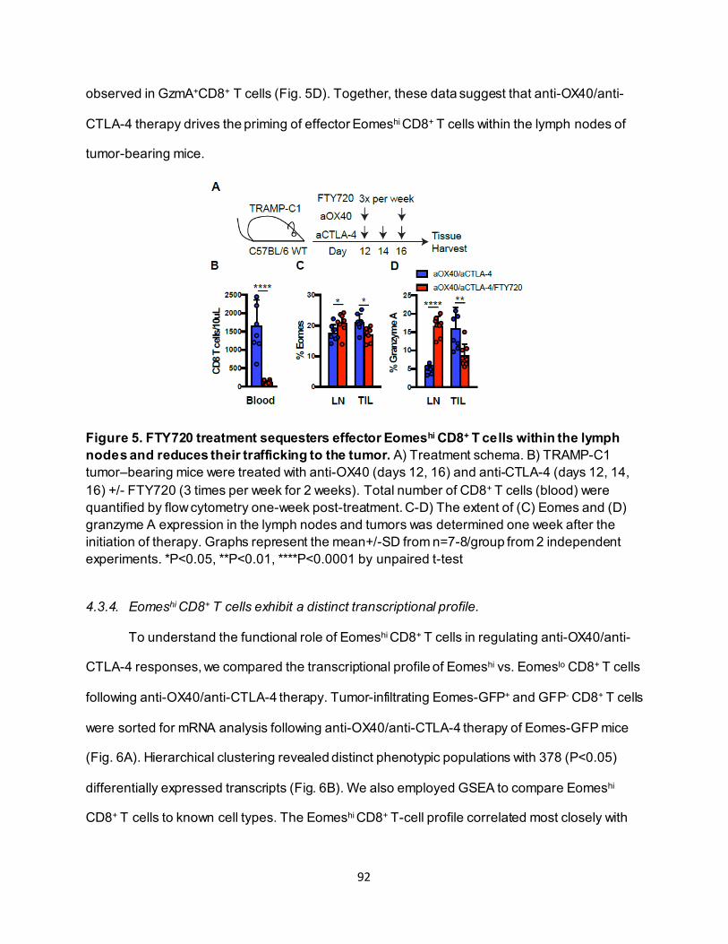
92
observed in GzmA+CD8+ T cells (Fig. 5D). Together, these data suggest that anti-OX40/anti-
CTLA-4 therapy drives the priming of effector Eomeshi CD8+ T cells within the lymph nodes of
tumor-bearing mice.
Figure 5. FTY720 treatment sequesters effector Eomeshi CD8+ T cells within the lymph nodes and reduces their trafficking to the tumor. A) Treatment schema. B) TRAMP-C1 tumor–bearing mice were treated with anti-OX40 (days 12, 16) and anti-CTLA-4 (days 12, 14, 16) +/- FTY720 (3 times per week for 2 weeks). Total number of CD8+ T cells (blood) were quantified by flow cytometry one-week post-treatment. C-D) The extent of (C) Eomes and (D) granzyme A expression in the lymph nodes and tumors was determined one week after the initiation of therapy. Graphs represent the mean+/-SD from n=7-8/group from 2 independent experiments. *P<0.05, **P<0.01, ****P<0.0001 by unpaired t-test
4.3.4. Eomeshi CD8+ T cells exhibit a distinct transcriptional profile.
To understand the functional role of Eomeshi CD8+ T cells in regulating anti-OX40/anti-
CTLA-4 responses, we compared the transcriptional profile of Eomeshi vs. Eomeslo CD8+ T cells
following anti-OX40/anti-CTLA-4 therapy. Tumor-infiltrating Eomes-GFP+ and GFP- CD8+ T cells
were sorted for mRNA analysis following anti-OX40/anti-CTLA-4 therapy of Eomes-GFP mice
(Fig. 6A). Hierarchical clustering revealed distinct phenotypic populations with 378 (P<0.05)
differentially expressed transcripts (Fig. 6B). We also employed GSEA to compare Eomeshi
CD8+ T cells to known cell types. The Eomeshi CD8+ T-cell profile correlated most closely with
93
that of memory T cells, suggesting that Eomeshi CD8+ T cells have a more memory-like than
effector-like phenotype (Fig. 6C).
We next compared the relative expression of Eomeshi CD8+ T cell–associated genes to
naïve, effector, and effector memory CD8+ T-cell populations responding to acute LCMV
infection available in the Gene Expression Omnibus (GEO) database (Fig. 6D-G) (377). We
found that, with the exception of PD1 expression, our Eomeshi CD8+ T-cell phenotype most
closely resembled effector memory cells (Fig. 6D). For example, TCF1 was increased in
Eomeshi CD8+ T cells to similar levels as observed in effector memory cells, suggesting self-
renewal memory formation (378) (Fig. 6E). We also observed increased CD62L and granzyme
A expression, which was similar to the levels seen in the effector memory population (Fig. 6F-
G). Analysis of CD62L and CD44 showed that Eomes was most highly expressed in the central
memory (CD44hi, CD62Lhi) and effector/effector memory (CD44hi, CD62Llo) CD8+ T-cell
population in the lymph nodes and tumors following anti-OX40/anti-CTLA-4 therapy (Fig. 7A-B).
Thus, Eomeshi CD8+ T cells induced by anti-OX40/anti-CTLA-4 therapy have a phenotypic
profile similar to central memory CD8+ T cells with strong effector potential. In addition, Eomeshi
CD8+ T cells expressed significantly lower levels of critical inhibitory and exhaustion markers
including PD1, LAG-3, and TIM-3 compared with Eomeslo CD8+ T cells (Fig. 6H). We confirmed
these markers by protein expression, including verification of checkpoint inhibitors (PD1, TIM-3,
LAG-3), proliferation (Ki-67), and effector cytokine production (granzyme A, polyfunctional
IFNγ/TNFα population) (Fig. 6I).
High granzyme A expression among Eomeshi compared with Eomeslo CD8+ T cells
following anti-OX40/anti-CTLA-4 therapy raised concern over whether they might be functionally
exhausted, as increased Eomes expression has been associated with the development of CD8+
T-cell exhaustion (61, 379). Therefore, the cytolytic activity of Eomeshi and Eomeslo CD8+ T cells
isolated from anti-OX40/anti-CTLA-4-treated tumor-bearing mice was determined. Eomeshi
CD8+ T cells trended toward lysing more target cells (tumor) compared with Eomeslo cells (Fig.
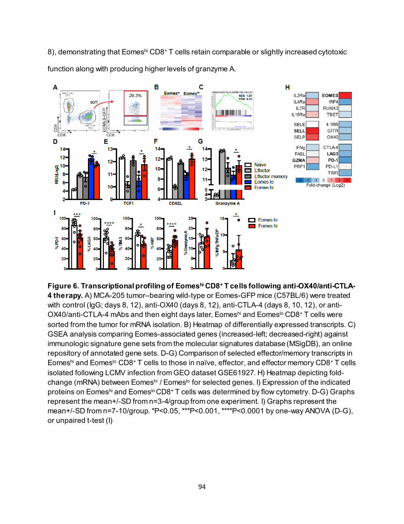
94
8), demonstrating that Eomeshi CD8+ T cells retain comparable or slightly increased cytotoxic
function along with producing higher levels of granzyme A.
Figure 6. Transcriptional profiling of Eomeshi CD8+ T cells following anti-OX40/anti-CTLA-4 therapy. A) MCA-205 tumor–bearing wild-type or Eomes-GFP mice (C57BL/6) were treated with control (IgG; days 8, 12), anti-OX40 (days 8, 12), anti-CTLA-4 (days 8, 10, 12), or anti-OX40/anti-CTLA-4 mAbs and then eight days later, Eomeshi and Eomeslo CD8+ T cells were sorted from the tumor for mRNA isolation. B) Heatmap of differentially expressed transcripts. C) GSEA analysis comparing Eomes-associated genes (increased-left; decreased-right) against immunologic signature gene sets from the molecular signatures database (MSigDB), an online repository of annotated gene sets. D-G) Comparison of selected effector/memory transcripts in Eomeshi and Eomeslo CD8+ T cells to those in naïve, effector, and effector memory CD8+ T cells isolated following LCMV infection from GEO dataset GSE61927. H) Heatmap depicting fold-change (mRNA) between Eomeshi / Eomeslo for selected genes. I) Expression of the indicated proteins on Eomeshi and Eomeslo CD8+ T cells was determined by flow cytometry. D-G) Graphs represent the mean+/-SD from n=3-4/group from one experiment. I) Graphs represent the mean+/-SD from n=7-10/group. *P<0.05, ***P<0.001, ****P<0.0001 by one-way ANOVA (D-G), or unpaired t-test (I)
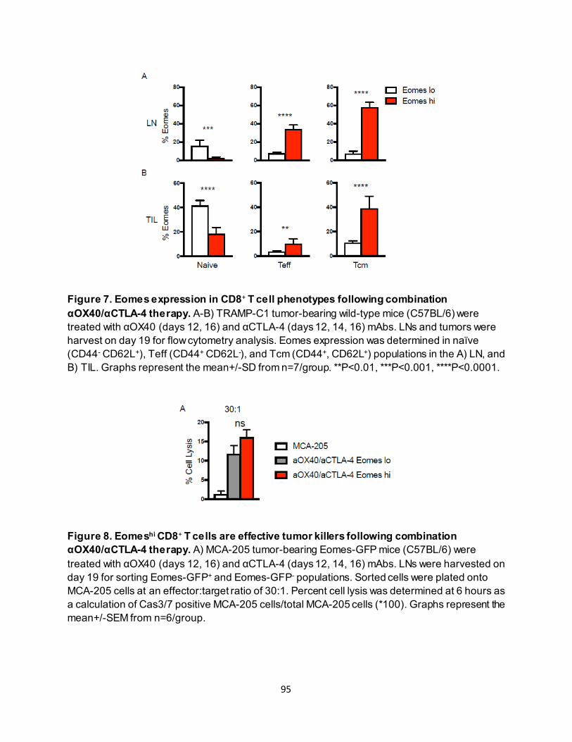
95
Figure 7. Eomes expression in CD8+ T cell phenotypes following combination αOX40/αCTLA-4 therapy. A-B) TRAMP-C1 tumor-bearing wild-type mice (C57BL/6) were treated with αOX40 (days 12, 16) and αCTLA-4 (days 12, 14, 16) mAbs. LNs and tumors were harvest on day 19 for flow cytometry analysis. Eomes expression was determined in naïve (CD44- CD62L+), Teff (CD44+ CD62L-), and Tcm (CD44+, CD62L+) populations in the A) LN, and B) TIL. Graphs represent the mean+/-SD from n=7/group. **P<0.01, ***P<0.001, ****P<0.0001.
Figure 8. Eomeshi CD8+ T cells are effective tumor killers following combination αOX40/αCTLA-4 therapy. A) MCA-205 tumor-bearing Eomes-GFP mice (C57BL/6) were treated with αOX40 (days 12, 16) and αCTLA-4 (days 12, 14, 16) mAbs. LNs were harvested on day 19 for sorting Eomes-GFP+ and Eomes-GFP- populations. Sorted cells were plated onto MCA-205 cells at an effector:target ratio of 30:1. Percent cell lysis was determined at 6 hours as a calculation of Cas3/7 positive MCA-205 cells/total MCA-205 cells (*100). Graphs represent the mean+/-SEM from n=6/group.
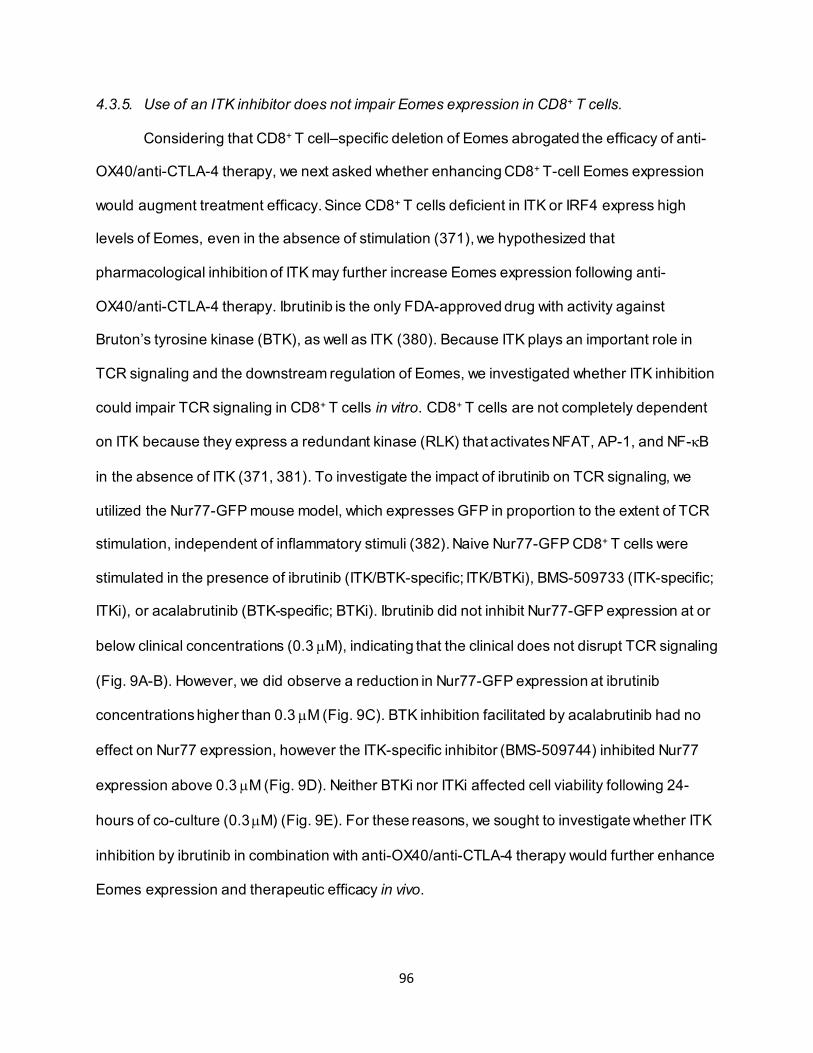
96
4.3.5. Use of an ITK inhibitor does not impair Eomes expression in CD8+ T cells.
Considering that CD8+ T cell–specific deletion of Eomes abrogated the efficacy of anti-
OX40/anti-CTLA-4 therapy, we next asked whether enhancing CD8+ T-cell Eomes expression
would augment treatment efficacy. Since CD8+ T cells deficient in ITK or IRF4 express high
levels of Eomes, even in the absence of stimulation (371), we hypothesized that
pharmacological inhibition of ITK may further increase Eomes expression following anti-
OX40/anti-CTLA-4 therapy. Ibrutinib is the only FDA-approved drug with activity against
Bruton’s tyrosine kinase (BTK), as well as ITK (380). Because ITK plays an important role in
TCR signaling and the downstream regulation of Eomes, we investigated whether ITK inhibition
could impair TCR signaling in CD8+ T cells in vitro. CD8+ T cells are not completely dependent
on ITK because they express a redundant kinase (RLK) that activates NFAT, AP-1, and NF-κB
in the absence of ITK (371, 381). To investigate the impact of ibrutinib on TCR signaling, we
utilized the Nur77-GFP mouse model, which expresses GFP in proportion to the extent of TCR
stimulation, independent of inflammatory stimuli (382). Naive Nur77-GFP CD8+ T cells were
stimulated in the presence of ibrutinib (ITK/BTK-specific; ITK/BTKi), BMS-509733 (ITK-specific;
ITKi), or acalabrutinib (BTK-specific; BTKi). Ibrutinib did not inhibit Nur77-GFP expression at or
below clinical concentrations (0.3 µM), indicating that the clinical does not disrupt TCR signaling
(Fig. 9A-B). However, we did observe a reduction in Nur77-GFP expression at ibrutinib
concentrations higher than 0.3 µM (Fig. 9C). BTK inhibition facilitated by acalabrutinib had no
effect on Nur77 expression, however the ITK-specific inhibitor (BMS-509744) inhibited Nur77
expression above 0.3 µM (Fig. 9D). Neither BTKi nor ITKi affected cell viability following 24-
hours of co-culture (0.3 µM) (Fig. 9E). For these reasons, we sought to investigate whether ITK
inhibition by ibrutinib in combination with anti-OX40/anti-CTLA-4 therapy would further enhance
Eomes expression and therapeutic efficacy in vivo.
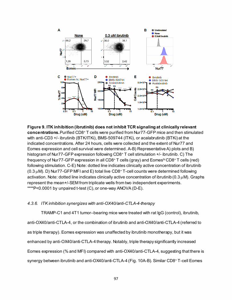
97
Figure 9. ITK inhibition (ibrutinib) does not inhibit TCR signaling at clinically relevant concentrations. Purified CD8+ T cells were purified from Nur77-GFP mice and then stimulated with anti-CD3 +/- ibrutinib (BTK/ITKi), BMS-509744 (ITKi), or acalabrutinib (BTKi) at the indicated concentrations. After 24 hours, cells were collected and the extent of Nur77 and Eomes expression and cell survival were determined. A-B) Representative A) plots and B) histogram of Nur77-GFP expression following CD8+ T cell stimulation +/- ibrutinib. C) The frequency of Nur77-GFP expression in all CD8+ T cells (gray) and Eomeshi CD8+ T cells (red) following stimulation. C-E) Note: dotted line indicates clinically active concentration of ibrutinib (0.3 µM). D) Nur77-GFP MFI and E) total live CD8+ T-cell counts were determined following activation. Note: dotted line indicates clinically active concentration of ibrutinib (0.3 µM). Graphs represent the mean+/-SEM from triplicate wells from two independent experiments. ****P<0.0001 by unpaired t-test (C), or one-way ANOVA (D-E).
4.3.6. ITK inhibition synergizes with anti-OX40/anti-CTLA-4-therapy
TRAMP-C1 and 4T1 tumor–bearing mice were treated with rat IgG (control), ibrutinib,
anti-OX40/anti-CTLA-4, or the combination of ibrutinib and anti-OX40/anti-CTLA-4 (referred to
as triple therapy). Eomes expression was unaffected by ibrutinib monotherapy, but it was
enhanced by anti-OX40/anti-CTLA-4 therapy. Notably, triple therapy significantly increased
Eomes expression (% and MFI) compared with anti-OX40/anti-CTLA-4, suggesting that there is
synergy between ibrutinib and anti-OX40/anti-CTLA-4 (Fig. 10A-B). Similar CD8+ T-cell Eomes
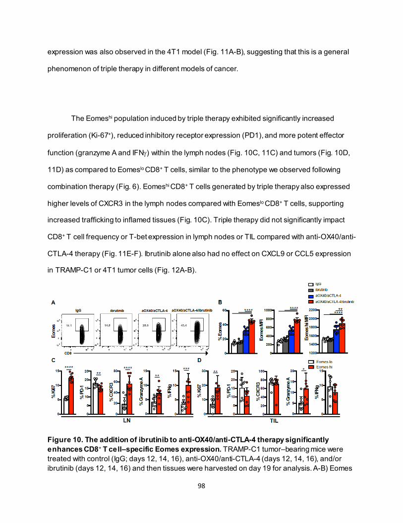
98
expression was also observed in the 4T1 model (Fig. 11A-B), suggesting that this is a general
phenomenon of triple therapy in different models of cancer.
The Eomeshi population induced by triple therapy exhibited significantly increased
proliferation (Ki-67+), reduced inhibitory receptor expression (PD1), and more potent effector
function (granzyme A and IFNγ) within the lymph nodes (Fig. 10C, 11C) and tumors (Fig. 10D,
11D) as compared to Eomeslo CD8+ T cells, similar to the phenotype we observed following
combination therapy (Fig. 6). Eomeshi CD8+ T cells generated by triple therapy also expressed
higher levels of CXCR3 in the lymph nodes compared with Eomeslo CD8+ T cells, supporting
increased trafficking to inflamed tissues (Fig. 10C). Triple therapy did not significantly impact
CD8+ T cell frequency or T-bet expression in lymph nodes or TIL compared with anti-OX40/anti-
CTLA-4 therapy (Fig. 11E-F). Ibrutinib alone also had no effect on CXCL9 or CCL5 expression
in TRAMP-C1 or 4T1 tumor cells (Fig. 12A-B).
Figure 10. The addition of ibrutinib to anti-OX40/anti-CTLA-4 therapy significantly enhances CD8+ T cell–specific Eomes expression. TRAMP-C1 tumor–bearing mice were treated with control (IgG; days 12, 14, 16), anti-OX40/anti-CTLA-4 (days 12, 14, 16), and/or ibrutinib (days 12, 14, 16) and then tissues were harvested on day 19 for analysis. A-B) Eomes
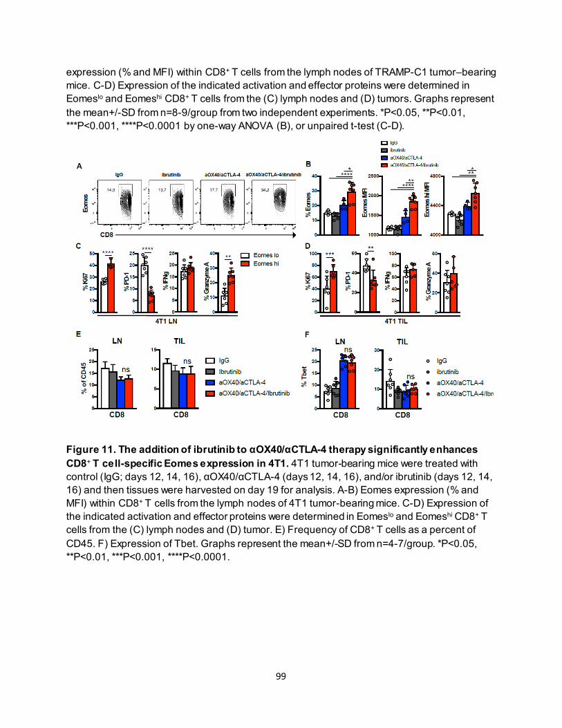
99
expression (% and MFI) within CD8+ T cells from the lymph nodes of TRAMP-C1 tumor–bearing mice. C-D) Expression of the indicated activation and effector proteins were determined in Eomeslo and Eomeshi CD8+ T cells from the (C) lymph nodes and (D) tumors. Graphs represent the mean+/-SD from n=8-9/group from two independent experiments. *P<0.05, **P<0.01, ***P<0.001, ****P<0.0001 by one-way ANOVA (B), or unpaired t-test (C-D).
Figure 11. The addition of ibrutinib to αOX40/αCTLA-4 therapy significantly enhances CD8+ T cell-specific Eomes expression in 4T1. 4T1 tumor-bearing mice were treated with control (IgG; days 12, 14, 16), αOX40/αCTLA-4 (days 12, 14, 16), and/or ibrutinib (days 12, 14, 16) and then tissues were harvested on day 19 for analysis. A-B) Eomes expression (% and MFI) within CD8+ T cells from the lymph nodes of 4T1 tumor-bearing mice. C-D) Expression of the indicated activation and effector proteins were determined in Eomeslo and Eomeshi CD8+ T cells from the (C) lymph nodes and (D) tumor. E) Frequency of CD8+ T cells as a percent of CD45. F) Expression of Tbet. Graphs represent the mean+/-SD from n=4-7/group. *P<0.05, **P<0.01, ***P<0.001, ****P<0.0001.
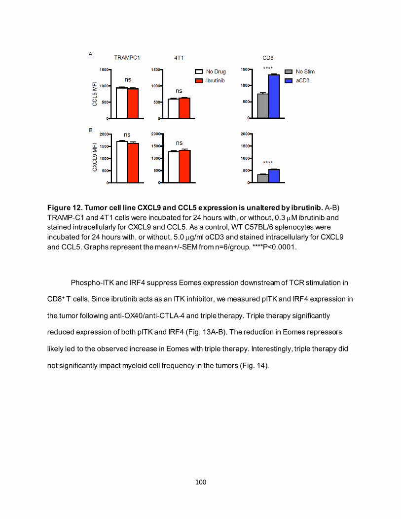
100
Figure 12. Tumor cell line CXCL9 and CCL5 expression is unaltered by ibrutinib. A-B) TRAMP-C1 and 4T1 cells were incubated for 24 hours with, or without, 0.3 µM ibrutinib and stained intracellularly for CXCL9 and CCL5. As a control, WT C57BL/6 splenocytes were incubated for 24 hours with, or without, 5.0 µg/ml αCD3 and stained intracellularly for CXCL9 and CCL5. Graphs represent the mean+/-SEM from n=6/group. ****P<0.0001.
Phospho-ITK and IRF4 suppress Eomes expression downstream of TCR stimulation in
CD8+ T cells. Since ibrutinib acts as an ITK inhibitor, we measured pITK and IRF4 expression in
the tumor following anti-OX40/anti-CTLA-4 and triple therapy. Triple therapy significantly
reduced expression of both pITK and IRF4 (Fig. 13A-B). The reduction in Eomes repressors
likely led to the observed increase in Eomes with triple therapy. Interestingly, triple therapy did
not significantly impact myeloid cell frequency in the tumors (Fig. 14).
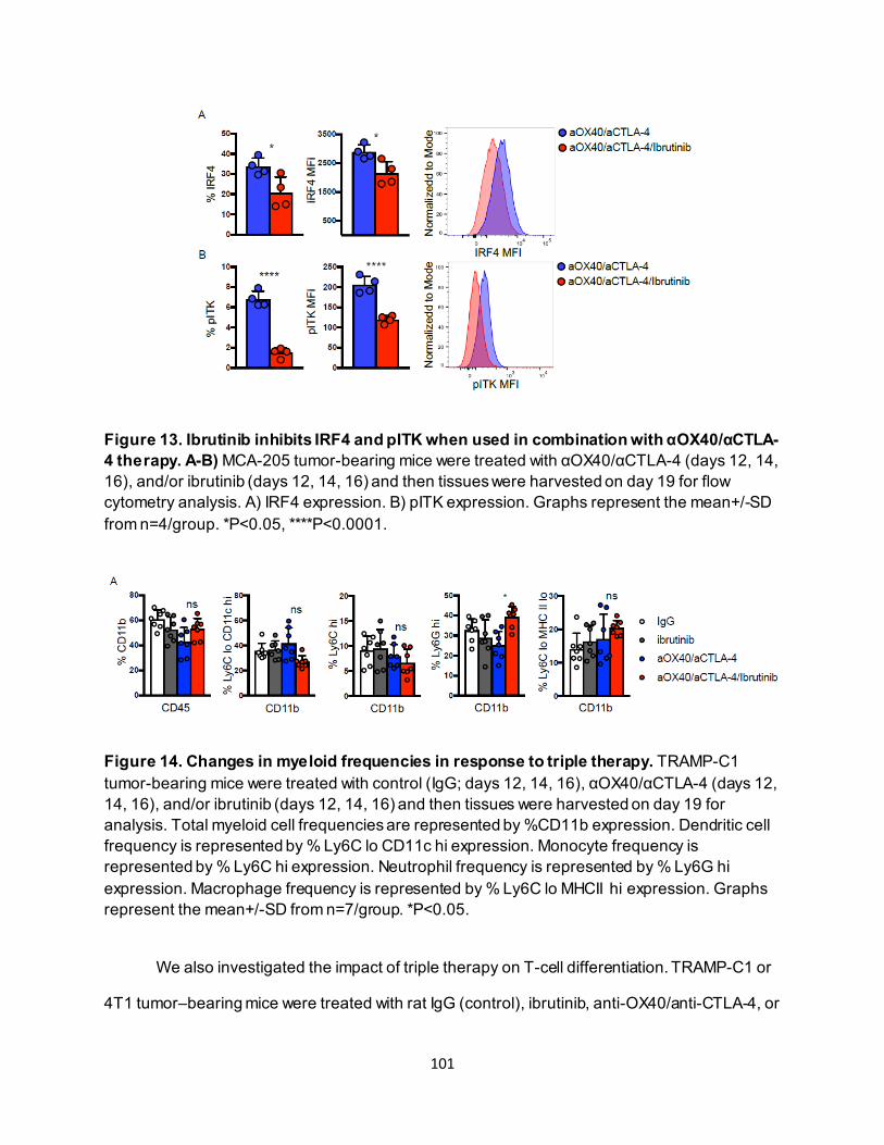
101
Figure 13. Ibrutinib inhibits IRF4 and pITK when used in combination with αOX40/αCTLA-4 therapy. A-B) MCA-205 tumor-bearing mice were treated with αOX40/αCTLA-4 (days 12, 14, 16), and/or ibrutinib (days 12, 14, 16) and then tissues were harvested on day 19 for flow cytometry analysis. A) IRF4 expression. B) pITK expression. Graphs represent the mean+/-SD from n=4/group. *P<0.05, ****P<0.0001.
Figure 14. Changes in myeloid frequencies in response to triple therapy. TRAMP-C1 tumor-bearing mice were treated with control (IgG; days 12, 14, 16), αOX40/αCTLA-4 (days 12, 14, 16), and/or ibrutinib (days 12, 14, 16) and then tissues were harvested on day 19 for analysis. Total myeloid cell frequencies are represented by %CD11b expression. Dendritic cell frequency is represented by % Ly6C lo CD11c hi expression. Monocyte frequency is represented by % Ly6C hi expression. Neutrophil frequency is represented by % Ly6G hi expression. Macrophage frequency is represented by % Ly6C lo MHCII hi expression. Graphs represent the mean+/-SD from n=7/group. *P<0.05.
We also investigated the impact of triple therapy on T-cell differentiation. TRAMP-C1 or
4T1 tumor–bearing mice were treated with rat IgG (control), ibrutinib, anti-OX40/anti-CTLA-4, or
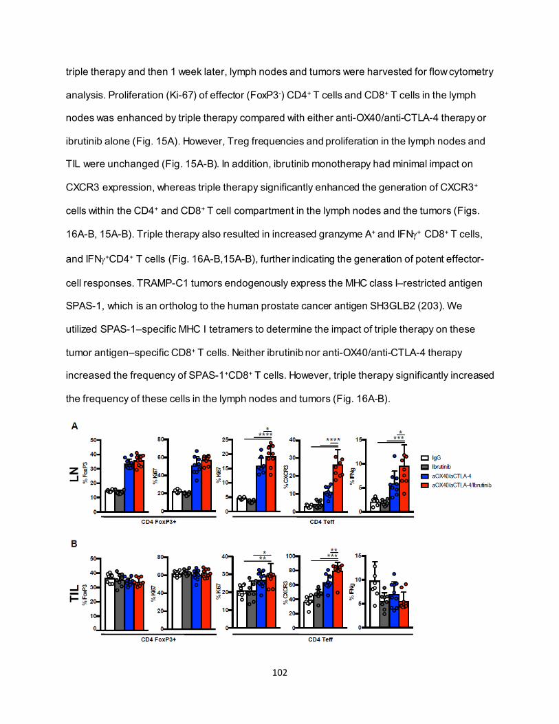
102
triple therapy and then 1 week later, lymph nodes and tumors were harvested for flow cytometry
analysis. Proliferation (Ki-67) of effector (FoxP3-) CD4+ T cells and CD8+ T cells in the lymph
nodes was enhanced by triple therapy compared with either anti-OX40/anti-CTLA-4 therapy or
ibrutinib alone (Fig. 15A). However, Treg frequencies and proliferation in the lymph nodes and
TIL were unchanged (Fig. 15A-B). In addition, ibrutinib monotherapy had minimal impact on
CXCR3 expression, whereas triple therapy significantly enhanced the generation of CXCR3+
cells within the CD4+ and CD8+ T cell compartment in the lymph nodes and the tumors (Figs.
16A-B, 15A-B). Triple therapy also resulted in increased granzyme A+ and IFNγ+ CD8+ T cells,
and IFNγ+CD4+ T cells (Fig. 16A-B,15A-B), further indicating the generation of potent effector-
cell responses. TRAMP-C1 tumors endogenously express the MHC class I–restricted antigen
SPAS-1, which is an ortholog to the human prostate cancer antigen SH3GLB2 (203). We
utilized SPAS-1–specific MHC I tetramers to determine the impact of triple therapy on these
tumor antigen–specific CD8+ T cells. Neither ibrutinib nor anti-OX40/anti-CTLA-4 therapy
increased the frequency of SPAS-1+CD8+ T cells. However, triple therapy significantly increased
the frequency of these cells in the lymph nodes and tumors (Fig. 16A-B).
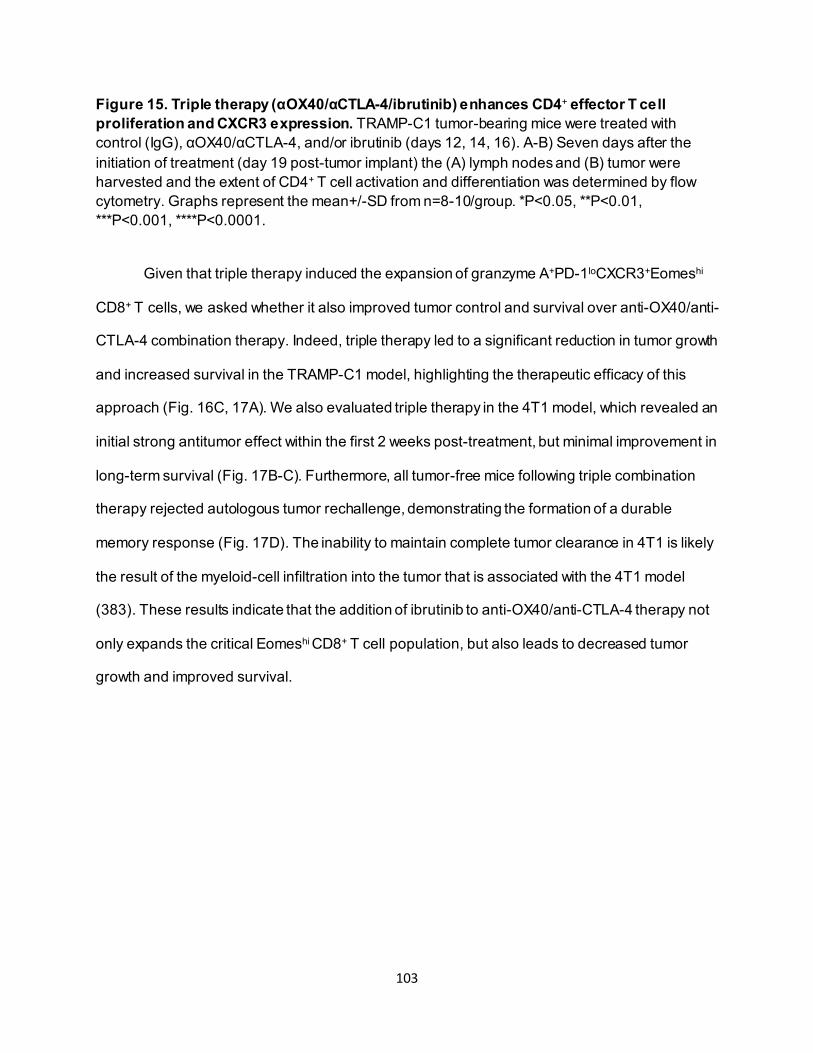
103
Figure 15. Triple therapy (αOX40/αCTLA-4/ibrutinib) enhances CD4+ effector T cell proliferation and CXCR3 expression. TRAMP-C1 tumor-bearing mice were treated with control (IgG), αOX40/αCTLA-4, and/or ibrutinib (days 12, 14, 16). A-B) Seven days after the initiation of treatment (day 19 post-tumor implant) the (A) lymph nodes and (B) tumor were harvested and the extent of CD4+ T cell activation and differentiation was determined by flow cytometry. Graphs represent the mean+/-SD from n=8-10/group. *P<0.05, **P<0.01, ***P<0.001, ****P<0.0001.
Given that triple therapy induced the expansion of granzyme A+PD-1loCXCR3+Eomeshi
CD8+ T cells, we asked whether it also improved tumor control and survival over anti-OX40/anti-
CTLA-4 combination therapy. Indeed, triple therapy led to a significant reduction in tumor growth
and increased survival in the TRAMP-C1 model, highlighting the therapeutic efficacy of this
approach (Fig. 16C, 17A). We also evaluated triple therapy in the 4T1 model, which revealed an
initial strong antitumor effect within the first 2 weeks post-treatment, but minimal improvement in
long-term survival (Fig. 17B-C). Furthermore, all tumor-free mice following triple combination
therapy rejected autologous tumor rechallenge, demonstrating the formation of a durable
memory response (Fig. 17D). The inability to maintain complete tumor clearance in 4T1 is likely
the result of the myeloid-cell infiltration into the tumor that is associated with the 4T1 model
(383). These results indicate that the addition of ibrutinib to anti-OX40/anti-CTLA-4 therapy not
only expands the critical Eomeshi CD8+ T cell population, but also leads to decreased tumor
growth and improved survival.
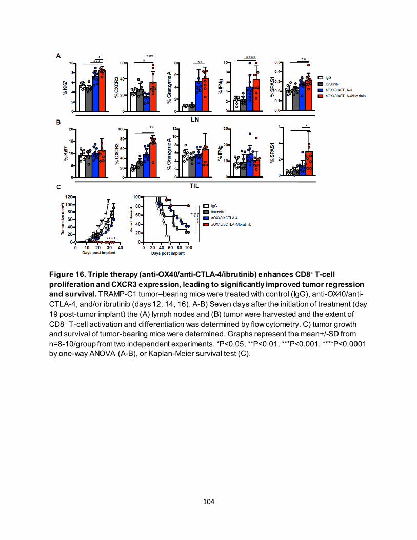
104
Figure 16. Triple therapy (anti-OX40/anti-CTLA-4/ibrutinib) enhances CD8+ T-cell proliferation and CXCR3 expression, leading to significantly improved tumor regression and survival. TRAMP-C1 tumor–bearing mice were treated with control (IgG), anti-OX40/anti-CTLA-4, and/or ibrutinib (days 12, 14, 16). A-B) Seven days after the initiation of treatment (day 19 post-tumor implant) the (A) lymph nodes and (B) tumor were harvested and the extent of CD8+ T-cell activation and differentiation was determined by flow cytometry. C) tumor growth and survival of tumor-bearing mice were determined. Graphs represent the mean+/-SD from n=8-10/group from two independent experiments. *P<0.05, **P<0.01, ***P<0.001, ****P<0.0001 by one-way ANOVA (A-B), or Kaplan-Meier survival test (C).
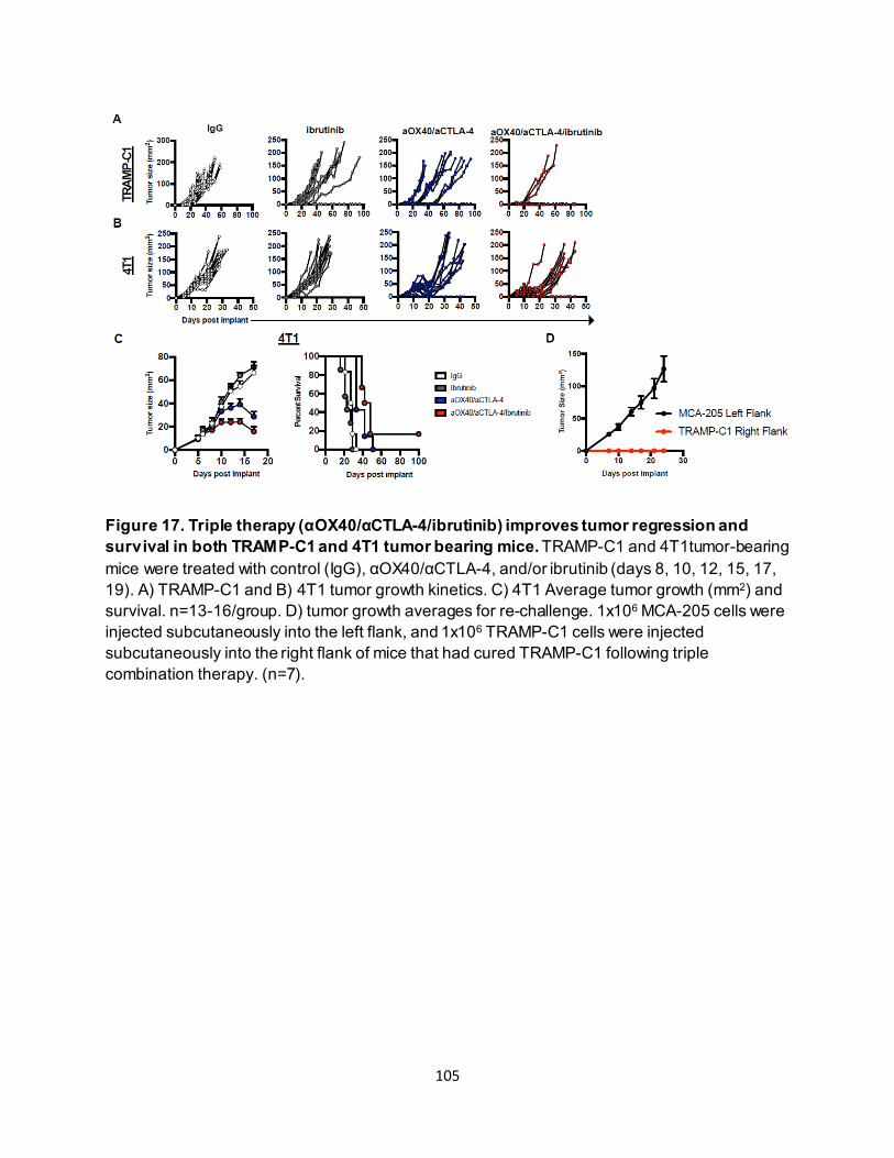
105
Figure 17. Triple therapy (αOX40/αCTLA-4/ibrutinib) improves tumor regression and survival in both TRAMP-C1 and 4T1 tumor bearing mice. TRAMP-C1 and 4T1tumor-bearing mice were treated with control (IgG), αOX40/αCTLA-4, and/or ibrutinib (days 8, 10, 12, 15, 17, 19). A) TRAMP-C1 and B) 4T1 tumor growth kinetics. C) 4T1 Average tumor growth (mm2) and survival. n=13-16/group. D) tumor growth averages for re-challenge. 1x10⁶ MCA-205 cells were injected subcutaneously into the left flank, and 1x10⁶ TRAMP-C1 cells were injected subcutaneously into the right flank of mice that had cured TRAMP-C1 following triple combination therapy. (n=7).
106
4.4. Discussion
Developing rational combinatorial immunotherapy treatments with synergistic effects on
antitumor immunity is critical to provide clinical benefit for a greater percentage of patients. In
the current study, we demonstrated that anti-OX40/anti-CTLA-4 therapy synergized to enhance
the survival of tumor-bearing mice across multiple tumor models. This increased efficacy
corresponded with the generation of Eomeshi CD8+ T cells and was abrogated in the absence of
CD8+ T cell–specific Eomes expression, which correlated with significantly decreased effector
function (granzyme A) in EomesCKO as compared to WT mice, highlighting the critical role of
these cells in mediating antitumor responses. Several questions were raised by these findings
such as what is the expression profile and function of Eomeshi CD8+ T cells and does a further
increase in Eomeshi CD8+ T cells improve therapeutic efficacy? The increased expression of
Eomes in CD8+ T cell may be a unique property of the anti-OX40/anti-CTLA-4 combination, as
anti-OX40/anti-PD1 did not elicit the same response, indicating that anti-CTLA-4 has unique
properties when used concurrently with anti-OX40. This may reflect PD1 blockade functioning
through the rescue of exhausted T cells, whereas anti-OX40 and anti-CTLA-4-mediated
therapies promote effector T-cell priming and, in some cases, Treg depletion (353, 384, 385).
Thus, it seems likely that T-cell priming and TNFR stimulation may be critical for the generation
of Eomeshi CD8+ T cells. Future studies will investigate whether ligation of other TNFR family
members in the presence of ICB similarly induces Eomeshi CD8+ T cells. Our findings suggest
that the benefit of anti-CTLA-4 therapy may be enhanced through anti-OX40 stimulation and the
generation of Eomeshi CD8+ T cells, which may be an effective alternative approach for treating
PD1 refractory patients.
The observed synergy between these two therapies may be a unique property of how
they signal and influence co-stimulatory receptor expression. CTLA-4 blockade aids in T-cell
stimulation through enhanced CD28 co-stimulatory receptor signaling by blocking CTLA-4 from
binding to the ligands of CD28 (B7-1, B7-2). The CD28–B7 ligand interaction directly regulates
107
OX40 receptor expression through enhanced IL2 production, while also promoting OX40
receptor expression (296, 386). OX40 signaling, in turn, sensitizes tumor-reactive CD8+ T cells
to the direct effects of CTLA-4 blockade by increasing CTLA-4 expression on tumor-reactive
CD8+ T cells (385, 387). Our data demonstrated that when T-cell lymph node egress was
inhibited by FTY720 treatment, Eomeshi CD8+ T cells accumulated in the lymph node and were
diminished in number in the tumor, further suggesting that Eomeshi CD8+ T cells were driven by
T-cell activation and priming in the lymph nodes prior to traffic to the tumor site (Fig. 5C). This
finding may have implications for lymphadenectomy, common in surgery for many types of
cancer, which may be removing critical sites of Eomeshi CD8+ T-cell generation.
Compared with their Eomeslo counterparts, Eomeshi CD8+ T cells had lower RNA and
protein expression of several key inhibitory receptors including PD1, LAG-3, and TIM-3, as well
as higher expression of several key effector markers such as Ki-67, granzyme A, TNFα, and
IFNγ, indicating a unique effector memory phenotype. IRF4, a key negative regulator of Eomes
expression in the TCR signaling pathway (371), was also reduced in Eomeshi CD8+ T cells,
suggesting that Eomes expression may be regulated by TCR signaling as a result of anti-
OX40/anti-CTLA-4 therapy. Additionally, we observed increased expression of TCF1 in Eomeshi
CD8+ T cells. TCF1 is a transcription factor that identifies CD8+ T cells that are stimulated by
antigen. TCF1 maintains lymphoid recirculation and self-renewal potential of T cells and
positively regulates the transcription of Eomes (388). TCF1 has also been linked to
responsiveness to PD1 blockade immunotherapy through the development of memory T-cell
responses (140). This was supported by our finding of high Eomes expression in central
memory CD8+ T cells and the ability of these cells to kill tumor cells in vitro, which indicates that
this subset is not exhausted.
TCR ligation signals downstream through ITK and inhibits Eomes through IRF4.
Importantly, ITK is redundant to RLK signaling in CD8+ T cells. Ibrutinib covalently binds to BTK
108
and ITK, but does not bind to RLK in CD8+ T cells (389). Ibrutinib’s effects on treating B-cell
malignancies through BTK inhibition are well documented, however its effects mediated by ITK
blockade, including enhancing T-cell function, have only recently been appreciated (390, 391).
For example, ITK inhibition was shown to improve T-cell function, number, and TCR diversity in
chronic lymphocytic leukemia (CLL) patients receiving long-term ibrutinib therapy (392, 393).
This effect was the result of the ITK-specific effects of ibrutinib (but not the BTK-specific inhibitor
acalabrutinib) to increase T-cell numbers in CLL patients, suggesting a specific role for ITK
inhibition in enhancing T-cell function in cancer therapy (372).
Ibrutinib has primarily been utilized in trials focused on BTK-expressing cancers such as
CLL, diffuse large B-cell lymphoma, follicular lymphoma, and mantle cell lymphoma. However,
the immune-modulating activity of ibrutinib provides a strong rationale for its combination with
immunotherapies. In preclinical murine models, ibrutinib plus anti-PD-L1 leads to a greater
frequency of antigen-experienced CD8+ T cells (372). Our studies revealed that ibrutinib plus
anti-OX40/anti-CTLA-4 therapy led to a synergistic increase in Eomes expression, reduced pITK
and IRF4 expression, enhanced therapeutic efficacy, and markedly enhanced tumor regression.
Triple therapy also enhanced several aspects of T-cell effector function including
proliferation of CD8+ T cells and effector (FoxP3-) CD4+ T cells, which was associated with a
striking increase in CXCR3 expression. The induction of CXCR3 is typically associated with type
1 helper T cell (Th1)-polarized immune responses, which are critical to an effective anticancer
response through enhanced tumor trafficking via the CXCR3 ligands CXCL9 and CXCL10 (20).
Ibrutinib alone had a small, but significant, impact on CXCR3 expression in FoxP3- CD4+ T cells
and CD8+ T cells within the tumor, which likely stems from ITK-mediated regulation of CXCR3
expression, as Itk-/- mice are characterized by high expression of CXCR3 and Eomes (371). The
generation of a Th1-polarized population by triple therapy was further supported by increased
expression of IFNγ, TNFα, and granzyme A by effector CD4+ T cells and CD8+ T cells. ITK
inhibition likely plays a role in the generation of Th1 immunity beyond its role in regulating
109
Eomes. For Th2-polarized CD4+ T cells, ITK is a required TEC kinase as Th2 cells do not
express the redundant RLK found in Th1 CD4+ and CD8+ T cells. As a result, inhibition of ITK by
ibrutinib may inhibit the formation of Th2 CD4+ T cells while leaving Th1-polarized CD4+ and
CD8+ T cell populations intact.
ITK signaling acts as a rheostat for T-cell function and differentiation. The quantity of
phosphorylated ITK (pITK) is determined by LCK and Zap70-mediated phosphorylation through
stable TCR complexes based on antigen binding affinity. The downstream effects of pITK can
be divided into digital and graded responses (394). The activation of the NFAT and NF-κB
pathways are digital responses, meaning that any activity of pITK triggers these pathways. AP-1
activation, however, is a graded response, meaning the amount of upstream pITK determines
the extent to which AP-1 is activated. By inhibiting ITK with ibrutinib, we are likely shifting CD8+
T cells receiving strong TCR stimulation to expresses higher levels of Eomes. This functional
shift would occur without inhibiting NF-κB and NFAT signaling. Recently, it has been shown that
submaximal TCR affinity stimulation leads to a survival benefit through Eomes expression (395).
This in turn allows for Eomeshi CD8+ T cells to compete for a memory niche that would otherwise
be occupied by high affinity cells in the absence of Eomes. We are currently investigating the
role of TCR stimulation in the Eomes response to understand how the strength of TCR signal
determines and influences CD8+ T cell function and response to immunotherapy.
As new cancer immunotherapies continue to emerge, opportunities for novel
combinations of therapies have expanded. While a greater percentage of patients may benefit
from combination immunotherapy, particular attention needs to be given to the development of
adverse events that may result from new combinations. Adverse effects may be ameliorated, in
part, by finding combinations that have synergistic, rather than additive or antagonist effects.
Our studies revealed unique synergy between ibrutinib and anti-OX40/anti-CTLA-4
immunotherapy, through a CD8+ T cell Eomes-specific mechanism. How ibrutinib may combine
110
and possibly synergize with the plethora of other immunotherapies currently being evaluated
clinically (e.g., vaccines, oncolytic viruses, checkpoint inhibitors, T cell agonists, cytokines, and
myeloid-targeted therapies) remains unclear. However, our data demonstrates that
ibrutinib/anti-OX40/anti-CTLA-4 therapy warrants further investigation and evaluation in the
clinic.
111
Chapter 5: Future Directions
5.1. Summary of my research and overview of questions raised
Treating cancer patients by utilizing the immune system to eliminate cancer has been
groundbreaking in its advancement of cancer therapy in the recent decades. Immunotherapy, a
class of treatments that utilize and enhance immune function in patients, consists of several
different strategies to augment the immune response, as covered in previous chapters. One of
these strategies enhances the function of T cells through targeting specific T cell receptors
using monoclonal antibodies. There are multiple strategies regarding the targeting of stimulatory
and inhibitory receptors, typically through immune checkpoint blockade or agonist binding to co-
stimulatory receptors (396, 397). Immune checkpoint blockade works by releasing the inhibitory
“brakes” on T cells by preventing ligation of inhibitory receptors by their binding partners.
Clinically, immune checkpoint blockade has had success in the treatment of several cancers
including melanoma, head and neck squamous cell carcinoma, Hodgkin’s lymphoma, bladder,
kidney, and lung, as well as cancers with microsatellite instability or mismatch repair defects
(265, 354, 355, 398). Three immune checkpoint blockade therapies have found clinical success
(αCTLA-4, αPD-1, αPD-L1). However, the efficacy of these therapies alone has been limited to
a subset of patients, and further augmentation of the response has required combining immune
checkpoint blockade with additional therapies (399). One combination therapy approach is to
pair immune checkpoint blockade with agonist co-stimulation. Co-stimulating monoclonal
antibodies effectively “step on the gas” of T cells to augment and enhance their survival and
cytotoxicity by binding to a co-stimulatory receptor in a way that simulates binding by the native
ligand (358, 400). One target for agonist co-stimulatory therapies is members of the tumor
necrosis factor receptor (TNFR) family. For example, targeting the TNFR OX40, with αOX40
has shown success pre-clinically but has had limited success thus far in the clinic (401, 402). As
112
monotherapies of either checkpoint blockade or agonist co-stimulation have had limited
success, the development of rational immunotherapy combinations is critical to providing clinical
benefit to the highest number of patients.
My research revealed a novel mechanism driving improved anti-cancer immunity
through the combination of αOX40 and αCTLA-4 therapies, which together enhance a
population of CD8+ T cells expressing the transcription factor, Eomesodermin. An understanding
of the mechanisms by which an immunotherapy functions is critical to further enhance that
combination therapy; for example, a mechanistic understanding of how a therapy works might
influence changes to the timing or dosage of the therapy, its utilization against specific cancers,
and the development of novel therapies that build on the functional properties of previous
therapies. My research demonstrated that αOX40/αCTLA-4 therapy synergized to enhanced
survival in tumor-bearing mice, whereas αOX40 or αCTLA-4 alone had very limited efficacy
against several cancers, as shown in MCA-205 sarcoma, TRAMP-C1 prostate adenocarcinoma,
4T1 mammary carcinoma (403). I worked out both the mechanism by which these two
therapies synergize and how that mechanism can be enhanced. While αOX40/αCTLA-4 therapy
had many effects on T cell function in terms of cytokines produced and receptors expressed,
one of the more interesting findings was a change in which master transcription factors were
expressed. The expression of Eomes in CD8+ T cells correlated closely with outcomes. Both
αOX40 or αCTLA-4 alone had little effect of the expression of Eomes, however, αOX40/αCTLA-
4 therapy led to a synergistic increase in CD8+ T cell-specific Eomes expression (increase in
Eomeshi cells). We then hypothesized that Eomes expression in CD8+ T cells could explain the
increased efficacy of this combination.
Investigations into the role and function of Eomeshi CD8+ T cells generated by
αOX40/αCTLA-4 therapy demonstrated they may play a unique role in the anti-tumor response
in the context of this therapy. In the absence of Eomes expression in CD8+ T cells, the efficacy
113
of αOX40/αCTLA-4 therapy was abrogated, suggesting that this population of CD8+ T cells is
critical to the functionality of this therapy. Eomeshi CD8+ T cells generated following
αOX40/αCTLA-4 therapy had a unique transcriptional profile characterized by lower checkpoint
receptor expression, central memory characteristics, and increased cytotoxicity. These findings
were verified by flow cytometry and suggest that Eomeshi CD8+ T cells may play a critical role in
prolonged survival, resistance to tumor and immune suppression, and tumor cytotoxicity.
With an understanding of the functional role of Eomeshi CD8+ T cells in the anti-tumor
immune response induced by αOX40/αCTLA-4 therapy, I next investigated whether Eomes
expression could be further enhanced, and if this increased expression would lead to better
outcomes in tumor-bearing mice. While the generation of Eomes expression is complex and
multi-faceted, I sought to find molecules that could be targeted by existing drugs. TCR ligation to
peptide-bound MHC signals downstream through Inducible T cell Kinase (ITK). ITK
phosphorylation/activation inhibits Eomes expression, mediated by interferon regulatory factor 4
(IRF4). Because ITK plays such an important role in Eomes expression, it suggests a potential
therapeutic target that can be blocked, which might enhance Eomes expression by reducing
suppression of Eomes. Ibrutinib, a small molecule kinase inhibitor that is FDA approved for the
treatment of several B cell malignancies such as chronic lymphocytic leukemia, mantle cell
lymphoma, and Waldenstrom’s macroglobulinemia, works through covalently modifying Bruton’s
tyrosine kinase (BTK) (404). Additionally, ibrutinib has similar activity against ITK since ITK and
BTK share a homologous ATP binding structure (389). The combination of ibrutinib with
immunotherapy checkpoint blockade has previously been explored in the form of ibrutinib and
PD-L1, however the mechanism describing how ibrutinib synergizes with immunotherapy has
not previously been defined (372). My studies revealed that ibrutinib plus αOX40/αCTLA-4
therapy led to a synergistic increase in CD8+ T cell Eomes expression, characterized by
reduced pITK and IRF4 expression. This novel triple combination therapy also had enhanced
114
therapeutic efficacy across multiple tumor models, leading to enhanced tumor regression and
improved survival.
A characterization of the response to triple therapy led to several significant findings and
raised important questions regarding T cell signaling and function in response to
immunotherapy. Triple therapy led to enhanced proliferation of CD8+ and effector CD4+ T cell
beyond αOX40/αCTLA-4 but did not enhance Treg proliferation beyond αOX40/αCTLA-4
without ibrutinib. The enhanced CD8+ and effector CD4+ T cells were characterized by high
expression of CXCR3, an important chemokine receptor for trafficking to inflamed tissues and
tumor sites, as well as high expression of effector cytokines such as IFN-γ, TNF-α, and
granzyme A. However, many questions still remain regarding the application of triple therapy
and the use of ibrutinib in immunotherapy in general. As ibrutinib is designed as a BTK inhibitor,
how does it impact cells expressing BTK such as B cells, specific myeloid cells, and possibly
cancer cells? How does ibrutinib impact CD4+ T cells that also express ITK, and how does
inhibiting an important TCR signaling kinase not negatively impact T cell activation and
function? What would be the applications of a pure ITK inhibitor, and are there other points of
intervention for enhancing Eomes expression beyond ITK inhibition? How does ibrutinib in triple
combination therapy impact and augment the development of antigen specificity in T cells?
Beyond these questions regarding the role of ibrutinib in αOX40/αCTLA-4 therapy, many
other questions remain about the function and role of Eomeshi CD8+ T cells in the context of
immunotherapy and general immunology. What is the mechanism by which αOX40/αCTLA-4
leads to Eomes expression? We have seen that so far this combination may be unique in its
ability to induce Eomes expression, as αPD-1/αOX40 had no effect. However, targeting other
TNFRs like 4-1BB might induce Eomes expression if used in combination with αCTLA-4. What
exactly is Eomes expression doing in terms of the genes that it activates, and is this gene set
different dependent on how Eomes expression is stimulated? What role does Eomes play in
115
CD8+ T cell priming considering its role and activation/induction in the lymph node? How do my
findings fit into, and contribute to, the field of cancer immunotherapy, and the broader field of
immunology? All these questions and more will be addressed in the following sections.
5.2. Future directions
5.2.1. αOX40 and αCTLA-4 signaling pathways elicit CD8+ T cell Eomes expression in the
lymph node
For T cells to achieve optimal activation and cytotoxic response they need to be
stimulated through the T cell receptor, followed by co-stimulation, and finally cytokines binding
to cytokine receptors, which tailor and direct the T cell response. Several TNFRs are expressed
following TCR stimulation, including OX40, 4-1BB, CD27, and GITR, which augments T cell
cytotoxicity, survival, proliferation, and differentiation to varying degrees (358). Additionally,
αOX40 therapy has shown promise in the clinic and in pre-clinical models. In a phase I clinical
trial for the treatment of patients with metastatic melanoma, αOX40 mAb therapy led to a
regression of at least one metastatic site in 12 out of 30 patients, leading to enhanced T cell
functions in those patients (402). Thus, αOX40 monotherapy has been shown to have potential
in modulating and enhancing the immune response, however it is lacking in potency to fully
eliminate cancer on its own.
No matter how potent αOX40 or other TNFR targeting mAbs may be, activated T cells
are still susceptible to tumor and immune suppression through CTLA-4 and PD-1 signaling.
αCTLA-4, αPD-1, and αPD-L1 mAbs have all been FDA approved for the treatment of a variety
of cancers (397). These mAbs function by blocking T cell inhibition. Additionally, αCTLA-4 may
not only prevent CTLA-4 inhibition but may also lead to enhanced CD28 and B7 ligand binding.
CD28-B7 binding is a critical connection that must be made to achieve proper T cell activation.
116
αCTLA-4 is a potent therapy to combine with αOX40 due to the unique mechanistic interactions
that occur between how they signal and affect cell function. Further, αOX40 has been shown to
uniquely sensitize tumor-reactive CD8+ T cells to the effects of CTLA-4 blockade. Expression of
OX40 and CTLA-4 on CD8+ T cells is necessary to promote optimal CD8+ T cell activation
following αOX40/αCTLA-4 therapy (362). However, currently the full mechanism by which
αOX40 synergizes with αCTLA-4 is not fully understood.
My research aimed to investigate one aspect of the synergy between αOX40 and
αCTLA-4 therapy; the synergistic development of Eomeshi CD8+ T cells. In my research, the
effects of αOX40/αCTLA-4 on transcription factors besides Eomes, such as T-bet, were far less
pronounced and less significant in both CD4+ and CD8+ T cells compared to the effect on
Eomes. Why are Eomeshi CD8+ T cells induced preferentially in the lymph node by
αOX40/αCTLA-4? It should be noted that while increases to CD8+ T cell Eomes expression in
the tumor were generally not observed under these conditions, it is possible that the Eomeshi
CD8+ T cells had not yet trafficked to the tumor at the time of tissue harvest. This could make
sense considering that murine tumor tissues were harvested one week following the start of
treatment, which might be too soon to observe the full and lasting effects of the therapy in the
tumor. Unfortunately, probing at later timepoints becomes more difficult with tumor-bearing
mice. Typically, either the tumor responds to therapy and regresses, leaving no tumor tissue to
harvest, or the tumor does not respond and is no longer representative of a productive immune
response to the therapy. My experiments into the timing and tracking of induced Eomeshi CD8+
T cells would suggest that more Eomeshi cells are produced in the lymph node compared to the
tumor, and that they likely traffic to the tumor from the lymph node. When lymph node egress
was blocked using FTY720, a SP1R agonist, in concurrence with αOX40/αCTLA-4 therapy in
tumor-bearing mice, mice receiving FTY720 had significantly more Eomeshi CD8+ T cell in the
lymph nodes, and significantly fewer Eomeshi CD8+ T cells in the tumor compared to mice not
117
receiving FTY720. We have yet to address what Eomeshi CD8+ T cells are doing in the lymph
node. Are Eomeshi CD8+ T cells important for priming other cells? To address this question, I
would treat WT and Eomes KO MCA-205-OVA tumor bearing mice with αOX40/αCTLA-4. The
use of an OVA expressing tumor would allow for tracking of MHC I and MHC II OVA specific
cells by tetramer staining. One week after treatment I would look in the tumor for tetramer+
CD4+ and CD8+ T cells indicating tumor specificity. If Eomeshi CD8+ T cells are critical for
priming other cells, then I would expect the Eomes KO mouse to have reduced tetramer+ cells
in the tumor. If Eomeshi cells are not critical for priming in the lymph node, then I would expect
similar numbers of tetramer+ cells between the two groups.
How does αOX40/αCTLA-4 specifically synergize to produce Eomeshi CD8+ T cells in the
lymph nodes? OX40 receptor activation, and to some extent other TNFR activation, likely
contributes through which signaling molecules they activate. OX40, as well as 4-1BB, signal
through TNF receptor-associated factor 2 (TRAF2), which in turn activates canonical NF-kB
signaling (405). NF-kB signaling is critical for Eomes activation (369). As discussed in earlier
chapters, TCR stimulation is two sided in its role in Eomes activation as it can activate or inhibit
Eomes expression through PKC and eventually NF-kB, or NFAT and IRF4, respectively (394).
Whether the TCR activates or inhibits Eomes is determined by the strength of TCR ligation and
mediated through ITK. When the TCR binds strongly to its cognate antigen expressed on MHC,
the complex is stabilized for longer, leading to more phosphorylated ITK. pITK in turn inhibits
Eomes expression through activating IRF4. However, at lower levels of TCR stimulation by
antigens of slightly lower binding affinity, NF-kB is still activated at the small levels as with a
high affinity antigen, but pITK activation is significantly reduced (394). This shifts the scale in
favor of Eomes expression. The addition of OX40 receptor stimulation to TCR stimulation may
further shift the scale in favor of Eomes expression. However, this only partially explains our in-
vivo observations of αOX40 monotherapy. Typically, we observe a small (and only sometimes
118
significant) increase in CD8+ T cell Eomes expression as a result of αOX40 monotherapy.
Perhaps the role that αCTLA-4 plays in contribution to Eomes expression is more critical and
key to understanding Eomes induction.
The exact signaling pathways of CTLA-4 are still being debated. It is known how CTLA-4
plays a role in binding to B7 ligands with high affinity, which prevents B7 ligands from binding to
CD28. However, any cell intrinsic effects of CTLA-4 are controversial. Is the contribution of
αCTLA-4 to Eomes expression simply that it allows for more CD28 signaling? This may partially
explain the synergy observed by αOX40/αCTLA-4 therapy. αCD28 does add to NF-kB signaling
through PCK signaling (406). Perhaps more importantly, CD28 signaling may indirectly inhibit
NFAT expression. CD28 signals through AKT, AKT inhibits GSK3b, and GSK3b activated
NFAT. This would make sense that T cell activation through CD28 would contribute to Eomes, a
transcription factor that drives survival, memory, and cytotoxicity. While this is a possible
explanation for cell intrinsic Eomes expression generated by αOX40/αCTLA-4, other immune
cells and signaling pathways are involved, as well as CD4+ T cells and antigen presenting cells.
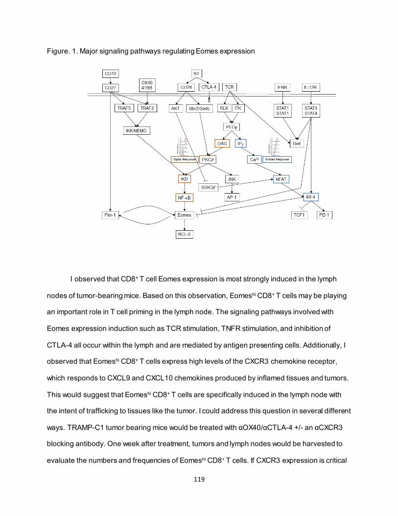
119
Figure. 1. Major signaling pathways regulating Eomes expression
I observed that CD8+ T cell Eomes expression is most strongly induced in the lymph
nodes of tumor-bearing mice. Based on this observation, Eomeshi CD8+ T cells may be playing
an important role in T cell priming in the lymph node. The signaling pathways involved with
Eomes expression induction such as TCR stimulation, TNFR stimulation, and inhibition of
CTLA-4 all occur within the lymph and are mediated by antigen presenting cells. Additionally, I
observed that Eomeshi CD8+ T cells express high levels of the CXCR3 chemokine receptor,
which responds to CXCL9 and CXCL10 chemokines produced by inflamed tissues and tumors.
This would suggest that Eomeshi CD8+ T cells are specifically induced in the lymph node with
the intent of trafficking to tissues like the tumor. I could address this question in several different
ways. TRAMP-C1 tumor bearing mice would be treated with αOX40/αCTLA-4 +/- an αCXCR3
blocking antibody. One week after treatment, tumors and lymph nodes would be harvested to
evaluate the numbers and frequencies of Eomeshi CD8+ T cells. If CXCR3 expression is critical
120
for Eomeshi CD8+ T cells to traffic to the tumor, then the αCXCR3 treated cohort would have
reduced Eomes in the tumor and possibly increased Eomes in the lymph node or peripheral
blood. If CXCR3 is insignificant to tumor trafficking by Eomeshi CD8+ T cells then no change in
Eomes expression in the tumor would be observed between the two cohorts. Additionally, the
TME could be evaluated for CXCL9 and CXCL10 expression by cytokine bead array to
determine if the TRAMP-C1 TME, or other tumor lines, are producing these chemokines.
5.2.2. CD8+ T cell Eomes expression drives a beneficial immune cancer response
Eomes, and the related molecule T-bet, are members of the T-box transcription factor
family. Many of the proteins in this family play important roles in embryonic and neurological
development with several being embryonically lethal if missing (407). Eomes and T-bet play
important roles in immunological cellular development. Eomes, also known as T-box brain
protein 2 (TBR2), binds to a specific genetic sequence known as the T-box. The DNA
consensus binding sequence consists of a minimum of 8 core nucleic acids, also called the T-
box half site or T-box binding element (TBE). T-box binding proteins have larger binding regions
than just the core consensus sequence, adding to variability in the genes activated by the
transcription factor. Additionally, T-box transcription factor expression is highly specific to cell
types and developmental regions. For example, the related T-box transcription factor Brachyury
is expressed in the posterior mesoderm during gastrulation development. Deletion of Eomes is
embryonically lethal as it disrupts both trophoblast and cardiac mesoderm development. The
exact outcomes of Eomes DNA binding in immune cells however is not well defined.
Studies looking at the effects of T cell specific deletions of Eomes and/or T-bet have
provided insight into how these two transcription factors regulate T cell function and
development. Through these knockout experiments, Eomes has been linked with several
121
important CD8+ T cell functions. Eomes has been linked with CD8+ T cell cytotoxicity, such as
production of IFN-g, TNF-a, and a variety of granzymes (408). Eomes has been linked to cell
trafficking through expression of CD44, CD62L, and CXCR3 (376). Additionally, these trafficking
proteins have helped to define Eomes CD8+ T cells as having memory like properties (409).
Fitting in with T cell memory is the expression of several pro-survival Bcl molecules (395).
Conversely, T-bet has overlapping and differing functions to Eomes in CD8+ T cells. T-bet also
plays an important role in the production of cytotoxic molecules such as IFN-γ, TNF-α, and
granzymes (410). However, T-bet expressing CD8+ T cells are often referred to as short lived
effector cells (363).
Fitting into the role of Eomes expression in CD8+ T cells is the related role of several
other transcription factors. The transcription factors T-cell factor 1 (TCF1), Tox, and Pim-1 all
play important roles in the regulation of Eomes and in how they effect Eomes-related cellular
function. TCF1 shares a lot of similarities with Eomes. TCF1 expression marks antigen
experienced CD8+ T cells that have properties of self-renewal and re-circulation (411).
Expression of TCF1 is also most frequently found in memory T cells, not effector CD8+ T cells.
Eomes and TCF1 also share similar upstream regulators. IRF4, which is a negative regulator of
Eomes expression and often determines Eomes expression, is also a negative regulator of
TCF1. On the other hand, Tox is a transcription factor that marks the exhausted T cell
phenotype in cancer and chronic viral infections (412). Tox expression is driven by NFAT,
similarly to expression of IRF4. Expression of Tox leads to upregulation of inhibitory receptors
and downregulation of cytotoxicity and cytokine production. In my studies, we observed that
Eomeshi CD8+ T cells induced by αOX40/αCTLA-4 therapy have lower Tox expression
compared to Eomeslo cells. Pim-1, while not a transcription factor, also plays an important role
in Eomes regulation and expression (369). Pim-1 is serine/threonine kinase expressed and
activated following CD27 ligation. CD27 itself is a TNF receptor expressed on most T cells and
122
contributes to NF-κB signaling through TRAF2. What makes the addition of Pim-1 signaling
downstream of CD27 different compared to other TNFRs is that Pim-1 and Eomes positively
signal back and forth on each other. Pim-1 inhibition results in reduced Eomes expression, and
reduced expression of BCL anti-apoptotic molecules downstream of Eomes such as Bcl-2 and
Bcl-xL. All of these signaling pathways point to the TCR and subsequent pITK/IRF4 expression
as being critical to the expression of Eomes.
A closer investigation of the mechanism by which Eomes expression is induced following
αOX40/αCTLA-4 therapy could be looked at in-vitro using a model of αOX40/αCTLA-4
stimulation, and inhibitors of specific signaling molecules. I have seen that Eomes expression
can be induced by αOX40/αCTLA-4 when lymph node derived lymphocytes are stimulated by
plate bound αCD3 Figure 1. To test the mechanism of induced Eomes expression I would use
inhibitors for NF-κB (IKKb), ITK, IRF4, and PIM-1 to probe the significance of each of these
signaling molecules to Eomes expression. I would hypothesize the chemical inhibition of ITK
and IRF4 will lead to enhanced Eomes expression, and inhibition of NF-κB or PIM-1 will lead to
decreased Eomes expression in αCD3/αOX40/αCTLA-4 stimulated CD8+ T cells from
lymphocytes.
Previous work looking at the role of Eomes expression in CD8+ T cells has suggested
that it contributes to overall T cell exhaustion. Li et al. looked at exhausted CD8+ T cells in
thymic lymphoma tumors and observed high expression of Eomes to accompany PD-1 and Tim-
3 expression (379). However, they found that CD8+ T cells lost cytotoxicity when Eomes was
deleted. Additionally, these experiments were in non-treated, tumor bearing WT mice and are
not a direct comparison to Eomeshi CD8+ T cells induced by αOX40/αCTLA-4 therapy. In their
study, not only was Eomes expressed in exhausted cells but was also highly expressed in
memory and effector populations. In the context of their experiments, it is possible that in the
absence of T cell stimulation, which would be provided by αOX40/αCTLA-4 therapy, CD8+ T cell
123
populations progress towards exhaustion in the tumor at a faster rate than I observed. In my
observations, the Eomeshi CD8+ T cells were cytotoxic and did not express high levels of
exhaustion markers. Other studies that suggest a role of Eomes in CD8+ T cell exhaustion are
examining the response to chronic viral infection such as LCMV (413). In these studies, it was
also found that Eomes is expressed in the effector and memory populations as well, with
increasing expression in the exhausted population increasing over time. Perhaps if Eomeshi
cells are subjected to a chronic infection without additional stimulation, they will transition into an
exhausted phenotype. However, relevant studies looking at the role of Eomes expression in
clinical cancer patients paints a different picture. Eomeshi CD8+ T cells have been shown to be
clinically relevant, as demonstrated by the presence of Eomeshi T cells in biopsies associated
with better clinical outcomes following αPD-1 or αPD-1/αCTLA-4 immunotherapy (366), and the
presence of Eomeshi CD8+ T cells within the tumor correlated with long-term survival in patients
with pancreatic cancer (414, 415).
The Eomeshi CD8+ T cells induced by αOX40/αCTLA-4 therapy had high expression of
CD44 and CD62L, placing them in the central memory phenotype. This finding was supported
by Eomeshi CD8+ T cell transcriptome GSEA analysis. What is the significance of Eomeshi CD8+
T cells resembling Tcm, and is this be a benefit to the overall anti-cancer response? Nicholas
Restifo has performed extensive research on the role of central memory and effector memory T
cells in cancer (416). Adoptive transfer of in-vitro generated of Tcm CD8+ T cell led to
eradication of large established tumors. However, adoptive transfer of in-vitro generated Tem
CD8+ T cells were far less effective at eliminating tumors. Their findings indicated that homing to
secondary lymphoid tissues prior to tumor trafficking was required for optimal treatment and
efficacy. The impact of the memory role that Eomeshi CD8+ T cells may be playing in
αOX40/αCTLA-4 therapy has yet to be determined. To test the hypothesis that Eomeshi CD8+ T
cells following αOX40/αCTLA-4 exhibit memory potential and secondary memory responses, we
124
could utilize the Eomes-GFP model for adoptive transfer experiments. Eomes-GFP+ or Eomes-
GFP- CD8+ T cells would be harvested from the lymph nodes of TRAMP-C1 tumor bearing
αOX40/αCTLA-4 mice, one week after treatment. These cells on the Thy1.2 C57BL/6
background would be adoptively transferred into WT C57BL/6 Thy1.1 mice. Four weeks after
adoptive transfer, the memory phenotype and frequency of Eomes expression would be
evaluated by flow cytometry from harvested secondary lymphoid organs as tracked by Thy1.2.
We would hypothesize that that Eomes-GFP+ transfer will have greater persistence compared
to the Eomes-GFP- transfer, as well as stability of Eomes-GFP expression. Additionally, using
this same experimental set up, four weeks after adoptive transfer mice will be challenged with
TRAMP-C1 or B16 tumor cells as a negative control. Effector function and memory phenotype
of Thy1.2 cells will be evaluated one week after tumor challenge. We would hypothesize that the
Eomes-GFP+ Thy1.2 cells to have greater effector function and memory phenotype compared
to the Eomes-GFP- Thy1.2 transferred cells.
5.2.3. Multifaceted roles of ibrutinib in αOX40/αCTLA-4 therapy
As the evidence mounted that Eomeshi CD8+ T cells were critical to the anti-tumor
efficacy of αOX40/αCTLA-4 therapy, it became important to investigate if CD8+ T cell Eomes
expression could be further enhanced. An analysis of the pathways involved in Eomes
expression suggested that the TCR signaling pathway, and specifically ITK, could be a point of
therapeutic intervention. As described previously in this chapter and in chapter 4, ITK plays a
critical role as a rheostat for how a T cell is going to react to TCR stimulation. Specifically, in the
case of CD8+ T cells, pITK results in reduced/inhibited Eomes expression as mediated by IRF4.
We chose ITK as a potential therapeutic target for a number of reasons. ITK is a signaling
kinase and could be targeted for inhibition by a small molecule kinase inhibitor. ITK expression
is conserved among T cells and not highly expressed in other tissues. There already is an FDA-
125
approved ITK inhibitor in the form of ibrutinib, and other drugs exist that target ITK as well. For
these reasons I investigated if ibrutinib used in combination with αOX40/αCTLA-4 therapy would
enhance CD8+ T cell Eomes expression, and what the effects this novel triple therapy would be.
The triple therapy had a synergistic effect with αOX40/αCTLA-4 on the expression of Eomes in
CD8+ T cells, while ibrutinib alone had no effect on Eomes expression. This is likely due to the
pathways leading to high Eomes expression such as strong T cell activation and blockade of
CTLA-4 were not present in these T cells. However, when ibrutinib was given to tumor-bearing
mice along with αOX40/αCTLA-4 it led to further increased Eomes expression. Both pITK and
IRF4 expression was significantly reduced in mice receiving triple therapy compared to
αOX40/αCTLA-4, suggesting that the effect of ibrutinib functioned through ITK inhibition.
I observed that triple therapy led to an increase in tumor antigen-specific CD8+ T cells as
determined by SPAS1 tetramer staining compared to αOX40/αCTLA-4 or control treated mice.
Similarly, Sagiv-Barfi et al showed an increase in tetramer+ CD8+ T cells in ibrutinib/αPD-L1
treated mice (372). Is ibrutinib therapy in combination with checkpoint blockade/co-stim agonists
enhancing the frequency and/or diversity of antigen specific CD8+ T cells? We could use TCR-
seq to test the hypothesis that ibrutinib is enhancing the clonality and tumor reactivity of CD8+ T
cells for αOX40/αCTLA-4 treatment. CD8+ T cells would be isolated from the blood and tumor
one week after treatment from TRAMP-C1 tumor bearing mice for TCR sequencing. Groups
would be compared between WT, ibrutinib, αOX40/αCTLA-4, and αOX40/αCTLA-4/ibrutinib
cohorts. This approach will allow us to determine TCR diversity and clonal abundance in CD8+ T
cells responding to each therapy. We would hypothesize that triple therapy treated mice would
have expanded clonal diversity, and enhanced abundance of specific tumor reactive clones.
However, triple therapy had wider effects than just enhancing Eomes expression. There
was also an overall increase in both CD8+ and CD4+ T cell function. Some of the effects on the
CD8+ T cell compartment can be explained by enhanced Eomes expression, such as higher
126
proliferation, TNF-α, IFN-γ, and CXCR3 expression. However, this does not explain the similar
increased function seen in CD4+ T cells. There are a few important differences between how
CD8+ and CD4+ T cells might respond to ITK inhibition. Both CD8+ and Th1 CD4+ T cells have
redundant signaling kinases in the TCR signaling pathway that can compensate for the loss of
ITK signaling. This redundant kinase is RLK, which is not inhibited by ibrutinib at the
concentrations used. Additionally, RLK does not signal through IRF4 in CD8+ T cells, and thus
does not inhibit Eomes expression. Th2 CD4+ T cells on the other hand do not express RLK and
are dependent on ITK signaling for T cell activation. Byrd et al. investigated the effects of
ibrutinib on CD4+ T cell polarization in patients receiving ibrutinib for the treatments of CLL
(389). They found that patients receiving ibrutinib had inhibited the activation of Th2 CD4+ T
cells and that they skewed towards a Th1 dominant phenotype. Additionally, Levy et al.
investigated the effects of ibrutinib on T cells in the context of αPD-L1 cancer therapy (372) and
found that this combination had therapeutic efficacy across multiple tumor models, leading to
enhanced numbers of tumor-specific T cells. However, a mechanism of action was not fully
explained. The effects of αOX40/αCTLA-4/ibrutinib triple therapy may be multi-faceted and work
through a combination of enhancing Eomes expression in CD8+ T cells and inhibiting Th2 CD4+
T cell polarization.
While ibrutinib has thus far been discussed in the context of its effect on ITK expression
and function in T cells, it has to be noted that ibrutinib is a drug designed to inhibit BTK
expression in B cells. A concern of using ibrutinib as a means of inhibiting ITK would be that it
could potentially inhibit BTK expression as well, the consequences of which could be
detrimental to the therapy. In the case of inhibition of B cells, there appears to be mounting
evidence that ibrutinib does not have a significant effect on mature antigen experienced B cells
(417). Additionally, I found that B cells were dispensable to the efficacy of triple therapy. Another
concern of using ibrutinib is its effect on the myeloid compartment as some myeloid cells
127
express BTK, such as monocytes and macrophages (418). While this may be a concern, I saw
no significant change to overall myeloid cells or their function in mice receiving triple therapy
compared to αOX40/αCTLA-4. One further concern with ibrutinib therapy is if BTK inhibition has
a direct effect on the tumor growth or the way the tumor tailors the TME. However, if this is the
case it would likely lead to further tumor inhibition. Some groups have even proposed BTK as a
potential therapeutic target in prostate cancer (419). In my experiments looking at the effect of
ibrutinib on any of the tumor cell lines I used for in-vivo experiments, ibrutinib had no effect on
tumor cell growth, even at concentrations 100X higher than used in-vivo. These concerns might
further be abrogated with the use of an ITK specific inhibitor such as the BMS compound
509744, however further research would be needed. Regardless, ITK specific inhibitors have
great therapeutic potential.
To further evaluate global changes and unique signatures associated with
αOX40/aCLTA-4 therapy, and the impact of ibrutinib, we could utilize single-cell RNA
sequencing. TRAMP-C1 tumor bearing mice would be treated with control IgG, αOX40/αCTLA-
4/ or triple therapy. One week after treatment, CD45+ cells would be sorted out of tumors by
FACS for scRNAseq analysis. Unique cell types will be clustered using UMAP. We will
determine differentially expressed genes associated with therapy, and unique pathways
associated with Eomes regulation.
I have shown that ibrutinib has strong anti-cancer efficacy when used in combination
with αOX40/αCTLA-4 therapy. However, ibrutinib may synergize with other therapies, as was
shown with PD-L1 (372). Considering that ibrutinib enhanced CD8+ T cell Eomes expression
under strong T cell activating conditions and is known to skew CD4+ T cells towards a Th1
phenotype, perhaps we should look at therapies that would drive Eomes expression or be
limited by Th2 polarization. For example, other anti-TNFR mAbs, such as a4-1BB, have been
suggested to play a role in the induction of Eomes (420). However, targeting other TNFRs might
128
run into similar issues as αOX40 in that Eomes was not highly expressed without the addition of
αCTLA-4. While one report showed that αPD-L1 therapy synergized with ibrutinib to reduce
tumor growth, my studies revealed that αPD-1 or αOX40/αPD-1 did not induce Eomes
expression, which strongly suggested an important role for αCTLA-4 (372). Targeting ICOS may
also be efficacious because it plays a role to CD4+ T cell polarization and skewing towards Th2.
Additionally, therapies that enhance TCR signaling like cancer vaccines and therapies that
enhance NF-kB signaling, like TLR agonists, may also work well with ibrutinib to further CD8+ T
cell Eomes expression.
There are several ongoing clinical trials relevant to my research. One trial is looking at
the efficacy of αOX40 and αCTLA-4 in combination. A phase I/II trial is investigating the
combination of BMS-986178 by itself or in combination with pembrolizumab and/or ipilimumab
for patients with advanced solid tumors (NCT02737475). There is an ongoing OX40 trail looking
at the combination of a hexavalent OX40 agonist in combination with pembrolizumab for the
treatment of advanced metastatic solid tumors (NCT04198766).
IRF4 inhibition in the form of an antisense oligonucleotide against IRF4 has shown
efficacy in the treatment of myeloma (421). Myeloma tumor bearing mice treated with the
antisense oligonucleotide against IRF4 had decreased tumor burden and increased overall
survival. No analysis was done on the direct effects on the immune cell populations. The use of
a drug to inhibit IRF4 rather than upstream ITK may be a cleaner way of tailoring the T cell
response but may lose the Th2 inhibitor effects of ITK inhibition (422).
Ibrutinib (Imbruvica) is being investigated in clinical trials for use in combination with
immunotherapeutic drugs. Ibrutinib and pembrolizumab are being evaluated in a phase II trial
for high-risk lymphocytic leukemia with a focus on evaluating improved immune function
(NCT03514017). A phase I trial is looking at the combination of ibrutinib and nivolumab for
patients with metastatic solid tumors (NCT03525925). Preliminary results from this phase I trials
129
indicated that T cell function improved throughout the study for patients receiving combination
ibrutinib and nivolumab. Four of the 16 patients had a partial response and another 4 of 16
patients had stable disease. The combination was well tolerated. A Phase Ib/II trial looking at
the combination of ibrutinib and durvalumab, a PD-L1 blocking antibody, has been evaluated for
patients with pre-treated relapsed/refractory solid tumors (NCT02401048). The combination was
well tolerated but had limited anti-tumor activity in the cohort. Other clinical trials looking at the
combination of ibrutinib with αPD-1 are ongoing. Many of these trials are for the treatment of
refractory lymphomas and leukemias (NCT02332980, NCT03204188). Some attention has been
given towards the use of ibrutinib for the treatment of prostate cancer, however ibrutinib has yet
to be evaluated in combination with immunotherapy. A phase II trial examined the effects of
ibrutinib given pre-surgery to prostate cancer patients, with the intention of inhibiting tumor cell
growth and enhancing the patient’s anti-tumor immune response prior to surgery
(NCT02643667).
5.3. Conclusions
As has been iterated many times in this body of work, checkpoint blockade has great
potential but limited success in treating a variety of cancers. From murine studies and clinical
trials, it has been determined that monotherapy checkpoint blockade has only limited
therapeutic efficacy and that combination immunotherapy may be an answer to better clinical
outcomes as demonstrated by the enhanced efficacy of αPD-1/αCTLA-4 therapy. However,
there still is a lack of knowledge regarding mechanisms of how certain immunotherapies
function and the mechanisms by which combination immunotherapies may synergize. With that
knowledge from pre-clinical murine studies, we can inform upon and rationally design novel
immunotherapy combinations.
130
My research aimed to advance immunotherapy design through investigating the
combination of a checkpoint inhibitor (αCTLA-4) and a co-stimulatory agonist (αOX40) in order
to better understand how they these two specifically interact, what aspects of this combination
drives anti-tumor immunity, and how this therapy might be enhanced for clinical use. An
important finding from my research was the critical nature of Eomeshi CD8+ T cells following
αOX40/αCTLA-4 therapy. This raises the question as to whether Eomeshi CD8+ T cells are as
critical following other cancer therapies or if this is a unique property of αOX40/αCTLA-4. My
research profiling the function of Eomeshi CD8+ T cells demonstrated they play an important
memory T cell role. As evidenced by my research and studies investigating Eomes expression
in human cancers, it would seem the generation of Eomes expression is beneficial and may be
critical to the anti-tumor immune response. I used this novel understanding of Eomeshi CD8+ T
cells to inform therapy design by investigating the role of ITK inhibition on Eomeshi generation.
The combination of αOX40/αCTLA-4/ibrutinib therapy was investigated to test the hypothesis
that if the generation of Eomeshi T cells enhances αOX40/αCTLA-4 therapy, then further
enhancing Eomes expression with ibrutinib would improve the overall therapy. My novel finding
was that the ITK inhibitor effects of ibrutinib further enhanced Eomeshi CD8+ T cells in
αOX40/αCTLA-4 therapy leading to reduced tumor growth and enhanced survival in tumor
bearing mice compared to αOX40/αCTLA-4 alone. This is a significant and impactful finding, as
ibrutinib is FDA-approved, orally available, and has limited side effects. Ongoing clinical trials
investigating ibrutinib in combination with checkpoint blockade have shown an acceptable safety
profile. The knowledge generated by my research on the generation and function of Eomeshi
CD8+ T cells, as well as the use of ibrutinib as an ITK inhibitor, has strong potential for
therapeutic design and translation to the clinic.
My research also raised several important questions that would be valuable for the field
to address in the future, such as a need for further investigation into the mechanism of Eomes
131
expression in CD8+ T cells, which may inform upon other therapeutic points of intervention for
better therapy design. The use of a pure ITK inhibitor such a BMS-509744 may be a more
specific way of inhibiting ITK to generate Eomes expression, or perhaps targeting IRF4 may be
a better option as it has potential for direct αtumor effects as well. Further studies are needed
regarding the expression and function of Eomeshi CD8+ T cells in humans. Overall, my research
added valuable knowledge to the field of translational immunotherapy, and suggests the use of
ibrutinib and modulating of Eomes for future research and clinical therapy design.
132
6. References
1. O. Takeuchi, S. Akira, Pattern recognition receptors and inflammation. Cell 140, 805-820 (2010). 2. C. Hernandez, P. Huebener, R. F. Schwabe, Damage-associated molecular patterns in cancer: a
double-edged sword. Oncogene 35, 5931-5941 (2016). 3. T. Kawasaki, T. Kawai, Toll-like receptor signaling pathways. Front Immunol 5, 461 (2014). 4. E. W. Hewitt, The MHC class I antigen presentation pathway: strategies for viral immune
evasion. Immunology 110, 163-169 (2003). 5. J. P. Ting, J. Trowsdale, Genetic control of MHC class II expression. Cell 109 Suppl, S21-33 (2002). 6. I. Livneh, V. Cohen-Kaplan, C. Cohen-Rosenzweig, N. Avni, A. Ciechanover, The life cycle of the
26S proteasome: from birth, through regulation and function, and onto its death. Cell Res 26, 869-885 (2016).
7. R. Abele, R. Tampe, The ABCs of immunology: structure and function of TAP, the transporter associated with antigen processing. Physiology (Bethesda) 19, 216-224 (2004).
8. Z. Liu, P. A. Roche, Macropinocytosis in phagocytes: regulation of MHC class-II-restricted antigen presentation in dendritic cells. Front Physiol 6, 1 (2015).
9. P. Bertolino, C. Rabourdin-Combe, The MHC class II-associated invariant chain: a molecule with multiple roles in MHC class II biosynthesis and antigen presentation to CD4+ T cells. Crit Rev Immunol 16, 359-379 (1996).
10. M. Embgenbroich, S. Burgdorf, Current Concepts of Antigen Cross-Presentation. Front Immunol 9, 1643 (2018).
11. S. K. Wculek et al., Dendritic cells in cancer immunology and immunotherapy. Nat Rev Immunol 20, 7-24 (2020).
12. C. Geisler, J. K. Larsen, T. Plesner, Identification of alpha beta and gamma delta T cell receptor-positive cells. Scand J Immunol 28, 741-745 (1988).
13. K. W. Wucherpfennig, E. Gagnon, M. J. Call, E. S. Huseby, M. E. Call, Structural biology of the T-cell receptor: insights into receptor assembly, ligand recognition, and initiation of signaling. Cold Spring Harb Perspect Biol 2, a005140 (2010).
14. M. S. Krangel, Mechanics of T cell receptor gene rearrangement. Curr Opin Immunol 21, 133-139 (2009).
15. H. Takaba, H. Takayanagi, The Mechanisms of T Cell Selection in the Thymus. Trends Immunol 38, 805-816 (2017).
16. N. Kurd, E. A. Robey, T-cell selection in the thymus: a spatial and temporal perspective. Immunol Rev 271, 114-126 (2016).
17. S. Halle, O. Halle, R. Forster, Mechanisms and Dynamics of T Cell-Mediated Cytotoxicity In Vivo. Trends Immunol 38, 432-443 (2017).
18. I. Voskoboinik, J. C. Whisstock, J. A. Trapani, Perforin and granzymes: function, dysfunction and human pathology. Nat Rev Immunol 15, 388-400 (2015).
19. R. V. Luckheeram, R. Zhou, A. D. Verma, B. Xia, CD4(+)T cells: differentiation and functions. Clin Dev Immunol 2012, 925135 (2012).
20. K. L. Knutson, M. L. Disis, Tumor antigen-specific T helper cells in cancer immunity and immunotherapy. Cancer Immunol Immunother 54, 721-728 (2005).
21. L. Ni, J. Lu, Interferon gamma in cancer immunotherapy. Cancer Med 7, 4509-4516 (2018). 22. H. Wajant, K. Pfizenmaier, P. Scheurich, Tumor necrosis factor signaling. Cell Death Differ 10, 45-
65 (2003). 23. L. Wang, F. Du, X. Wang, TNF-alpha induces two distinct caspase-8 activation pathways. Cell 133,
693-703 (2008).
133
24. H. E. Kohrt et al., Profile of immune cells in axillary lymph nodes predicts disease-free survival in breast cancer. PLoS Med 2, e284 (2005).
25. K. L. Dennis, N. R. Blatner, F. Gounari, K. Khazaie, Current status of interleukin-10 and regulatory T-cells in cancer. Curr Opin Oncol 25, 637-645 (2013).
26. Y. Ohue, H. Nishikawa, Regulatory T (Treg) cells in cancer: Can Treg cells be a new therapeutic target? Cancer Sci 110, 2080-2089 (2019).
27. E. Batlle, J. Massague, Transforming Growth Factor-beta Signaling in Immunity and Cancer. Immunity 50, 924-940 (2019).
28. S. K. Mittal, K. J. Cho, S. Ishido, P. A. Roche, Interleukin 10 (IL-10)-mediated Immunosuppression: MARCH-I INDUCTION REGULATES ANTIGEN PRESENTATION BY MACROPHAGES BUT NOT DENDRITIC CELLS. J Biol Chem 290, 27158-27167 (2015).
29. B. M. Olson, J. A. Sullivan, W. J. Burlingham, Interleukin 35: a key mediator of suppression and the propagation of infectious tolerance. Front Immunol 4, 315 (2013).
30. J. R. Hwang, Y. Byeon, D. Kim, S. G. Park, Recent insights of T cell receptor-mediated signaling pathways for T cell activation and development. Exp Mol Med 52, 750-761 (2020).
31. A. H. Courtney, W. L. Lo, A. Weiss, TCR Signaling: Mechanisms of Initiation and Propagation. Trends Biochem Sci 43, 108-123 (2018).
32. M. Karin, Z. Liu, E. Zandi, AP-1 function and regulation. Curr Opin Cell Biol 9, 240-246 (1997). 33. T. Liu, L. Zhang, D. Joo, S. C. Sun, NF-kappaB signaling in inflammation. Signal Transduct Target
Ther 2 (2017). 34. F. Macian, NFAT proteins: key regulators of T-cell development and function. Nat Rev Immunol
5, 472-484 (2005). 35. D. M. Sansom, CD28, CTLA-4 and their ligands: who does what and to whom? Immunology 101,
169-177 (2000). 36. D. Gardner, L. E. Jeffery, D. M. Sansom, Understanding the CD28/CTLA-4 (CD152) pathway and
its implications for costimulatory blockade. Am J Transplant 14, 1985-1991 (2014). 37. R. Elgueta et al., Molecular mechanism and function of CD40/CD40L engagement in the immune
system. Immunol Rev 229, 152-172 (2009). 38. B. J. Laidlaw, J. E. Craft, S. M. Kaech, The multifaceted role of CD4(+) T cells in CD8(+) T cell
memory. Nat Rev Immunol 16, 102-111 (2016). 39. V. Kalia, S. Sarkar, Regulation of Effector and Memory CD8 T Cell Differentiation by IL-2-A
Balancing Act. Front Immunol 9, 2987 (2018). 40. K. A. Frauwirth, M. L. Alegre, C. B. Thompson, Induction of T cell anergy in the absence of CTLA-
4/B7 interaction. J Immunol 164, 2987-2993 (2000). 41. G. L. Xu, X. H. Zhu, B. Guo, Y. Z. Wu, Involvement of CTLA-4 in T-cell anergy induced by
staphylococcal enterotoxin A in vitro. Mol Immunol 41, 1-8 (2004). 42. N. L. Alves, F. A. Arosa, R. A. van Lier, Common gamma chain cytokines: dissidence in the details.
Immunol Lett 108, 113-120 (2007). 43. S. H. Ross, D. A. Cantrell, Signaling and Function of Interleukin-2 in T Lymphocytes. Annu Rev
Immunol 36, 411-433 (2018). 44. E. R. Mehlhop-Williams, M. J. Bevan, Memory CD8+ T cells exhibit increased antigen threshold
requirements for recall proliferation. J Exp Med 211, 345-356 (2014). 45. D. Skrombolas, J. G. Frelinger, Challenges and developing solutions for increasing the benefits of
IL-2 treatment in tumor therapy. Expert Rev Clin Immunol 10, 207-217 (2014). 46. T. A. Waldmann, S. Dubois, M. D. Miljkovic, K. C. Conlon, IL-15 in the Combination
Immunotherapy of Cancer. Front Immunol 11, 868 (2020). 47. T. O. Robinson, K. S. Schluns, The potential and promise of IL-15 in immuno-oncogenic therapies.
Immunol Lett 190, 159-168 (2017).
134
48. D. C. Wilson, S. Matthews, G. S. Yap, IL-12 signaling drives CD8+ T cell IFN-gamma production and differentiation of KLRG1+ effector subpopulations during Toxoplasma gondii Infection. J Immunol 180, 5935-5945 (2008).
49. R. Morris, N. J. Kershaw, J. J. Babon, The molecular details of cytokine signaling via the JAK/STAT pathway. Protein Sci 27, 1984-2009 (2018).
50. Y. Y. Wan, R. A. Flavell, Regulatory T cells, transforming growth factor-beta, and immune suppression. Proc Am Thorac Soc 4, 271-276 (2007).
51. R. I. Nurieva, Y. Chung, Understanding the development and function of T follicular helper cells. Cell Mol Immunol 7, 190-197 (2010).
52. J. Zhu, H. Yamane, W. E. Paul, Differentiation of effector CD4 T cell populations (*). Annu Rev Immunol 28, 445-489 (2010).
53. A. Malyshkina et al., Fas Ligand-mediated cytotoxicity of CD4+ T cells during chronic retrovirus infection. Sci Rep 7, 7785 (2017).
54. R. M. Dunham et al., CD127 and CD25 expression defines CD4+ T cell subsets that are differentially depleted during HIV infection. J Immunol 180, 5582-5592 (2008).
55. R. de Waal Malefyt et al., CD2/LFA-3 or LFA-1/ICAM-1 but not CD28/B7 interactions can augment cytotoxicity by virus-specific CD8+ cytotoxic T lymphocytes. Eur J Immunol 23, 418-424 (1993).
56. M. L. Dustin, E. O. Long, Cytotoxic immunological synapses. Immunol Rev 235, 24-34 (2010). 57. I. Voskoboinik, M. A. Dunstone, K. Baran, J. C. Whisstock, J. A. Trapani, Perforin: structure,
function, and role in human immunopathology. Immunol Rev 235, 35-54 (2010). 58. D. Kaiserman et al., The major human and mouse granzymes are structurally and functionally
divergent. J Cell Biol 175, 619-630 (2006). 59. M. D. Martin, V. P. Badovinac, Defining Memory CD8 T Cell. Front Immunol 9, 2692 (2018). 60. J. C. Nolz, G. R. Starbeck-Miller, J. T. Harty, Naive, effector and memory CD8 T-cell trafficking:
parallels and distinctions. Immunotherapy 3, 1223-1233 (2011). 61. E. J. Wherry, M. Kurachi, Molecular and cellular insights into T cell exhaustion. Nat Rev Immunol
15, 486-499 (2015). 62. J. S. Yi, M. A. Cox, A. J. Zajac, T-cell exhaustion: characteristics, causes and conversion.
Immunology 129, 474-481 (2010). 63. B. Youngblood et al., Chronic virus infection enforces demethylation of the locus that encodes
PD-1 in antigen-specific CD8(+) T cells. Immunity 35, 400-412 (2011). 64. A. Schietinger, P. D. Greenberg, Tolerance and exhaustion: defining mechanisms of T cell
dysfunction. Trends Immunol 35, 51-60 (2014). 65. W. X. Huff, J. H. Kwon, M. Henriquez, K. Fetcko, M. Dey, The Evolving Role of CD8(+)CD28(-)
Immunosenescent T Cells in Cancer Immunology. Int J Mol Sci 20 (2019). 66. J. Ye et al., Human regulatory T cells induce T-lymphocyte senescence. Blood 120, 2021-2031
(2012). 67. J. Ye et al., Tumor-derived gammadelta regulatory T cells suppress innate and adaptive
immunity through the induction of immunosenescence. J Immunol 190, 2403-2414 (2013). 68. R. Nurieva, J. Wang, A. Sahoo, T-cell tolerance in cancer. Immunotherapy 5, 513-531 (2013). 69. M. C. Hunter, A. Teijeira, C. Halin, T Cell Trafficking through Lymphatic Vessels. Front Immunol 7,
613 (2016). 70. J. Kranich, N. J. Krautler, How Follicular Dendritic Cells Shape the B-Cell Antigenome. Front
Immunol 7, 225 (2016). 71. S. K. Bromley, S. Y. Thomas, A. D. Luster, Chemokine receptor CCR7 guides T cell exit from
peripheral tissues and entry into afferent lymphatics. Nat Immunol 6, 895-901 (2005).
135
72. M. Matloubian et al., Lymphocyte egress from thymus and peripheral lymphoid organs is dependent on S1P receptor 1. Nature 427, 355-360 (2004).
73. T. H. Pham, T. Okada, M. Matloubian, C. G. Lo, J. G. Cyster, S1P1 receptor signaling overrides retention mediated by G alpha i-coupled receptors to promote T cell egress. Immunity 28, 122-133 (2008).
74. H. Harjunpaa, M. Llort Asens, C. Guenther, S. C. Fagerholm, Cell Adhesion Molecules and Their Roles and Regulation in the Immune and Tumor Microenvironment. Front Immunol 10, 1078 (2019).
75. P. T. Kuo et al., The Role of CXCR3 and Its Chemokine Ligands in Skin Disease and Cancer. Front Med (Lausanne) 5, 271 (2018).
76. N. L. Harris, V. Watt, F. Ronchese, G. Le Gros, Differential T cell function and fate in lymph node and nonlymphoid tissues. J Exp Med 195, 317-326 (2002).
77. C. B. Blackadar, Historical review of the causes of cancer. World J Clin Oncol 7, 54-86 (2016). 78. C. M. Croce, Oncogenes and cancer. N Engl J Med 358, 502-511 (2008). 79. D. Hanahan, R. A. Weinberg, Hallmarks of cancer: the next generation. Cell 144, 646-674 (2011). 80. U. Testa, E. Pelosi, G. Castelli, Colorectal cancer: genetic abnormalities, tumor progression,
tumor heterogeneity, clonal evolution and tumor-initiating cells. Med Sci (Basel) 6 (2018). 81. A. O. Kamphorst et al., Rescue of exhausted CD8 T cells by PD-1–targeted therapies is CD28-
dependent. Science (New York, N.Y.) 355, 1423-1427 (2017). 82. D. L. Barber et al., Restoring function in exhausted CD8 T cells during chronic viral infection.
Nature 439, 682-687 (2006). 83. W. Jiang et al., Exhausted CD8+T Cells in the Tumor Immune Microenvironment: New Pathways
to Therapy. Front Immunol 11, 622509 (2020). 84. E. J. Wherry, T cell exhaustion. Nature Immunology 12, 492 (2011). 85. P. Savas et al., Single-cell profiling of breast cancer T cells reveals a tissue-resident memory
subset associated with improved prognosis. Nature Medicine 24, 986-993 (2018). 86. E. Peranzoni et al., Macrophages impede CD8 T cells from reaching tumor cells and limit the
efficacy of anti-PD-1 treatment. P Natl Acad Sci USA 115, E4041-E4050 (2018). 87. F. Azimi et al., Tumor-Infiltrating Lymphocyte Grade Is an Independent Predictor of Sentinel
Lymph Node Status and Survival in Patients With Cutaneous Melanoma. Journal of Clinical Oncology 30, 2678-2683 (2012).
88. B. Mlecnik et al., Histopathologic-based prognostic factors of colorectal cancers are associated with the state of the local immune reaction. J Clin Oncol 29, 610-618 (2011).
89. F. Pages et al., Effector memory T cells, early metastasis, and survival in colorectal cancer. N Engl J Med 353, 2654-2666 (2005).
90. P. C. Tumeh et al., PD-1 blockade induces responses by inhibiting adaptive immune resistance. Nature 515, 568-571 (2014).
91. B. Beutler, E. T. Rietschel, Innate immune sensing and its roots: the story of endotoxin. Nature Reviews Immunology 3, 169 (2003).
92. T. Kawai, S. Akira, The role of pattern-recognition receptors in innate immunity: update on Toll-like receptors. Nature Immunology 11, 373 (2010).
93. A. Williams, C. A. Peh, T. Elliott, The cell biology of MHC class I antigen presentation. Tissue Antigens 59, 3-17 (2002).
94. J. E. Smith-Garvin, G. A. Koretzky, M. S. Jordan, T Cell Activation. Annual Review of Immunology 27, 591-619 (2009).
95. P. Guermonprez, J. Valladeau, L. Zitvogel, C. Théry, S. Amigorena, Antigen Presentation and T Cell Stimulation by Dendritic Cells. Annual Review of Immunology 20, 621-667 (2002).
136
96. A. Brewitz et al., CD8(+) T Cells Orchestrate pDC-XCR1(+) Dendritic Cell Spatial and Functional Cooperativity to Optimize Priming. Immunity 46, 205-219 (2017).
97. J. Lieberman, The ABCs of granule-mediated cytotoxicity: new weapons in the arsenal. Nature Reviews Immunology 3, 361 (2003).
98. Y. Yang et al., High intratumoral CD8(+) T-cell infiltration is associated with improved survival in prostate cancer patients undergoing radical prostatectomy. Prostate 81, 20-28 (2021).
99. X. Li et al., Infiltration of CD8(+) T cells into tumor cell clusters in triple-negative breast cancer. Proc Natl Acad Sci U S A 116, 3678-3687 (2019).
100. M. W. Teng, S. F. Ngiow, A. Ribas, M. J. Smyth, Classifying Cancers Based on T-cell Infiltration and PD-L1. Cancer Res 75, 2139-2145 (2015).
101. P. Matzinger, The Danger Model: A Renewed Sense of Self. Science 296, 301 (2002). 102. S. Gallucci, M. Lolkema, P. Matzinger, Natural adjuvants: Endogenous activators of dendritic
cells. Nature Medicine 5, 1249 (1999). 103. B. Sauter et al., Consequences of Cell Death. The Journal of Experimental Medicine 191, 423
(2000). 104. D. E. Orange et al., Effective antigen cross-presentation by prostate cancer patients'
dendritic cells: implications for prostate cancer immunotherapy. Prostate Cancer And Prostatic Diseases 7, 63 (2004).
105. A. L. Ackerman, P. Cresswell, Cellular mechanisms governing cross-presentation of exogenous antigens. Nature Immunology 5, 678 (2004).
106. S. Jung et al., In vivo depletion of CD11c+ dendritic cells abrogates priming of CD8+ T cells by exogenous cell-associated antigens. Immunity 17, 211-220 (2002).
107. M. Haniffa et al., Human tissues contain CD141hi cross-presenting dendritic cells with functional homology to mouse CD103+ nonlymphoid dendritic cells. Immunity 37, 60-73 (2012).
108. S. Spranger, D. Dai, B. Horton, T. F. Gajewski, Tumor-Residing Batf3 Dendritic Cells Are Required for Effector T Cell Trafficking and Adoptive T Cell Therapy. Cancer Cell 31, 711-+ (2017).
109. C. H. GeurtsvanKessel et al., Clearance of influenza virus from the lung depends on migratory langerin+CD11b- but not plasmacytoid dendritic cells. The Journal of experimental medicine 205, 1621-1634 (2008).
110. M. L. Broz et al., Dissecting the tumor myeloid compartment reveals rare activating antigen-presenting cells critical for T cell immunity. Cancer Cell 26, 638-652 (2014).
111. I. Treilleux et al., Dendritic Cell Infiltration and Prognosis of Early Stage Breast Cancer. Clinical Cancer Research 10, 7466 (2004).
112. E. W. Roberts et al., Critical Role for CD103+/CD141+Dendritic Cells Bearing CCR7 for Tumor Antigen Trafficking and Priming of T Cell Immunity in Melanoma. Cancer Cell 30, 324-336 (2016).
113. M. Mercader et al., T cell infiltration of the prostate induced by androgen withdrawal in patients with prostate cancer. Proceedings of the National Academy of Sciences 98, 14565 (2001).
114. S. J. A. M. Santegoets et al., Myeloid derived suppressor and dendritic cell subsets are related to clinical outcome in prostate cancer patients treated with prostate GVAX and ipilimumab. Journal for immunotherapy of cancer 2, 31 (2014).
115. P. Stoitzner et al., Inefficient presentation of tumor-derived antigen by tumor-infiltrating dendritic cells. Cancer Immunology, Immunotherapy 57, 1665-1673 (2008).
116. B.-Z. Qian, J. W. Pollard, Macrophage Diversity Enhances Tumor Progression and Metastasis. Cell 141, 39-51 (2010).
117. B. Ruffell et al., Macrophage IL-10 blocks CD8+ T cell-dependent responses to chemotherapy by suppressing IL-12 expression in intratumoral dendritic cells. Cancer Cell 26, 623-637 (2014).
137
118. L. de Chaisemartin et al., Characterization of Chemokines and Adhesion Molecules Associated with T cell Presence in Tertiary Lymphoid Structures in Human Lung Cancer. Cancer Research 71, 6391-6399 (2011).
119. E. N. McNamee, J. Rivera-Nieves, Ectopic Tertiary Lymphoid Tissue in Inflammatory Bowel Disease: Protective or Provocateur? Frontiers in immunology 7, 308 (2016).
120. M. C. Dieu-Nosjean, J. Goc, N. A. Giraldo, C. Sautes-Fridman, W. H. Fridman, Tertiary lymphoid structures in cancer and beyond. Trends in Immunology 35, 571-580 (2014).
121. M.-C. Dieu-Nosjean et al., Long-Term Survival for Patients With Non–Small-Cell Lung Cancer With Intratumoral Lymphoid Structures. Journal of Clinical Oncology 26, 4410-4417 (2008).
122. J. Goc, W. H. Fridman, C. Sautes-Fridman, M. C. Dieu-Nosjean, Characteristics of tertiary lymphoid structures in primary cancers. Oncoimmunology 2, e26836 (2013).
123. M. D. L. L. Garcia-Hernandez et al., A Unique Cellular and Molecular Microenvironment Is Present in Tertiary Lymphoid Organs of Patients with Spontaneous Prostate Cancer Regression. Frontiers in immunology 8 (2017).
124. J. Goc et al., Dendritic cells in tumor-associated tertiary lymphoid structures signal a Th1 cytotoxic immune contexture and license the positive prognostic value of infiltrating CD8+ T cells. Cancer Res 74, 705-715 (2014).
125. L. A. M. Pozzi, J. W. Maciaszek, K. L. Rock, Both Dendritic Cells and Macrophages Can Stimulate Naive CD8 T Cells In Vivo to Proliferate, Develop Effector Function, and Differentiate into Memory Cells. The Journal of Immunology 175, 2071-2081 (2005).
126. K. Ley, M1 Means Kill; M2 Means Heal. J Immunol 199, 2191-2193 (2017). 127. G. Comito et al., Cancer-associated fibroblasts and M2-polarized macrophages synergize during
prostate carcinoma progression. Oncogene 33, 2423 (2013). 128. S. R. Gordon et al., PD-1 expression by tumour-associated macrophages inhibits phagocytosis
and tumour immunity. Nature 545, 495-499 (2017). 129. Y. Komohara et al., Macrophage infiltration and its prognostic relevance in clear cell renal cell
carcinoma. Cancer science 102, 1424-1431 (2011). 130. M. J. Campbell et al., Proliferating macrophages associated with high grade, hormone receptor
negative breast cancer and poor clinical outcome. Breast Cancer Res Tr 128, 703-711 (2011). 131. V. Bronte et al., Recommendations for myeloid-derived suppressor cell nomenclature and
characterization standards. Nature Communications 7 (2016). 132. L. Fong et al., Activated lymphocyte recruitment into the tumor microenvironment following
preoperative sipuleucel-T for localized prostate cancer. J Natl Cancer Inst 106 (2014). 133. P. W. Kantoff et al., Sipuleucel-T immunotherapy for castration-resistant prostate cancer. N Engl
J Med 363, 411-422 (2010). 134. E. J. Wherry et al., Molecular signature of CD8+ T cell exhaustion during chronic viral infection.
Immunity 27, 670-684 (2007). 135. A. J. Zajac et al., Viral Immune Evasion Due to Persistence of Activated T Cells Without Effector
Function. The Journal of Experimental Medicine 188, 2205-2213 (1998). 136. G. J. Freeman, E. J. Wherry, R. Ahmed, A. H. Sharpe, Reinvigorating exhausted HIV-specific T cells
via PD-1–PD-1 ligand blockade. 203, 2223-2227 (2006). 137. L. Baitsch et al., Exhaustion of tumor-specific CD8+ T cells in metastases from melanoma
patients. The Journal of Clinical Investigation 121, 2350-2360 (2011). 138. A. Schietinger et al., Tumor-Specific T Cell Dysfunction Is a Dynamic Antigen-Driven
Differentiation Program Initiated Early during Tumorigenesis. Immunity 45, 389-401 (2016). 139. S. J. Im et al., Defining CD8+ T cells that provide the proliferative burst after PD-1 therapy.
Nature 537, 417-421 (2016).
138
140. I. Siddiqui et al., Intratumoral Tcf1(+)PD-1(+)CD8(+) T Cells with Stem-like Properties Promote Tumor Control in Response to Vaccination and Checkpoint Blockade Immunotherapy. Immunity 50, 195-211 e110 (2019).
141. S. Kurtulus et al., Checkpoint Blockade Immunotherapy Induces Dynamic Changes in PD-1(-)CD8(+) Tumor-Infiltrating T Cells. Immunity 50, 181-194 e186 (2019).
142. J. Brummelman et al., High-dimensional single cell analysis identifies stem-like cytotoxic CD8(+) T cells infiltrating human tumors. J Exp Med 215, 2520-2535 (2018).
143. H. Li et al., Dysfunctional CD8 T Cells Form a Proliferative, Dynamically Regulated Compartment within Human Melanoma. Cell 176, 775-789.e718 (2019).
144. J. P. Bottcher, C. Reis e Sousa, The Role of Type 1 Conventional Dendritic Cells in Cancer Immunity. Trends Cancer 4, 784-792 (2018).
145. W. J. Catalona, S. Loeb, Prostate cancer screening and determining the appropriate prostate-specific antigen cutoff values. J Natl Compr Canc Netw 8, 265-270 (2010).
146. J. A. Antenor et al., Preoperative PSA and progression-free survival after radical prostatectomy for Stage T1c disease. Urology 66, 156-160 (2005).
147. S. Loeb et al., Pathological characteristics of prostate cancer detected through prostate specific antigen based screening. J Urol 175, 902-906 (2006).
148. A. Amayo, W. Obara, Serum prostate specific antigen levels in men with benign prostatic hyperplasia and cancer of prostate. East Afr Med J 81, 22-26 (2004).
149. E. Elkord, A. W. Rowbottom, H. Kynaston, P. E. Williams, Correlation between CD8+ T cells specific for prostate-specific antigen and level of disease in patients with prostate cancer. Clin Immunol 120, 91-98 (2006).
150. E. Elkord, P. E. Williams, H. Kynaston, A. W. Rowbottom, Differential CTLs specific for prostate-specific antigen in healthy donors and patients with prostate cancer. Int Immunol 17, 1315-1325 (2005).
151. R. A. Madan, P. M. Arlen, M. Mohebtash, J. W. Hodge, J. L. Gulley, Prostvac-VF: a vector-based vaccine targeting PSA in prostate cancer. Expert Opin Investig Drugs 18, 1001-1011 (2009).
152. J. W. Hodge et al., A triad of costimulatory molecules synergize to amplify T-cell activation. Cancer Res 59, 5800-5807 (1999).
153. P. W. Kantoff et al., Overall survival analysis of a phase II randomized controlled trial of a Poxviral-based PSA-targeted immunotherapy in metastatic castration-resistant prostate cancer. J Clin Oncol 28, 1099-1105 (2010).
154. A. S. Japp et al., Dysfunction of PSA-specific CD8+ T cells in prostate cancer patients correlates with CD38 and Tim-3 expression. Cancer Immunol Immunother 64, 1487-1494 (2015).
155. S. Muniyan et al., Human prostatic acid phosphatase: structure, function and regulation. Int J Mol Sci 14, 10438-10464 (2013).
156. M. Quiroz-Munoz et al., Mechanisms of Osteoblastic Bone Metastasis in Prostate Cancer: Role of Prostatic Acid Phosphatase. J Endocr Soc 3, 655-664 (2019).
157. A. Taira, G. Merrick, K. Wallner, M. Dattoli, Reviving the acid phosphatase test for prostate cancer. Oncology (Williston Park) 21, 1003-1010 (2007).
158. B. M. Olson et al., HLA-A2-restricted T-cell epitopes specific for prostatic acid phosphatase. Cancer Immunol Immunother 59, 943-953 (2010).
159. T. Igawa, F. F. Lin, P. Rao, M. F. Lin, Suppression of LNCaP prostate cancer xenograft tumors by a prostate-specific protein tyrosine phosphatase, prostatic acid phosphatase. Prostate 55, 247-258 (2003).
160. L. Fong, C. L. Ruegg, D. Brockstedt, E. G. Engleman, R. Laus, Induction of tissue-specific autoimmune prostatitis with prostatic acid phosphatase immunization: implications for immunotherapy of prostate cancer. J Immunol 159, 3113-3117 (1997).
139
161. D. G. McNeel et al., Safety and immunological efficacy of a DNA vaccine encoding prostatic acid phosphatase in patients with stage D0 prostate cancer. J Clin Oncol 27, 4047-4054 (2009).
162. M. A. Cheever, C. S. Higano, PROVENGE (Sipuleucel-T) in prostate cancer: the first FDA-approved therapeutic cancer vaccine. Clin Cancer Res 17, 3520-3526 (2011).
163. E. J. Small et al., Placebo-controlled phase III trial of immunologic therapy with sipuleucel-T (APC8015) in patients with metastatic, asymptomatic hormone refractory prostate cancer. J Clin Oncol 24, 3089-3094 (2006).
164. D. A. Silver, I. Pellicer, W. R. Fair, W. D. Heston, C. Cordon-Cardo, Prostate-specific membrane antigen expression in normal and malignant human tissues. Clin Cancer Res 3, 81-85 (1997).
165. G. L. Wright, Jr. et al., Upregulation of prostate-specific membrane antigen after androgen-deprivation therapy. Urology 48, 326-334 (1996).
166. J. S. Ross et al., Correlation of primary tumor prostate-specific membrane antigen expression with disease recurrence in prostate cancer. Clin Cancer Res 9, 6357-6362 (2003).
167. S. S. Taneja, ProstaScint(R) Scan: Contemporary Use in Clinical Practice. Rev Urol 6 Suppl 10, S19-28 (2004).
168. D. Ma et al., Potent antitumor activity of an auristatin-conjugated, fully human monoclonal antibody to prostate-specific membrane antigen. Clin Cancer Res 12, 2591-2596 (2006).
169. S. A. Rajasekaran et al., A novel cytoplasmic tail MXXXL motif mediates the internalization of prostate-specific membrane antigen. Mol Biol Cell 14, 4835-4845 (2003).
170. P. Wolf et al., Preclinical evaluation of a recombinant anti-prostate specific membrane antigen single-chain immunotoxin against prostate cancer. J Immunother 33, 262-271 (2010).
171. G. Zuccolotto et al., PSMA-specific CAR-engineered T cells eradicate disseminated prostate cancer in preclinical models. PLoS One 9, e109427 (2014).
172. S. P. Santoro et al., T cells bearing a chimeric antigen receptor against prostate-specific membrane antigen mediate vascular disruption and result in tumor regression. Cancer Immunol Res 3, 68-84 (2015).
173. R. E. Reiter et al., Prostate stem cell antigen: a cell surface marker overexpressed in prostate cancer. Proc Natl Acad Sci U S A 95, 1735-1740 (1998).
174. G. Bahrenberg, A. Brauers, H. G. Joost, G. Jakse, Reduced expression of PSCA, a member of the LY-6 family of cell surface antigens, in bladder, esophagus, and stomach tumors. Biochem Biophys Res Commun 275, 783-788 (2000).
175. R. E. Reiter et al., Coamplification of prostate stem cell antigen (PSCA) and MYC in locally advanced prostate cancer. Genes Chromosomes Cancer 27, 95-103 (2000).
176. Z. Gu et al., Prostate stem cell antigen (PSCA) expression increases with high gleason score, advanced stage and bone metastasis in prostate cancer. Oncogene 19, 1288-1296 (2000).
177. Z. Zhigang, S. Wenlu, Complete androgen ablation suppresses prostate stem cell antigen (PSCA) mRNA expression in human prostate carcinoma. Prostate 65, 299-305 (2005).
178. A. Jain, A. Lam, I. Vivanco, M. F. Carey, R. E. Reiter, Identification of an androgen-dependent enhancer within the prostate stem cell antigen gene. Mol Endocrinol 16, 2323-2337 (2002).
179. A. Morgenroth et al., Targeting of tumor cells expressing the prostate stem cell antigen (PSCA) using genetically engineered T-cells. Prostate 67, 1121-1131 (2007).
180. A. K. Thomas-Kaskel et al., Vaccination of advanced prostate cancer patients with PSCA and PSA peptide-loaded dendritic cells induces DTH responses that correlate with superior overall survival. Int J Cancer 119, 2428-2434 (2006).
181. Y. Waeckerle-Men et al., Dendritic cell-based multi-epitope immunotherapy of hormone-refractory prostate carcinoma. Cancer Immunol Immunother 55, 1524-1533 (2006).
140
182. E. S. Antonarakis et al., Phase I rapid dose-escalation study of AGS-1C4D4, a human anti-PSCA (prostate stem cell antigen) monoclonal antibody, in patients with castration-resistant prostate cancer: a PCCTC trial. Cancer Chemother Pharmacol 69, 763-771 (2012).
183. P. J. Cozzi et al., MUC1, MUC2, MUC4, MUC5AC and MUC6 expression in the progression of prostate cancer. Clin Exp Metastasis 22, 565-573 (2005).
184. V. Genitsch, I. Zlobec, G. N. Thalmann, A. Fleischmann, MUC1 is upregulated in advanced prostate cancer and is an independent prognostic factor. Prostate Cancer Prostatic Dis 19, 242-247 (2016).
185. H. Rajabi et al., MUC1-C oncoprotein confers androgen-independent growth of human prostate cancer cells. Prostate 72, 1659-1668 (2012).
186. E. S. Gabitzsch et al., The generation and analyses of a novel combination of recombinant adenovirus vaccines targeting three tumor antigens as an immunotherapeutic. Oncotarget 6, 31344-31359 (2015).
187. A. A. Jungbluth et al., Immunohistochemical analysis of NY-ESO-1 antigen expression in normal and malignant human tissues. Int J Cancer 92, 856-860 (2001).
188. T. Kurashige et al., Ny-ESO-1 expression and immunogenicity associated with transitional cell carcinoma: correlation with tumor grade. Cancer Res 61, 4671-4674 (2001).
189. M. T. Bethune et al., Isolation and characterization of NY-ESO-1-specific T cell receptors restricted on various MHC molecules. Proc Natl Acad Sci U S A 115, E10702-E10711 (2018).
190. Y. Oshima et al., NY-ESO-1 autoantibody as a tumor-specific biomarker for esophageal cancer: screening in 1969 patients with various cancers. J Gastroenterol 51, 30-34 (2016).
191. J. Yuan et al., CTLA-4 blockade enhances polyfunctional NY-ESO-1 specific T cell responses in metastatic melanoma patients with clinical benefit. Proc Natl Acad Sci U S A 105, 20410-20415 (2008).
192. M. Essand et al., High expression of a specific T-cell receptor gamma transcript in epithelial cells of the prostate. Proc Natl Acad Sci U S A 96, 9287-9292 (1999).
193. F. R. Fritzsche et al., Diagnostic and prognostic value of T-cell receptor gamma alternative reading frame protein (TARP) expression in prostate cancer. Histol Histopathol 25, 733-739 (2010).
194. S. Varambally et al., Integrative genomic and proteomic analysis of prostate cancer reveals signatures of metastatic progression. Cancer Cell 8, 393-406 (2005).
195. L. True et al., A molecular correlate to the Gleason grading system for prostate adenocarcinoma. Proc Natl Acad Sci U S A 103, 10991-10996 (2006).
196. S. Oh et al., Human CTLs to wild-type and enhanced epitopes of a novel prostate and breast tumor-associated protein, TARP, lyse human breast cancer cells. Cancer Res 64, 2610-2618 (2004).
197. M. Epel et al., Targeting TARP, a novel breast and prostate tumor-associated antigen, with T cell receptor-like human recombinant antibodies. Eur J Immunol 38, 1706-1720 (2008).
198. C. D. Wolfgang, M. Essand, B. Lee, I. Pastan, T-cell receptor gamma chain alternate reading frame protein (TARP) expression in prostate cancer cells leads to an increased growth rate and induction of caveolins and amphiregulin. Cancer Res 61, 8122-8126 (2001).
199. C. J. Best et al., Molecular alterations in primary prostate cancer after androgen ablation therapy. Clin Cancer Res 11, 6823-6834 (2005).
200. L. V. Wood et al., TARP vaccination is associated with slowing in PSA velocity and decreasing tumor growth rates in patients with Stage D0 prostate cancer. Oncoimmunology 5, e1197459 (2016).
201. A. van Elsas, A. A. Hurwitz, J. P. Allison, Combination immunotherapy of B16 melanoma using anti-cytotoxic T lymphocyte-associated antigen 4 (CTLA-4) and granulocyte/macrophage colony-
141
stimulating factor (GM-CSF)-producing vaccines induces rejection of subcutaneous and metastatic tumors accompanied by autoimmune depigmentation. J Exp Med 190, 355-366 (1999).
202. A. van Elsas et al., Elucidating the autoimmune and antitumor effector mechanisms of a treatment based on cytotoxic T lymphocyte antigen-4 blockade in combination with a B16 melanoma vaccine: comparison of prophylaxis and therapy. J Exp Med 194, 481-489 (2001).
203. M. Fasso et al., SPAS-1 (stimulator of prostatic adenocarcinoma-specific T cells)/SH3GLB2: A prostate tumor antigen identified by CTLA-4 blockade. Proc Natl Acad Sci U S A 105, 3509-3514 (2008).
204. J. Lapointe et al., Gene expression profiling identifies clinically relevant subtypes of prostate cancer. Proc Natl Acad Sci U S A 101, 811-816 (2004).
205. R. S. Hubert et al., STEAP: a prostate-specific cell-surface antigen highly expressed in human prostate tumors. Proc Natl Acad Sci U S A 96, 14523-14528 (1999).
206. I. M. Gomes, C. J. Maia, C. R. Santos, STEAP proteins: from structure to applications in cancer therapy. Mol Cancer Res 10, 573-587 (2012).
207. I. M. Gomes, P. Arinto, C. Lopes, C. R. Santos, C. J. Maia, STEAP1 is overexpressed in prostate cancer and prostatic intraepithelial neoplasia lesions, and it is positively associated with Gleason score. Urol Oncol 32, 53 e23-59 (2014).
208. S. Rausch, C. Schwentner, A. Stenzl, J. Bedke, mRNA vaccine CV9103 and CV9104 for the treatment of prostate cancer. Hum Vaccin Immunother 10, 3146-3152 (2014).
209. M. Krupa et al., Immunization with recombinant DNA and modified vaccinia virus Ankara (MVA) vectors delivering PSCA and STEAP1 antigens inhibits prostate cancer progression. Vaccine 29, 1504-1513 (2011).
210. D. C. Danila et al., A phase I study of DSTP3086S, an antibody-drug conjugate (ADC) targeting STEAP-1, in patients (pts) with metastatic castration-resistant prostate cancer (CRPC). Journal of Clinical Oncology 32, 5024-5024 (2014).
211. J. R. Gingrich, N. M. Greenberg, A transgenic mouse prostate cancer model. Toxicol Pathol 24, 502-504 (1996).
212. J. R. Gingrich et al., Androgen-independent prostate cancer progression in the TRAMP model. Cancer Res 57, 4687-4691 (1997).
213. T. Chiaverotti et al., Dissociation of epithelial and neuroendocrine carcinoma lineages in the transgenic adenocarcinoma of mouse prostate model of prostate cancer. Am J Pathol 172, 236-246 (2008).
214. A. A. Hurwitz, B. A. Foster, J. P. Allison, N. M. Greenberg, E. D. Kwon, The TRAMP mouse as a model for prostate cancer. Curr Protoc Immunol Chapter 20, Unit 20 25 (2001).
215. E. D. Kwon et al., Elimination of residual metastatic prostate cancer after surgery and adjunctive cytotoxic T lymphocyte-associated antigen 4 (CTLA-4) blockade immunotherapy. Proc Natl Acad Sci U S A 96, 15074-15079 (1999).
216. S. Kasper et al., Development, progression, and androgen-dependence of prostate tumors in probasin-large T antigen transgenic mice: a model for prostate cancer. Lab Invest 78, i-xv (1998).
217. S. J. Patel, A. A. Molinolo, S. Gutkind, N. P. Crawford, Germline genetic variation modulates tumor progression and metastasis in a mouse model of neuroendocrine prostate carcinoma. PLoS One 8, e61848 (2013).
218. S. Bai et al., A positive role of c-Myc in regulating androgen receptor and its splice variants in prostate cancer. Oncogene 38, 4977-4989 (2019).
219. K. Ellwood-Yen et al., Myc-driven murine prostate cancer shares molecular features with human prostate tumors. Cancer Cell 4, 223-238 (2003).
142
220. T. Iwata et al., MYC overexpression induces prostatic intraepithelial neoplasia and loss of Nkx3.1 in mouse luminal epithelial cells. PLoS One 5, e9427 (2010).
221. M. S. Song, L. Salmena, P. P. Pandolfi, The functions and regulation of the PTEN tumour suppressor. Nat Rev Mol Cell Biol 13, 283-296 (2012).
222. L. C. Cantley, B. G. Neel, New insights into tumor suppression: PTEN suppresses tumor formation by restraining the phosphoinositide 3-kinase/AKT pathway. Proc Natl Acad Sci U S A 96, 4240-4245 (1999).
223. M. Yoshimoto et al., Interphase FISH analysis of PTEN in histologic sections shows genomic deletions in 68% of primary prostate cancer and 23% of high-grade prostatic intra-epithelial neoplasias. Cancer Genet Cytogenet 169, 128-137 (2006).
224. I. N. Holcomb et al., Comparative analyses of chromosome alterations in soft-tissue metastases within and across patients with castration-resistant prostate cancer. Cancer Res 69, 7793-7802 (2009).
225. S. Wang et al., Prostate-specific deletion of the murine Pten tumor suppressor gene leads to metastatic prostate cancer. Cancer Cell 4, 209-221 (2003).
226. L. C. Trotman et al., Pten dose dictates cancer progression in the prostate. PLoS Biol 1, E59 (2003).
227. X. Wang et al., A luminal epithelial stem cell that is a cell of origin for prostate cancer. Nature 461, 495-500 (2009).
228. N. Floc'h et al., Dual targeting of the Akt/mTOR signaling pathway inhibits castration-resistant prostate cancer in a genetically engineered mouse model. Cancer Res 72, 4483-4493 (2012).
229. Z. Chen et al., Crucial role of p53-dependent cellular senescence in suppression of Pten-deficient tumorigenesis. Nature 436, 725-730 (2005).
230. J. Nemunaitis, Vaccines in cancer: GVAX, a GM-CSF gene vaccine. Expert Rev Vaccines 4, 259-274 (2005).
231. A. D. Simmons et al., GM-CSF-secreting cancer immunotherapies: preclinical analysis of the mechanism of action. Cancer Immunol Immunother 56, 1653-1665 (2007).
232. T. L. Walunas et al., CTLA-4 can function as a negative regulator of T cell activation. Immunity 1, 405-413 (1994).
233. E. D. Kwon et al., Ipilimumab versus placebo after radiotherapy in patients with metastatic castration-resistant prostate cancer that had progressed after docetaxel chemotherapy (CA184-043): a multicentre, randomised, double-blind, phase 3 trial. Lancet Oncol 15, 700-712 (2014).
234. L. Cabel et al., Long-term complete remission with Ipilimumab in metastatic castrate-resistant prostate cancer: case report of two patients. J Immunother Cancer 5, 31 (2017).
235. D. Y. Oh et al., Immune Toxicities Elicted by CTLA-4 Blockade in Cancer Patients Are Associated with Early Diversification of the T-cell Repertoire. Cancer Res 77, 1322-1330 (2017).
236. T. Powles et al., Atezolizumab versus chemotherapy in patients with platinum-treated locally advanced or metastatic urothelial carcinoma (IMvigor211): a multicentre, open-label, phase 3 randomised controlled trial. Lancet 391, 748-757 (2018).
237. R. J. Motzer et al., Nivolumab versus Everolimus in Advanced Renal-Cell Carcinoma. N Engl J Med 373, 1803-1813 (2015).
238. A. M. Martin et al., Paucity of PD-L1 expression in prostate cancer: innate and adaptive immune resistance. Prostate Cancer Prostatic Dis 18, 325-332 (2015).
239. J. N. Graff et al., Early evidence of anti-PD-1 activity in enzalutamide-resistant prostate cancer. Oncotarget 7, 52810-52817 (2016).
240. A. S. Malamas, S. R. Gameiro, K. M. Knudson, J. W. Hodge, Sublethal exposure to alpha radiation (223Ra dichloride) enhances various carcinomas' sensitivity to lysis by antigen-specific cytotoxic
143
T lymphocytes through calreticulin-mediated immunogenic modulation. Oncotarget 7, 86937-86947 (2016).
241. R. E. Kontermann, U. Brinkmann, Bispecific antibodies. Drug Discov Today 20, 838-847 (2015). 242. P. Buhler et al., A bispecific diabody directed against prostate-specific membrane antigen and
CD3 induces T-cell mediated lysis of prostate cancer cells. Cancer Immunol Immunother 57, 43-52 (2008).
243. S. A. Rosenberg, N. P. Restifo, J. C. Yang, R. A. Morgan, M. E. Dudley, Adoptive cell transfer: a clinical path to effective cancer immunotherapy. Nat Rev Cancer 8, 299-308 (2008).
244. V. Hillerdal, B. Nilsson, B. Carlsson, F. Eriksson, M. Essand, T cells engineered with a T cell receptor against the prostate antigen TARP specifically kill HLA-A2+ prostate and breast cancer cells. Proc Natl Acad Sci U S A 109, 15877-15881 (2012).
245. W. R. Heath, F. R. Carbone, Cross-presentation, dendritic cells, tolerance and immunity. Annu Rev Immunol 19, 47-64 (2001).
246. D. J. Lenschow, T. L. Walunas, J. A. Bluestone, CD28/B7 system of T cell costimulation. Annu Rev Immunol 14, 233-258 (1996).
247. A. M. Cornel, I. L. Mimpen, S. Nierkens, MHC Class I Downregulation in Cancer: Underlying Mechanisms and Potential Targets for Cancer Immunotherapy. Cancers (Basel) 12 (2020).
248. B. Burkholder et al., Tumor-induced perturbations of cytokines and immune cell networks. Biochim Biophys Acta 1845, 182-201 (2014).
249. S. P. Patel, R. Kurzrock, PD-L1 Expression as a Predictive Biomarker in Cancer Immunotherapy. Mol Cancer Ther 14, 847-856 (2015).
250. D. J. Hicklin, F. M. Marincola, S. Ferrone, HLA class I antigen downregulation in human cancers: T-cell immunotherapy revives an old story. Mol Med Today 5, 178-186 (1999).
251. L. A. Emens et al., Cancer immunotherapy: Opportunities and challenges in the rapidly evolving clinical landscape. European journal of cancer 81, 116-129 (2017).
252. M. D. Hellmann, C. F. Friedman, J. D. Wolchok, Combinatorial Cancer Immunotherapies. Adv Immunol 130, 251-277 (2016).
253. A. H. Sharpe, K. E. Pauken, The diverse functions of the PD1 inhibitory pathway. Nat Rev Immunol 18, 153-167 (2018).
254. F. Garrido, N. Aptsiauri, E. M. Doorduijn, A. M. Garcia Lora, T. van Hall, The urgent need to recover MHC class I in cancers for effective immunotherapy. Curr Opin Immunol 39, 44-51 (2016).
255. M. Tagliamonte, A. Petrizzo, M. L. Tornesello, F. M. Buonaguro, L. Buonaguro, Antigen-specific vaccines for cancer treatment. Hum Vaccin Immunother 10, 3332-3346 (2014).
256. S. H. van der Burg, R. Arens, F. Ossendorp, T. van Hall, C. J. Melief, Vaccines for established cancer: overcoming the challenges posed by immune evasion. Nat Rev Cancer 16, 219-233 (2016).
257. J. F. Brunet et al., A new member of the immunoglobulin superfamily--CTLA-4. Nature 328, 267-270 (1987).
258. C. E. Rudd, A. Taylor, H. Schneider, CD28 and CTLA-4 coreceptor expression and signal transduction. Immunol Rev 229, 12-26 (2009).
259. R. J. Greenwald, G. J. Freeman, A. H. Sharpe, The B7 family revisited. Annu Rev Immunol 23, 515-548 (2005).
260. B. A. Inman, X. Frigola, H. Dong, E. D. Kwon, Costimulation, coinhibition and cancer. Curr Cancer Drug Targets 7, 15-30 (2007).
261. D. R. Leach, M. F. Krummel, J. P. Allison, Enhancement of antitumor immunity by CTLA-4 blockade. Science 271, 1734-1736 (1996).
144
262. M. S. Kuhns, V. Epshteyn, R. A. Sobel, J. P. Allison, Cytotoxic T lymphocyte antigen-4 (CTLA-4) regulates the size, reactivity, and function of a primed pool of CD4+ T cells. Proc Natl Acad Sci U S A 97, 12711-12716 (2000).
263. J. D. Wolchok et al., Development of ipilimumab: a novel immunotherapeutic approach for the treatment of advanced melanoma. Ann N Y Acad Sci 1291, 1-13 (2013).
264. Anonymous, Tremelimumab. Drugs R D 10, 123-132 (2010). 265. F. S. Hodi et al., Improved survival with ipilimumab in patients with metastatic melanoma. N
Engl J Med 363, 711-723 (2010). 266. A. Ribas et al., Phase III randomized clinical trial comparing tremelimumab with standard-of-care
chemotherapy in patients with advanced melanoma. J Clin Oncol 31, 616-622 (2013). 267. T. Okazaki, S. Chikuma, Y. Iwai, S. Fagarasan, T. Honjo, A rheostat for immune responses: the
unique properties of PD-1 and their advantages for clinical application. Nat Immunol 14, 1212-1218 (2013).
268. B. Homet Moreno, A. Ribas, Anti-programmed cell death protein-1/ligand-1 therapy in different cancers. Br J Cancer 112, 1421-1427 (2015).
269. S. P. Kang et al., Pembrolizumab KEYNOTE-001: an adaptive study leading to accelerated approval for two indications and a companion diagnostic. Annals of Oncology 28, 1388-1398 (2017).
270. M. Reck et al., Pembrolizumab versus Chemotherapy for PD-L1-Positive Non-Small-Cell Lung Cancer. N Engl J Med 375, 1823-1833 (2016).
271. J. Bauml et al., Pembrolizumab for Platinum- and Cetuximab-Refractory Head and Neck Cancer: Results From a Single-Arm, Phase II Study. J Clin Oncol 35, 1542-1549 (2017).
272. E. D. Deeks, Nivolumab: a review of its use in patients with malignant melanoma. Drugs 74, 1233-1239 (2014).
273. F. C. Santini, C. M. Rudin, Atezolizumab for the treatment of non-small cell lung cancer. Expert Rev Clin Pharmacol 10, 935-945 (2017).
274. T. Powles et al., Efficacy and Safety of Durvalumab in Locally Advanced or Metastatic Urothelial Carcinoma: Updated Results From a Phase 1/2 Open-label Study. JAMA Oncol 3, e172411 (2017).
275. J. L. Gulley et al., Avelumab for patients with previously treated metastatic or recurrent non-small-cell lung cancer (JAVELIN Solid Tumor): dose-expansion cohort of a multicentre, open-label, phase 1b trial. Lancet Oncol 18, 599-610 (2017).
276. S. Yu et al., Recent advances of bispecific antibodies in solid tumors. J Hematol Oncol 10, 155 (2017).
277. J. Wu, J. Fu, M. Zhang, D. Liu, Blinatumomab: a bispecific T cell engager (BiTE) antibody against CD19/CD3 for refractory acute lymphoid leukemia. J Hematol Oncol 8, 104 (2015).
278. M. A. Postow et al., Nivolumab and ipilimumab versus ipilimumab in untreated melanoma. N Engl J Med 372, 2006-2017 (2015).
279. Y. Song, P. Buchwald, TNF superfamily protein-protein interactions: feasibility of small- molecule modulation. Curr Drug Targets 16, 393-408 (2015).
280. P. Xie, TRAF molecules in cell signaling and in human diseases. J Mol Signal 8, 7 (2013). 281. H. Walczak, Death receptor-ligand systems in cancer, cell death, and inflammation. Cold Spring
Harb Perspect Biol 5, a008698 (2013). 282. A. Kretschmer, R. Schwanbeck, T. Valerius, T. Rosner, Antibody Isotypes for Tumor
Immunotherapy. Transfus Med Hemother 44, 320-326 (2017). 283. M. L. Chiu, G. L. Gilliland, Engineering antibody therapeutics. Curr Opin Struct Biol 38, 163-173
(2016).
145
284. M. Bruggemann et al., Human antibody production in transgenic animals. Arch Immunol Ther Exp (Warsz) 63, 101-108 (2015).
285. D. M. Czajkowsky, J. Hu, Z. Shao, R. J. Pleass, Fc-fusion proteins: new developments and future perspectives. EMBO Mol Med 4, 1015-1028 (2012).
286. P. Bansal-Pakala, B. S. Halteman, M. H. Y. Cheng, M. Croft, Costimulation of CD8 T Cell Responses by OX40. The Journal of Immunology 172, 4821-4825 (2004).
287. I. Gramaglia, A. D. Weinberg, M. Lemon, M. Croft, Ox-40 ligand: a potent costimulatory molecule for sustaining primary CD4 T cell responses. Journal of immunology 161, 6510-6517 (1998).
288. Y. Bulliard et al., OX40 engagement depletes intratumoral Tregs via activating FcgammaRs, leading to antitumor efficacy. Immunol Cell Biol 92, 475-480 (2014).
289. T. So, M. Croft, Cutting Edge: OX40 Inhibits TGF- - and Antigen-Driven Conversion of Naive CD4 T Cells into CD25+Foxp3+ T cells. The Journal of Immunology 179, 1427-1430 (2007).
290. M. D. Vu et al., OX40 costimulation turns off Foxp3+ Tregs. Blood 110, 2501-2510 (2007). 291. K. Murata et al., Impairment of antigen-presenting cell function in mice lacking expression of
OX40 ligand. J Exp Med 191, 365-374 (2000). 292. D. J. Paterson et al., Antigens of activated rat T lymphocytes including a molecule of 50,000 Mr
detected only on CD4 positive T blasts. Mol Immunol 24, 1281-1290 (1987). 293. Y. Tanaka, T. Inoi, H. Tozawa, N. Yamamoto, Y. Hinuma, A glycoprotein antigen detected with
new monoclonal antibodies on the surface of human lymphocytes infected with human T-cell leukemia virus type-I (HTLV-I). Int J Cancer 36, 549-555 (1985).
294. A. D. Weinberg, K. W. Wegmann, C. Funatake, R. H. Whitham, Blocking OX-40/OX-40 ligand interaction in vitro and in vivo leads to decreased T cell function and amelioration of experimental allergic encephalomyelitis. Journal of immunology 162, 1818-1826 (1999).
295. A. D. Weinberg et al., Engagement of the OX-40 Receptor In Vivo Enhances Antitumor Immunity. The Journal of Immunology 164, 2160-2169 (2000).
296. W. L. Redmond, S. N. Linch, M. J. Kasiewicz, Combined targeting of costimulatory (OX40) and coinhibitory (CTLA-4) pathways elicits potent effector T cells capable of driving robust antitumor immunity. Cancer Immunol Res 2, 142-153 (2014).
297. S. Salek-Ardakani, M. Moutaftsi, S. Crotty, A. Sette, M. Croft, OX40 Drives Protective Vaccinia Virus-Specific CD8 T Cells. The Journal of Immunology 181, 7969-7976 (2008).
298. W. L. Redmond, M. J. Gough, B. Charbonneau, T. L. Ratliff, A. D. Weinberg, Defects in the acquisition of CD8 T cell effector function after priming with tumor or soluble antigen can be overcome by the addition of an OX40 agonist. Journal of immunology 179, 7244-7253 (2007).
299. B. D. Curti et al., OX40 is a potent immune-stimulating target in late-stage cancer patients. Cancer research 73, 7189-7198 (2013).
300. E. Carbone et al., A new mechanism of NK cell cytotoxicity activation: the CD40-CD40 ligand interaction. J Exp Med 185, 2053-2060 (1997).
301. S. A. Quezada, L. Z. Jarvinen, E. F. Lind, R. J. Noelle, CD40/CD154 interactions at the interface of tolerance and immunity. Annu Rev Immunol 22, 307-328 (2004).
302. S. A. Haxhinasto, G. A. Bishop, Synergistic B cell activation by CD40 and the B cell antigen receptor: role of B lymphocyte antigen receptor-mediated kinase activation and tumor necrosis factor receptor-associated factor regulation. J Biol Chem 279, 2575-2582 (2004).
303. N. Zhu et al., CD40 Signaling in B Cells Regulates the Expression of the Pim-1 Kinase Via the NF- B Pathway. The Journal of Immunology 168, 744-754 (2002).
304. S. Danese, The CD40/CD40L costimulatory pathway in inflammatory bowel disease. Gut 53, 1035-1043 (2004).
305. C. M. Smith et al., Cognate CD4(+) T cell licensing of dendritic cells in CD8(+) T cell immunity. Nat Immunol 5, 1143-1148 (2004).
146
306. S. P. Schoenberger, R. E. Toes, E. I. van der Voort, R. Offringa, C. J. Melief, T-cell help for cytotoxic T lymphocytes is mediated by CD40-CD40L interactions. Nature 393, 480-483 (1998).
307. L. J. Sigal, S. Crotty, R. Andino, K. L. Rock, Cytotoxic T-cell immunity to virus-infected non-haematopoietic cells requires presentation of exogenous antigen. Nature 398, 77-80 (1999).
308. P. Guermonprez et al., ER-phagosome fusion defines an MHC class I cross-presentation compartment in dendritic cells. Nature 425, 397-402 (2003).
309. R. H. Vonderheide, M. J. Glennie, Agonistic CD40 antibodies and cancer therapy. Clin Cancer Res 19, 1035-1043 (2013).
310. A. Khong, M. D. Brown, J. B. Vivian, B. W. Robinson, A. J. Currie, Agonistic anti-CD40 antibody therapy is effective against postoperative cancer recurrence and metastasis in a murine tumor model. J Immunother 36, 365-372 (2013).
311. A. L. Tutt et al., T Cell Immunity to Lymphoma Following Treatment with Anti-CD40 Monoclonal Antibody. The Journal of Immunology 168, 2720-2728 (2002).
312. R. H. Vonderheide et al., Phase I study of recombinant human CD40 ligand in cancer patients. J Clin Oncol 19, 3280-3287 (2001).
313. R. H. Vonderheide et al., Clinical activity and immune modulation in cancer patients treated with CP-870,893, a novel CD40 agonist monoclonal antibody. J Clin Oncol 25, 876-883 (2007).
314. J. C. Byrd et al., Phase I study of the anti-CD40 humanized monoclonal antibody lucatumumab (HCD122) in relapsed chronic lymphocytic leukemia. Leuk Lymphoma 53, 2136-2142 (2012).
315. J. Ishihara et al., Improving Efficacy and Safety of Agonistic Anti-CD40 Antibody Through Extracellular Matrix Affinity. Mol Cancer Ther 17, 2399-2411 (2018).
316. D. S. Vinay, B. S. Kwon, 4-1BB signaling beyond T cells. Cell Mol Immunol 8, 281-284 (2011). 317. W. Dawicki, T. H. Watts, Expression and function of 4-1BB during CD4 versus CD8 T cell
responses in vivo. Eur J Immunol 34, 743-751 (2004). 318. D. S. Vinay, B. S. Kwon, Immunotherapy of cancer with 4-1BB. Mol Cancer Ther 11, 1062-1070
(2012). 319. S. Middendorp et al., Mice deficient for CD137 ligand are predisposed to develop germinal
center-derived B-cell lymphoma. Blood 114, 2280-2289 (2009). 320. D. S. Vinay, J. H. Choi, J. D. Kim, B. K. Choi, B. S. Kwon, Role of endogenous 4-1BB in the
development of systemic lupus erythematosus. Immunology 122, 394-400 (2007). 321. I. Melero et al., Monoclonal antibodies against the 4-1BB T-cell activation molecule eradicate
established tumors. Nat Med 3, 682-685 (1997). 322. V. Gauttier et al., Agonistic anti-CD137 antibody treatment leads to antitumor response in mice
with liver cancer. Int J Cancer 135, 2857-2867 (2014). 323. B. Li, J. Lin, M. Vanroey, M. Jure-Kunkel, K. Jooss, Established B16 tumors are rejected following
treatment with GM-CSF-secreting tumor cell immunotherapy in combination with anti-4-1BB mAb. Clin Immunol 125, 76-87 (2007).
324. Q. Li, A. Carr, F. Ito, S. Teitz-Tennenbaum, A. E. Chang, Polarization effects of 4-1BB during CD28 costimulation in generating tumor-reactive T cells for cancer immunotherapy. Cancer Res 63, 2546-2552 (2003).
325. A. Morales-Kastresana et al., Essential complicity of perforin-granzyme and FAS-L mechanisms to achieve tumor rejection following treatment with anti-CD137 mAb. J Immunother Cancer 1, 3 (2013).
326. W. Lin et al., Fc-dependent expression of CD137 on human NK cells: insights into "agonistic" effects of anti-CD137 monoclonal antibodies. Blood 112, 699-707 (2008).
327. H. E. Kohrt et al., CD137 stimulation enhances the antilymphoma activity of anti-CD20 antibodies. Blood 117, 2423-2432 (2011).
147
328. A. W. Tolcher et al., Phase Ib Study of Utomilumab (PF-05082566), a 4-1BB/CD137 Agonist, in Combination with Pembrolizumab (MK-3475) in Patients with Advanced Solid Tumors. Clin Cancer Res 23, 5349-5357 (2017).
329. J. Shimizu, S. Yamazaki, T. Takahashi, Y. Ishida, S. Sakaguchi, Stimulation of CD25(+)CD4(+) regulatory T cells through GITR breaks immunological self-tolerance. Nat Immunol 3, 135-142 (2002).
330. R. S. McHugh et al., CD4(+)CD25(+) immunoregulatory T cells: gene expression analysis reveals a functional role for the glucocorticoid-induced TNF receptor. Immunity 16, 311-323 (2002).
331. Y. Tone et al., Gene expression in the Gitr locus is regulated by NF-kappaB and Foxp3 through an enhancer. J Immunol 192, 3915-3924 (2014).
332. L. M. Snell et al., CD8 T cell-intrinsic GITR is required for T cell clonal expansion and mouse survival following severe influenza infection. J Immunol 185, 7223-7234 (2010).
333. E. M. Esparza, T. Lindsten, J. M. Stockhausen, R. H. Arch, Tumor necrosis factor receptor (TNFR)-associated factor 5 is a critical intermediate of costimulatory signaling pathways triggered by glucocorticoid-induced TNFR in T cells. J Biol Chem 281, 8559-8564 (2006).
334. S. Ronchetti et al., Glucocorticoid-induced tumour necrosis factor receptor-related protein: a key marker of functional regulatory T cells. J Immunol Res 2015, 171520 (2015).
335. D. A. Schaer et al., GITR pathway activation abrogates tumor immune suppression through loss of regulatory T cell lineage stability. Cancer Immunol Res 1, 320-331 (2013).
336. A. D. Cohen et al., Agonist anti-GITR monoclonal antibody induces melanoma tumor immunity in mice by altering regulatory T cell stability and intra-tumor accumulation. PLoS One 5, e10436 (2010).
337. M. Tone et al., Mouse glucocorticoid-induced tumor necrosis factor receptor ligand is costimulatory for T cells. Proc Natl Acad Sci U S A 100, 15059-15064 (2003).
338. G. L. Stephens et al., Engagement of glucocorticoid-induced TNFR family-related receptor on effector T cells by its ligand mediates resistance to suppression by CD4+CD25+ T cells. J Immunol 173, 5008-5020 (2004).
339. S. Ronchetti et al., GITR, a member of the TNF receptor superfamily, is costimulatory to mouse T lymphocyte subpopulations. Eur J Immunol 34, 613-622 (2004).
340. C. Liu, C. J. Workman, D. A. Vignali, Targeting regulatory T cells in tumors. FEBS J 283, 2731-2748 (2016).
341. B. Shang, Y. Liu, S. J. Jiang, Y. Liu, Prognostic value of tumor-infiltrating FoxP3+ regulatory T cells in cancers: a systematic review and meta-analysis. Sci Rep 5, 15179 (2015).
342. N. J. Tigue et al., MEDI1873, a potent, stabilized hexameric agonist of human GITR with regulatory T-cell targeting potential. Oncoimmunology 6, e1280645 (2017).
343. A. Hutloff et al., ICOS is an inducible T-cell co-stimulator structurally and functionally related to CD28. Nature 397, 263-266 (1999).
344. S. Yao et al., B7-h2 is a costimulatory ligand for CD28 in human. Immunity 34, 729-740 (2011). 345. S. Wang et al., Costimulation of T cells by B7-H2, a B7-like molecule that binds ICOS. Blood 96,
2808-2813 (2000). 346. S. K. Yoshinaga et al., T-cell co-stimulation through B7RP-1 and ICOS. Nature 402, 827-832
(1999). 347. L. So, D. A. Fruman, PI3K signalling in B- and T-lymphocytes: new developments and therapeutic
advances. Biochem J 442, 465-481 (2012). 348. M. Löhning et al., Expression of ICOS In Vivo Defines CD4+Effector T Cells with High
Inflammatory Potential and a Strong Bias for Secretion of Interleukin 10. The Journal of Experimental Medicine 197, 181-193 (2003).
148
349. X. Fan, S. A. Quezada, M. A. Sepulveda, P. Sharma, J. P. Allison, Engagement of the ICOS pathway markedly enhances efficacy of CTLA-4 blockade in cancer immunotherapy. J Exp Med 211, 715-725 (2014).
350. H. Chen et al., CD4 T cells require ICOS-mediated PI3K signaling to increase T-Bet expression in the setting of anti-CTLA-4 therapy. Cancer Immunol Res 2, 167-176 (2014).
351. T. Fu, Q. He, P. Sharma, The ICOS/ICOSL pathway is required for optimal antitumor responses mediated by anti-CTLA-4 therapy. Cancer research 71, 5445-5454 (2011).
352. M. A. Postow, M. K. Callahan, J. D. Wolchok, Immune Checkpoint Blockade in Cancer Therapy. J Clin Oncol 33, 1974-1982 (2015).
353. K. S. Peggs, S. A. Quezada, C. A. Chambers, A. J. Korman, J. P. Allison, Blockade of CTLA-4 on both effector and regulatory T cell compartments contributes to the antitumor activity of anti-CTLA-4 antibodies. J Exp Med 206, 1717-1725 (2009).
354. J. Wolchok, How recent advances in immunotherapy are changing the standard of care for patients with metastatic melanoma. Ann Oncol 23 Suppl 8, viii15-21 (2012).
355. J. G. Egen, M. S. Kuhns, J. P. Allison, CTLA-4: new insights into its biological function and use in tumor immunotherapy. Nat Immunol 3, 611-618 (2002).
356. S. L. Topalian et al., Safety, activity, and immune correlates of anti-PD-1 antibody in cancer. N Engl J Med 366, 2443-2454 (2012).
357. S. M. Ansell et al., PD-1 blockade with nivolumab in relapsed or refractory Hodgkin's lymphoma. N Engl J Med 372, 311-319 (2015).
358. T. H. Watts, TNF/TNFR family members in costimulation of T cell responses. Annu Rev Immunol 23, 23-68 (2005).
359. W. L. Redmond, C. E. Ruby, A. D. Weinberg, The role of OX40-mediated co-stimulation in T-cell activation and survival. Crit Rev Immunol 29, 187-201 (2009).
360. M. Croft, Control of immunity by the TNFR-related molecule OX40 (CD134). Annu Rev Immunol 28, 57-78 (2010).
361. J. D. Wolchok et al., Nivolumab plus ipilimumab in advanced melanoma. N Engl J Med 369, 122-133 (2013).
362. S. N. Linch et al., Combination OX40 agonism/CTLA-4 blockade with HER2 vaccination reverses T-cell anergy and promotes survival in tumor-bearing mice. Proc Natl Acad Sci U S A 113, E319-327 (2016).
363. N. S. Joshi et al., Inflammation directs memory precursor and short-lived effector CD8(+) T cell fates via the graded expression of T-bet transcription factor. Immunity 27, 281-295 (2007).
364. G. Li et al., T-Bet and Eomes Regulate the Balance between the Effector/Central Memory T Cells versus Memory Stem Like T Cells. PLoS One 8, e67401 (2013).
365. E. L. Pearce et al., Control of effector CD8+ T cell function by the transcription factor Eomesodermin. Science 302, 1041-1043 (2003).
366. T. N. Gide et al., Distinct Immune Cell Populations Define Response to Anti-PD-1 Monotherapy and Anti-PD-1/Anti-CTLA-4 Combined Therapy. Cancer Cell 35, 238-255 e236 (2019).
367. C. Young Hwan, T. Tsujikawa, A. Margolin, L. M. Coussens, J. W. Gray, Multiplexed immunohistochemistry image analysis using sparse coding. Conf Proc IEEE Eng Med Biol Soc 2017, 4046-4049 (2017).
368. S. A. Carty, G. A. Koretzky, M. S. Jordan, Interleukin-4 regulates eomesodermin in CD8+ T cell development and differentiation. PloS one 9, e106659 (2014).
369. K. M. Knudson et al., NFkappaB-Pim-1-Eomesodermin axis is critical for maintaining CD8 T-cell memory quality. Proc Natl Acad Sci U S A 114, E1659-E1667 (2017).
370. R. Nayar et al., Graded levels of IRF4 regulate CD8+ T cell differentiation and expansion, but not attrition, in response to acute virus infection. J Immunol 192, 5881-5893 (2014).
149
371. R. Nayar et al., TCR signaling via Tec kinase ITK and interferon regulatory factor 4 (IRF4) regulates CD8+ T-cell differentiation. Proc Natl Acad Sci U S A 109, E2794-2802 (2012).
372. I. Sagiv-Barfi et al., Therapeutic antitumor immunity by checkpoint blockade is enhanced by ibrutinib, an inhibitor of both BTK and ITK. Proc Natl Acad Sci U S A 112, E966-972 (2015).
373. Z. Guo et al., PD-1 blockade and OX40 triggering synergistically protects against tumor growth in a murine model of ovarian cancer. PLoS One 9, e89350 (2014).
374. F. Polesso, A. D. Weinberg, A. E. Moran, Late-Stage Tumor Regression after PD-L1 Blockade Plus a Concurrent OX40 Agonist. Cancer Immunol Res 7, 269-281 (2019).
375. Y. Maekawa et al., Notch2 integrates signaling by the transcription factors RBP-J and CREB1 to promote T cell cytotoxicity. Nat Immunol 9, 1140-1147 (2008).
376. Y. Zhu et al., T-bet and eomesodermin are required for T cell-mediated antitumor immune responses. Journal of immunology 185, 3174-3183 (2010).
377. M. Quinn et al., Memory T cells specific for murine cytomegalovirus re-emerge after multiple challenges and recapitulate immunity in various adoptive transfer scenarios. J Immunol 194, 1726-1736 (2015).
378. X. Zhou et al., Differentiation and persistence of memory CD8(+) T cells depend on T cell factor 1. Immunity 33, 229-240 (2010).
379. J. Li, Y. He, J. Hao, L. Ni, C. Dong, High Levels of Eomes Promote Exhaustion of Anti-tumor CD8(+) T Cells. Front Immunol 9, 2981 (2018).
380. A. Berglof et al., Targets for Ibrutinib Beyond B Cell Malignancies. Scand J Immunol 82, 208-217 (2015).
381. L. O. Atherly et al., The Tec family tyrosine kinases Itk and Rlk regulate the development of conventional CD8+ T cells. Immunity 25, 79-91 (2006).
382. A. E. Moran et al., T cell receptor signal strength in Treg and iNKT cell development demonstrated by a novel fluorescent reporter mouse. J Exp Med 208, 1279-1289 (2011).
383. S. A. DuPre, D. Redelman, K. W. Hunter, Jr., The mouse mammary carcinoma 4T1: characterization of the cellular landscape of primary tumours and metastatic tumour foci. Int J Exp Pathol 88, 351-360 (2007).
384. H. Arasanz et al., PD1 signal transduction pathways in T cells. Oncotarget 8, 51936-51945 (2017). 385. S. N. Linch, W. L. Redmond, Combined OX40 ligation plus CTLA-4 blockade: More than the sum
of its parts. Oncoimmunology 3, e28245 (2014). 386. W. L. Redmond, T. Triplett, K. Floyd, A. D. Weinberg, Dual anti-OX40/IL-2 therapy augments
tumor immunotherapy via IL-2R-mediated regulation of OX40 expression. PLoS One 7, e34467 (2012).
387. S. N. Linch, M. J. McNamara, W. L. Redmond, OX40 Agonists and Combination Immunotherapy: Putting the Pedal to the Metal. Front Oncol 5, 34 (2015).
388. M. A. Paley, E. J. Wherry, TCF-1 flips the switch on Eomes. Immunity 33, 145-147 (2010). 389. J. A. Dubovsky et al., Ibrutinib is an irreversible molecular inhibitor of ITK driving a Th1-selective
pressure in T lymphocytes. Blood 122, 2539-2549 (2013). 390. R. H. Advani et al., Bruton tyrosine kinase inhibitor ibrutinib (PCI-32765) has significant activity
in patients with relapsed/refractory B-cell malignancies. J Clin Oncol 31, 88-94 (2013). 391. J. Barrientos, K. Rai, Ibrutinib: a novel Bruton's tyrosine kinase inhibitor with outstanding
responses in patients with chronic lymphocytic leukemia. Leuk Lymphoma 54, 1817-1820 (2013). 392. M. Long et al., Ibrutinib treatment improves T cell number and function in CLL patients. J Clin
Invest 127, 3052-3064 (2017). 393. Q. Yin et al., Ibrutinib Therapy Increases T Cell Repertoire Diversity in Patients with Chronic
Lymphocytic Leukemia. J Immunol 198, 1740-1747 (2017).
150
394. J. M. Conley, M. P. Gallagher, L. J. Berg, T Cells and Gene Regulation: The Switching On and Turning Up of Genes after T Cell Receptor Stimulation in CD8 T Cells. Front Immunol 7, 76 (2016).
395. I. Kavazovic et al., Eomes broadens the scope of CD8 T-cell memory by inhibiting apoptosis in cells of low affinity. PLoS Biol 18, e3000648 (2020).
396. S. C. Wei, C. R. Duffy, J. P. Allison, Fundamental Mechanisms of Immune Checkpoint Blockade Therapy. Cancer Discov 8, 1069-1086 (2018).
397. D. A. Emerson, W. L. Redmond, Overcoming Tumor-Induced Immune Suppression: From Relieving Inhibition to Providing Costimulation with T Cell Agonists. BioDrugs 32, 221-231 (2018).
398. X. Zang, J. P. Allison, The B7 family and cancer therapy: costimulation and coinhibition. Clin Cancer Res 13, 5271-5279 (2007).
399. J. Duraiswamy, K. M. Kaluza, G. J. Freeman, G. Coukos, Dual blockade of PD-1 and CTLA-4 combined with tumor vaccine effectively restores T-cell rejection function in tumors. Cancer Res 73, 3591-3603 (2013).
400. M. Croft, Co-stimulatory members of the TNFR family: keys to effective T-cell immunity? Nat Rev Immunol 3, 609-620 (2003).
401. W. L. Redmond, A. D. Weinberg, Targeting OX40 and OX40L for the treatment of autoimmunity and cancer. Crit Rev Immunol 27, 415-436 (2007).
402. B. D. Curti et al., OX40 is a potent immune-stimulating target in late-stage cancer patients. Cancer Res 73, 7189-7198 (2013).
403. D. A. Emerson, A. S. Rolig, W. L. Redmond, Enhancing the Generation of Eomes(hi) CD8(+) T Cells Augments the Efficacy of OX40- and CTLA-4-Targeted Immunotherapy. Cancer Immunol Res 9, 430-440 (2021).
404. L. A. Honigberg et al., The Bruton tyrosine kinase inhibitor PCI-32765 blocks B-cell activation and is efficacious in models of autoimmune disease and B-cell malignancy. Proc Natl Acad Sci U S A 107, 13075-13080 (2010).
405. R. H. Arch, C. B. Thompson, 4-1BB and Ox40 are members of a tumor necrosis factor (TNF)-nerve growth factor receptor subfamily that bind TNF receptor-associated factors and activate nuclear factor kappaB. Mol Cell Biol 18, 558-565 (1998).
406. P. Riha, C. E. Rudd, CD28 co-signaling in the adaptive immune response. Self Nonself 1, 231-240 (2010).
407. V. E. Papaioannou, The T-box gene family: emerging roles in development, stem cells and cancer. Development 141, 3819-3833 (2014).
408. F. Cruz-Guilloty et al., Runx3 and T-box proteins cooperate to establish the transcriptional program of effector CTLs. J Exp Med 206, 51-59 (2009).
409. A. Banerjee et al., Cutting edge: The transcription factor eomesodermin enables CD8+ T cells to compete for the memory cell niche. J Immunol 185, 4988-4992 (2010).
410. B. M. Sullivan, A. Juedes, S. J. Szabo, M. von Herrath, L. H. Glimcher, Antigen-driven effector CD8 T cell function regulated by T-bet. Proc Natl Acad Sci U S A 100, 15818-15823 (2003).
411. R. Kratchmarov, A. M. Magun, S. L. Reiner, TCF1 expression marks self-renewing human CD8(+) T cells. Blood Adv 2, 1685-1690 (2018).
412. O. Khan et al., TOX transcriptionally and epigenetically programs CD8(+) T cell exhaustion. Nature 571, 211-218 (2019).
413. M. A. Paley et al., Progenitor and terminal subsets of CD8+ T cells cooperate to contain chronic viral infection. Science 338, 1220-1225 (2012).
414. A. M. Intlekofer et al., Effector and memory CD8+ T cell fate coupled by T-bet and eomesodermin. Nat Immunol 6, 1236-1244 (2005).
151
415. C. Young Hwan, T. Tsujikawa, A. Margolin, L. M. Coussens, J. W. Gray, Multiplexed immunohistochemistry image analysis using sparse coding. Annu Int Conf IEEE Eng Med Biol Soc 2017, 4046-4049 (2017).
416. C. A. Klebanoff et al., Central memory self/tumor-reactive CD8+ T cells confer superior antitumor immunity compared with effector memory T cells. Proc Natl Acad Sci U S A 102, 9571-9576 (2005).
417. S. Schliffke et al., Dynamic changes of the normal B lymphocyte repertoire in CLL in response to ibrutinib or FCR chemo-immunotherapy. Oncoimmunology 7, e1417720 (2018).
418. A. P. de Porto et al., Btk inhibitor ibrutinib reduces inflammatory myeloid cell responses in the lung during murine pneumococcal pneumonia. Mol Med 25, 3 (2019).
419. L. Kokabee et al., Bruton's tyrosine kinase is a potential therapeutic target in prostate cancer. Cancer Biol Ther 16, 1604-1615 (2015).
420. M. A. Curran et al., Systemic 4-1BB activation induces a novel T cell phenotype driven by high expression of Eomesodermin. J Exp Med 210, 743-755 (2013).
421. P. K. Mondala et al., Selective antisense oligonucleotide inhibition of human IRF4 prevents malignant myeloma regeneration via cell cycle disruption. Cell Stem Cell 28, 623-636 e629 (2021).
422. P. K. Mondala et al., Selective antisense oligonucleotide inhibition of human IRF4 prevents malignant myeloma regeneration via cell cycle disruption. Cell Stem Cell 10.1016/j.stem.2020.12.017 (2021).