Temporal lobe lesions and semantic impairment: a comparison of herpes simplex virus encephalitis and...
10
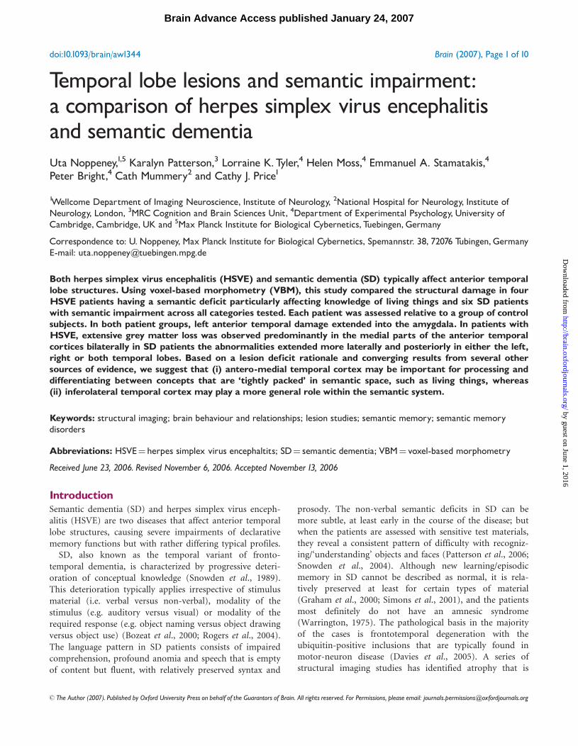
Temporal lobe lesions and semantic impairment: a comparison of herpes simplex virus encephalitis and semantic dementia Uta Noppeney, 1,5 Karalyn Patterson, 3 Lorraine K. Tyler, 4 Helen Moss, 4 Emmanuel A. Stamatakis, 4 Peter Bright, 4 Cath Mummery 2 and Cathy J. Price 1 1 Wellcome Department of Imaging Neuroscience, Institute of Neurology, 2 National Hospital for Neurology, Institute of Neurology, London, 3 MRC Cognition and Brain Sciences Unit, 4 Department of Experimental Psychology, University of Cambridge, Cambridge, UK and 5 Max Planck Institute for Biological Cybernetics, Tuebingen, Germany Correspondence to: U. Noppeney, Max Planck Institute for Biological Cybernetics, Spemannstr. 38, 72076 Tubingen, Germany E-mail: [email protected] Both herpes simplex virus encephalitis (HSVE) and semantic dementia (SD) typically affect anterior temporal lobe structures. Using voxel-based morphometry (VBM), this study compared the structural damage in four HSVE patients having a semantic deficit particularly affecting knowledge of living things and six SD patients with semantic impairment across all categories tested. Each patient was assessed relative to a group of control subjects. In both patient groups, left anterior temporal damage extended into the amygdala. In patients with HSVE, extensive grey matter loss was observed predominantly in the medial parts of the anterior temporal cortices bilaterally in SD patients the abnormalities extended more laterally and posteriorly in either the left, right or both temporal lobes. Based on a lesion deficit rationale and converging results from several other sources of evidence, we suggest that (i) antero-medial temporal cortex may be important for processing and differentiating between concepts that are ‘tightly packed’ in semantic space, such as living things, whereas (ii) inferolateral temporal cortex may play a more general role within the semantic system. Keywords: structural imaging; brain behaviour and relationships; lesion studies; semantic memory; semantic memory disorders Abbreviations: HSVE ¼ herpes simplex virus encephaltits; SD ¼ semantic dementia; VBM ¼ voxel-based morphometry Received June 23, 2006. Revised November 6, 2006. Accepted November 13, 2006 Introduction Semantic dementia (SD) and herpes simplex virus enceph- alitis (HSVE) are two diseases that affect anterior temporal lobe structures, causing severe impairments of declarative memory functions but with rather differing typical profiles. SD, also known as the temporal variant of fronto- temporal dementia, is characterized by progressive deteri- oration of conceptual knowledge (Snowden et al., 1989). This deterioration typically applies irrespective of stimulus material (i.e. verbal versus non-verbal), modality of the stimulus (e.g. auditory versus visual) or modality of the required response (e.g. object naming versus object drawing versus object use) (Bozeat et al., 2000; Rogers et al., 2004). The language pattern in SD patients consists of impaired comprehension, profound anomia and speech that is empty of content but fluent, with relatively preserved syntax and prosody. The non-verbal semantic deficits in SD can be more subtle, at least early in the course of the disease; but when the patients are assessed with sensitive test materials, they reveal a consistent pattern of difficulty with recogniz- ing/‘understanding’ objects and faces (Patterson et al., 2006; Snowden et al., 2004). Although new learning/episodic memory in SD cannot be described as normal, it is rela- tively preserved at least for certain types of material (Graham et al., 2000; Simons et al., 2001), and the patients most definitely do not have an amnesic syndrome (Warrington, 1975). The pathological basis in the majority of the cases is frontotemporal degeneration with the ubiquitin-positive inclusions that are typically found in motor-neuron disease (Davies et al., 2005). A series of structural imaging studies has identified atrophy that is doi:10.1093/brain/awl344 Brain (2007), Page 1 of 10 ß The Author (2007). Published by Oxford University Press on behalf of the Guarantors of Brain. All rights reserved. For Permissions, please email: [email protected] Brain Advance Access published January 24, 2007 by guest on June 1, 2016 http://brain.oxfordjournals.org/ Downloaded from
-
Upload
independent -
Category
Documents
-
view
3 -
download
0
Transcript of Temporal lobe lesions and semantic impairment: a comparison of herpes simplex virus encephalitis and...
Temporal lobe lesions and semantic impairment:a comparison of herpes simplex virus encephalitisand semantic dementiaUta Noppeney,1,5 Karalyn Patterson,3 Lorraine K.Tyler,4 Helen Moss,4 Emmanuel A. Stamatakis,4
Peter Bright,4 Cath Mummery2 and Cathy J. Price1
1Wellcome Department of Imaging Neuroscience, Institute of Neurology, 2National Hospital for Neurology, Institute ofNeurology, London, 3MRC Cognition and Brain Sciences Unit, 4Department of Experimental Psychology, University ofCambridge, Cambridge, UK and 5Max Planck Institute for Biological Cybernetics, Tuebingen,Germany
Correspondence to: U. Noppeney, Max Planck Institute for Biological Cybernetics, Spemannstr. 38, 72076 Tubingen,GermanyE-mail: [email protected]
Both herpes simplex virus encephalitis (HSVE) and semantic dementia (SD) typically affect anterior temporallobe structures.Using voxel-based morphometry (VBM), this study compared the structural damage in fourHSVE patients having a semantic deficit particularly affecting knowledge of living things and six SD patientswith semantic impairment across all categories tested. Each patient was assessed relative to a group of controlsubjects. In both patient groups, left anterior temporal damage extended into the amygdala. In patients withHSVE, extensive grey matter loss was observed predominantly in the medial parts of the anterior temporalcortices bilaterally in SD patients the abnormalities extended more laterally and posteriorly in either the left,right or both temporal lobes. Based on a lesion deficit rationale and converging results from several othersources of evidence, we suggest that (i) antero-medial temporal cortex may be important for processing anddifferentiating between concepts that are ‘tightly packed’ in semantic space, such as living things, whereas(ii) inferolateral temporal cortex may play a more general role within the semantic system.
Keywords: structural imaging; brain behaviour and relationships; lesion studies; semantic memory; semantic memorydisorders
Abbreviations: HSVE¼herpes simplex virus encephaltits; SD¼ semantic dementia; VBM¼ voxel-based morphometry
Received June 23, 2006. Revised November 6, 2006. Accepted November 13, 2006
IntroductionSemantic dementia (SD) and herpes simplex virus enceph-alitis (HSVE) are two diseases that affect anterior temporallobe structures, causing severe impairments of declarativememory functions but with rather differing typical profiles.SD, also known as the temporal variant of fronto-
temporal dementia, is characterized by progressive deteri-oration of conceptual knowledge (Snowden et al., 1989).This deterioration typically applies irrespective of stimulusmaterial (i.e. verbal versus non-verbal), modality of thestimulus (e.g. auditory versus visual) or modality of therequired response (e.g. object naming versus object drawingversus object use) (Bozeat et al., 2000; Rogers et al., 2004).The language pattern in SD patients consists of impairedcomprehension, profound anomia and speech that is emptyof content but fluent, with relatively preserved syntax and
prosody. The non-verbal semantic deficits in SD can bemore subtle, at least early in the course of the disease; butwhen the patients are assessed with sensitive test materials,they reveal a consistent pattern of difficulty with recogniz-ing/‘understanding’ objects and faces (Patterson et al., 2006;Snowden et al., 2004). Although new learning/episodicmemory in SD cannot be described as normal, it is rela-tively preserved at least for certain types of material(Graham et al., 2000; Simons et al., 2001), and the patientsmost definitely do not have an amnesic syndrome(Warrington, 1975). The pathological basis in the majorityof the cases is frontotemporal degeneration with theubiquitin-positive inclusions that are typically found inmotor-neuron disease (Davies et al., 2005). A series ofstructural imaging studies has identified atrophy that is
doi:10.1093/brain/awl344 Brain (2007), Page 1 of 10
� The Author (2007). Published by Oxford University Press on behalf of the Guarantors of Brain. All rights reserved. For Permissions, please email: [email protected]
Brain Advance Access published January 24, 2007 by guest on June 1, 2016
http://brain.oxfordjournals.org/D
ownloaded from
bilateral but often particularly pronounced in the lefthemisphere, encompassing the temporal pole, the inferior/middle temporal and fusiform gyri, the amygdaloidcomplex and sometimes the ventromedial frontal cortex(Levy et al., 2004; Mummery et al., 2000; Rosen et al.,2002). Using fluid registration, longitudinal studies havedemonstrated that grey matter atrophy spreads from a leftanterior temporal focus both anteriorly and posteriorly withprogressive involvement of the right hemisphere (Whitwellet al., 2004). Using volumetric methods, two studies havealso reported hippocampal involvement (Chan et al., 2001;Galton et al., 2001). Similarly, functional studies havedemonstrated hypometabolism in the anterior temporallobes spreading posteriorly (Diehl et al., 2004) andextending into the hippocampus (Nestor et al., 2006).HSVE is the most common viral encephalitis in humans
(Kennedy and Chaudhuri, 2002). Pathological and structuralimaging studies have demonstrated damage in a widespreadtemporo–limbic–diencephalic system encompassing theamygdala, hippocampus, peri-/entorhinal, parahippocampaland orbitofrontal cortex, insula and cingulate gyri(Gitelman et al., 2001). The long-term neuropsychology ischaracterized by dense anterograde amnesia, and sometimesbut less commonly by impairments of semantic memoryand/or executive functions (Kapur et al., 1994). Importantly,whilst the diagnosis of SD is based on both anteriortemporal lobe atrophy and progressive semantic impair-ments, diagnosis of HSVE is based on positive virologyirrespective of the cognitive consequences.SD has often been described as involving primarily
antero-lateral temporal damage and mainly semantic-memory impairment, whereas HSVE has been said toentail primarily antero-medial temporal damage andmainly episodic-memory impairment. This contrast has ledat least one group of researchers (Levy et al., 2004) toconclude that medial temporal lobe structures supportformation of declarative (particularly episodic) memorywhile lateral temporal lobe structures underpin representa-tion of semantic knowledge. This conclusion, however, wasbased on qualitatively descriptive methods rather thanstandardized quantitative procedures that would enable adirect comparison of the lesion extents. Indeed, this doubledissociation between medial and lateral temporal loberemains disputed in the literature. Thus, other studies havedemonstrated a correlation between semantic knowledge andvolume of the perirhinal cortex and implicated medialtemporal lobe structures in semantic knowledge (Davieset al., 2004). It should be noted that human perirhinal cortexhas a complex anatomy: it occupies the banks of thecollateral sulcus and medial aspect of the temporal lobe but,because it is cytoarchitectonically continuous with tempor-opolar cortex, these two areas should probably be consideredas part of the same cortical region in terms of connectivity(Hodges et al., 2006; Insausti et al., 1998).The present study takes a different perspective and
compares SD and HSVE patients to gain insight into the
organization of semantic memory. Using a lesion-deficitapproach, we asked whether different regions of the temporallobe were associated with distinct types or qualitiesof semantic information. In particular, as is often the casefollowing HSVE (Gainotti et al., 1995; Capitani et al., 2003;Warrington and Shallice, 1984; Laiacona et al., 2003),the encephalitic patients in this study had a ‘category-specific’ pattern characterized by a semantic deficit forconcepts from the domain of living things, with relativesparing of non-living items or artefacts. In contrast, as istypically the case (Bozeat et al., 2000; Lambon-Ralph et al.,2003; Moss et al., 2005), the SD patients studied hereexhibited a non-category-specific semantic impairmentaffecting both living things and artefacts. Hence, relatingthe lesions in HSVE and SD patients to their distinctpatterns of semantic disorder might provide insight into theneural organization of semantic memory. In order to achievequantitative characterization of the lesion patterns in SDand HSVE patients, we combined structural imaging withvoxel-based morphometry, a whole-brain unbiased objectivetechnique that tests for regional changes in grey (or white)matter volume (Ashburner and Friston, 2000, 2003).
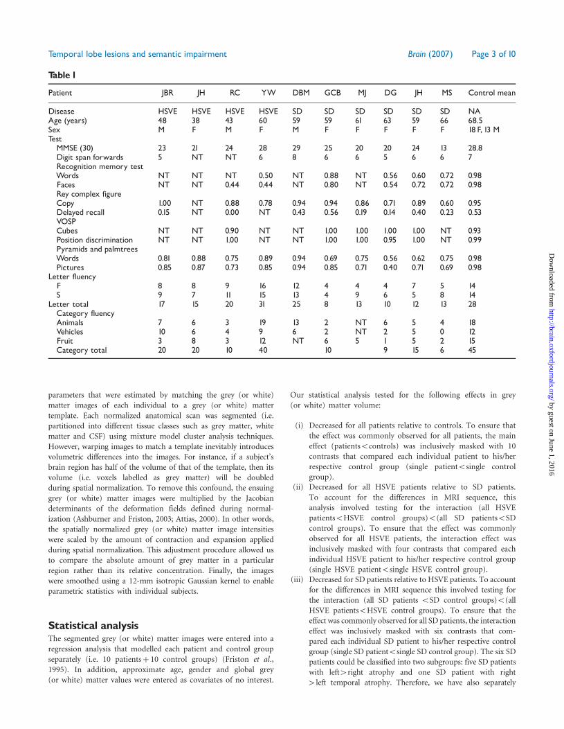
Material and methodsSubjectsFour patients with a history of HSVE, six patients with a diagnosis ofSD and 89 control subjects participated in this study. The diagnosisof HSVE was based on standard measures including EEG,neuroimaging and virology. The diagnosis of SD was based on thecurrently accepted criteria including anomia and semantic impair-ments. The control subjects were divided into 10 groups with eachassigned to one particular patient. This procedure allowed us tomatch the control group to the patient imaging data with respect toage and gender. Furthermore, it enabled us to make inferences aboutregionally specific effects that were common to all patients, as eachcomparison (patients versus control group) was based on indepen-dent data. Table 1 presents demographic information and back-ground neuropsychological test scores for the patients individuallyand for a group of control participants from the participant panel atthe MRC Cognition and Brain Sciences Unit.
MRI scanningWhole brain structural images were acquired from all participantsusing a 2 T MRI scanner (Magnetom Vision; Siemens, Erlangen,Germany). Two different T1-weighted scanning sequences (voxelsize: 1� 1� 1.5–3) were used for HSVE, SD patients and controlgroups [for further details, see Mummery et al. (2000) andGitelman et al. (2001)].
Data preprocessingAll image processing and statistical analyses were performed inSPM2 (Wellcome Department of Imaging Neuroscience, London,UK). The images were preprocessed according to the optimizedVBM protocol (Good et al., 2001). This involved initial affineregistration and segmentation. The structural images were thenspatially normalized into standard MNI space using normalization
Page 2 of 10 Brain (2007) U. Noppeney et al.
by guest on June 1, 2016http://brain.oxfordjournals.org/
Dow
nloaded from
parameters that were estimated by matching the grey (or white)
matter images of each individual to a grey (or white) matter
template. Each normalized anatomical scan was segmented (i.e.
partitioned into different tissue classes such as grey matter, white
matter and CSF) using mixture model cluster analysis techniques.
However, warping images to match a template inevitably introduces
volumetric differences into the images. For instance, if a subject’s
brain region has half of the volume of that of the template, then its
volume (i.e. voxels labelled as grey matter) will be doubled
during spatial normalization. To remove this confound, the ensuing
grey (or white) matter images were multiplied by the Jacobian
determinants of the deformation fields defined during normal-
ization (Ashburner and Friston, 2003; Attias, 2000). In other words,
the spatially normalized grey (or white) matter image intensities
were scaled by the amount of contraction and expansion applied
during spatial normalization. This adjustment procedure allowed us
to compare the absolute amount of grey matter in a particular
region rather than its relative concentration. Finally, the images
were smoothed using a 12-mm isotropic Gaussian kernel to enable
parametric statistics with individual subjects.
Statistical analysisThe segmented grey (or white) matter images were entered into a
regression analysis that modelled each patient and control group
separately (i.e. 10 patientsþ 10 control groups) (Friston et al.,
1995). In addition, approximate age, gender and global grey
(or white) matter values were entered as covariates of no interest.
Our statistical analysis tested for the following effects in grey
(or white) matter volume:
(i) Decreased for all patients relative to controls. To ensure that
the effect was commonly observed for all patients, the main
effect (patients5controls) was inclusively masked with 10
contrasts that compared each individual patient to his/her
respective control group (single patient5single control
group).(ii) Decreased for all HSVE patients relative to SD patients.
To account for the differences in MRI sequence, this
analysis involved testing for the interaction (all HSVE
patients5HSVE control groups)5(all SD patients5SD
control groups). To ensure that the effect was commonly
observed for all HSVE patients, the interaction effect was
inclusively masked with four contrasts that compared each
individual HSVE patient to his/her respective control group
(single HSVE patient5single HSVE control group).(iii) Decreased for SD patients relative to HSVE patients. To account
for the differences in MRI sequence this involved testing for
the interaction (all SD patients 5SD control groups)5(all
HSVE patients5HSVE control groups). To ensure that the
effect was commonly observed for all SD patients, the interaction
effect was inclusively masked with six contrasts that com-
pared each individual SD patient to his/her respective control
group (single SD patient5single SD control group). The six SD
patients could be classified into two subgroups: five SD patients
with left4right atrophy and one SD patient with right
4left temporal atrophy. Therefore, we have also separately
Table 1
Patient JBR JH RC YW DBM GCB MJ DG JH MS Control mean
Disease HSVE HSVE HSVE HSVE SD SD SD SD SD SD NAAge (years) 48 38 43 60 59 59 61 63 59 66 68.5Sex M F M F M F F F F F 18F, 13 MTestMMSE (30) 23 21 24 28 29 25 20 20 24 13 28.8Digit span forwards 5 NT NT 6 8 6 6 5 6 6 7Recognition memory testWords NT NT NT 0.50 NT 0.88 NT 0.56 0.60 0.72 0.98Faces NT NT 0.44 0.44 NT 0.80 NT 0.54 0.72 0.72 0.98Rey complex figureCopy 1.00 NT 0.88 0.78 0.94 0.94 0.86 0.71 0.89 0.60 0.95Delayed recall 0.15 NT 0.00 NT 0.43 0.56 0.19 0.14 0.40 0.23 0.53VOSPCubes NT NT 0.90 NT NT 1.00 1.00 1.00 1.00 NT 0.93Position discrimination NT NT 1.00 NT NT 1.00 1.00 0.95 1.00 NT 0.99Pyramids and palmtreesWords 0.81 0.88 0.75 0.89 0.94 0.69 0.75 0.56 0.62 0.75 0.98Pictures 0.85 0.87 0.73 0.85 0.94 0.85 0.71 0.40 0.71 0.69 0.98
Letter fluencyF 8 8 9 16 12 4 4 4 7 5 14S 9 7 11 15 13 4 9 6 5 8 14
Letter total 17 15 20 31 25 8 13 10 12 13 28Category fluencyAnimals 7 6 3 19 13 2 NT 6 5 4 18Vehicles 10 6 4 9 6 2 NT 2 5 0 12Fruit 3 8 3 12 NT 6 5 1 5 2 15Category total 20 20 10 40 10 9 15 6 45
Temporal lobe lesions and semantic impairment Brain (2007) Page 3 of 10
by guest on June 1, 2016http://brain.oxfordjournals.org/
Dow
nloaded from

masked for these two subgroups. To fully characterize the three
different effects described earlier, we also identified:(iv) Decreased for all HSVE patients relative to controls. To
ensure that the effect was commonly observed for all
patients, the main effect (HSVE5controls) was inclusively
masked with four contrasts that compared each individual
patient to his/her respective control group (single
HSVE5single control group).(v) Decreased for all SD patients relative to controls. To ensure
that the effect was commonly observed for all patients, the
main effect (SD5controls) was inclusively masked with six
contrasts that compared each individual patient to his/her
respective control group (single SD5single control group).(vi) The direct comparison of all six SD patients with all four
HSVE patients.
Statistical thresholdUnless otherwise stated, we report grey (or white) matter volume
changes at P50.05 corrected for the entire brain using extent
thresholds of40 voxels for grey matter and4100 voxels for white
matter in the tables and the text. To provide a more detailed
characterization of the white matter damage, an extent threshold
of40 voxels was used in all the figures. The inclusive masks were
applied at P50.05 uncorrrected. The inclusive masking option
was used to identify grey (or white) matter differences that
were commonly observed in all patients within a group. This
procedure enabled us to remove non-systematic artefacts that may
result from normalizing and segmenting lesioned brains. To assign
the anatomic differences to the proper structure and ensure that
the observed effects were due to differences in grey (or white)
matter rather than registration and misclassification (e.g. displace-
ment) errors, we referenced them to visual inspection of each
patient’s brain. We only interpret and discuss those effects that
qualified as differences in grey (or white) matter per se according
to visual inspection.
ResultsBehavioural resultsTable 2 displays scores (in proportion correct) for eachindividual patient and as averages for each patient groupon two different semantic measures: picture naming andword–picture matching. These two tests, from theCambridge semantic battery (Bozeat et al., 2000), includethe same 64 items, and in each case consist of 32 naturalkinds or living things (from the subcategories of animals,birds and fruits) and 32 non-living things or artefacts(subcategories: household items, vehicles and tools). Fornaming, the patient is presented with each line drawingindividually and asked to name it; for each item in theword–picture matching test, the patient hears the name ofa target picture in conjunction with a visual array of 10pictures (the target and nine other items from the samecategory) and is asked to point to the target. Previousstudies in Cambridge with these test materials (Bozeat et al.,2000) have established that normal controls in the agerange of the SD patients score essentially at ceiling on both
naming (mean¼ 62.3/64, SD¼ 1.6) and word–picturematching (mean¼ 63.7/64, SD¼ 0.5).
In an assessment of performance accuracy for thepatients in the current study, a three-way ANOVA withpatient group (HSVE versus SD), semantic category (livingversus non-living) and task (naming versus word–picturematching) identified main effects of task and category butno main effect of patient group after Greenhouse–Geissercorrection: the patients were more successful at word–picture matching than naming [F(1,8)¼ 29.3; P50.001];this effect was consistently observed for both HSVE[F(1,3)¼ 17.3; P50.05] and SD [F(1,5)¼ 21.7;P50.01] groups. Furthermore, performance was betteron artefacts than living things [F(1,8)¼ 37.5; P50.01].Most importantly, a significant interaction between patientgroup and semantic category [F(1,8)¼ 23.2; P50.01] wasidentified, with a much bigger artefact 4 living advantagefor HSVE than SD. Thus, repeated measurement ANOVAperformed separately for each subject group revealed aneffect of semantic category only for the HSVE group[F(1,3)¼ 23.1; P50.05].
Voxel-based morphometry resultsGreymatter volume(i) Decreased for all patients relative to controls: the left
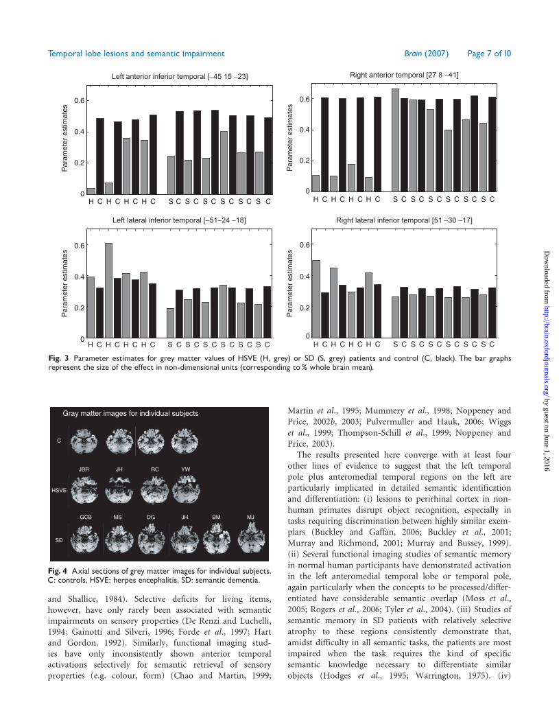
anterior temporal lobe, extending into the amygdalaand insula, and the caudate nucleus showed decreasedgrey matter volume for all HSVE and SD patientsrelative to their control groups (see Fig. 3 forconsistency across subjects).
(ii) Decreased for all HSVE relative to SD: decreased graymatter volume for the HSVE patients relative to boththeir control group and SD patients was observedpredominantly in the medial parts of the anteriortemporal cortices bilaterally.
Table 2 Behavioural data for HSVE and SD patients:proportion correct on naming and word^picture matchingfor a common set of 64 items, half living or natural kinds(e.g. rabbit, strawberry) and half non-living or artefacts(e.g. train, watering-can)
Patient Disease Naming Word^picture matching
Living Non-living Living Non-living
JBR HSVE 0.16 0.63 0.31 0.97JH HSVE 0.53 0.72 0.63 0.97RC HSVE 0.22 0.66 0.38 0.88YW HSVE 0.59 0.84 0.66 0.88Mean 0.38 0.71 0.49 0.92DBM SD 0.75 0.66 0.92 1.00GCB SD 0.41 0.44 0.69 0.63MJ SD 0.16 0.13 0.63 0.81DG SD 0.16 0.31 0.34 0.44JH SD 0.13 0.16 0.25 0.41MS SD 0.06 0.06 0.42 0.42Mean 0.28 0.29 0.54 0.62
Page 4 of 10 Brain (2007) U. Noppeney et al.
by guest on June 1, 2016http://brain.oxfordjournals.org/
Dow
nloaded from
(iii) Decreased for SD patients relative to HSVE: decreasedgrey matter was observed common to all SD patientsin the right lateral inferior temporal lobe. Maskingwith the five left4right atrophy SD patients relative totheir control group revealed decreased grey mattervolume in the left inferior temporal gyrus. Likewise,masking with the one right4left atrophy SD caserelative to her control group produced decreased greymatter volume in the right inferior temporal gyrus(limited to one single voxel). Therefore, in SD patientsrelative to HSVE patients, damage spreads moreposteriorly and involves lateral inferior temporalareas that can be detected on the left or right depend-ing on the balance of left/right atrophy in theindividual patient.
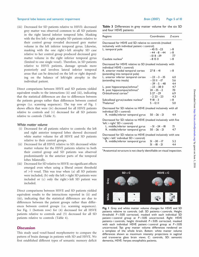
Direct comparisons between HSVE and SD patients yieldedequivalent results to the interactions (ii) and (iii), indicatingthat the statistical differences are due to differences betweenthe patients groups rather than differences between controlgroups (i.e. scanning sequences). The top row of Fig. 1shows effects that were (iv) decreased for all HSVE patientsrelative to controls and (v) decreased for all SD patientsrelative to controls (Table 3).
White matter volume(i) Decreased for all patients relative to controls: the left
and right anterior temporal lobes showed decreasedwhite matter volume for all HSVE and SD patientsrelative to their control groups.
(ii) Decreased for all HSVE relative to SD: decreased whitematter volume for the HSVE patients relative to boththeir control group and SD patients was observedpredominantly in the anterior parts of the temporallobes bilaterally.
(iii) Decreased for SD relative to HSVE: no significant effectsemerged even when using a liberal extent thresholdof 40 voxel. This was true when (a) all SD patientswere included, (b) only the left4right SD patients wereincluded or (c) only the right4left SD patient wasincluded.
Direct comparisons between HSVE and SD patients yieldedequivalent results to the interactions reported in (ii) and(iii), indicating that the statistical differences are due todifferences between the patient groups rather than differ-ences between control groups (i.e. scanning sequences).See Fig. 1 (bottom row) for (4) decreased for all HSVEpatients relative to controls and (5) decreased for all SDpatients relative to controls (Table 4).
DiscussionThis study used voxel-based morphometry to compare thepattern of brain damage in patients with SD and HSVE. Wefirst established different types of semantic memory deficit
SD < C HSVE < C
Wh
ite
Gra
y
Fig. 1 Grey and white matter volume changes for HSVE and SDpatients relative to controls. Left: SD patients5controls. Heightthreshold: P50.05 corrected, masked with each individual SDpatient5control group at P50.05 uncorrected. Right: HSVEpatients5controls; height threshold: P50.05 corrected, maskedwith each individual HSVE patient5control group at P50.05uncorrected. Top: grey matter volume differences rendered ona template of the whole brain. Bottom: white matter volumedifferences shown as maximum intensity projections in sagittaland transverse glass brain views. C: controls, SD: semanticdementia, HSVE: herpes encephalitis patients.
Table 3 Differences in grey matter volume for the six SDand four HSVE patients
Regions Coordinates Z-score
Decreased for HSVE and SD relative to controls (maskedinclusively with individual patient5control)L. temporal pole ^45 15 ^23 48
^44 ^8 ^44 48^51 8 ^39 7.7
Caudate nucleusa ^8 15 0 48
Decreased for HSVE relative to SD (masked inclusively withindividual HSVE5control)R. anterior medial temporal cortex 27 8 �41 48(extending into temporal pole)L. anterior inferior temporal cortex �23 �3 �35 6.0(extending into insula) �3311 �17 5.6
�39 �5 �48 4.4L. post hippocampus/isthmusa �23 �38 0 4.7R. post hippocampus/isthmusa 24 �35 �2 7.0Orbitofrontal cortexa 5 27 �29 6.9
�2 23 �23 4.3Subcallosal gyrus/caudate nucleusa 9 18 �3 48Thalamusa 5 �12 11 5.0
Decreased for SD relative to HSVE (masked inclusively with allindividual SD5control)
R. middle/inferior temporal gyrus 50 �26 �21 4.4
Decreased for SD relative to HSVE (masked inclusively with five‘left4right’ SD5control)L. middle/inferior temporal gyrus �51 �24 �18 7.1R. middle/inferior temporal gyrus 50 �26 �21 4.7
Decreased for SD relative to HSVE (masked inclusively with one‘right4left’ individual SD5control)R. middle/inferior temporal gyrus 51 �30 �17 5.2
51 �18 �21 4.4
aAnatomical structure is not clearly identifiable on visual inspection.
Temporal lobe lesions and semantic impairment Brain (2007) Page 5 of 10
by guest on June 1, 2016http://brain.oxfordjournals.org/
Dow
nloaded from
in the two groups: all of the HSVE patients showed anotably category-selective pattern with substantially greaterimpairment for living things than artefacts in both namingand comprehension; in contrast, all of the SD patients hadsevere semantic deficits that were of roughly equal magni-tude for the two types of concepts, on both production andcomprehension tests. We then measured the lesion extentsin the HSVE and SD individuals and groups, and relatedthese to the distinct cognitive patterns, in an attempt togain some insight into the neuroanatomical structure ofsemantic memory.Both patient groups, relative to controls, consistently
showed reduced grey matter volume in the left anteriortemporal lobe extending into the amygdala. Regionalvolume loss selective or increased for the HSVE patientswas observed predominantly in the medial parts of theanterior temporal cortices bilaterally. Regional volume lossselective for the SD patients occurred in the inferior tem-poral gyri, spreading laterally and posteriorly from the pole,although the extent to which this effect applied to leftversus right temporal lobe depended on the patient (Fig. 2).As usual, lesion extent and location varied considerably
across patients, even within the SD and HSVE groups.Nevertheless, assuming a consistent functional neuroanat-omy across subjects, it seems reasonable to draw a numberof tentative conclusions. We will first discuss the conclu-sions relating to a category-selective organization of seman-tic memory and then turn to the neuroanatomy of generalsemantic functions.
Category-selective organization ofsemantic memoryThe anterior and medial left temporal areas that werehighly abnormal in both HSVE and SD patients maybe particularly important for semantic knowledge ofliving items. This is probably not specifically because
these concepts have the semantic featureþ living. Instead,it is more likely to be due to the type of processingrequired by living items. It has been proposed that livingthings (or more generally, natural kinds) are characterizedby many shared and highly intercorrelated features withrelatively few distinctive features (McRae et al., 1997;Randall et al., 2004; Rogers et al., 2004; Tyler et al., 2000).This difference between living things and artefactsis particularly salient at the ‘basic’ level, which is theconceptual level tested in the behavioural assessments ofnaming and word–picture matching used in the currentstudy. Basic level refers to names and concepts like dog orcar: at this level, two animal concepts (such as dog andgoat) typically have more features in common than, forexample, two vehicle concepts (such as car and aeroplane).Concepts that are more tightly packed in semantic space,like basic-level living things, place increased demands onidentification and differentiation processes at the level ofboth semantic and perceptual processing during objectrecognition (Ikeda et al., 2006; Moss et al., 2005; Tyler andMoss, 2001; Tyler et al., 2004).
Alternatively or additionally, it has been suggested thatliving items are primarily characterized by sensory featuresthat may be supported by the anterior temporal lobe(Farah and McClelland, 1991; Shallice, 1988; Warrington
Table 4 Differences in white matter volume for the six SDand four HSVE patients
Regions Coordinates Z-score
Decreased for HSVE and SD relative to controls (maskedinclusively with individual patient5control)R. ant. temporal lobe 41 �8 �21 48L. ant. temporal lobe �41 �3 �30 48
Decreased for HSVE relative to SD (masked inclusively withindividual HSVE5control)L. medial orbitofrontal �14 27 �11 48R. medial orbitofrontal 14 26 �12 7.6R. ant. temporal lobe 45 �11 �26 48L. ant. temporal lobe �47 �11 �32 5.4R. capsula interna 11 5 0 6.2Corpus callosum �8 6 29 5.3
Decreased for SD relative to HSVE (masked inclusively with all, five‘left4right’ SD5control, one right4left individual SD5control)None
x = +27 y = +8 z = −41HSVE < SD
SD < HSVE
L
L
R
Lx = −51 y = −24 z = −18
x = +45 y = −11 z = −26HSVE < SD(B)
(A)
R L
Fig. 2 (A) Grey matter volume differences presented on sagittal,coronal and axial sections of a canonical brain. Top: HSVEpatients5SD patients (interaction); height threshold: P50.05corrected, masked with each individual HSVE patient5controlgroup at P50.05 uncorrected. Bottom: SD patients5HSVE patients(interaction). Height threshold: P50.05 corrected, masked witheach individual SD patient5control group at P50.05 uncorrected.(B) White matter volume differences presented on sagittal, coro-nal and axial sections of a canonical brain. HSVE patients5SDpatients (interaction); height threshold: P50.05 corrected,masked with each individual HSVE patient5control group atP50.05 uncorrected. C: controls, SD: semantic dementia, HSVE:herpes encephalitis patients
Page 6 of 10 Brain (2007) U. Noppeney et al.
by guest on June 1, 2016http://brain.oxfordjournals.org/
Dow
nloaded from
and Shallice, 1984). Selective deficits for living items,however, have only rarely been associated with semanticimpairments on sensory properties (De Renzi and Luchelli,1994; Gainotti and Silveri, 1996; Forde et al., 1997; Hartand Gordon, 1992). Similarly, functional imaging stud-ies have only inconsistently shown anterior temporalactivations selectively for semantic retrieval of sensoryproperties (e.g. colour, form) (Chao and Martin, 1999;
Martin et al., 1995; Mummery et al., 1998; Noppeney andPrice, 2002b, 2003; Pulvermuller and Hauk, 2006; Wiggset al., 1999; Thompson-Schill et al., 1999; Noppeney andPrice, 2003).
The results presented here converge with at least fourother lines of evidence to suggest that the left temporalpole plus anteromedial temporal regions on the left areparticularly implicated in detailed semantic identificationand differentiation: (i) lesions to perirhinal cortex in non-human primates disrupt object recognition, especially intasks requiring discrimination between highly similar exem-plars (Buckley and Gaffan, 2006; Buckley et al., 2001;Murray and Richmond, 2001; Murray and Bussey, 1999).(ii) Several functional imaging studies of semantic memoryin normal human participants have demonstrated activationin the left anteromedial temporal lobe or temporal pole,again particularly when the concepts to be processed/differ-entiated have considerable semantic overlap (Moss et al.,2005; Rogers et al., 2006; Tyler et al., 2004). (iii) Studies ofsemantic memory in SD patients with relatively selectiveatrophy to these regions consistently demonstrate that,amidst difficulty in all semantic tasks, the patients are mostimpaired when the task requires the kind of specificsemantic knowledge necessary to differentiate similarobjects (Hodges et al., 1995; Warrington, 1975). (iv)
Right anterior temporal [27 8 −41]
Left lateral inferior temporal [−51−24 −18]
Left anterior inferior temporal [−45 15 −23]
Right lateral inferior temporal [51 −30 −17]
H C H C H C H C S C S C S C S C S C S C0
0.2
0.4
0.6
Par
amet
er e
stim
ates
H C H C H C H C S C S C S C S C S C S C0
0.2
0.4
0.6
Par
amet
er e
stim
ates
H C H C H C H C S C S C S C S C S C S C0
0.2
0.4
0.6
Par
amet
er e
stim
ates
H C H C H C H C S C S C S C S C S C S C0
0.2
0.4
0.6
Par
amet
er e
stim
ates
Fig. 3 Parameter estimates for grey matter values of HSVE (H, grey) or SD (S, grey) patients and control (C, black). The bar graphsrepresent the size of the effect in non-dimensional units (corresponding to% whole brain mean).
C
HSVE
SD
Gray matter images for individual subjects
JBR JH RC YW
GCB MS DG JH BM MJ
Fig. 4 Axial sections of grey matter images for individual subjects.C: controls, HSVE: herpes encephalitis, SD: semantic dementia.
Temporal lobe lesions and semantic impairment Brain (2007) Page 7 of 10
by guest on June 1, 2016http://brain.oxfordjournals.org/
Dow
nloaded from
Identification of people from their faces is probablya supreme example of the kind of semantic task requiringsuch fine differentiation; this ability (a) is typically devas-tated in both HSVE and SD (Snowden et al., 2004; Tranelet al., 1997), and (b) results in anterior temporal activa-tion in functional imaging studies of normal participants(Damasio et al., 1996; Gorno-Tempini et al., 1998;Grabowski et al., 2001; Rotshtein et al., 2005).The association of the anteromedial temporal lobes
with the recognition of living things more than artefactsis consistent with functional neuroimaging evidence (Devlinet al., 2002; Moss et al., 2005). It is perhaps surprising thatthe semantic advantage for artefacts4natural kinds has notbeen observed more frequently in SD. The literaturecontains two reports of SD patients who did demonstratethis pattern (Barbarotto et al., 1995; Lambon-Ralph et al.,2003), and it is possibly worth noting that both of thesecases had right4left temporal atrophy, like DG (Table 2)who probably came the closest of any SD patient in thecurrent study to a degree (though non-significant) of thiscategory differential (Fig. 4). The anteromedial focus ofdamage observed in HSVE in association with a living-things deficit might predict the observation of thisbehavioural phenomenon in early-stage SD if atrophywere limited to the medial temporal lobes (Brambatiet al., 2006). If atrophy starts more laterally beforespreading to the medial temporal lobe structures, however,then we would predict a parallel degradation of conceptualknowledge even in the early stages, as is typical in SD.
The neural basis of semantic retrievalThe more widespread lateral inferior temporal lobeabnormalities in SD compared to HSVE may indicate theimportance of this larger temporal region in semanticprocessing more generally. Inferior temporal together withfrontal activation is frequently reported during semanticretrieval or naming tasks in functional imaging studies(Vandenberghe et al., 1996; Roskies et al., 2001; Noppeneyand Price, 2002a; Sabsevitz et al., 2005; Tranel et al., 2005;Gold et al., 2006). These semantic activations can beobserved irrespective of stimulus material (e.g. pictures orwords (Vandenberghe, et al., 1996), modality (e.g. sounds orpictures (Buckner et al., 2000; Tranel et al., 2005) or semanticcontent (Noppeney et al., 2003). Thus, lesion and functionalimaging results suggest a more general role for the lateralinferior temporal cortex in semantic processing that maydepend on interactions with frontal cortex (Wagner et al.,2001; Gold and Buckner 2002; Noppeney et al., 2004).Finally, five out of the six SD patients studied here had
bilateral anterior temporal damage, consistent with previousclaims that semantic memory is underpinned by a bilateralnetwork of regions (Damasio, 1989; Damasio et al., 1996;Hodges et al., 2006). Although unilateral lesions can disruptsome aspects of semantic processing or manipulation(Jefferies and Lambon-Ralph, 2006), damage to one side
of the brain—even the left—rarely if ever results ingenuinely degraded conceptual knowledge.
Despite their plausibility and support from other linesof evidence, these proposed lesion-deficit or structure–function relationships can only—at this stage of ourknowledge—be hypotheses. One important issue challeng-ing the direct comparison of SD and HSVE patients isthat their gray-matter losses result from very differentpathological processes: neurodegenerative in the formercase, viral/inflammatory in the latter. Identical volume lossin SD and HSVE may therefore not be functionallyequivalent. Furthermore, the gross temporal lobe distor-tions in HSVE and SD may lead to some degree ofambiguity when interpreting the statistical comparisonbetween the two patient populations (Gitelman et al.,2001). Thus, decreased regional grey matter volume in thepatients may not only be caused by regional grey matterloss per se, but also by misclassification errors due tochanges in grey matter intensity values or registrationdifficulties. Nevertheless, the medial temporal and inferiortemporal abnormalities identified by our VBM analysiscould be referenced consistently to the appropriate anato-mical structures in each patient’s brain. The consistency ofour results with the known histopathology provides furtherface validity.
It is so rare to find groups of these different patienttypes tested on the same, or even comparable, assessmentmeasures that we offer our current observations in the hopethat this study will be followed by more and better researchof a similar kind.
AcknowledgementsThis work was funded by the Wellcome Trust. U.N. issupported by the DFG, L.K.T. by the MRC and NewtonTrust. We are grateful to Professor John R. Hodges forpermission to publish details of these six SD cases,Dr Sharon Davies for help with data for the SD patientsand controls and Professor Andrew Mayes for referring theHSVE patients to us.
ReferencesAshburner J, Friston KJ. Voxel-based morphometry—the methods.
Neuroimage 2000; 11: 805–21.
Ashburner J, Friston KJ. Morphometry. In: Frackowiak R, Friston K,
Price C, Frith CD, Ashburner J, Penny W, editors. Human brain
function. London: Elsevier; 2003.
Attias H. A variational Bayesian framework for graphical models. Neural
Inf Process Syst 2000; 12: 209–15.
Barbarotto R, Capitani E, Laiacona M. Slowly progressive semantic
impairment with category-specificity. Neurocase 1995; 1: 107–19.
Bozeat S, Lambon Ralph MA, Patterson K, Garrard P, Hodges JR.
Non-verbal semantic impairment in semantic dementia.
Neuropsychologia 2000; 38: 1207–15.
Brambati S, Meyers D, Ogar J, Dronkers NF, Miller B, Gorno-Tempini M.
The anatomy of category-specific object naming in neurodegenerative
diseases. J Cogn Neurosci 2006; 18: 1644–53.
Page 8 of 10 Brain (2007) U. Noppeney et al.
by guest on June 1, 2016http://brain.oxfordjournals.org/
Dow
nloaded from

Buckley MJ, Booth MC, Rolls ET, Gaffan D. Selective perceptual
impairments after perirhinal cortex ablation. J Neurosci 2001; 21:
9824–36.
Buckley MJ, Gaffan D. Perirhinal cortical contributions to object
perception. Trends Cogn Sci 2006; 10: 100–7.
Buckner RL, Koutstaal W, Schacter DL, Rosen BR. Functional MRI
evidence for a role of frontal and inferior temporal cortex in amodal
components of priming. Brain 2000; 123: 620–40.
Capitani E, Laiacona M, Mahon B, Caramazza A. What are the facts of
semantic category-specific deficits? A critical review of the clinical
evidence. Cogn Neuropsychol 2003; 20: 213–62.
Chan D, Fox NC, Scahill RI, et al. Patterns of temporal lobe atrophy in
semantic dementia and Alzheimer’s disease. Ann Neurol 2001; 49:
433–42.
Chao LL, Martin A. Cortical regions associated with perceiving, naming,
and knowing about colors. J Cogn Neurosci 1999; 11: 25–35.
Damasio AR. Time-locked multiregional retroactivation: a systems-level
proposal for the neural substrates of recall and recognition. Cognition
1989; 33: 25–62.
Damasio H, Grabowski TJ, Tranel D, Hichwa RD, Damasio AR. A neural
basis for lexical retrieval [see comments] [published erratum appears in
Nature 1996 Jun 27; 381: 810]. Nature 1996; 380: 499–505.
Davies RR, Graham KS, Xuereb JH, Williams GB, Hodges JR. The human
perirhinal cortex and semantic memory. Eur J Neurosci 2004; 20:
2441–6.
Davies RR, Hodges JR, Kril JJ, Patterson K, Halliday GM, Xuereb JH.
The pathological basis of semantic dementia. Brain 2005; 128: 1984–95.
De Renzi E, Luchelli F. Are semantic systems separately represented in the
human brain? The case of living category impairment. Cortex 1994; 30:
3–25.
Devlin JT, Moore CJ, Mummery CJ, et al. Anatomic constraints
on cognitive theories of category specificity. Neuroimage 2002; 15:
675–85.
Diehl J, Grimmer T, Drzezga A, Riemenschneider M, Forstl H, Kurz A.
Cerebral metabolic patterns at early stages of frontotemporal dementia
and semantic dementia. A PET study. Neurobiol Aging 2004; 25:
1051–6.
Farah MJ, McClelland JI. A computational model of semantic memory
impairment: modality specificity and emergent category specificity.
J Exp Psychol 1991; 120: 339–57.
Forde EME, Francis D, Riddoch MJ, Rumialati RI, Humphreys GW.
On the links between visual knowledge and naming: a single case of
a patient with a category-specific impairment for living things. Cogn
Neuropsychol 1997; 14: 403–58.
Friston KJ, Holmes A, Worsley KJ, Poline JB, Frith CD, Frackowiak R.
Statistical parametric mapping: a general linear approach. Hum Brain
Mapp 1995; 2: 189–210.
Gainotti G, Giustolisi L, Daniele A, Silveri MC. Neuroanatomical
correlates of category-specific semantic disorders: a critical survey.
Memory 1995; 3: 247–64.
Gainotti G, Silveri MC. Cognitive and anatomical locus of lesion in a
patient with a category-specific semantic impairment for living beings.
Cogn Neuropsychol 1996; 13: 357–89.
Galton CJ, Patterson K, Graham K, et al. Differing patterns of temporal
atrophy in Alzheimer’s disease and semantic dementia. Neurology 2001;
57: 216–25.
Gitelman DR, Ashburner J, Friston KJ, Tyler LK, Price CJ. Voxel-based
morphometry of herpes simplex encephalitis. Neuroimage 2001; 13:
623–31.
Gold BT, Balota DA, Jones SJ, Powell DK, Smith CD, Andersen AH.
Dissociation of automatic and strategic lexical-semantics: functional
magnetic resonance imaging evidence for differing roles of multiple
frontotemporal regions. J Neurosci 2006; 26: 6523–32.
Gold BT, Buckner RL. Common prefrontal regions coactivate with
dissociable posterior regions during controlled semantic and phonolo-
gical tasks. Neuron 2002; 35: 803–12.
Good CD, Johnsrude IS, Ashburner J, Henson RN, Friston KJ,
Frackowiak RS. A voxel-based morphometric study of ageing in 465
normal adult human brains. Neuroimage 2001; 14: 21–36.
Gorno-Tempini ML, Price CJ, Josephs O, et al. The neural systems
sustaining face and proper-name processing [published errata appear in
Brain 1998; 121: 2402 and 2000; 123: 419]. Brain 1998; 121: 2103–18.
Grabowski TJ, Damasio H, Tranel D, Ponto LL, Hichwa RD, Damasio AR.
A role for left temporal pole in the retrieval of words for unique entities.
Hum Brain Mapp 2001; 13: 199–212.
Graham KS, Simons JS, Pratt KH, Patterson K, Hodges JR. Insights from
semantic dementia on the relationship between episodic and semantic
memory. Neuropsychologia 2000; 38: 313–24.
Hart J Jr, Gordon B. Neural subsystems for object knowledge. Nature
1992; 359: 60–4.
Hodges JR, Davies RR, Patterson K. Semantic dementia, or, a little
knowledge is a dangerous thing. In: Miller B, Boeve B, editors. The
behavioural neurology of dementia. 2006.
Hodges JR, Graham N, Patterson K. Charting the progression in semantic
dementia: implications for the organisation of semantic memory.
Memory 1995; 3: 463–95.
Ikeda M, Patterson K, Graham KS, Ralph MA, Hodges JR. A horse of a
different colour: do patients with semantic dementia recognise different
versions of the same object as the same? Neuropsychologia 2006; 44:
566–75.
Insausti R, Juottonen K, Soininen H, et al. MR volumetric analysis of the
human entorhinal, perirhinal, and temporopolar cortices. AJNR Am J
Neuroradiol 1998; 19: 659–71.
Jefferies E, Lambon-Ralph MA. Semantic impairment in stroke aphasia vs
semantic dementia: a case series comparison. Brain 2006; 129: 2132–47.
Kapur N, Barker S, Burrows EH, et al. Herpes-simplex encephalitis -
long-term magnetic-resonance-imaging and neuropsychological profile.
J Neurol Neurosurg Psychiatry 1994; 57: 1334–42.
Kennedy PGE, Chaudhuri A. Herpes simplex encephalitis. J Neurol
Neurosurg Psychiatry 2002; 73: 237–38.
Laiacona M, Capitani E, Caramazza A. Category-specific semantic deficits
do not reflect the ‘sensory/functional’ organization of the brain: a test of
the sensory quality hypothesis. Neurocase 2003; 9: 221–31.
Lambon-Ralph MA, Patterson K, Garrard P, Hodges JR. Semantic
dementia with category-specificity: a comparative case-series study.
Cogn Neuropsychol 2003; 20: 307–26.
Levy DA, Bayley PJ, Squire LR. The anatomy of semantic knowledge:
medial vs. lateral temporal lobe. Proc Natl Acad Sci USA 2004; 101:
6710–5.
Martin A, Haxby JV, Lalonde FM, Wiggs CL, Ungerleider LG. Discrete
cortical regions associated with knowledge of color and knowledge of
action. Science 1995; 270: 102–5.
McRae K, Desa VR, Seidenberg MS. On the nature and scope of
featural representations of word meaning. J Exp Psychol Gen 1997; 126:
99–130.
Moss HE, Rodd JM, Stamatakis EA, Bright P, Tyler LK. Anteromedial
temporal cortex supports fine-grained differentiation among objects.
Cereb Cortex 2005; 15: 616–27.
Mummery CJ, Patterson K, Hodges JR, Price CJ. Functional neuroanatomy
of the semantic system: divisible by what? J Cogn Neurosci 1998; 10:
766–77.
Mummery CJ, Patterson K, Price CJ, Ashburner J, Frackowiak RS,
Hodges JR. A voxel-based morphometry study of semantic dementia:
relationship between temporal lobe atrophy and semantic memory. Ann
Neurol 2000; 47: 36–45.
Murray EA, Bussey TJ. Perceptual-mnemonic functions of the perirhinal
cortex. Trends Cogn Sci 1999; 3: 142–51.
Murray EA, Richmond BJ. Role of perirhinal cortex in object perception,
memory, and associations. Curr Opin Neurobiol 2001; 11: 188–93.
Nestor PJ, Fryer TD, Hodges JR. Declarative memory impairments in
Alzheimer’s disease and semantic dementia. Neuroimage 2006; 30:
1010–20.
Temporal lobe lesions and semantic impairment Brain (2007) Page 9 of 10
by guest on June 1, 2016http://brain.oxfordjournals.org/
Dow
nloaded from
Noppeney U, Friston K, Price C. Effects of visual deprivation on the
organisation of the semantic system. Brain 2003; 126: 1620–7.
Noppeney U, Phillips JA, Price C. The neural areas that control the
retrieval and selection of semantics. Neuropsychologia 2004; 42:
1269–80.
Noppeney U, Price CJ. A PET study of stimulus- and task-induced
semantic processing. Neuroimage 2002a; 15: 927–35.
Noppeney U, Price CJ. Retrieval of visual, auditory, and abstract
semantics. Neuroimage 2002b; 15: 917–26.
Noppeney U, Price CJ. Functional imaging of the semantic system:
retrieval of sensory-experienced and verbally-learnt knowledge. Brain
Lang 2003; 84: 120–33.
Patterson K, Lambon Ralph MA, Jefferies E, et al. ‘Presemantic’ cognition
in semantic dementia: six deficits in search of an explanation. J Cogn
Neurosci 2006; 18: 169–83.
Pulvermuller F, Hauk O. Category-specific conceptual processing of color
and form in left fronto-temporal cortex. Cereb Cortex 2006; 16:
1193–201.
Randall B, Moss HE, Rodd JM, Greer M, Tyler LK. Distinctiveness and
correlation in conceptual structure: behavioral and computational
studies. J Exp Psychol Learn Mem Cogn 2004; 30: 393–406.
Rogers TT, Hocking J, Noppeney U, et al. The anterior temporal cortex
and semantic memory: reconciling findings from neuropsychology and
functional imaging. Cogn Affect Behav Neurosci 2006, in press.
Rogers TT, Lambon Ralph MA, Garrard P, et al. Structure and
deterioration of semantic memory: a neuropsychological and computa-
tional investigation. Psychol Rev 2004; 111: 205–35.
Rosen HJ, Gorno-Tempini ML, Goldman WP, et al. Patterns of brain
atrophy in frontotemporal dementia and semantic dementia. Neurology
2002; 58: 198–208.
Roskies AL, Fiez JA, Balota DA, Raichle ME, Petersen SE. Task-dependent
modulation of regions in the left inferior frontal cortex during semantic
processing. J Cogn Neurosci 2001; 13: 829–43.
Rotshtein P, Henson RN, Treves A, Driver J, Dolan RJ. Morphing Marilyn
into Maggie dissociates physical and identity face representations in the
brain. Nat Neurosci 2005; 8: 107–13.
Sabsevitz DS, Medler DA, Seidenberg M, Binder JR. Modulation of
the semantic system by word imageability. Neuroimage 2005; 27:
188–200.
Shallice T. From neuropsychology to mental structure. Cambridge:
Cambridge University Press; 1988.
Simons JS, Graham KS, Galton CJ, Patterson K, Hodges JR. Semantic
knowledge and episodic memory for faces in semantic dementia.
Neuropsychology 2001; 15: 101–14.
Snowden J, Goulding P, Neary D. Semantic dementia: a form of
circumscribed brain atrophy. Behav Neurol 1989; 2: 167–82.
Snowden JS, Thompson JC, Neary D. Knowledge of famous faces and
names in semantic dementia. Brain 2004; 127: 860–72.
Thompson-Schill SL, Aguirre GK, D’Esposito M, Farah MJ. A neural basis
for category and modality specificity of semantic knowledge.
Neuropsychologia 1999; 37: 671–6.
Tranel D, Damasio H, Damasio AR. A neural basis for the retrieval of
conceptual knowledge. Neuropsychologia 1997; 35: 1319–27.
Tranel D, Grabowski TJ, Lyon J, Damasio H. Naming the same entities
from visual or from auditory stimulation engages similar regions of left
inferotemporal cortices. J Cogn Neurosci 2005; 17: 1293–305.
Tyler LK, Moss HE. Towards a distributed account of conceptual
knowledge. Trends Cogn Sci 2001; 5: 244–52.
Tyler LK, Moss HE, Durrant-Peatfield MR, Levy JP. Conceptual structure
and the structure of concepts: a distributed account of category-specific
deficits. Brain Lang 2000; 75: 195–231.
Tyler LK, Stamatakis EA, Bright P, et al. Processing objects at different
levels of specificity. J Cogn Neurosci 2004; 16: 351–62.
Vandenberghe R, Price C, Wise R, Josephs O, Frackowiak RS. Functional
anatomy of a common semantic system for words and pictures
[see comments]. Nature 1996; 383: 254–6.
Wagner AD, Pare-Blagoev EJ, Clark J, Poldrack RA. Recovering meaning:
left prefrontal cortex guides controlled semantic retrieval. Neuron 2001;
31: 329–38.
Warrington EK. The selective impairment of semantic memory. Q J Exp
Psychol 1975; 27: 635–57.
Warrington EK, Shallice T. Category specific semantic impairments. Brain
1984; 107: 829–54.
Whitwell JL, Anderson VM, Scahill RI, Rossor MN, Fox NC. Longitudinal
patterns of regional change on volumetric MRI in frontotemporal lobar
degeneration. Dement Geriatr Cogn Disord 2004; 17: 307–10.
Wiggs CL, Weisberg J, Martin A. Neural correlates of semantic and
episodic memory retrieval. Neuropsychologia 1999; 37: 103–18.
Page 10 of 10 Brain (2007) U. Noppeney et al.
by guest on June 1, 2016http://brain.oxfordjournals.org/
Dow
nloaded from