Development of electrochemical immunosensors based on different serum antibody immobilization...
7
Development of electrochemical immunosensors based on different serum antibody immobilization methods for detection of Japanese encephalitis virus This content has been downloaded from IOPscience. Please scroll down to see the full text. Download details: IP Address: 221.130.17.49 This content was downloaded on 01/10/2013 at 02:24 Please note that terms and conditions apply. 2012 Adv. Nat. Sci: Nanosci. Nanotechnol. 3 015012 (http://iopscience.iop.org/2043-6262/3/1/015012) View the table of contents for this issue, or go to the journal homepage for more Home Search Collections Journals About Contact us My IOPscience
-
Upload
independent -
Category
Documents
-
view
1 -
download
0
Transcript of Development of electrochemical immunosensors based on different serum antibody immobilization...

Development of electrochemical immunosensors based on different serum antibody
immobilization methods for detection of Japanese encephalitis virus
This content has been downloaded from IOPscience. Please scroll down to see the full text.
Download details:
IP Address: 221.130.17.49
This content was downloaded on 01/10/2013 at 02:24
Please note that terms and conditions apply.
2012 Adv. Nat. Sci: Nanosci. Nanotechnol. 3 015012
(http://iopscience.iop.org/2043-6262/3/1/015012)
View the table of contents for this issue, or go to the journal homepage for more
Home Search Collections Journals About Contact us My IOPscience

IOP PUBLISHING ADVANCES IN NATURAL SCIENCES: NANOSCIENCE AND NANOTECHNOLOGY
Adv. Nat. Sci.: Nanosci. Nanotechnol. 3 (2012) 015012 (6pp) doi:10.1088/2043-6262/3/1/015012
Development of electrochemicalimmunosensors based on different serumantibody immobilization methods fordetection of Japanese encephalitis virusQuang Huy Tran1,2, Thi Hong Hanh Nguyen1, Anh Tuan Mai2,Thi Thuy Nguyen2, Quang Khue Vu3 and Thi Nga Phan1
1 National Institute of Hygiene and Epidemiology (NIHE), 1 Yersin, Hanoi, Vietnam2 International Training Institute for Materials Science (ITIMS), Hanoi University of Scienceand Technology (HUST), 1 Dai Co Viet, Hanoi, Vietnam3 Bac Ninh Vocational College of Technology and Economics, Bac Ninh City, Vietnam
E-mail: [email protected] and [email protected]
Received 6 November 2011Accepted for publication 6 January 2012Published 6 March 2012Online at stacks.iop.org/ANSN/3/015012
AbstractThis paper describes the development of electrochemical immunosensors based on humanserum antibodies with different immobilization methods for detection of Japanese encephalitisvirus (JEV). Human serum containing anti-JEV antibodies was used to immobilize onto thesurface of silanized interdigitated electrodes by four methods: direct adsorption(APTES-serum), covalent binding with a cross linker of glutaraldehyde (APTES-GA-serum),covalent binding with a cross linker of glutaraldehyde combined with anti-human IgG(APTES-GA-anti-HIgG-serum) and covalent binding with a cross linker of glutaraldehydecombined with a bioaffinity of protein A (APTES-GA-PrA-serum). Atomic force microscopywas used to verify surface characteristics of the interdigitated electrodes before and aftertreatment with serum antibodies. The output signal of the immunosensors was measured bythe change of conductivity resulting from the specific binding of JEV antigens and serumantibodies immobilized on the electrodes, with the help of horseradish peroxidase(HRP)-labeled secondary antibody against JEV. The results showed that theAPTES-GA-PrA-serum method provided the highest signal of the electrochemicalimmunosensor for detection of JEV antigens, with the linear range from 25 ng ml−1 to1 µg ml−1, and the limit of detection was about 10 ng ml−1. This study shows a potentialdevelopment of novel electrochemical immunosensors applied for virus detection in clinicalsamples in case of possible outbreaks.
Keywords: electrochemical immunosensors, serum antibody, protein A, virus detection
Classification numbers: 2.05, 6.09, 6.10
1. Introduction
Japanese encephalitis virus (JEV) is a leading cause ofchildhood encephalitis in Asia. It has high mortality andhigh risk for subsequent infections. It is the most significantmosquito-borne viral encephalitis in several Asian countries.Twenty-five countries are at risk from Japanese encephalitis
(JE), and approximately three billion people, includingmore than 700 million children under the age of 15 live inrisk areas [1, 2]. Several conventional diagnostic methodshave been developed for detection of JEV infection suchas: immunoglobulin M (IgM) assay, plaque reductionneutralization test, reverse transcription polymerase chainreaction (RT-PCR) or virus isolation [2]. However, these
2043-6262/12/015012+06$33.00 1 © 2012 Vietnam Academy of Science & Technology
Adv. Nat. Sci.: Nanosci. Nanotechnol. 3 (2012) 015012 Q H Tran et al
developed diagnosis techniques require a pre-treatmentsample, biological products, and time-consuming analysisto yield an answer. Therefore, the development of rapiddiagnostic tests always plays a crucial role for the control andprevention of JE outbreak.
In recent decades, biosensors/biochips have beenenvisaged to compensate and complement conventionaldiagnostic methods due to their easy operation and transport;they require no expensive reagents and provide results ina few minutes [3–5]. Among them, immunosensors basedon electrochemical detection have the advantage of beinghighly sensitive, rapid, inexpensive and highly amenable tomicro-fabrication and it is also easy to measure the changesin electrical/electrochemical properties resulting from theantigen-antibody reaction on the surface of the sensor [4, 5].
However, most immunosensors normally use purifiedantibodies as probes for detecting pathogens [6]. In outbreaks,it is not easy to dispose of specific antibodies against thesepathogens, especially against unknown pathogens within ashort period of time and screened human serum becomes aneffective choice to develop serum antibody-based biosensorsfor preliminary pathogen screening.
A major problem in the development of immunosensorsis to overcome the complexity of binding antibody to thesensor surface. In fact, the immobilization of antibody is akey for the success of electrochemical immunosensors in virusdetection. So, antibodies need to be immobilized onto surfaceswith a high density and good orientation in order to detectviral antigens easily with the usage of a small volume ofsample solution. The influence of nonspecific bindings shouldbe minimized to improve the detection performance. Severalimmobilizing methods of antibody onto the sensor surfaceswere proposed such as physical adsorption, entrapmentin a gel, covalent binding, cross-linking, electrochemicalpolymerization and conducting polymers [6–10]. In spite ofthese studies, there is no standard immobilization with resultsthat can be reproduced in order to use them to evaluate newmethods [11]. This is because all methods depend on thesurface of developed sensors as well as the nature, origin andhistory of the used antibody. In our study, a suitable serumantibody immobilization method will offer a high throughput,high ratio of signal per noise, reproducible results, highsensitivity and specificity.
This study aims to compare the detection performances ofelectrochemical immunosensors developed using differentserum antibody immobilization methods for detection ofJapanese encephalitis virus in solution. The use of interdigi-tated sensors designed with two separate micro-electroderegions of the working and reference electrodes wasconvenient for electrochemical measurements, and the changein conductivity caused by the binding of JEV antigens toworking electrodes, under the help of horseradish peroxidase(HRP)-labeled secondary antibody against JEV resulting inthe difference of the output signals.
2. Experimental
2.1. Materials and reagents
Human serum containing antibodies against JEV (testedfor non-cross reactivity with other flaviviruses by
IgM antibody-capture enzyme-linked immunosorbentassay (MAC-ELISA) kits), inactivated JEV, horseradishperoxidase-labeled anti-JEV antibody (Ab-HRP), Denguevirus (Dengue antigens) and healthy mouse serum wereprovided by the National Institute of Hygiene andEpidemiology (NIHE) of Vietnam. These biological productswere stored at −20 ◦C before use.
Goat anti-human immunoglobulin G polyclonal antibody(anti-HIgG), bovine serum albumin (BSA), protein A (PrA),3-aminopropyl-triethoxy-silane (APTES), glutaraldehyde(GA), potassium iodide (KI), sodium chloride (NaCl) andhydrogen peroxide (H2O2) were purchased from Sigma,USA. All other chemicals were of analytical grade.
The interdigitated sensors were designed and fabricatedat the Hanoi University of Science and Technology (HUST).The fingers of interdigitated electrodes were 10 µm wide andtheir gap size was 10 µm, by sputtering 10 nm Ti and 200 nmPt on a 100 nm thermally thick silicon dioxide (SiO2) layergrown on top of a silicon wafer. The detailed configuration ofthis sensor and a diagram of measuring principle have beendescribed in previous publications [3, 10].
2.2. Serum antibodies immobilization methods
Sensors were immersed in KOH/MeOH (1 : 1) solution for30 min for surface cleaning and functionalization. Thesesensors were then rinsed in de-ionized (DI) water andnitrogen-dried. The silanization process was conducted in5% APTES/MeOH for 1 h to create amino groups (NH2),allowing binding between the antibodies and interdigitatedsurface. A drop of acetic acid was added during thesilanization to orientate amino groups outward from theinterdigitated surface. Sensors were then washed three timesin DI water, nitrogen-dried and annealed thermally at 120 ◦Cfor 6–8 min to completely remove excess water molecules onthe surface [10]. The silanized sensors were kept in a dry boxat room temperature before use.
In this work, four serum antibody immobilizationmethods were performed on the interdigitated electrodes todevelop immunosensors for optimizing conditions of JEVdetection.
2.2.1. APTES-serum antibody (APTES–serum). Silanizedsensor was incubated in 1 mg ml−1 JEV serum antibodies for1 h, respectively. The unsaturated and non-specific bindingsites on the surface were blocked with 2% BSA in phosphatebuffer saline (PBS) for 30 min, washed with PBS (pH 7.0),and air-dried.
2.2.2. APTES-glutaraldehyde-serum antibody (APTES-GA-serum). The silanized sensor was dipped in 5% glutaral-dehyde solution for 30 min. The sensor was washeds threetime in DI water, followed by incubation in 1 mg ml−1 JEVserum antibodies for 1 h, respectively. The unsaturatedand non-specific binding sites on the surface wereblocked with 2% BSA/PBS for 30 min, washed with PBS(pH 7.0) and air-dried.
2

Adv. Nat. Sci.: Nanosci. Nanotechnol. 3 (2012) 015012 Q H Tran et al
2.2.3. APTES-glutaraldehyde-antihuman IgG-serum antibody(APTES-GA-antiHIgG-serum). The silanized sensor wasdipped in 5% glutaraldehyde for 30 min and washed inDI water three times. Next, 1 mg ml goat anti-humanimmunoglobulin G polyclonal antibody was added to thesurface for 30 min, then washed in PBS (pH 7.0) followedby incubation in 1 mg ml JEV serum antibodies for 1 h,respectively. The unsaturated and non-specific binding siteson the surface were blocked with 2% BSA/PBS for 30 min,washed in PBS (pH 7.0) and air-dried.
2.2.4. APTES-glutaraldehyde-protein A-serum antibody(APTES-GA-PrA-serum). The silanized sensor was dippedin 5% glutaraldehyde for 30 min and washed in DI waterthree times. Next, 5 µl of PrA solution [1 mg PrA/1 ml PBS(pH 7.0)] was added to the surface for 30 min, then washedin PBS (pH 7.0) followed by incubation in 1 mg ml−1 JEVserum antibodies for 1 h, respectively. The unsaturated andnon-specific binding sites on the surface were blocked with2% BSA/PBS for 30 min, washed with PBS (pH 7.0) andair-dried.
The surface morphology of the interdigitated electrodesbefore and after immobilization methods was observed byatomic force microscopy (Multimode, Vecco). Other surfacecharacteristics and detailed immobilization methods havebeen reported in our previous publication [10].
2.3. Principle and measurements
Prior to measurement, immunosensors were incubated withJEV antigens diluted for 45 min, then washed carefullyin PBS followed by incubation in 0.5 µg ml−1 horseradishperoxidase-labeled-JEV antibody for 45 min. A potentialof 100 mV with a fixed frequency of 10 kHz was appliedto electrodes using an RS830 Lock-in amplifier (StanfordResearch Systems, USA) for measurements and the voltagedrop across two 1 k� resistors. Conductimetric measurementswere then performed at room temperature (27◦ C ± 2◦ C)by immersing the immunosensors in glass cells, each cellcontaining a total volume of 200 µl of 0.02 M PBS (pH = 7.0)containing 0.05 M KI, 80 µM H2O2 and 0.15 M NaCl. Thedifferential signal between working electrodes and referenceelectrodes was calculated from 1S = Sw–Sr, where Sw is theconductive value obtained on working electrodes resultingfrom JEV antigens-serum antibody binding, and Sr is theconductive value obtained on reference electrodes resultingfrom the free binding or non-specific bindings. The principleof conductimetric measurements is illustrated via the methodof APTES-GA-PrA-serum in figure 1.
For specificity, the non-specific reactions were testedusing a closely related viral antigen-Dengue virus and healthymouse serum and BSA under the same conditions as for JEVantigens.
3. Results and discussion
3.1. Morphology of the interdigitated electrodes
AFM was used to investigate the change in morphology ofinterdigitated electrodes before and after treatment with serum
Figure 1. The principle of conductimetric measurements ofelectrochemical immunosensors based on serum antibodyimmobilization via protein A (APTES-GA-PrA-serum method)immobilization of serum antibody, (b) incubation with JEV antigensand (c) incubation with a secondary anti-JEV antibody conjugatedwith horseradish peroxidase.
antibodies via different immobilization methods. Figure 2(A)shows the surface of bare electrodes before silanizationwith uniform grains and defined boundaries. The meansurface roughness is determined as approximately 1.2 ±
0.5 nm in height. The characteristic AFM image of thesurface after silanization with APTES becomes more uniform(figure 2(B)), and the average surface roughness decreasesto 0.6 ± 0.08 nm in height due to the formation of silanizedlayer on the electrodes. This is because APTES moleculeshave been deposited into the gaps in-between the grainsto fill some of the grain boundaries resulted in the overallsmoothening of the surface [12]. After treatments with serumantibodies, the surface of the electrodes have become rougherdepending on the immobilization method (figures 2(C)–(F)).Figure 2(C) shows the rough surface of the electrode resultingfrom the APTES-serum method. This image reveals big grainsof 3–4 nm in height, and may come from the direct covalentbinding or physical adsorption of the random clusters of serumantibodies on the electrode tested. Figure 2(D) shows thatthe cross-linker of GA could help to improve the bindingof serum antibodies over the surface of the electrode, andthe size of grains to decrease [13]. However, this methodcould not help to select specific antibodies against JEVin serum for the immobilization on the electrode, becauseGA would bind both serum antibodies and other proteinsremaining in serum and lead to the formation of clusters.Figure 2(E) shows the change in geography of the surfacewhen using antiHIgG to select IgG molecules in serum forthe development of the immunosensor, and there are lots ofsmall peaks like waves on the electrode. However, a drawbackof antiHIgG mediated immobilization is the lack of control onthe orientation of antiHIgG itself; it may lead to the disorder inorientation of serum antibodies. Therefore, another advancedmethod has been developed to achieve higher orientation
3

Adv. Nat. Sci.: Nanosci. Nanotechnol. 3 (2012) 015012 Q H Tran et al
Figure 2. AFM images of the sensor surfaces: (A) before silanization; (B) after silanization with APTES; (C) APTES-serum method;(D) APTES-GA-serum method; (E) APTES-GA-antiHIgG-serum method; (F) APTES-GA-PrA-serum method.
control upon immobilization. Figure 2(F) shows many shapepeaks arranged on the surface of the electrode when usingprotein A to bind and orient serum antibodies. In fact, IgGmolecules are the main immunoglobulins, constituting 75%of the total immunoglobulins in human serum [14]. They arealso major factors responsible for the detection of antigensin immunosensor applications. In these experiments, PrA wasused to immobilize serum antibodies on the silanized surface.PrA can bind with high affinity to immunoglobulins (Ig),especially to the Fc region of human IgG1 and IgG2, bindswith moderate affinity to human IgM, IgA and IgE, but notreact with human IgG3, IgD or other proteins in human serum.This binding of PrA to immunoglobulin molecules does notinfluence their binding sites to antigens [15]. Moreover, PrAis also often immobilized onto a solid support and used asa reliable method for purifying total IgG from crude proteinmixtures [16]. Hopefully, PrA would be the best choicefor selection and orientation of IgG antibodies immobilizedon the electrode, and lead to the possibility of serumantibodies immobilized to detect JEV antigens significantly insolution.
3.2. Response mechanism of the electrochemicalimmunosensor
In the configuration of electrochemical immunosensors, thespecific binding of serum antibodies immobilized on theelectrode and JEV antigens detected in the solution would
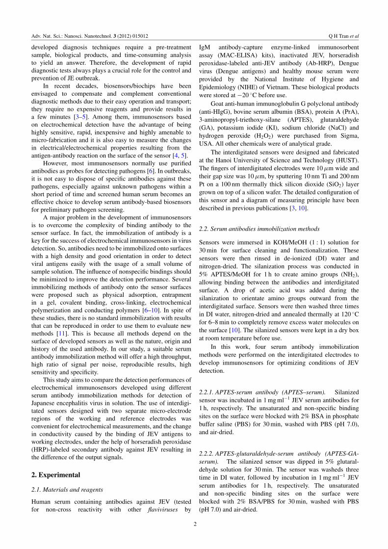
Figure 3. Bioactivity of the carried HRP versus concentration ofH2O2 measured in 0.02 M PBS (pH 7.0) containing 0.05 M KI and0.15 M NaCl using immunosensors developed after incubation with50 ng ml−1 JEV antigens: (a) incubation with healthy mouse serum,(b) APTES-serum method, (c) APTES-GA-antiHIgG-serum method,(d) APTES-GA-serum method and (e) APTES-GA-PrA-serummethod. Error bars were calculated from the mean values, n = 3.
lead to binding with the secondary anti-JEV antibody labeledHRP by the ‘sandwich’ mechanism. This configuration wassimilar to an enzyme immune-bioassay format to detect JEV
4
Adv. Nat. Sci.: Nanosci. Nanotechnol. 3 (2012) 015012 Q H Tran et al
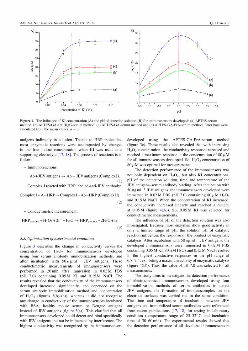
Figure 4. The influence of KI concentration (A) and pH of detection solution (B) for immunosensors developed: (a) APTES-serummethod; (b) APTES-GA-antiHIgG-serum method; (c) APTES-GA-serum method and (d) APTES-GA-PrA-serum method. Error bars werecalculated from the mean values, n = 3.
antigens indirectly in solution. Thanks to HRP molecules,most enzymatic reactions were accompanied by changesin the free iodine concentration when KI was used as asupporting electrolyte [17, 18]. The process of reactions is asfollows:
– Immunoreactions:
Ab + JEV antigens → Ab − JEV antigens (Complex I).(1)
Complex I reacted with HRP labeled-anti-JEV antibody:
Complex I + A−HRP→Complex I−Ab−HRP (Complex II).
(2)
– Conductimetric measurement:
HRPreactant + H2O2 + 2I− + H2O → HRPproduct + 2H2O + I2.
(3)
3.3. Optimization of experimental conditions
Figure 3 describes the change in conductivity versus theconcentration of H2O2 for immunosensors developedusing four serum antibody immobilization methods, andafter incubation with 50 µg ml−1 JEV antigens. Theseconductimetric measurements of immunosensors wereperformed in 20 min after immersion in 0.02 M PBS(pH 7.0) containing 0.05 M KI and 0.15 M NaCl. Theresults revealed that the conductivity of the immunosensorsdeveloped increased significantly, and depended on theserum antibody immobilization method and concentrationof H2O2 (figures 3(b)–(e)), whereas it did not recognizeany change in conductivity of the immunosensors incubatedwith BSA, healthy mouse serum or Dengue antigensinstead of JEV antigens (figure 3(a)). This clarified that allimmunosensors developed could detect and bind specificallywith JEV antigens and not be influenced by interference. Thehighest conductivity was recognized by the immunosensors
developed using the APTES-GA-PrA-serum method(figure 3e). These results also revealed that with increasingH2O2 concentration, the conductivity response increased andreached a maximum response at the concentration of 80 µMfor all immunosensors developed. So, H2O2 concentration of80 µM was optimal for measurements.
The detection performance of the immunosensors wasnot only dependent on H2O2, but also KI concentrations,pH of the detection solution, time and temperature of theJEV antigens–serum antibody binding. After incubation with50 ng ml−1 JEV antigens, the immunosensors developed wereimmersed in 0.02 M PBS (pH 7.0) containing 80 µM H2O2
and 0.15 M NaCl. When the concentration of KI increased,the conductivity increased linearly and reached a plateauat 0.05 M (figure 4(A)). So, 0.05 M KI was selected forconductimetric measurements.
The influence of pH of the detection solution was alsoinvestigated. Because most enzymes show good activity inonly a limited range of pH, the solution pH of catalyticreaction influences the response of the product of enzymaticcatalysis. After incubation with 50 ng ml−1 JEV antigens, thedeveloped immunosensors were immersed in 0.02 M PBScontaining 0.05 M KI, 80 µM H2O2 and 0.15 M NaCl resultedin the highest conductive responses in the pH range of6.8–7.4, exhibiting a maximum activity of enzymatic catalysis(figure 4(B)). Thus, the value of pH 7.0 was selected for allmeasurements.
The study aims to investigate the detection performanceof electrochemical immunosensors developed using fourimmobilization methods of serum antibodies to detectJEV antigens, the formation of immunocomplex on theelectrode surfaces was carried out in the same condition.The time and temperature of incubation between JEVantigens and immobilized serum antibodies were referencedfrom recent publications [17, 18] for testing in laboratorycondition (temperature range of 25–32◦ C and incubationtime of 30–60 min). The experimental results showed thatthe detection performance of all developed immunosensors
5

Adv. Nat. Sci.: Nanosci. Nanotechnol. 3 (2012) 015012 Q H Tran et al
Figure 5. The differential voltage output versus concentrationof JEV antigens from immunosensors developed using differentimmobilization methods of serum antibodies: (a) APTES-serum method, (b) APTES-GA-antiHIgG-serum method, (c)APTES-GA-serum method and (d) APTES-GA-PrA-serum method.Error bars were calculated from the mean values, n = 3.
was stable in these conditions. In fact, the temperature of27±2◦ C and incubation time of 45 min were selected for mostmeasurements.
3.4. Detection of JEV antigens
The detection signals have been found stable for 20 min,since immersion of immunosensors developed in 0.02 M PBS(pH 7.0) containing 0.05 M KI, 80 µM H2O2 and 0.15 MNaCl. The change in conductivity comes from the workingelectrodes in comparison with reference electrodes, thesesignals could be converted into the differential voltage inthe output of RS 830 Lock-in amplifier. Figure 5 showsthe differential voltages of immunosensors developed bydifferent concentration of JEV antigens in 0.02 M PBS(pH 7.0) containing 0.05 M KI, 80 µM H2O2 and0.15 M NaCl. Figure 5(d) shows that the electrochemicalimmunosensor developed using the APTES-GA-PrA-serummethod obtained the highest signal for detection of JEVantigens in the linear range from 25 ng ml−1 to 1 µg ml−1,and the limit of detection (LOD) was about 10 ng ml−1 [19].
This demonstrated that the use of protein A is the bestchoice for selection and orientation of serum antibodiesimmobilized on the electrodes for the development ofelectrochemical immunosensors in virus detection.
4. Conclusion
This study presented the development of electrochemicalimmunosensors based on serum antibodies via four
immobilization methods to detect JEV antigens. Thedetection process was carried out indirectly by conductimetricmeasurements in 0.02 M PBS (pH 7.0) containing 0.05 MKI, 80 µM H2O2 and 0.15 M NaCl with the support ofHRP-labeled secondary antibody against JEV. The resultsshowed that the electrochemical immunosensor developedusing the APTES-GA-PrA-serum method obtained thehighest signal for detection of JEV antigens in the linearrange from 25 ng ml−1 to 1 µg ml−1, and the limit ofdetection was about 10 ng ml−1. The study showed apotential development of useful and powerful devices forvirus detection from clinical samples in case of possibleoutbreaks.
Acknowledgments
This work was financially supported by Vietnam’s NationalFoundation for Science and Technology Development(NAFOSTED), project code: 106.16.181.09.
References
[1] Coker R J, Hunter B M, Rudge J W, Liverani M andHanvoravongchai P 2011 Lancet 377 599
[2] WHO Manual for the Laboratory Diagnosis of JapaneseEncephalitis Virus Infection 2007 http://www.who.int/immunization monitoring/Manual lab diagnosis JE.pdf
[3] Tam P D, Tuan M A, Huy T Q, Anh L T and Hieu N V 2010Mater. Sci. Eng. C 30 1145
[4] Labib M, Martic S, Shipman P O and Kraatz H-B 2011Talanta 85 770
[5] Huy T Q, Hanh N T H, Thuy N T, Chung P V, Nga P T andTuan M A 2011 Talanta 86 271
[6] Pejcic B, Marco D R and Parkinson 2006 Analyst131 1079
[7] Paul P et al 2003 Anal. Biochem. 312 113[8] Das R D, Maji S, Das S and Roy C C 2010 Appl. Surf. Sci.
256 5867[9] Ahuja T, Mir I A, Kumar D and Rajesh 2007 Biomaterials
28 791[10] Huy T Q, Hanh N T H, Chung P V, Anh D D, Nga P T and
Tuan M A 2011 Appl. Surf. Sci. 257 7090[11] Ramírez N B, Salgado A M and Valdman B 2009 Bras. J.
Chem. Eng. 26 227[12] Corso C D, Dickherber A and Hunt H D 2008 Biosens.
Bioelectron. 24 805[13] Nagare G D and Mukherji S 2009 Appl. Surf. Sci.
255 3696[14] Junqueira L C and Jose C 2003 Basic Histology (Los Altos:
CA: McGraw-Hill)[15] Surolia A, Pain D and Khan M I 1982 Trend. Biochem. Sci.
7 74[16] Page M and Thorpe R 2002 The Protein Protocols Handbook
part VII ed J M Walker (Totowa, NJ: Humana Press) p 993[17] Liu H, Yang Y, Chen P and Zhong Z 2009 Biochem. Eng. J.
45 107[18] Liang K-Z, Qui I-S, Mu W-J and Liu Z-X 2009 Bioprocess
Biosyst. Eng. 32 353[19] Armbruster A D and Pry T 2008 Clin. Biochem. Rev.
29 (Suppl. i) S49
6