BASED ELECTROCHEMICAL BIOSENSOR ROSWANI BINTI ...
45
INTEGRATION OF GLUTARALDEHYDE ONTO IMMUNO-MEMBRANE FOR POLYANILINE- BASED ELECTROCHEMICAL BIOSENSOR ROSWANI BINTI SHAIMI UNIVERSITI SAINS MALAYSIA 2013
-
Upload
khangminh22 -
Category
Documents
-
view
5 -
download
0
Transcript of BASED ELECTROCHEMICAL BIOSENSOR ROSWANI BINTI ...

i
INTEGRATION OF GLUTARALDEHYDE ONTO
IMMUNO-MEMBRANE FOR POLYANILINE-
BASED ELECTROCHEMICAL BIOSENSOR
ROSWANI BINTI SHAIMI
UNIVERSITI SAINS MALAYSIA
2013

ii
INTEGRATION OF GLUTARALDEHYDE ONTO IMMUNO-MEMBRANE
FOR POLYANILINE-BASED ELECTROCHEMICAL BIOSENSOR
by
ROSWANI BINTI SHAIMI
Thesis submitted in fulfillment of the
requirements for the degree of
Master of Science
DECEMBER 2013

ii
ACKNOWLEDGEMENT
In the name of Allah S.W.T. the most gracious and most merciful, Lord of
the universe, with His permission, Alhamdulillah the project has been completed.
Praise to Prophet Muhammad S.A.W., His companions and to those on the path as
what He preached upon, might Allah Almighty keep us His blessing and tenders.
A special gratitude to my beloved parents, Mr. Shaimi bin Umat and Mrs.
Siti Fatimah binti Hussin, my family members and also my partner, Che Amir
Rajhan bin Che Jaffar for their endless support and prays.
I would like to express deepest gratitude to my beloved supervisor, Dr. Low
Siew Chun for her excellent knowledge guidance, encouragement, valuable
suggestion, guidance support and advises rendered throughout my research. Also, I
would like to express my gratitude to my co-supervisor, Prof. Abdul Latif Ahmad
for sharing his knowledge, opinion and assistance.
Not forgetting, thanks a lot to all technicians and staffs of School of
Chemical Engineering for their cooperation. Deepest thank to all my friends for the
help and moral support.
Last but not least, I would like to express my gratitude to whom that
involved directly or indirectly for their unselfish advice and assistance toward
performing in finishing this research within time.
Thank you so much and may Allah S.W.T. the Almighty be with us all the time.

iii
TABLE OF CONTENTS
Page
ACKNOWLEDGEMENT
ii
LIST OF TABLES
viii
LIST OF FIGURES
ix
LIST OF PLATES
xii
LIST OF SYMBOLS
xiii
LIST OF ABBREVIATIONS
xv
ABSTRAK
xvii
ABSTRACT
xix
CHAPTER 1: INTRODUCTION
1.1 Biomedical Technology
1.2 Biosensor
1.3 Membrane for bio-sensing applications
1.4 Membrane modification by glutaraldehyde
1.5 Problem statement and importance of the research
1.6 Research objectives
1.7 Scope of study
1.8 Organization of the thesis
1
3
5
6
7
8
9
11
CHAPTER 2: LITERATURE REVIEW
2.1 Emerging waterborne pathogens
2.2 Conventional methods in pathogen and viruses detection
13
15

iv
2.3 Biosensor in pathogen and viruses detection
2.4 Biomolecule Immobilization
2.4.1 Classical method of biomolecule immobilization
2.4.2 New trend of biomolecule immobilization
2.5 Biosensors detecting systems based on different transducer properties
2.5.1 Optical transducer
2.5.2 Electrochemical transducer
2.5.2.1 Amperometric
2.5.2.2 Potentiometric
2.5.2.3 Conductometric
2.5.3 Mass-based transducer
2.6 Electrochemical transduction method in the development of biosensor
2.7 Implementation of conducting polymers in development of
electrochemical based biosensor
2.8 Magnetic polymer nanoparticle in development of biosensor
2.9 Polymeric membrane in sensing application
2.10 Future direction
17
19
19
20
29
30
31
31
31
32
34
36
38
46
49
52
CHAPTER 3: RESEARCH METHODOLOGY
3.1 Introduction
3.2 Materials and chemicals
3.3 Lateral flow membrane selection
3.3.1 Quantification of protein binding ability
3.3.2 Lateral wicking time
53
55
58
58
59
v
3.4 Characterization of the membrane and conjugated biomolecules
3.4.1 Attenuated Total Reflectance Fourier Transform Infrared
(ATR-FTIR)
3.4.2 Field Emission Scanning Electron Microscope (FESEM)
3.4.3 Atomic Force Microscope (AFM)
3.4.4 Thermo-Gravimetric Analysis (TGA)
3.4.5 Dynamic Laser Scattering (DLS)
3.4.6 Porosity
3.5 Membrane modification: Integration of glutaraldehyde (GA) onto
polymeric membranes
3.5.1 Setting of GA concentration
3.5.2 Setting of number of integration layers
3.5.3 Setting of integration time
3.6 Optimization of GA operational integration by Response Surface
Methodology (RSM)
3.6.1 Design of experiment (CCD)
3.6.2 Data analysis
3.6.3Validation of the experiment
3.7 Preparation of complex polyaniline-Fe2O3 conjugated biotinylated
IgG
3.7.1Synthesis of polyaniline (PANI) as an electrical signal
transducer
3.7.2 Conjugation of PANI to Fe2O3 (PANI- Fe2O3)
60
60
60
61
61
61
62
62
63
63
64
64
65
66
66
66
66
67
vi
3.7.3 Conjugation of PANI-Fe2O3- GA with biotinylated IgG
3.8 Performance Test via Pulse-Mode Measurement
3.8.1 Assembly of electrochemical biosensor
3.8.2 Configuration of electrochemical biosensor
3.8.3 Performance of the polyaniline-electrochemical biosensor
68
69
69
70
71
CHAPTER 4: RESULTS AND DISCUSSION
4.1 Selection of the potential transport media by quantitatively determine
the binding capacity and lateral wicking time of polymeric membranes
4.1.1 Effect of membrane‟s material on biosensor performances
4.1.2 Integration effects of GA on protein immobilization
4.2 Modification of NC membrane surface using glutaraldehyde to
enhance the protein immobilization ability
4.2.1Effect of concentration of GA as a cross-linker in protein binding
4.2.2 Effect of number of GA deposited layers
4.2.3 Effect of integration time
4.2.4 Thermal stability of GA-modified membrane
4.3 Optimization of the integration operational procedure of GA
using RSM
4.3.1 Experimental design and analysis
4.3.2 Analysis of variance (ANOVA)
4.3.3 Interaction of independent variables on response (protein
binding ability)
4.3.4 Response surface model in 3D for independent variables
73
74
79
84
85
86
89
91
94
94
96
101
104
vii
4.3.5 Model validation and confirmation of the optimized result
4.4 Preparation of complex polyaniline-Fe2O3 nanocomposite conjugated
with biotin
4.4.1 Synthesis of polyaniline
4.4.2 Conjugation of PANI to Fe2O3 (PANI- Fe2O3)
4.4.3 Conjugation of PANI-Fe2O3 with biotinylated IgG
4.5 Performance Test via Pulse-Mode Measurement
4.5.1 Configuration of Biosensor
4.5.2 Conductivity Performance
111
114
114
117
123
125
125
129
CHAPTER 5: CONCLUSIONS AND RECOMMENDATIONS
5.1 Conclusions
5.2 Recommendations
132
134
REFERENCES 135
APPENDICES
APPENDIX A
APPENDIX B
APPENDIX C
APPENDIX D
APPENDIX E
148
149
149
150
151
List of publications and conferences 152

viii
LIST OF TABLES
Page
Table 2.1 Importance of the glutaraldehyde as cross-linker for improved
membrane performances
24
Table 2.2 Role of polyaniline in bio-sensing application 41
Table 2.3 Procedures for synthesis of PANI through chemical oxidative
polymerization
42
Table 3.1 List of materials and chemicals 55
Table 3.2 Independent variables and their selected values based on the
CCD design
65
Table 4.1 Design layout and corresponding response (Protein binding
ability)
95
Table 4.2 Analysis of the variance (ANOVA) for the fit of the
experimental data
97
Table 4.3 Validation of the model between predicted and experimental
value
112
Table 4.4 Zeta charge 118
Table 4.5 Biosensor test results at different configuration of biosensor 130

ix
LIST OF FIGURES
Page
Figure 1.1 Growing technology in detecting pathogen 2
Figure 1.2 Biosensor transduction methods in pathogen detection 3
Figure 1.3 Schematic diagram showing the main component of an
electrochemical biosensor
4
Figure 2.1 Three representation of a molecule of monomeric
glutaraldehyde
22
Figure 2.2 Reaction of GA with amino groups of protein 23
Figure 2.3 Classification of Biosensor 29
Figure 2.4 Overview of conductometric based-biosensor assembly
and (a) schematic representation of the biosensor detection
system (b)
34
Figure 2.5 Antibody-based virus of surface acoustic wave (SAW)
biosensor
36
Figure 2.6 Some of conducting polymers 40
Figure 2.7 Aniline oxidative polymerization 45
Figure 2.8 Preparation of electrically active polyaniline conjugated
magnetic (EAPM) nanoparticle
49
Figure 3.1 Flow chart for the experimental procedures 54
Figure 3.2 Schematic diagram of biosensor configuration 70
Figure 4.1 Comparison of pure binding ability in NC (NC-90, NC-
135, NC-180), CA, nylon and PVDF membranes
74
x
Figure 4.2 ATR-FTIR spectra of (a) NC membrane and (b) Nylon
membrane
75
Figure 4.3 Performance of lateral wicking time (min) in NC (NC-90,
NC-135, NC-180), CA, nylon and PVDF membranes
77
Figure 4.4 Porosity of NC (NC-90, NC-135, NC-180), CA, nylon and
PVDF membranes
79
Figure 4.5 Protein binding ability in varying membranes at condition
with or without integration of GA (1 wt. %)
80
Figure 4.6 Stability of protein bound on NC-180 membrane treated
with and without GA. Concentration of GA was set at 0.5
wt. %
81
Figure 4.7 ATR-FTIR spectra of (a) pure GA, (b) Neat NC membrane
and (c) Integrated NC membrane with GA
84
Figure 4.8 Effect of concentration of GA as cross-linker in protein
binding
86
Figure 4.9 Effect of Layer by Layer modification of GA 88
Figure 4.10 ATR-FTIR spectra of NC modified with GA with different
number of layers a) single b) double and c) triple layers
89
Figure 4.11 Effect of integration time 91
Figure 4.12 Thermal stability of GA-modified membrane 93
Figure 4.13 TGA analysis for NC and NC integrated with GA 94
Figure 4.14 Normal probability plot of the studentized residual for
protein binding ability
99

xi
Figure 4.15 The studentized residuals and predicted response plot 100
Figure 4.16 Predicted versus actual protein binding values 101
Figure 4.17 Interaction graphs between (a) number of layer (factor B)
and integration time (factor C) at 3 wt. % of GA, (b)
concentration of GA (factor A) and integration time (factor
C) at l layer
103
Figure 4.18 Response surface plotted on (a) factor C (integration time):
factor A (concentration of GA) at a fixed single layer; (b)
factor B (number of layer): factor C (integration time) at a
fixed 3 wt. % of GA concentration
105
Figure 4.19 Response surface plotted on integration time (factor C):
concentration of GA (factor A) at (a) single layer (b)
double layer (c) triple layer of GA
110
Figure 4.20 ATR-FTIR spectrum of polyaniline 116
Figure 4.21 Synthesis mechanism of polyaniline conjugated maghemite
nanoparticles (PANI-Fe2O3)
119
Figure 4.22 ATR-FTIR spectra of polyaniline and polyaniline
conjugated maghemite nanoparticles
122
Figure 4.23 Absorbance of PANI, PANI-Fe2O3, PANI-Fe2O3-GA with
biotin
124
Figure 4.24 Cross-section of biosensor to illustrate electric signal
generation
130
xii
LIST OF PLATES
Page
Plate 3.1 Lateral wicking test setup 59
Plate 3.2 Pulse mode measurement test setup 72
Plate 4.1 Schematic diagram of chemical bonds between GA, NC
membrane and protein
82
Plate 4.2 AFM for NC membrane surface a) NC membrane b) NC
membrane modified with GA
108
Plate 4.3 SEM for the NC membrane surface a) NC membrane b) NC
integrated with GA
113
Plate 4.4 TEM image of polyaniline prepared in 1M HCl 117
Plate 4.5 TEM images of (a) unmodified Fe2O3 nanoparticles (b)
synthesized of PANI-Fe2O3 nanocomposites
121
Plate 4.6 Difference in staining on biosensor configuration: (a) BSA
lining followed by silver paste (b) Silver paste followed by
BSA lining
126
Plate 4.7 Different configuration of biosensor observed by light
microscope (a) BSA lining followed by silver paste (b)
Silver paste followed by BSA lining
128
xiii
LIST OF SYMBOLS
oC Temperature
wt. % weight percentage
μg Microgram
cm3 centimeter cubic
% Percentage
mS milli Semen
M Ω Mega Ohm
ml Milliliter
nm Nanometer
M Molar
e.g Example
i.e. which is
h Hour
min Minutes
rpm revolution per minutes
µm Micrometer
cm Centimeter
VE membrane‟s existent volume
VA membrane‟s apparent volume
Ɛ Porosity
mm Millimeter
g Gram

xiv
< less than
v/v volume/ volume
mV Unit for zeta potential
kΩ Kilo Ohm
xv
LIST OF ABBREVIATIONS
PANI Polyaniline
BSA Bovine Serum Albumin
HF High Flow
NC Nitrocellulose
GA Glutaraldehyde
RSM Response Surface methodology
DLS Dynamic Laser Scattering
PCR Polymerase Chain Reaction
CA Cellulose Acetate
PVDF Polyvinylidene Fluoride
FESEM Field Emission Scanning Electron Microscope
ATR-FTIR Attenuated Total Reflectance Fourier Transform Infrared
TGA Thermo-Gravimetric Analysis
AFM Atomic Force Microscope
CCD Central Composite Design
E. coli Escherichia coli
APS Ammonium Persulfate
SPR Surface Plasmon Resonance
PPy Polypyrrole
HCl Hydrochloric acid
DMF Dimethylformamide
PBS Phosphate Buffer Saline
xvi
LiCl Lithium Chloride
EAPM Electrically active polyaniline magnetic
BCA Bicinchoninic acid
Eq Equation
ANOVA Analysis of variance
xvii
INTEGRASI GLUTARALDEHID KE ATAS IMMUNO-MEMBRAN UNTUK
BIOPENDERIA ELEKTROKIMIA BERASASKAN POLIANILINA
ABSTRAK
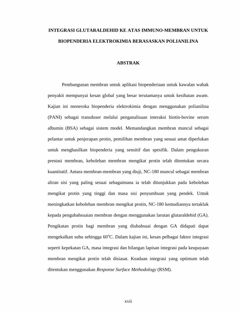
Pembangunan membran untuk aplikasi biopenderiaan untuk kawalan wabak
penyakit mempunyai kesan global yang besar terutamanya untuk kesihatan awam.
Kajian ini meneroka biopenderia elektrokimia dengan menggunakan polianilina
(PANI) sebagai transduser melalui penganalisaan interaksi biotin-bovine serum
albumin (BSA) sebagai sistem model. Memandangkan membran muncul sebagai
pelantar untuk penjerapan protin, pemilihan membran yang sesuai amat diperlukan
untuk menghasilkan biopenderia yang sensitif dan spesifik. Dalam pengukuran
prestasi membran, kebolehan membran mengikat protin telah ditentukan secara
kuantitatif. Antara membran-membran yang diuji, NC-180 muncul sebagai membran
aliran sisi yang paling sesuai sebagaimana ia telah ditunjukkan pada kebolehan
mengikat protin yang tinggi dan masa sisi penyumbuan yang pendek. Untuk
meningkatkan kebolehan membran mengikat protin, NC-180 kemudiannya tertakluk
kepada pengubahsuaian membran dengan menggunakan larutan glutaraldehid (GA).
Pengikatan protin bagi membran yang diubahsuai dengan GA didapati dapat
mengekalkan suhu sehingga 60oC. Dalam kajian ini, kesan pelbagai faktor integrasi
seperti kepekatan GA, masa integrasi dan bilangan lapisan integrasi pada keupayaan
membran mengikat protin telah disiasat. Keadaan integrasi yang optimum telah
ditentukan menggunakan Response Surface Methodology (RSM).
xviii
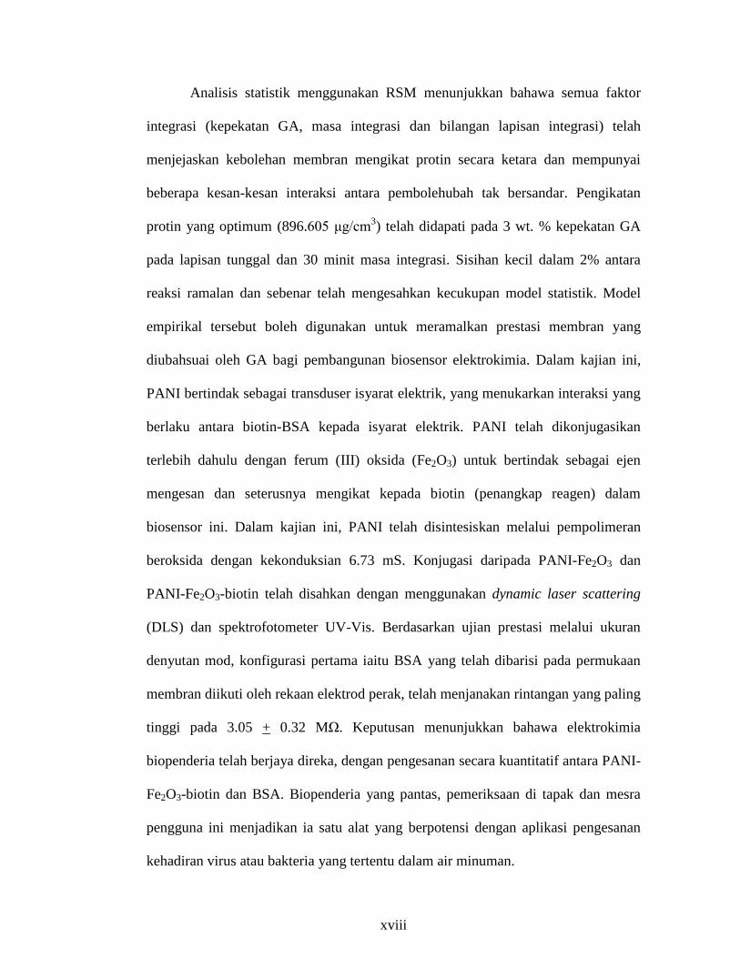
Analisis statistik menggunakan RSM menunjukkan bahawa semua faktor
integrasi (kepekatan GA, masa integrasi dan bilangan lapisan integrasi) telah
menjejaskan kebolehan membran mengikat protin secara ketara dan mempunyai
beberapa kesan-kesan interaksi antara pembolehubah tak bersandar. Pengikatan
protin yang optimum (896.605 μg/cm3) telah didapati pada 3 wt. % kepekatan GA
pada lapisan tunggal dan 30 minit masa integrasi. Sisihan kecil dalam 2% antara
reaksi ramalan dan sebenar telah mengesahkan kecukupan model statistik. Model
empirikal tersebut boleh digunakan untuk meramalkan prestasi membran yang
diubahsuai oleh GA bagi pembangunan biosensor elektrokimia. Dalam kajian ini,
PANI bertindak sebagai transduser isyarat elektrik, yang menukarkan interaksi yang
berlaku antara biotin-BSA kepada isyarat elektrik. PANI telah dikonjugasikan
terlebih dahulu dengan ferum (III) oksida (Fe2O3) untuk bertindak sebagai ejen
mengesan dan seterusnya mengikat kepada biotin (penangkap reagen) dalam
biosensor ini. Dalam kajian ini, PANI telah disintesiskan melalui pempolimeran
beroksida dengan kekonduksian 6.73 mS. Konjugasi daripada PANI-Fe2O3 dan
PANI-Fe2O3-biotin telah disahkan dengan menggunakan dynamic laser scattering
(DLS) dan spektrofotometer UV-Vis. Berdasarkan ujian prestasi melalui ukuran
denyutan mod, konfigurasi pertama iaitu BSA yang telah dibarisi pada permukaan
membran diikuti oleh rekaan elektrod perak, telah menjanakan rintangan yang paling
tinggi pada 3.05 + 0.32 MΩ. Keputusan menunjukkan bahawa elektrokimia
biopenderia telah berjaya direka, dengan pengesanan secara kuantitatif antara PANI-
Fe2O3-biotin dan BSA. Biopenderia yang pantas, pemeriksaan di tapak dan mesra
pengguna ini menjadikan ia satu alat yang berpotensi dengan aplikasi pengesanan
kehadiran virus atau bakteria yang tertentu dalam air minuman.
xix
INTEGRATION OF GLUTARALDEHYDE INTO IMMUNO-MEMBRANE
FOR POLYANILINE-BASED ELECTROCHEMICAL BIOSENSOR
ABSTRACT
Development of membrane for bio-sensing applications for epidemics
control has a huge global impact especially for public health. This research explored
the electrochemical biosensor with polyaniline (PANI) as a transducer through
analyzing the biotin-bovine serum albumin (BSA) interaction as a model system. As
membrane appeared as the platform for protein immobilization, selection of suitable
membrane is required to create a sensitive and specific biosensor. In measurement of
membrane performance, membrane protein binding abilities were quantitatively
determined. Among the tested membranes, NC-180 appeared as the most suitable
lateral flow membrane as it performed high protein binding ability and short lateral
wicking time. To increase the membrane protein binding ability, NC-180 was then
subjected to membrane modification using glutaraldehyde (GA) solution. The
protein binding of the modified membrane with GA was able to retain up to
temperature of 60oC. In the present study, the effect of various integration factors
such as the concentration of GA, integration time and number of the integration
layer on membrane‟s protein binding ability was investigated. The optimum
integration conditions were determined using response surface methodology (RSM).
The statistical analysis using RSM showed that all the integration factors
(concentration of GA, integration time and number of the integration layer) had
significantly affected the membrane‟s protein binding ability and had some
xx
interaction effects among the independent variables. The optimum protein binding
(896.605 µg/cm3) was obtained at 3 wt. % of GA concentration at single layer and
30 min of integration time. A small error within 2 % between predicted and actual
responses confirmed the adequacy of the statistical model. This empirical model is
applicable to predict the performance of the GA-modified membranes for the
development of electrochemical biosensor. In this research, PANI served as an
electric signal transducer, converting the interaction occurred between biotin-BSA to
electrical signal. The PANI was first conjugated with iron (III) oxide (Fe2O3) to
serve as the detecting agent and then further bound to biotin (target reagent) in the
biosensor. In this work, the PANI was synthesized through oxidative polymerization
with the conductivity of 6.73 mS. The conjugations of PANI-Fe2O3 and PANI-
Fe2O3-biotin were confirmed using dynamic laser scattering (DLS) and UV-Vis
spectrophotometer. Based on the performance test via pulse mode measurement, the
first configuration i.e. the BSA was lined on the membrane surface followed by
fabrication of the silver electrode, generated the highest resistance at 3.05 + 0.32
MΩ. Results indicated that the electrochemical biosensor was successfully
fabricated, with quantitative detection between PANI-Fe2O3-biotin and BSA. This
rapid, on-site examination and user friendly biosensor make it a promising device
with application in detecting the presence of selected viruses or bacteria in drinking
water.
1
CHAPTER 1
INTRODUCTION
1.1 Biomedical Technology
Modern medical technology has undeniably achieved lots of advancement
and sophistication, especially in the developed countries for providing treatment and
controlling the spread of diseases. However, millions of people die annually due to
waterborne outbreaks especially in third world countries (Bouwer, 2000). The root
cause to the loss of millions human life is mainly due to the unavailability or
inaccessible of diagnostics facilities in the underdeveloped or developing countries.
Existing diagnostics techniques do not address the needs of bottom billions.
It was reported that 40 % of the approximately 50 million total annual deaths world-
wide because of the infectious diseases (Leonard et al., 2003). Of these infectious
diseases deaths, 10-20 million was contributed from waterborne pathogens while
non-fatal infection was more than 200 million people each year (Leonard et al.,
2003). Most of the major food-borne and water-borne diseases are caused of
Escherichia coli (e.g., E. coli O157:H7). This pathogen is known to cause diarrhea,
hemolytic uremic syndrome (HUS) and hemorrhagic colitis in humans (Zhu et al.,
2005). The outbreaks of illness are mainly due to ingestion of meats, water, and
uncooked fruits and vegetables.
As popular awareness of the importance of health is increases, a cost
effective approach would need to be considered to develop a rapid, on-site
examination and user friendly device in monitoring and detecting the presence of
selected viruses or bacteria in drinking water. In the past, polymerase chain reaction

2
(PCR) has been a common practice for the detection and identification of pathogen.
This conventional method is sensitive and can be used to amplify small quantities of
pure samples in detecting the presence of pathogen and viruses in drinking water.
However, it needs relatively longer time to get the result, skilled personnel and
advanced infrastructure to carry out the analysis (Waswa et al., 2007), thus the focus
has shifted to the development of biosensor. Biosensor appeared as one of the fastest
growing pathogen detection tools with equally reliable results in faster time and on-
site examination. In recent year, the biosensor possesses the fastest growth compared
to other conventional methods, as illustrated in Figure 1.1.
Figure 1.1: Growing technology in detecting pathogen (Lazcka et al., 2007).
Year

3
1.2 Biosensor
Several biosensors approaches based on electrochemical, optical and
piezoelectric detection principles are developed as illustrated in Figure 1.2. The
primary aim of those configurations are of low sample consumption, fast response
time and high throughput (Zhang et al., 2011). Among these approaches, the
electrochemical biosensors present to be an ideal choice compared to the other
transduction methods: (1) the electrochemical electrodes are relatively low cost for
production which promising for practical use (2) the applied voltage are sufficiently
small, thus minimize the sensor‟s power consumption, (3) easily integrated with
electronic devices and less susceptible to environmental effects and contaminants
and (4) reduce response time due to the smaller diffusion distances.
Figure 1.2: Biosensor transduction methods in pathogen detection
(Lazcka et al., 2007).
4
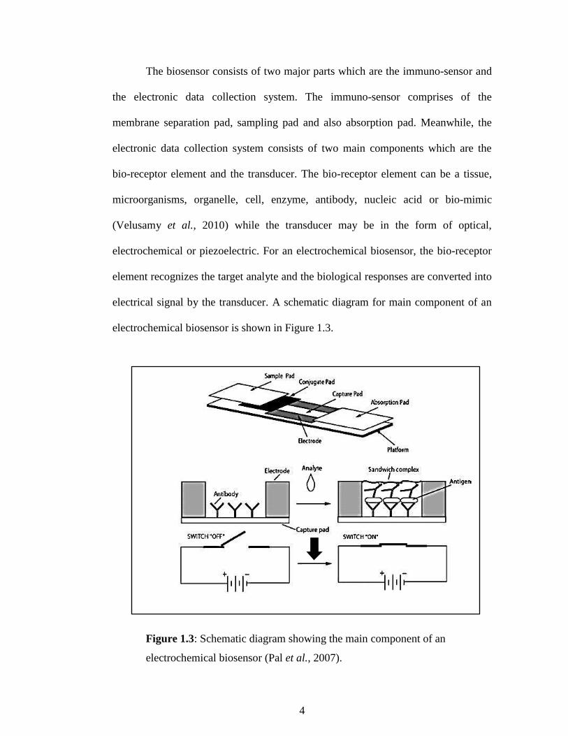
The biosensor consists of two major parts which are the immuno-sensor and
the electronic data collection system. The immuno-sensor comprises of the
membrane separation pad, sampling pad and also absorption pad. Meanwhile, the
electronic data collection system consists of two main components which are the
bio-receptor element and the transducer. The bio-receptor element can be a tissue,
microorganisms, organelle, cell, enzyme, antibody, nucleic acid or bio-mimic
(Velusamy et al., 2010) while the transducer may be in the form of optical,
electrochemical or piezoelectric. For an electrochemical biosensor, the bio-receptor
element recognizes the target analyte and the biological responses are converted into
electrical signal by the transducer. A schematic diagram for main component of an
electrochemical biosensor is shown in Figure 1.3.
Figure 1.3: Schematic diagram showing the main component of an
electrochemical biosensor (Pal et al., 2007).

5
1.3 Membrane for bio-sensing applications
The unique separation principle of a membrane, namely selective transport
processes and efficient separation makes it an ideal candidate as the key functional
element in a biosensor. Membrane provides the capillary action and induces a flow
of aqueous medium along the lateral side of the strip that transports the target
analyte to the immobilized binding partner. The binding reaction between antigen
and antibody takes place on the surface and unbound molecules are subsequently
separated by the medium flow. Selection of membrane as the support material to
bind with bio-molecules oftentimes a protein is not a trivial task. A wide range of
porous membranes such as cellulose acetate membrane (CA), polyvinylidene
fluoride membrane (PVDF), nylon membrane and nitrocellulose membrane (NC)
have found to be compatible in bio-sensing development (Singh et al., 2008, Fang et
al., 2011). NC membrane has long occupied a position of central importance in
medical and immunological analysis due to its excellent wetting properties, high
binding capacity and low background staining (Morais et al., 1999).
Protein is the most common reagent to be applied onto the membrane
surface in an immunoassay. The interaction between protein and membrane is
generally affected by the membrane morphology, and eventually reflected the
effectiveness of the biosensor. Protein immobilization onto the membrane material
remains a key issue in development of the lateral flow biosensor (Kim et al., 2000,
Minett et al., 2002, Cho et al., 2007, Apostolova et al., 2011).

6
1.4 Membrane modification by glutaraldehyde
Immobilization of protein by covalent bonding onto membrane surface is one
of the effective methods in development of biosensor. To assure the required
sensitivity level of a biosensor, the membrane surface is always modified using the
cross-linker reagent. Aldehyde has found to be an excellent cross-linking agent in
fields such as medicine, electron microscopy study of proteins, cells, and etc.
(Fathima et al., 2004). Among the aldehyde groups, glutaraldehyde (GA) has found
great success in protein immobilization due to its thermally and chemically stable
(Migneault et al., 2004). The cross-linked protein by GA presents a stable, compact
protein structure (Emre et al., 2011), low impact on the activities recoveries and
improves the stability (Alonso et al., 2005). As a result, more stable and high
sensitivity biosensor is obtained.
Glutaraldehyde, a bi-functional aldehyde serves covalent bonding to the
polymer‟s backbone and protein (Busto et al., 1997). Two identical reactive groups
react with the residual free amino acid groups of protein and the polymer‟s
backbone, make the protein immobilization in development of biosensor to achieve
easily (Romero et al., 2008). The three-dimensional linking between protein and
polymer‟s backbone through intramolecular and intermolecular cross-linking
resulting the modified protein to completely insoluble in water and can be adsorbed
onto the support (Albareda-Sirvent et al., 2000).

7
1.5 Problem statement and importance of the research
Although sophisticated medical technology has undeniably achieved a lot of
advancement, however, millions of people die due to waterborne outbreak,
especially in the third world countries. The root cause to the loss of millions human
life is mainly due to the unavailability or inaccessible of diagnostics facilities in the
underdeveloped or developing countries. Existing diagnostics techniques do not
address the needs of bottom billions. To address this problem, the biosensor has
been found to be promising device for the rapid detection of pathogen and viruses in
drinking water.
Membrane is the most important key element in the development of
biosensor as it serves capillary action for the target analyte to flow and place for the
interaction between target analyte and capture reagent that is immobilized onto
membrane surface. Biosensor will not fully function without applying the membrane
as a support material. As membrane shows its importance, selection of the
membrane should be focused and studied for the development of bio-sensing
applications. The suitable membrane could contribute towards the high effectiveness
of the developed biosensor.
The main factor on the sensitivity of biosensor is based on the protein
immobilization technique onto the membrane surface. Adsorption technique is
commonly applied for protein immobilization. However, the leaking of the bio-
receptor and possible diffusion barriers are the common problems that occur by
applying this conventional immobilization technique, which eventually rendered the
low sensitivity of the biosensor. A sensitive biosensor could be obtained if some
improvement on protein immobilization technique is performed. Modification by
8
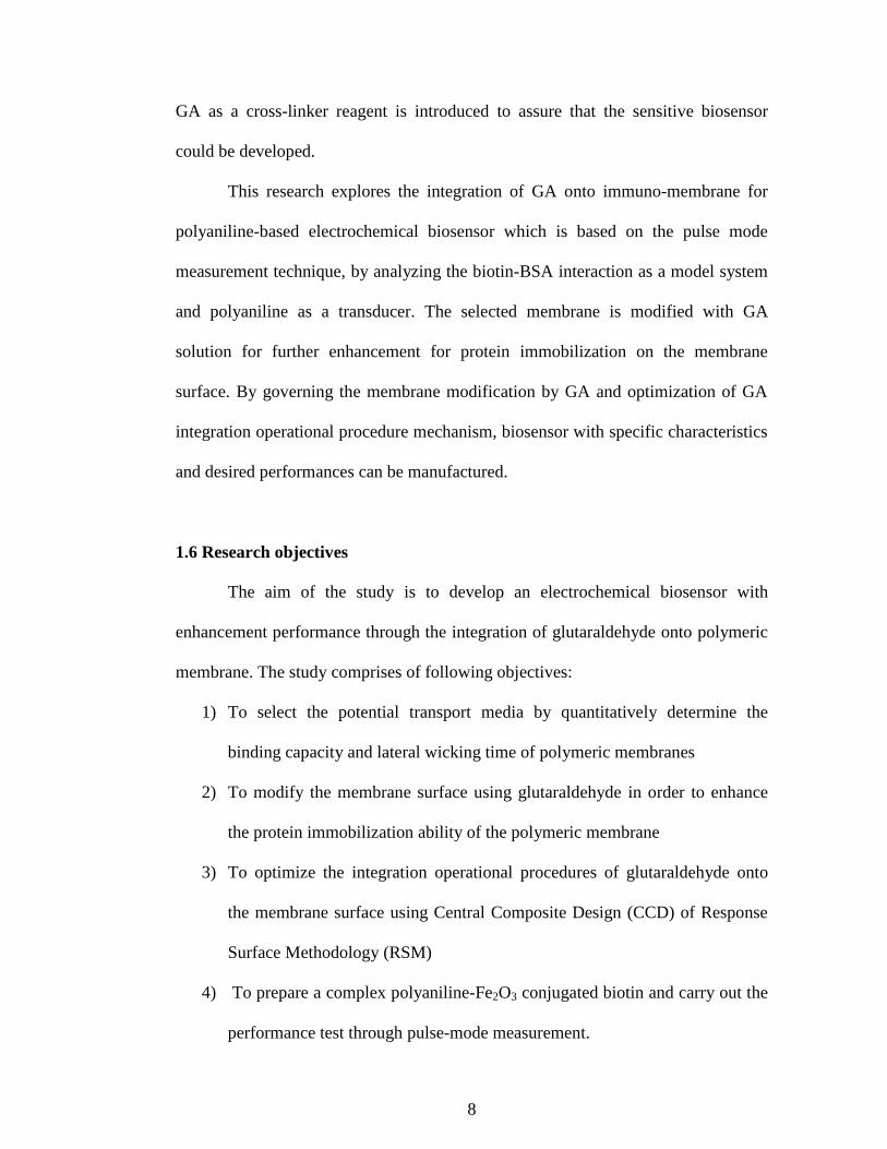
GA as a cross-linker reagent is introduced to assure that the sensitive biosensor
could be developed.
This research explores the integration of GA onto immuno-membrane for
polyaniline-based electrochemical biosensor which is based on the pulse mode
measurement technique, by analyzing the biotin-BSA interaction as a model system
and polyaniline as a transducer. The selected membrane is modified with GA
solution for further enhancement for protein immobilization on the membrane
surface. By governing the membrane modification by GA and optimization of GA
integration operational procedure mechanism, biosensor with specific characteristics
and desired performances can be manufactured.
1.6 Research objectives
The aim of the study is to develop an electrochemical biosensor with
enhancement performance through the integration of glutaraldehyde onto polymeric
membrane. The study comprises of following objectives:
1) To select the potential transport media by quantitatively determine the
binding capacity and lateral wicking time of polymeric membranes
2) To modify the membrane surface using glutaraldehyde in order to enhance
the protein immobilization ability of the polymeric membrane
3) To optimize the integration operational procedures of glutaraldehyde onto
the membrane surface using Central Composite Design (CCD) of Response
Surface Methodology (RSM)
4) To prepare a complex polyaniline-Fe2O3 conjugated biotin and carry out the
performance test through pulse-mode measurement.

9
1.7 Scope of study
In the development of lateral flow membrane for polyaniline-based
electrochemical biosensor, this research was consisted of four major investigations.
i.e. the selection of potential transport media (membrane) which governs the
performance of the lateral flow biosensor, modification of selected membrane with
glutaraldehyde (cross-linker reagent), optimization of GA integration operational
procedure using RSM and the last section was the preparation of a complex
polyaniline-Fe2O3 conjugated biotin and subsequently performance test via pulse-
mode measurement.
In this research, the interest was focused on determining the protein binding
ability and lateral wicking time for NC, Nylon, CA and PVDF membranes. Both
factors reflect the effectiveness of the biosensor in direct and eventually, developed
an in-depth understanding of membrane selection for the development of biosensor.
The characterization of the membrane such as membrane morphology, pore structure
and bonding characteristic were performed with the aid of different analytical
instruments such as field emission scanning electron microscope (FESEM) and
attenuated total reflectance fourier transform infrared (ATR-FTIR).
The selected membrane was subjected to membrane modification using GA
solution. In this section, we studied the integration operational procedure of GA onto
the membrane surface. The effects of GA concentration (0.5 wt. % to 5.0 wt. %),
integration time (10 min-90 min), the number of integration layer (single to triple) as
well as the thermal effects of integration (40oC-70
oC) were taken into
considerations. For the measurement of membrane performance, membrane protein
10
binding abilities were quantitatively determined. Furthermore, the functionality of
the GA-modified membrane was verified through ATR-FTIR and TGA analyses.
The interactive effects among the independent variables and optimizing the
GA integrated were presented through central composite design (CCD) of RSM.
Same effects as preliminary study (GA concentration, number of integration layers
and integration time) were taken into considerations. In measuring the membrane
performances, the membrane protein binding abilities were quantitatively
determined. In this section, the topographic of the GA-modified NC membrane was
determined through FESEM and AFM analyses.
In the last section, the biosensor was assembled by using BSA as the bio-
receptor element and polyaniline (PANI) nanowires as the molecular transducer. The
working principle of the developed biosensor was based on the antigen-antibody
interaction where the target reagent (biotinylated IgG) was conjugated with PANI
maghemite nanoparticles. PANI, an electric signal transducer for monitoring of the
bio-specific binding event between biotinylated IgG-BSA, was synthesized through
the oxidative polymerization of aniline monomer in the presence of ammonium
persulfate as oxidizing agent. The synthesized PANI was then integrated with
maghemite nanoparticles and subsequently, conjugated with biotinylated IgG. Next,
the biosensors were assembled into different biosensor assembly configurations, and
the performance of this polyaniline-based biosensor was demonstrated using the
pulse mode measurement technique.
.

11
1.8 Organization of the thesis
The thesis comprises of five chapters providing all the details and findings of
the research. Chapter One (Introduction) focuses on the biomedical technology that
implement the conventional method in pathogen detection, then eventually shift to
the development of biosensor. The chapter also included the application of the
membrane as a platform in protein immobilization and then further, the
enhancement of protein immobilization by modification of membrane using GA
solution. The problem statement and importance of the research are also highlighted
in this chapter. Besides, the research objectives of present study are elaborated in
detail together with the scope of the study to be covered. The thesis organization is
given in the last section of this chapter.
Chapter Two (Literature Review) reviews all the important literatures from
the researchers especially in the development of biosensor in detection of pathogen
and viruses in drinking water. At first, the chapter reviews the problem of
waterborne pathogens and the conventional method to detect the pathogens or
viruses in this waterborne problem. Biosensor is introduced to overcome the
problems of using the conventional method. As biomolecule immobilization is the
main stage in the development of biosensor, hence next section discusses on the
biomolecule immobilization.
Besides, this chapter also demonstrates the biosensor detecting systems
based on different transducer properties and then the implementation of the
electrochemical transduction method in development of biosensor. The rapid growth
of conducting polymers which lead to an innovation in development of
electrochemical based biosensor is also presented while the interesting of magnetic

12
polymer nanoparticle that is employed as magnetic concentrator and signal
transducer in biosensor is discussed in the next section. The polymeric membrane
that serves as platform for protein immobilization is reported and then, the final
section in the chapter, a future direction is given to illustrate the future study in
development of biosensor.
Chapter Three (Research Methodology) presents the experimental
procedures that were performed in this study. All details on materials and chemicals
and experimental procedures regarding the selection of lateral flow membrane,
modification of polymeric membrane by GA solution, optimization of GA
integration, preparation of complex PANI-Fe2O3 conjugated biotinylated IgG as well
as performance test were described. Detail equipment used together with the
analysis methods is incorporated in this section toward to achieve the objectives of
the study.
Chapter Four (Results and Discussion) presents the experimental results and
discussion obtained from the study. This chapter is divided into five sub-chapter that
is generally based on each objectives studied. The results and discussion covers the
selection of membrane as the potential transport media, membrane modification
using glutaraldehyde, optimization of GA integration operational procedures,
preparation of complex PANI-Fe2O3 conjugated biotinylated IgG and further the
performance test via pulse mode measurement.
Chapter Five (Conclusion and Recommendation) concludes the finding in
current study. In the second part of this chapter, several recommendations related in
this field are reported for improvement in future work.
13
CHAPTER 2
LITERATURE REVIEW
2.1 Emerging waterborne pathogens
Microorganisms such as bacteria and viruses are widely found in
environment like food, marine and estuarine waters, soil and intestinal tracts of
human and animals. These microorganisms play a few essential functions, especially
important in the biotechnological applications such as the in vitro evolution of the
enzyme usage as whole cell bioconversion, pesticide detoxification, removal of
heavy metal and traditional food fermentation process (Kim et al., 2005). Besides,
the microorganisms were also employed as a host in manufacturing the recombinant
proteins (Serrano-Heras et al., 2005, Choi et al., 2006, Westers et al., 2004), as well
as used for the biological control of plant diseases caused by microbial infections
(Hwang and Wang, 2012). However several of these organisms are harmful which
are profound to become negative effects to both human and animals. These harmful
microorganisms can contaminate food and water, eventually causes a plethora of
infectious diseases in both animals and humans. It is estimated that infectious
diseases caused about 40 % of the approximately 50 million total annual deaths
world-wide. Waterborne pathogens caused 10-20 million of these deaths with
additional non-fatal infection of more than 200 million people each year (Leonard et
al., 2003).
Enterohemorrhagic Escherichia coli (e.g., E. coli O157:H7) is a major food-
borne and water-borne pathogen. This pathogen is known to cause diarrhea and
hemolytic uremic syndrome (HUS) and hemorrhagic colitis in humans (Zhu et al.,
14
2005). E. coli O157:H7 is a facultative gram negative bacillus that has been
implicated in outbreaks of illness due to ingestion of meats, water and uncooked
fruits and vegetables. The strain of Shiga toxin-producing E. coli is the most
common outbreaks in many developed countries include Canada, Europe, Australia,
and Japan (Tims and Lim, 2003). It is estimated that E. coli O157:H7 causes nearly
75,000 human infections in the U.S. each year by The Center for Disease Control
and Prevention (Zhu et al., 2005) .
Another pathogen that is responsible for numerous waterborne outbreaks is
Protozoan parasite Cryptosporidium parvum that causes diarrheal disease. The
organism poses a significant public health threat because of the resistance of oocysts
to chlorine treatment and large number of unfiltered surface water supplies.
Generally, the C. parvum infection is initiated by the ingestion of oocysts, which
then undergo excystation to release sporozoites. Crucial primary steps in the
pathogenesis of cryptosporidiosis is by attachment of sporozoites to host epithelial
cells and subsequent invasion (Kang et al., 2006). There are no effective drug
treatments for this infection and the routine chlorination treatment could not
eradicate the dormant Cryptosporidium organisms in drinking water (Wang et al.,
1997).
The third example of common pathogen is the white-spot syndrome virus
(WSSV). Samanman et al. (2011) reported that the cultivation and exportation of
shrimp is the most common outbreaks affected by this infectious virus. This virus
causes high mortality rates, reaching 100% within 3–10 days. Water is one of the
major pathways for the WSSV pathogen to enter the aquaculture facilities.

15
2.2 Conventional methods in pathogen and viruses detection
In the past, polymerase chain reaction (PCR) has been a common practice for
detection and identification of any pathogen and viruses. This conventional method
is sensitive and can be used to amplify small quantities of pure samples in detecting
the presence of pathogen and viruses in drinking water (Ivnitski et al., 1999).
However, the PCR method requires pure samples and need longer time for
processing as well as expertise in molecular biology (Leonard et al., 2003). For
example, the PCR method provided high sensitivity for toxigenic V. cholerae
detection but they required pre-enrichment, molecule labeling, high skill operator
and multiple detection steps (Sappat et al., 2010). Although this technique is
sensitive but it is a time consuming method and is not able to detect the viable but
non-cultureable form of organisms, hence made it a big obstacle for the PCR
development (Sappat et al., 2010).
Chan et al. (1994) reported on the use of PCR kinetics by using a positive
internal control target to quantitatively detect cytomegalovirus (CMV) target
sequences. In this report, CMV PCR primer pair was used to detect the native
sequence target of CMV. The detection was carried out under co-amplification
conditions by a plasmid containing CMV control target. The quantitation of PCR
using co-amplification PCR was feasible, however, this co-amplification had
introduced a number of kinetic and analytical issues that need to be overcome before
this PCR method could be applied in clinical diagnostic testing. Further study on
PCR method was carried out by Chen et al. (1997) on the specificity and sensitivity
of the assay for pure cultures of Salmonella and Salmonella-contaminated food
samples. This PCR method presented a significant step in food microbiology due to

16
its automatable and direct means of analyzing PCR products. However,
simplification of the DNA isolation procedures was needed to be carried out before
it could be implemented in routine lab.
In another study by Riyaz-Ul-Hassan et al. (2004), primers based on
Salmonella enterotoxin gene were used to develop a specific PCR assay for the
detection of Salmonella. This method was developed for rapid detection in blood,
milk and water and took for 75–90 min as to complete the PCR reaction. However,
proper procedures for template preparation need to be developed before this method
could be implemented in food and clinical samples. As discussed earlier, E. coli
appeared as the most common infectious disease. Ellingson et al. (2005) published
an article describing the PCR based assay in detection of enterohemorrhagic E. coli
in bovine food products and feces. This assay could detect and confirm the presence
of E. coli O157:H7 with a great sensitivity but only able to provide result after
around 12 h upon receipt of sample.
In summary, PCR method had shown to have some weakness including
longer time to complete the PCR reaction (due to extensive washing and incubation
steps), need skilled personnel as well as advanced infrastructure. Moreover, proper
procedures for DNA isolation and template preparation should be considered in
applying this conventional method. This is why it is difficult to consider these
immunoassays as rapid diagnostic test. Therefore, a device that performs rapid
detection and on-site examination should be developed to replace this conventional
method.

17
2.3 Biosensor in pathogen and viruses detection
As popular awareness of the importance of health increases, the devices and
methods which can perform quick and accurate tests are becoming more and more
compelling. Biosensor appeared as one of the fastest grown pathogen detection
tools. Several biosensors approaches based on electrochemical, optical and
piezoelectric (mass-based/mass-sensitive) detection principles are developed (Zhang
et al., 2011). A biosensor is an analytical instrument that converts a biological
response into a measurable signal. The major potentials are on its specificity,
sensitivity, reliability, portability, real time analysis and simplicity of operation.
Generally, a biosensor is a device incorporating a biological molecular
recognition component connected to a transducer that can output a measurable signal
proportional to the concentration of the analyte being sensed (Zhang et al., 2006).
Recently, biosensors are increasingly utilized for the detection of point mutations to
facilitate the early diagnosis of genetic diseases. Specifically, the antibody-based
fiber-optic biosensors have received a lot of attention. Advances in luminescence
detection, research in biotechnology, and fiber-optics have opened opportunities for
developing biosensors for environmental monitoring (Ajit, 1998). In addition,
biosensor has also become an attractive research area with a wide variety of
important biomedical applications, ranging from diagnostics of cells, DNA and
protein micro-arrays, to novel materials for biosensors, tissue engineering, surface
modification, systems for drug delivery, and so on (Sang and Witte, 2010).
There are many types of biosensors that are used for pathogens detection in
water. For example, Banerjee and Bhunia (2010) have introduced cell-based
biosensor for rapid screening of pathogens and toxins. The biosensor detecting
18
analyte is interacted with mammalian cells and distinguish pathogenic from non-
pathogenic and active from inactive toxins, rendering accurate estimation of the risk
associated with the agents. Besides, Baeumner et al. (2003) have introduced the
RNA biosensor for the rapid detection of viable E. coli in water. The high
specificity, rapid, ease of use and high sensitivity make this biosensor detects as few
as 40 viable E. coli in water.
Another report by Wang et al. (1997), demonstrated electrochemical
biosensor for the detection of DNA sequences from the pathogenic protozoan
Cryptosporidium parvum. The sensor relied on the immobilization of a 38-mer
oligonucleotide unique to the Cryptosporidium DNA onto the carbon-paste
transducer. LaGier et al. (2007) also reported the detection of microbes in
environment sample using electrochemical biosensors. The research was conducted
by using Karenia brevis, fecal-indicating bacteria (Enterococcus spp.), markers
indicative of human sources of fecal pollution (human cluster Bacteroides and the
esp gene of Enterococcus faecium), bacterial pathogens (E. coli 0157:H7,
Salmonella spp., Campylobacter jejuni, Staphylococcus aureus), and a viral
pathogen (adenovirus) as the model. The integration waveguide biosensor has been
reported by Zhu et al. (2005) for the detection of water-borne E. coli O157. The
principle of the integrating waveguide biosensor was based on a fluorescent
sandwich immunoassay performed inside a glass capillary waveguide. The genomic
DNA of captured E. coli O157 cells was extracted and quantitative real-time PCR
subsequently performed to assess biosensor-capture efficiency.

19
2.4 Biomolecule Immobilization
2.4.1 Classical method of biomolecule immobilization
The main step in the construction of bio-sensing devices is the
immobilization of the sensing bio-receptor, which recognizes the analyte onto a
transducing surface. The immobilization method that applied should be simple to
carry out and highly reproducible (more to large scale production of biosensor).
Immobilization is not only helps in forming the required close proximity between
the biomaterial and the transducer, but also helps in stabilizing the immobilized
biomolecules. Non–specific binding and extreme environmental condition should be
avoided during the immobilization processes. The immobilized biomolecules must
be easily accessible after immobilization and chemically inert towards the host
structure. The immobilization could be performed through adsorption, entrapment,
covalent binding, cross-linking or a combination of all these techniques (Kumar and
D'Souza, 2008). The proteins are immobilized onto the polymeric carrier surface
such as membranes or microsphere beads via the above stated methods through
physical adsorption or covalent binding, which creates avenues for viable processes
involving the use of biomolecules (Yong et al., 2010).
The simplest method to immobilize biomolecules is by physical adsorption
technique. The electrostatic nature of physical adsorption makes it susceptible to pH.
The binding forces include hydrogen bonds, multiple salt linkages and van der
Waal‟s forces. However, this technique can cause easy desorption of the bio-
receptor from the surface and eventually leach to the sample solution during
measurement. This is because the adsorption originates from the weak electrostatic
binding (Teles and Fonseca, 2008).
20
2.4.2 New trend of biomolecule immobilization
Adsorption of protein solution onto solid surfaces plays a major role in
biological systems for development of bio-sensing applications. The immobilization
of the bio-receptor i.e. tissue, microorganisms, organelle, cell, enzyme, antibody,
nucleic acid as well as bio-mimic appeared as one of the main stages in the design
and development of a biosensor. The main challenge in this process is to retain the
protein active conformation after the immobilization procedure and avoiding several
limitations such as the leaking of bio-receptor and the occurrence of diffusion
barriers. These problems rendered low sensitivity, un-stable and longer response
time of the developed biosensor.
Cross-linking procedures was introduced to overcome these problems which
tend to create better biomolecule activity and greater stability. Different types of
cross-linkers are commonly studied to modify physicochemical properties of bio-
polymers materials. Heat-treatment, chemical method, irradiation and physical
cross-linking were also been considered in these cross-linking methods (Pedram et
al., 2013).
Chemical cross-linking is a new trend method to modify polymers to
improve mechanical, thermal and chemical stability to the immobilized materials
(Reis et al., 2006). Several cross-linker containing aldehyde groups such as
glutaraldehyde (GA), formaldehyde as well as glyoxal were considered to cross-link
with the biomolecules (Gerrard et al., 2005). Reis et al. (2006) has reported the
possibility utilization of the chemical cross-linking agents such as GA,
formaldehyde, glyoxal, adipic aldehyde, acrolein as well as terephthaldehyde in their
preparation of PVA membrane. As reported (Reis et al., 2006), the prepared PVA
21
membrane that cross-linked with GA showed no necessity to any thermal treatments
and the produced membrane demonstrated less swollen.
Previous study has shown the usage of GA as bifunctional agent to
coimmobilized 6-phospho-d-gluconate dehydrogenase and bovine serum albumin
onto screen-printed electrodes (SPCEs) by cross-linking method (Román et al.,
2013). This study presented an interesting alternative to determine gluconic acid
using amperometric biosensor. According to Jin et al. (2013), the interaction
between soy proteins and intercalated montmorillonite (MMT) platelets coated with
soy proteins was enhanced through the chemical cross-linking of GA. In this
research, the incorporation of the intercalated MMT using GA has significantly
promoted the development of the elastic modulus of nanocomposites hydrogels.
Moreover, Beppu et al. (2007) has reported on the use of GA as a cross-linking
agent in preparing chitosan membrane. A drastic change was found in some macro
and micro properties such as water absorption, ion permeability, chemical as well as
mechanical properties for the chitosan membrane that modified using
glutaraldehyde.
The enhancing usage of GA as a cross-linker agent was also reported by
Wilson et al. (2013). They reported a study on development of terpolymer (i.e.three-
component) sorbent materials by cross-linking of β-cyclodextrin (b-CD) and
chitosan with glutaraldehyde. This incorporation of the β-CD into the copolymer
framework promoted the potential GA in tuning the uptake efficiency of
inorganic/organic anion species.
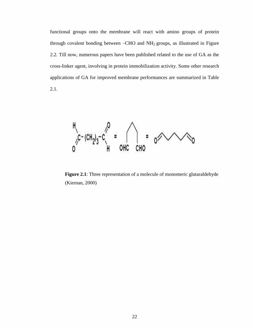
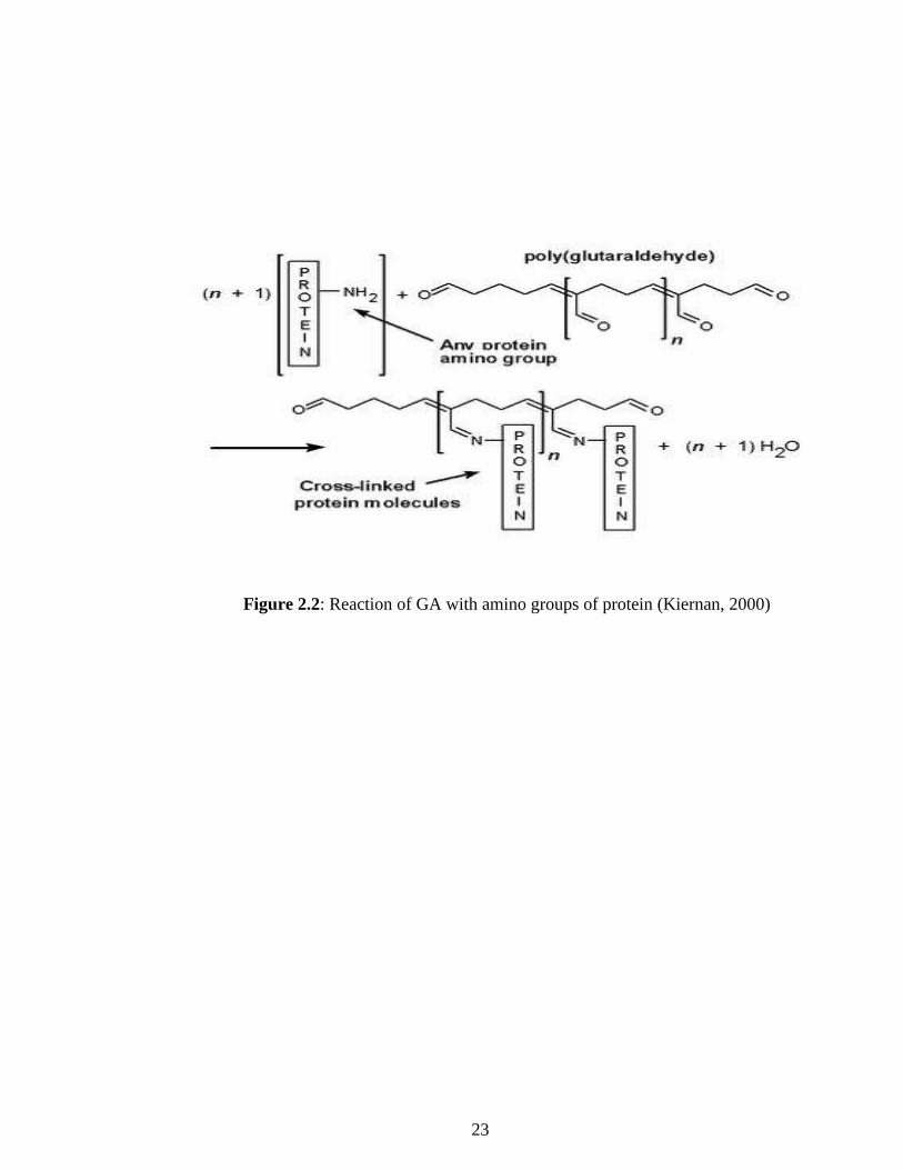
Figure 2.1 shows the three representation of a molecule of monomeric GA
structure that shows the bi-functional aldehyde groups. The deposited –CHO
22
functional groups onto the membrane will react with amino groups of protein
through covalent bonding between –CHO and NH2 groups, as illustrated in Figure
2.2. Till now, numerous papers have been published related to the use of GA as the
cross-linker agent, involving in protein immobilization activity. Some other research
applications of GA for improved membrane performances are summarized in Table
2.1.
Figure 2.1: Three representation of a molecule of monomeric glutaraldehyde
(Kiernan, 2000)
23
Figure 2.2: Reaction of GA with amino groups of protein (Kiernan, 2000)

24
Table 2.1: Importance of the glutaraldehyde as cross-linker for improved membrane performances
References Description Advantages of cross-linking
Pedram et al.
(2013)
Cross-linking of diethanolamine (DEA)
impregnated poly(vinyl alcohol) (PVA) on
polytetrafluoroethylene (PTFE) by
glutaraldehyde (GA). The membranes were
evaluated for CO2/CH4 separation.
(1) The glass transition temperature for cross-linking of GA with PVA
was dramatically increased.
(2) The homogeneous pattern was created and no macro-voids were
observed.
(3) The highest selectivity was achieved in PVA/ GA (1 wt. %)/PTFE
membrane.
Jin et al.
(2013)
Improvement of the mechanical strength of
composite hydrogels with constant soy
protein and montmorillonite (MMT)
concentrations by studying cross-linking
variables of GA concentration, pH, and
temperature.
(1) Can be beneficial for preparing bio nanocomposite materials with
enhanced mechanical properties.