PAB: a newly designed potentiometric alternating biosensor system
Upload
independentCategory
view
0download
0
A
cmg0ssar
aabw©
K
1
yfsdmdyt(fa
0d
Analytica Chimica Acta 582 (2007) 329–334
Development of a biosensor for caffeine
V.R. Sarath Babu a, S. Patra a, N.G. Karanth a, M.A. Kumar b, M.S. Thakur a,∗a Fermentation Technology and Bioengineering Department, Central Food Technological Research Institute, Mysore 570020, India
b Central Instrument facilities and Services Department, Central Food Technological Research Institute, Mysore, India
Received 14 July 2006; received in revised form 7 September 2006; accepted 9 September 2006Available online 27 September 2006
bstract
We have utilized a microbe, which can degrade caffeine to develop an Amperometric biosensor for determination of caffeine in solutions. Wholeells of Pseudomonas alcaligenes MTCC 5264 having the capability to degrade caffeine were immobilized on a cellophane membrane with aolecular weight cut off (MWCO) of 3000–6000 by covalent crosslinking method using glutaraledhyde as the bifunctional crosslinking agent and
elatin as the protein based stabilizing agent (PBSA). The biosensor system was able to detect caffeine in solution over a concentration range of.1 to 1 mg mL−1. With read-times as short as 3 min, this caffeine biosensor acts as a rapid analysis system for caffeine in solutions. Interestingly,uccessful isolation and immobilization of caffeine degrading bacteria for the analysis of caffeine described here was enabled by a novel selectiontrategy that incorporated isolation of caffeine degrading bacteria capable of utilizing caffeine as the sole source of carbon and nitrogen from soilsnd induction of caffeine degrading capacity in bacteria for the development of the biosensor. This biosensor is highly specific for caffeine andesponse to interfering compounds such as theophylline, theobromine, paraxanthine, other methyl xanthines and sugars was found to be negligible.
Although a few biosensing methods for caffeine are reported, they have limitations in application for commercial samples. The development andpplication of new caffeine detection methods remains an active area of investigation, particularly in food and clinical chemistry. The optimum pH
nd temperature of measurement were 6.8 and 30 ± 2 ◦C, respectively. Interference in analysis of caffeine due to different substrates was observedut was not considerable. Caffeine content of commercial samples of instant tea and coffee was analyzed by the biosensor and the results comparedell with HPLC analysis.2006 Elsevier B.V. All rights reserved.senso
me
hcidcw
a
eywords: Caffeine; Biosensor; Amperometric; Immobilization; Microbial bio
. Introduction
Demand for biosensors has increased markedly in recentears, driven by needs in many commercial and research sectorsor specific sensors that are capable of rapid, reliable mea-urements [1]. Development of biosensors is of interest foriverse applications ranging from biochemical profiling of nor-al and diseased cells (metabolomics), clinical diagnostics,
rug discovery and biodefense, to more straightforward anal-ses such as fermentation, process monitoring, environmentalesting and quality control of foods and beverages. Caffeine
1,3,7-trimethylxanthine) is a natural alkaloid occurring in cof-ee, cocoa beans, cola nuts and tea leaves. It is mildly stimulatingnd is used as a therapeutic agent [2]. It is a white compound,∗ Corresponding author. Tel.: +91 821 251 5792; fax: +91 821 251 7233.E-mail address: [email protected] (M.S. Thakur).
qecm
([
003-2670/$ – see front matter © 2006 Elsevier B.V. All rights reserved.oi:10.1016/j.aca.2006.09.017
r; High-performance liquid chromatography
oderately soluble in water and organic solvents like ethanol,thyl acetate, methanol, benzene, etc.
While being a stimulant to the central nervous system, it canave some adverse effects on health. If consumed in excess itan cause adverse mutation effects [3,4]. It is teratogenic, causesnhibition of DNA repair [5], inhibition of cyclic AMP phospho-iesterase activity [5] and inhibits seed germination. It can be aause of cancer, heart diseases [6] and complications in pregnantomen and aging [7].Coffee is one of the most popular beverages across the world
nd its caffeine content has an important role in determining theuality of coffee beverages. Further, on account of the harmfulffects of caffeine its efficient measurement is relevant. In thisontext, the development of a sensitive, rapid and cost effective
ethod for monitoring caffeine is greatly needed.Conventionally, high-performance liquid chromatographyHPLC) separation [8] and UV-spectrophotometric detection9], methods are applied to both regular and decaffeinated

3 Chimi
gntpaaHafttHtesfd[tldagfcsfutbs
1d
stddlsccc
C
2
afndM
(As
lwime(b
2
bim
h9Tcfmbo3wf
2
The biosensor comprises the biological sensing element,the transducer, amplification and detector systems as shown inFig. 1, which was earlier reported [19].
30 V.R.S. Babu et al. / Analytica
reen and roasted coffee beans for caffeine content determi-ations. Other methods such as capillary electrophoresis [10],hin layer chromatography (TLC) [8] and gas chromatogra-hy (GC) [11], are used for separation of caffeine in thenalysis of mixtures, combined with detection methods suchs mass spectroscopy [11] and FTIR spectrophotometry [12].owever, expensive instrumentation, highly skilled technicians
nd complicated and time-consuming procedures are requiredor such methods. Another possible technique is flow injec-ion immunoassay using a solid phase reactor, which makeshe assay faster because no separation step is needed [13].owever, the time and cost for monoclonal antibody produc-
ion and purification, and the need for their manipulation withxtreme care, are the disadvantages of this approach. A biosen-or based on inhibition of 3,5-cyclic phosphodiesterase (CPDE)rom bovine heart in combination with a pH electrode for theetection of caffeine in coffee was reported by Pizzariello et al.14]. The development and application of new caffeine detec-ion methods remains an active area of investigation, particu-arly in food and clinical chemistry. Significant research andevelopment activity has been devoted to preparing compactnalytical devices comprising a bioactive sensing element inte-rated with a suitable transducing system, known as biosensors,or determination of various inorganic, organic and biologi-al substances. The main advantages of these devices are theirpecificity, sensitivity and ease of sample preparation, and theact that no other reagents besides a buffer and a standard aresually required [15]. With these advantages in view, investiga-ions have been carried out in this work to develop a microbialiosensor for the estimation of caffeine in food and beverageamples.
.1. Principle of microbial based biosensor for theetection of caffeine
Oxidase enzymes utilize molecular oxygen for oxidation ofubstrate. This oxygen consumption can be monitored whenhese enzymatic reactions are brought about in the vicinity of aissolved oxygen electrode. In microorganisms, the enzymaticegradation of caffeine is brought about by sequential demethy-ation by an oxygenase, into theobromine or paraxanthine. Atoichiometric relation exists between the amount of caffeineonverted by the microorganisms and the amount of oxygenonsumed based on which, the amount of caffeine in the samplean be determined.
affeine + O2Enzyme−→ Product
. Materials and methods
Caffeine, theophylline, theobromine, gelatin, glutaraldehydend para chloro mercuri benzoic acid (Ultra Pure) were obtained
rom Sigma Chemicals, St. Louis, U.S.A. Analytical grade, di-itro phenyl hydrazine, polyvinyl alcohol and polyvinyl pyrroli-one were procured from Sisco Research Laboratory Chemicals,umbai, India. Dehydrated nutrient agar and nutrient brothca Acta 582 (2007) 329–334
AR) were obtained from M/s Himedia Labs, Mumbai, India.ll other chemicals were of high purity and were procured from
tandard sources.A microbe previously isolated and characterized in our
aboratory as Pseudomonas alcaligenes MTCC 5264 [16],hich was found to have potent caffeine degradation capabil-
ty was used for the studies. The isolate was cultivated in aodified nutrient broth containing peptone (1.5 g L−1), beef
xtract (5 g L−1), yeast extract (1.5 g L−1), sodium chloride5 g L−1) and caffeine (0.3 g L−1) adjusted to pH 7.2 to obtainiomass.
.1. Induction of the organism for caffeine degradation
The caffeine degrading enzyme system has been reported toe highly inducible [17]. The induction was effected by harvest-ng the biomass and incubating in M-9 medium [18], which was
odified by incorporating 0.1% (w/v) caffeine.The microbial cells accumulated in the nutrient broth were
arvested towards the end of logarithmic phase of growth (about0 h) by centrifugation at 16,000 × g and 4 ◦C for 30 min.he biomass thus obtained as a pellet was washed thrice withold Phosphate buffer (100 mM), pH 7.2 and aseptically trans-erred into a 500-mL flask containing 100 mL of caffeine liquidedium (CLM) containing 1 g L−1 of caffeine. This was incu-
ated at 30 ◦C in an orbital rotary shaker (150-rpm) for a periodf 48 h. The cells were then harvested by centrifugation for0 min at 16,000 × g and 4 ◦C. The pellet of microbial cells wasashed 4–5 times with phosphate buffer and stored at 4 ◦C until
urther use.
.2. Construction of the caffeine biosensor
Fig. 1. Schematic diagram of the whole cell electrode used for the biosensor.

Chimi
2
bas
(b
t(ichcbmaf
owarb
2
ctsmibacbdotcpc
2s
t(oocc
2c
acbobtD
sXb3
2
ftogowp
2
2
somo
2
df(r
2
at
2i
V.R.S. Babu et al. / Analytica
.3. Immobilization of whole cells
Immobilization of microbial cells was done with three immo-ilizing agents namely gelatin [19], polyvinyl alcohol (PVA)nd polyvinyl pyrrolidone (PVP) [21], using the followingteps.
The harvested microbial cells were washed 4–5 times. A 10%w/v) suspension of the induced cells was prepared in phosphateuffer (pH 6.8).
Immobilization of the cells using gelatin was done by pipet-ing 50 �L of cell suspension onto a cellophane membrane2 cm × 2 cm) and 50 �L of a 10% (w/v) of gelatin and mix-ng well with a fine glass rod by avoiding air bubbles. To thisell matrix mixture 25 �L of 5% aqueous solution of glutaralde-yde was added and mixed well. The cells were allowed torosslink with the immobilizing agent (gelatin) on the membraney incubation at room temperature for 1 h. The immobilized cellembranes were then washed thoroughly with distilled water
nd phosphate buffer (100 mM, pH 6.8) and stored at 4 ◦C tillurther use.
For immobilization of the cells using PVP and PVA, 50 �Lf the cell suspension and 100 �L of PVA and PVP, respectively,ere added to the membrane and mixed with a glass rod to formfine layer of the cells. The membranes were allowed to set at
oom temperature for 1 h. These membranes were preserved inuffer at 4 ◦C till further use.
.4. The biological sensing element
In order to construct the biosensor for caffeine, immobilizedells having caffeine degrading activity sandwiched betweenwo semi permeable membranes were fixed on to the electrodeurface by using an ‘O’ ring shown in Fig. 1 [19]. Gas per-eable teflon membrane (WTW, Germany) was used as the
nner membrane in the sensor element and cellophane mem-rane (3000–6000 MWCO; Gambro, Lund, Sweden) was useds the outer membrane. Electrode poisoning due to electro-hemically interfering compounds like metal ions and ascor-ic acid and metal chelating agents like citric acid, is avoideduring real sample analysis due to the selective permeabilityf the teflon membrane to gases only. A 10 mL glass con-ainer with 2.5 mL phosphate buffer was used as the sampleell. Air was continuously bubbled using a simple aquariumump to keep the contents mixed as well as oxygen suppliedontinuously.
.5. Construction of the transducer-amplifier-detectorystem
In order to apply the potential to the electrode and to processhe signal from the electrode, a Clarke type dissolved oxygenDO) sensor (EDT Instruments, UK) was used. The reduction
f oxygen at the cathode due to the biochemical reaction gives anutput voltage/current, which is proportional to the oxygen con-entration in solution, which can be correlated with the analyteoncentration.b0dt
ca Acta 582 (2007) 329–334 331
.6. Measurement of response by immobilized microbialells for caffeine
Microbial (immobilized) cells having caffeine degradingctivity were taken for construction of microbial biosensor foraffeine analysis in tea and coffee samples. Immobilized mem-rane was tied on the tip of the DO probe and bubbled continu-usly. Initially, the electrode kept in the sample-cell containinguffer was polarized for 1 h. Then 100 �L of the sample con-aining caffeine (0.1%, w/v) was injected and the decrease inO with time was recorded.The response of the DO electrode as decrease of DO at steady
tate was plotted against the caffeine concentration (%, w/v) on-axis and electrode response (� %DO) on Y-axis. The immo-ilized cell membranes retained 50% of the initial activity after0 days when stored at 4 ◦C.
.7. Response of xanthine oxidase to caffeine
The enzyme involved in the first step of degradation of caf-eine has not been characterized, but reports are available onhe further degradation of caffeine to xanthine, by xanthinexidase, uricase, allantiocase, allantoinase, glyoxylate dehydro-enase and urease [5]. To check for the interference of xanthinexidase activity (XOD) in the determination of caffeine, XODas immobilized and its response to caffeine, theobromine andaraxanthine (0.1%, w/v) was recorded.
.8. Optimization of parameters for the biosensor
.8.1. pHEffect of pH on the activity of the immobilized cells was
tudied by incubating the membrane in buffers in the pH rangef 5.0–8.0 with an increment of 0.5 pH units and response of theembrane to 1% (w/v) of caffeine was recorded as depletion of
xygen.
.8.2. TemperatureThe optimum temperature for the caffeine response was
etermined by incubating the immobilized membranes at dif-erent temperatures (32, 35, 40, 45 and 50 ◦C) in shaking waterJulabo bath) and the response to 1% (w/v) of caffeine wasecorded.
.8.3. Interference of different sugars in caffeine estimationThe response of immobilized (P. alcaligenes) to different sug-
rs viz. sucrose, glucose and fructose was studied by injectinghe samples (1%, w/v) and the response was recorded.
.8.4. Studies on inhibition of glucose uptake by themmobilized cells
Three known glucose uptake inhibitors viz., P-chloromercuri
enzoate, sodium bisulphate and Dinitrophenyl hydrazine at.1% (w/v) concentration were injected into the sample celluring analysis of caffeine and the response of the biosensoro caffeine was recorded.
3 Chimica Acta 582 (2007) 329–334
2
o
c10tosa
c10tpp
2
1to(Stc
2c
eoNwoi
stp
E
3
3
3
pbaiiw
Table 1Response of the biosensor using different immobilizing agents
Stabilizing agent %DO depletion shown bythe biosensor after 3 min(1%, w/v)
No. of analyses withone sensor element
Gelatin 1.7 1PP
ic
3
s
tcf
b
y
x
wc
tcp
c
y
This can be rewritten as
x = y−20.323
4453(4)
32 V.R.S. Babu et al. / Analytica
.8.5. Calibration for caffeine using HPLC and biosensorCalibration graphs for caffeine were plotted for the analysis
f caffeine in samples by the biosensor and by HPLC.Calibration graph for caffeine analysis by the biosensor in the
oncentration range of 0.01–0.1% (w/v) was plotted by injecting00 �L of standard caffeine solution at concentrations of 0.01,.02, 0.04, 0.06, 0.08 and 0.1% (w/v) of caffeine and recordinghe decrease in dissolved oxygen (%DO drop) concentrationf the sample. A graph of concentration versus decrease in dis-olved oxygen was plotted with caffeine concentration on X-axisnd %DO drop on Y-axis.
Calibration graph for caffeine analysis by HPLC in the con-entration range of 0.01–0.1% (w/v) was plotted by injecting0 �L of standard caffeine solution at concentrations of 0.01,.02, 0.04, 0.06, 0.08 and 0.1% (w/v) of caffeine and recordinghe peak area of the sample. A graph of concentration versuseak area was plotted with caffeine concentration on X-axis andeak area on Y-axis.
.8.6. Analysis of caffeine in real samples by HPLCHPLC analysis of caffeine was done using a Shimadzu-LC
0A fitted with a C18 Column (300 mm × 4.6 mm) with ace-onitrile and water (10:90%) as mobile phase set at a flow ratef 1 mL min−1 and a UV detector set at 273 nm. Real samplescoffee and tea sample) were analyzed for their caffeine content.amples of tea and coffee were prepared as described below and
he concentration was determined by recording the peak area andomparing with the calibration graph.
.8.7. Preparation of real samples for determination ofaffeine
Instant coffee and tea were prepared by dissolving 0.5 g ofach in 125 mL boiling water [20] and were kept for 30 minn a magnetic stirrer and filtered using a Whatman filter Papero.1. A 100 �L of the sample was then injected and the responseas recorded. The commercial cola samples were injected with-ut any treatment. For HPLC analysis 10 �L of the sample wasnjected and the peak area was recorded.
In order to reduce the interference of glucose present in theample, the sample was passed through a packed column con-aining glucose oxidase enzyme immobilized on glass beadsrior to injection to the biosensor.
The caffeine content in the samples was calculated using theqs. (2) and (4) for biosensor and HPLC, respectively.
. Results and discussion
.1. Construction of caffeine biosensor
.1.1. Immobilization of whole cellsFor immobilization of cells, different agents viz. gelatin,
olyvinyl pyrrolidone and polyvinyl alcohol were tried. Theiosensor gave good response for caffeine (1%, w/v) showing
steady state depletion of 53.2% DO concentration as shownn Table 1. It was observed that polyvinyl alcohol is a bettermmobilizing agent in terms of a higher response and reusability,hich may be due to a higher oxygen permeability and diffusiv-
olyvinyl pyrrolidone 1.6 2olyvinyl alcohol 53.2 15
ty [21]. With PVA as stabilizing agent, about 15–25 analysesould be done with a single membrane.
.1.2. Response of biosensor to caffeineThe response of the biosensor to 1% (w/v) of caffeine is
hown in Fig. 2.For the determination of caffeine content in the samples using
he immobilized cells based biosensor, a calibration graph foraffeine in the concentration range of 0.01–0.1% (w/v) of caf-eine was plotted with a regression value of 0.992.
The concentration of caffeine in the sample can be calculatedy using Eq. (2)
= 189.53x + 6.1134 (1)
This can be rewritten as
= y − 6.1134
189.53(2)
here ‘y’ is the % dissolved oxygen consumed and ‘x’ is theoncentration of caffeine in the sample.
For the comparison of the results of analysis of caffeine byhe biosensor with a standard method, a calibration graph foraffeine in the concentration range of 0.01–0.1% (w/v) was alsolotted with a regression value of 0.996 using HPLC.
The concentration of caffeine present in the sample can bealculated by using Eq. (4) derived from the calibration curve:
= 4453x − 20.323 (3)
Fig. 2. Response of the biosensor to 1% (w/v) of pure caffeine.
V.R.S. Babu et al. / Analytica Chimica Acta 582 (2007) 329–334 333
wc
3
s
(umbicm(
fbac
Table 2Effect of interfering agents on caffeine biosensor response
Serial no. Interfering agent (1%, w/v) Response (%DO drop)
1 Theophylline 2.002 Theobromine 3.503 Sucrose 4.004 Glucose 5.005 Fructose 0.206 Glycine 1.207 Tyrosine 0.608 Histidine 1.109 Ascorbic acid 1.50
10 Catechin 1.20
*Response to 1.0% (w/v) caffeine = 20.6.
Ft
3e
fiio
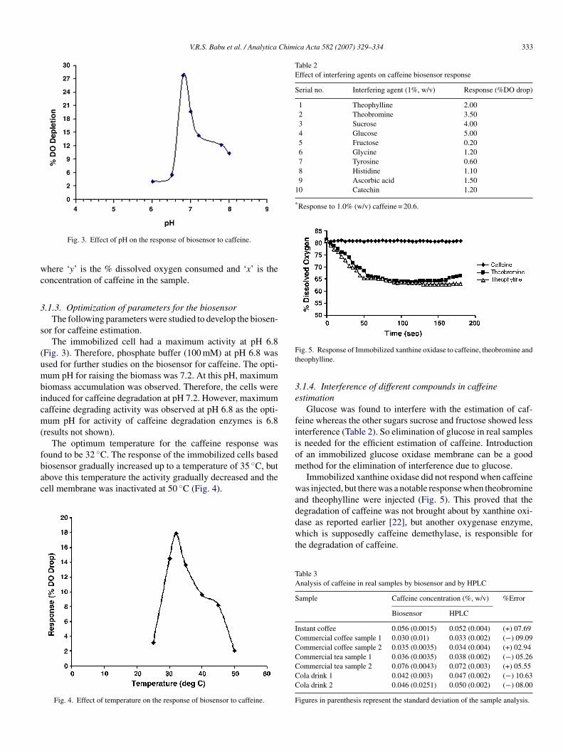
Fig. 3. Effect of pH on the response of biosensor to caffeine.
here ‘y’ is the % dissolved oxygen consumed and ‘x’ is theoncentration of caffeine in the sample.
.1.3. Optimization of parameters for the biosensorThe following parameters were studied to develop the biosen-
or for caffeine estimation.The immobilized cell had a maximum activity at pH 6.8
Fig. 3). Therefore, phosphate buffer (100 mM) at pH 6.8 wassed for further studies on the biosensor for caffeine. The opti-um pH for raising the biomass was 7.2. At this pH, maximum
iomass accumulation was observed. Therefore, the cells werenduced for caffeine degradation at pH 7.2. However, maximumaffeine degrading activity was observed at pH 6.8 as the opti-um pH for activity of caffeine degradation enzymes is 6.8
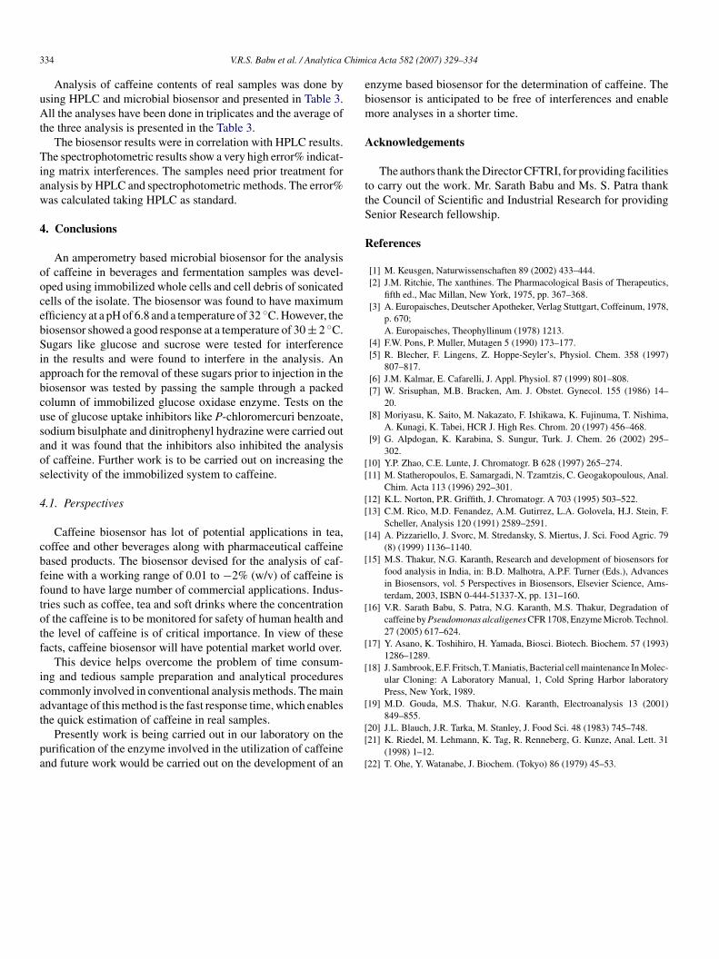
results not shown).The optimum temperature for the caffeine response was
ound to be 32 ◦C. The response of the immobilized cells based
iosensor gradually increased up to a temperature of 35 ◦C, butbove this temperature the activity gradually decreased and theell membrane was inactivated at 50 ◦C (Fig. 4).Fig. 4. Effect of temperature on the response of biosensor to caffeine.
m
waddwt
TA
S
ICCCCCC
F
ig. 5. Response of Immobilized xanthine oxidase to caffeine, theobromine andheophylline.
.1.4. Interference of different compounds in caffeinestimation
Glucose was found to interfere with the estimation of caf-eine whereas the other sugars sucrose and fructose showed lessnterference (Table 2). So elimination of glucose in real sampless needed for the efficient estimation of caffeine. Introductionf an immobilized glucose oxidase membrane can be a goodethod for the elimination of interference due to glucose.Immobilized xanthine oxidase did not respond when caffeine
as injected, but there was a notable response when theobrominend theophylline were injected (Fig. 5). This proved that theegradation of caffeine was not brought about by xanthine oxi-
ase as reported earlier [22], but another oxygenase enzyme,hich is supposedly caffeine demethylase, is responsible forhe degradation of caffeine.
able 3nalysis of caffeine in real samples by biosensor and by HPLC
ample Caffeine concentration (%, w/v) %Error
Biosensor HPLC
nstant coffee 0.056 (0.0015) 0.052 (0.004) (+) 07.69ommercial coffee sample 1 0.030 (0.01) 0.033 (0.002) (−) 09.09ommercial coffee sample 2 0.035 (0.0035) 0.034 (0.004) (+) 02.94ommercial tea sample 1 0.036 (0.0035) 0.038 (0.002) (−) 05.26ommercial tea sample 2 0.076 (0.0043) 0.072 (0.003) (+) 05.55ola drink 1 0.042 (0.003) 0.047 (0.002) (−) 10.63ola drink 2 0.046 (0.0251) 0.050 (0.002) (−) 08.00
igures in parenthesis represent the standard deviation of the sample analysis.
3 Chimi
uAt
Tiaw
4
oocebSiabcusaos
4
cbfftotf
icat
pa
ebm
A
ttS
R
[[
[[
[
[
[
[
[
[
34 V.R.S. Babu et al. / Analytica
Analysis of caffeine contents of real samples was done bysing HPLC and microbial biosensor and presented in Table 3.ll the analyses have been done in triplicates and the average of
he three analysis is presented in the Table 3.The biosensor results were in correlation with HPLC results.
he spectrophotometric results show a very high error% indicat-ng matrix interferences. The samples need prior treatment fornalysis by HPLC and spectrophotometric methods. The error%as calculated taking HPLC as standard.
. Conclusions
An amperometry based microbial biosensor for the analysisf caffeine in beverages and fermentation samples was devel-ped using immobilized whole cells and cell debris of sonicatedells of the isolate. The biosensor was found to have maximumfficiency at a pH of 6.8 and a temperature of 32 ◦C. However, theiosensor showed a good response at a temperature of 30 ± 2 ◦C.ugars like glucose and sucrose were tested for interference
n the results and were found to interfere in the analysis. Anpproach for the removal of these sugars prior to injection in theiosensor was tested by passing the sample through a packedolumn of immobilized glucose oxidase enzyme. Tests on these of glucose uptake inhibitors like P-chloromercuri benzoate,odium bisulphate and dinitrophenyl hydrazine were carried outnd it was found that the inhibitors also inhibited the analysisf caffeine. Further work is to be carried out on increasing theelectivity of the immobilized system to caffeine.
.1. Perspectives
Caffeine biosensor has lot of potential applications in tea,offee and other beverages along with pharmaceutical caffeineased products. The biosensor devised for the analysis of caf-eine with a working range of 0.01 to −2% (w/v) of caffeine isound to have large number of commercial applications. Indus-ries such as coffee, tea and soft drinks where the concentrationf the caffeine is to be monitored for safety of human health andhe level of caffeine is of critical importance. In view of theseacts, caffeine biosensor will have potential market world over.
This device helps overcome the problem of time consum-ng and tedious sample preparation and analytical proceduresommonly involved in conventional analysis methods. The maindvantage of this method is the fast response time, which enables
he quick estimation of caffeine in real samples.Presently work is being carried out in our laboratory on theurification of the enzyme involved in the utilization of caffeinend future work would be carried out on the development of an
[[
[
ca Acta 582 (2007) 329–334
nzyme based biosensor for the determination of caffeine. Theiosensor is anticipated to be free of interferences and enableore analyses in a shorter time.
cknowledgements
The authors thank the Director CFTRI, for providing facilitieso carry out the work. Mr. Sarath Babu and Ms. S. Patra thankhe Council of Scientific and Industrial Research for providingenior Research fellowship.
eferences
[1] M. Keusgen, Naturwissenschaften 89 (2002) 433–444.[2] J.M. Ritchie, The xanthines. The Pharmacological Basis of Therapeutics,
fifth ed., Mac Millan, New York, 1975, pp. 367–368.[3] A. Europaisches, Deutscher Apotheker, Verlag Stuttgart, Coffeinum, 1978,
p. 670;A. Europaisches, Theophyllinum (1978) 1213.
[4] F.W. Pons, P. Muller, Mutagen 5 (1990) 173–177.[5] R. Blecher, F. Lingens, Z. Hoppe-Seyler’s, Physiol. Chem. 358 (1997)
807–817.[6] J.M. Kalmar, E. Cafarelli, J. Appl. Physiol. 87 (1999) 801–808.[7] W. Srisuphan, M.B. Bracken, Am. J. Obstet. Gynecol. 155 (1986) 14–
20.[8] Moriyasu, K. Saito, M. Nakazato, F. Ishikawa, K. Fujinuma, T. Nishima,
A. Kunagi, K. Tabei, HCR J. High Res. Chrom. 20 (1997) 456–468.[9] G. Alpdogan, K. Karabina, S. Sungur, Turk. J. Chem. 26 (2002) 295–
302.10] Y.P. Zhao, C.E. Lunte, J. Chromatogr. B 628 (1997) 265–274.11] M. Statheropoulos, E. Samargadi, N. Tzamtzis, C. Geogakopoulous, Anal.
Chim. Acta 113 (1996) 292–301.12] K.L. Norton, P.R. Griffith, J. Chromatogr. A 703 (1995) 503–522.13] C.M. Rico, M.D. Fenandez, A.M. Gutirrez, L.A. Golovela, H.J. Stein, F.
Scheller, Analysis 120 (1991) 2589–2591.14] A. Pizzariello, J. Svorc, M. Stredansky, S. Miertus, J. Sci. Food Agric. 79
(8) (1999) 1136–1140.15] M.S. Thakur, N.G. Karanth, Research and development of biosensors for
food analysis in India, in: B.D. Malhotra, A.P.F. Turner (Eds.), Advancesin Biosensors, vol. 5 Perspectives in Biosensors, Elsevier Science, Ams-terdam, 2003, ISBN 0-444-51337-X, pp. 131–160.
16] V.R. Sarath Babu, S. Patra, N.G. Karanth, M.S. Thakur, Degradation ofcaffeine by Pseudomonas alcaligenes CFR 1708, Enzyme Microb. Technol.27 (2005) 617–624.
17] Y. Asano, K. Toshihiro, H. Yamada, Biosci. Biotech. Biochem. 57 (1993)1286–1289.
18] J. Sambrook, E.F. Fritsch, T. Maniatis, Bacterial cell maintenance In Molec-ular Cloning: A Laboratory Manual, 1, Cold Spring Harbor laboratoryPress, New York, 1989.
19] M.D. Gouda, M.S. Thakur, N.G. Karanth, Electroanalysis 13 (2001)
849–855.20] J.L. Blauch, J.R. Tarka, M. Stanley, J. Food Sci. 48 (1983) 745–748.21] K. Riedel, M. Lehmann, K. Tag, R. Renneberg, G. Kunze, Anal. Lett. 31
(1998) 1–12.22] T. Ohe, Y. Watanabe, J. Biochem. (Tokyo) 86 (1979) 45–53.
Copyright © 2022 FDOKUMEN