Caffeine and Chlorogenic Acid Combination Attenuate Early ...
Upload
khangminh22Category
view
1download
0
ARTICLE
Caffeine-inducible gene switches controllingexperimental diabetesDaniel Bojar 1, Leo Scheller 1, Ghislaine Charpin-El Hamri2, Mingqi Xie1 & Martin Fussenegger 1,3
Programming cellular behavior using trigger-inducible gene switches is integral to synthetic
biology. Although significant progress has been achieved in trigger-induced transgene
expression, side-effect-free remote control of transgenes continues to challenge cell-based
therapies. Here, utilizing a caffeine-binding single-domain antibody we establish a caffeine-
inducible protein dimerization system, enabling synthetic transcription factors and cell-
surface receptors that enable transgene expression in response to physiologically relevant
concentrations of caffeine generated by routine intake of beverages such as tea and coffee.
Coffee containing different caffeine concentrations dose-dependently and reversibly
controlled transgene expression by designer cells with this caffeine-stimulated advanced
regulators (C-STAR) system. Type-2 diabetic mice implanted with microencapsulated,
C-STAR-equipped cells for caffeine-sensitive expression of glucagon-like peptide 1 showed
substantially improved glucose homeostasis after coffee consumption compared to untreated
mice. Biopharmaceutical production control by caffeine, which is non-toxic, inexpensive and
only present in specific beverages, is expected to improve patient compliance by integrating
therapy with lifestyle.
DOI: 10.1038/s41467-018-04744-1 OPEN
1 Department of Biosystems Science and Engineering, ETH Zurich, Mattenstrasse 26, 4058 Basel, Switzerland. 2 IUT, Département Génie Biologique, InstitutUniversitaire de Technologie, F-69622 Villeurbanne Cedex, France. 3 Faculty of Life Science, University of Basel, Mattenstrasse 26, CH-4058 Basel,Switzerland. Correspondence and requests for materials should be addressed to M.F. (email: [email protected])
NATURE COMMUNICATIONS | (2018) 9:2318 | DOI: 10.1038/s41467-018-04744-1 |www.nature.com/naturecommunications 1
1234
5678
90():,;

In recent years, synthetic biology, the fusion between engi-neering and biology1, has brought the rational and predictableconstruction of sophisticated gene circuits into the forefront of
biomedical research. Plug-and-play combinations of carefullydesigned biological modules have enabled major advances intherapies for personalized medicine2,3, as well as in the challen-ging endeavor of lineage control4,5, bringing achievements in thelaboratory ever closer to rewarding real-world applications6,7. Inthis context, cell-based therapies capitalizing on the complexity ofmammalian cells are taking the lead in the advent of personalizedmedicine8, as exemplified by applications of chimeric antigenreceptor (CAR)—T cells9 or designer cell implants to treat var-ious common diseases2,10.
Controlling the dynamics of gene expression is essential for thefunctionality of synthetic gene circuits. This is especially relevantin synthetic biology-inspired therapies, where gene expressionregulation determines the dosage of the produced therapeutic andallows for considerable control over the designer cell implant.Therefore, gene expression in most circuits is controlled either atthe transcriptional or translational level. At the transcriptionallevel, promoters responsive to specific triggers are controlled bytranscription factors11, whereas at the translational level, ribo-zymes or riboswitches responsive to specific triggers controlprotein translation12. In recent years, the quest for better inducershas progressed rapidly. Initially, antibiotics such as tetracycline ordoxycycline13 were used for the control of gene expression,raising issues such as antibiotic resistance14 and side effects15.The next generation of inducers were designed to be safe andorthogonal, such as the apple tree leaf metabolite phloretin16 orthe food additives benzoate and vanillic acid17. However, theseinducers still suffer from potential side effects, especially in long-term applications, and have to be exogenously added. Tracelessinducers, such as light18 or temperature19, have recently beendeveloped, but ambient light and ambient temperature makethem less orthogonal than would be desirable. The ideal inducerwould be inexpensive, would have no side effects, and would bepresent in only a specific set of known sources.
Here, we report a bioengineering approach for the induction ofgene expression in mammalian designer cells by caffeine. Thesmall molecule caffeine is non-toxic20, cheap to produce21, andonly present in specific beverages, such as coffee and tea. Everyday, more than two billion cups of coffee are being consumedworldwide, making coffee one of the most popular beverages afterwater, and one of the most important cash crops in the world22.Currently available caffeine-responsive gene switches requireenzymatic conversion of caffeine to theophylline to providetranslation control in yeast23. However, due to its low sensitivitythe yeast system is unsuitable for sensing physiologically relevantcaffeine concentrations in mammalian cells. To engineer fullysynthetic caffeine-inducible gene switches with user-definedsensitivity and functionality, we established a caffeine-mediatedprotein dimerization system in mammalian cells based on asingle-domain VHH camelid antibody (referred to as aCaffVHH)that has high affinity (Kd= 500 nM) and homodimerizes in thepresence of caffeine24–26. By fusing aCaffVHH to the intracellularsignaling domains of different mammalian receptor classes, wecreated fully synthetic receptors that sense caffeine at physiolo-gically relevant levels (e.g., the amount provided in a cup ofcoffee). The robustness of these caffeine receptors, which we callC-STAR (caffeine-stimulated advanced regulators), is demon-strated in vitro with pure caffeine and with a diverse array ofeveryday sources of caffeine, such as black tea, coffee, and energydrinks, as well as in vivo in two mouse models of experimentalType-2 diabetes.
Type-2 diabetes mellitus (T2D) affects more than 400 millionpeople worldwide27 and associated health costs amount to about
825 billion US dollars per year28. As T2D is characterized by asustained increase in blood glucose levels after each meal, wewanted to capture the natural dynamics of caffeine uptake aftereach major meal to achieve a novel therapeutic approach to theacute phase of T2D by using designer cells equipped with C-STAR to deliver synthetic human glucagon-like peptide 1(shGLP-1) in response to dietary intake of coffee or othercaffeine-containing beverages. Capitalizing on routine culturalhabits, therapies based on such systems should seamlessly inte-grate into people’s lifestyle, and therefore could be a key pillarupon which the new generation of personalized medicine canbuild.
ResultsDesign of a caffeine-inducible gene switch. After drinking anaverage cup of coffee, blood levels of caffeine peak in the lowmicromolar range29,30, so for the present purpose, we required anovel caffeine sensor system for non-toxic (SupplementaryFig. 1), physiologically relevant concentrations. To capture theseconcentrations, we established a caffeine-inducible proteindimerization system in mammalian cells to create different typesof gene switches. (i) Fusion of the caffeine-binding single-domainantibody aCaffVHH to DNA-binding and transactivationdomains reconstitutes synthetic transcription factors drivingchimeric target promoters in a caffeine-responsive manner. (ii)Fusion of the caffeine-binding single-domain antibodyaCaffVHH to intracellular signaling domains of different mam-malian receptor classes reconstitutes synthetic signaling cascadesand allows caffeine to dose-dependently activate differentpathway-specific promoters (Fig. 1a).
To design an aCaffVHH-dependent transcription factor-basedgene switch, we C-terminally fused aCaffVHH to the DNA-binding TetR-domain (PSV40-TetR-aCaffVHH-pASV40, pDB307),as well as N-terminally to four repeats of a transactivating12-amino-acid peptide (VPmin, PCAG-aCaffVHH-VPminx4-pAβG,pDB335). In this design, the presence of caffeine should dimerizethe DNA-binding domain with the transactivating VPmin domainand lead to gene expression (Fig. 1b). Utilizing the reporter genehuman placental-secreted alkaline phosphatase (SEAP) controlledby a TetR-dependent promoter (PtetO7-SEAP-pASV40, pMF111),we observed clear caffeine-dependent gene expression in thepresence of 100 µM caffeine (Fig. 1b).
We reasoned that this low sensitivity to caffeine might be dueto the absence of signal amplification in this split transcriptionfactor setup. Therefore, we applied the caffeine-inducibledimerization system to different signaling pathway-specific signaltransduction domains. First, we fused aCaffVHH N-terminally tothe transmembrane domain of interleukin 13 receptor subunitalpha 1 (IL13Rα1, PhCMV-aCaffVHH-IL13Rα1-pAbGH, pDB323),as well as interleukin 4 receptor subunit alpha (IL4Rα, PhCMV-aCaffVHH-IL4Rα-pAbGH, pDB324). Addition of caffeine shouldinduce heterodimerization of these receptors and activate signaltransducer and activator of transcription 6 (STAT6) signaling.Indeed, when we co-transfected STAT6 (PhCMV-STAT6-pAbGH,pLS16) and a STAT6-responsive reporter construct (PSTAT6-SEAP-pASV40, pLS12), we could see caffeine-dependent geneexpression starting from 1 µM caffeine (Fig. 1c), a considerableimprovement in sensitivity compared to the split transcriptionfactor setup using pDB307 and pDB335. However, the absoluteoutput strength of this setup in SEAP units was limited,necessitating a more powerful system.
To overcome the output strength issue, we fused aCaffVHH C-terminally to the intracellular part of the murine fibroblastgrowth factor receptor 1 (mFGFR1, PhCMV-mFGFR1405-822-aCaffVHH-pAbGH, pDB395)31. The presence of caffeine should
ARTICLE NATURE COMMUNICATIONS | DOI: 10.1038/s41467-018-04744-1
2 NATURE COMMUNICATIONS | (2018) 9:2318 | DOI: 10.1038/s41467-018-04744-1 | www.nature.com/naturecommunications

homodimerize mFGFR1405-822-aCaffVHH and lead to MAPKsignaling, which we re-routed to TetR-dependent pMF111 byco-transfecting TetR-Elk1 (PhCMV-TetR-Elk1-pAbGH, MKp37).The signal amplification of the MAPK signaling cascade32 yieldeda strong and sensitive gene expression response in the presence ofas little as 0.01 µM caffeine (Fig. 1d). However, this extraordinarysensitivity to caffeine may be detrimental in a therapeutic setting,since even trace amounts of caffeine would induce the genecircuit. Additionally, the requirement of there-routing protein TetR-Elk1 meant that transfection of threeplasmids was necessary for this system.
Improving on the mFGFR1-dependent system, we fusedaCaffVHH N-terminally to an erythropoietin receptor derivative(EpoR, PhCMV-aCaffVHH-EpoRm-IL-6RBm-pAbGH, pDB306)33,34,leading to homodimerization of the receptor in the presence ofcaffeine and subsequent JAK/STAT signaling through STAT3. AsHEK-293T cells endogenously express STAT3, we only needed totransfect pDB306 and a STAT3-dependent reporter plasmid(PSTAT3-SEAP-pASV40, pLS13). This setup yielded a strong andsensitive gene expression system with a maximal response at 1µM caffeine (Fig. 1e).
P
PSTAT3 SEAP
P
SEAP
PSTAT6 SEAP
SEAP
PtetO7 SEAP
SEAP
TetR VPmin
VPmin
VPmin
VPmin
PtetO7 SEAP
SEAP
TetR
Elk1P
PP
P
MAPKP
b c
ed
0 0.01 0.1 1 10 1000
5
10
15
20
25
µM Caffeine
SE
AP
(U
/L)
STAT6
STAT6
STAT3P
STAT3P
aCaffVHH
aCaffVHH
aCaffVHH
aCaffVHH
mFGFR1
IL4Ra IL13Ra1
EpoRm-IL6RBm
0 0.01 0.1 1 10 100
0
5
10
15
20
25
30
µM Caffeine
SE
AP
(U
/L)
0 0.01 0.1 1 10 1000
25
50
75
100
125
µM Caffeine
SE
AP
(U
/L)
0 0.01 0.1 1 10 1000
25
50
75
100
125
µM Caffeine
SE
AP
(U
/L)
a
Caffeine-induced dimerization system
Caffeine
O
O
N
N N
N
aCaffVHH
Signaltransduction
PP
P
DNA bindingdomain
Transactivatordomain
Signalingdomain A
Signalingdomain B
Transcription
PP
P
P
P
P
NATURE COMMUNICATIONS | DOI: 10.1038/s41467-018-04744-1 ARTICLE
NATURE COMMUNICATIONS | (2018) 9:2318 | DOI: 10.1038/s41467-018-04744-1 |www.nature.com/naturecommunications 3

Overall, caffeine-dependent STAT3-signaling proved to be thebest fit in terms of potency, sensitivity to physiological caffeinelevels, and number of components, and so it was used for allfurther experiments. Due to receptor homodimerization andendogenous STAT3 expression, we only needed to transfect twocomponents to obtain a full C-STAR system. Since the presentedgene expression systems had different sensitivities and relied onorthogonal promoters, they could be used for endowing designercells with a nonlinear response to caffeine by expressing multiplereceptors (Supplementary Fig. 2a, b).
Characterization of the caffeine-inducible C-STAR system.Functionality of the C-STAR system was also demonstrated inhuman telomerase reverse transcriptase-immortalized humanmesenchymal stem cells (hMSC-hTERT) (Fig. 2a). However,HEK-293T cells showing higher caffeine sensitivity and proteinsecretion capacity were used in all follow-up experiments. Forlong-term experiments, the C-STAR receptor (PhEF-1α-aCaffVHH-EpoRm-IL-6RBm-pASV40, pDB326) was stably inte-grated into the genome of HEK-293T cells, creating the designercell line C-STARDB1. The caffeine dose-response relationship ofthis polyclonal cell line was similar to that of the transientlytransfected cells (Fig. 2b). However, selection of monoclonalC-STAR cell lines yielded clones with different sensitivities forcaffeine (Supplementary Fig. 3a–d). All further in vitro experi-ments were conducted with the C-STARDB1 cell line.
To capture the time window of high caffeine concentration inthe blood, an in vivo C-STAR system would need to induce geneexpression after brief exposure to the inducer. Exposure tophysiologically relevant concentrations of caffeine induced a half-maximal response of the C-STAR system within just one hour,and a maximal response was obtained after six hours of exposure(Fig. 2c). Since the half-life of caffeine in human blood isapproximately five hours30, in vivo activation of the C-STARsystem by caffeine should be feasible. Among the caffeine analogstested in vitro, only theophylline showed modest cross-activationof C-STAR at 1 µM concentration (Supplementary Fig. 4), whichis unlikely to be reached in the physiological situation35,36. Theresponse time of the C-STAR system after caffeine addition wasassessed and the C-STAR system responded in a timely mannerto the presence of caffeine, yielding detectable amounts ofreporter protein at 12 h, whereas no induction of SEAPexpression was seen in the negative control lacking caffeine(Fig. 2d). Testing the reversibility of the gene circuit, C-STARDB1
cells were incubated with physiologically relevant concentrationsof caffeine or the equivalent amount of H2O (mock), with anexchange of caffeine to mock, or vice versa, every day (Fig. 2e). Asexpected, the system was shut off by the removal of caffeine and
could be activated again by the renewed addition of caffeine to thecells, indicating reversibility after removal or degradation ofcaffeine.
Caffeine quantification in commercial beverages usingC-STAR. Caffeine is a component of various beverages. There-fore, to broaden the range of available beverages for the inductionof the C-STAR system, and to establish the specificity of thesynthetic biology-inspired caffeine-sensing system,C-STARDB1 cells were challenged with 26 products, includingNespresso Grand Cru®, Starbucks® coffee, Red Bull®, CuidaTe® tea capsule, and Coca-Cola® (Fig. 3a). Several NespressoGrand Cru® capsules were also tested in their decaffeinated ver-sion as negative controls (Vivalto lungo decaffeinato®, Vollutodecaffeinato®, Decaffeinato intenso®, and Arpeggio decaffeinato®).As three of these beverage samples also have caffeinated versions(Vivalto lungo®, Volluto®, and Arpeggio®), which are claimed bythe manufacturer to be identical to the respective decaffeinatedversions except for the caffeine content, they allowed us to con-firm that caffeine itself upregulates gene expression and not anyother of the hundreds of chemical compounds present in coffee22.Overall, our beverage samples covered a wide range of caffeineconcentrations from 0 to 4.8 g L−1. A standarddose-response curve was obtained with pure caffeine. Thisenabled us to convert the SEAP values from C-STARDB1 cellsincubated with beverage samples into caffeine concentrations.
For all samples tested, caffeine concentrations indicated by thevendor corresponded well to those measured with C-STARDB1
cells (Fig. 3b, c). As expected, decaffeinated beverage samplesshowed very low activation of the C-STAR system (Fig. 3b, c).These results indicate that C-STAR reproducibly generates adose-dependent, caffeine-specific response.
C-STAR treatment for obesity-induced Type-2 diabetes. Thefunctionality of the designed C-STAR system in vascularizedmicrocontainers was first confirmed in vitro with pure caffeine(Supplementary Fig. 5). After validating the immunoprotectivefunction of microcapsule implants for drug delivery in vivo(Supplementary Fig. 6), mice implanted with the designer cellcapsules were given room temperature Volluto® coffee (NespressoGrand Cru®), or H2O to drink. As expected, only mice graftedwith the C-STAR system showed reversible, coffee-induced SEAPexpression (Supplementary Fig. 7a, b). The same mice were re-stimulated a few days later and showed the same response as inthe initial experiment (Supplementary Fig. 7c, d).
Next, in order to examine whether this system could be utilizedfor caffeine-induced treatment of obesity-induced T2D, wereplaced the reporter gene SEAP with the gene coding for
Fig. 1 Synthetic biology-inspired genetic circuits for caffeine-induced gene expression. a Caffeine-inducible protein dimerization system based on thecamelid-derived single-domain antibody aCaffVHH. aCaffVHH homodimerizes in the presence of caffeine and can be used to reconstitute synthetictranscription factors or signaling cascades that fine-tune caffeine-responsive gene expression. b Caffeine-sensing circuit based on the heterodimerizationof aCaffVHH-TetR (pDB307) and aCaffVHH-VPminx4 (pDB335), leading to direct transcriptional activation. The caffeine dose-response relationship wasquantified with the reporter gene SEAP (PtetO7-SEAP-pASV40, pMF111). c Caffeine-sensing circuit based on the IL13 receptor and the JAK/STAT6 pathway.Caffeine-induced heterodimerization of aCaffVHH-IL13Rα1 (pDB323) and aCaffVHH-IL4Rα (pDB324) leads to phosphorylation of STAT6 (pLS16) by JAKkinases and subsequent transcriptional activation of the STAT6-responsive promoter PSTAT6. The caffeine dose-response relationship was quantified withthe reporter gene SEAP (PSTAT6-SEAP-pASV40, pLS12). d Caffeine-sensing circuit based on the MAPK pathway. Caffeine-induced homodimerization ofmFGFR1405-822-aCaffVHH (pDB395) led to phosphorylation of MEK1/2 and downstream signaling of the MAPK cascade. Rewiring the signaling cascadethrough the hybrid transcription factor TetR-Elk1 (MKp37) led to expression of the reporter gene SEAP (PtetO7-SEAP-pASV40, pMF111), enablingquantification of the caffeine dose-response relationship. e Caffeine-sensing circuit based on the Epo receptor and the JAK/STAT3 pathway. Caffeine-induced homodimerization of aCaffVHH-EpoRm-IL-6RBm (pDB306) leads to phosphorylation of STAT3 by JAK kinases and subsequent transcriptionalactivation of the STAT3-responsive promoter PSTAT3. The caffeine dose-response relationship was quantified with the reporter gene SEAP(PSTAT3-SEAP-pASV40, pLS13). Data in (b–e) are shown as the mean in bar graphs and symbols indicate individual data points. The data displayed representthree independent experiments (n= 3)
ARTICLE NATURE COMMUNICATIONS | DOI: 10.1038/s41467-018-04744-1
4 NATURE COMMUNICATIONS | (2018) 9:2318 | DOI: 10.1038/s41467-018-04744-1 | www.nature.com/naturecommunications

synthetic human glucagon-like peptide coupled to mouse IgG(shGLP-1, PSTAT3-shGLP-1-pASV40, pDB387), an engineeredprotein clinically licensed for the treatment of T2D37. Experi-ments in vitro with the C-STARDB6 cell line incorporating theresulting construct validated the caffeine-dependent expression ofshGLP-1 (Supplementary Fig. 8a, b). Pharmacokinetic analyses ofcaffeine and shGLP-1 in mice confirmed the potential ofC-STARDB6 for cell-based diabetes therapy; a single oraladministration of coffee resulted in a transient surge of caffeinein the bloodstream38 that was sufficient to trigger sustainedshGLP-1 activity (Supplementary Fig. 9). Importantly,
hypoglycemic side effects were not observed following higherlevels of caffeine-dependent shGLP-1 production (SupplementaryFig. 10), confirming the inherent inactivity of GLP-1 innormoglycemic environments39,40. Then, we examined theefficacy of these cells in two T2D mouse models with impairedinsulin sensitivity. For this purpose, diet-induced obesity41 (DIO;Fig. 4) and leptin receptor-deficient41 (db/db; Fig. 5) mice wereimplanted intraperitoneally with capsules containing C-STARDB6
cells or with control capsules containing cells equipped with onlythe output module pDB387 (mock). All mice received regular oraldoses of Volluto® coffee. DIO mice treated with C-STARDB6 cells
a b
c d
e
0 0.01 0.1 1 10 1000
5
10
15
µM Caffeine
SE
AP
(U
/L)
hMSC-hTERT
0 0.01 0.1 1 10 1000
10
20
30
40
µM Caffeine
C-STARDB1
0 10 0 10 0 100
10
20
30
40
SE
AP
(U
/L)
SE
AP
(U
/L)
SE
AP
(U
/L)
1 h
6 h
24 h
µM Caffeine
0 10 20 30 40 50 600
2
4
6
8
Time in h
SE
AP
(U
/L)
Mock – Caffeine – Mock
Caffeine – Mock – Caffeine
0 20 40 60 800
20
40
60
80
100
Time in h
0 µM Caffeine
0.01 µM Caffeine
100 µM Caffeine0.1 µM Caffeine
1 µM Caffeine
10 µM Caffeine
Fig. 2 Characterization of the caffeine-induced gene switch C-STAR. a Functionality of C-STAR in hMSC-hTERT cells. hMSC-hTERT cells were transientlytransfected with pDB306 (PhCMV-aCaffVHH-EpoRm-IL-6RBm-pAbGH) and pLS13 (PSTAT3-SEAP-pASV40). Sixteen hours after transfection, the cells wereexposed to increasing concentrations of caffeine in standard cell culture medium. The caffeine dose-response relationship was quantified in terms of SEAPexpression after 24 h. The data displayed represent three independent experiments (n= 3). b Caffeine-responsiveness of polyclonal C-STARDB1 cells.Polyclonal C-STARDB1 cells were exposed to increasing caffeine concentrations to examine their sensitivity. Supernatant levels of SEAP were quantifiedafter 24 h. The data displayed represent three independent experiments (n= 3). c Caffeine exposure time needed for the activation of the C-STAR system.C-STARDB1 cells were exposed to H2O or 10 µM caffeine in standard cell culture medium for different periods of time to determine the minimum exposuretime needed for induction. After the indicated time, the caffeinated medium was replaced with standard cell culture medium and SEAP expressionproceeded for 24 h before quantification. Data in a–c are shown as the mean in bar graphs and symbols indicate individual data points. The data displayedrepresent three independent experiments (n= 3). d Response time of the C-STAR system to caffeine. C-STARDB1 cells were exposed to H2O or increasingconcentrations of caffeine in standard cell culture medium to determine the response time of the system. Supernatant samples containing SEAP were takenevery 12 h for 72 h. The data displayed represent the means ± s.d. of three independent experiments (n= 3). e Reversibility of the C-STAR system.C-STARDB1 cells were alternately exposed to H2O and 10 µM caffeine in standard cell culture medium to show the reversibility of the system. Supernatantsamples containing SEAP were taken every three hours for nine hours per day. The data displayed represent the means ± s.d. of three independentexperiments (n= 3)
NATURE COMMUNICATIONS | DOI: 10.1038/s41467-018-04744-1 ARTICLE
NATURE COMMUNICATIONS | (2018) 9:2318 | DOI: 10.1038/s41467-018-04744-1 |www.nature.com/naturecommunications 5

exhibited lower fasting blood glucose values throughout a two-week experimental time course compared to the untreated controlgroup (Fig. 4a). To demonstrate improved glycemic control in C-STARDB6-treated T2D mice, a glucose tolerance test wasconducted to simulate a meal response. As expected,C-STARDB6-triggered GLP-1 production (Fig. 4b) increased theinsulin levels of DIO mice (Fig. 4c) and established near-
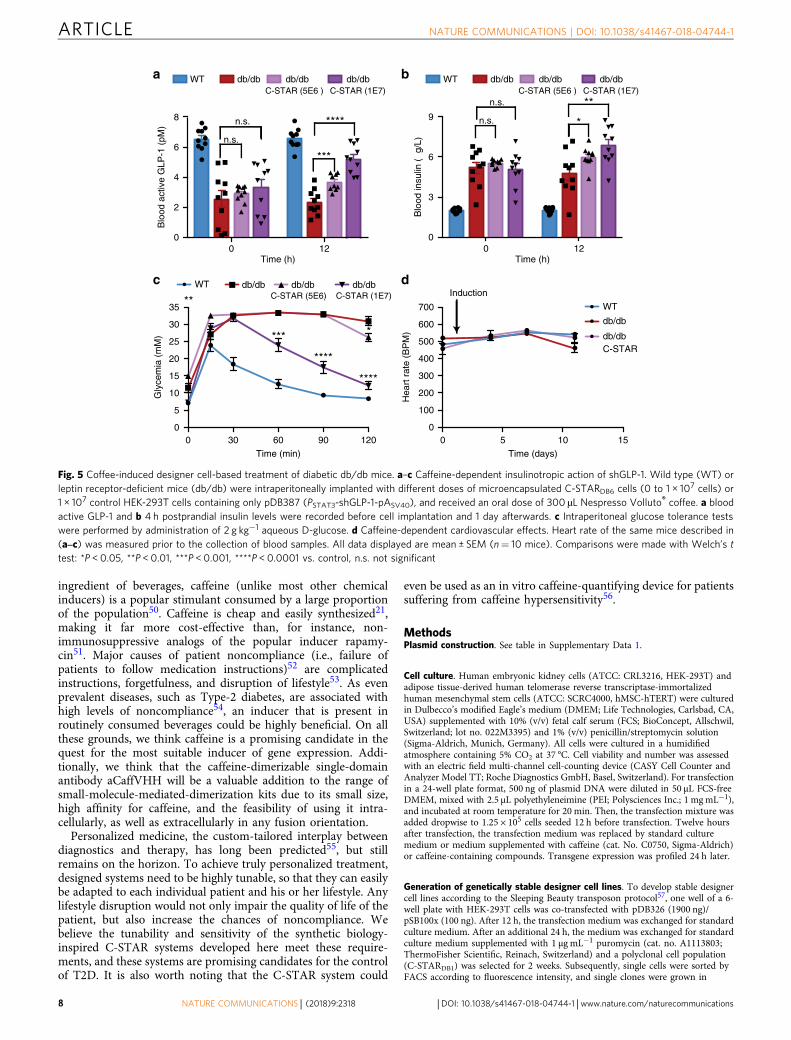
homeostatic postprandial glucose metabolism in coffee-treateddiabetic mice (Fig. 4d). For db/db mice, which develop increasedhyperinsulinemia compared to DIO mice42 (Figs. 4c and 5b),GLP-1-dependent insulinotropic action (Fig. 5a, b) and glucosetolerance (Fig. 5c) were also restored, but required a higher doseof implanted C-STARDB6 cells (Fig. 5a–c). Importantly, thiscoffee-triggered C-STARDB6-based diabetes therapy did not
a
0 3 6 9 12 15 18 21 24
3×10⁴
2×10⁴
1×10⁴
7.5×10³
5.0×10³
2.5×10³
0.0
Compounds
Caf
fein
e (µ
M)
Nes
quik
®
Cui
da T
e® H
erba
l Tea
Nes
pres
so®
Viv
alto
Lun
go d
ecaf
Nes
pres
so®
Vol
luto
dec
af
Nes
pres
so®
Dec
af in
tens
o
Nes
pres
so®
Arp
eggi
o de
caf
Coc
a C
ola®
Cui
da T
e® G
reen
Tea
1
Cui
da T
e® G
reen
Tea
2
Cui
da T
e® B
lack
Tea
Sta
rbuc
ks®
Cof
fee
Fra
ppuc
chin
o
Sta
rbuc
ks®
Car
amel
Mac
chia
to
Red
Bul
l®
Nes
pres
so®
Buk
eela
ka
Eth
iopi
a
Nes
pres
so®
Viv
alto
Lun
go
Sta
rbuc
ks®
Cof
fee
Nes
pres
so®
Cap
ricci
o
Nes
pres
so®
Liva
nto
Nes
pres
so®
Apf
elst
rude
l
Nes
pres
so®
Vol
luto
Nes
pres
so®
Rom
a
Nes
pres
so®
Arp
eggi
o
Nes
pres
so®
Ris
tret
to
Nes
pres
so®
Dha
rkan
Mili
tary
Ene
rgy®
Nes
pres
so®
Kaz
aar
Gum
®
b c
3×1042.0×104
1.8×10⁴
1.6×10⁴
1.4×10⁴
1.2×10⁴
1.0×10⁴
5×10³
4×10³
3×10³
2×10³
1×10³
2.5×10⁴
2.0×10⁴
1.5×10⁴
1.0×10⁴
5.0×10³
0.0
Viv
alto
lung
ode
caf
Viv
alto
lung
o
Vol
luto
dec
af
Vol
luto
Cap
ricci
o
Liva
nto
Apf
elst
rude
l
Dec
afin
tens
o
Rom
a
Arp
eggi
ode
caf
Arp
eggi
o
Ris
tret
to
Dha
rkan
Kaz
aar
Caffeine (µM)
Her
bal T
ea
Nes
quik
Coc
aCol
a
Gre
en T
ea 1
Gre
en T
ea 2
Bla
ck T
ea
Cof
fee
Fra
ppuc
cino
Car
amel
Mac
chia
to
Red
Bul
l
Sta
rbuc
ksC
offe
e
Caf
fein
eB
ubbl
egum
0
Caffeine (µM)
Buk
eela
ka E
thio
pia
Fig. 3 In vitro caffeine quantification in commercial caffeine sources. a Illustration of the tested solutions with their respective caffeine concentration. Fromleft to right, the boxes correspond to Nesquik® capsules, Forest Fruits® (herbal tea), Vivalto lungo decaffeinato®, Volluto decaffeinato®, Decaffeinatointenso®, Arpeggio decaffeinato®, Coca-Cola®, Mediterranean® (green tea), Marrakech® (green tea), Earl Grey® (black tea), Starbucks® Coffee Frappuccino,Starbucks® Caramel Macchiato, Red Bull®, Bukeela ka Ethiopia®, Vivalto lungo®, Starbucks® Coffee, Capriccio®, Livanto®, Apfelstrudel®, Volluto®, Roma®,Arpeggio®, Ristretto®, Dharkan®, Military Energy Gum®, and Kazaar®. The indicated caffeine concentrations were calculated from the specifications of thevendor regarding the amount of caffeine in each beverage. b, c Quantification of the caffeine concentration in coffee from Nespresso Grand Cru® capsules(b) and other commercially available caffeine sources (c). Caffeine-containing samples were added to C-STARDB1 cells with a dilution of 1:50,000. Astandard curve obtained with pure caffeine enabled conversion of the quantified SEAP levels to caffeine concentrations in the original samples. Eachbeverage was prepared or bought on three separate occasions and the data represent the quantification of each replicate in triplicate (n= 3). Data in (b, c)are shown as the mean in bar graphs and symbols indicate individual data points. The caffeine concentration indicated by the vendor is shown in blue
ARTICLE NATURE COMMUNICATIONS | DOI: 10.1038/s41467-018-04744-1
6 NATURE COMMUNICATIONS | (2018) 9:2318 | DOI: 10.1038/s41467-018-04744-1 | www.nature.com/naturecommunications

impact on the heart rate of treated animals (Figs 4e and 5d), butreduced the body weight of diet-induced obese mice after 2 weeks(Fig. 4f).
DiscussionThe C-STAR system developed here extends previous efforts23 toinduce gene expression with caffeine by enabling engineeredmammalian cells to directly sense caffeine at physiologicallyrelevant concentrations, thereby making it possible to fine-tunetherapeutic transgene expression in response to routine intake ofbeverages, such as tea and coffee without supplementation of anyadditional chemicals. Receptor setups with differing sensitivity(Fig. 1), as well as different monoclonal cell lines generated fromthe C-STARDB1 system (Supplementary Fig. 3), could be useful toaccommodate different lifestyles of patients, who may consumedifferent amounts of caffeine per day. As the C-STAR system
responds dose-dependently to caffeine, a variety of caffeine-containing beverages, ranging from coffee or tea to energy drinks,can be used to trigger the system. Importantly, decaffeinatedcoffee did not activate the C-STAR system, so C-STAR-treatedpatients could still enjoy decaffeinated drinks without activatingtheir implants. Additionally, coffee capsules such as NespressoGrand Cru® are highly standardized and could allow for pre-dictable dosing. The persistence, efficacy, and immunoprotectivefunctions of alginate-based microcontainers used in this studyhave already been demonstrated in diabetes clinical trials, pavingthe way for the application of C-STAR cells in humans43.
Potential side effects of caffeine are minimal and well known,even in the case of lifelong consumption44,45. Indeed, normaldoses of caffeine in the form of coffee are reported to have healthbenefits46–49. Even caffeine doses of up to 400 mg per day have noadverse effect in adults20, so that a broad range of caffeine con-sumption is available for the control of C-STAR cells. As a natural
a b
c d
e
WT DIO DIO C-STAR
WT
DIO
DIOC-STAR
****
****
****
***
0 5 10 15400
500
600
700
Hea
rt r
ate
(BP
M)
WT DIO DIO C-STAR
Induction
f
WT DIO DIO C-STAR
WT DIO DIO C-STAR
0 5 10 150
2
4
6
8
Blo
od a
ctiv
e G
LP-1
(pM
)
*******
0 5 10 150
1
2
3
4
5
6
7
Blo
od in
sulin
(µg
/L)
**
**** ****
Time (days)
Time (days)
Time (days)
0 5 10 150
5
10
15
Time (days)
Fas
ting
glyc
emia
(m
M)
****
** **** ****
n.s.
0 30 60 90 1200
5
10
15
20
25
30
35
Time (min)G
lyce
mia
(m
M)
******
WT DIO DIOC-STAR
–6
–4
–2
0
2
Wei
ght l
oss
(g)
**
Fig. 4 Coffee-induced designer cell-based treatment of diabetic diet-induced obese mice. a–d Caffeine-dependent insulinotropic action of shGLP-1. Wildtype (WT) or diet-induced obese mice (DIO) were intraperitoneally implanted with microencapsulated C-STARDB6 cells or control HEK-293T cellscontaining only pDB387 (PSTAT3-shGLP-1-pASV40) and received daily oral doses of 300 µL Nespresso Volluto® coffee. a Fasting glycemia, b blood activeGLP-1, and c 4 h postprandial insulin levels were recorded for 14 days. d Intraperitoneal glucose tolerance tests were performed by administration of 2 g kg−1 aqueous D-glucose. e Caffeine-dependent cardiovascular effects. Heart rate of the same mice shown in (b, c) was measured prior to the collection ofblood samples. f Caffeine-triggered shGLP-1-mediated effects on body weight. On day 15, the body weights of individual mice shown in (a–e) werecompared to their initial body weights (day 1; prior to first coffee intake). The confidence interval of the balance is indicated by a gray box. All datadisplayed are mean ± SEM (n= 10 mice). Comparisons were made with Welch’s t test: *P < 0.05, **P < 0.01, ***P < 0.001, ****P < 0.0001 vs. control, n.s.not significant. The range of homeostatic fasting glycemia is indicated with a red box
NATURE COMMUNICATIONS | DOI: 10.1038/s41467-018-04744-1 ARTICLE
NATURE COMMUNICATIONS | (2018) 9:2318 | DOI: 10.1038/s41467-018-04744-1 |www.nature.com/naturecommunications 7
ingredient of beverages, caffeine (unlike most other chemicalinducers) is a popular stimulant consumed by a large proportionof the population50. Caffeine is cheap and easily synthesized21,making it far more cost-effective than, for instance, non-immunosuppressive analogs of the popular inducer rapamy-cin51. Major causes of patient noncompliance (i.e., failure ofpatients to follow medication instructions)52 are complicatedinstructions, forgetfulness, and disruption of lifestyle53. As evenprevalent diseases, such as Type-2 diabetes, are associated withhigh levels of noncompliance54, an inducer that is present inroutinely consumed beverages could be highly beneficial. On allthese grounds, we think caffeine is a promising candidate in thequest for the most suitable inducer of gene expression. Addi-tionally, we think that the caffeine-dimerizable single-domainantibody aCaffVHH will be a valuable addition to the range ofsmall-molecule-mediated-dimerization kits due to its small size,high affinity for caffeine, and the feasibility of using it intra-cellularly, as well as extracellularly in any fusion orientation.
Personalized medicine, the custom-tailored interplay betweendiagnostics and therapy, has long been predicted55, but stillremains on the horizon. To achieve truly personalized treatment,designed systems need to be highly tunable, so that they can easilybe adapted to each individual patient and his or her lifestyle. Anylifestyle disruption would not only impair the quality of life of thepatient, but also increase the chances of noncompliance. Webelieve the tunability and sensitivity of the synthetic biology-inspired C-STAR systems developed here meet these require-ments, and these systems are promising candidates for the controlof T2D. It is also worth noting that the C-STAR system could
even be used as an in vitro caffeine-quantifying device for patientssuffering from caffeine hypersensitivity56.
MethodsPlasmid construction. See table in Supplementary Data 1.
Cell culture. Human embryonic kidney cells (ATCC: CRL3216, HEK-293T) andadipose tissue-derived human telomerase reverse transcriptase-immortalizedhuman mesenchymal stem cells (ATCC: SCRC4000, hMSC-hTERT) were culturedin Dulbecco’s modified Eagle’s medium (DMEM; Life Technologies, Carlsbad, CA,USA) supplemented with 10% (v/v) fetal calf serum (FCS; BioConcept, Allschwil,Switzerland; lot no. 022M3395) and 1% (v/v) penicillin/streptomycin solution(Sigma-Aldrich, Munich, Germany). All cells were cultured in a humidifiedatmosphere containing 5% CO2 at 37 °C. Cell viability and number was assessedwith an electric field multi-channel cell-counting device (CASY Cell Counter andAnalyzer Model TT; Roche Diagnostics GmbH, Basel, Switzerland). For transfectionin a 24-well plate format, 500 ng of plasmid DNA were diluted in 50 µL FCS-freeDMEM, mixed with 2.5 µL polyethyleneimine (PEI; Polysciences Inc.; 1 mgmL−1),and incubated at room temperature for 20 min. Then, the transfection mixture wasadded dropwise to 1.25 × 10⁵ cells seeded 12 h before transfection. Twelve hoursafter transfection, the transfection medium was replaced by standard culturemedium or medium supplemented with caffeine (cat. No. C0750, Sigma-Aldrich)or caffeine-containing compounds. Transgene expression was profiled 24 h later.
Generation of genetically stable designer cell lines. To develop stable designercell lines according to the Sleeping Beauty transposon protocol57, one well of a 6-well plate with HEK-293T cells was co-transfected with pDB326 (1900 ng)/pSB100x (100 ng). After 12 h, the transfection medium was exchanged for standardculture medium. After an additional 24 h, the medium was exchanged for standardculture medium supplemented with 1 µg mL−1 puromycin (cat. no. A1113803;ThermoFisher Scientific, Reinach, Switzerland) and a polyclonal cell population(C-STARDB1) was selected for 2 weeks. Subsequently, single cells were sorted byFACS according to fluorescence intensity, and single clones were grown in
0 120
2
4
6
8
Time (h)
Blo
od a
ctiv
e G
LP-1
(pM
)
a
n.s.
n.s. ****
***
0 120
3
6
9
Time (h)
Blo
od in
sulin
(µg
/L)
b
n.s.
n.s. **
*
0 30 60 90 1200
5
10
15
20
25
30
35
Time (min)
Gly
cem
ia (
mM
)
c
**
***
****
****
*
d
0 5 10 150
100
200
300
400
500
600
700
Hea
rt r
ate
(BP
M)
WT
db/db
db/dbC-STAR
Induction
Time (days)
db/dbWT db/dbC-STAR (5E6 )
db/dbC-STAR (1E7)
db/dbWT db/dbC-STAR (5E6)
db/dbC-STAR (1E7)
db/dbWT db/dbC-STAR (5E6 )
db/dbC-STAR (1E7)
Fig. 5 Coffee-induced designer cell-based treatment of diabetic db/db mice. a–c Caffeine-dependent insulinotropic action of shGLP-1. Wild type (WT) orleptin receptor-deficient mice (db/db) were intraperitoneally implanted with different doses of microencapsulated C-STARDB6 cells (0 to 1 × 10⁷ cells) or1 × 107 control HEK-293T cells containing only pDB387 (PSTAT3-shGLP-1-pASV40), and received an oral dose of 300 µL Nespresso Volluto® coffee. a bloodactive GLP-1 and b 4 h postprandial insulin levels were recorded before cell implantation and 1 day afterwards. c Intraperitoneal glucose tolerance testswere performed by administration of 2 g kg−1 aqueous D-glucose. d Caffeine-dependent cardiovascular effects. Heart rate of the same mice described in(a‒c) was measured prior to the collection of blood samples. All data displayed are mean ± SEM (n= 10 mice). Comparisons were made with Welch’s ttest: *P < 0.05, **P < 0.01, ***P < 0.001, ****P < 0.0001 vs. control, n.s. not significant
ARTICLE NATURE COMMUNICATIONS | DOI: 10.1038/s41467-018-04744-1
8 NATURE COMMUNICATIONS | (2018) 9:2318 | DOI: 10.1038/s41467-018-04744-1 | www.nature.com/naturecommunications

conditioned HEK-293T culture medium. Monoclonal cell populations werescreened for caffeine-responsive SEAP expression and CSTARDB3 was chosen asthe best performer. The polyclonal stable cell line C-STARDB6 was similarly gen-erated with the plasmid pDB387, using 100 µg mL−1 zeocin (cat. no. R25005;ThermoFisher Scientific, Reinach, Switzerland) as the selecting reagent.
CCK-8 assay. Cell viability was quantified with the Cell Counting Kit-8 (CCK-8;Dojindo Laboratories; cat. no. CK04) according to the manufacturer’s instructionsin Corning® 96 black well plates with a clear bottom (cat. no. CLS3603,Sigma-Aldrich). Briefly, 12 h after transfection, standard culture medium supple-mented with or without caffeine was added to the cells. After 24 h, the medium wasexchanged for standard culture medium supplemented with 10% (v/v) CellCounting Kit-8. After an incubation period of one hour at 37 °C, absorbance wasmeasured at 450 nm with an EnVision 2104 multilabel reader (PerkinElmer),yielding a surrogate for cell viability.
SEAP assay. For the quantification of human placental-secreted alkaline phos-phatase (SEAP), cell culture supernatant was heat-inactivated for 30 min at 65 °C.Then, 80 µL supernatant was mixed with 100 µL 2 × SEAP buffer (20 mM homo-arginine, 1 mM MgCl2, 21% (v/v) diethanolamine, pH 9.8) and 20 μL of substratesolution containing 20 mM pNPP (Acros Organics BVBA). Measurement was thenperformed at 405 nm using an EnVision 2104 multilabel reader (PerkinElmer).SEAP production in vivo was quantified with the chemiluminescence SEAPreporter gene assay (cat. no. 11779842001, Sigma-Aldrich) according to themanufacturer’s instructions.
Mouse IgG ELISA. Mouse IgG levels in samples containing shGLP1-mIgG werequantified using the Mouse IgG ELISA Kit (cat. no. E-90G, ICL Lab), according tothe manufacturer’s instructions. The absorbance was quantified at 450 nm with anEnVision 2104 multilabel reader (PerkinElmer) and the mouse IgG levels wereinterpolated with a standard curve.
Glucose tolerance test. Mice were challenged by intraperitoneal injection ofglucose (2 g kg−1 body weight in H2O) and the glycemic profiles were generated bymeasurement of blood glucose levels with a glucometer (Contour® Next; BayerHealthCare, Leverkusen, Germany) every 15 or 30 min for 120 min.
Insulin ELISA. Insulin blood levels in tested mice were assessed with the Ultra-sensitive Mouse Insulin ELISA (cat. no. 10-1132-01, Mercodia) according to themanufacturer’s instructions. The absorbance was quantified at 450 nm with anEnVision 2104 multilabel reader (PerkinElmer).
shGLP-1 ELISA. Blood levels of GLP-1 in tested mice were measured with the HighSensitivity GLP-1 Active ELISA Kit, Chemiluminescent (cat. no. EZGLPHS-35K,Merck) according to the manufacturer’s instructions. The absorbance was quan-tified at 450 nm with an EnVision 2104 multilabel reader (PerkinElmer).
Caffeine samples. Coffee (Nespresso Grand Cru®) and tea samples (Cuida Te®)were prepared on a Nespresso Capri Automatic Sand machine (Koenig®). Star-bucks coffee samples were obtained from a local Starbucks®. Coca-Cola® and RedBull® samples were purchased from a local supermarket. Nesquik® (Nescafé DolceGusto®) was prepared on a Circolo Automatic EDG605B EX:1 (Nescafé DolceGusto®, DeLonghi). Military energy gum® (MarketRight Inc.) was mechanicallycrushed, covered with 40 mL water, and shaken for several hours at 37 °C tosimulate chewing. Unless indicated otherwise, volumes of prepared beverages werethose recommended by the respective manufacturer. All samples were diluted1:50,000 in standard culture medium and added to the designer cells for quanti-fication of caffeine.
Animal experiments. Encapsulated HEK-293T and C-STAR-derivative cells forthe intraperitoneal implants were generated with an Inotech Encapsulator ResearchUnit IE-50R (EncapBioSystems Inc., Greifensee, Switzerland). Coherent alginate-poly-(L-lysine)-beads (400 µm diameter, 500 cells per capsule) were generated withthe following parameters: 200-µm nozzle with a vibration frequency of 1025 Hz;25-mL syringe operated at a flow rate of 410 units; 1.12 kV bead dispersionvoltage58. Female C57BL/6 (14 weeks old) or T2D mice were injected with 1–2 mLof serum-free DMEM containing 1 × 10⁴ capsules. As genetically disposed T2Dmice, db/db mice (female, 8 weeks old) were purchased from Janvier Labs. For thediet-induced obesity (DIO) model of T2D, C57BL/6 J mice (Janvier Labs, female,4 weeks old) were fed for 10 weeks with a 10-kcal% or a 60-kcal% fat diet (TestDiet,cat. no. T-58Y1-58126) before C-STAR-controlled treatment. Blood glucose con-centration was measured with a glucometer (Contour® Next; Bayer HealthCare,Leverkusen, Germany). Serum was collected using microtainer serum separatingtubes (cat. no. 365967; Becton Dickinson, Plymouth, UK) according to the man-ufacturer’s instructions. Experiments involving animals were carried out inaccordance with the directive of the European Union by Ghislaine Charpin-El
Hamri (No. 69266309; project No. DR2013–01 (v2)) at the Institut Universitaire deTechnologie, UCB Lyon 1, F-69622 Villeurbanne Cedex, France.
Data availability. Requests for materials should be made to the correspondingauthor. All plasmids generated in this study are available upon request. All data areavailable upon reasonable request.
Received: 28 June 2017 Accepted: 8 May 2018
References1. Ausländer, S., Ausländer, D. & Fussenegger, M. Synthetic biology-the
synthesis of biology. Angew. Chem. Int. Ed. 56, 6396–6419 (2017).2. Xie, M. et al. β-cell-mimetic designer cells provide closed-loop glycemic
control. Science 354, 1296–1301 (2016).3. Bai, P. et al. A synthetic biology-based device prevents liver injury in mice. J.
Hepatol. 65, 84–94 (2016).4. Guye, P. et al. Genetically engineering self-organization of human pluripotent
stem cells into a liver bud-like tissue using Gata6. Nat. Commun. 7, 10243(2016).
5. Saxena, P. et al. A programmable synthetic lineage-control network thatdifferentiates human IPSCs into glucose-sensitive insulin-secreting beta-likecells. Nat. Commun. 7, 11247 (2016).
6. Ruder, W. C., Lu, T. & Collins, J. J. Synthetic biology moving into the clinic.Science 333, 1248–1252 (2011).
7. Bojar, D. & Fussenegger, M. The best of both worlds: reaping the benefits frommammalian and bacterial therapeutic circuits. Curr. Opin. Chem. Biol. 34,11–19 (2016).
8. Weber, W. & Fussenegger, M. Emerging biomedical applications of syntheticbiology. Nat. Rev. Genet. 13, 21–35 (2012).
9. Roybal, K. T. et al. Precision tumor recognition by T cells with combinatorialantigen-sensing circuits. Cell 164, 770–779 (2016).
10. Saxena, P., Charpin-El Hamri, G., Folcher, M., Zulewski, H. & Fussenegger,M. Synthetic gene network restoring endogenous pituitary-thyroid feedbackcontrol in experimental Graves’ disease. Proc. Natl Acad. Sci. USA 113,1244–1249 (2016).
11. Ausländer, S. & Fussenegger, M. From gene switches to mammalian designercells: present and future prospects. Trends Biotechnol. 31, 155–168 (2013).
12. Ausländer, S. & Fussenegger, M. Synthetic RNA-based switches formammalian gene expression control. Curr. Opin. Biotechnol. 48, 54–60 (2017).
13. Gossen, M. & Bujard, H. Tight control of gene expression in mammalian cellsby tetracycline-responsive promoters. Proc. Natl Acad. Sci. USA 89,5547–5551 (1992).
14. Chait, R., Palmer, A. C., Yelin, I. & Kishony, R. Pervasive selection for andagainst antibiotic resistance in inhomogeneous multistress environments. Nat.Commun. 7, 10333 (2016).
15. Valentín, S., Morales, A., Sánchez, J. L. & Rivera, A. Safety and efficacy ofdoxycycline in the treatment of rosacea. Clin. Cosmet. Investig. Dermatol. 2,129–140 (2009).
16. Gitzinger, M., Kemmer, C., El-Baba, M. D., Weber, W. & Fussenegger, M.Controlling transgene expression in subcutaneous implants using a skin lotioncontaining the apple metabolite phloretin. Proc. Natl Acad. Sci. USA 106,10638–10643 (2009).
17. Xie, M., Ye, H., Hamri, G. C.-E. & Fussenegger, M. Antagonistic control of adual-input mammalian gene switch by food additives. Nucleic Acids Res. 42,e116 (2014).
18. Müller, K. et al. Multi-chromatic control of mammalian gene expression andsignaling. Nucleic Acids Res. 41, e124 (2013).
19. Yamaguchi, M., Ito, A., Ono, A., Kawabe, Y. & Kamihira, M. Heat-induciblegene expression system by applying alternating magnetic field to magneticnanoparticles. ACS Synth. Biol. 3, 273–279 (2014).
20. Wikoff, D. et al. Systematic review of the potential adverse effects of caffeineconsumption in healthy adults, pregnant women, adolescents, and children.Food Chem. Toxicol. 109, 585–648 (2017).
21. Zajac, M. A., Zakrzewski, A. G., Kowal, M. G. & Narayan, S. A novel methodof caffeine synthesis from Uracil. Synth. Commun. 33, 3291–3297 (2003).
22. Gaascht, F., Dicato, M. & Diederich, M. Coffee provides a natural multitargetpharmacopeia against the hallmarks of cancer. Genes Nutr. 10, 51 (2015).
23. Michener, J. K. & Smolke, C. D. High-throughput enzyme evolution inSaccharomyces cerevisiae using a synthetic RNA switch. Metab. Eng. 14,306–316 (2012).
24. Ladenson, R. C., Crimmins, D. L., Landt, Y. & Ladenson, J. H. Isolation andcharacterization of a thermally stable recombinant anti-caffeine heavy-chainantibody fragment. Anal. Chem. 78, 4501–4508 (2006).
NATURE COMMUNICATIONS | DOI: 10.1038/s41467-018-04744-1 ARTICLE
NATURE COMMUNICATIONS | (2018) 9:2318 | DOI: 10.1038/s41467-018-04744-1 |www.nature.com/naturecommunications 9

25. Sonneson, G. J. & Horn, J. R. Hapten-induced dimerization of a single-domainVHH camelid antibody. Biochem. 48, 6693–6695 (2009).
26. Franco, E. J. et al. Production and characterization of a genetically engineeredanti-caffeine camelid antibody and its use in immunoaffinity chromatography.J. Chromatogr. B 878, 177–186 (2010).
27. Tuomilehto, J. & Bahijri, S. Epidemiology: lifetime risk of diabetesmellitus–how high? Nat. Rev. Endocrinol. 12, 127–128 (2016).
28. Risk, N. C. D. Factor collaboration (NCD-RisC). Worldwide trends in diabetessince 1980: a pooled analysis of 751 population-based studies with 4.4 millionparticipants. Lancet 387, 1513–1530 (2016).
29. Noguchi, K. et al. Effect of caffeine contained in a cup of coffee onmicrovascular function in healthy subjects. J. Pharmacol. Sci. 127, 217–222(2015).
30. Teekachunhatean, S., Tosri, N., Rojanasthien, N., Srichairatanakool, S. &Sangdee, C. Pharmacokinetics of caffeine following a single administration ofcoffee enema versus oral coffee consumption in healthy male subjects. ISRNPharmacol. 2013, 1–7 (2013).
31. Grusch, M. et al. Spatio-temporally precise activation of engineered receptortyrosine kinases by light. EMBO J. 33, 1713–1726 (2014).
32. Huang, C. Y. & Ferrell, J. E. Ultrasensitivity in the mitogen-activated proteinkinase cascade. Proc. Natl Acad. Sci. USA 93, 10078–10083 (1996).
33. Kawahara, M. et al. A growth signal with an artificially induced erythropoietinreceptor-gp130 cytoplasmic domain heterodimer. J. Biochem. 130, 305–312(2001).
34. Scheller, L., Strittmatter, T., Fuchs, D., Bojar, D. & Fussenegger, M.Generalized extracellular molecule sensor platform for programming cellularbehavior. Nat. Chem. Biol. https://doi.org/10.1038/s41589-018-0046-z (2018).
35. Hicks, M. B., Hsieh, Y.-H. P. & Bell, L. N. Tea preparation and its influence onmethylxanthine concentration. Food Res. Int. 29, 325–330 (1996).
36. Hackett, J., Telepchak, M. J. & Coyer, M. J. Analysis of total caffeine and otherxanthines in specialty coffees using mixed mode solid-phase extraction andliquid chromatography-diode-array detection after microwave digestion. J.Anal. Toxicol. 32, 695–701 (2008).
37. Holz, G. G. IV, Kiihtreiber, W. M. & Habener, J. F. Pancreatic beta-cells arerendered glucose-competent by the insulinotropic hormone glucagon-likepeptide-1(7-37). Nature 361, 362–365 (1993).
38. Xu, K. et al. Neuroprotection by caffeine in the MPTP model of parkinson’sdisease and its dependence on adenosine A 2A receptors. Neuroscience 322,129–137 (2016).
39. Doyle, M. E. & Egan, J. M. Mechanisms of action of glucagon-like peptide 1 inthe pancreas. Pharmacol. Ther. 113, 546–593 (2007).
40. Meier, J. J. et al. Normalization of glucose concentrations and deceleration ofgastric emptying after solid meals during intravenous glucagon-like peptide 1in patients with type 2 diabetes. J. Clin. Endocrinol. Metab. 88, 2719–2725(2003).
41. King, A. J. F. The use of animal models in diabetes research. Br. J. Pharmacol.166, 877–894 (2012).
42. Ye, H. et al. Self-adjusting synthetic gene circuit for correcting insulinresistance. Nat. Biomed. Eng. 1, 0005 (2016).
43. Jacobs-Tulleneers-Thevissen, D. et al. Sustained function of alginate-encapsulated human islet cell implants in the peritoneal cavity of mice leadingto a pilot study in a type 1 diabetic patient. Diabetologia 56, 1605–1614 (2013).
44. Poole, R. et al. Coffee consumption and health: umbrella review ofmeta-analyses of multiple health outcomes. BMJ 359, j5024 (2017).
45. Grosso, G. et al. Long-term coffee consumption is associated with decreasedincidence of new-onset hypertension: a dose–response meta-analysis.Nutrients 9, 890 (2017).
46. Kennedy, O. J. et al. Coffee, including caffeinated and decaffeinated coffee, andthe risk of hepatocellular carcinoma: a systematic review and dose–responsemeta-analysis. BMJ Open 7, e013739 (2017).
47. Eskelinen, M. H. & Kivipelto, M. Caffeine as a protective factor in dementiaand Alzheimer’s disease. J. Alzheimers Dis. 20, S167–S174 (2010).
48. Muley, A., Muley, P. & Shah, M. Coffee to reduce risk of type 2 diabetes?: asystematic review. Curr. Diabetes Rev. 8, 162–168 (2012).
49. Grosso, G., Godos, J., Galvano, F. & Giovannucci, E. L. Coffee, caffeine, andhealth outcomes: an umbrella review. Annu. Rev. Nutr. 37, 131–156 (2017).
50. Román, S., Sánchez-Siles, L. M. & Siegrist, M. The importance of foodnaturalness for consumers: results of a systematic review. Trends Food Sci.Technol. 67, 44–57 (2017).
51. Hoyle, M. et al. Cost-effectiveness of temsirolimus for first line treatment ofadvanced renal cell carcinoma. Value Health J. Int. Soc. Pharm. Outcomes Res.13, 61–68 (2010).
52. Hugtenburg, J. G., Timmers, L., Elders, P. J., Vervloet, M. & van Dijk, L.Definitions, variants, and causes of nonadherence with medication: achallenge for tailored interventions. Patient Prefer. Adherence 7, 675–682(2013).
53. Martin, L. R., Williams, S. L., Haskard, K. B. & DiMatteo, M. R. The challengeof patient adherence. Ther. Clin. Risk. Manag. 1, 189–199 (2005).
54. Blackburn, D. F., Swidrovich, J. & Lemstra, M. Nonadherence in type 2diabetes: practical considerations for interpreting the literature. Patient Prefer.Adherence 7, 183–189 (2013).
55. Chan, I. S. & Ginsburg, G. S. Personalized medicine: progress and promise.Annu. Rev. Genom. Hum. Genet. 12, 217–244 (2011).
56. Hinrichs, R. et al. Caffeine hypersensitivity. Allergy 57, 859–860 (2002).57. Mátés, L. et al. Molecular evolution of a novel hyperactive Sleeping Beauty
transposase enables robust stable gene transfer in vertebrates. Nat. Genet. 41,753–761 (2009).
58. Ye, H. et al. Pharmaceutically controlled designer circuit for the treatment ofthe metabolic syndrome. Proc. Natl Acad. Sci. USA 110, 141–146 (2013).
AcknowledgementsWe thank Pratik Saxena, David Fuchs, and Ryosuke Kojima for their generous adviceand critical comments on the manuscript. We thank Ted Abel for providing MKp37.This work was supported by the National Centre of Competence in Research (NCCR),Molecular Systems Engineering.
Author contributionsD.B., M.X., and M.F. designed the project, analyzed the results, and wrote the manu-script, D.B. and L.S. conducted the in vitro work, G.C.-E.H. performed the animalexperiments.
Additional informationSupplementary Information accompanies this paper at https://doi.org/10.1038/s41467-018-04744-1.
Competing interests: The authors declare no competing interests.
Reprints and permission information is available online at http://npg.nature.com/reprintsandpermissions/
Publisher's note: Springer Nature remains neutral with regard to jurisdictional claims inpublished maps and institutional affiliations.
Open Access This article is licensed under a Creative CommonsAttribution 4.0 International License, which permits use, sharing,
adaptation, distribution and reproduction in any medium or format, as long as you giveappropriate credit to the original author(s) and the source, provide a link to the CreativeCommons license, and indicate if changes were made. The images or other third partymaterial in this article are included in the article’s Creative Commons license, unlessindicated otherwise in a credit line to the material. If material is not included in thearticle’s Creative Commons license and your intended use is not permitted by statutoryregulation or exceeds the permitted use, you will need to obtain permission directly fromthe copyright holder. To view a copy of this license, visit http://creativecommons.org/licenses/by/4.0/.
© The Author(s) 2018
ARTICLE NATURE COMMUNICATIONS | DOI: 10.1038/s41467-018-04744-1
10 NATURE COMMUNICATIONS | (2018) 9:2318 | DOI: 10.1038/s41467-018-04744-1 | www.nature.com/naturecommunications
Copyright © 2022 FDOKUMEN