Electrochemical Sunset Yellow Biosensor Based on ...
17
sensors Article Electrochemical Sunset Yellow Biosensor Based on Photocured Polyacrylamide Membrane for Food Dye Monitoring Normazida Rozi 1 , Amalina Ahmad 1 , Lee Yook Heng 1 , Loh Kee Shyuan 2 and Sharina Abu Hanifah 1,3, * 1 School of Chemical Sciences and Food Technology, Faculty of Science and Technology, Universiti Kebangsaan Malaysia, Bangi 43600, Selangor, Malaysia; [email protected] (N.R.); [email protected] (A.A.); [email protected] (L.Y.H.) 2 Fuel Cell Institute, Universiti Kebangsaan Malaysia, Bangi 43600, Selangor, Malaysia; [email protected] 3 Polymer Research Center, Faculty of Science and Technology, Universiti Kebangsaan Malaysia, Bangi 43600, Selangor, Malaysia * Correspondence: [email protected]; Tel.: +603-8921-3902 Received: 4 November 2017; Accepted: 15 December 2017; Published: 1 January 2018 Abstract: An enzyme-based electrochemical biosensor was investigated for the analysis of Sunset Yellow synthetic food dye. A glassy carbon electrode was coated with a poly(acrylamide-co-ethyl methacrylate) membrane to immobilize laccase using a single-step photopolymerization procedure. Poly(acrylamide-co-ethyl methacrylate) membrane was demonstrated to have acceptable water absorption and suitable for biosensor application. Sunset Yellow biosensor exhibited a linear response range from 0.08 to 10.00 μM with a detection limit of 0.02 μM. This biosensor was successfully used to determine Sunset Yellow in soft drinks with recoveries of 99.0–101.6%. The method was validated using high-performance liquid chromatography, indicating the biosensor can be as a promising alternative method for Sunset Yellow detection. Keywords: electrochemical biosensor; Sunset Yellow; synthetic food dye; laccase; photopolymerization 1. Introduction In recent years, the use of synthetic dyes in food and drink has become an important food safety issue because of their effects on human health. Synthetic dyes have been used to improve and maintain the appearance, color, and texture of food during processing and storage [1,2]. However, if the intake of a dye exceeds a certain threshold then it may be pathogenic [3]. Sunset Yellow contains an azo functional group (N=N) and an aromatic ring structure, which can be harmful to human health and cause hyperactivity in children [4–7]. Excessive intake can also result in allergies, asthma, and migraines [8]. Moreover, studies have shown that azo dyes can cause bladder cancer in men and hepatocarcinoma in mice [9]. Nevertheless, Sunset Yellow is frequently used in food products because it is less expensive and more stable than natural dyes. Therefore, synthetic food dyes are restricted by the United Nations’ Food and Agricultural Organization (FAO) and World Health Organization (WHO) [10]. According to the WHO, the accepted daily intake (ADI) value for Sunset Yellow is 0–4 mg kg -1 [11]. Moreover, the concentration of Sunset Yellow in non-alcoholic beverages should not exceed 50 mg L -1 [1]. In order to regulate food quality and provide safety assurances for consumers, appropriate and effective methods to detect synthetic dyes are indispensable [5,12]. Various methods have been used for food dyes analyses, including spectroscopy [1], high-performance liquid chromatography (HPLC) [13], liquid-liquid extraction (LLE) [14] and electrophoresis [15]. However, these complicated methods do Sensors 2018, 18, 101; doi:10.3390/s18010101 www.mdpi.com/journal/sensors
-
Upload
khangminh22 -
Category
Documents
-
view
0 -
download
0
Transcript of Electrochemical Sunset Yellow Biosensor Based on ...
sensors
Article
Electrochemical Sunset Yellow Biosensor Based onPhotocured Polyacrylamide Membrane for FoodDye Monitoring
Normazida Rozi 1, Amalina Ahmad 1, Lee Yook Heng 1, Loh Kee Shyuan 2 andSharina Abu Hanifah 1,3,*
1 School of Chemical Sciences and Food Technology, Faculty of Science and Technology,Universiti Kebangsaan Malaysia, Bangi 43600, Selangor, Malaysia; [email protected] (N.R.);[email protected] (A.A.); [email protected] (L.Y.H.)
2 Fuel Cell Institute, Universiti Kebangsaan Malaysia, Bangi 43600, Selangor, Malaysia; [email protected] Polymer Research Center, Faculty of Science and Technology, Universiti Kebangsaan Malaysia,
Bangi 43600, Selangor, Malaysia* Correspondence: [email protected]; Tel.: +603-8921-3902
Received: 4 November 2017; Accepted: 15 December 2017; Published: 1 January 2018
Abstract: An enzyme-based electrochemical biosensor was investigated for the analysis of SunsetYellow synthetic food dye. A glassy carbon electrode was coated with a poly(acrylamide-co-ethylmethacrylate) membrane to immobilize laccase using a single-step photopolymerization procedure.Poly(acrylamide-co-ethyl methacrylate) membrane was demonstrated to have acceptable waterabsorption and suitable for biosensor application. Sunset Yellow biosensor exhibited a linear responserange from 0.08 to 10.00 µM with a detection limit of 0.02 µM. This biosensor was successfully usedto determine Sunset Yellow in soft drinks with recoveries of 99.0–101.6%. The method was validatedusing high-performance liquid chromatography, indicating the biosensor can be as a promisingalternative method for Sunset Yellow detection.
Keywords: electrochemical biosensor; Sunset Yellow; synthetic food dye; laccase; photopolymerization
1. Introduction
In recent years, the use of synthetic dyes in food and drink has become an important food safetyissue because of their effects on human health. Synthetic dyes have been used to improve and maintainthe appearance, color, and texture of food during processing and storage [1,2]. However, if the intakeof a dye exceeds a certain threshold then it may be pathogenic [3]. Sunset Yellow contains an azofunctional group (N=N) and an aromatic ring structure, which can be harmful to human healthand cause hyperactivity in children [4–7]. Excessive intake can also result in allergies, asthma, andmigraines [8]. Moreover, studies have shown that azo dyes can cause bladder cancer in men andhepatocarcinoma in mice [9]. Nevertheless, Sunset Yellow is frequently used in food products becauseit is less expensive and more stable than natural dyes. Therefore, synthetic food dyes are restrictedby the United Nations’ Food and Agricultural Organization (FAO) and World Health Organization(WHO) [10]. According to the WHO, the accepted daily intake (ADI) value for Sunset Yellow is0–4 mg kg−1 [11]. Moreover, the concentration of Sunset Yellow in non-alcoholic beverages should notexceed 50 mg L−1 [1].
In order to regulate food quality and provide safety assurances for consumers, appropriate andeffective methods to detect synthetic dyes are indispensable [5,12]. Various methods have been used forfood dyes analyses, including spectroscopy [1], high-performance liquid chromatography (HPLC) [13],liquid-liquid extraction (LLE) [14] and electrophoresis [15]. However, these complicated methods do
Sensors 2018, 18, 101; doi:10.3390/s18010101 www.mdpi.com/journal/sensors

Sensors 2018, 18, 101 2 of 17
not permit the rapid detection of food dyes [4,16]. Several chemosensor devices have been reportedfor the analysis of food dyes. Yu et al. [17] reported a glassy carbon electrode (GCE) modified withplatinum nanoparticles and a cetrimonium bromide (CTAB)/graphene composite for Sunset Yellowanalysis. Ghoreishi et al. [2] used a screen-printed electrode modified with gold nanoparticles for thesame purpose. Recently, Wang et al. [4] reported a GCE based on polypyrrole/single-carbon nanotubesfor Sunset Yellow determination. Although these chemosensors enabled the rapid analysis of SunsetYellow, they exhibited higher redox potential. At higher potentials, the sensor was vulnerable tointerference from many other electroactive substances.
In the present study, a Sunset Yellow biosensor was developed by immobilizing laccasein a photocurable poly(acrylamide-co-ethylmethacrylate) (AAm-co-EMA) membrane. AAm is abiocompatible, water-soluble polymer which can be used directly as an enzyme support [18–20].EMA is a hydrophobic polymer that is used as a peelable membrane [21–23] and to minimize enzymeleaching [24] by controlling the hydrophilicity of membrane. Previously, laccase was used as abiosensor for the detection of commercially reactive dyes [18]; methyl orange, an azo dye, was usedas a model and compared with other commercially reactive dyes. To the best of our knowledge, alaccase-based Sunset Yellow biosensor has not previously been reported. Most of the previous studiesfocused on laccase-based biosensor for the detection of phenolic compounds [25,26].
2. Materials and Methods
2.1. Apparatus and Reagents
Laccase (E.C. 1.10.3.2) (0.5 µg, Sigma Aldrich, St. Louis, MO, USA), Sunset Yellow (98%, SigmaAldrich), acrylamide (98%, Sigma Aldrich), ethyl methacrylate (97%, Sigma Aldrich), 2,2-dimethoxy-2-phenylacetophenone (99%, Sigma Aldrich), potassium dihydrogen phosphate (99%, Systerm,Shah Alam, Malaysia), alumina (Autolab, Ultrecht, The Netherlands), and dipotassium hydrogenphosphate (99%, Systerm) were used as received without further purification. The biosensor responsewas measured using a potentiostat (DropSens, Asturias, Spain) and GCE (Autolab).
2.2. Preparation of Poly(AAm) and Poly(AAm-co-EMA)
Preparation of poly(AAm) and poly(AAm-co-EMA) were conducted using a photopolymerizationtechnique by ultraviolet exposure unit (UV). The poly(AAm-co-EMA) membranes used in the presentstudy were composed of 90% (w/w) AAm and 10% (w/w) EMA. An amount of 100% (w/w) of AAmwas used for poly(AAm) membrane and 1.6% w/w of 2,2 dimethoxy-2-phenylacetophenone (DMPP)photoinitiator was added into monomer mixture. For the preparation of the membrane, 10 mL ofmonomer mixture was loaded on the petri dish and exposed to ultraviolet radiation for 10 min inatmospheric gaseous nitrogen. As a result, a thin film of polymer was produced.
2.3. Preparation of Biosensor Membrane
Preparation of a copolymer membrane for the immobilization of laccase was conducted usinga photopolymerization technique using an ultraviolet exposure unit (UV). A host monomer, AAm,was mixed with EMA to increase the hydrophilicity of AAm monomers. The poly(AAm-co-EMA)membranes used in the present study were composed of 90% AAm and 10% EMA. First, 0.9 g ofAAm powder was dissolved in 799 µL of deionized water. Subsequently, 100 µL of EMA was addeddropwise into the AAm solution. Then, 1.6% w/w of 2,2-dimethoxy-2-phenylacetophenone (DMPP)was added to the mixture.
Additionally, 10 mg/mL of laccase solution was prepared, which was then pipetted into 10 µL ofthe monomer mixture. The bare GC electrode was polished with alumina slurry until a mirror-likesurface was evident. It was then washed with anhydrous alcohol and distilled water in an ultrasonicbath for 3 min and then dried via nitrogen gas blowing [17]. The active surface of a GCE (0.20 cm2

Sensors 2018, 18, 101 3 of 17
surface area) was dripped with 10 µL of the 1:1 ratio of laccase:monomer solution and then exposed toultraviolet radiation (60 W) for 10 min under a nitrogen atmosphere [19].
2.4. Water Absorption Test
Water absorption test for poly(AAm) and poly(AAm-co-EMA) membranes were measured byimmersing these membranes in different beakers. Each beaker contained distilled water at roomtemperature. In every two minutes, the membranes were weighed before and after the immersion andcalculated by using an Equation (1) below [27]:
Percentage of water absorption (%) =We − Wo
Wo× 100% (1)
where We = weight of swollen polymer, g; Wo = weight of dry polymer, g.
2.5. Electrochemical Measurement of Sunset Yellow Using Laccase Immobilized in aPoly(AAm-co-EMA) Membrane
Cyclic voltammetry (CV) was carried out using a DropSens potentiostat to investigate theelectrochemical properties of Sunset Yellow. The CV electrochemical cell potential was fixed from−1.500 to 1.500 V and 0.150 to 0.400 V, with a 0.05 V/s scan rate. A GC electrode coated with laccaseimmobilized into poly(AAm-co-EMA) was used as the working electrode. A GCE (Metrohm) was usedas the auxiliary electrode, and Ag/AgCl (3 M KCl) was used as the reference electrode. The workingelectrode was placed between the other two electrodes in a beaker containing 0.05 M phosphatebuffered saline (PBS) (pH 5).
2.6. Optimization of Poly(AAm-co-EMA)/Lac/GCE Biosensor Membrane
The optimization of parameters such as pH, enzyme loading, accumulation time and stability ofthe biosensor was conducted using differential pulse voltammetry (DPV). The electrochemical cellpotential was fixed from 0.150 to 0.400 V with a scan rate of 0.05 V/s. A GCE (Metrohm) was used as theauxiliary electrode, Ag/AgCl (3 M KCl) as the reference electrode, and poly(AAm-co-EMA)/Lac/GCEas the working electrode. These three electrodes were immersed in a beaker containing 10 mL of buffersolution and 10 µM Sunset Yellow; the analyte concentration was fixed for all of the experiments.The effects of pH were studied by conducting measurements across a pH range of 1–9. Enzymeloadings of 0.025–0.500 mg/cm2, accumulation times of 0–5 min and stability of biosensor up to21 days were studied.
2.7. Linear Range and Limit of Detection
Linear range was determined under optimal conditions with a Sunset Yellow concentrationrange of 0.08–30.00 µM. To determine the detection limit, a buffer solution was used as a blank.Both of the DPV experiments were conducted from 0.15 to 0.40 V with a scan rate of 0.05 V/s using aDropsens potentiostat.
2.8. Interference Study of Biosensor
Interference study for Sunset Yellow biosensor was carried out by using several substances suchas citric acid, glucose, ascorbic acid, and tartrazine. These substances are usually being added togetherwith Sunset Yellow in soft drinks. The concentration of Sunset Yellow used was 6.00 µM and 1-, 10-, 50-,or 100-fold concentration of citric acid, glucose, ascorbic acid, or tartrazine was added to the solution.Biosensor response towards Sunset Yellow without the presence of interfering compounds also wasrecorded to obtain the percentage of interference when the interfering substances were present.
Sensors 2018, 18, 101 4 of 17
2.9. Determination of Sunset Yellow in Soft Drink
An electrode fabricated with poly(AAm-co-EMA)/Lac was used for the quantitative determinationof Sunset Yellow in a soft drink purchased from a local market. First, 50 mL of the soft drink wasboiled to remove carbon dioxide [20] before being diluted with buffer solution. Then, 10 µL of thetreated sample was added to a buffer solution in a 10-mL beaker. Sunset Yellow was determined usingthe DPV method under optimal conditions. Recovery tests were performed with 0.08–2.00 µM SunsetYellow. The concentration of Sunset Yellow was validated using an HPLC method [28].
3. Results and Discussion
3.1. Characterization of Poly(AAm) and Poly(AAm-co-EMA) by Fourier-Transform Infrared Red (FTIR)
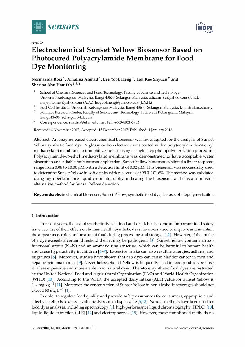
Figure 1 shows the FTIR spectra of poly(AAm) and poly(AAm-co-EMA). Poly(AAm) andpoly(AAm-co-EMA) showed peaks at 3336, 3191 and 3415 cm−1 which attributed to the primary aminestretch [29,30]. The peak that corresponded to the stretching of –CH functional group at 2925 cm−1
in poly(AAm-co-EMA) seem more intense compared to poly(AAm) at 2919 cm−1. The peaks for C=Owas also observed at 1739 cm−1 and 1649 cm−1 for poly(AAm). In addition, peak near 1651 and1607 cm−1 in both spectra were assigned to the –NH bend. In addition, the peaks at 1464 cm−1 and1449 cm−1 associated with the stretching vibration of –CN of poly(AAm-co-EMA) and poly(AAm) [29].The most important peaks that attributed to –CH3 and –CO functional groups in the copolymerwere contributed by EMA at 1377 and 1166 cm−1. Pavia et al. (2010) [31] reported that peak of–CH3 and –CO functional groups usually have bending vibration of approximately 1375 cm−1 and1300 cm−1–1000 cm−1. These two important peaks prove the copolymerization occurred as they onlyappeared in poly(AAm-co-EMA) spectra [32].
Sensors 2018, 18, 101 4 of 17
2.9. Determination of Sunset Yellow in Soft Drink
An electrode fabricated with poly(AAm-co-EMA)/Lac was used for the quantitative
determination of Sunset Yellow in a soft drink purchased from a local market. First, 50 mL of the soft
drink was boiled to remove carbon dioxide [20] before being diluted with buffer solution. Then, 10 µL
of the treated sample was added to a buffer solution in a 10-mL beaker. Sunset Yellow was determined
using the DPV method under optimal conditions. Recovery tests were performed with 0.08–2.00 μM
Sunset Yellow. The concentration of Sunset Yellow was validated using an HPLC method [28].
3. Results and Discussion
3.1. Characterization of poly(AAm) and poly(AAm-co-EMA) by Fourier-Transform Infrared Red (FTIR)
Figure 1 shows the FTIR spectra of poly(AAm) and poly(AAm-co-EMA). Poly(AAm) and
poly(AAm-co-EMA) showed peaks at 3336, 3191 and 3415 cm−1 which attributed to the primary
amine stretch [29,30]. The peak that corresponded to the stretching of –CH functional group at 2925
cm−1 in poly(AAm-co-EMA) seem more intense compared to poly(AAm) at 2919 cm−1. The peaks for
C=O was also observed at 1739 cm−1 and 1649 cm−1 for poly(AAm). In addition, peak near 1651 and
1607 cm−1 in both spectra were assigned to the –NH bend. In addition, the peaks at 1464 cm−1 and
1449 cm−1 associated with the stretching vibration of –CN of poly(AAm-co-EMA) and poly(AAm)
[29]. The most important peaks that attributed to –CH3 and –CO functional groups in the copolymer
were contributed by EMA at 1377 and 1166 cm−1. Pavia et al. (2010) [31] reported that peak of –CH3
and –CO functional groups usually have bending vibration of approximately 1375 cm−1 and
1300 cm−1–1000 cm−1. These two important peaks prove the copolymerization occurred as they only
appeared in poly(AAm-co-EMA) spectra [32].
5001000150020002500300035004000
T (%
)
Wavenumber (cm-1)
1651
34152925
3336
2919
16491607
1449
1739
1377
1166
1464
3191
1349
(b)
(a)
Figure 1. Fourier-Transform Infrared Red (FTIR) spectra of (a) poly(AAM) and (b)
poly(AAM-co-EMA). Reproduced from [AIP Conference Proceedings 1784, 030017 (2016);
doi:10.1063/1.4966755] with permission of AIP Publishing [32].
Figure 1. Fourier-Transform Infrared Red (FTIR) spectra of (a) poly(AAM) and (b) poly(AAM-co-EMA).Reproduced from [AIP Conference Proceedings 1784, 030017 (2016); doi:10.1063/1.4966755] withpermission of AIP Publishing [32].

Sensors 2018, 18, 101 5 of 17
3.2. Morphological Characterization
Figure 2 shows the micrographs of poly(AAm) and poly(AAm-co-EMA) at two differentmagnifications, 100× and 500×. There was good miscibility between AAm and EMA monomers.These micrographs also showed that poly(AAm) exhibited more porous surface. Poly(AAm-co-EMA)showed a smoother surface than poly(AAm). This finding was supported by Li et al. (2008) [33] andGowda and Betageri (2011) [34] that poly(AAm) often has porous structure networks that will allowsolute diffuse through the polymer structure [33].
Sensors 2018, 18, 101 5 of 17
3.2. Morphological Characterization
Figure 2 shows the micrographs of poly(AAm) and poly(AAm-co-EMA) at two different
magnifications, 100× and 500×. There was good miscibility between AAm and EMA monomers. These
micrographs also showed that poly(AAm) exhibited more porous surface. Poly(AAm-co-EMA)
showed a smoother surface than poly(AAm). This finding was supported by Li et al. (2008) [33] and
Gowda and Betageri (2011) [34] that poly(AAm) often has porous structure networks that will allow
solute diffuse through the polymer structure [33].
Figure 2. Micrographs of (a) poly(AAm) and (b) poly(AAm-co-EMA) at 100× of magnification, (c)
Poly (AAm) and (d) poly (AAm-co-EMA) at 500× of magnification. Reproduced from [AIP
Conference Proceedings 1784, 030017 (2016); doi:10.1063/1.4966755] with permission of AIP
Publishing [32].
3.3. Water Absorption Test
Figure 3 shows the water absorption percentage of poly(AAm) and poly(AAm-co-EMA) were
approximately 94.0% and 96.2% respectively. Poly(AAm-co-EMA) had slightly higher water
absorption percentage compared to poly(AAm). However, poly(AAm) required a shorter time (8
min) to reach equilibrium compared to poly(AAm-co-EMA) (10 min). Its means, an introduction of
hydrophobic EMA monomer into poly(AAm) membrane was able to control the hydrophilicity
properties of copolymer. Some degradation of both polymers also was observed in the distilled
water and they had difficulty in maintaining original shape after reach the equilibrium state.
Overall, water absorption for poly (AAm) membrane was slightly higher than poly(AAm-co-EMA)
thus the highest ability to absorb water [32]. It had been reported that polar substitution groups such
as –OH and –NH in a structure of polymer will cause problem in controlling the interaction of
molecule with water-based solution. Amines group can ionize in media which are at pH below pKb
of ionization species and increasingly hydrophilic and highly swell [35]. Furthermore, the more
porous surface of poly(AAm) also contributed to the higher water absorption. This will lead to
enzyme leaching problem. It means poly(AAm-co-EMA) has better potential to be studied further
for the application of enzyme immobilization and biosensor.
Figure 2. Micrographs of (a) poly(AAm) and (b) poly(AAm-co-EMA) at 100× of magnification,(c) Poly(AAm) and (d) poly(AAm-co-EMA) at 500× of magnification. Reproduced from [AIP ConferenceProceedings 1784, 030017 (2016); doi:10.1063/1.4966755] with permission of AIP Publishing [32].
3.3. Water Absorption Test
Figure 3 shows the water absorption percentage of poly(AAm) and poly(AAm-co-EMA) wereapproximately 94.0% and 96.2% respectively. Poly(AAm-co-EMA) had slightly higher water absorptionpercentage compared to poly(AAm). However, poly(AAm) required a shorter time (8 min) to reachequilibrium compared to poly(AAm-co-EMA) (10 min). Its means, an introduction of hydrophobic EMAmonomer into poly(AAm) membrane was able to control the hydrophilicity properties of copolymer.Some degradation of both polymers also was observed in the distilled water and they had difficulty inmaintaining original shape after reach the equilibrium state. Overall, water absorption for poly(AAm)membrane was slightly higher than poly(AAm-co-EMA) thus the highest ability to absorb water [32].It had been reported that polar substitution groups such as –OH and –NH in a structure of polymerwill cause problem in controlling the interaction of molecule with water-based solution. Amines groupcan ionize in media which are at pH below pKb of ionization species and increasingly hydrophilic andhighly swell [35]. Furthermore, the more porous surface of poly(AAm) also contributed to the higherwater absorption. This will lead to enzyme leaching problem. It means poly(AAm-co-EMA) has betterpotential to be studied further for the application of enzyme immobilization and biosensor.

Sensors 2018, 18, 101 6 of 17
The poly(AAm-co-EMA) membrane was composed of 90.0% (w/w) AAm and 10% (w/w) EMAalthough water absorption test only 2.0% differences from poly(AAm). Further increase in EMAmonomer cannot be tolerated in this study. It is because the deterioration of the sensitivities of thebiosensors can be occurred with less hydrophilic membranes due to slow diffusion process. When thehydrophilicity is the lowest, the loss in sensitivity is thus the most severe [19]. Moreover, from the SEMimage in Section 3.2, it clearly showed that poly(AAm-co-EMA) has smoother surface than poly(AAm).An amount of 10.0% of EMA monomer already covered most of the porous surface of the poly(AAm).Further increase in EMA monomer can cause more porous surface to be covered and contributed tolower water absorption as well as creating an interference to the analyte and contributed to a lowersensitivity of biosensor.
Sensors 2018, 18, 101 6 of 17
The poly(AAm-co-EMA) membrane was composed of 90.0% (w/w) AAm and 10% (w/w) EMA
although water absorption test only 2.0% differences from poly(AAm). Further increase in EMA
monomer cannot be tolerated in this study. It is because the deterioration of the sensitivities of the
biosensors can be occurred with less hydrophilic membranes due to slow diffusion process. When
the hydrophilicity is the lowest, the loss in sensitivity is thus the most severe [19]. Moreover, from
the SEM image in Section 3.2, it clearly showed that poly(AAm-co-EMA) has smoother surface than
poly(AAm). An amount of 10.0% of EMA monomer already covered most of the porous surface of
the poly(AAm). Further increase in EMA monomer can cause more porous surface to be covered and
contributed to lower water absorption as well as creating an interference to the analyte and
contributed to a lower sensitivity of biosensor.
0
20
40
60
80
100
120
0 2 4 6 8 10 12 14 16
Wat
er A
bsor
ptio
n (%
)
Time (min)
p(AAm) p(AAm-co-EMA)
Figure 3. Water absorption percentage as a function of time for the membrane poly(AAm) and
poly(AAm-co-EMA). Reproduced from [AIP Conference Proceedings 1784, 030017 (2016);
doi:10.1063/1.4966755] with permission of AIP Publishing [32].
3.4. Electrochemical Behaviour of Sunset Yellow Using poly(AAm-co-EMA)/Lac/GC Electrode
The electrochemical behavior of the Sunset Yellow biosensor was analyzed using 30 µM Sunset
Yellow in 0.05 M PBS (pH 5) using a bare GC electrode (GCE), a poly(AAm-co-EMA) coated
electrode, and an electrode modified with laccase (producing a poly(AAm-co-EMA)/Lac/GCE). The
analyses were performed using CV with a potential window from −1.500 to 1.500 V. In the presence
of Sunset Yellow, neither oxidation nor reduction peaks were observed from either the bare GCE or
poly(AAm-co-EMA/GCE) (Figure 4). However, after laccase was introduced via a
poly(AAm-co-EMA)/Lac membrane, a pair of well-defined and partially reversible redox peaks were
observed in the cyclic voltammograms (Ipa = 2.132 µA, Epa = 0.296 V; Ipc = −3.766 μM, Epc = −0.806 V).
These peaks indicated that the modified electrode significantly improved the redox reaction of
Sunset Yellow, and therefore is suitable for use in a biosensor. As reported previously, anodic peak
potentials of 0.6–0.95 V were recorded for Sunset Yellow [2,8,17]. However, the oxidation potential
of the Sunset Yellow biosensor developed in the present study was lower than previously reported
chemical sensors. Yang and Li [21] also reported a lower oxidation potential at 0.020 V (SCE
references electrode). A biosensor with a low oxidation potential is favourable, because it can avoid
oxidation of potential interferents that may coexist with Sunset Yellow in real samples. Sunset
Yellow contains an –OH group attached to the benzene ring structure that accommodates an
electron and facilitates proton transfer. This suggests that the redox reaction of Sunset Yellow
occurred at the –OH group, with a potential of 0.296 V for oxidation and −0.806 V for reduction.
Figure 3. Water absorption percentage as a function of time for the membrane poly(AAm)and poly(AAm-co-EMA). Reproduced from [AIP Conference Proceedings 1784, 030017 (2016);doi:10.1063/1.4966755] with permission of AIP Publishing [32].
3.4. Electrochemical Behaviour of Sunset Yellow Using Poly(AAm-co-EMA)/Lac/GC Electrode
The electrochemical behavior of the Sunset Yellow biosensor was analyzed using 30 µM SunsetYellow in 0.05 M PBS (pH 5) using a bare GC electrode (GCE), a poly(AAm-co-EMA) coatedelectrode, and an electrode modified with laccase (producing a poly(AAm-co-EMA)/Lac/GCE).The analyses were performed using CV with a potential window from −1.500 to 1.500 V. In thepresence of Sunset Yellow, neither oxidation nor reduction peaks were observed from either thebare GCE or poly(AAm-co-EMA/GCE) (Figure 4). However, after laccase was introduced via apoly(AAm-co-EMA)/Lac membrane, a pair of well-defined and partially reversible redox peaks wereobserved in the cyclic voltammograms (Ipa = 2.132 µA, Epa = 0.296 V; Ipc = −3.766 µM, Epc = −0.806 V).These peaks indicated that the modified electrode significantly improved the redox reaction of SunsetYellow, and therefore is suitable for use in a biosensor. As reported previously, anodic peak potentialsof 0.6–0.95 V were recorded for Sunset Yellow [2,8,17]. However, the oxidation potential of the SunsetYellow biosensor developed in the present study was lower than previously reported chemical sensors.Yang and Li [21] also reported a lower oxidation potential at 0.020 V (SCE references electrode).A biosensor with a low oxidation potential is favourable, because it can avoid oxidation of potentialinterferents that may coexist with Sunset Yellow in real samples. Sunset Yellow contains an –OH groupattached to the benzene ring structure that accommodates an electron and facilitates proton transfer.This suggests that the redox reaction of Sunset Yellow occurred at the –OH group, with a potential of0.296 V for oxidation and −0.806 V for reduction.

Sensors 2018, 18, 101 7 of 17
Sensors 2018, 18, 101 7 of 17
Figure 4. Cyclic voltammograms of Sunset Yellow 30 µM in 0.05 M and pH 5 of phosphate buffer
solution at 5 mm diameter are (a) poly(AAm-co-EMA)/Lac/GCE working electrode, (b) poly
(AAm-co-EMA)/GCE working electrode and (c) blank GCE working electrode with glassy carbon
electrode as auxillary electrode and Ag/AgCl as reference electrode. S/N = 3 (S/N: ratio of mean to
standard deviation of a measurement).
3.5. Effect of pH
The electrochemical behavior of Sunset Yellow in 0.05 M PBS was studied across a range of pH
values. The effect of pH on the response of the Sunset Yellow biosensor poly(AAm-co-EMA)/Lac/GCE
was measured using DPV. Based on Figures 5 and 6, the oxidation peak current increased from pH 1
to 4, and increased dramatically at pH 5. The current slowly decreased from pH 6 to 9. This decline
was attributed to the loss of enzyme catalytic activity [23]. The Sunset Yellow signal was highest in
pH 5 buffer; this condition was chosen for further studies. Similar results were reported by Li et al.
[24], who entrapped laccase in silica spheres to detect dopamine in pH 5 PBS. In addition, these
results correspond to the optimum pH range (3.5–5.0) of free laccase. Thus, the enzyme immobilization
procedure was unlikely to affect the enzyme activity [36,37].
Figure 5. DPV for the effect of pH value on the oxidation peak potential of 10 µM SY in 0.05 PBS
(pH range from 1 to 9). S/N = 3.
Figure 4. Cyclic voltammograms of Sunset Yellow 30 µM in 0.05 M and pH 5 of phosphatebuffer solution at 5 mm diameter are (a) poly(AAm-co-EMA)/Lac/GCE working electrode, (b)poly(AAm-co-EMA)/GCE working electrode and (c) blank GCE working electrode with glassy carbonelectrode as auxillary electrode and Ag/AgCl as reference electrode. S/N = 3 (S/N: ratio of mean tostandard deviation of a measurement).
3.5. Effect of pH
The electrochemical behavior of Sunset Yellow in 0.05 M PBS was studied across a range of pHvalues. The effect of pH on the response of the Sunset Yellow biosensor poly(AAm-co-EMA)/Lac/GCEwas measured using DPV. Based on Figures 5 and 6, the oxidation peak current increased from pH 1 to4, and increased dramatically at pH 5. The current slowly decreased from pH 6 to 9. This decline wasattributed to the loss of enzyme catalytic activity [23]. The Sunset Yellow signal was highest in pH 5buffer; this condition was chosen for further studies. Similar results were reported by Li et al. [24],who entrapped laccase in silica spheres to detect dopamine in pH 5 PBS. In addition, these resultscorrespond to the optimum pH range (3.5–5.0) of free laccase. Thus, the enzyme immobilizationprocedure was unlikely to affect the enzyme activity [36,37].
Sensors 2018, 18, 101 7 of 17
Figure 4. Cyclic voltammograms of Sunset Yellow 30 µM in 0.05 M and pH 5 of phosphate buffer
solution at 5 mm diameter are (a) poly(AAm-co-EMA)/Lac/GCE working electrode, (b) poly
(AAm-co-EMA)/GCE working electrode and (c) blank GCE working electrode with glassy carbon
electrode as auxillary electrode and Ag/AgCl as reference electrode. S/N = 3 (S/N: ratio of mean to
standard deviation of a measurement).
3.5. Effect of pH
The electrochemical behavior of Sunset Yellow in 0.05 M PBS was studied across a range of pH
values. The effect of pH on the response of the Sunset Yellow biosensor poly(AAm-co-EMA)/Lac/GCE
was measured using DPV. Based on Figures 5 and 6, the oxidation peak current increased from pH 1
to 4, and increased dramatically at pH 5. The current slowly decreased from pH 6 to 9. This decline
was attributed to the loss of enzyme catalytic activity [23]. The Sunset Yellow signal was highest in
pH 5 buffer; this condition was chosen for further studies. Similar results were reported by Li et al.
[24], who entrapped laccase in silica spheres to detect dopamine in pH 5 PBS. In addition, these
results correspond to the optimum pH range (3.5–5.0) of free laccase. Thus, the enzyme immobilization
procedure was unlikely to affect the enzyme activity [36,37].
Figure 5. DPV for the effect of pH value on the oxidation peak potential of 10 µM SY in 0.05 PBS
(pH range from 1 to 9). S/N = 3. Figure 5. DPV for the effect of pH value on the oxidation peak potential of 10 µM SY in 0.05 PBS (pHrange from 1 to 9). S/N = 3.

Sensors 2018, 18, 101 8 of 17Sensors 2018, 18, 101 8 of 17
Figure 6. Effect of pH value on the oxidation peak potential of 10.00 µM SY in 0.05 PBS (pH range
from 1 to 9). S/N = 3.
3.6. Influence of Laccase Loading
To characterize the influence of laccase loading (0.025–0.500 mg/cm2) on the detection of Sunset
Yellow, the electrochemical behavior of 10.00 µM Sunset Yellow in 0.05 M PBS was examined using
poly(AAm-co-EMA)/Lac/GCE as a working electrode. It has been reported that the loading of
immobilized enzyme on an electrode surface significantly affects biosensor sensitivity, detection
limit, linear range, and substrate conversion [23]. As expected, the oxidation peak current of Sunset
Yellow was strongly dependent on the enzyme loading (Figure 7A,B). Generally, the oxidation peak
current increased with increasing enzyme loading. The maximum oxidation peak current was
observed when the enzyme loading was 0.250 mg/cm2. However, increasing the enzyme loading
from 0.250 to 0.500 mg/cm2 resulted in a significant decrease in the oxidation peak current of Sunset
Yellow. This may be due to the photopolymerization technique, which results in a high-density
crosslinked polymer with low interstitial space. Hence, the active site of the enzyme is also insulated,
and not fully available to catalyze the redox reaction at high enzyme loadings, thereby decreasing
laccase activity [28].
0
10
20
30
40
50
0.025 0.125 0.250 0.375 0.500
Cu
rren
t (μ
A)
Enzyme loading (mg/cm2)
(A)
Figure 6. Effect of pH value on the oxidation peak potential of 10.00 µM SY in 0.05 PBS (pH range from1 to 9). S/N = 3.
3.6. Influence of Laccase Loading
To characterize the influence of laccase loading (0.025–0.500 mg/cm2) on the detection of SunsetYellow, the electrochemical behavior of 10.00 µM Sunset Yellow in 0.05 M PBS was examined usingpoly(AAm-co-EMA)/Lac/GCE as a working electrode. It has been reported that the loading ofimmobilized enzyme on an electrode surface significantly affects biosensor sensitivity, detection limit,linear range, and substrate conversion [23]. As expected, the oxidation peak current of Sunset Yellowwas strongly dependent on the enzyme loading (Figure 7A,B). Generally, the oxidation peak currentincreased with increasing enzyme loading. The maximum oxidation peak current was observedwhen the enzyme loading was 0.250 mg/cm2. However, increasing the enzyme loading from 0.250 to0.500 mg/cm2 resulted in a significant decrease in the oxidation peak current of Sunset Yellow. This maybe due to the photopolymerization technique, which results in a high-density crosslinked polymer withlow interstitial space. Hence, the active site of the enzyme is also insulated, and not fully available tocatalyze the redox reaction at high enzyme loadings, thereby decreasing laccase activity [28].
Sensors 2018, 18, 101 8 of 17
Figure 6. Effect of pH value on the oxidation peak potential of 10.00 µM SY in 0.05 PBS (pH range
from 1 to 9). S/N = 3.
3.6. Influence of Laccase Loading
To characterize the influence of laccase loading (0.025–0.500 mg/cm2) on the detection of Sunset
Yellow, the electrochemical behavior of 10.00 µM Sunset Yellow in 0.05 M PBS was examined using
poly(AAm-co-EMA)/Lac/GCE as a working electrode. It has been reported that the loading of
immobilized enzyme on an electrode surface significantly affects biosensor sensitivity, detection
limit, linear range, and substrate conversion [23]. As expected, the oxidation peak current of Sunset
Yellow was strongly dependent on the enzyme loading (Figure 7A,B). Generally, the oxidation peak
current increased with increasing enzyme loading. The maximum oxidation peak current was
observed when the enzyme loading was 0.250 mg/cm2. However, increasing the enzyme loading
from 0.250 to 0.500 mg/cm2 resulted in a significant decrease in the oxidation peak current of Sunset
Yellow. This may be due to the photopolymerization technique, which results in a high-density
crosslinked polymer with low interstitial space. Hence, the active site of the enzyme is also insulated,
and not fully available to catalyze the redox reaction at high enzyme loadings, thereby decreasing
laccase activity [28].
0
10
20
30
40
50
0.025 0.125 0.250 0.375 0.500
Cu
rren
t (μ
A)
Enzyme loading (mg/cm2)
(A)
Figure 7. Cont.
Sensors 2018, 18, 101 9 of 17
Sensors 2018, 18, 101 9 of 17
Figure 7. (A) Influence of Laccase enzyme loading in range 0.025 to 0.500 mg/cm2 on the oxidation
peak currents of 10 µM SY in 0.05 M PBS. S/N = 3; (B) DPV for influence of laccase enzyme loading
study.
3.7. Effect of Accumulation Time
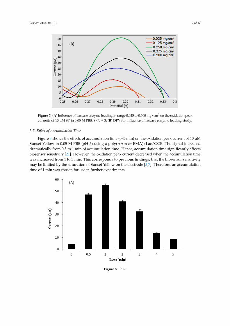
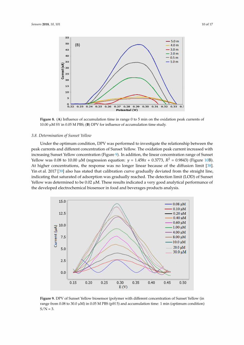
Figure 8 shows the effects of accumulation time (0–5 min) on the oxidation peak current of
10 µM Sunset Yellow in 0.05 M PBS (pH 5) using a poly(AAm-co-EMA)/Lac/GCE. The signal
increased dramatically from 0.5 to 1 min of accumulation time. Hence, accumulation time
significantly affects biosensor sensitivity [21]. However, the oxidation peak current decreased when
the accumulation time was increased from 1 to 5 min. This corresponds to previous findings, that the
biosensor sensitivity may be limited by the saturation of Sunset Yellow on the electrode [5,7].
Therefore, an accumulation time of 1 min was chosen for use in further experiments.
(B)
Figure 7. (A) Influence of Laccase enzyme loading in range 0.025 to 0.500 mg/cm2 on the oxidation peakcurrents of 10 µM SY in 0.05 M PBS. S/N = 3; (B) DPV for influence of laccase enzyme loading study.
3.7. Effect of Accumulation Time
Figure 8 shows the effects of accumulation time (0–5 min) on the oxidation peak current of 10 µMSunset Yellow in 0.05 M PBS (pH 5) using a poly(AAm-co-EMA)/Lac/GCE. The signal increaseddramatically from 0.5 to 1 min of accumulation time. Hence, accumulation time significantly affectsbiosensor sensitivity [21]. However, the oxidation peak current decreased when the accumulation timewas increased from 1 to 5 min. This corresponds to previous findings, that the biosensor sensitivitymay be limited by the saturation of Sunset Yellow on the electrode [5,7]. Therefore, an accumulationtime of 1 min was chosen for use in further experiments.
Sensors 2018, 18, 101 9 of 17
Figure 7. (A) Influence of Laccase enzyme loading in range 0.025 to 0.500 mg/cm2 on the oxidation
peak currents of 10 µM SY in 0.05 M PBS. S/N = 3; (B) DPV for influence of laccase enzyme loading
study.
3.7. Effect of Accumulation Time
Figure 8 shows the effects of accumulation time (0–5 min) on the oxidation peak current of
10 µM Sunset Yellow in 0.05 M PBS (pH 5) using a poly(AAm-co-EMA)/Lac/GCE. The signal
increased dramatically from 0.5 to 1 min of accumulation time. Hence, accumulation time
significantly affects biosensor sensitivity [21]. However, the oxidation peak current decreased when
the accumulation time was increased from 1 to 5 min. This corresponds to previous findings, that the
biosensor sensitivity may be limited by the saturation of Sunset Yellow on the electrode [5,7].
Therefore, an accumulation time of 1 min was chosen for use in further experiments.
(B)
Figure 8. Cont.
Sensors 2018, 18, 101 10 of 17
Sensors 2018, 18, 101 10 of 17
Figure 8. (A) Influence of accumulation time in range 0 to 5 min on the oxidation peak currents of
10.00 µM SY in 0.05 M PBS; (B) DPV for influence of accumulation time study.
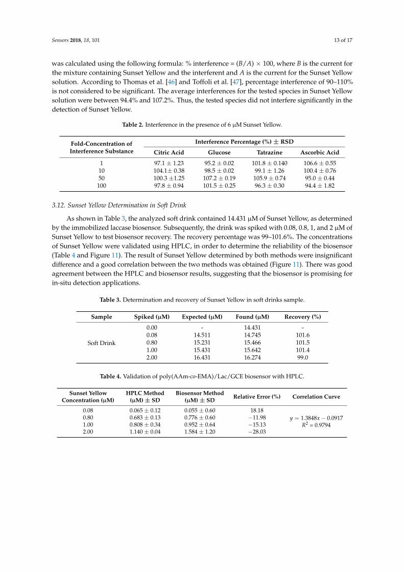
3.8. Determination of Sunset Yellow
Under the optimum condition, DPV was performed to investigate the relationship between the
peak currents and different concentration of Sunset Yellow. The oxidation peak current increased
with increasing Sunset Yellow concentration (Figure 9). In addition, the linear concentration range of
Sunset Yellow was 0.08 to 10.00 µM (regression equation: y = 1.458x + 0.3773, R2 = 0.9843) (Figure 10
(B)). At higher concentrations, the response was no longer linear because of the diffusion limit [38].
Yin et al. 2017 [39] also has stated that calibration curve gradually deviated from the straight line,
indicating that saturated of adsorption was gradually reached. The detection limit (LOD) of Sunset
Yellow was determined to be 0.02 μM. These results indicated a very good analytical performance of
the developed electrochemical biosensor in food and beverages products analysis.
Figure 9. DPV of Sunset Yellow biosensor (polymer with different concentration of Sunset Yellow (in
range from 0.08 to 30.0 µM) in 0.05 M PBS (pH 5) and accumulation time: 1 min (optimum condition)
S/N = 3.
(B)
Figure 8. (A) Influence of accumulation time in range 0 to 5 min on the oxidation peak currents of10.00 µM SY in 0.05 M PBS; (B) DPV for influence of accumulation time study.
3.8. Determination of Sunset Yellow
Under the optimum condition, DPV was performed to investigate the relationship between thepeak currents and different concentration of Sunset Yellow. The oxidation peak current increased withincreasing Sunset Yellow concentration (Figure 9). In addition, the linear concentration range of SunsetYellow was 0.08 to 10.00 µM (regression equation: y = 1.458x + 0.3773, R2 = 0.9843) (Figure 10B).At higher concentrations, the response was no longer linear because of the diffusion limit [38].Yin et al. 2017 [39] also has stated that calibration curve gradually deviated from the straight line,indicating that saturated of adsorption was gradually reached. The detection limit (LOD) of SunsetYellow was determined to be 0.02 µM. These results indicated a very good analytical performance ofthe developed electrochemical biosensor in food and beverages products analysis.
Sensors 2018, 18, 101 10 of 17
Figure 8. (A) Influence of accumulation time in range 0 to 5 min on the oxidation peak currents of
10.00 µM SY in 0.05 M PBS; (B) DPV for influence of accumulation time study.
3.8. Determination of Sunset Yellow
Under the optimum condition, DPV was performed to investigate the relationship between the
peak currents and different concentration of Sunset Yellow. The oxidation peak current increased
with increasing Sunset Yellow concentration (Figure 9). In addition, the linear concentration range of
Sunset Yellow was 0.08 to 10.00 µM (regression equation: y = 1.458x + 0.3773, R2 = 0.9843) (Figure 10
(B)). At higher concentrations, the response was no longer linear because of the diffusion limit [38].
Yin et al. 2017 [39] also has stated that calibration curve gradually deviated from the straight line,
indicating that saturated of adsorption was gradually reached. The detection limit (LOD) of Sunset
Yellow was determined to be 0.02 μM. These results indicated a very good analytical performance of
the developed electrochemical biosensor in food and beverages products analysis.
Figure 9. DPV of Sunset Yellow biosensor (polymer with different concentration of Sunset Yellow (in
range from 0.08 to 30.0 µM) in 0.05 M PBS (pH 5) and accumulation time: 1 min (optimum condition)
S/N = 3.
(B)
Figure 9. DPV of Sunset Yellow biosensor (polymer with different concentration of Sunset Yellow (inrange from 0.08 to 30.0 µM) in 0.05 M PBS (pH 5) and accumulation time: 1 min (optimum condition)S/N = 3.

Sensors 2018, 18, 101 11 of 17Sensors 2018, 18, 101 11 of 17
Figure 10. Calibration graph of Sunset Yellow biosensor (polymer with different concentration of
Sunset Yellow (in range from (A) 0.08 to 30.00 µM and (B) 0.08 to 10.00 µM) in 0.05 M PBS (pH 5) and
accumulation time: 1 min (optimum condition). S/N = 3.
3.9. Comparison with Previous Reported Sunset Yellow Sensors
Some of the analytical characteristics obtained in the present study were compared to those
from previous reports (Table 1). To date only this study that based on enzymatic biosensor to detect
Sunset Yellow has been reported. Some novel electrochemical properties of Sunset Yellow were
discovered in this work. Most of the reported findings on sensors commonly focused on designation
of conductive materials based sensor to detect Sunset Yellow. They used conductive materials such
as MWCNT, gold nanoparticles, graphene and conductive polymer in their sensor to promote direct
electron transfer but has contributed to a higher oxidation potential for example 0.600–0.950 V
[2,8,17,39,40]. At such potentials, sensor will detect interference from many other electroactive
substances. In comparison, the oxidation of Sunset Yellow was dramatically shifted to lower
potential using poly(AAm-co-EMA)/Lac/GCE biosensor. The decrease in overpotentials for Sunset
Yellow oxidation was achieved due to extraordinary redox capability of laccase enzyme.
Moreover, compared to this study, most sensors in Table 1 used conductive material in order to
improve sensitivity and lower detection limit of the sensors. The conductive materials have ability to
assist better electron transfer process. From the fact, our biosensor has comparable result to these
conductive material based chemical sensors (Table 1) although without the presence of conductive
material, shows that biosensor has its own capability to oxidize Sunset Yellow. Biosensor also has
Figure 10. Calibration graph of Sunset Yellow biosensor (polymer with different concentration ofSunset Yellow (in range from (A) 0.08 to 30.00 µM and (B) 0.08 to 10.00 µM) in 0.05 M PBS (pH 5) andaccumulation time: 1 min (optimum condition). S/N = 3.
3.9. Comparison with Previous Reported Sunset Yellow Sensors
Some of the analytical characteristics obtained in the present study were compared to thosefrom previous reports (Table 1). To date only this study that based on enzymatic biosensor todetect Sunset Yellow has been reported. Some novel electrochemical properties of Sunset Yellowwere discovered in this work. Most of the reported findings on sensors commonly focused ondesignation of conductive materials based sensor to detect Sunset Yellow. They used conductivematerials such as MWCNT, gold nanoparticles, graphene and conductive polymer in their sensor topromote direct electron transfer but has contributed to a higher oxidation potential for example0.600–0.950 V [2,8,17,39,40]. At such potentials, sensor will detect interference from many otherelectroactive substances. In comparison, the oxidation of Sunset Yellow was dramatically shiftedto lower potential using poly(AAm-co-EMA)/Lac/GCE biosensor. The decrease in overpotentials forSunset Yellow oxidation was achieved due to extraordinary redox capability of laccase enzyme.
Moreover, compared to this study, most sensors in Table 1 used conductive material in order toimprove sensitivity and lower detection limit of the sensors. The conductive materials have abilityto assist better electron transfer process. From the fact, our biosensor has comparable result to theseconductive material based chemical sensors (Table 1) although without the presence of conductive

Sensors 2018, 18, 101 12 of 17
material, shows that biosensor has its own capability to oxidize Sunset Yellow. Biosensor also hasexhibited better detection limit and sensitivity compared to gold nanoparticles and MWCNT basedsensor reported by Ghoreishi et al. [2] and Rovina et al. [40]. In Table 1, although the sensors reportedby Yu et al. [17] and Ye et al. [8] exhibited lower LOD than the sensor developed in the present study,their sensors had lower sensitivities. Furthermore, sensor reported by Chao and Ma [41] exhibiteda higher LOD and sensitivity than the present biosensor. The LOD obtained in this study reachedbelow the maximum level of Sunset Yellow in non-alcoholic beverages recommended by the EuropeanCommission 2011 (50 mg/L) [42]. This means the LOD value recorded in this study also still acceptablealthough much higher than LOD recorded in previous studies (Table 1). Overall, the LOD of ourbiosensor was comparable to previously reported findings, which indicates its feasibility for SunsetYellow detection in real samples.
Table 1. Comparison of electrochemical methods for Sunset Yellow detection.
Types of Matrix Used pH Potential (V) LinearRange (µM) LOD (µM) Sensitivity
(µA/µM) References
Screen-printed electrode modified with goldnanoparticles. 4.0 0.750 (ox) 0.10–2.00 0.0300 1.490 [2]
β-cyclodextrin-layeredpoly(diallyldimethylammonium
chloride)-graphene composite membrane.5.0 0.820 (ox) 0.05–200.0 0.0120 0.476 [8]
Platinum nanoparticle andCTAB/graphene-composite-modified GCE. 3.0 0.811 (ox) * 0.08–10.00 0.0042 0.749 [17]
CTAB/Graphene/MWCNT-modified GCE. 6.0 −0.019 (ox) * 0.01–20.00 0.0100 0.260 [21]
Chitosan/CaONP/MWCNT/gold electrode. 7.0 1.00 (ox) 1.99–22.11 1.7685 1.326 [40]
Polydopamine-coated-MWCNT/GCE. 6.0 0.619 (ox) * 0.0022–4.64 0.0014 17.112 [39]
ZnONF/CPE. 5.0 0.691 (ox) * 0.001–0.020.02–0.15 0.0002 0.0046 [43]
Poly(AAm-co-EMA)/Lac/GCE. 5.0 0.296 (ox) 0.08–10.00 0.0200 1.458 This work
* ox: oxidation potential; * References [17,21,39,43] that used SCE as reference electrode, the potential values havebeen corrected by applied scale conversion between references electrodes.
3.10. Reproducibility, Stability and Repeatability
The long-term stability of the modified electrode was evaluated through the DPV response of10 µM Sunset Yellow. The poly(AAm-co-EMA)/Lac/GCE biosensor exhibited good reproducibility,repeatability, and stability. Five modified electrodes were used to measure 10 µM Sunset Yellow usingthe DPV method. The oxidation peak current of DPV was almost constant and the relative standarddeviation (RSD) was 0.5–1.9%, confirming that the biosensor exhibited good repeatability. The RSDof Sunset Yellow (10 µM) determination using a poly(AAm-co-EMA)/Lac/GCE was 0.6–0.8% (n = 3).These results demonstrate that this biosensor has good repeatability and reproducibility. Moreover, theoxidation peak current for 10.00 µM Sunset Yellow was only reduced to 92.8% of its initial value afterthe modified GC electrodes were stored for seven days at 4 ◦C, indicating that the poly(AAm-co-EMA)matrix enhanced the stability and activity of immobilized laccase [44]. The similar electrode also beingused to detect Sunset Yellow and biosensor response still retained up to 51.7% of its original valueafter 21 days of storage at 4 ◦C. It was due to the ability of the photocured hydrophilic membrane toretain water and hence maintaining the enzyme activity [45].
3.11. Interference Study
An interference study was conducted to ensure that other synthetic dyes, or substances such ascitric acid, glucose, ascorbic acid, and tartrazine, would not interfere with Sunset Yellow detectionin real samples. Therefore, 6 µM Sunset Yellow was prepared in 0.05 M PBS (pH 5), and a 1-, 10-,50-, or 100-fold concentration of citric acid, glucose, ascorbic acid, or tartrazine was added to thesolution [17]. Interferences were analyzed by DPV, and the results are shown in Table 2. Interference
Sensors 2018, 18, 101 13 of 17
was calculated using the following formula: % interference = (B/A) × 100, where B is the current forthe mixture containing Sunset Yellow and the interferent and A is the current for the Sunset Yellowsolution. According to Thomas et al. [46] and Toffoli et al. [47], percentage interference of 90–110%is not considered to be significant. The average interferences for the tested species in Sunset Yellowsolution were between 94.4% and 107.2%. Thus, the tested species did not interfere significantly in thedetection of Sunset Yellow.
Table 2. Interference in the presence of 6 µM Sunset Yellow.
Fold-Concentration ofInterference Substance
Interference Percentage (%) ± RSD
Citric Acid Glucose Tatrazine Ascorbic Acid
1 97.1 ± 1.23 95.2 ± 0.02 101.8 ± 0.140 106.6 ± 0.5510 104.1± 0.38 98.5 ± 0.02 99.1 ± 1.26 100.4 ± 0.7650 100.3 ±1.25 107.2 ± 0.19 105.9 ± 0.74 95.0 ± 0.44100 97.8 ± 0.94 101.5 ± 0.25 96.3 ± 0.30 94.4 ± 1.82
3.12. Sunset Yellow Determination in Soft Drink
As shown in Table 3, the analyzed soft drink contained 14.431 µM of Sunset Yellow, as determinedby the immobilized laccase biosensor. Subsequently, the drink was spiked with 0.08, 0.8, 1, and 2 µM ofSunset Yellow to test biosensor recovery. The recovery percentage was 99–101.6%. The concentrationsof Sunset Yellow were validated using HPLC, in order to determine the reliability of the biosensor(Table 4 and Figure 11). The result of Sunset Yellow determined by both methods were insignificantdifference and a good correlation between the two methods was obtained (Figure 11). There was goodagreement between the HPLC and biosensor results, suggesting that the biosensor is promising forin-situ detection applications.
Table 3. Determination and recovery of Sunset Yellow in soft drinks sample.
Sample Spiked (µM) Expected (µM) Found (µM) Recovery (%)
Soft Drink
0.00 - 14.431 -0.08 14.511 14.745 101.60.80 15.231 15.466 101.51.00 15.431 15.642 101.42.00 16.431 16.274 99.0
Table 4. Validation of poly(AAm-co-EMA)/Lac/GCE biosensor with HPLC.
Sunset YellowConcentration (µM)
HPLC Method(µM) ± SD
Biosensor Method(µM) ± SD Relative Error (%) Correlation Curve
0.08 0.065 ± 0.12 0.055 ± 0.60 18.18y = 1.3848x − 0.0917
R2 = 0.97940.80 0.683 ± 0.13 0.776 ± 0.60 −11.981.00 0.808 ± 0.34 0.952 ± 0.64 −15.132.00 1.140 ± 0.04 1.584 ± 1.20 −28.03
Sensors 2018, 18, 101 14 of 17Sensors 2018, 18, 101 14 of 17
Figure 11. A comparison between the Sunset Yellow biosensor and HPLC method for the determination
of Sunset Yellow.
4. Conclusions
A sensitive electrochemical method was developed for Sunset Yellow detections, based on a
poly(AAm-co-EMA)/Lac/GCE system. The selection of poly(AAm-co-EMA) was carried out based on
the morphology and swelling properties. The biosensor membrane coated onto the GCE effectively
improved the redox response of Sunset Yellow, thereby increasing the sensitivity of the detection
method. Under optimized conditions, the anodic peak current was linear in a Sunset Yellow
concentration range of 0.08 to 10.00 μM; the detection limit was 0.02 μM. This biosensor showed
excellent activity, which was retained to 92.8% after 7 days of dry storage at 4 °C. Furthermore, the
poly(AAm-co-EMA)/Lac/GCE-based biosensor exhibited good reproducibility. The biosensor was
validated in real samples using HPLC. The validation results were well correlated, and exhibited
high sample recoveries. Hence, the laccase-based biosensor developed in this study is useful for the
determination of Sunset Yellow in soft drinks, and is expected to facilitate the development of
various electrochemical biosensors for detecting other food additives.
Acknowledgments: This work was supported by the Chemical Sensor and Biosensor Research Group,
Universiti Kebangsaan Malaysia, Research University Grant (GUP-2016-061) and a grant from the Ministry of
Higher Education of Malaysia (FRGS/1/2016/TK07/UKM/02/2). We also thank Universiti Kebangsaan Malaysia
for providing research facilities.
Author Contributions: Rozi, N. and Abu Hanifah, S. conceived and designed the experiments; Rozi, N.
performed the experiments and analyzed the data; Rozi, N. and Ahmad, A. drafted the manuscript;
Abu Hanifah, S., Heng, L.Y. and Loh, K.S. contributed reagents/materials/analysis tools. All authors discussed
the results and commented on the manuscript.
Conflicts of Interest: The authors declare no conflict of interest.
References
1. Llamas, N.E.; Garrido, M.; Di Nezio, M.S.; Fernandez Band, B.S. Second order advantage in the
determination of amaranth, sunset yellow fcf and tatrazine by uv-vis and multivariate curve
resolution-alternating least squares. Anal. Chim. Acta 2009, 655, 38–42.
2. Ghoreishi, S.M.; Behpour, M.; Golestaneh, M. Simultaneous determination of Sunset Yellow and
Tartrazine in soft drinks using gold nanoparticles carbon paste electrode. Food Chem. 2012, 132, 637–641.
3. Chen, X.; Wu, K.; Sun, Y.; Song, X. Highly sensitive electrochemical sensor for sunset yellow based on the
enhancement effect of alumina microfibers. Sens. Actuators B 2013, 185, 582–586.
4. Wang, M.; Sun, Q.; Gao, Y.; Yang, X.; Zhao, J. Determination of sunset yellow in foods based on a facile
electrochemical sensor. Anal. Methods 2014, 6, 8760–8766.
y = 1.3848x - 0.0917
R² = 0.9794
-0.200
0.000
0.200
0.400
0.600
0.800
1.000
1.200
1.400
1.600
1.800
0.000 0.200 0.400 0.600 0.800 1.000 1.200
Co
nce
ntr
atio
n S
un
set
Yel
low
det
ecte
d b
y b
iose
nso
r m
eth
od
(μM
)
Concentration of Sunset Yellow detected by HPLC method (μM)
Figure 11. A comparison between the Sunset Yellow biosensor and HPLC method for the determinationof Sunset Yellow.
4. Conclusions
A sensitive electrochemical method was developed for Sunset Yellow detections, based ona poly(AAm-co-EMA)/Lac/GCE system. The selection of poly(AAm-co-EMA) was carried outbased on the morphology and swelling properties. The biosensor membrane coated onto the GCEeffectively improved the redox response of Sunset Yellow, thereby increasing the sensitivity of thedetection method. Under optimized conditions, the anodic peak current was linear in a Sunset Yellowconcentration range of 0.08 to 10.00 µM; the detection limit was 0.02 µM. This biosensor showedexcellent activity, which was retained to 92.8% after 7 days of dry storage at 4 ◦C. Furthermore, thepoly(AAm-co-EMA)/Lac/GCE-based biosensor exhibited good reproducibility. The biosensor wasvalidated in real samples using HPLC. The validation results were well correlated, and exhibitedhigh sample recoveries. Hence, the laccase-based biosensor developed in this study is useful for thedetermination of Sunset Yellow in soft drinks, and is expected to facilitate the development of variouselectrochemical biosensors for detecting other food additives.
Acknowledgments: This work was supported by the Chemical Sensor and Biosensor Research Group, UniversitiKebangsaan Malaysia, Research University Grant (GUP-2016-061) and a grant from the Ministry of HigherEducation of Malaysia (FRGS/1/2016/TK07/UKM/02/2). We also thank Universiti Kebangsaan Malaysia forproviding research facilities.
Author Contributions: Normazida Rozi and Sharina Abu Hanifah conceived and designed the experiments;Normazida Rozi performed the experiments and analyzed the data; Normazida Rozi and Amalina Ahmaddrafted the manuscript; Sharina Abu Hanifah, Lee Yook Heng and Loh Kee Shyuan contributedreagents/materials/analysis tools. All authors discussed the results and commented on the manuscript.
Conflicts of Interest: The authors declare no conflict of interest.
References
1. Llamas, N.E.; Garrido, M.; Di Nezio, M.S.; Fernandez Band, B.S. Second order advantage in the determinationof amaranth, sunset yellow fcf and tatrazine by uv-vis and multivariate curve resolution-alternating leastsquares. Anal. Chim. Acta 2009, 655, 38–42. [CrossRef] [PubMed]
2. Ghoreishi, S.M.; Behpour, M.; Golestaneh, M. Simultaneous determination of Sunset Yellow and Tartrazinein soft drinks using gold nanoparticles carbon paste electrode. Food Chem. 2012, 132, 637–641. [CrossRef][PubMed]
3. Chen, X.; Wu, K.; Sun, Y.; Song, X. Highly sensitive electrochemical sensor for sunset yellow based on theenhancement effect of alumina microfibers. Sens. Actuators B 2013, 185, 582–586. [CrossRef]
4. Wang, M.; Sun, Q.; Gao, Y.; Yang, X.; Zhao, J. Determination of sunset yellow in foods based on a facileelectrochemical sensor. Anal. Methods 2014, 6, 8760–8766. [CrossRef]
Sensors 2018, 18, 101 15 of 17
5. Wang, M.G.; Sun, Q.; Zhao, J. Sensitively simultaneous determination of sunset yellow and tatrazine in foodsbased on polyrrole modifies oxidized single-walled carbon nanotubes. J. Electrochem. Soc. 2014, 161, 297–304.[CrossRef]
6. Xu, J.; Zhang, Y.; Zhou, H.; Wang, M.; Xu, P.; Zhang, J. An amperometric sensor for sunset yellow fcf detectionbased on molecularly imprinted polypyrrole. Sci. Res. Eng. 2012, 5, 159–162. [CrossRef]
7. Zhang, W.; Liu, T.; Zheng, X.; Huang, W.; Wan, C. Surfaced-enhanced oxidation and detection of sunsetyellow and tatrazine using multi-walled carbon nanotubes film-modified electrode. Colloids Surf. B 2009, 74,28–31. [CrossRef] [PubMed]
8. Ye, X.; Du, Y.; Lu, D.; Wang, C. Fabrication of β-cyclodextrin-coated poly(diallyldimethylammoniumchloride)-functionalized graphene composite film modified glassy carbon-rotating disk electrode andits application for simultaneous electrochemical determination colorants of sunset yellow and tatrazine.Anal. Chim. Acta 2013, 779, 22–34. [PubMed]
9. Srivinivasan, G.P.; Sikkanthar, A.; Elamaran, A.; Delma, C.R.; Subramaniyam, K.; Somasundaran, S.T.Biodegradation of carcinogetic textile azo dye using bacterial isolates of mangrove sediment. J. Coast.Life Med. 2014, 2, 154–162.
10. Gan, T.; Sun, J.; Cao, S.; Gao, F.; Zhang, Y.; Yang, Y. One-step electrochemical approach fo the preparation ofgraphene wrapped-phosphotungstic acids hydrid and its application for simulataneous determination ofsunset yellow and tatrazine. Electrochim. Acta 2012, 74, 151–157. [CrossRef]
11. Aguilar, F.; Boon, P.E.; Crebelli, R.; Dusemund, B.; Gott, D.; Hallas-Moller, T.; Konig, J.; Lindtner, O.;Marzin, D.; Meyland, I.; et al. Reconsideration of the temporary ADI and refined exposure assessmet forsunset yellow FCF (E 110). EFSA J. 2014, 12, 1–39. [CrossRef]
12. Medeiros, R.A.; Lourencao, B.C.; Rocha-Filho, R.C.; Fatibello-Filho, O. Simulataneous voltammetricdetermination of synthetic colorants in food using a cathodically pretreated boron-doped diamond electrode.Talanta 2012, 97, 291–297. [CrossRef] [PubMed]
13. Gosetti, F.; Gianotti, V.; Polati, S.; Gennaro, M.C. HPLC-MS degradation study of E110 Sunset Yellow FCF inacommercial beverage. J. Chrom. A 2005, 1090, 107–115. [CrossRef]
14. Sharma, Y.C. Optimization of parameters for adsorption of methylene blue on a low cost activated carbon.J. Chem. Eng. 2010, 55, 435–439. [CrossRef]
15. Zhang, Y.; Chang, C.; Guo, Q.; Cao, H.; Bai, Y.; Liu, H. Analysis of tartrazine and sunset yellow aluminiumlake in foods by capillary zone electrophoresis. Chin. J. Chrom. 2014, 32, 438–442. [CrossRef]
16. Song, Y.Z. Electrochemical reduction of sunset yellow at a multi-walled carbon nanotube (MWCNT)-modifiedglassy carbon electrode and its analytical applications. Can. J. Chem. 2010, 88, 676–681. [CrossRef]
17. Yu, L.; Shi, M.; Yue, X.; Qu, L. A novel and sensitive hexadecyltrimethyl ammonium bromide functionalizedgraphene supported platinum nanoparticles composite modified glassy carbon electrode for determinationof sunset yellow. Sens. Actuators B 2015, 209, 1–8. [CrossRef]
18. Munteanu, F.D.; Paulo-Cavaro, A. Biosensors based on laccase for detection of commercially reactive dyes.Anal. Lett. 2010, 43, 1126–1131. [CrossRef]
19. Hanifah, S.A.; Heng, L.Y.; Ahmad, M. Effect of gold nanoparticles on the response of phenol biosensorcontaining photocurable membrane with tyrosinase. Sensors 2008, 8, 6407–6416.
20. Zhang, K.; Luo, P.; Wu, J.; Wang, W.; Ye, B. Highly sensitive determination of sunset yellow in drink using apoly(L-cysteine) modified glassy carbon electrode. Anal. Methods 2013, 5, 5044–5050. [CrossRef]
21. Yang, J.Y.; Li, W. CTAB functionalized graphene oxide/multi-walled carbon nanotube composite modifiedelectrode for the simultaneous determination of sunset yellow and tartrazine. Russ. J. Electrochem. 2015, 51,218–226. [CrossRef]
22. Manole, A.; Herea, D.; Chiriac, H.; Melnig, V. Laccase activity determination. Cuza Uni. Sci. Ann. Lasi 2008,4, 17–24.
23. Portaccio, M.; Tuoro, D.D.; Arduini, F.; Moscone, D.; Cammarota, M.; Mita, D.G.; Lepore, M. Laccasebiosensor based on screen-printed electrode modified with thionine-carbon black nanocomposite forBisphenol A detection. Electrochim. Acta 2013, 109, 340–347. [CrossRef]
24. Li, Y.; Li, Z.; Li, M.; Pan, Z.; Li, D. A disposable biosensor based on immobilization of laccase with silicaspheres on the MWCNTs-doped screen printed electrode. Chem. Cent. J. 2012, 6, 1–8. [CrossRef] [PubMed]
Sensors 2018, 18, 101 16 of 17
25. Palanisamy, S.; Ramaraj, S.K.; Chen, S.M.; Yang, T.C.K.; Yi-Fan, P.; Chen, T.W.; Velusamy, V.; Selvam, S.A novel laccase biosensor based on laccase immobilized graphene-cellulose microfiber composite modifiedscreen-printed carbon electrode for sensitive determination of catechol. Sci. Rep. 2017, 7, 1–12. [CrossRef][PubMed]
26. De Oliveira Neto, J.R.; Rezende, S.G.; Lobon, G.S.; Garcia, T.A.; Macedo, L.F.; Garcia, L.F.; Alves, V.F.;Torres, I.M.S.; Santiago, M.F.; Schmidt, F.; et al. Electroanalysis and laccase-based biosensor on thedetermination of phenolic content and antioxidant power of honey samples. Food Chem. 2017, 237, 1118–1123.[CrossRef] [PubMed]
27. Yoon, S.D.; Park, M.H.; Byun, H.S. Mechanical and water barrier properties of starch/PVA composite filmsby adding nanosized poly(methyl methacrylate-co-acrylamide) particle. Carbohydr. Polym. 2012, 87, 676–686.[CrossRef]
28. Cai, Y.; Jessop, J.L.P. Decreased oxygen inhibition in photopolumerized acrylate/epoxide hydrid polymercoatings as demonstrated by Raman spectroscopy. Polymer 2006, 47, 6560–6566. [CrossRef]
29. Rifa, A.A.; Hashim, S.; Muhamad, I.I. Synthesis and characterization of poly(acrylamide-co-acrylic acid)-grafted-poly(styrene-co-methyl) microgels by emulsion polymerization. J. Adv. Mater. Sci. 2015, 5, 21–26.
30. Karthikeyan, S.; Anandan, C.; Subramanian, J.; Sekaran, G. Characterization of iron impregnatedpolyacrylamide catalyst and its application to the treatment of municipal wastewater. RSC Adv. 2013,35, 15044–15057. [CrossRef]
31. Pavia, D.L.; Lampman, G.M.; Kriz, G.S.; Vyvyan, J.R. Spectroscopy, 4th ed.; Brook/Cole Cengage Learning:Belmont, CA, USA, 2010; ISBN 13 978-0-495-11478-9.
32. Rozi, N.; Hanifah, S.A.; Heng, L.Y.; Loh, K.S. Characterization of ultraviolet-photocured acrylamide basedpolymer. AIP Conf. Proc. 2016, 1784, 030017. [CrossRef]
33. Li, S.; Liu, X. Synthesis, characterization and evaluation of semi-IPN hydrogels consisted of poly(methacrylicacid) and guar gum for colon-specific drug delivery. Poly. Adv. Tech. 2008, 19, 371–376. [CrossRef]
34. Gowda, V.; Betageri, G.V. Water soluble polymers for pharmaceutical applications. Polymers 2011, 3,1972–2009.
35. Nesrinne, S.; Djamel, A. Synthesis, characterization and rheological behavior of pH sensitive poly(acrylamide-co-acrylic acid) hydrogels. Arab. J. Chem. 2013, 10, 1–9. [CrossRef]
36. Makas, Y.G.; Kalkan, N.A.; Aksoy, S.; Altinok, H.; Hasirci, N. Immobilization of laccase in k-carrageenanbased semi-interpenetrating networks. J. Biotechnol. 2010, 148, 216–220. [CrossRef] [PubMed]
37. Morozova, O.V.; Shumakovich, G.P.; Gorbacheva, M.A.; Shieev, S.V.; Yaropolov, A.I. Blue laccase. Biochemistry2007, 72, 11–36. [CrossRef]
38. Rajesh; Kaneto, K. A new tyrosinase biosensor based on covalent immobilization of enzyme on N-(3-aminopropyl) pyrrole polymer film. Curr. Appl. Phys. 2005, 5, 178–183. [CrossRef]
39. Yin, Z.Z.; Cheng, S.W.; Xu, L.B.; Liu, H.Y.; Huang, K.; Li, L.; Zhai, Y.Y.; Zeng, Y.B.; Liu, H.Q.;Shao, Y.; et al. Highly sensitive and selective sensor for sunset yellow based on molecularly imprintedpolydopamine-coated multi-walled carbon nanotubes. Biosens. Bioelectron. 2017, 100, 565–570. [CrossRef][PubMed]
40. Rovina, K.; Siddiquee, S.; Shaarani, S.M. Higly sensitive determination of sunset yellow FCF(E110) in foodproducts based on chitosan/nanoparticles/MWCNTs with modified gold electrode. IOP Sci. 2016, 36, 1–7.[CrossRef]
41. Chao, M.; Ma, X. Convenient electrochemical determination of sunset yellow and tatrazine in food sampleusing a poly(L-Phenylalanine)-modified glassy carbon electrode. Food Anal. Meth. 2015, 8, 130–138.[CrossRef]
42. Konig, J. Chapter 2: Food colour additives of synthetic origin (pp. 35–60). In Colour Additives for Foods andBeverages, 1st ed.; Scotter, M.J., Ed.; Woodhead Publishing: Cambridge, UK, 2015; pp. 1–260. ISBN 9781782420118.
43. Ya, Y.; Jiang, C.; Li, T.; Liao, J.; Fan, Y.; Wei, Y.; Yan, F.; Xie, L. A zinc oxide nanoflower-based electrochemicalsensor for trace detection of sunset yellow. Sensors 2017, 17, 545. [CrossRef] [PubMed]
44. Zhang, D.H.; Yuwen, L.X.; Peng, L.J. Parameters affecting the performance of immobilized enzyme. J. Chem.2013, 2013, 946248. [CrossRef]
45. Mohammad, R.; Ahmad, M.; Heng, L.Y. An amperometric biosensor utilizing a ferrocene-mediatedhorseradish peroxidase preaction for the determination of capsaicin. Sensors 2013, 12, 10014–10026.[CrossRef] [PubMed]

Sensors 2018, 18, 101 17 of 17
46. Thomas, T.; Petrie, K.; Shim, J.; Abildshov, K.M.; Westhoff, C.L.; Cremers, S. A UPLC-MS/MS method fortherapeutic drug monitoring of etonogestrel. Ther. Drug. Monit. 2013, 35. [CrossRef] [PubMed]
47. Toffoli, G.; Rothman, N.; Ferguson, R.; Nagourney, S.; Robinson, D.; Sanguiliano, J.; Worby, P. Data QualityAssessment and Data Usability Evaluation Technical; New Jersey Department of Environment Protection:Trenton, NJ, USA, 2014; pp. 1–138.
© 2018 by the authors. Licensee MDPI, Basel, Switzerland. This article is an open accessarticle distributed under the terms and conditions of the Creative Commons Attribution(CC BY) license (http://creativecommons.org/licenses/by/4.0/).


![Sunset Seed & Plant Co. (Sherwood Hall Nursery Co.) : [catalog]](https://static.fdokumen.com/doc/165x107/63266583e491bcb36c0ac5ab/sunset-seed-plant-co-sherwood-hall-nursery-co-catalog.jpg)


