Survey of the year 2006 commercial optical biosensor literature
67
Review Survey of the year 2006 commercial optical biosensor literature Rebecca L. Rich and David G. Myszka * Center for Biomolecular Interaction Analysis, University of Utah, Salt Lake City, Utah, USA We identified 1219 articles published in 2006 that described work performed using commercial optical biosensor platforms. It is interesting to witness how the biosensor market is maturing with an increased number of instrument manufacturers offering a wider variety of platforms. However, it is clear from a review of the results presented that the advances in technology are outpacing the skill level of the average biosensor user. While we can track a gradual improvement in the quality of the published work, we clearly have a long way to go before we capitalize on the full potential of biosensor technology. To illustrate what is right with the biosensor literature, we highlight the work of 10 groups who have their eye on the ball. To help out the rest of us who have the lights on but nobody home, we use the literature to address common myths about biosensor technology. Copyright # 2007 John Wiley & Sons, Ltd. Keywords: affinity; Biacore; biomolecular interaction analysis; evanescent wave; IAsys; kinetics; Surface Plasmon Resonance; resonant mirror Received 10 September 2007; accepted 14 September 2007 INTRODUCTION Using a number of internet-based literature search engines, we uncovered 1219 articles published in 2006 that reported using optical biosensor technology. We would classify about 100 of these articles as reviews of the technology, methodology, and theoretical discussions, and the remaining 1100 or so as primary research articles. We considered only papers which utilized commercial instruments and we purposefully avoided specified techniques, such as the use of nanoparticles with surface plasmon resonance, that would require their own reviews. We continue to see a 10% increase in the number of articles published each year utilizing biosensor technology. This increase is due in part to the range of instruments now available from 24 different manufacturers. This diversity in platforms, from the lower cost, bench-top end to the higher throughput, high-sensitivity end, demonstrates the market’s growing need for a full range of sensor technology. Unfortunately, in many ways the technology’s brawn outweighs the user’s brain. Our thorough review of the literature uncovered a number of key areas in which the general user needs to improve in terms of experimental design and data analysis. In order to remain positive, we have selected the work from 10 different teams who provide shining examples of how to properly implement, as well as report, biosensor analyses. By following their lead, we can all learn how to take full advantage of what the technology has to offer. Finally, we take a look at some myths and misconceptions surrounding biosensors and we use the literature to help dispel some of the more commonly expressed misgivings about the technology. REVIEWS, THEORY, AND METHODS Reviews In the reference list, we subdivided the reviews into articles that (a) focus on optical biosensor technology and its contributions in various applications [1–16], (b) discuss optical biosensors alongside other interaction technologies [17–48], and (c) describe how optical biosensor-based analyses, in concert with other methods, have advanced a particular field of study [49–92]. Optical biosensors. Each year we see optical biosensors’ increased impact in both basic and applied research efforts. To meet this need, established biosensor companies continue to produce next-generation platforms and new manufac- turers are emerging in the biosensor market. References 5,15 and 16 illustrate the number and diversity of platforms now available or under development, with Cooper’s [5] article in particular providing details of the latest instru- ments available from several of the more familiar manufacturers. We recommend two particularly noteworthy references to every biosensor user. The book, Surface Plasmon Resonance Based Sensors edited by Homola [9], spans aspects of SPR from its theoretical basis to assay development and specific applications. Book chapters include in-depth discussions of how plasmons are generated and used to create sensing devices, introductions to molecular interactions and mass transport effects, and examples of the diversity of biosensor JOURNAL OF MOLECULAR RECOGNITION J. Mol. Recognit. 2007; 20: 300–366 Published online in Wiley InterScience (www.interscience.wiley.com) DOI:10.1002/jmr.862 *Correspondence to: D. G. Myszka, University of Utah School of Medicine 4A417, 50 N. Medical Drive, Salt Lake City, UT 84132, USA. E-mail: [email protected] Copyright # 2007 John Wiley & Sons, Ltd.
-
Upload
khangminh22 -
Category
Documents
-
view
1 -
download
0
Transcript of Survey of the year 2006 commercial optical biosensor literature

Review
Survey of the year 2006 commercial opticalbiosensor literature
Rebecca L. Rich and David G. Myszka*
Center for Biomolecular Interaction Analysis, University of Utah, Salt Lake City, Utah, USA
We identified 1219 articles published in 2006 that described work performed using commercial opticalbiosensor platforms. It is interesting to witness how the biosensor market is maturing with an increasednumber of instrument manufacturers offering a wider variety of platforms. However, it is clear from areview of the results presented that the advances in technology are outpacing the skill level of the averagebiosensor user. While we can track a gradual improvement in the quality of the published work, we clearlyhave a long way to go before we capitalize on the full potential of biosensor technology. To illustrate what isright with the biosensor literature, we highlight the work of 10 groups who have their eye on the ball. To helpout the rest of us who have the lights on but nobody home, we use the literature to address common mythsabout biosensor technology. Copyright # 2007 John Wiley & Sons, Ltd.
Keywords: affinity; Biacore; biomolecular interaction analysis; evanescent wave; IAsys; kinetics; Surface PlasmonResonance; resonant mirror
Received 10 September 2007; accepted 14 September 2007
INTRODUCTION
Using a number of internet-based literature search engines,we uncovered 1219 articles published in 2006 that reportedusing optical biosensor technology. Wewould classify about100 of these articles as reviews of the technology,methodology, and theoretical discussions, and the remaining1100 or so as primary research articles. We considered onlypapers which utilized commercial instruments and wepurposefully avoided specified techniques, such as the use ofnanoparticles with surface plasmon resonance, that wouldrequire their own reviews.
We continue to see a �10% increase in the number ofarticles published each year utilizing biosensor technology.This increase is due in part to the range of instruments nowavailable from 24 different manufacturers. This diversity inplatforms, from the lower cost, bench-top end to the higherthroughput, high-sensitivity end, demonstrates the market’sgrowing need for a full range of sensor technology.Unfortunately, in many ways the technology’s brawnoutweighs the user’s brain.
Our thorough review of the literature uncovered a numberof key areas in which the general user needs to improve interms of experimental design and data analysis. In order toremain positive, we have selected the work from 10 differentteams who provide shining examples of how to properlyimplement, as well as report, biosensor analyses. Byfollowing their lead, we can all learn how to take fulladvantage of what the technology has to offer.
Finally, we take a look at some myths and misconceptionssurrounding biosensors and we use the literature to help
dispel some of the more commonly expressed misgivingsabout the technology.
REVIEWS, THEORY, AND METHODS
Reviews
In the reference list, we subdivided the reviews into articlesthat (a) focus on optical biosensor technology and itscontributions in various applications [1–16], (b) discussoptical biosensors alongside other interaction technologies[17–48], and (c) describe how optical biosensor-basedanalyses, in concert with other methods, have advanced aparticular field of study [49–92].
Optical biosensors. Each year we see optical biosensors’increased impact in both basic and applied research efforts.Tomeet this need, established biosensor companies continueto produce next-generation platforms and new manufac-turers are emerging in the biosensor market. References5,15 and 16 illustrate the number and diversity of platformsnow available or under development, with Cooper’s [5]article in particular providing details of the latest instru-ments available from several of the more familiarmanufacturers.
We recommend two particularly noteworthy references toevery biosensor user. The book, Surface Plasmon ResonanceBased Sensors edited by Homola [9], spans aspects of SPRfrom its theoretical basis to assay development and specificapplications. Book chapters include in-depth discussions ofhow plasmons are generated and used to create sensingdevices, introductions to molecular interactions and masstransport effects, and examples of the diversity of biosensor
JOURNAL OF MOLECULAR RECOGNITIONJ. Mol. Recognit. 2007; 20: 300–366Published online in Wiley InterScience(www.interscience.wiley.com) DOI:10.1002/jmr.862
*Correspondence to: D. G. Myszka, University of Utah School of Medicine
4A417, 50 N. Medical Drive, Salt Lake City, UT 84132, USA.
E-mail: [email protected]
Copyright # 2007 John Wiley & Sons, Ltd.

analyses, ranging from identifying binding partners, rankinganalyte libraries, and epitope mapping to examining kineticsand thermodynamics. Also, this book provides particularlyuseful suggestions regarding the various methods availablefor coupling ligands to the sensor surface and highlights thetechnology’s increasing impact in environment monitoring,food safety, and medical diagnostics. Huber and Mueller’s[10] review article, although targeted toward the drugdiscovery community, is widely applicable to the generalbiosensor user. These authors explain the fundamentalphysics of SPR, describe how to set up an experiment,summarize different immobilization strategies, outline howto analyze data to obtain stoichiometry, specificity, kinetic,affinity, and/or thermodynamic information, and highlightthe biosensor’s application in characterizing biopharma-ceuticals and small molecules.
Other authors provided brief summaries of SPR detection,flow cell construction, surface chemistries, assay formats,and data collection [4,11,15]. Indyk’s [11] review alsoemphasized the technology’s range of biological appli-cations while others detailed how biosensors are used in foodscience [1,7,8,12,14,15] and drug discovery [6]. Also, in oursurvey last year [16], we highlighted excellent biosensorwork published by 10 groups in 2005. We also providedside-by-side examples of good and bad data sets from the2005 literature to illustrate some common experimentalmistakes, as well as to demonstrate the effort required toobtain high-quality data.
Interaction technologies. A wide range of optical andnon-optical interaction technologies is now commerciallyavailable. References 17–48 describe how optical biosensorscompare with other technologies to characterize bindingevents. For example, several authors summarized howbiosensors are now used in health care to diagnose disease[24,43], track blood coagulation 25, and monitor theimmune system 17 and in the environmental/food/veterinarysciences to evaluate food and water quality 18,26,38,39,40,48, and detect drug metabolites 46. Patel 37 reviewedmany of the biosensor technologies applied throughout thefood industry while Cooper 22, Goodnow 27, Ince andNarayanaswamy 29, and Ma et al. [35] described how theseinstruments are advancing drug discovery. Other authorsoutlined the various biosensors’ detection systems used toexamine proteins 20,23,30,33,44, self-assembled mono-layers and polymers [34,42], and antibodies 36. And,References 19,21,28,31,32,41,45, and 47 provided over-views of the recent achievements in developing interactiontechnologies in array formats.
Biological applications. A number of review articlesillustrate how optical biosensors, along with complementarybiochemical and biophysical techniques, have furthered ourunderstanding of protein interactions [50,56–58,61,63,67,79,80,86], as well as the characterization of carbohydrates[55,78,82], oligonucleotides [91], membranes, lipids, films,and other biological interfaces [53,59,68,74,75,83,87,92].Other reviews demonstrate the biosensor’s contribution inbasic research of small molecules [73] and antibodies[62,65,71,88], as well as the development of small-moleculedrugs and protein therapeutics [49,52,54,89,90]. Severalauthors demonstrate the biosensor’s escalating role in two
areas: (1) food composition/contamination and environ-ment/food safety [51,64,70,72] and (2) detection of virusesand bacteria as emerging disease elements and biologicalwarfare agents [66,76,77,81,85]. Downard [60] andNedelkov [84] described how the biosensor can be usedin tandem with mass spectrometry to identify bindingpartners, an application that unfortunately has not yet beenwidely adapted by biosensor users.
Theory
References 93–101 discuss theoretical analyses of bothinstrument construction and the signals produced by bindingevents. For example, Zhu [101] compared biosensordetection systems based on SPR and oblique-incidencereflectivity difference from a theoretical viewpoint whileother investigators modeled various features of sampledelivery to the flow cell surface [94–97,99]. Also, Anderssonand Hamalainen [93] modeled the rate constant informationuseful in structure–activity relationships and Snopok andKostyukevich [100] described aspects of protein adsorptionto the sensor surface.
Methods
References 102–107 provide protocols for the biosensor-based characterization of specific biological systems,although many of the tips these authors provide can beapplied to biosensor analyses in general. Sambrook andRussell [107] outlined a common antibody capture/antigenbinding strategy and Nedelkov and Nelson [106] describedthe steps required in a SPR/MS experiment. Three authorsoutlined methods for characterizing selectin/ligand [102],protein/DNA [103], and protein/lipid [105] interactionsusing traditional Biacore technology. Using DNA/proteinand peptide/protein examples, Goodrich [104] detailed whatis required for, and the data obtainable from, an interactionanalysis using the array biosensor platform manufactured byGWC Technologies.
PRIMARY RESEARCH ARTICLES
We found 1112 primary research articles published in 2006that included work performed using optical biosensors.Although the year’s biosensor work was most oftenpublished in J. Biol. Chem. (13%), Biochemistry (4%),and J. Mol. Biol. (4%), this collection of articles was culledfrom more than 300 journals.
The year 2006 was a watershed year with regards to theinstruments available to the biosensor community. Asdemonstrated in Table 1, we found articles describing workperformed using 39 platforms developed by 24 manufac-turers. While studies using Biacore biosensors weredescribed in 86% of the year’s literature, results reportedfrom the variety of instruments available demonstrates thereare now sensor options to meet the wide range of users’experimental needs and cost requirements.
Of the articles that mentioned which Biacore platformwas used, the majority described using the 3000 (36%), 2000
Copyright # 2007 John Wiley & Sons, Ltd. J. Mol. Recognit. 2007; 20: 300–366
DOI: 10.1002/jmr
2006 OPTICAL BIOSENSOR LITERATURE 301

(27%), X (14%), and 1000 (5%) platforms, with a fewinvestigators describing work performed using Bialite[810], C [1016], J [205,323,325,326,696,752], Q [771,1025,1026,1031,1034,1036,1037], S51 [194,395,591,828,887,902,917,919,929,932,933,937], and Flexchip [135,494,665,1059]. Also, in 2006 several articles describingBiacore’s recently released T100 [363,435,629,695,719,929,937] and A100 [295,455] platforms were published.
In the reference list, articles that describe Biacore-basedstudies are subdivided by application area. Biacoretechnology continues to be used primarily to examinediverse proteins [108–327], antibodies [328–509], receptors[510–634], and peptides [635–729], although its impact inlipid/self-assembled monolayer/polymer studies [730–817]is increasing rapidly. The technology’s breadth of appli-cations is further illustrated by the range of articlesinvestigating oligonucleotides [818–898], small molecules[899–944], carbohydrates [945–981], and extracellularmatrix components [982–1007]. In addition, the numberof articles describing biosensor work in clinical support[1008–1024], food/agricultural/veterinary/environmentalsciences [1025–1038], and membrane/virus/cell character-ization [1039–1050] demonstrate Biacore’s emergingcontributions in these fields. Biacore’s more unusualapplications, including the characterization of fibrils andthe analysis of crude analytes are summarized in References1051–1066.
An overview of these research articles revealed how usersare implementing biosensors and presenting their results.For example:
Analysis type
Seventy per cent of the research groups took advantage ofthe technology’s capability to measure binding constants;the remaining 30% used biosensors in qualitative analyses(e.g., epitope mapping, identifying binders, and comparingrelative binding parameters). Almost half of the quantitativeexperiments yielded kinetic rate constants and the remainderproduced affinity constants (from equilibrium or compe-tition analyses).
Immobilization methods
For the majority of applications that employed standarddextran surface (i.e., CM5 and CM4 sensor chips), directimmobilization was used 60% of the time and indirectcapture 40% of the time. Users overwhelmingly preferred toimmobilize the ligand via amine coupling (96%), although afew groups used thiol coupling (3%) or developed novelimmobilization chemistries [e.g., Reference 717]. Incapturing assays, ligands were most often tagged withbiotin (63%), Hisx (10%), or GST (8%), although severalauthors captured using anti-IgG [353,364,372], protein A, G,or A/G [217,294,388,474,487,542,557], or ligand-specificantibodies [252,558,561]. Clark et al. [143] and Le Pogamet al. [919] minimally biotinylated proteins for streptavidincapture and Zaman et al. [943] blocked a target’s active sitewith a small-molecule binder during immobilization toensure the target was not coupled via this site [943]. More
Table 1. Commercial optical biosensor technologies
Manufacturer Platforms References
Biacore AB Biacore, Bialite, C, J, Q, X, 1000, 2000, 3000,Flexchip, S51, T100, A100
108–1066
Affinity Sensors IAsys, IAsysþ, IAsys Auto 142, 357, 1067–1107Texas Instruments Spreeta 1030, 1108–1123EcoChemie Autolab Espirit 1124–1138Nippon Laser & Electronics SPR-670 1139–1150Analytical m-systems BIOSUPLAR, BIOSUPLAR-2 1151–1159Optrel Multiskop 1160–1167GWC Technologies SPRimager II 292, 1168–1174Sensia b-SPR 1175–1179Artificial Sensing OWLS 1180–1183Genoptics SPRi-Array 1184–1187Reichert Analytical Instruments SR7000 1188–1191Resonant Probes SPRTM 1145, 1192–1195Toyobo MultiSPRinter 1196–1199Corning Epic 1200–1202Farfield Sensors Analight Bio200 1203–1205SRU Biosystems BIND 1206–1208Bio-Rad ProteOn XPR36 1209, 1210DKK-TOA SPR-20 1211, 1212Lumera Proteomic Processor 1213, 1214Nanofilm Technology EP3 1215, 1216Axela Biosensors dotLab 1217HSS Systeme Plasmonic 1218K-mac SPR LAB 254, 1219
Copyright # 2007 John Wiley & Sons, Ltd. J. Mol. Recognit. 2007; 20: 300–366
DOI: 10.1002/jmr
302 R. L. RICH AND D. G. MYSZKA

than 30 research groups used more than one immobilizationmethod, and a handful of investigators directly comparedmultiple immobilization and capture approaches [e.g.,Reference [558]. For the most part, these researchersdiscovered that one method better preserved ligand activityand/or produced higher density surfaces, but the optimalimmobilization method needed to be determined empiricallyfor each system.
Data presentation
We find figures of binding responses essential for evaluatingboth the quality of the experiment and interpretation of thedata. Unfortunately, only 70% of the year’s literatureincluded at least one figure of binding responses and only20 articles (<2%) showed overlaid responses from replicateanalyte injections. Also, few of the quantitative analysesindicated how accurately the interaction model described theresponses. For example, only one-fourth of the kineticanalyses showed the data overlaid with the fit of the selectedinteraction model.
While we are disheartened by the disappointing quality ofmuch of the biosensor work described in the literature, weremain optimistic. Over the past few years, we have seen agradual improvement in the biosensor community’s level ofexpertise, as well as the increased inclusion of figures ofprimary data and descriptions of experimental details in theliterature. In 2006, we estimate that 15% of the year’s reportsincluded reliable biosensor data. From this pool, wehighlight examples that illustrate the versatility of thetechnology and the quality of data we expect to see from abiosensor experiment. In addition, we choose otherexamples from this pool to debunk several of the biosensormyths we see in the literature.
2006 LITERATURE HIGHLIGHTS
We are always pleased to find research articles that describewell-performed biosensor experiments. We focus onparticularly exceptional work published by 10 groups.One way these articles stand out from the rest of the year’sliterature is that they all show data. More importantly, theauthors of these articles do the experiments right. Inaddition, several of these articles demonstrate how thetechnology can be used for applications beyond kinetics(e.g., thermodynamics and stoichiometry), and/or describenew assay designs and novel applications.
High-quality experiments
The first three articles we selected are excellent examples ofbiosensor-based experiments. This collection demonstratesthat high-quality data can be obtained for a variety ofbiological interactions; here we spotlight well-performedRNA/protein, small-molecule inhibitor/enzyme, and proteinligand/receptor studies. Also, each of these articles includesmany figures of biosensor data, which reveal much moreabout an interaction (and the quality of the experiment) thana table of numbers. And finally, these authors provided
important details regarding their assay design and datainterpretation.
To investigate how both structure and sequence contributeto target recognition by RNA-binding proteins, Law et al.[852,853] characterized the interaction between U1Aprotein and its target, the hairpin II of U1snRNA (U1hpII).These researchers prepared panels of mutations in bothbinding partners and determined how each mutation affectedU1A binding to biotinylated U1hpII captured on streptavi-din-coated surfaces. Figure 1 shows how mutating either theRNA (Figure 1A) or protein (Figure 1B) altered theinteraction kinetics.
We consider this analysis to be a particularly well-performedbiosensor study since Law et al. optimized several experi-mental design parameters. First, low densities of RNAwerecaptured on the biosensor surface, which minimized masstransport and avidity effects. Second, the concentrationrange of U1A produced responses spanning from almost nodetectable binding to nearly surface saturation. This wideanalyte concentration range is important in fitting the datasince responses from the higher concentrations provideinformation about surface capacity while responses fromthe lower concentrations contribute to determining the rateconstants. Third, the lengths of the U1A injection time(1min) and wash-out time (5min) were long enough todetect both curvature in the association phase and decay inthe dissociation phase for even the most tightly boundcomplexes. Fourth, each U1A concentration was tested threetimes for binding to the RNA surface. The responses fromthese triplicate injections overlay, demonstrating the bindingpartners were stable throughout the experiment and theregeneration conditions were appropriate. And finally, the fitof a 1:1 interaction model that included a mass transportterm is superimposed over each data set. The agreementbetween the responses and the model indicates the reportedrate constants accurately describe the interaction.
The biosensor-based characterization of this RNA/proteinpair revealed a number of features required for its high-affinity interaction. As illustrated in Figure 1A, lengtheningthe RNA stem did not significantly alter the interaction,whereas other structure and sequence changes dramaticallyaffected the binding kinetics. The responses shown inFigure 1B revealed that a few positively charged residues inthe protein play critical roles in both forming and maintain-ing the U1A/U1hpII complex. These studies advanced theauthors’ understanding of the U1A/U1hpII binding mechan-ism and serve as a model for other protein/RNA interactions.
The role of positively charged amino acids andelectrostatic interactions in the complex of U1Aprotein and U1 hairpin II RNA. Law MJ et al. (2006)Nuc. Acids Res. 34: 275–285.
The role of RNA structure in the interaction of U1Aprotein with U1 hairpin II RNA. Law MJ et al. (2006)RNA 12: 1168–1178.
Interaction kinetic characterization of HIV-1 reversetranscriptase non-nucleoside inhibitor resistance. Geit-mann M et al. (2006) J. Med. Chem. 49: 2375–2387.
Copyright # 2007 John Wiley & Sons, Ltd. J. Mol. Recognit. 2007; 20: 300–366
DOI: 10.1002/jmr
2006 OPTICAL BIOSENSOR LITERATURE 303

Non-nucleoside reverse transcriptase inhibitors (NNRTIs)block HIV-1 reverse transcriptase (RT) activity but do notbind at this polymerase’s active site. Instead, thesecompounds bind at a separate site and structurally distortthe enzyme. But several RT mutants, identified in
patients treated with NNRTIs, display high levels of drugresistance. To investigate the mechanism of NNRTIresistance, Geitmann et al. [915] characterized a series ofstructurally related inhibitors binding to both wild type andmutant RT.
Figure 1. Kinetic analyses of a protein/RNA interaction. (A) Responses for U1A protein binding to wild type and mutant U1hpII RNAs.RNA structures are shown on the right. (B) Responses for wild type andmutant U1A binding to U1hp11. The protein/RNA structure, withresidues of particular interest highlighted, is shown on the right. In each panel, responses (black lines) are overlaid with the fit of a 1:1interactionmodel that includes amass transport term (red lines). Figures reproduced fromReferences 852 and 853with permission fromthe authors and the RNA Society # 2006.
Copyright # 2007 John Wiley & Sons, Ltd. J. Mol. Recognit. 2007; 20: 300–366
DOI: 10.1002/jmr
304 R. L. RICH AND D. G. MYSZKA

Figure 2 shows the responses and global kinetic fits of 12compounds binding to seven RTs amine-coupled to thesensor surface. From a technical viewpoint, this figurereveals the quality of the experiment. For example, in eachpanel the response intensities are proportional to the injecteddrug concentration and the binding profiles can be described
by exponentials. Also, the overlay of each data set with themodel fit establishes the reliability of the reported rateconstants. From a biological viewpoint, Figure 2 demon-strates how both enzyme mutations and inhibitor structureaffect the kinetics of NNRTI/RT interactions. Looking downa column of data sets we can see how different functional
Figure 2. Kinetic analysis of 12 small-molecule inhibitors binding to seven enzyme variants. In each panel, responses(colored lines) are overlaid with the fit of the interaction model (black lines). Reproduced from Reference 915 withpermission from the American Chemical Society # 2006.
Copyright # 2007 John Wiley & Sons, Ltd. J. Mol. Recognit. 2007; 20: 300–366
DOI: 10.1002/jmr
2006 OPTICAL BIOSENSOR LITERATURE 305

groups in the NNRTIs alter binding to one RT variant.Looking across one row we can compare how different RTvariants recognize one compound.
This kinetic analysis of wild type and mutant RT aids ininterpreting the role of specific amino acids in drug binding,as well as designing drug therapies against specific enzymevariants. For example, the drug panel in general dissociatesfastest from (and has the weakest affinity for) the K103N/L1001 RT mutant, suggesting this mutation is most resistantto NNRTI therapy. Also, the biosensor-based characteriz-ation allows for kinetic profiling of the inhibitors since theresearchers could identify which compounds form high-affinity, stable complexes with each enzyme variant. Thiswork also shows the progress made in development of theseinhibitors: overall, the first commercial NNRTI, nevirapine,displays the weakest RTaffinities, while modifications in themore recently developed inhibitors have in fact resulted inhigher affinity interactions. Perhaps most importantly, thiswork demonstrates the need to take a combinatorialapproach, changing elements in both enzyme and drugstructure, toward understanding this interaction.
Expanding on the idea that specific amino acids (hotspots) contribute significantly to the energetics of a bindinginterface, Moza et al. proposed these residues can beclustered into ‘hot regions’ and cooperative effects aregreater between than within the hot regions [588]. Theseresearchers demonstrated this concept using a T cell receptorsubunit (b chain variable domain 2.1, hVb2.1)/toxic shocksyndrome toxin 1 (TSST-1) binding pair.
The hVb2.1 wild type analog (EP-8), a variant (D1) thatcontains 14 residue changes compared to EP-8 and displaysan affinity 30 000 fold tighter than EP-8, and various otherreceptor mutants were each tested for binding to immobil-ized TSST-1. To identify residues that contribute to D1’shigher affinity, each of the 14 residues of interest were singlymutated in the EP-8 background and characterized using thebiosensor. Figure 3A shows the kinetic and equilibriumanalyses of several mutants. Compared to theWT interaction(upper left panel), the most dramatic changes in kineticsoccurred when glutamic acid was replaced with valine atposition 61 (lower right panel). The left panel inFigure 3B summarizes how each mutation affected bindingfree energy. Four residues are energetically significant.Structural studies revealed two hot regions: three of the fourresidues (51, 52a, 53) are located in one loop (CDR2) and thefourth (61) in a loop (FR3) �23 A away (Figure 3B, rightpanel). Responses for EP-8 injected across TSST-1 mutantspredicted to eliminate binding are shown in Figure 3C.These studies confirmed that the orientation of the structuralmodel shown in Figure 3B was correct. Figure 3D showsintra- and inter-hot region mutants binding to TSST-1 andFigure 3E shows several hydrophobic mutations at position61 did not disrupt the binding interface. From this extensivebiosensor analysis, Moza et al. calculated DGcoop for eachmutant and determined the hot regions were not energeti-cally isolated.
As illustrated throughout Figure 3, Moza et al. werecareful in both their experimental design and data analysis.For example, Moza et al. recognized that for very stablecomplexes (e.g., forD1 and S52aF/K53N/E61Vin Figure 3D),dissociation information needed to be collected until notice-able decay was detected in the signal (i.e., the dissociationphase was monitored for two hours). In addition, theseresearchers performed equilibrium analyses only for datasets in which responses for every analyte concentrationreached equilibrium by the end of the association phase(e.g., in five of the six panels in Figure 3A). All too often wesee data published in which equilibrium constants weredetermined from data sets like that shown for E61V inFigure 3A. Fitting these responses to a binding isothermwould yield an erroneous KD.
Beyond kinetics
Traditionally, determining kinetic parameters has beenconsidered the pinnacle of biosensor experiments. However,much information in addition to ka/kd pairs is extractablefrom advanced biosensor kinetic analyses. For example,determining kinetic parameters under different salt concen-tration and/or temperatures reveals details about the bindingmechanism. The three examples we highlight here demons-trate how the biosensor can be applied to interpretinteraction thermodynamics, electrostatics, and stoichi-ometry.
IkBa sequesters NF-kB outside the nucleus to preventunwanted transcriptional activity. The IkBa/NF-kB complexwas determined in vivo to form a very stable complex havinga half-life of >2 days. As illustrated in Figure 4A, theinteraction is complicated since NF-kB exists as a hetero-dimer and each monomer subunit is composed of severaldomains. Even though the structure of the IkBa/NF-kBcomplex has been solved, the structure did not reveal whichelements regulated this high-affinity interaction. To deter-mine how the individual domains of p65 contribute tothe IkBa/NF-kB interaction, Bergqvist et al. [123] exami-ned IkBa binding to a variety of biotinylated NF-kBconstructs captured on streptavidin-coated sensor surfaces.
Figure 4B shows the responses for IkBa binding to twoNF-kB constructs at two temperatures. In the left panel,IkBa was injected across the nearly intact NF-kB at 378Cand the kinetic fit yielded KD� 40 pM. The middle panelshows how the loss of the NF-kB N-terminal domaindecreased the interaction affinity to �320 pM. For com-parison, data in the right panel illustrate how the dissociationrate slowed fourfold as the temperature was decreased from37 to 258C. The effects of deleting the NF-kB C-terminalhelix 4 are shown in Figure 4C: this truncation increases thedissociation rate constant by more than 1000-fold (KD�460 nM at 258C and�160 nM at 158C), indicating this helixis responsible for the stability of the IkBa/NF-kB complex.
Bergqvist et al. also investigated this interaction usingisothermal calorimetry (ITC) and demonstrated how these
Long-range cooperative binding effects in a T cellreceptor variable domain. Moza B et al. (2006) Proc.Natl Acad. Sci. USA 103: 9867–9872.
Thermodynamics reveal that helix four in the NLS ofNF-kB anchors IkBa, forming a very stable complex.Bergqvist S et al. (2006) J. Mol. Biol. 360: 421–434.
Copyright # 2007 John Wiley & Sons, Ltd. J. Mol. Recognit. 2007; 20: 300–366
DOI: 10.1002/jmr
306 R. L. RICH AND D. G. MYSZKA

Figure 3. Kinetic analysis of a receptor/ligand complex. (A) Equilibrium and/or kinetic analysis of wild type (EP-8) and single-sitemutants of human T cell receptor b chain variable domain 2.1 (hVb2.1) binding to immobilized TSST-1. (B) Left: change in freeenergy for each of the single-site hVb2.1 mutants binding to TSST-1. The dotted red line indicates the threshold value used todistinguish energetically significant mutations. Right: Two hot regions for TSST-1 interaction in hVb2.1. In the top panel, theCDR2 hot region is shown in red and FR3 hot region in blue. The b-strand that connects the two hot regions is shown in green. Inthe model of the hVb2.1/TSST-1 complex (bottom), the hVb2.1 hot spots are shown interfacing the TSST-1 hot spots (purple andorange). (C) Equilibrium analysis of TSST-1mutants binding to hVb2.1 EP-8. (D) Kinetic (and equilibrium for E51Q/K53N) analysisofmulti-site hVb2.1mutants binding to TSST-1. (E) Kinetic analysis ofmutations at position 61 in hVb2.1. In each kinetic analysis,the responses are fit to a 1:1 interaction model (shown in black) and the corresponding residual values are plotted below eachdata set. In each equilibrium analysis, the responses at the end of the injection are plotted against analyte concentration and fit toa simple binding isotherm (shown in the insets). Reproduced fromReference 588 with permission from TheNational Academy ofSciences USA # 2006. This figure is available in colour online at www.interscience.wiley.com/journal/jmr
Copyright # 2007 John Wiley & Sons, Ltd. J. Mol. Recognit. 2007; 20: 300–366
DOI: 10.1002/jmr
2006 OPTICAL BIOSENSOR LITERATURE 307

two functional methods complemented each other andsupported the structural analyses of IkBa/NF-kB. Theirkinetic studies quantitated the slow dissociation rate ofthe IkBa/NF-kB complex, which explains the complex’slong half-life observed in vivo, and revealed that theNF-kB C-terminal helix was required to form a high-affinitycomplex. Their thermodynamic measurements indicatedhow IkBa maintains its stability when complexed withNF-kb but is otherwise quickly degraded.
Fox-1 is an unusual protein since it binds with high affi-nity (KD� 1 nM) to a short nucleotide sequence, GCAUG.Auweter et al. [819] employed both structural and functionalstudies to interpret RNA recognition by Fox-1 protein. Asdetermined by NMR, the RNA sequence, unstructured insolution, binds the protein in a bent conformation(Figure 5A). In addition, the structural studies suggestedthe binding events are regulated primarily by electrostaticand hydrophobic interactions. These researchers used thebiosensor in various assay formats to confirm the affinity ofthis RNA/protein interaction, as well as to characterizethe molecular mechanism by which Fox-1 binds RNAsequences.
In the first set of analyses, a concentration series of Fox-1was injected over a short (10-mer) biotinylated sequence thatcontained the GCAUG sequence and was captured on astreptavidin surface. As shown in the top right panel ofFigure 5B, the protein bound the surface-tethered oligonu-cleotide with high affinity (KD¼ 0.49 nM at 150mM NaCl).In addition, mutational analyses established that one residue(F126) is essential for RNA recognition. Next, the kineticsof the protein/RNA interaction were determined inbuffers having different NaCl concentrations. As shownin Figure 5B, the interaction affinity was strongly dependenton the salt concentration. Furthermore, the linearity of the
plots of logKD, logkon, and logkoff, versus logf� indicatedelectrostatic interactions affected the rate-limiting step inboth complex association and dissociation (Figure 5C). In athird experiment, Auweter et al. mapped the contributions ofindividual hydrogen bonds (both intermolecular andintra-RNA) to complex formation. Mutant oligonucleotidesequences were added to the Fox-1 solution and the mixturewas injected across the GCAUG surface. By measuring howthe mutant sequences competed with wild type for proteinbinding, the researchers could calculate how each mutationaffected the free binding energy in the structure and therebyidentify which hydrogen bonds were lost.
These experiments provide another example of howbiosensor studies can complement structural analyses. Bycharacterizing the interactions of both wild type and mutantprotein and RNA constructs, as well as evaluating thecomplex’s dependence on salt concentration, Auweter et al.determined the extent to which electrostatics and hydrogenbonds regulate Fox-1’s RNA-binding mechanism.
Nucleocapsid (NC) of HIV-1 binds nucleic acids,particularly the repeating d(TG)n sequence, at several stepsduring retroviral replication. Using biosensor technology, inconcert with other biophysical characterization methods,Fisher et al. examined the kinetics and stoichiometry of NCbinding to a simple oligonucleotide (TG)4 and developed aprotocol to detect lower affinity NC/oligonucleotide intera-tions [828]. In these series of biosensor experiments, NCwasinjected under different conditions across biotinylated (TG)4captured at a range of densities on streptavidin surfaces.
At the very low (TG)4 density (�5RU), in which theestimated distance between oligonucleotides on the sensorsurface is approximately twice the length of a fully extendedNC, no cross-linking can occur so it is not surprising that the
Figure 3. (Continued)
Molecular basis of RNA recognition by the humanalternative splicing factor Fox-1. Auweter SD et al.(2006) EMBO J. 25: 163–173.
Complex interactions of HIV-1 nucleocapsid proteinwith oligonucleotides. Fisher RJ et al. (2006) Nuc.Acids Res. 34: 472–484.
Copyright # 2007 John Wiley & Sons, Ltd. J. Mol. Recognit. 2007; 20: 300–366
DOI: 10.1002/jmr
308 R. L. RICH AND D. G. MYSZKA

Figure 4. Kinetic and equilibrium analyses of a protein/protein interaction. (A) Schematic illustrating the domain structure of theNF-kB p50/p65 heterodimer. (B) Kinetic analyses of IkBa constructs binding to streptavidin-captured NF-kB at two temperatures.Cartoons of the IkBa constructs used in each experiment are shown in the insets. (C) Kinetic and equilibriumanalyses of an IkBa constructthat does not contain helix 4 binding to NF-kB at two temperatures. Cartoons of the IkBa construct used in this experiment are shown inthe insets. Reproduced from Reference 123 with permission from Elsevier # 2006. This figure is available in colour online atwww.interscience.wiley.com/journal/jmr
Copyright # 2007 John Wiley & Sons, Ltd. J. Mol. Recognit. 2007; 20: 300–366
DOI: 10.1002/jmr
2006 OPTICAL BIOSENSOR LITERATURE 309

responses for the NC/(TG)4 interaction are well described bya simple interaction model and display 1:1 stoichiometry(Figure 6A, left panel). In addition, this interaction isspecific, as demonstrated by the lack of NC binding tosurface-tethered (A)8 olignucleotide (Figure 6A, rightpanel). This protein/oligonucleotide interaction, however,becomes more complicated at higher (TG)4 surface densi-ties. Figure 6B shows the responses for NC binding to a150RU (TG)4 surface. First, the different profile shapes inthe left panels of Figure 6A and 6B indicate NC can bindmultiple (TG)4 sequences when the oligonucleotide surfacedensity is high enough. Second, in Figure 6B the responseintensities for NC binding are greater than what would beestimated for a 1:1 interaction, indicating the protein canbind the NC/(TG)4 complex. Together, these featuressuggest that both binding partners are multivalent. Further
characterizing this complicated interaction, Fisher et al.tested the salt dependence of NC binding to the 150RU(TG)4 surface to determine the electrostatic contributions tobinding. The decrease in kinetics and stoichiometry withincreased ionic strength (compare the two panels ofFigure 6B) demonstrates that electrostatic forces play asignificant role in this multivalent protein/oligonucleotideinteraction.
Fisher et al. also used a competition approach tocharacterize low-affinity NC/oligonucleotide interactions.In these competition experiments, a standard curve was firstconstructed and then the competitive effects of oligonucleo-tides in solution were monitored. When NC was injectedover sufficiently high densities (>300RU) of (TG)4, theinitial binding rates were linear (Figure 6C, left panel) andthe slopes of these initial response were plotted against
Figure 5. Characterization of the electrostatic contributions in a oligonucleotide/protein interaction. (A) Structure of the Fox-1 RNA-binding domain (RBD) in complex with UGCAUGU. (B) Biosensor-based salt dependence of RNA binding to Fox-1. Responses for Fox-1RBD binding to streptavidin-captured biotin-50-CUCGCAUGU-30 at four NaCl concentrations overlaid with the fit of a 1:1 interactionmodel that includes a mass transport term. (C) Plot of logKD, logkoff, and logkon versus logf�, where f� is the electrostatic contribution(linked to the ionic strength). Reproduced from Reference 819 with permission from the European Molecular Biology Organization #2006. This figure is available in colour online at www.interscience.wiley.com/journal/jmr
Copyright # 2007 John Wiley & Sons, Ltd. J. Mol. Recognit. 2007; 20: 300–366
DOI: 10.1002/jmr
310 R. L. RICH AND D. G. MYSZKA

Figure 6. Biosensor-based characterization of HIV-1 NC binding to (TG)4 oligodeoxynucleotide. (A)Responses for 1.7–200nM NC injected across low-density (5–6RU) surfaces of (TG)4 (left) and (A)8(right). In the left panel, responses are overlaid with the fit of a 1:1 interactionmodel (grey lines). (B)Responses for �1–1000nM NC injected across 155–160RU (TG)4 in buffer containing 150mM (left)and 250mM (right) NaCl. (C) Standard curve generation for oligonucleotide competition exper-iments. Left: responses for 1–200nM NC injected across a 340RU (TG)4 surface. Right: NC bindingrate as a function of concentration. Initial slopes from the left panel were plotted against NCconcentration and fit using linear regression. (D) Competition between oligonucleotides in solutionand immobilized (TG)4 for binding to NC. NC solutions (100nM) incubated with varying amounts ofdifferent oligonucleotideswere injected across a�330RU (TG)4 surface. Initial slopes of the bindingresponses were plotted against competitor concentration and fit to an isotherm to obtain IC50
values. Reproduced from Reference 828 with permission from the authors # 2006.
Copyright # 2007 John Wiley & Sons, Ltd. J. Mol. Recognit. 2007; 20: 300–366
DOI: 10.1002/jmr
2006 OPTICAL BIOSENSOR LITERATURE 311

injected NC concentration to generate the standard curve(Figure 6C, right panel). Then, aliquots of NC at oneconcentration were mixed with a range of concentrations ofa competing oligonucleotide and the mixture was injectedacross the (TG)4 surface. Figure 6D summarizes thecompetition data obtained for a panel of oligonucleotidesand demonstrates this method can be used to obtaininformation about both low- and high-affinity NC binders.
Using other biophysicalmeasurements (tryptophan fluore-scence quenching, fluorescence anisotropy, isothermalcalorimetry, and mass spectrometry), Fisher et al. validatedthe affinity, kinetics, and stoichiometric parameters deter-mined from the biosensor. From this multi-facetedcharacterization of the mechanism underlying NC/(TG)4complex formation, this research team developed a bindingmodel applicable to diverse multivalent interactions. Whilethis information should prove important to understandingHIV biology, it also serves as a well-outlined example ofhow to thoroughly examine complexes that exhibit non-equimolar stoichiometry.
Assay development
In this section we focus on novel approaches to improvingligand activity and increasing analyte throughput. We alsohighlight two alternatives to traditional biosensor analysesthat circumvent the need to regenerate the ligand surface.
Phosphatases are attractive therapeutic targets and thebiosensor’s capabilities in characterizing small-molecule/macromolecular interactions are well established. Theproblem that many groups have faced when setting up drugstudies against phosphatases and related proteins is that thebiosensor assay requires protein preparations that maintainhigh levels of activity after immobilization and throughout thedrug binding study. Usually this entails immobilizing veryhigh densities of the protein target (�1000RU) and mayrequire identifying which coupling condition best maintainsthe target’s integrity. Unfortunately, identifying conditions thatmaintain these enzymes’ activities has proven challenging.However, Stenlund et al. [937] developed a methodicalapproach to optimize enzyme activity and obtain reliable smallmolecule binding responses. To demonstrate the feasibility oftheir approach, this group examined small molecules bindingto PP1 and PP2B, serine/threonine phosphatases, and PTP1B,a tyrosine phosphatase.
First, Stenlund et al. investigated ways to maintainenzyme activity during and after immobilization. Since thethree phosphatases all remained active to some degree afterbeing amine-coupled to the surface, these researchersmodified the solution used in the ligand coupling. Theytested how a variety of additives and a range of pHs affectedeach immobilized enzyme’s activity. For example,Figure 7A shows the responses for a compound bindingto PP1 that was immobilized using four different couplingbuffers. In this example, the addition of DTT, Mn2þ, and anactive-site inhibitor, ocadaic acid, to the standard immobil-
ization buffer (10mM acetate) increased this enzyme’sactivity more than 10-fold. While the addition of DTT, areducing agent necessary in phosphatase studies, and Mn2þ,which serves as a cofactor, each improved the enzymeactivity slightly, addition of the inhibitor, which presumablyacted as a protecting compound to prevent amine coupling atthe binding site, made the most significant difference. Theseresearchers discovered the best immobilization conditionneeded to be identified individually for each phosphastaseand the three enzymes retained activity under slightlydifferent conditions.
By determining the effects of different buffering agentsand ionic strengths, as well as the addition of detergent,Stenlund et al. also optimized the assay buffer conditions toobtain reliable binding responses. For example, they did notconsider using phosphate buffers since precipitates mayform when the divalent cations required for enzyme activitywere added to the buffer. HEPES-based buffers producedunreliable, drifting baselines. In their experience, Tris-basedbuffers were best. In addition, at low ionic strengths(<150mM NaCl), the phosphatase inhibitors bound non-specifically to the unmodified reference surface. The toppanels in Figure 7B shows how the addition of a detergent,Tween-20, improved the data quality for one compound/phosphatase interaction. Again, these conditions weredetermined empirically for each of the three enzymes anddifferent assay buffers proved optimal for maintaining theactivity of each enzyme.
The bottom panels in Figure 7B illustrate how the kineticsof one enzyme/inhibitor pair changed as the assaytemperature increased from 25 to 378C. Since kinetics aretemperature dependent, Stenlund et al. suggested investi-gators consider using temperatures above 258C to increasesampling throughput (since the analyte will dissociate fasterfrom the enzyme surface at higher temperature) and usingtemperatures below 258C to slow down binding events whenthe kinetics are too fast to be determined under standardtemperature conditions.
Once the experimental conditions were optimized,Stenlund et al. examined small-molecule inhibitors bindingto the three phosphatases and identified different mechan-isms of binding. For example, Figure 7C shows theresponses (overlaid with the fit of 1:1 interaction model)for concentration series of two inhibitors binding to onephosphatase. The kinetic characterization of these two PP1inhibitors emphasizes the importance of determining rateconstants rather than simply measuring interaction affinities.Both the on- and off-rates of these compounds binding toPP1 vary by several orders of magnitude and, althoughocadaic acid has a higher affinity for PP1 compared tocantharidin (KD (ocadaic acid)¼ 12 nM vs. KD (canthar-idin)¼ 2.6mM), cantharidin forms a more stable complexwith PP1 (kd (cantharidin)¼ 2.1� 10�4 s�1 vs. kd (ocadaicacid)¼ 0.02 s�1)). Expanding these kinetic studies, theseresearchers examined the interactions of the inhibitor panelwith the three phosphatases. As shown in Figure 7D, somecompounds exhibited binding to all three enzymes whileothers were much more specific. In addition, somecompounds (e.g., calyculin A) bound with different kineticsto the three enzymes. Using this approach, Stenlund et al.could determine inhibitor specificity as well as discriminatebetween interaction mechanisms. This characterization may
Studies of small molecule interactions with proteinphosphatases using biosensor technologies. StenlundP et al. (2006) Anal. Biochem. 353: 217–225.
Copyright # 2007 John Wiley & Sons, Ltd. J. Mol. Recognit. 2007; 20: 300–366
DOI: 10.1002/jmr
312 R. L. RICH AND D. G. MYSZKA

Figure 7. Assay development for a small-molecule/protein target interaction. (A) Optimization of amine-couplingconditions. Responses (normalized for immobilization level) for 250nM okadaic acid binding to PP1 immobilized in(1) 10mM acetate pH 5.5, (2) 10mM acetate, 1mM DTT, 2mMMnCl2, pH 5.5, (3) 10mM acetate, 0.5mM TCEP, 2mMMnCl2, pH 5.5, and (4) 10mM acetate, 1mM DTT, 2mM MnCl2, 5 uM ocadaic acid, pH 5.5. (B) Optimization of assayconditions. Top: Responses for 0–50mM cantharidin binding to PP1 without (left) and with (right) Tween-20detergent added to the assay buffer. Bottom: Responses for 1.5–25mM cantharidin binding to PP1 at 258C (left) and378C (right). (C) Kinetic analyses of 8–250nM ocadaic acid (left) and 1.6–25mM cantharidin (right) binding to PP1.Responses are overlaid with the fit of a 1:1 interaction model. For okadaic acid, ka¼ 1.6� 106M�1 s�1, kd¼0.02 s�1,KD¼ 12nM; for cantharidin, ka¼8.0� 102M�1 s�1, kd¼ 2.1� 10�4 s�1, KD¼2.6mM. (D) Inhibitor specificity analyses.Inhibitors were tested in concentration series (or at single concentrations when the dissociation was slow) forbinding to three phosphatases. Reproduced from Reference 937 with permission from Elsevier # 2006.
Copyright # 2007 John Wiley & Sons, Ltd. J. Mol. Recognit. 2007; 20: 300–366
DOI: 10.1002/jmr
2006 OPTICAL BIOSENSOR LITERATURE 313

be used to develop highly specific, high-affinity phosphataseinhibitors, and in a broader context, serves as a template fordeveloping methodologies to optimize activity in othersmall-molecule/target analyses.
To streamline their phage display library screening pro-cess, Steukers et al. required a high-throughput assay to rankclones based on affinity and kinetics. Recognizing a biosensor-based approach would be suitable (since it requires verylittle material, is compatible with high-throughput analysis,and is amenable to characterizing interactions in crudepreparations), this group developed a two-step assay thatpermitted the kinetic parameters of clones expressed assoluble Fab fragments in Escherichia coli to be determined[1066]. Using Fabs against Tie-1 receptor kinase as a modelsystem, Steukers et al. first determined the Fab concen-tration in each bacterial extract and then determined bindingconstants for each Fab/Tie-1 interaction.
Knowing the Fab concentration was required for thisgroup’s detailed kinetic analysis of the Fab/receptor interac-tions. So, Steukers et al. tested the panel of Fab-containingextracts, as well as a concentration series of a well-charac-terized purified Fab, for binding to high-density (�2000 and�5000RU) Protein A/G surfaces. These high-densitysurfaces promoted mass transport, which produced linearbinding responses in the early part of the association phase(left panel of Figure 8A). The concentration of Fab in eachbacterial extract could be determined using a calibration plotconstructed for the purified Fab (right panel of Figure 8B).This methodical concentration determination proved invalu-able since the Fab concentrations in the extracts varied bymore than 10-fold across the panel.
Next, Tie-1 was immobilized at three densities and thebacterial extracts were diluted to 100 nM Fab and flowedacross these surfaces. Kinetic rate constants could bedetermined from the single sensorgram generated by eachFab (Figure 8B), revealing the Fabs in this screen hadaffinities spanning a>100-fold range (illustrated in the ka vs.kd plot shown in Figure 8C). Figure 8C also demonstrates theimportance of determining kinetic rate constants instead ofonly measuring affinity. The highest-affinity Fabs (circled inFigure 8C) all bound the receptor with affinities of �1 nM,but the kinetics (both the ka and kd) varied by more than10-fold. This kinetic information, which would not beobtainable from equilibrium-based assays, indicates arange of different binding mechanisms were observed forthese Fab/Tie-1 interactions. Finally, to demonstrate thebinding parameters obtained in this kinetic screen werevalid, these researchers purified a subset of the Fabs,performed a thorough kinetic analysis (Figure 8D), andobtained rate constants that agreed with those determinedfrom the kinetic screen.
Steukers et al.’s Fab characterization assay is both rapidand rigorous. They report that over 100 clones can bekinetically screened per week and the few high-affinityclones present in a large phage display panel can beidentified early in the development process. In addition,several features of these experiments demonstrate the carethis group took to obtain reliable binding responses. First,
they used two high-density Protein A/G surfaces in theconcentration determinations and three low-density receptorkinase surfaces in the kinetic analyses. Second, for thekinetic analyses, they collected association phase data longenough to observe curvature and dissociation phase datalong enough to detect significant decay in the responses.Also, the detailed kinetic characterizations (Figure 8D)incorporated a wide range of Fab concentrations (each testedin replicate) that were fit to a 1:1 interaction model (overlaidatop the responses in each panel of Figure 8D) and wereperformed at four different flow rates to assess the effects ofmass transport.
For a different biological system, Takeda et al. [295]developed a method that also examines interactions in crudematerial while increasing sampling throughput. This researchteam’s kinase assay is particularly novel since they trackedenzyme activity, an uncommon application of biosensortechnology; Takeda et al. indirectly detected enzymeactivity by monitoring phosphotyrosine antibody bindingafter the enzymatic tyrosine phosphorylation reaction.In vitro tyrosine phosporylation was performed off-line inmicrotiter plates and these plates, containing the crudereaction milieu, were transferred directly into the platestacker of a Biacore A100 biosensor, which automaticallyextracted the substrate out of the reaction, washed awaycontaminating material, monitored antibody binding, andstripped the antibody/substrate complex from the surface.
Figure 9A depicts a schematic of the three steps involvedin the biosensor phase of this strategy and Figure 9B showsthe sensorgram for a typical binding cycle. First, FLAG-tagged kinase substrate from each microtiter plate well wasstably captured on an anti-FLAG surface while othercomponents of the reaction mixture (including the tyrosinekinases) pass undetected through the flow cell. Second, thelevel of phosphotyrosine (pTyr) was detected with anti-pTyrantibody. Third, the anti-FLAG surfaces were regenerated.With this method, Takeda et al., observed only a slightdecrease in capture levels over hundreds of binding cycles.
To demonstrate the viability of their approach, Takedaet al. tracked the phosphorylation of poly(Glu4Tyr)10peptide (Figure 9C). The peptide was incubated withdifferent concentrations of a tyrosine kinase and evaluated inthe biosensor assay for the levels of phosphorylated tyrosineresidues (Figure 9C, top panel). The linear correlation in theamount of kinase added to the reaction and the anti-pTyrbinding level (Figure 9C, bottom panel) indicated that bothphosphorylation and its detection by SPR were quantitiative.With this biosensor assay, peptides can be screened toidentify the primary sequence recognized by specifickinases.
Using six different protein systems, Takeda et al. alsoestablished the utility of this assay for monitoring proteinphosphorylation; Figure 9D shows the responses theyobtained for phosphorylation of rabbit muscle enolase andbovine brain myelin basic protein (these biosensor-obtainedresults were confirmed by radioisotope studies). This protein
Rapid kinetic-based screening of human Fab frag-ments. Steukers M et al. (2006) J. Immunol. Meth.310: 126–135.
High-throughput kinase assay based on surfaceplasmon resonance suitable for native protein sub-strates. Takeda H et al. (2006) Anal. Biochem. 357:262–271.
Copyright # 2007 John Wiley & Sons, Ltd. J. Mol. Recognit. 2007; 20: 300–366
DOI: 10.1002/jmr
314 R. L. RICH AND D. G. MYSZKA

screening assay allows investigators to distinguish betweendifferent kinases’ substrate specificities.
Takeda et al.’s kinase screening method is extremelyefficient and flexible. By using Biacore A100 (which permits
eight samples to be analyzed in one binding cycle), the assaythroughput was almost 1000 samples per day. In addition,capturing the tagged substrate on the biosensor surface (1)reduced the background signal of potentially contaminating
Figure 8. Kinetic screening of Fabs from bacterial extracts. (A) Concentration analysis using immobilizedProtein A/G. Left: responses for 0–500nM control Fab (purified anti-streptavidin) binding to a high-density(5200RU) Protein A/G surface. Right: slope of the initial binding rate plotted against anti-streptavidin Fabconcentration and fit to a straight line to generate a standard curve. In this example, the data from two differenthigh-density Protein A/G surfaces demonstrate the reproducibility of the analysis. (B) Periplasmic extractsdiluted to an anti-Tie-1 Fab concentration of 100nMwere injected across immobilized Tie-1. Responses were fitto a 1:1 interaction model. (C) kon versus koff plot based on the kinetics determined from the crude extracts. Theaffinity (KD) is indicated by the dotted lines. The six highest-affinity clones (KD �1nM) are circled in green. (D)Kinetic analysis of four purified Fabs (3.1–100nM) binding to immobilized Tie-1. The black lines represent theglobal fit of a 1:1 interaction model. Reproduced from Reference 1066 with permission from Elsevier # 2006.This figure is available in colour online at www.interscience.wiley.com/journal/jmr
Copyright # 2007 John Wiley & Sons, Ltd. J. Mol. Recognit. 2007; 20: 300–366
DOI: 10.1002/jmr
2006 OPTICAL BIOSENSOR LITERATURE 315

materials (e.g., tyrosine kinases, which may be autopho-sphorylated) and (2) allowed the same chip and capturingagent to be used for hundreds of binding cycles. Similarassays could be developed to study serine/threoninephosphorylation, thereby further expanding its applicabilityin screening kinase activity, substrates, and inhibitors.
The classical approach to measuring kinetics is to injectseveral analyte concentrations over one ligand surface and
Figure 9. High-throughput kinase assay. (A) Schematic of the substrate capture, anti-pTyr binding, and regeneration events in eachbinding cycle. In vitro phosphorylation (Step 1) occurs in microtiter plates prior to loading the plates in the biosensor. (B) Typicalsensorgram for the detection of protein substrate phosphorylation. (C) Detection of peptide phosphorylation. Top: overlaid sensorgramsfor poly(Glu4Tyr)10 peptide phosphorylated by different amounts of EphB1 kinase. Bottom: plot of EphB1 concentration versus tyrosinephosphorylation of the substrate peptide. (D) Detection of protein phosphorylation. Top: overlaid sensorgrams for enolase and MBPphosphorylated by different concentration of EphB1 kinase. Bottom: plots of EphB1 concentration versus tyrosine phosphorylation ofthe substrate proteins. Reproduced from Reference 295 with permission from Elsevier# 2006. This figure is available in colour online atwww.interscience.wiley.com/journal/jmr
Analyzing a kinetic titration series using affinitybiosensors. Karlsson R et al. (2006) Anal. Biochem.349: 136–147.
Copyright # 2007 John Wiley & Sons, Ltd. J. Mol. Recognit. 2007; 20: 300–366
DOI: 10.1002/jmr
316 R. L. RICH AND D. G. MYSZKA

regenerate this surface after each binding cycle. This methodproduces binding profiles like those shown in the left panelof Figure 10A. By regenerating the surface, the samenumber of ligand binding sites is available for each analytebinding cycle. But, finding the appropriate regenerationcondition is sometimes difficult.
Karlsson et al. described the ‘kinetic titration’ method, inwhich an analyte is injected (usually from lowest to highestconcentration) without a regeneration step between eachinjection [194]. This method produces a sawtooth-shapedbinding profile since bound analyte from an injection has notcompletely dissociated from the surface before the nextinjection begins (Figure 10A, right panel). The depletion ofavailable binding sites can be accounted for mathematicallyand, as demonstrated by the examples highlighted inFigure 10, the responses can be fit to simple interactionmodels to obtain kinetic and affinity information. And, byperforming side-by-side analyses of several systems usingboth the classical and kinetic titration methods, theseresearchers confirmed the rate constants obtained by thisnovel approach are valid.
Kinetic titration can be used to examine a variety ofbiological systems that exhibit a wide range of binding rates.For example, Figure 10B shows the responses obtained fortwo slowly dissociating complexes, an antibody/antigen pair(left panel) and a small-molecule/target (right panel), whileFigure 10C shows the responses for rapidly dissociatingsmall-molecule/target interactions. Figure 10D depicts morerigorous analyses of two small-molecule/target interactions,in which the analyte was injected across two target surfacesof different densities. In addition, the right panel inFigure 10D illustrates that the analyte injection series doesnot need to not be sequential.
Also, kinetic titration is faster than the classical kineticmethod. Since the data are collected in a single bindingcycle, the time required to regenerate and wash the surface iseliminated. Also, it is not necessary to collect muchdissociation information for each analyte concentration.Monitoring the signal decay in the dissociation phase of onlythe highest analyte concentration often provides enoughinformation for data fitting, so the dissociation phases for thelower concentrations can be very short (e.g., Figure 10D, leftpanel). Because of its increased sampling throughput andlack of regeneration requirements, the kinetic titrationmethod is being adopted quickly by the biosensorcommunity [298,478].
Tackling throughput and regeneration issues via adifferent approach, Bravman et al. [1209] describe kineticanalyses performed using ProteOn XPR36, an opticalbiosensor recently released by Bio-Rad. This instrumentincorporates six parallel flow channels and flow path that canrotate 908. As illustrated in Figure 11A, up to six targets arefirst simultaneously immobilized in the six channels, andthen the flow path is rotated 908 and six analytes are injectedacross the surface at one time. In this ‘one-shot’ kineticsapproach, responses are collected for an array of 36
interactions (as well as from the unmodified regions betweentarget channels that serve as reference spots). Therefore,with the simultaneous injection of six analyte concen-trations, a user can collect data for a full kinetic analysiswithout regenerating the surface.
The data sets shown in Figure 11B demonstrate theProteOn XPR36’s reliability and sensitivity. An enzyme wasimmobilized at five densities and with six simultaneousanalyte injections, a small-molecule inhibitor (201Da) wastested at six concentrations. Binding of this small moleculewas detectable on every surface, the response intensitiescorrelated with immobilization density, and each analyteconcentration series could be fit to a simple 1:1 interactionmodel to obtain kinetic parameters.
Using this one-shot approach, Bravman et al. examinedthe kinetics of nine small-molecule inhibitors (mw¼ 157–341Da) binding to the enzyme surfaces (Figure 11C). Thedifferences in kinetics across the inhibitor panel are readilyapparent and the affinities determined for each of these nineinteractions agreed well with the values measured using theclassical kinetic approach with Biacore technology, as wellas with the values obtained from ITC studies. Theseresearchers also demonstrated the ProteOn XPR36’s utilityin thermodynamic studies by characterizing four of thecompounds at a range of temperatures (15–358C). Asillustrated in Figure 11D, this biosensor could track howchanges in temperature affected the kinetics of eachinhibitor/enzyme interaction differently. This figure alsoreveals the speed and reproducibility of the ProteOnXPR36’s data collection: the 1200 binding profiles shownin Figure 11D were obtained from only 40 injections. For themost part, the overlaid duplicate responses from eachanalyte concentration are indistinguishable.
The ProteOn XPR36 is poised to impact a wide variety ofapplications. In high-resolution one-shot kinetic analyses,the parallel collection of responses from different analyteconcentrations not only eliminates the need to regenerate thesurface, but it also decreases sampling time. In one-shotscreening assays, investigators can rapidly optimize both thetarget(s) and analytes. For example, a target could beimmobilized using six chemistries to compare how differentcoupling methods affect its activity. Alternatively, six analytescould be tested against six targets to identify binders andobtain preliminary kinetic and affinity information.
BIOSENSOR MYTHS
In the TV seriesMythBustersTM, a team of scientists tests thevalidity of various urban legends. By the end of each epi-sode, they separate fact from fiction and each myth isclassified ‘busted’, or ‘plausible’, or ‘confirmed’. Here wedo the same for several rumors we often encounter regardingbiosensor technology.
Myth #1: every biosensor signalis biologically meaningful
With today’s automated technologies, it is easy to obtainbiosensor data. The hard part, however, is interpreting theseresponses. Signals arising from experimental artifacts suchas non-specific interactions, bulk shifts, and instrument drift
Exploring ‘‘one-shot’’ kinetics and small moleculeanalysis using the ProteOn XPR36 array biosensor.Bravman Tet al. (2006) Anal. Biochem. 358: 281–288.
Copyright # 2007 John Wiley & Sons, Ltd. J. Mol. Recognit. 2007; 20: 300–366
DOI: 10.1002/jmr
2006 OPTICAL BIOSENSOR LITERATURE 317

Figure 10. Kinetic titration method. (A) Binding responses produced using classical (left) and titration(right) approaches to determining kinetic parameters. (B) Titration binding profiles for antibody/antigen(left) and small-molecule/protein target (right) interactions that did not completely dissociate during thedissociation phase. (C) Titration binding profiles for two small-molecule/protein target interactions thatrapidly dissociate. (D) Small-molecule/target titrations using two surface densities and sequentiallyincreasing analyte concentrations (left) and an irregular analyte concentration series (right). In eachpanel, the binding responses (black) are overlaid with the fit of a 1:1 interaction model (red). Reproducedfrom Reference 194 with permission from Elsevier # 2006. This figure is available in colour online atwww.interscience.wiley.com/journal/jmr
Copyright # 2007 John Wiley & Sons, Ltd. J. Mol. Recognit. 2007; 20: 300–366
DOI: 10.1002/jmr
318 R. L. RICH AND D. G. MYSZKA

Figure 11. ‘One-shot’ kinetics using Bio-Rad’s ProteOn XPR36. (A) Schematic of the instrument’scrisscrossing flow system that produces a 36-spot array. Left: Target is immobilized in six parallel flowchannels. Unmodified regions between the immobilization channels serve as reference spots. Right:After the flow system is rotated 908, six analytes are injected across the sensor surface (in this example,one analyte was injected at six concentrations). (B) Responses for six concentrations of a small-molecule inhibitor binding to an enzyme immobilized at five densities (labeled 1–5), as well as anunmodified surface (6). (C) Kinetic data sets for nine small-molecule inhibitors (labeled A–I) binding toone enzyme surface. (D) Temperature-dependent responses for four compounds. Each data set showsthe overlaid responses for duplicate analyses of each analyte concentration. In panels B–D,the responses (black) are overlaid with the fit of a 1:1 interaction model (red). Reproduced fromReference 1209 with permission from Elsevier # 2006. This figure is available in colour online atwww.interscience.wiley.com/journal/jmr
Copyright # 2007 John Wiley & Sons, Ltd. J. Mol. Recognit. 2007; 20: 300–366
DOI: 10.1002/jmr
2006 OPTICAL BIOSENSOR LITERATURE 319

are too often attributed to significant binding events. Forexample, the responses in panels A and B in Figure 12 arepredominantly due to bulk shift and drift. In addition, thenegative responses in the dissociation phase ofFigure 12A suggest the analyte bound more to the referencesurface than to the ligand. In Figure 12C, actual bindingevents are overwhelmed by the mismatch between sampleand running buffer. And, although binding is apparent inFigure 12D, these responses cannot be described by simpleexponentials, suggesting the quality of the binding partnersand/or the instrument maintenance were suboptimal. Thisyear’s literature is rife with other examples of biosensor datathat display inadequate assay optimization and/or dataprocessing, which led the authors to convoluted biologicalinterpretations. If the binding responses are unusual, it ismostly likely because of the experiment, not the biology.The bottom line is that a complex binding response may notbe the result of interesting biology. Rather, it is more likelydue to poor experimental design. We call this mythBUSTED.
Myth #2: ‘immobilizing one binding partner altersthe interaction’
In most biosensor experiments, the ligand is not actuallyimmobilized onto a flat surface. Instead, it is tethered to a
dextran (or dextran-like) layer that coats the sensor surface.The dextran layer essentially suspends the ligand in solution.Figure 13 depicts the correlation in affinities determined inseveral side-by-side biosensor and solution-based exper-iments, which demonstrates that a molecule immobilized onthe surface can interact like it would in solution.
Using Biacore and ITC, Papalia et al. [929] establishedthat the affinities of 10 small-molecule inhibitors binding toan enzyme were the same whether the enzyme wasimmobilized or free in solution. Similarly, Wear andWalkinshaw [719] demonstrated the Biacore- and ITC-determined affinities measured at a range of temperatures fora peptide/protein system agreed well (Figure 13B). Evenmore impressive is Navratilova et al.’s [591] comparison ofKDs determined using the biosensor and KIs from whole-cellexperiments (Figure 13C). The agreement between theKDs and KIs for 19 small-molecule inhibitors binding to aseven-transmembrane receptor indicates the receptor on thesensor surface behaves as it does embedded in the cellmembrane.
In Figure 13D, we compiled the affinities reported by fourother groups who performed both biosensor and solution-based experiments [527,629,828,829]. This figure re-affirmsthe correlation in affinities obtainable for a range ofbiological systems if, of course, both analyses are performedcorrectly. In addition, the agreement between the affinitiesobtained for two small-molecule inhibitor/enzyme pairs
Figure 12. Panels A–D depict examples of responses dominated by mismatches between sample and running buffer,instrument drift, and non-specific binding. Reproduced from References 850, 250, 320 and 663 with permission fromthe American Society for Biochemistry and Molecular Biology, Elsevier, Federation of European Biology, and ColdSpring Harbor Laboratory Press # 2006. This figure is available in colour online at www.interscience.wiley.com/journal/jmr
Copyright # 2007 John Wiley & Sons, Ltd. J. Mol. Recognit. 2007; 20: 300–366
DOI: 10.1002/jmr
320 R. L. RICH AND D. G. MYSZKA
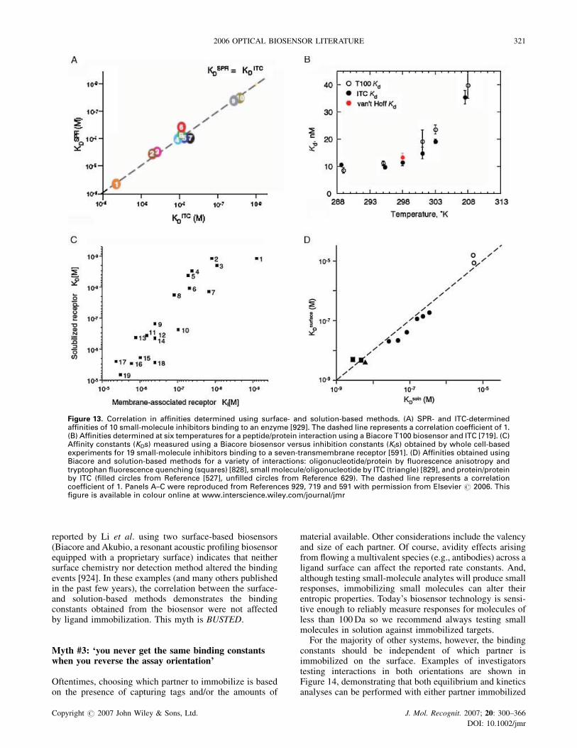
reported by Li et al. using two surface-based biosensors(Biacore and Akubio, a resonant acoustic profiling biosensorequipped with a proprietary surface) indicates that neithersurface chemistry nor detection method altered the bindingevents [924]. In these examples (and many others publishedin the past few years), the correlation between the surface-and solution-based methods demonstrates the bindingconstants obtained from the biosensor were not affectedby ligand immobilization. This myth is BUSTED.
Myth #3: ‘you never get the same binding constantswhen you reverse the assay orientation’
Oftentimes, choosing which partner to immobilize is basedon the presence of capturing tags and/or the amounts of
material available. Other considerations include the valencyand size of each partner. Of course, avidity effects arisingfrom flowing a multivalent species (e.g., antibodies) across aligand surface can affect the reported rate constants. And,although testing small-molecule analytes will produce smallresponses, immobilizing small molecules can alter theirentropic properties. Today’s biosensor technology is sensi-tive enough to reliably measure responses for molecules ofless than 100Da so we recommend always testing smallmolecules in solution against immobilized targets.
For the majority of other systems, however, the bindingconstants should be independent of which partner isimmobilized on the surface. Examples of investigatorstesting interactions in both orientations are shown inFigure 14, demonstrating that both equilibrium and kineticsanalyses can be performed with either partner immobilized
Figure 13. Correlation in affinities determined using surface- and solution-based methods. (A) SPR- and ITC-determinedaffinities of 10 small-molecule inhibitors binding to an enzyme [929]. The dashed line represents a correlation coefficient of 1.(B) Affinities determined at six temperatures for a peptide/protein interaction using a Biacore T100 biosensor and ITC [719]. (C)Affinity constants (KDs) measured using a Biacore biosensor versus inhibition constants (KIs) obtained by whole cell-basedexperiments for 19 small-molecule inhibitors binding to a seven-transmembrane receptor [591]. (D) Affinities obtained usingBiacore and solution-based methods for a variety of interactions: oligonucleotide/protein by fluorescence anisotropy andtryptophan fluorescence quenching (squares) [828], small molecule/oligonucleotide by ITC (triangle) [829], and protein/proteinby ITC (filled circles from Reference [527], unfilled circles from Reference 629). The dashed line represents a correlationcoefficient of 1. Panels A–C were reproduced from References 929, 719 and 591 with permission from Elsevier # 2006. Thisfigure is available in colour online at www.interscience.wiley.com/journal/jmr
Copyright # 2007 John Wiley & Sons, Ltd. J. Mol. Recognit. 2007; 20: 300–366
DOI: 10.1002/jmr
2006 OPTICAL BIOSENSOR LITERATURE 321

Figure 14. Interactions examined in both orientations. (A) Equilibrium analysis ofthe HLA-G (a MHCI molecule) monomer/leukocyte Ig-like receptor B1 (LILRB1)interaction. Left: responses and binding isotherm (black) for the HLA-G monomerbinding to the immobilized receptor. Right: responses and binding isotherm(green) for the receptor in solution binding to immobilized HLA-G. The isothermsincluded in the bottom panels for other constructs are explained in Reference 612.(B) Kinetic analysis of the human serum albumin (HSA)/MHC-related Fc receptorfor IgG (FcRn) using immobilized receptor (top) or immobilized HSA (bottom).Responses overlaid with the fit of a 1:1 interaction model are shown in the leftpanels and the effect of pH on the rate constants is summarized in the right panels.Reproduced from References 527 and 612 with permission from the AmericanChemical Society and The American Society for Biochemistry and MolecularBiology # 2006. This figure is available in colour online at www.interscience.wiley.com/journal/jmr
Copyright # 2007 John Wiley & Sons, Ltd. J. Mol. Recognit. 2007; 20: 300–366
DOI: 10.1002/jmr
322 R. L. RICH AND D. G. MYSZKA

to obtain similar binding parameters (and, the overlay of thefit and responses in Figure 14B suggest that this experimentcould be further optimized so that the rate constantsdetermined in the two orientations may agree even more)[527,612]. Whenever feasible, we encourage researchers toexamine interactions in both orientations (and explore avariety of different immobilization methods). We declarethis myth BUSTED.
Myth #4: ‘kinetics can provide detailed informationabout binding interactions’
In addition to the example shown in Figure 8C, Figure 15provides additional evidence that supports the importance ofdetermining rate constants. For example, Arnold and
Fremont [518] kinetically characterized the chemokineCCL3 binding to wild type and 12 mutant EVM1(Ectromelia virus, Moscow strain) proteins (two data setswith overlaid fits of a 1:1 interaction model are shown in theinset of Figure 15A). A kinetic distribution plot of theseinteractions revealed two kinetically distinct classes thatcluster based on kd (Figure 15A main panel). Since the twoclasses of interactions display similar (and relatively narrow)ranges in ka, the differences in affinity (clustered at18–140 pM and 0.72–10 nM) can be attributed to how eachmutation affected the stability of the CCL3/EVM1 complex.
Panels B–D in Figure 15 further emphasize the wealth ofinformation available from kinetic analyses. In Figure 15B,several compounds cluster along the KD¼ 10�6M iso-affinity line, but display different rate constants. Likewise,the kinetic distribution plots of two antibody/antigen studies
Figure 15. Kinetic distribution plots. (A) ka versus kd plotted for the chemokine CCL3 binding towild type andmutant EVM1proteins. Thetwo classes of interactions are shown in green and red. The insets show the responses and fits of example interactions (corresponding tothe unfilled circles in the main panel) from the two kinetic classes. Rate constants plotted in the main panel were published in Reference518. (B) ka versus kd plotted for 15–19 replicate studies of 10 small-molecule inhibitors (labeled 1–10) binding to an enzyme. (C) ka versuskd plotted for 90 positivemAbs selected froma screenof 384 hybridoma samples. (D) ka versus kd plotted for Fab showing the progressivekinetic effects during affinity maturation. Panels B–D and the insets in panel A were reproduced from References 518, 929, 455 and 435with permission from the American Society of Microbiology and Elsevier # 2006. This figure is available in colour online at www.interscience.wiley.com/journal/jmr
Copyright # 2007 John Wiley & Sons, Ltd. J. Mol. Recognit. 2007; 20: 300–366
DOI: 10.1002/jmr
2006 OPTICAL BIOSENSOR LITERATURE 323

(Figure 15C and 15D) emphasize the mechanistic differ-ences between antibodies displaying similar antigenaffinities (e.g., the data points marked with an asterisk inFigure 15C). These differences in kinetics should not beoverlooked when selecting which antibodies and com-pounds to pursue as therapeutic candidates and in drugdevelopment. These examples are only a few of the manykinetic experiments that turn this myth into a FACT.
Myth #5: ‘optical biosensors cannot measure theaffinities of weak interactions’
Much of the interaction technology at our disposal (e.g.,ELISA, pull-downs, fluorescence, and radio-ligand assays)is geared toward measuring high-affinity systems. Unfortu-nately, these technologies often have restrictions when itcomes to measuring low affinities. These separation assaysrequire washing steps before a signal is recorded. When theinteraction is weak, the complex may dissociate and bewashed away before being detected. And, although othersolution methods (e.g., NMR, AUC, and ITC) measure theamount of complex formed in the presence of free materialand therefore can be used to study weak interactions, theytypically require large amounts of sample and/or have lowthroughput.
Optical biosensors, on the other hand, are well suited toanalyzing weak interactions since they directly measure theamount of complex in the presence of free material, requireno labels, consume little sample, and have comparativelyhigh throughput. Also, the biosensor surface itself can beused to extract a binding partner from crude samples, whichdramatically reduces sample preparation time. The factorsthat may limit biosensor-based measurements of weakinteractions (i.e., high analyte concentrations and lownon-specific binding) are not inherent to this technology;instead, these are limiting factors for any method used toexamine transient binding events.
Figure 16 depicts two group’s biosensor-based charac-terization of very weak (KDs� 250–2000mM) interactions.In two separate experiments, Kato et al. [661,662] measuredthe responses for a protein (at concentrations up to 482mM)binding to a peptide surface to obtain KD¼ 260� 20mM(Figure 16A). Figure 16B shows the responses and isothermsfor a protein, ubiquitin (at concentrations up to 2500mM),binding to a panel of immobilized protein binding partners(the wild type partner and four mutants) [108]. Since theresponses for ubiquitin binding by the wild type protein andthe L10D mutant provide information about surfacecapacity, the affinities of each of the interactions could bedetermined. References 114 and 213 provide other examplesof Sundquist and co-workers’ studies of particularlylow-affinity interactions. This myth is BUSTED.
Myth #6: ‘mass transport makes it impossible toresolve kinetics’
For years now, modeling tools have been available thatextract binding parameters from mass transport-influencedresponses. Perhaps this myth arises when users do notrecognize mass transport effects in their data sets and try to
fit the responses to a simple 1:1 interaction model. In bindingresponse profiles, mass transport’s most apparent effect isthe early portion of the association phase becomes morelinear. In addition, the dissociation of the complex willappear slower than expected based on the kd determined bymodel fitting. These effects are illustrated in Figure 17A:mass transport-influenced responses are shown in the leftpanels while the responses in the right panels do not displaymass transport effects [432].
Figure 17B shows two examples of responses obtainedunder particularly mass transport-limiting conditions [897].To illustrate what the responses would look like if masstransport was not occurring, these data sets are overlaid witha set of simulated responses (shown in green) we modeledusing the reported rate constants. This overlay emphasizeshow mass transport slows down the apparent rate in both theassociation and dissociation phases. (Hint: we recommendalways fitting responses to a 1:1 interaction model thatincludes a mass transport term. If the interaction is notinfluenced by mass transport, the mass transport parameterin the fit will approach infinity and the intrinsic rateconstants will be unaffected.) This myth is so BUSTED.
Myth #7: optical biosensors can be used to detectwhole cells binding to immobilized targets
Traditionally, we have been leary about using the biosensorto examine whole cells binding to immobilized targets forseveral reasons. First, the delivery of cells to the sensorsurface is difficult since under flow larger particles will befocused midstream rather than diffuse to the edges of thechannel. Second, quantitative cell binding analyses wouldneed to account for avidity effects since cells will bindmultivalently to the surface. And, the biosensor datapublished in most of the reports of cell binding experimentshas been dubious at best. Too often the responses were oddlyshaped and/or very low (<20RU). Since cells are very largeanalytes, we expect the binding signals produced to be verylarge. Three studies published in 2006 suggest thatbiosensor-based cell binding studies may in fact be feasible.Each of the experiments shown in Figure 18 showreasonably shaped binding profiles and the responses aremore on the order of what we would expect for largeanalytes. In addition, each analysis included several controlexperiments to confirm the specificity and reproducibility ofthe cell/ligand interaction.
Using the biosensor, Oli et al. [1043] compared how threeStreptococcus mutans preparations recognize salivaryagglutinin. Cells expressing the surface adhesion P1 boundto the immobilized agglutinin, while a P1-negative mutantand a sonicated supernatant did not (Figure 18A). Byregenerating the ligand surface and retesting cell bindingseveral times, these researchers demonstrated the reprodu-cibility of these cell binding responses.
To investigate the structure of bacterial outer-membraneproteins (Omps), Visudtiphole et al. [1049] examined apanel of E. coli expressing various OmpG mutants thatcontained inserted FLAG sequences. Biosensor analysesrevealed which FLAG sequences (and neighboring OmpGregions) were exposed to the environment or inaccessible ineither the membrane or the periplasmic space (Figure 18B).
Copyright # 2007 John Wiley & Sons, Ltd. J. Mol. Recognit. 2007; 20: 300–366
DOI: 10.1002/jmr
324 R. L. RICH AND D. G. MYSZKA

To confirm these findings, this group compared the whole-cell responses with those obtained after disrupting the cells.
Hearty et al. [1041] developed an assay to characterizeListeria monocytogenes binding to an antibody that recog-nizes a surface-localized protein expressed in L. monocyto-
genes but not in non-L. monocytogenes cells (Figure 18C).As control experiments, this group showed that heatinactivation of the cells eliminated binding, a panel ofnon-L. monocytogenes cells did not bind to the antibodysurface, and the L. monocytogenes responses were concen-
Figure 16. Biosensor-determined affinities of weak interactions. (A) Two independent bio-sensor measurements of the affinity of a WW domain protein binding to its immobilizedpeptide binding partner. Reproduced from References 661 and 662with permission from TheAmerican Society for Biochemistry and Molecular Biology and Bentham Science PublishersLtd.# 2006. (B) Response data and isotherms for ubiquitin binding to GST-tagged wild typeand fourmutant constructs derived from the ESCRT-II protein captured on the sensor surface.This figure was provided by the authors (personal communication) and are summarized inSupplemental Table 2 of Reference 108. This figure is available in colour online at www.interscience.wiley.com/journal/jmr
Copyright # 2007 John Wiley & Sons, Ltd. J. Mol. Recognit. 2007; 20: 300–366
DOI: 10.1002/jmr
2006 OPTICAL BIOSENSOR LITERATURE 325

tration dependent. These studies served as the basis foridentifying and isolating the L. monocytogenes surfaceprotein that may serve as a marker in analyses to detectL. monocytogenes and other pathogens in food samples.
Based on these reports, we may be witnessing theearly stages of assay development to obtain reliable cellbinding data using the biosensor. We find this mythPLAUSIBLE.
Figure 17. Binding responses influenced by mass transport. (A) Responses for concentration series of two polyamides binding toimmobilized double-stranded hairpin DNA containing the sequences TGCACA (left) and AGCACA (right). Each data set is overlaidwith the fit of a 1:1 interaction model (the model used to fit data in the left panels included a mass transport term). (B) Responses(black lines) for a concentration (HEL) binding to immobilized anti-HEL mAbs HC1 (left), HC2 (right). The red lines depict the fit of a1:1 interaction model that includes a mass transport term. Green lines are simulations that depict the expected responses if masstransport did not occur. Reproduced from References 432 and 897 with permission from Blackwell Publishing and the AmericanChemical Society # 2006. This figure is available in colour online at www.interscience.wiley.com/journal/jmr
Copyright # 2007 John Wiley & Sons, Ltd. J. Mol. Recognit. 2007; 20: 300–366
DOI: 10.1002/jmr
326 R. L. RICH AND D. G. MYSZKA

Myth #8: ‘always immobilize the multivalentbinding partner’
Most of the time we caution against testing the multivalentbinding partner in solution since this can complicate theanalysis by introducing avidity. However, there are timeswhen we may want to mimic the avidity events that occurin vivo; for example, when antibodies bind to receptorsexposed on the cell surface (Figure 19A). The biosensorsurface can be used to mimic this behaviour by testingantibody in solution binding to immobilized receptors(Figure 19B). We look forward to seeing good examplespublished that describe this approach to using biosensors andwe call this myth BUSTED.
Myth #9: ‘optical biosensors can be used to measureconformational changes’
Actually, this myth began in 1995 when we first introducedthe concept of using numerical integration, which permitsone to create any reaction mechanism (Anal. Biochem. 227:176–185). Then, we showed this two-step conformational-change model (AþB¼AB¼AB�) could describe a variety
Figure 18. Intact cells binding to ligand surfaces. (A) S. mutans(wild type, mutant, and sonicated) binding to immobilized sali-vary agglutinin. (B) Responses for E. coli expressing a panel ofFLAG-tagged Omp mutants binding to immobilized anti-FLAGantibody. (C) Responses for L. monocytogenes cells binding toan immobilized L.monocytogenes-specificmAb before and afterheat inactivation of the cells. Reproduced from References 1041and 1043, and 1049 with permission from Elsevier # 2006.
Figure 19. Example of using the biosensor to mimic biologicallyrelevant avidity events. (A) In vivo, a bivalent antibody wouldbind simultaneously to two receptors presented on the cell sur-face. (B) Biosensor assay design thatmimics antibody binding tocell surface-presented receptors: antibody in solution is testedfor binding to immobilized receptors. This figure is available incolour online at www.interscience.wiley.com/journal/jmr
Figure 20. Monitoring self-association events using optical bio-sensors. (A) Assay design for a biosensor experiment intended tomonitor self-association between molecules in solution andimmobilized on the sensor surface. (B) Since the dextran layeris non-rigid and analyte should be tested at concentrations nearKD, the immobilized molecules, as well as those in solution, canassociate, thereby blocking any surface/solution interaction. Thisfigure is available in colour online at www.interscience.wiley.com/journal/jmr
Copyright # 2007 John Wiley & Sons, Ltd. J. Mol. Recognit. 2007; 20: 300–366
DOI: 10.1002/jmr
2006 OPTICAL BIOSENSOR LITERATURE 327

Figure 21. Kinetic analyses of an antibody/antigen interaction performed by 22 study participants (A–V). (A) Except for V, each panelincludes the responses obtained for antigen binding to antibody immobilized at three densities. The three left data sets depict antigenconcentration series examined using a relatively short dissociation time and the right three data sets show responses collected for thehighest antigen concentration using amuch longer dissociation time. The responses (black) are overlaid with the fit a of a 1:1 interactionmodel (red). (B) Distribution of the kinetic rates and equilibrium binding constants determined by the participants. The average value ineach histogram ismarked by a dotted line and the standard deviation is shown in gray. Reproduced from Reference 395 with permissionfrom Elsevier # 2006. This figure is available in colour online at www.interscience.wiley.com/journal/jmr
Copyright # 2007 John Wiley & Sons, Ltd. J. Mol. Recognit. 2007; 20: 300–366
DOI: 10.1002/jmr
328 R. L. RICH AND D. G. MYSZKA

Figure 22. Kinetic analyses of 10 smallmolecules (labeled 1–10 across the top row) binding toone protein target interaction performed by 26 study participants (B–AA). The responses(black) are overlaid with the fit a of a 1:1 interactionmodel (red). Some data sets could not befit to obtain binding parameters; individual data sets omitted from the analysis are indicatedby red Xs and data from participants A, J, K, N, O, P, R, and S were excluded entirely.Reproduced from Reference 929 with permission from Elsevier # 2006. This figure isavailable in colour online at www.interscience.wiley.com/journal/jmr
Copyright # 2007 John Wiley & Sons, Ltd. J. Mol. Recognit. 2007; 20: 300–366
DOI: 10.1002/jmr
2006 OPTICAL BIOSENSOR LITERATURE 329

of complex response profiles—not necessarily only thosewith a conformational change. Unfortunately, we often findgroups applying this model just because ‘it fits the data’.
In 2006, several authors used the conformational-changemodel. If you look carefully at their results, the confor-mational change was predicted to occur on a time scale ofminutes to hours. We find it hard to believe that abiologically important conformational change would takethis long to complete. (When you look at classically studiedconformational-change systems, you see that these eventsoccur on the nanosecond to millisecond time scale, which istoo fast for biosensors to measure). In today’s literature wefind too many groups go fishing to find the model that fitstheir data and become somehow fixated on the conforma-tional-change model. Instead, it is much more likely thatwhat they are seeing in their sensor data is a reflection ofaggregation, avidity, or heterogeneity. Until we seeconvincing evidence that a half-life of 60min is biologicallyrelevant, we recommend you stay away from this model andcall this myth BUSTED.
Myth #10: ‘optical biosensors can be usedto monitor self-association’
We think biosensor technology is powerful but we admit thatit does have some limits. Nothing makes us chuckle morethan seeing groups apply sensor technology to study aself-associating events like dimerization. This will not workfor several reasons because, under the conditions used in abiosensor assay, the molecules on the surface (and those insolution) will self-associate even before the analyteinjection. First, let us assume you immobilize a proteinonto a dextran surface (which is the most commonapproach). Numerous groups have shown how the dextransurface is fluid and mobile. This means that one immobilizedprotein molecule can easily find another immobilizedbinding partner to self-associate with. The next problemis that once immobilized, the protein’s local concentration isvery high. So, the immobilized protein is mobile and itsbinding partner is very close by, which helps drive complexformation. Therefore, the most thermodynamically stablestate is for the surface-immobilized molecules to self-associate. Finally, for a significant population of ligand/analyte complexes to form, the analyte needs to be injectedat concentrations near (or above) the KD of the interaction. Ifthe self-associating protein is in solution at a concentrationnear its KD, by definition half of the protein molecules willbe self-associated and therefore not available to interact withthe sensor surface. So, in this example you would be in-jecting a self-associated protein over a surface that is alreadyself-associated and you will see no binding. (Figure 20)
In the 17 years of commercial biosensor literature, youwill not find a single example of where someone has taken awell-characterized dimeric system and used the biosensor toextract the correct binding constants. Our point is that thereare times when other technologies are better suited to answera question (Hint: to characterize self-association, useanalytical ultracentrifugation), so we call this myth BUSTED.
Myth #11: ‘there must be some trickto getting good data’
We suspect that much of the bad biosensor data we see resultfrom dirty instruments, poorly behaved reagents, suboptimalassay design, and/or sloppy experimental technique. Todemonstrate that biosensor users ranging in expertiselevel and using a variety of biosensors could obtainhigh-quality data, we oversaw two studies that determinedthe reliablity of biosensor data obtained by differentexperimentalists. For these comparative studies, 22 partici-pants examined a high-affinity antibody/antigen interactionand 26 participants examined 10 small molecules bindingto one protein target. To gauge only the variability betweenthe participants’ techniques, we provided each with thematerials required for the analyses as well as a detailedprotocol.
The data obtained from these studies are summarized inFigures 21 and 22. As shown in Figure 21A, each of the 22participants obtained interpretable responses for theantibody/antigen interaction (monitored at three surfacedensities of antibody). Each data set was well described by a1:1 interaction model and the variability in the reported rateconstants was�14% (Figure 21B). In the second study, 19 ofthe 26 participants were able to measure interactionparameters for most, if not all, of the 10 compoundsbinding to the protein target (Figure 22). The experimentalstandard error averaged 31% for the small-molecule study.The higher standard error in this study was not unexpectedsince this was a more challenging experiment (the variabilityin these rate constants is demonstrated in the kineticdistribution plot shown in Figure 15B.).
These studies, together with other similar comparativestudies, emphasize that anyone using a well-maintainedinstrument, characterizing a well-behaved biological sys-tem, and employing careful experimental design can obtaindata of similar quality to that shown in Figures 21 and 22.This myth is BUSTED.
SUMMARY
Perhaps we will look back on 2006 as a major turning pointin the optical biosensor field. The technology’s breadth ofapplications has expanded in ways we could not havepredicted a few years ago. Recent innovations in instru-mentation have increased sampling throughput and sensi-tivity while the results obtained from SPR array platformssuggest we will see widespread adoption of this imagingformat in the near future. Now we are approaching a criticalmass of users. As the skill level within the communityimproves, we foresee more investigators publishing high-quality data and more manuscript reviewers recognizing theelements required in a well-performed biosensor exper-iment. Without a doubt, the increasing contributions fromoptical biosensors will continue to advance basic researchand drug discovery, making sure none of us are left home inthe dark.
Copyright # 2007 John Wiley & Sons, Ltd. J. Mol. Recognit. 2007; 20: 300–366
DOI: 10.1002/jmr
330 R. L. RICH AND D. G. MYSZKA
REFERENCES
Reviews
Optical biosensors1. Bergwerff AA, van Knapen F. 2006. Surface plasmon reson-
ance biosensors for detection of pathogenic microorgan-isms: strategies to secure food and environmental safety.J. AOAC Int. 89: 826–831.
2. Besenicar M, Macek P, Lakey JH, Anderluh G. 2006. Surfaceplasmon resonance in protein-membrane interactions.Chem. Phys. Lipids 141: 169–178.
3. Boozer C, Kim G, Cong S, Guan H, Londergan T. 2006.Looking towards label-free biomolecular interaction analysisin a high-throughput format: a review of new surface plas-mon resonance technologies. Curr. Opin. Biotechnol. 17:400–405.
4. Calander N. 2006. Molecular detection and analysis byusing surface plasmon resonances. Curr. Anal. Chem. 2:203–211.
5. Cooper MA. 2006. Optical biosensors: where next and howsoon? Drug Discov. Today 11: 1061–1067.
6. Fang Y. 2006. Label-free cell-based assays with optical bio-sensors in drug discovery. Assay Drug Devel. Technol. 4:583–595.
7. Gambari R, Feriotto G. 2006. Surface plasmon resonance fordetection of genetically modified organisms in the foodsupply. J. AOAC Int. 89: 893–897.
8. Haughey SA, Baxter GA. 2006. Biosensor screening forveterinary drug residues in foodstuffs. J. AOAC Int. 89:862–867.
9. Homola J (ed.). 2006. Surface Plasmon Resonance BasedSensors. Springer Series on Chemical Sensors and Biosen-sors, vol. 4. Springer: Berlin.
10. Huber W, Mueller F. 2006. Biomolecular interaction analysisin drug discovery using surface plasmon resonance technol-ogy. Curr. Pharm. Des. 12: 3999–4021.
11. Indyk HE. 2006. Optical biosensors: making sense of inter-actions. Chem. New Zealand July: 42–46.
12. Jonsson H, Eriksson A, Yman IM. 2006. Detecting foodallergens with a surface plasmon resonance immunoassay.InDetectingAllergens in Food, KoppelmanSJ, HefleSL (eds).Woodhead Publishing Ltd: Cambridge, UK; 158–174.
13. Koga H, Kyo M, Usui-Aoki K, Inamori K. 2006. A chip-basedminiaturized format for protein-expression profiling: theexploitation of comprehensively produced antibodies. Elec-trophoresis 27: 3676–3683.
14. Lacy A, Dunne L, Fitzpatrick B, Daly S, Keating G, Baxter A,Hearty S, O’Kennedy R. 2006. Rapid analysis of coumarinsusing surface plasmon resonance. J. AOAC Int. 89: 884–892.
15. Pattnaik P, Srivastav A. 2006. Surface plasmon resonance—applications in food science research: a review. J. Food Sci.Technol. 43: 329–336.
16. Rich RL, Myszka DG. 2006. Survey of the year 2005 com-mercial optical biosensor literature. J. Mol. Recognit. 19:478–534.
Interaction technologies
17. Aponte VM, Finch DS, Klaus DM. 2006. Considerations fornon-invasive in-flight monitoring of astronaut immune sta-tus with potential use of MEMS and NEMS devices. Life Sci.79: 1317–1333.
18. Arora K, Chand S, Malhotra BD. 2006. Recent developmentsin bio-molecular electronics techniques for food pathogens.Anal. Chim. Acta 568: 259–274.
19. Bally M, Halter M, Voros J, Grandin HM. 2006. Opticalmicroarray biosensing techniques. Surf. Interface Anal. 38:1442–1458.
20. Bilitewski U. 2006. Protein-sensing assay formats anddevices. Anal. Chim. Acta 568: 232–247.
21. Bonetta L. 2006. Microarrays: branching out from expressionanalysis. Nat. Meth. 3: 571–578.
22. Cooper MA. 2006. Current biosensor technologies in drugdiscovery. Drug Discov. World Summer: 68–82.
23. Cummings MD, Farnum MA, Nelen MI. 2006. Universalscreening methods and applications of ThermoFluor1.J. Biomol. Screen. 11: 854–863.
24. Eshaque B, Dixon B. 2006. Technology platforms for mol-ecular diagnosis of cystic fibrosis. Biotechnol. Adv. 24:86–93.
25. Evans PA, Hawkins K, Williams PR. 2006. Rheometry forblood coagulation studies. In Rheology Reviews 2006, Bind-ing DM, Waters K (eds). The British Society of Rheology:Cambridge, UK; 255–291.
26. Goldschmidt MC. 2006. The use of biosensor andmicroarraytechniques in the rapid detection and identification of Sal-monellae. J. AOAC Int. 89: 530–537.
27. Goodnow RA Jr. 2006. Hit and lead identification: integratedtechnology-based approaches. Drug Discov. Today: Tech-nol. 3: 367–375.
28. Hultschig C, Kreutzberger J, Seitz H, Konthur Z, Bussow K,Lehrach H. 2006. Recent advances of protein microarrays.Curr. Opin. Chem. Biol. 10: 4–10.
29. Ince R, Narayanaswamy R. 2006. Analysis of the perform-ance of interferometry, surface plasmon resonance andluminescence as biosensors and chemosensors.Anal. Chim.Acta 569: 1–20.
30. Ivanov YD, Govorun VM, Bykov VA, Archakov AI. 2006.Nanotechnologies in proteomics. Proteomics 6: 1399–1414.
31. Jackson DB, Stein MA, Merino A, Eils R. 2006. Microarraysmeet the Voltaire challenge: drug discovery on a chip? DrugDiscov. Today: Technol. 3: 153–161.
32. Kricka LJ, Master SR, Joos TO, Fortina P. 2006. Currentperspectives in protein array technology. Ann. Clin.Biochem. 43: 457–467.
33. Kuroda K, Kato M, Mima J, Ueda M. 2006. Systems for thedetection and analysis of protein-protein interactions. Appl.Microbiol. Biotechnol. 71: 127–136.
34. Ludden MJW, Reinhoudt DN, Huskens J. 2006. Molecularprintboards: versatile platforms for the creation and posi-tioning of supramolecular assemblies and materials. Chem.Soc. Rev. 35: 1122–1134.
35. MaH, Horiuchi KY. 2006. Chemicalmicroarray: a new tool fordrug screening and discovery. Drug Discov. Today 11:661–668.
36. Marquette CA, Blum LJ. 2006. State of the art and recentadvances in immunoanalytical systems. Biosens. Bioelec-tron. 21: 1424–1433.
37. Patel PD. 2006. Overview of affinity biosensors in foodanalysis. J. AOAC Int. 89: 805–818.
38. Rasooly A, Herold KE. 2006. Biosensors for the analysis offood- and waterborne pathogens and their toxins. J. AOACInt. 89: 873–883.
39. Rodriguez-Mozaz S, Lopez de Alda MJ, Barcelo D. 2006.Biosensors as useful tools for environmental analysis andmonitoring. Anal. Bioanal. Chem. 386: 1025–1041.
40. Rogers KR. 2006. Recent advances in biosensor techniquesfor environmental monitoring. Anal. Chim. Acta 568:222–231.
41. Schaferling M, Nagl S. 2006. Optical technologies for theread out and quality control of DNA and proteinmicroarrays.Anal. Bioanal. Chem. 385: 500–517.
42. Shin SK, Yoon H-J, Jung YJ, Park JW. 2006. Nanoscalecontrolled self-assembled monolayers and quantum dots.Curr. Opin. Chem. Biol. 10: 423–429.
43. Soper SA, Brown K, Ellington A, Frazier B, Garcia-Manero G,Gau V, Gutman SI, Hayes DF, Korte B, Landers JL, Larson D,Ligler F, Majumdar A, Mascini M, Nolte D, Rosenzweig Z,Wang J, Wilson D. 2006. Point-of-care biosensor systems forcancer diagnostics/prognostics. Biosens. Bioelectron. 21:1932–1942.
44. Starodub NF. 2006. Biosensors for the evaluation of lipaseactivity. J. Mol. Catalysis B 40: 155–160.
Copyright # 2007 John Wiley & Sons, Ltd. J. Mol. Recognit. 2007; 20: 300–366
DOI: 10.1002/jmr
2006 OPTICAL BIOSENSOR LITERATURE 331

45. Timlin JA. 2006. Scanningmicroarrays: currentmethods andfuture directions. Meth. Enzymol. 411: 79–98.
46. Toldra F, Reig M. 2006. Methods for rapid detection ofchemical and veterinary drug residues in animal foods.Trends Food Sci. Technol. 17: 482–489.
47. Yu X, Xu D, Cheng Q. 2006. Label-free detectionmethods forprotein microarrays. Proteomics 6: 5493–5503.
48. Zheng MZ, Richard JL, Binder J. 2006. A review of rapidmethods for the analysis of mycotoxins. Mycopathologia161: 261–273.
Biological applications49. Bartoli S, Fincham CI, Fattori D. 2006. The fragment-
approach: an update. Drug Discov. Today: Technol. 3:425–431.
50. Bossen C, Schneider P. 2006. BAFF, APRIL and their recep-tors: structure, function and signaling. Semin. Immunol. 18:263–275.
51. Campbell CG, Borglin SE, Green FB, Grayson A, Wozei E,Stringfellow WT. 2006. Biologically directed environmentalmonitoring, fate, and transport of estrogenic endocrine dis-rupting compounds in water: a review. Chemosphere 65:1265–1280.
52. Chen S, Chen L, Chen K, Shen X, Jiang H. 2006. Techniquesused for the discovery of therapeutic compounds: thecase of SARS. Drug Discov. Today: Technol. 3: 277–283.
53. Cho W, Stahelin RV. 2006. Membrane binding and subcel-lular targeting of C2 domains. Biochim. Biophys. Acta 1761:838–849.
54. Copeland RA, Pompliano DL, Meek TD. 2006. Drug-targetresidence time and its implications for lead optimization.Nat. Rev. Drug Discov. 5: 730–739.
55. Coullerez G, Seeberger PH, Textor M. 2006. Mergingorganic and polymer chemistries to create glycomaterialsfor glycomics applications. Macromol. Biosci. 6: 634–647.
56. Cretich M, Damin F, Pirri G, Chiari M. 2006. Protein andpeptide arrays: recent trends and new directions. Biomol.Eng. 23: 77–88.
57. Dillon SR, Gross JA, Ansell SM, Novak AJ. 2006. An APRIL toremember: novel TNF ligands as therapeutic targets. Nat.Rev. Drug Discov. 5: 235–246.
58. Dinarello CA, Kim S-H. 2006. IL-32, a novel cytokine with apossible role in disease. Ann Rheum. Dis. 65(Suppl. III):iii61–iii64.
59. DiNitto JP, Lambright DG. 2006. Membrane and juxtamem-brane targeting by PH and PTB domains. Biochim. Biophys.Acta 1761: 850–867.
60. Downard KM. 2006. Ions of the interactome: the role ofMS inthe study of protein interactions in proteomics and structuralbiology. Proteomics 6: 5374–5384.
61. Dzugaj A. 2006. Localization and regulation of muscle fruc-tose-1,6-bisphosphatase, the key enzyme of glyconeogen-esis. Adv. Enzyme Reg. 46: 51–71.
62. EisensteinM. 2006. ‘Chain gang’ delivers for hard labor.Nat.Meth. 3: 753–762.
63. Fajkus J. 2006. Detection of telomerase activity by the TRAPassay and its variants and alternatives. Clin. Chim. Acta 371:25–31.
64. Glynn B, Lahiff S, Wernecke M, Barry T, Smith TJ, Maher M.2006. Current and emerging molecular diagnostic technol-ogies applicable to bacterial food safety. Int. J. Dairy Tech-nol. 59: 126–139.
65. Goodchild S, Love T, Hopkins N, Mayers C. 2006. Engineer-ing antibodies for biosensor technologies. Adv. Appl. Micro-biol. 58: 185–226.
66. Gooding JJ. 2006. Biosensor technology for detecting bio-logical warfare agents: recent progress and future trends.Anal. Chim. Acta 559: 137–151.
67. Haab BB, Paulovich AG, Anderson NL, Clark AM, DowningGJ, Hermjakob H, LaBaer J, Uhlen M. 2006. A reagentresource to identify proteins and peptides of interestfor the cancer community. Mol. Cell. Proteomics 5:1996–2007.
68. He L, Dexter AF, Middelberg APJ. 2006. Biomolecularengineering at interfaces. Chem. Engi. Sci. 61: 989–1003.
69. Howell JM, Winstone TL, Coorssen JR, Turner RJ. 2006.An evaluation of in vitro protein-protein interaction tech-niques: assessing contaminating background proteins. Pro-teomics 6: 2050–2069.
70. Kalman A, Caelen I, Svorc J. 2006. Vitamin and pseudovita-min analysis with biosensors in food products—a review.J. AOAC Int. 89: 819–825.
71. Kamada M, Maegawa M, Daitoh T, Mori K, Yamamoto S,Nakagawa K, Yamano S, Irahara M, Aono T, Mori T. 2006.Sperm-zona pellucida interaction and immunological infer-tility. Reprod. Med. Biol. 5: 95–104.
72. Konings EJ. 2006. Water-soluble vitamins. J. AOAC Int. 89:285–288.
73. Kruppa M, Konig B. 2006. Reversible coordinative bonds inmolecular recognition. Chem. Rev. 106: 3520–3560.
74. Kurata S, Ariki S, Kawabata S-i. 2006. Recognition of patho-gens and activation of immune responses in Drosophila andhorseshoe crab innate immunity. Immunobiology 211:237–249.
75. Lee T-H, Aguilar M-I. 2006. Trends in the development andapplication of functional biomembrane surfaces.Biotechnol.Ann. Rev. 12: 85–136.
76. Ler SG, Lee FK, Gopalakrishnakone P. 2006. Trends in detec-tion of warfare agents: detection methods for ricin, staphy-lococcal enterotoxin B and T-2 toxin. J. Chromatogr. A 1133:1–12.
77. ŁosM. 2006. Virus detection today.Mod. Bacteriophage Biol.Biotechnol. 37: 131–152.
78. Mast SW, Moremen KW. 2006. Family 47 a-mannosidases inN-glycan processing. Meth. Enzymol. 415: 31–46.
79. Mattei S, Klein G, Satre M, Aubry L. 2006. Trafficking anddevelopmental signaling: Alix at the crossroads. Eur. J. CellBiol. 85: 925–936.
80. Moll D, Zimmerman B, Gesellchen F, Herberg FW. 2006.Current developments for the in vitro characterization ofprotein interactions. In Proteomics in Drug Research,Hamacher M, Marcus K, Stuhler K, van Hall A, WarscheidB, Meyer HE (eds). Wiley-VCH Verlay GmbH & Co.:Weinheim, Germany, 159–172.
81. Mothershed EA, Whitney AM. 2006. Nucleic acid-basedmethods for the detection of bacterial pathogens: presentand future considerations for the clinical laboratory. Clin.Chim. Acta 363: 206–220.
82. Nandini CD, Sugahara K, Nicola V. 2006. Role of the sulfationpattern of chondroitin sulfate in its biological activities and inthe binding of growth factors. Adv. Pharmacol. 53: 253–279.
83. Narayan K, Lemmon MA. 2006. Determining selectivity ofphosphoinositide-binding domains. Methods 39: 122–133.
84. Nedelkov D. 2006. Mass spectrometry-based immunoassaysfor the next phase of clinical applications. Expert Rev. Pro-teomics 3: 631–640.
85. Pejcic B, De Marco R, Parkinson G. 2006. The role of biosen-sors in the detection of emerging infectious diseases.Analyst 131: 1079–1090.
86. Sciarretta KL, Gordon DJ, Meredith SC. 2006. Peptide-basedinhibitors of amyloid assembly. Meth. Enzymol. 413:273–312.
87. Snook CF, Jones JA, Hannun YA. 2006. Sphingolipid-binding proteins. Biochim. Biophys. Acta 1761: 927–946.
88. Socher I, Kroll H, Santoso S. 2006. Heterogeneity of plateletalloantigens and alloantibodies: new insights into structureand function. Transfus. Med. Hemother. 33: 244–253.
89. Swanson SJ. 2006. Immunogenicity issues in drug develop-ment. J. Immunotoxicol. 3: 165–172.
90. Wadhwa M, Thorpe R. 2006. Strategies and assays for theassessment of unwanted immunogenicity. J. Immuno-toxicol. 3: 115–121.
91. Willis B, Arya DP. 2006. An expanding view of aminoglyco-side-nucleic acid recognition. Adv. Carbohydr. Chem.Biochem. 60: 251–302.
Copyright # 2007 John Wiley & Sons, Ltd. J. Mol. Recognit. 2007; 20: 300–366
DOI: 10.1002/jmr
332 R. L. RICH AND D. G. MYSZKA

92. Zheng LZ, Yao X, Li JH. 2006. Layer-by-layer assembly filmsand their applications in electroanalytical chemistry. Curr.Anal. Chem. 2: 279–296.
Theory
93. Andersson K, Hamalainen MD. 2006. Replacing affinity withbinding kinetics in QSAR studies resolves otherwise con-founded effects. J. Chemometrics 20: 370–375.
94. Edwards DA. 2006. Convection effects in the BIAcore dextranlayer: surface reaction model. Bull. Math. Biol. 68: 627–654.
95. Edwards DA. 2006. Convection effects in thin reaction zones:applications to BIAcore. Siam J. Appl. Math. 66: 1978–1997.
96. Gervais T, Jensen KF. 2006. Mass transport and surfacereactions in microfluidic systems. Chem. Eng. Sci. 61:1102–1121.
97. Lebedev K, Mafe S, Stroeve P. 2006. Convection, diffusionand reaction in a surface-based biosensor: modeling ofcooperativity and binding site competition on the surfaceand in the hydrogel. J. Colloid Interface Sci. 296: 527–537.
98. Nag A, Dinner AR. 2006. Enhancement of diffusion-controlled reaction rates by surface-induced orientationalrestriction. Biophys. J. 90: 896–902.
99. Roper DK. 2006. Enhancing lateralmass transport to improvethe dynamic range of adsorption rates measured by surfaceplasmon resonance. Chem. Eng. Sci. 61: 2557–2564.
100. Snopok BA, Kostyukevich EV. 2006. Kinetic studies of pro-tein-surface interactions: a two-stage model of surfa-ce-induced protein transitions in adsorbed biofilms. Anal.Biochem. 348: 222–231.
101. Zhu XD. 2006. Comparison of two optical techniques forlabel-free detection of biomolecular microarrays on solids.Opt. Commun. 259: 751–753.
Methods
102. Beauharnois ME, Neelamegham S, Matta KL. 2006. Quan-titativemeasurement of selectin-ligand interactions: assaysto identify a sweet pill in a library of carbohydrates. Meth.Mol. Biol. 347: 343–358.
103. Binz SK, DicksonAM, Haring SJ,WoldMS. 2006. Functionalassays for replication protein A (RPA). Meth. Enzymol. 409:11–38.
104. Goodrich TT, Wark AW, Corn RM, Lee HJ. 2006. Surfaceplasmon resonance imaging measurements of proteininteractions with biopolymer microarrays. Meth. Mol. Biol.328: 113–130.
105. Lopez PH, Schnaar RL. 2006. Determination of glycolipid-protein interaction specificity. Meth. Enzymol. 417:205–220.
106. Nedelkov D, Nelson RW. 2006. Surface plasmon resonancemass spectrometry for protein analysis. Meth. Mol. Biol.328: 131–139.
107. Sambrook J, Russell DW. 2006. Analysis of interactingproteins with SPR spectroscopy using BIAcore. Stage 1:preparation of the capture surface and test binding. CSHProtocols. DOI:10.1101/pdb.prot3894
Primary Research Articles
Biacore
Proteins108. AlamSL, Langelier C,Whitby FG, Koirala S, RobinsonH, Hill
CP, SundquistWI. 2006. Structural basis for ubiquitin recog-nition by the human ESCRT-II EAP45 GLUE domain. Nat.Struct. Mol. Biol. 13: 1029–1030.
109. Allan GJ, Tonner E, Szymanowska M, Shand JH, Kelly SM,Phillips K, Clegg RA, Gow IF, Beattie J, Flint DJ. 2006.Cumulative mutagenesis of the basic residues in the201-218 region of insulin-like growth factor (IGF)-binding
protein-5 results in progressive loss of both IGF-I bindingand inhibition of IGF-I biological action. Endocrinology 147:338–349.
110. Alvarado D, Evans TA, Sharma R, Lemmon MA, Duffy JB.2006. Argos mutants define an affinity threshold for Spitzinhibition in vivo. J. Biol. Chem. 281: 28993–29001.
111. Amstutz P, Koch H, Binz HK, Deuber SA, Pluckthun A. 2006.Rapid selection of specific MAP kinase-binders fromdesigned ankyrin repeat protein libraries. Prot. Eng. 19:219–229.
112. Andersson FI, Blakytny R, Kirstein J, Turgay K, Bukau B,Mogk A, Clarke AK. 2006. Cyanobacterial ClpC/HSP100protein displays intrinsic chaperone activity. J. Biol. Chem.281: 5468–5475.
113. Antolik C, Catino DH, Resneck WG, Bloch RJ. 2006. Thetetratricopeptide repeat domains of rapsyn bind directly tocytoplasmic sequences of the muscle-specific kinase.Neuroscience 141: 87–100.
114. Arrigoni R, Alam SL, Wamstad JA, Bardwell VJ, SundquistWI, Schreiber-Agus N. 2006. The Polycomb-associatedprotein Rybp is a ubiquitin binding protein. FEBS Lett.580: 6233–6241.
115. Aukrust I, Evensen L, Hollas H, Berven F, Atkinson RA,Trave G, Flatmark T, Vedeler A. 2006. Engineering, bio-physical characterisation and binding properties of asoluble mutant form of annexin A2 domain IV that adoptsa partially folded conformation. J. Mol. Biol. 363: 469–481.
116. Badura A, Esper B, Ataka K, Grunwald C, Woll C, KuhlmannJ, Heberle J, Rogner M. 2006. Light-driven water splittingfor (bio-)hydrogen production: photosysterm 2 as the cen-tral part of a bioelectrochemical device. Photochem. Photo-biol. 82: 1385–1390.
117. Bækgaard L, Luoni L, De Michelis MI, Palmgren MG. 2006.The plant plasma membrane Ca2þ pump ACA8 containsoverlapping as well as physically separated autoinhibitoryand calmodulin-binding domains. J. Biol. Chem. 281:1058–1065.
118. Bajaj SP, Schmidt AE, Agah S, Bajaj MS, Padmanabhan K.2006. High resolution structures of p-aminobenzamidine-and benzamidine-VIIa/soluble tissue factor. J. Biol. Chem.281: 24873–24888.
119. Baraquet C, Theraulaz L, Guiral M, Lafitte D, Mejean V,Jourlin-Castelli C. 2006. TorT, a member of a new periplas-mic binding protein family, triggers induction of the Torrespiratory system upon trimethylamine N-oxide electron-acceptor binding in Escherichia coli. J. Biol. Chem. 281:38189–38199.
120. Becker A-K, Zeppenfeld T, Staab A, Seitz S, Boos W, MoritaT, Aiba H, Mahr K, Titgemeyer F, Jahreis K. 2006. YeeI, anovel protein involved in modulation of the activity of theglucose-phosphotransferase system in Escherichia coliK-12. J. Bacteriol. 188: 5439–5449.
121. Beissenhirtz MK, Scheller FW, Viezzoli MS, Lisdat F. 2006.Engineered superoxide dismutase monomers for super-oxide biosensor applications. Anal. Chem. 78: 928–935.
122. Bellare P, Kutach AK, Rines AK, Guthrie C, Sontheimer EJ.2006. Ubiquitin binding by a variant Jab1/MPN domain inthe essential pre-mRNA splicing factor Prp8p. RNA 12:292–302.
123. Bergqvist S, Croy CH, Kjaergaard M, Huxford T, Ghosh G,Komives EA. 2006. Thermodynamics reveal that helix fourin the NLS of NF-kB p65 anchors IkBa, forming a very stablecomplex. J. Mol. Biol. 360: 421–434.
124. Bernaudat F, Bulow L. 2006. Combined hydrophobic-metalbinding fusion tags for applications in aqueous two-phasepartitioning. Prot. Expr. Purif. 46: 438–445.
125. Bernini A, Spiga O, Ciutti A, Venditti V, Prischi F, Govern-atori M, Bracci L, Lelli B, Pileri S, Botta M, Barge A, Laschi F,Niccolai N. 2006. NMR studies of BPTI aggregation by usingparamagnetic relaxation reagents. Biochim. Biophys. Acta1764: 856–862.
126. Bertrand Y, Demeule M, Rivard G-E, Beliveau R. 2006.Stimulation of tPA-dependent provisional extracellularfibrin matrix degradation by human recombinant soluble
Copyright # 2007 John Wiley & Sons, Ltd. J. Mol. Recognit. 2007; 20: 300–366
DOI: 10.1002/jmr
2006 OPTICAL BIOSENSOR LITERATURE 333

melanotransferrin. Biochim. Biophys. Acta 1763: 1024–1030.
127. Bertsche U, Kast T, Wolf B, Fraipont C, Aarsman MEG,Kannenberg K, von Rechenberg M, Nguyen-Disteche M,den Blaauwen T, Holtje JV, Vollmer W. 2006. Interactionbetween two murein (peptidoglycan) synthases, PBP3and PBP1B, in Escherichia coli. Mol. Microbiol. 61: 675–690.
128. Boukhelifa M, Moza M, Johansson T, Rachlin A, Parast M,Huttelmaier S, Roy P, Jockusch BM, Carpen O, Karlsson R,Otey CA. 2006. The proline-rich protein palladin is a bindingpartner for profilin. FEBS J. 273: 26–33.
129. Bovenschen N, van Stempvoort G, Voorberg J, Mertens K,Meijer AB. 2006. Proteolytic cleavage of factor VIII heavychain is required to expose the binding-site for low-densitylipoprotein receptor-related protein within the A2 domain.J. Thromb. Haemost. 4: 1487–1493.
130. Bozzi M, Sciandra F, Ferri L, Torreri P, Pavoni E, Petrucci TC,Giardina B, Brancaccio A. 2006. Concerted mutation of Pheresidues belonging to the b-dystroglycan ectodomainstrongly inhibits the interaction with a-dystroglycanin vitro. FEBS J. 273: 4929–4943.
131. Brett TJ, Legendre-Guillemin V, McPherson PS, FremontDH. 2006. Structural definition of the F-actin-bindingTHATCH domain from HIP1R. Nat. Struct. Mol. Biol. 13:121–130.
132. Bruck S, Huber TB, Ingham RJ, Kim K, Niederstrasser H,Allen PM, Pawson T, Cooper JA, Shaw AS. 2006. Identifi-cation of a novel inhibitory actin-capping protein bindingmotif in CD2-associated protein. J. Biol. Chem. 281:19196–19203.
133. Bustos VH, Ferrarese A, Venerando A, Marin O, Allende JE,Pinna LA. 2006. The first armadillo repeat is involved in therecognition and regulation of beta-catenin phosphorylationby protein kinase CK1. Proc. Natl Acad. Sci. USA 103:19725–19730.
134. Butcher AJ, Leroy J, Richards MW, Pratt WS, Dolphin AC.2006. The importance of occupancy rather than affinity ofCaVb subunits for the calcium channel I-II linker in relationto calcium channel function. J. Physiol. 574: 387–398.
135. Chang-Yen DA, Myszka DG, Gale BK. 2006. A novel PDMSmicrofluidic spotter for fabrication of protein chips andmicroarrays. J. Microelectromechan. Syst. 15: 1145–1151.
136. Chaudhari A, FialhoAM, Ratner D,Gupta P, HongCS, KahaliS, Yamada T, Haldar K, Murphy S, ChoW, Chauhan VS, DasGupta TK, Chakrabarty AM. 2006. Azurin, Plasmodium fal-ciparum malaria and HIV/AIDS: inhibition of parasitic andviral growth by azurin. Cell Cycle 5: 1642–1648.
137. Chen LH. 2006. Improvement of the specificity of surfaceplasmon resonance with BSA-modified chip. Chin. Chem.Lett. 17: 1619–1622.
138. Cheng Z-Z, Hellwage J, Seeberger H, Zipfel PF, Meri S,Jokiranta TS. 2006. Comparison of surface recognitionand C3b binding properties of mouse and human comp-lement factor H. Mol. Immunol. 43: 972–979.
139. Chien C-H, Tsai C-H, Lin C-H, Chou C-Y, Chen X. 2006.Identification of hydrophobic residues critical for DPP-IVdimerization. Biochemistry 45: 7006–7012.
140. Choi H-J, Huber AH, Weis WI. 2006. Thermodynamics ofb-catenin-ligand interactions: the roles of the N- andC-terminal tails in modulating binding affinity. J. Biol.Chem. 281: 1027–1038.
141. Choi I-D, Kim K-N, Yun C-W, Choi Y-J. 2006. Cloning andcharacterization of the HPr kinase/phosphorylase genefrom Bacillus stearothermophilus No. 236. Biosci. Biotech-nol. Biochem. 70: 1089–1101.
142. Chtcheglova LA, Vogel M, Gruber HJ, Dietler G, Haeberli A.2006. Kinetics of the interaction of desAABB-fibrin mono-mer with immobilized fibrinogen. Biopolymers 83: 69–82.
143. Clark RW, Ruggeri RB, Cunningham D, Bamberger MJ.2006. Description of the torcetrapib series of cholesterylester transfer protein inhibitors, including mechanism ofaction. J. Lipid Res. 47: 537–552.
144. Comoletti D, Flynn RE, BoucardAA, Demeler B, Schirf V, ShiJ, Jennings LL, Newlin HR, Sudhof TC, Taylor P. 2006. Gene
selection, alternative splicing, and post-translational pro-cessing regulate neuroligin selectivity for b-neurexins. Bio-chemistry 45: 12816–12827.
145. Craig SJ, Foong FC, Nordon R. 2006. Engineered proteinscontaining the cohesin and dockerin domains from Clos-tridium thermocellum provides a reversible, high affinityinteraction for biotechnology applications. J. Biotechnol.121: 165–173.
146. Croucher D, Saunders DN, Ranson M. 2006. The urokinase/PAI-2 complex: a new high affinity ligand for the endocy-tosis receptor low density lipoprotein receptor-relatedprotein. J. Biol. Chem. 281: 10206–10213.
147. De Felice M, Esposito L, Rossi M, Pisani FM. 2006. Bio-chemical characterization of two Cdc6/ORC1-like proteinsfrom the crenarchaeon Sulfolobus solfataricus. Extremo-philes 10: 61–70.
148. De Seranno S, Benaud C, Assard N, Khediri S, Gerke V,Baudier J, Delphin C. 2006. Identification of an AHNAKbinding motif specific for the annexin2/S100A10 tetramer.J. Biol. Chem. 281: 35030–35038.
149. Doke AM, Sadana A. 2006. Detection of glucose and relatedanalytes by biosensors: a fractal analysis. Biotechnol. Prog.22: 14–23.
150. Dragovic Z, Shomura Y, Tzvetkov N, Hartl FU, Bracher A.2006. Fes1p acts as a nucleotide exchange factor for theribosome-associated molecular chaperone Ssb1p. Biol.Chem. 387: 1593–1600.
151. DreuxM, Pietschmann T, Granier C, Voisset C, Ricard-BlumS, Mangeot P-E, Keck Z, Foung S, Vu-Dac N, Dubuisson J,Bartenschlager R, Lavillette D, Cosset F-L. 2006. Highdensity lipoprotein inhibits hepatitis C virus-neutralizingantibodies by stimulating cell entry via activation of thescavenger receptor BI. J. Biol. Chem. 281: 18285–18295.
152. Dumelin CE, Scheuermann J, Melkko S, Neri D. 2006.Selection of streptavidin binders from a DNA-encodedchemical library. Bioconjug. Chem. 17: 366–370.
153. Dunn EJ, Philippou H, Ariens RA, Grant PJ. 2006. Molecularmechanisms involved in the resistance of fibrin to clot lysisby plasmin in subjects with type 2 diabetes mellitus. Dia-betologia 49: 1071–1080.
154. Elhamdani A, Azizi F, Solomaha E, Palfrey HC, Artalejo CR.2006. Two mechanistically distinct forms of endocytosis inadrenal chromaffin cells: differential effects of SH3 domainsand amphiphysin antagonism. FEBS Lett. 580: 3263–3269.
155. Erbse A, Schmidt R, Bornemann T, Schneider-Mergener J,Mogk A, Zahn R, Dougan DA, Bukau B. 2006. ClpS is anessential component of the N-end rule pathway in Escher-ichia coli. Nature 439: 753–756.
156. Evans-Nguyen KM, Fuierer RR, Fitchett BD, Tolles LR, Con-boy JC, Schoenfisch MH. 2006. Changes in adsorbed fibri-nogen upon conversion to fibrin. Langmuir 22: 5115–5121.
157. Fiedler E, Fiedler M, Proetzel G, Scheuermann T, Fiedler U,Rudolph R. 2006. AffilinTM molecules novel ligands forbioseparation. Food Bioprod. Process. 84: 3–8.
158. Fribourg C, Meijer AB, Mertens K. 2006. The interfacebetween the EGF2 domain and the protease domain inblood coagulation factor IX contributes to factor VIII bind-ing and factor X activation. Biochemistry 45: 10777–10785.
159. Fry SA, Van de Steen PE, Royle L, Wormald MR, LeathemAJ, Opdenakker G, McDonnell JM, Dwek RA, Rudd PM.2006. Cancer-associated glycoforms of gelatinase B exhibita decreased level of binding to galectin-3. Biochemistry 45:15249–15258.
160. FukudaM. 2006. Distinct Rab27A binding affinities of Slp2-aand Slac2-a/melanophilin: hierarchy of Rab27A effectors.Biochem. Biophys. Res. Commun. 343: 666–674.
161. Fung W-T, Sze K-H, Lee K-F, Shaw P-C. 2006. Functionalstudies of the small subunit of EcoHK31I DNAmethyltrans-ferase. Biol. Chem. 387: 507–513.
162. Gaidukov L, Rosenblat M, Aviram M, Tawfik DS. 2006. The192R/Q polymorphs of serum paraoxonase PON1 differ inHDL binding, lipolactonase stimulation, and cholesterolefflux. J. Lipid Res. 47: 2492–2502.
163. Gallo SA,WangW, Rawat SS, JungG,Waring AJ, Cole AM,Lu H, Yan X, Daly NL, Craik DJ, Jiang S, Lehrer RI,
Copyright # 2007 John Wiley & Sons, Ltd. J. Mol. Recognit. 2007; 20: 300–366
DOI: 10.1002/jmr
334 R. L. RICH AND D. G. MYSZKA

Blumenthal R. 2006. u-defensins prevent HIV-1 Env-mediated fusion by binding gp41 and blocking 6-helixbundle formation. J. Biol. Chem. 281: 18787–18792.
164. Gao J, Lee K, Zhao M, Qiu J, Zhan X, Saxena A, Moore CJ,Cohen SN, Georgiou G. 2006. Differential modulation ofE. colimRNAabundance by inhibitory proteins that alter thecomposition of the degradosome. Mol. Microbiol. 61:394–406.
165. Gesellchen F, Prinz A, Zimmermann B, Herberg FW. 2006.Quantification of cAMP antagonist action in vitro and inliving cells. Eur. J. Cell Biol. 85: 663–672.
166. Gilyazova DG, Rosenkranz AA, Gulak PV, Lunin VG, Ser-gienko OV, Khramtsov YV, Timofeyev KN, Grin MA, Mir-onov AF, Rubin AB, Georgiev GP, Sobolev AS. 2006.Targeting cancer cells by novel engineered modular trans-porters. Cancer Res. 66: 10534–10540.
167. Goetzman ES, He M, Nguyen TV, Vockley J. 2006. Func-tional analysis of acyl-CoA dehydrogenase catalytic residuemutants using surface plasmon resonance and circulardichroism. Mol. Genet. Metab. 87: 233–242.
168. Gorbatyuk VY, Nosworthy NJ, Robson SA, Bains NPS,Maciejewski MW, dos Remedios CG, King GF. 2006. Map-ping the phosphoinositide-binding site on chick cofilinexplains how PIP2 regulates the cofilin-actin interaction.Mol. Cell 24: 511–522.
169. Hakansson P, Dahl L, Chilkova O, Domkin V, Thelander L.2006. The Schizosaccharomyces pombe replication inhibi-tor Spd1 regulates ribonucleotide reductase activity anddNTPs by binding to the large Cdc22 subunit. J. Biol. Chem.281: 1778–1783.
170. Hallier M, Desreac J, Felden B. 2006. Small protein B inter-acts with the large and the small subunits of a stalledribosome during trans-translation. Nuc. Acids. Res. 34:1935–1943.
171. Hamburger ZA, Hamburger AE, West AP Jr, Weis WI. 2006.Crystal structure of the S. cerevisiae exocyst componentExo70p. J. Mol. Biol. 356: 9–21.
172. HanW, KimKH, JoMJ, Lee JH, Yang J, Doctor RB,MoeOW,Lee J, Kim E, Lee MG. 2006. Shank2 associates with andregulates Naþ/Hþ exchanger 3. J. Biol. Chem. 281:1461–1469.
173. Hartmann K, Corvey C, Skerka C, Kirschfink M, Karas M,Brade V, Miller JC, Stevenson B, Wallich R, Zipfel PF,Kraiczy P. 2006. Functional characterization of BbCRASP-2,a distinct outer membrane protein of Borrelia burgdorferithat binds host complement regulators factor H and FHL-1.Mol. Microbiol. 61: 1220–1236.
174. Hayashida O, Uchiyama M. 2006. Cyclophane-basedtetra(resorcinarene) as a host for both histone andhydrophobic molecular guests. Tetrahedron Lett. 47:4091–4094.
175. Hermkes R, Funke S, Richter C, Kuhlmann J, SchunemannD. 2006. The a-helix of the second chromodomain of the 43kDa subunit of the chloroplast signal recognition particlefacilitates binding to the 54 kDa subunit. FEBS Lett. 580:3107–3111.
176. Higashimoto Y, Sato H, Sakamoto H, Takahashi K, PalmerG, Noguchi M. 2006. The reactions of heme- and verdohe-me-heme oxygenase-1 complexes with FMN-depletedNADPH-cytochrome P450 reductase. J. Biol. Chem. 281:31659–31667.
177. Higgins MK, Oprian DD, Schertler GFX. 2006. Recoverinbinds exclusively to an amphipathic peptide at the N ter-minus of rhodopsin kinase, inhibiting rhodopsin phos-phorylation without affecting catalytic activity of thekinase. J. Biol. Chem. 281: 19426–19432.
178. Hirano S, Kawasaki M, Ura H, Kato R, Raiborg C, StenmarkH, Wakatsuki S. 2006. Double-sided ubiquitin binding ofHrs-UIM in endosomal protein sorting. Nat. Struct. Mol.Biol. 13: 272–277.
179. Hogg T, Nagarajan K, Herzberg S, Chen L, Shen X, Jiang H,Wecke M, Blohmke C, Hilgenfeld R, Schmidt CL. 2006.Structural and functional characterization of falcipain-2, ahemoglobinase from the malarial parasite Plasmodiumfalciparum. J. Biol. Chem. 281: 25425–25437.
180. Holmfeldt P, Brannstrom K, Stenmark S, Gullberg M. 2006.Aneugenic activity of Op18/stathmin is potentiated by thesomatic Q18! E mutation in leukemic cells. Mol. Biol. Cell17: 2921–2930.
181. HourcadeDE. 2006. The role of properdin in the assembly ofthe alternative pathway C3 convertases of complement.J. Biol. Chem. 281: 2128–2132.
182. Huang Z, Park JI, Watson DS, Hwang P, Szoka FC Jr. 2006.Facile synthesis of multivalent nitrilotriacetic acid (NTA)and NTA conjugates for analytical and drug delivery appli-cations. Bioconjug. Chem. 17: 1592–1600.
183. IwamotoM, Shimizu H, Inoue F, Konno T, Sasaki YC, Oiki S.2006. Surface structure and its dynamic rearrangements ofthe KcsA potassium channel upon gating and tetrabuty-lammonium blocking. J. Biol. Chem. 281: 28379–28386.
184. Izard T, Van Nhieu GT, Bois PRJ. 2006. Shigella appliesmolecular mimicry to subvert vinculin and invade hostcells. J. Cell Biol. 175: 465–475.
185. Jeong EH, Son YM, Hah Y-S, Choi YJ, Lee KH, Song T, KimDR. 2006. RshA mimetic peptides inhibiting the transcrip-tion driven by a Mycobacterium tuberculosis sigmafactor SigH. Biochem. Biophys. Res. Commun. 339: 392–398.
186. Jeong MS, Jang SB. 2006. Full-length Fas-associated deathdomain protein interacts with short form of cellular FLICEinhibitory protein. Bull. Korean Chem. Soc. 27: 87–92.
187. Joachimiak LA, Kortemme T, Stoddard BL, Baker D. 2006.Computational design of a new hydrogen bond networkand at least a 300-fold specificity switch at a protein-proteininterface. J. Mol. Biol. 361: 195–208.
188. Jokiranta TS, Jaakola V-P, LehtinenMJ, ParepaloM,Meri S,Goldman A. 2006. Structure of complement factor H car-boxyl-terminus reveals molecular basis of atypical haemo-lytic uremic syndrome. EMBO J. 25: 1784–1794.
189. Jozsi M, Heinen S, Hartmann A, Ostrowicz CW, Halbich S,Richter H, Kunert A, Licht C, Saunders RE, Perkins SJ, ZipfelPF, Skerka C. 2006. Factor H and atypical hemolytic uremicsyndrome: mutations in the C-terminus cause structuralchanges and defective recognition functions. J. Am. Soc.Nephrol. 17: 170–177.
190. Juraja SM, Mulhern TD, Hudson PJ, Hattarki MK, Carmi-chael JA, Nuttall SD. 2006. Engineering of the Escherichiacoli Im7 immunity protein as a loop display scaffold. Prot.Eng. 19: 231–244.
191. KageyamaH,Nishiwaki T, NakajimaM, Iwasaki H, OyamaT,Kondo T. 2006. Cyanobacterial circadian pacemaker: Kaiprotein complex dynamics in the KaiC phosphorylationcycle in vitro. Mol. Cell 23: 161–171.
192. Kang HY, Lee S, Park SG, Yu J, Kim Y, Jung G. 2006.Phosphorylation of hepatitis B virus Cp at Ser87 facilitatescore assembly. Biochem. J. 398: 311–317.
193. Karim MM, Svitkin YV, Kahvejian A, De Crescenzo G, Cost-a-Mattioli M, Sonenberg N. 2006. A mechanism of transla-tional repression by competition of Paip2 with eIF4G forpoly(A) binding protein (PABP) binding. Proc. Natl Acad.Sci. USA 103: 9494–9499.
194. Karlsson R, Katsamba PS, Nordin H, Pol E, Myszka DG.2006. Analyzing a kinetic titration series using affinity bio-sensors. Anal. Biochem. 349: 136–147.
195. Kato K, Furihata K, Cheli Y, Radis-Baptista G, Kunicki TJ.2006. Effect of multimer size and a natural dimorphism onthe binding of convulxin to platelet glycoprotein (GP)VI.J. Thromb. Haemost. 4: 1107–1113.
196. KaweM, Forrer P, Amstutz P, Pluckthun A. 2006. Isolation ofintracellular proteinase inhibitors derived from designedankyrin repeat proteins by genetic screening. J. Biol. Chem.281: 40252–40263.
197. Khan F, He M, Taussig MJ. 2006. Double-hexahistidine tagwith high-affinity binding for protein immobilization, puri-fication, and detection on Ni-nitrilotriacetic acid surfaces.Anal. Chem. 78: 3072–3079.
198. Kim Y-K, Lee WK, Jin Y, Lee K-J, Jeon H, Yu YG. 2006.Doxorubicin binds to un-phosphorylated formof hNopp140and reduces protein kinase CK2-dependent phosphoryl-ation of hNopp140. J. Biochem. Mol. Biol. 39: 774–781.
Copyright # 2007 John Wiley & Sons, Ltd. J. Mol. Recognit. 2007; 20: 300–366
DOI: 10.1002/jmr
2006 OPTICAL BIOSENSOR LITERATURE 335

199. Kirstein J, Schlothauer T, Dougan DA, Lilie H, TischendorfG, Mogk A, Bukau B, Turgay K. 2006. Adaptor proteincontrolled oligomerization activates the AAAþ proteinClpC. EMBO J. 25: 1481–1491.
200. Kisiela D, Laskowska A, Sapeta A, Kuczkowski M, WieliczkoA, Ugorski M. 2006. Functional characterization of the FimHadhesin from Salmonella enterica serovar Enteritidis.Microbiology 152: 1337–1346.
201. Kiyonari S, Takayama K, Nishida H, Ishino Y. 2006. Identi-fication of a novel binding motif in Pyrococcus furiosusDNA ligase for the functional interaction with proliferatingcell nuclear antigen. J. Biol. Chem. 281: 28023–28032.
202. Klaavuniemi T, Ylanne J. 2006. Zasp/Cypher internalZM-motif containing fragments are sufficient to co-localizewitha-actinin—analysis of patientmutations.Exp. Cell Res.312: 1299–1311.
203. Kobayashi A, KangM-I, Watai Y, Tong KI, Shibata T, UchidaK, Yamamoto M. 2006. Oxidative and electrophilic stressesactivate Nrf2 through inhibition of ubiquitination activity ofKeap1. Mol. Cell. Biol. 26: 221–229.
204. Kokai E, Tantos A, Vissi E, Szoor B, Tompa P, Gausz J,Alphey L, Friedrich P, Dombradi V. 2006. CG15031/PPYR1 isan intrinsically unstructured protein that interacts withprotein phosphatase Y. Arch. Biochem. Biophys. 451:59–67.
205. Konishi K, Uyeda TQP, Kubo T. 2006. Genetic engineeringof a Ca2þ dependent chemical switch into the linear bio-motor kinesin. FEBS Lett. 580: 3589–3594.
206. Korotchkina LG, Sidhu S, Patel MS. 2006. Characterizationof testis-specific isoenzyme of human pyruvate dehydro-genase. J. Biol. Chem. 281: 9688–9696.
207. Krauss M, Kukhtina V, Pechstein A, Haucke V. 2006. Stimu-lation of phosphatidylinositol kinase type I-mediated phos-phatidylinositol (4,5)-bisphosphate synthesis byAP-2m-cargo complexes. Proc. Natl Acad. Sci. USA 103:11934–11939.
208. Kuang Z, Yao S, Keizer DW, Wang CC, Bach LA, Forbes BE,Wallace JC, Norton RS. 2006. Structure, dynamics andheparin binding of the C-terminal domain of insulin-likegrowth factor-binding protein-2 (IGFBP-2). J. Mol. Biol. 364:690–704.
209. Kwan JJ, Warner N, Maini J, Chan Tung KW, Zakaria H,Pawson T, Donaldson LW. 2006. Saccharomyces cerevisiaeSte50 binds the MAPKKK Ste11 through a head-to-tail SAMdomain interaction. J. Mol. Biol. 356: 142–154.
210. Lagal V, Portnoı D, Faure G, Postic D, Baranton G. 2006.Borrelia burgdorferi sensu stricto invasiveness is correlatedwith OspC-plasminogen affinity. Microbes Infect. 8:645–652.
211. Lam Y, Abu-Lail NI, Alam MS, Zauscher S. 2006. Usingmicrocantilever deflection to detect HIV-1 envelope glyco-protein gp120. Nanomed.: Nanotechnol. Biol. Med. 2:222–229.
212. Lambert B, BuckleM. 2006. Characterisation of the interfacebetween nucleophosmin (NPM) and p53: potential role inp53 stabilisation. FEBS Lett. 580: 345–350.
213. Langelier C, von Schwedler UK, Fisher RD, De Domenico I,White PL, Hill CP, Kaplan J, Ward D, Sundquist WI. 2006.Human ESCRT-II complex and its role in human immuno-deficiency virus type 1 release. J. Virol. 80: 9465–9480.
214. Lansky IB, Lukat-Rodgers GS, Block D, Rodgers KR, RatliffM, Wilks A. 2006. The cytoplasmic heme-binding protein(PhuS) from the heme uptake system of Pseudomonasaeruginosa is an intracellular heme-trafficking protein tothe d-regioselective heme oxygenase. J. Biol. Chem. 281:13652–13662.
215. Lebreton S, Andreescu S, Graciet E, Gontero B. 2006. Map-ping of the interaction site of CP12 with glyceraldehyde-3-phosphate dehydrogenase from Chlamydomonas rein-hardtii. FEBS J. 273: 3358–3369.
216. Lee D, Walsh JD, Mikhailenko I, Yu P, Migliorini M, Wu Y,Krueger S, Curtis JE, Harris B, Lockett S, Blacklow SC,Strickland DK, Wang Y-X. 2006. RAP uses a histidine switchto regulate its interactionwith LRP in the ER andGolgi.Mol.Cell 22: 423–430.
217. Lee HX, Ambrosio AL, Reversade B, De Robertis EM. 2006.Embryonic dorsal-ventral signaling: secreted frizzle-d-related proteins as inhibitors of tolloid proteinases. Cell124: 147–159.
218. Lee S, Tsai YC, Mattera R, Smith WJ, Kostelansky MS,Weissman AM, Bonifacino JS, Hurley JH. 2006. Structuralbasis for ubiquitin recognition and autoubiquitination byRabex-5. Nat. Struct. Mol. Biol. 13: 264–271.
219. Lee SD, Alani E. 2006. Analysis of interactions betweenmismatch repair initiation factors and the replication pro-cessivity factor PCNA. J. Mol. Biol. 355: 175–184.
220. Lee S-J, Marintcheva B, Hamdan SM, Richardson CC. 2006.The C-terminal residues of bacteriophage T7 gene 4 heli-case-primase coordinate helicase and DNA polymeraseactivities. J. Biol. Chem. 281: 25841–25849.
221. Lehmann C, Lechner H, Loer B, Knieps M, Herrmann S,Famulok M, Bauer R, Hoch M. 2006. Heteromerization ofinnexin gap junction proteins regulates epithelial tissueorganization in Drosophila. Mol. Biol. Cell 17: 1676–1685.
222. Leung E, Blom AM, Clemenza L, Isenman DE. 2006. Thecomplement regulator C4b-binding protein (C4BP) interactswith both the C4c and C4dg subfragments of the parent C4bligand: evidence for synergy in C4BP subsite binding. Bio-chemistry 45: 8378–8392.
223. Lewis W, Keshavarz-Moore E, Windust J, Bushell D, ParryN. 2006. Construction and evaluation of novel fusionproteins for targeted delivery of micro particles to cellulosesurfaces. Biotechnol. Bioeng. 94: 625–632.
224. Li Y-J, Bi L-J, Zhang X-E, Zhou Y-F, Zhang J-B, Chen Y-Y, LiW, Zhang Z-P. 2006. Reversible immobilization of proteinswith streptavidin affinity tags on a surface plasmon reson-ance biosensor chip. Anal. Bioanal. Chem. 386: 1321–1326.
225. Licht C, Heinen S, Jozsi M, Loschmann I, Saunders RE,Perkins SJ, Waldherr R, Skerka C, Kirschfink M, Hoppe B,Zipfel PF. 2006. Deletion of Lys224 in regulatory domain 4 ofFactor H reveals a novel pathomechanism for dense depositdisease (MPGN II). Kidney Int. 70: 42–50.
226. Lin Y-L, Wu P-F, Wu TT, Chang L-S. 2006. KChI P3:a bindingprotein for Taiwan banded krait b-bungarotoxin. Toxicon47: 265–270.
227. LindseyML, Escobar GP, Mukherjee R, Goshorn DK, SheatsNJ, Bruce JA, Mains IM, Hendrick JK, Hewett KW, GourdieRG, Matrisian LM, Spinale FG. 2006. Matrix metalloprotei-nase-7 affects connexin-43 levels, electrical conduction,and survival after myocardial infarction. Circulation 113:2919–2928.
228. Liu H, Wong C-H. 2006. Characterization of a transglycosy-lase domain of Streptococcus pneumoniae PBP1b. Bioorg.Med. Chem. 14: 7187–7195.
229. Lobito AA, Kimberley FC, Muppidi JR, KomarowH, JacksonAJ, Hull KM, Kastner DL, Screaton GR, Siegel RM. 2006.Abnormal disulfide-linked oligomerization results in ERretention and altered signaling by TNFR1 mutants inTNFR1-associated periodic fever syndrome (TRAPS). Blood108: 1320–1327.
230. Lowe ED, Hasan N, Trempe J-F, Fonso L, Noble MEM,Endicott JA, Johnson LN, Brown NR. 2006. Structures ofthe Dsk2 UBL and UBA domains and their complex. ActaCrystallogr. D 62: 177–188.
231. Luo H,WuD, Shen C, Chen K, Shen X, Jiang H. 2006. Severeacute respiratory syndrome coronavirusmembrane proteininteracts with nucleocapsid protein mostly through theircarboxyl termini by electrostatic attraction. Int. J. Biochem.Cell Biol. 38: 589–599.
232. Lusetti SL, HobbsMD, Stohl EA, Chitteni-Pattu S, InmanRB,Seifert HS, Cox MM. 2006. The RecF protein antagonizesRecX function via direct interaction. Mol. Cell 21: 41–50.
233. Mansuy-Schlick V, Delage-Mourroux R, Jouvenot M, Boir-eau W. 2006. Strategy of macromolecular grafting onto agold substrate dedicated to protein-protein interactionmeasurements. Biosens. Bioelectron. 21: 1830–1837.
234. Marchesini GR, Meulenberg E, Haasnoot W, Mizuguchi M,Irth H. 2006. Biosensor recognition of thyroid-disruptingchemicals using transport proteins. Anal. Chem. 78:1107–1114.
Copyright # 2007 John Wiley & Sons, Ltd. J. Mol. Recognit. 2007; 20: 300–366
DOI: 10.1002/jmr
336 R. L. RICH AND D. G. MYSZKA

235. Marintcheva B, Hamdan SM, Lee S-J, Richardson CC. 2006.Essential residues in the C terminus of the bacteriophage T7gene 2.5 single-stranded DNA-binding protein. J. Biol.Chem. 281: 25831–25840.
236. Martin V, Groenendyk J, Steiner SS, Guo L, Dabrowska M,Parker JM, Muller-Esterl W, Opas M, Michalak M. 2006.Identification bymutational analysis of amino acid residuesessential in the chaperone function of calreticulin. J. Biol.Chem. 281: 2338–2346.
237. Maurer MC, Trumbo TA, Isetti G, Turner BT Jr. 2006.Probing interactions between the coagulants thrombin,Factor XIII, and fibrin(ogen). Arch. Biochem. Biophys.445: 36–45.
238. McLaughlin SH, Sobott F, Yao Z-P, Zhang W, Nielsen PR,Grossmann JG, Laue ED, Robinson CV, Jackson SE. 2006.The co-chaperone p23 arrests the Hsp90 ATPase cycle totrap client proteins. J. Mol. Biol. 356: 746–758.
239. Mirecka EA, Rudolph R, Hey T. 2006. Expression and puri-fication of His-tagged HPV16 E7 protein active in pRb bind-ing. Prot. Expr. Purif. 48: 281–291.
240. Miyoshi H, Suehiro N, Tomoo K, Muto S, Takahashi T,Tsukamoto T, Ohmori T, Natsuaki T. 2006. Binding analysesfor the interaction between plant virus genome-linkedprotein (VPg) and plant translational initiation factors. Bio-chimie 88: 329–340.
241. Moll D, Prinz A, Gesellchen F, Drewianka S, Zimmermann B,Herberg FW. 2006. Biomolecular interaction analysis infunctional proteomics. J. Neural Transm. 113: 1015–1032.
242. NagaeM,NishiN,Murata T, Usui T, Nakamura T,WakatsukiS, Kato R. 2006. Crystal structure of the galectin-9N-terminal carbohydrate recognition domain from Musmusculus reveals the basic mechanism of carbohydraterecognition. J. Biol. Chem. 281: 35884–35893.
243. Narvaez AJ, VoevodskayaN, Thelander L, GraslundA. 2006.The involvement of Arg265 of mouse ribonucleotidereductase R2 protein in proton transfer and catalysis.J. Biol. Chem. 281: 26022–26028.
244. Neumann M, Schulte M, Junemann N, Stocklein W, Leim-kuhler S. 2006. Rhodobacter capsulatus XdhC is involved inmolybdenum cofactor binding and insertion into xanthinedehydrogenase. J. Biol. Chem. 281: 15701–15708.
245. Ney A, Booms P, Epple G, Morgelin M, Guo G, KettelgerdesG, Gessner R, Robinson PN. 2006. Calcium-dependent self-association of the C-type lectin domain of versican. Int. J.Biochem. Cell Biol. 38: 23–29.
246. Nielsen MJ, Petersen SV, Jacobsen C, Oxvig C, Rees D,Moller HJ, Moestrup SK. 2006. Haptoglobin-related proteinis a high-affinity hemoglobin-binding plasma protein.Blood 108: 2846–2849.
247. NishijimaH, Nakayama J-i, Yoshioka T, KusanoA, NishitaniH, Shibahara K-i, Nishimoto T. 2006. Nuclear RanGAP isrequired for the heterochromatin assembly and is recipro-cally regulated by histone H3 and Clr4 histone methyltrans-ferase in Schizosaccharomyces pombe. Mol. Biol. Cell 17:2524–2536.
248. Novick D, Rubinstein M, Azam T, Rabinkov A, Dinarello CA,Kim S-H. 2006. Proteinase 3 is an IL-32 binding protein.Proc. Natl Acad. Sci. USA 103: 3316–3321.
249. Nuara AA, Bai H, Chen N, Buller RM, Walter MR. 2006. Theunique C termini of orthopoxvirus gamma interferon bind-ing proteins are essential for ligand binding. J. Virol. 80:10675–10682.
250. Oh Y-T, Chun KH, Oh JI, Park JA, Kim YU, Lee SK. 2006.PKCd modulates p21WAF1/CIP1 ability to bind to Cdk2 duringTNFa-induced apoptosis. Biochem. Biophys. Res. Com-mun. 339: 1138–1147.
251. Okorokov AL, Sherman MB, Plisson C, Grinkevich V, Sig-mundsson K, Selivanova G, Milner J, Orlova EV. 2006. Thestructure of p53 tumour suppressor protein reveals thebasis for its functional plasticity. EMBO J. 25: 5191–5200.
252. Page S, Judson A, Melford K, Bensadoun A. 2006. Inter-action of lipoprotein lipase and receptor-associatedprotein.J. Biol. Chem. 281: 13931–13938.
253. Parish JL, Kowalczyk A, Chen H-T, Roeder GE, Sessions R,Buckle M, Gaston K. 2006. E2 proteins from high- andlow-risk human papillomavirus types differ in their abilityto bind p53 and induce apoptotic cell death. J. Virol. 80:4580–4590.
254. Park TJ, LeeSY, LeeSJ, Park JP, YangKS, LeeKB, KoS, ParkJB, Kim T, Kim SK, Shin YB, Chung BH, Ku SJ, KimDH, ChoiIS. 2006. Protein nanopatterns and biosensors using goldbinding polypeptide as a fusion partner. Anal. Chem. 78:7197–7205.
255. Pechik I, Yakovlev S, Mosesson MW, Gilliland GL, MedvedL. 2006. Structural basis for sequential cleavage of fibrino-peptides upon fibrin assembly. Biochemistry 45:3588–3597.
256. Pendu R, Terraube V, Christophe OD, Gahmberg CG,de Groot PG, Lenting PJ, Denis CV. 2006. P-selectin glyco-protein ligand 1 and b2-integrins cooperate in the adhesionof leukocytes to von Willebrand factor. Blood 108:3746–3752.
257. Pereira PM, Teixeira M, Xavier AV, Louro RO, Pereira IAC.2006. The Tmc complex from Desulfovibrio vulgaris Hil-denborough is involved in transmembrane electron trans-fer from periplasmic hydrogen oxidation. Biochemistry 45:10359–10367.
258. Pezza RJ, Petukhova GV, Ghirlando R, Camerini-Otero RD.2006. Molecular activities of meiosis-specific proteinsHop2, Mnd1, and the Hop2-Mnd1 complex. J. Biol. Chem.281: 18426–18434.
259. Pinkner JS, Remaut H, Buelens F, Miller E, Aberg V, Pem-berton N, Hedenstrom M, Larsson A, Seed P, Waksman G,Hultgren SJ, Almqvist F. 2006. Rationally designed smallcompounds inhibit pilus biogenesis in uropathogenic bac-teria. Proc. Natl Acad. Sci. USA 103: 17897–17902.
260. Poole E, King CA, Sinclair JH, Alcami A. 2006. The UL144gene product of human cytomegalovirus activatesNFkB viaa TRAF6-dependent mechanism. EMBO J. 25: 4390–4399.
261. Preston RJS, Ajzner E, Razzari C, Karageorgi S, Dua S,Dahlback B, Lane DA. 2006. Multifunctional specificity ofthe Protein C/activated Protein C Gla domain. J. Biol. Chem.281: 28850–28857.
262. Prijatelj P, Charnay M, Ivanovski G, Jenko Z, Pungercar J,Krizaj I, FaureG. 2006. The C-terminal and b-wing regions ofammodytoxin A, a neurotoxic phospholipase A2 fromVipera ammodytes ammodytes, are critical for binding tofactor Xa and for anticoagulant effect. Biochimie 88: 69–76.
263. Provan F, Haavik J, Lillo C. 2006. The regulatory phosphory-lated serine in full-length nitrate reductase is necessary foroptimal binding to a 14-3-3 protein. Plant Sci. 170: 394–398.
264. Qiao F, Harada B, Song H, Whitelegge J, Courey AJ, BowieJU. 2006. Mae inhibits pointed-P2 transcriptional activity byblocking its MAPK docking site. EMBO J. 25: 70–79.
265. Rajendran KS, Nagy PD. 2006. Kinetics and functional stu-dies on interaction between the replicase proteins ofTomato Bushy Stunt Virus: requirement of p33:p92 inter-action for replicase assembly. Virology 345: 270–279.
266. Raviol H, Sadlish H, Rodriguez F, Mayer MP, Bukau B. 2006.Chaperone network in the yeast cytosol: Hsp110 is revealedas an Hsp70 nucleotide exchange factor. EMBO J. 25:2510–2518.
267. Rentzsch F, Zhang J, Kramer C, SebaldW, HammerschmidtM. 2006. Crossveinless 2 is an essential positive feedbackregulator of Bmp signaling during zebrafish gastrulation.Development 133: 801–811.
268. Roman I, Figys J, Steurs G, Zizi M. 2006. Direct measure-ment of VDAC-actin interaction by surface plasmon reson-ance. Biochim. Biophys. Acta 1758: 479–486.
269. Sabbah EN, Druillennec S, Morellet N, Bouaziz S, KroemerG, Roques BP. 2006. Interaction between the HIV-1 proteinVpr and the adenine nucleotide translocator. Chem. Biol.Drug Des. 67: 145–154.
270. Sacco E, Metalli D, Busti S, Fantinato S, D’Urzo A,Mapelli V,Alberghina L, Vanoni M. 2006. Catalytic competence of theRas-GEF domain of hSos1 requires intra-REMdomain inter-actions mediated by phenylalanine 577. FEBS Lett. 580:6322–6328.
Copyright # 2007 John Wiley & Sons, Ltd. J. Mol. Recognit. 2007; 20: 300–366
DOI: 10.1002/jmr
2006 OPTICAL BIOSENSOR LITERATURE 337

271. Saito N, Xu J, Hosaka T, Okamoto S, Aoki H, Bibb MJ, OchiK. 2006. EshA accentuates ppGpp Accumulation and isconditionally required for antibiotic production in Strepto-myces coelicolor A3(2). J. Bacteriol. 188: 4952–4961.
272. Sakamoto K, Ito Y, Mori T, Sugimura K. 2006. Interaction ofhuman hactoferrin with cell adhesion molecules throughRGD motif elucidated by lactoferrin-binding epitopes.J. Biol. Chem. 281: 24472–24478.
273. Sambi BS, Hains MD, Waters CM, Connell MC, Willard FS,Kimple AJ, Pyne S, Siderovski DP, Pyne NJ. 2006. The effectof RGS12 on PDGFb receptor signalling to p42/p44mitogenactivated protein kinase inmammalian cells.Cell Signal. 18:971–981.
274. Sarafanov AG, Makogonenko EM, Pechik IV, Radtke K-P,Khrenov AV, Ananyeva NM, Strickland DK, Saenko EL.2006. Identification of coagulation factor VIII A2 domainresidues forming the binding epitope for low-density lipo-protein receptor-related protein. Biochemistry 45:1829–1840.
275. Sato S, Chiba T, Sakata E, Kato K, Mizuno Y, Hattori N,Tanaka K. 2006. 14-3-3h is a novel regulator of parkinubiquitin ligase. EMBO J. 25: 211–221.
276. Schumann G, Schleier S, Rosenkrands I, Nehmann N, Hal-bich S, Zipfel PF, de JongeMI, Cole ST, Munder T, MollmanU. 2006. Mycobacterium tuberculosis secreted proteinESAT-6 interacts with the human protein syntenin-1. Cent.Eur. J. Biol. 1: 183–202.
277. Seitz H, Hutschenreiter S, Hultschig C, Zeilinger C, Zimmer-mann B, Kleinjung F, Schuchhardt J, Eickhoff H, HerbergFW. 2006. Differential binding studies applying functionalprotein microarrays and surface plasmon resonance. Pro-teomics 6: 5132–5139.
278. Sendra V, Cannella D, Bersch B, Fieschi F, Menage S,Lascoux D, Coves J. 2006. CopH from Cupriavidus metalli-durans CH34.A novel periplasmic copper-binding protein.Biochemistry 45: 5557–5566.
279. Sengupta S, Shah M, Nagaraja V. 2006. Glutamate race-mase from Mycobacterium tuberculosis inhibits DNA gyr-ase by affecting its DNA-binding. Nuc. Acids Res. 34:5567–5576.
280. Serva S, Nagy PD. 2006. Proteomics analysis of the tom-busvirus replicase: Hsp70 molecular chaperone is associ-ated with the replicase and enhances viral RNA replication.J. Virol. 80: 2162–2169.
281. Shafqat J, Melles E, Sigmundsson K, Johansson BL, EkbergK, Alvelius G, Henriksson M, Johansson J, Wahren J,Jornvall H. 2006. Proinsulin C-peptide elicits disaggregationof insulin resulting in enhanced physiological insulineffects. Cell. Mol. Life Sci. 63: 1805–1811.
282. Sharma VA, Kan E, Sun Y, Lian Y, Cisto J, Frasca V, Hilt S,Stamatatos L, Donnelly JJ, Ulmer JB, Barnett SW, Srivas-tava IK. 2006. Structural characteristics correlate withimmune responses induced by HIV envelope glycoproteinvaccines. Virology 352: 131–144.
283. Singh AK, Mullick J, Bernet J, Sahu A. 2006. Functionalcharacterization of the complement control protein homo-log of Herpesvirus saimiri. J. Biol. Chem. 281: 23119– 23128.
284. Sjoberg AP, Trouw LA, McGrath FD, Hack CE, Blom AM.2006. Regulation of complement activation by C-reactiveprotein: targeting of the inhibitory activity of C4b-bindingprotein. J. Immunol. 176: 7612–7620.
285. Snyder JT, Jezyk MR, Gershburg S, Harden TK, Sondek J.2006. Regulation of PLCb isoforms by Rac. Meth. Enzymol.406: 272–280.
286. Sonesson AW, Elofsson UM, Brismar H, Callisen TH. 2006.Adsorption and mobility of a lipase at a hydrophobic sur-face in the presence of surfactants. Langmuir 22:5810–5817.
287. Sorrell AM, Shand JH, Tonner E, Gamberoni M, Accorsi PA,Beattie J, Allan GJ, Flint DJ. 2006. Insulin-like growth fac-tor-binding protein-5 activates plasminogen by interactionwith tissue plasminogen activator, independently of itsability to bind to plasminogen activator inhibitor-1, insulin-like growth factor-I, or heparin. J. Biol. Chem. 281:10883–10889.
288. Sourial S, Nilsson C, Warnmark A, Achour A, Harris RA.2006. Deletion of the V1/V2 region does not increase theaccessibility of the V3 region of recombinant gp125. Curr.HIV Res. 4: 229–237.
289. Srilatha NS, Murthy GS. 2006. Electrostatic interaction inBIAcore binding studies:a cause for anomaly. Curr. Sci. 90:677–682.
290. Stricher F, Martin L, Vita C. 2006. Design of miniproteins bythe transfer of active sites onto small-size scaffolds. Meth.Mol. Biol. 240: 113–149.
291. Su X-C, Schaeffer PM, Loscha KV, Gan PHP, Dixon NE,Otting G. 2006. Monomeric solution structure of the heli-case-binding domain of Escherichia coli DnaG primase.FEBS J. 273: 4997–5009.
292. Subramanian L, Walker TM, Takach JC, Polans AS. 2006.An improved surface plasmon resonance method fordetermining the molecular basis of the calcium-dependentinteractions of ALG-2. Calcium Bind. Prot. 1: 54–61.
293. Suzuki H, ShimaM,Nogami K, Sakurai Y, Nishiya K, SaenkoEL, Tanaka I, Yoshioka A. 2006. Factor V C2 domain containsa major thrombin-binding site responsible for thrombin-catalyzed factor V activation. J. Thromb. Haemost. 4:1354–1360.
294. Swencki-Underwood B, Cunningham MR, Heavner GA,Blasie C, McCarthy SG, Dougherty T, Brigham-Burke M,Gunn GR, Goletz TJ, Snyder LA. 2006. Engineering humanIL-18 with increased bioactivity and bioavailability. Cyto-kine 34: 114–124.
295. Takeda H, Fukumoto A, Miura A, Goshima N, Nomura N.2006. High-throughput kinase assay based on surface plas-mon resonance suitable for native protein substrates. Anal.Biochem. 357: 262–271.
296. Theilgaard-Monch K, Jacobsen LC, NielsenMJ, RasmussenT, Udby L, Gharib M, Arkwright PD, Gombart AF, Calafat J,Moestrup SK, Porse BT, Borregaard N. 2006. Haptoglobin issynthesized during granulocyte differentiation, stored inspecific granules, and released by neutrophils in responseto activation. Blood 108: 353–361.
297. Tomita Y, Marchenko N, Erster S, Nemajerova A, Dehner A,Klein C, Pan H, Kessler H, Pancoska P, Moll UM. 2006.WT p53, but not tumor-derived mutants, bind to Bcl2 viathe DNA binding domain and induce mitochondrial per-meabilization. J. Biol. Chem. 281: 8600–8606.
298. Trutnau HH. 2006. New multi-step kinetics using commonaffinity biosensors saves time and sample at full accessto kinetics and concentration. J. Biotechnol. 124: 191–195.
299. Tsuboi S. 2006. A complex of Wiskott-Aldrich syndromeprotein withmammalian verprolins plays an important rolein monocyte chemotaxis. J. Immunol. 176: 6576–6585.
300. Tsuboi S, Nonoyama S, Ochs HD. 2006. Wiskott-Aldrichsyndrome protein is involved in aIIbb3-mediated celladhesion. EMBO Rep. 7: 506–511.
301. Tsurupa G, Yakovlev S, Pechik I, Lamanuzzi LB, Angles-Cano E, Medved L. 2006. Interaction of fibrin(ogen) withapolipoprotein(a): further characterization and identifi-cation of a novel lysine-dependent apolipoprotein(a)-binding site within the g chain 287–411 region. Biochem-istry 45: 10624–10632.
302. Vergnes A, Pommier J, Toci R, Blasco F, Giordano G,Magalon A. 2006. NarJ chaperone binds on two distinctsites of the aponitrate reductase of Escherichia coli tocoordinate molybdenum cofactor insertion and assembly.J. Biol. Chem. 281: 2170–2176.
303. Verma A, Hellwage J, Artiushin S, Zipfel PF, Kraiczy P,Timoney JF, Stevenson B. 2006. LfhA, a novel factorH-binding protein of Leptospira interrogans. Infect. Immun.74: 2659–2666.
304. Vincze O, Tokesi N, Olah J, Hlavanda E, Zotter A, Horvath I,Lehotzky A, Tirian L, Medzihradszky KF, Kovacs J, Orosz F,Ovadi J. 2006. Tubulin polymerization promoting proteins(TPPPs): members of a new family with distinct structuresand functions. Biochemistry 45: 13818–13826.
305. von der Haar T, Oku Y, Ptushkina M, Moerke N, Wagner G,Gross JD, McCarthy JE. 2006. Folding transitions during
Copyright # 2007 John Wiley & Sons, Ltd. J. Mol. Recognit. 2007; 20: 300–366
DOI: 10.1002/jmr
338 R. L. RICH AND D. G. MYSZKA

assembly of the eukaryotic mRNA cap-binding complex.J. Mol. Biol. 356: 982–992.
306. Wang H, Karlsson A, Sjostrom I, Wiman B. 2006. Theinteraction between plasminogen and antiplasmin variantsas studied by surface plasmon resonance. Biochim. Bio-phys. Acta 1764: 1730–1734.
307. Wang L, Weber ANR, Atilano ML, Filipe SR, Gay NJ, Ligox-ygakis P. 2006. Sensing of gram-positive bacteria in Dro-sophila: GNBP1 is needed to process and presentpeptidoglycan to PGRP-SA. EMBO J. 25: 5005–5014.
308. Wang W, Mulakala C, Ward SC, Jung G, Luong H, Pham D,Waring AJ, Kaznessis Y, Lu W, Bradley KA, Lehrer RI. 2006.Retrocyclins kill bacilli and germinating spores of Bacillusanthracis and inactivate anthrax lethal toxin. J. Biol. Chem.281: 32755–32764.
309. Wei T, Kikuchi A, Moriyasu Y, Suzuki N, Shimizu T, Hagi-wara K, Chen H, Takahashi M, Ichiki-Uehara T, Omura T.2006. The spread of Rice dwarf virus among cells of itsinsect vector exploits virus-induced tubular structures.J. Virol. 80: 8593–8602.
310. Wieczorek A, McHenry CS. 2006. The NH2-terminal phpdomain of the a subunit of the Escherichia coli replicasebinds the e proofreading subunit. J. Biol. Chem. 281:12561–12567.
311. Willard FS, McCudden CR, Siderovski DP. 2006. G-proteinalpha subunit interaction and guanine nucleotide dis-sociation inhibitor activity of the dual GoLocomotif proteinPCP-2 (Purkinje cell protein-2). Cell. Signal. 18: 1226–1234.
312. Willard FS, Siderovski DP. 2006. Covalent immobilization ofhistidine-tagged proteins for surface plasmon resonance.Anal. Biochem. 353: 147–149.
313. WuSC,Wong SL. 2006. Intracellular production of a solubleand functional monomeric streptavidin in Escherichia coliand its application for affinity purification of biotinylatedproteins. Protein Expr. Purif. 46: 268–273.
314. Wu Y-Z, Manevich Y, Baldwin JL, Dodia C, Yu K, FeinsteinSI, Fisher AB. 2006. Interaction of surfactant protein A withperoxiredoxin 6 regulates phospholipase A2 activity.J. Biol. Chem. 281: 7515–7525.
315. Xie B, Tassi E, Swift MR,McDonnell K, Bowden ET,Wang S,Ueda Y, Tomita Y, Riegel AT, Wellstein A. 2006. Identifi-cation of the fibroblast growth factor (FGF)-interactingdomain in a secreted FGF-binding protein by phage display.J. Biol. Chem. 281: 1137–1144.
316. Xu C, Zhang J, Huang X, Sun J, Xu Y, Tang Y, Wu J, Shi Y,Huang Q, Zhang Q. 2006. Solution structure of humanpeptidyl prolyl isomerase-like protein 1 and insightsinto its interaction with SKIP. J. Biol. Chem. 281:15900–15908.
317. Yakubovskaya E, Chen Z, Carrodeguas JA, Kisker C, Bogen-hagen DF. 2006. Functional human mitochondrial DNApolymerase g forms a heterotrimer. J. Biol. Chem. 281:374–382.
318. Yamaguchi Y, TakatsukaY,Matsufuji S,MurakamiY, KamioY. 2006. Characterization of a counterpart to mammalianornithine decarboxylase antizyme in prokaryotes. J. Biol.Chem. 281: 3995–4001.
319. Yao NY, Johnson A, Bowman GD, Kuriyan J, O’Donnell M.2006. Mechanism of proliferating cell nuclear antigenclamp opening by replication factor C. J. Biol. Chem. 281:17528–17539.
320. YingM, Flatmark T. 2006. Binding of the viral immunogenicoctapeptide VSV8 to native glucose-regulated proteinGrp94 (gp96) and its inhibition by the physiological ligandsATP and Ca2þ. FEBS J. 273: 513–522.
321. Yokoyama H, Matsui E, Akiba T, Harata K, Matsui I. 2006.Molecular structure of a novel membrane protease specificfor a stomatin homolog from the hyperthermophilicarchaeon Pyrococcus horikoshii. J. Mol. Biol. 358:1152–1164.
322. Youn MR, Park M-H, Choi C-K, Ahn B-C, Kim HY, Kang SS,Hong Y-K, Joe YA, Kim J-S, You W-K, Lee H-S, Chung S-I,Chang S-I. 2006. Direct binding of recombinant plasmino-gen kringle 1-3to angiogenin inhibits angiogenin-induced
angiogenesis in the chick embryo CAM. Biochem. Biophys.Res. Commun. 343: 917–923.
323. Zako T, Murase Y, Iizuka R, Yoshida T, Kanzaki T, Ide N,Maeda M, Funatsu T, Yohda M. 2006. Localization of pre-foldin interaction sites in the hyperthermophilic group IIchaperonin and correlations between binding rate andprotein transfer rate. J. Mol. Biol. 364: 110–120.
324. Zanardelli S, Crawley JTB, Chan Kwo Chion CKN, Lam JK,Preston RJS, Lane DA. 2006. ADAMTS13 substrate recog-nition of von Willebrand factor A2 domain. J. Biol. Chem.281: 1555–1563.
325. Zhao C, Kumada Y, Imanaka H, Imamura K, Nakanishi K.2006. Cloning, overexpression, purification, and character-ization of O-acetylserine sulfhydrylase-B from Escherichiacoli. Protein Expr. Purif. 47: 607–613.
326. Zhao C, Moriga Y, Feng B, Kumada Y, Imanaka H, ImamuraK, Nakanishi K. 2006. On the interaction site of serineacetyltransferase in the cysteine synthase complex fromEscherichia coli. Biochem. Biophys. Res. Commun. 341:911–916.
327. Ziechner U, Schonherr R, Born A-K, Gavrilova-Ruch O,Glaser RW, Malesevic M, Kullertz G, Heinemann SH.2006. Inhibition of human ether a go-go potassium chan-nels by Ca2þ/calmodulin binding to the cytosolic N- andC-termini. FEBS J. 273: 1074–1086.
Antibodies328. Abe M, Shiosaki K, Hammar L, Sonoda K, Xing L, Kuzuhara
S, Kino Y, Cheng RH. 2006. Immunological equivalencebetween mouse brain-derived and Vero cell-derived Japa-nese encephalitis vaccines. Virus Res. 121: 152–160.
329. Adams GP, Tai M-S, McCartney JE, Marks JD, Stafford WFIII, Houston LL, Huston JS, Weiner LM. 2006. Avidity-mediated enhancement of in vivo tumor targeting by sin-gle-chain Fv dimers. Clin. Cancer Res. 12: 1599–1605.
330. Adamson PJ, Millard DJ, Hohmann AW, Mavrangelos C,Macardle PJ, Pilkington G, Mulhern TD, Tedder TF, Zola H,Nicholson IC. 2006. Improved antigen binding by aCD20-specific single-chain antibody fragment with amutation in CDRH1. Mol. Immunol. 43: 550–558.
331. Agniswamy J, NagiecMJ, LiuM, Schuck P,Musser JM, SunPD. 2006. Crystal structure of GroupAStreptococcusMac-1:insight into dimer-mediated specificity for recognition ofhuman IgG. Structure 14: 225–235.
332. Almagro JC, Quintero-Hernandez V, Ortiz-LeonM, VelandiaA, Smith SL, Becerril B. 2006. Design and validation of asynthetic VH repertoire with tailored diversity for proteinrecognition. J. Mol. Recognit. 19: 413–422.
333. Andreasson U, Flicker S, Lindstedt M, Valenta R, Greiff L,Korsgren M, Borrebaeck CAK, Ohlin M. 2006. The humanIgE-encoding transcriptome to assess antibody repertoiresand repertoire evolution. J. Mol. Biol. 362: 212– 227.
334. Astrom E ,Ohlson S. 2006. Detection of weakly interactinganti-carbohydrate scFv phages using surface plasmonresonance. J. Mol. Recognit. 19: 282–286.
335. Aurich K, Glockl G, Romanus E, Weber P, Nagel S, Weits-chies W. 2006. Magneto-optical relaxation measurementsfor the characterization of biomolecular interactions.J. Physics: Condens. Matter. 18: S2847–S2863.
336. Bachle D, Loers G, Guthohrlein EW, Schachner M, SewaldN. 2006. Glycomimetic cyclic peptides stimulate neuriteoutgrowth. Angew. Chem. Int. Ed. Engl. 45: 6582–6585.
337. Barderas R, Shochat S, Martınez-Torrecuadrada J, AltschuhD, Meloen R, Ignacio Casal J. 2006. A fast mutagenesisprocedure to recover soluble and functional scFvs contain-ing amber stop codons from synthetic and semisyntheticantibody libraries. J. Immunol. Meth. 312: 182–189.
338. Bedouelle H, Belkadi L, England P, Guijarro JI, Lisova O,Urvoas A, Delepierre M, Thullier P. 2006. Diversity andjunction residues as hotspots of binding energy in anantibody neutralizing the dengue virus. FEBS J. 273: 34–46.
339. Bellofiore P, Petronzelli F, De Martino T, Minenkova O,Bombardi V, Anastasi AM, Lindstedt R, Felici F, De SantisR, Verdoliva A. 2006. Identification and refinement of a
Copyright # 2007 John Wiley & Sons, Ltd. J. Mol. Recognit. 2007; 20: 300–366
DOI: 10.1002/jmr
2006 OPTICAL BIOSENSOR LITERATURE 339

peptide affinity ligand with unique specificity for a mono-clonal anti-tenascin-C antibody by screening of a phagedisplay library. J. Chromatogr. A 1107: 182–191.
340. Bernard C, Leduc A, Barbeau J, Saoudi B, Yahia L’H,De Crescenzo G. 2006. Validation of cold plasma treatmentfor protein inactivation: a surface plasmon resonance-based biosensor study. J. Phys. D: Appl. Phys. 39:3470–3478.
341. Bonroy K, Frederix F, Reekmans G, Dewolf E, De Palma R,Borghs G, Declerck P, Goddeeris B. 2006. Comparison ofrandomand oriented immobilisation of antibody fragmentson mixed self-assembled monolayers. J. Immunol. Meth.312: 167–181.
342. Brack SS, Silacci M, Birchler M, Neri D. 2006. Tumor-targeting properties of novel antibodies specific to the largeisoform of tenascin-C. Clin. Cancer Res. 12: 3200–3208.
343. Brunel FM, Zwick MB, Cardoso RM, Nelson JD, Wilson IA,Burton DR, Dawson PE. 2006. Structure-function analysis ofthe epitope for 4E10, a broadly neutralizing human immu-nodeficiency virus type 1 antibody. J. Virol. 80: 1680–1687.
344. Burgess T, CoxonA,Meyer S, Sun J, RexK, Tsuruda T, ChenQ, Ho S-Y, Li L, Kaufman S, McDorman K, Cattley RC, Sun J,Elliott G, Zhang K, Feng X, Jia X-C, Green L, Radinsky R,Kendall R. 2006. Fully human monoclonal antibodies tohepatocyte growth factor with therapeutic potential againsthepatocyte growth factor/c-Met- dependent humantumors. Cancer Res. 66: 1721–1729.
345. Cairns TM, Shaner MS, Zuo Y, Ponce-de-Leon M, BaribaudI, Eisenberg RJ, Cohen GH, Whitbeck JC. 2006. Epitopemapping of herpes simplex virus type 2 gH/gL definesdistinct antigenic sites, including some associated withbiological function. J. Virol. 80: 2596–2608.
346. Carty NC, Wilcock DM, Rosenthal A, Grimm J, Pons J,Ronan V, Gottschall PE, Gordon MN, Morgan D. 2006.Intracranial administration of deglycosylated C-terminal-specific anti-Ab antibody efficiently clears amyloid plaqueswithout activating microglia in amyloid-depositing trans-genic mice. J. Neuroinflamm. 3: 11.
347. Catimel B, Nice EC, Karrlander M, Ross J, Catimel J,Burgess AW, Faux M. 2006. Purification and characteriz-ation of a high specificity polyclonal antibody to the ade-nomatous polyposis coli tumour suppressor protein.Biomed. Chromatogr. 20: 569–575.
348. Chen Z, Earl P, Americo J, Damon I, Smith SK, Zhou Y-H, YuF, Sebrell A, Emerson S, Cohen G, Eisenberg RJ, Svitel J,Schuck P, Satterfield W, Moss B, Purcell R. 2006. Chimpan-zee/human mAbs to vaccinia virus B5 protein neutralizevaccinia and smallpox viruses and protect mice againstvaccinia virus. Proc. Natl Acad. Sci. USA 103: 1882–1887.
349. Christ D, Famm K, Winter G. 2006. Tapping diversity lost intransformations—in vitro amplification of ligation reac-tions. Nuc. Acids Res. 34: e108.
350. Clancy RM, Neufing PJ, Zheng P, O’Mahony M, Nimmer-jahn F, Gordon TP, Buyon JP. 2006. Impaired clearance ofapoptotic cardiocytes is linked to anti-SSA/Ro and -SSB/Laantibodies in the pathogenesis of congenital heart block.J. Clin. Invest. 116: 2413–2422.
351. Clark LA, Boriack-Sjodin PA, Eldredge J, Fitch C, FriedmanB, Hanf KJM, JarpeM, LiparotoSF, Li Y, LugovskoyA,MillerS, Rushe M, Sherman W, Simon K, Van Vlijmen H. 2006.Affinity enhancement of an in vivo matured therapeuticantibodyusing structure-based computational design.Prot.Sci. 15: 949–960.
352. Cox KM, Sterling JD, Regan JT, Gasdaska JR, Frantz KK,Peele CG, Black A, Passmore D, Moldovan-Loomis C,Srinivasan M, Cuison S, Cardarelli PM, Dickey LF. 2006.Glycan optimization of a human monoclonal antibody inthe aquatic plant Lemna minor. Nat. Biotechnol. 24:1591–1597.
353. Daigo K, Sugita S, Mochizuki Y, Iwanari H, Hiraishi K,Miyano K, Kodama T, Hamakubo T. 2006. A simple hybri-doma screening method for high-affinity monoclonal anti-bodies using the signal ratio obtained from time-resolvedfluorescence assay. Anal. Biochem. 351: 219–228.
354. Dall’Acqua WF, Cook KE, Damschroder MM, Woods RM,Wu H. 2006. Modulation of the effector functions of ahuman IgG1 through engineering of its hinge region.J. Immunol. 177: 1129–1138.
355. Dall’AcquaWF, Kiener PA,WuH. 2006. Properties of humanIgG1s engineered for enhanced binding to the neonatal Fcreceptor (FcRn). J. Biol. Chem. 281: 23514–23524.
356. Danger Y, Gadjou C, Devys A, Galons H, Blanchard D, FolleaG. 2006. Development of murine monoclonal antibodies tomethamphetamine and methamphetamine analogues.J. Immunol. Meth. 309: 1–10.
357. De Genst E, Silence K, Decanniere K, Conrath K, Loris R,Kinne J, Muyldermans S, Wyns L. 2006. Molecular basis forthe preferential cleft recognition by dromedary heavy-chainantibodies. Proc. Natl. Acad. Sci. USA 103: 4586–4591.
358. De Wall SL, Painter C, Stone JD, Bandaranayake R, WileyDC, Mitchison TJ, Stern LJ, DeDecker BS. 2006. Noblemetals strip peptides from class II MHC proteins. Nat.Chem. Biol. 2: 197–201.
359. Dennehy KM, Elias F, Zeder-Lutz G, Ding X, Altschuh D,Luhder F, Hunig T. 2006. Cutting edge: monovalency ofCD28 maintains the antigen dependence of T cell costimu-latory responses. J. Immunol. 176: 5725–5729.
360. Dias RLA, Fasan R, Moehle K, Renard A, Obrecht D, Robin-son JA. 2006. Protein ligand design: from phage display tosynthetic protein epitope mimetics in human antibodyFc-binding peptidomimetics. J. Am. Chem. Soc. 128:2726–2732.
361. Dimitrov JD, Ivanovska ND, Lacroix-Desmazes S, Doltch-inkova VR, Kaveri SV, Vassilev TL. 2006. Ferrous ions andreactive oxygen species increase antigen-binding and anti-inflammatory activities of immunoglobulin G. J. Biol.Chem. 281: 439–446.
362. Dooley H, Stanfield RL, Brady RA, Flajnik MF. 2006. Firstmolecular and biochemical analysis of in vivo affinitymatu-ration in an ectothermic vertebrate. Proc. Natl. Acad. Sci.USA 103: 1846–1851.
363. Dotzlaf J, Carpenter J, Luo S, Roberts EF, Solenberg PJ,Qian Y-W, Lin A, He X, Sandusky GE, McClure DB, Chen VJ,Zuckerman SH. 2006. Derivation and characterizationof a monoclonal antibody against human glycinamideribonucleotide formyltransferase. Hybridoma 25: 139–144.
364. Dupont D, Arnould C, Rolet-Repecaud O, Duboz G, Faurie F,Martin B, Beuvier E. 2006. Determination of bovine lacto-ferrin concentrations in cheese with specific monoclonalantibodies. Int. Dairy J. 16: 1081–1087.
365. Eda Y, Takizawa M, Murakami T, Maeda H, Kimachi K,Yonemura H, Koyanagi S, Shiosaki K, Higuchi H, MakizumiK, Nakashima T, Osatomi K, Tokiyoshi S, Matsushita S,Yamamoto N, Honda M. 2006. Sequential immunizationwith V3 peptides from primary human immunodeficiencyvirus type 1 produces cross-neutralizing antibodies againstprimary isolates with a matching narrow-neutralizationsequence motif. J. Virol. 80: 5552–5562.
366. Eren R, Landstein D, Terkieltaub D, Nussbaum O, Zauber-man A, Ben-Porath J, Gopher J, Buchnick R, Kovjazin R,Rosenthal-Galili Z, Aviel S, Ilan E, Shoshany Y, Neville L,Waisman T, Ben-Moshe O, Kischitsky A, Foung SKH, KeckZ-Y, Pappo O, Eid A, Jurim O, Zamir G, Galun E, Dagan S.2006. Preclinical evaluation of two neutralizing humanmonoclonal antibodies against hepatitis C virus (HCV): apotential treatment to prevent HCV reinfection in livertransplant patients. J. Virol. 80: 2654–2664.
367. Fellouse FA, Barthelemy PA, Kelley RF, Sidhu SS. 2006.Tyrosine plays a dominant functional role in the paratope ofa synthetic antibody derived from a four amino acid code.J. Mol. Biol. 357: 100–114.
368. Feng Y, Zhu Z, Xiao X, Choudhry V, Barrett JC, Dimitrov DS.2006. Novel human monoclonal antibodies to insulin-likegrowth factor (IGF)-II that potently inhibit the IGF receptortype I signal transduction function. Mol. Cancer Ther. 5:114–120.
369. Ferrara C, Stuart F, Sondermann P, Brunker P, Umana P.2006. The carbohydrate at FcgRIIIa Asn-162. An element
Copyright # 2007 John Wiley & Sons, Ltd. J. Mol. Recognit. 2007; 20: 300–366
DOI: 10.1002/jmr
340 R. L. RICH AND D. G. MYSZKA

required for high affinity binding to non-fucosylated IgGglycoforms. J. Biol. Chem. 281: 5032–5036.
370. Ferraz RM, Arıs A, Martınez MA, Villaverde A. 2006. High-throughput, functional screening of the anti-HIV-1 humoralresponse by an enzymatic nanosensor. Mol. Immunol. 43:2119–2123.
371. Fest S, Huebener N,Weixler S, BleekeM, Zeng Y, StrandsbyA, Volkmer-Engert R, Landgraf C, Gaedicke G, Riemer AB,Michalsky E, Jaeger IS, Preissner R, Forster-Wald E, Jen-senjarolim E, Lode HN. 2006. Characterization of GD2 pep-tidemimotopeDNAvaccines effective against spontaneousneuroblastoma metastases. Cancer Res. 66: 10567–10575.
372. Foehr ED, Lorente G, Kuo J, Ram R, Nikolich K, Urfer R.2006. Targeting of the receptor protein tyrosine phospha-tase bwith a monoclonal mntibody delays tumor growth ina glioblastoma model. Cancer Res. 66: 2271–2278.
373. Fontayne A, Vanhoorelbeke K, Pareyn I, Van Rompaey I,Meiring M, Lamprecht S, Roodt J, Desmet J, Deckmyn H.2006. Rational humanization of the powerful antithrombo-tic anti-GPIba antibody: 6B4. Thromb. Haemost. 96:671–684.
374. Fukuda I, Kojoh K, Tabata N, Doi N, Takashima H, Miyamo-to-Sato E, Yanagawa H. 2006. In vitro evolution of single-chain antibodies using mRNA display. Nuc. Acids Res. 34:e127.
375. Griffin H, Elston R, Jackson D, Ansell K, ColemanM, WinterG, Doorbar J. 2006. Inhibition of papillomavirus proteinfunction in cervical cancer cells by intrabody targeting.J. Mol. Biol. 355: 360–378.
376. Groves M, Lane S, Douthwaite J, Lowne D, Rees DG,Edwards B, Jackson RH. 2006. Affinity maturation of phagedisplay antibody populations using ribosome display.J. Immunol. Meth. 313: 129–139.
377. Guo Z, Bi F, Tang Y, Zhang J, Yuan D, Xia Z, Liu J-N. 2006.Preparation and characterization of scFv for affinity purifi-cation of reteplase. J. Biochem. Biophys. Meth. 67: 27–36.
378. Gurbaxani B, Dela Cruz LL, Chintalacharuvu K,Morrison SL.2006. Analysis of a family of antibodies with differenthalf-lives in mice fails to find a correlation between affinityfor FcRn and serum half-life.Mol. Immunol. 43: 1462–1473.
379. Gurbaxani BM, Morrison SL. 2006. Development of newmodels for the analysis of Fc-FcRn interactions.Mol. Immu-nol. 43: 1379–1389.
380. Hagay Y, Lahav J, Levanon A, Varon D, Brill A, Panet A.2006. Molecular characterization of an human monoclonalantibody that interacts with a sulfated tyrosine-containingepitope of the GPIb receptor and inhibits platelet functions.Mol. Immunol. 43: 443–453.
381. Harris A, Belnap DM,Watts NR, Conway JF, Cheng N, StahlSJ, Vethanayagam JG, Wingfield PT, Steven AC. 2006.Epitope diversity of hepatitis B virus capsids: qua-si-equivalent variations in spike epitopes and binding ofdifferent antibodies to the same epitope. J. Mol. Biol. 355:562–576.
382. Harvey BR, Shanafelt AB, Baburina I, Hui R, Vitone S,Iverson BL, Georgiou G. 2006. Engineering of recombinantantibody fragments to methamphetamine by anchoredperiplasmic expression. J. Immunol. Meth. 308: 43–52.
383. Hellstrom I, Raycraft J, Kanan S, Sardesai NY, Verch T, YangY, HellstromKE. 2006.Mesothelin variant 1 is released fromtumor cells as a diagnostic marker. Cancer Epidemiol.Biomarkers Prev. 15: 1014–1020.
384. Helmke SM, Lu SM, Harmon M, Glasford JW, Larsen TD,Kwok SC, Hodges RS, Perryman MB. 2006. Myotonic dys-trophy protein kinase monoclonal antibody generationfrom a coiled-coil template. J. Mol. Recognit. 19: 215–226.
385. Huang B-c, Davern S, Kennel SJ. 2006. Mono and bivalentbinding of a scFv and covalent diabody tomurine laminin-1using radioiodinated proteins and SPR measurements:effects on tissue retention in vivo. J. Immunol. Meth.313: 149–160.
386. Inoue T, Shima M, Takeyama M, Matsumoto T, Nishiya K,Tanaka I, Sakurai Y, Giddings JC, Yoshioka A. 2006. Higherrecovery of factor VIII (FVIII) with intermediate FVIII/vonWillebrand factor concentrate than with recombinant FVIII
in a haemophilia A patient with an inhibitor. Haemophilia12: 110–113.
387. Jacquemin M, Radcliffe CM, Lavend’homme R, WormaldMR, Vanderelst L, Wallays G, Dewaele J, Collen D, Vermy-len J, Dwek RA, Saint-Remy J-M, Rudd PM, Dewerchin M.2006. Variable region heavy chain glycosylation determinesthe anticoagulant activity of a factor VIII antibody.J. Thromb. Haemost. 4: 1047–1055.
388. Janssens R, Dekker S, Hendriks RW, Panayotou G, Remoor-tere Av, San JK-a, Grosveld F, DrabekD. 2006.Generation ofheavy-chain-only antibodies in mice. Proc. Natl. Acad. Sci.USA 103: 15130–15135.
389. Jin C, Bencurova M, Borth N, Ferko B, Jensen-Jarolim E,Altmann F, Hantusch B. 2006. Immunoglobulin G specifi-cally binding plant N-glycans with high affinity could begenerated in rabbits but not in mice. Glycobiology 16:349–357.
390. Johansson R, Ohlin M, Jansson B, Ohlson S. 2006. Transi-ently binding antibody fragments against Lewis x andsialyl-Lewis x. J. Immunol. Meth. 312: 20–26.
391. Jurado P, de Lorenzo V, Fernandez LA. 2006. Thioredoxinfusions increase folding of single chain Fv antibodies in thecytoplasm of Escherichia coli: evidence that chaperoneactivity is the prime effect of thioredoxin. J. Mol. Biol.357: 49–61.
392. Kacskovics I, Kis Z, Mayer B, West AP Jr, Tiangco NE,TilahunM, Cervenak L, Bjorkman PJ, Goldsby RA, Szenci O,Hammarstrom L. 2006. FcRn mediates elongated serumhalf-life of human IgG in cattle. Int. Immunol. 18: 525–536.
393. KakitaM, Takahashi T, Komiya T, Iba Y, Tsuji T, KurosawaY,Takahashi M. 2006. Isolation of a human monoclonal anti-body with strong neutralizing activity against diphtheriatoxin. Infect. Immun. 74: 3682–3683.
394. Kakuta M, Takahashi H, Kazuno S, Murayama K, Ueno T,Tokeshi M. 2006. Development of the microchip-basedrepeatable immunoassay system for clinical diagnosis.Meas. Sci. Technol. 17: 3189–3194.
395. Katsamba PS, Navratilova I, Calderon-Cacia M, Fan L,Thornton K, Zhu M, Vanden Bos T, Forte C, Friend D,Laird-Offringa I, Tavares G, Whatley J, Shi E, Widom A,Lindquist KC, Klakamp S, Drake A, Bohmann D, Roell M,Rose L, Dorocke J, Roth B, Luginbuhl B, Myszka DG. 2006.Kinetic analysis of a high-affinity antibody/antigen inter-action performed bymultiple Biacore users.Anal. Biochem.352: 208–221.
396. Kim Y-R, Kim J-S, Lee S-H, Lee W-R, Sohn J-N, Chung Y-C,Shim H-K, Lee S-C, Kwon M-H, Kim Y-S. 2006. Heavy andlight chain variable single domains of an anti-DNA bindingantibody hydrolyze both double- and single-stranded DNAswithout sequence specificity. J. Biol. Chem. 281:15287–15295.
397. Kiss C, Fisher H, Pesavento E, Dai M, Valero R, Ovecka M,Nolan R, Phipps ML, Velappan N, Chasteen L, Martinez JS,Waldo GS, Pavlik P, Bradbury ARM. 2006. Antibody bindingloop insertions as diversity elements. Nuc. Acids Res. 34:e132.
398. Kobayashi N, Iwakami K, Kotoshiba S, Niwa T, Kato Y,Mano N, Goto J. 2006. Immunoenzymometric assay for asmallmolecule, 11-deoxycortisol, with attomole-range sen-sitivity employing an scFv-enzyme fusion protein and anti-idiotype antibodies. Anal. Chem. 78: 2244–2253.
399. Koch H, Grafe N, Schiess R, Pluckthun A. 2006. Directselection of antibodies from complex libraries with theprotein fragment complementation assay. J. Mol. Biol.357: 427–441.
400. Kopsidas G, Roberts AS, Coia G, Streltsov VA, Nuttall SD.2006. In vitro improvement of a shark IgNARantibody byQbreplicase mutation and ribosome display mimics in vivoaffinity maturation. Immunol. Lett. 107: 163–168.
401. Krinner E-M, Hepp J, Hoffmann P, Bruckmaier S, Petersen L,Petsch S, Parr L, Schuster I, Mangold S, Lorenczewski G,Lutterbuse P, Buziol S, Hochheim I, Volkland J, Mølhøj M,Sriskandarajah M, Strasser M, Itin C, Wolf A, Basu A, YangK, Filpula D, Sørensen P, Kufer P, Baeuerle P, Raum T. 2006.A highly stable polyethylene glycol-conjugated human
Copyright # 2007 John Wiley & Sons, Ltd. J. Mol. Recognit. 2007; 20: 300–366
DOI: 10.1002/jmr
2006 OPTICAL BIOSENSOR LITERATURE 341

single-chain antibody neutralizing granulocyte-macrophage colony stimulating factor at low nanomolarconcentration. Prot. Eng. Des. Select. 19: 461–470.
402. Kruszynski M, Stowell N, Das A, Seideman J, Tsui P, Brigh-am-Burke M, Nemeth JF, Sweet R, Heavner GA. 2006.Synthesis and biological characterization of human mono-cyte chemoattractant protein 1 (MCP-1) and its analogs.J. Pept. Sci. 12: 25–32.
403. Kuan C-T, Wakiya K, Dowell JM, Herndon JE II, ReardonDA, GranerMW, RigginsGJ,Wikstrand CJ, Bigner DD. 2006.Glycoprotein nonmetastatic melanoma protein B, a poten-tial molecular therapeutic target in patients with glioblas-toma multiforme. Clin. Cancer Res. 12: 1970–1982.
404. Kubetzko S, Balic E, Waibel R, Zangemeister-Wittke U,Pluckthun A. 2006. PEGylation and multimerization of theanti-p185HER-2 single chain Fv fragment 4D5. J. Biol. Chem.281: 35186–35201.
405. LazarGA, DangW, Karki S, VafaO, Peng JS, Hyun L, ChanC,Chung HS, Eivazi A, Yoder SC, Vielmetter J, Carmichael DF,Hayes RJ, Dahiyat BI. 2006. Engineered antibody Fc variantswith enhanced effector function. Proc. Natl. Acad. Sci. USA103: 4005–4010.
406. Le Buanec H, Delavallee L, Bessis N, Paturance S, Bizzini B,Gallo R, Zagury D, Boissier M-C. 2006. TNF{alpha} kinoidvaccination-induced neutralizing antibodies to TNF{alpha}protect mice from autologous TNF{alpha}-driven chronicand acute inflammation. Proc. Natl. Acad. Sci. USA 103:19442–19447.
407. Lee CV, Hymowitz SG, Wallweber HJ, Gordon NC, BilleciKL, Tsai S-P, Compaan DM, Yin J, Gong Q, Kelley RF,DeForge LE, Martin F, Starovasnik MA, Fuh G. 2006. Syn-thetic anti-BR3 antibodies that mimic BAFF binding andtarget both human and murine B cells. Blood 108:3103–3111.
408. Levites Y, Smithson LA, Price RW, Dakin RS, Yuan B, SierksMR, Kim J, McGowan E, Reed DK, Rosenberry TL, Das P,Golde TE. 2006. Insights into the mechanisms of action ofanti-Ab antibodies in Alzheimer’s disease mouse models.FASEB J. 20: 2576–2578.
409. Liang W-C, Wu X, Peale FV, Lee CV, Meng YG, Gutierrez J,Fu L, Malik AK, Gerber H-P, Ferrara N, Fuh G. 2006. Cross-species vascular endothelial growth factor (VEGF)-blockingantibodies completely inhibit the growth of human tumorxenografts and measure the contribution of stromal VEGF.J. Biol. Chem. 281: 951–961.
410. Liao H-X, Sutherland LL, Xia S-M, Brock ME, Scearce RM,Vanleeuwen S, Alam SM, McAdams M, Weaver EA, Cama-cho ZT, Ma B-J, Li Y, Decker JM, Nabel GJ, Montefiori DC,Hahn BH, Korber BT, Gao F, Haynes BF. 2006. A group Mconsensus envelope glycoprotein induces antibodies thatneutralize subsets of subtype B and C HIV-1 primaryviruses. Virology 353: 268–282.
411. Lightwood DJ, Carrington B, Henry AJ, McKnight AJ, CrookK, Cromie K, Lawson AD. 2006. Antibody generationthrough B cell panning on antigen followed by in situculture and direct RT-PCR on cells harvested enmasse fromantigen-positive wells. J. Immunol. Meth. 316: 133–143.
412. Lindman S, Xue W-F, Szczepankiewicz O, Bauer MC, Nils-son H, Linse S. 2006. Salting the charged surface: pH andsalt dependence of protein G B1 stability. Biophys. J. 90:2911–2921.
413. Liu J, Shao H, Tao Y, Yang B, Qian L, Yang X, Cao B, Hu G,Tachibana H, Cheng X. 2006. Production of an anti-severeacute respiratory syndrome (SARS) coronavirus humanmonoclonal antibody Fab fragment by using a combinator-ial immunoglobulin gene library derived from patients whorecovered from SARS. Clin. Vaccine Immunol. 13: 594–597.
414. Luftig MA, Mattu M, Di Giovine P, Geleziunas R, Hrin R,Barbato G, Bianchi E, Miller MD, Pessi A, Carfı A. 2006.Structural basis for HIV-1 neutralization by a gp41 fusionintermediate-directed antibody. Nat. Struct. Mol. Biol. 13:740–747.
415. Luginbuhl B, Kanyo Z, Jones RM, Fletterick RJ, Prusiner SB,Cohen FE, Williamson RA, Burton DR, Pluckthun A. 2006.Directed evolution of an anti-prion protein scFv fragment to
an affinity of 1 pM and its structural interpretation. J. Mol.Biol. 363: 75–97.
416. Luken BM, Kaijen PHP, Turenhout EAM, Kremer HovingaJA, van Mourik JA, Fijnheer R, Voorberg J. 2006. MultipleB-cell clones producing antibodies directed to the spacerand disintegrin/thrombospondin type-1 repeat 1 (TSP1)of ADAMTS13 in a patient with acquired thrombotic throm-bocytopenic purpura. J. Thromb. Haemost. 4: 2355–2364.
417. Lundquist R, Nielsen LK, Jafarshad A, SoeSoe D, Christen-sen LH, Druilhe P, Dziegiel MH. 2006. Human recombinantantibodies against Plasmodium falciparum merozoite sur-face protein 3 cloned from peripheral blood leukocytes ofindividuals with immunity to malaria demonstrate antipar-asitic properties. Infect. Immun. 74: 3222–3231.
418. Main S, Handy R, Wilton J, Smith S, Williams L, Du Fou L,Andrews J, Conroy LA, May R, Anderson I, Vaughan TJ.2006. A potent human anti-eotaxin1 antibody, CAT-213:isolation by phage display and in vitro and in vivo efficacy.J. Pharm. Exp. Ther. 11: 1059–1068.
419. Masuda K, Sakamoto K, Kojima M, Aburatani T, Ueda T,Ueda H. 2006. The role of interface framework residues indetermining antibody VH/VL interaction strength and anti-gen-binding affinity. FEBS J. 273: 2184–2194.
420. McGuinness CR, Mantis NJ. 2006. Characterization of anovel high-affinity monoclonal immunoglobulin G anti-body against the ricin B subunit. Infect. Immun. 74:3463–3470.
421. McMullen ME, Hart ML, Walsh MC, Buras J, Takahashi K,Stahl GL. 2006. Mannose-binding lectin binds IgM to acti-vate the lectin complement pathway in vitro and in vivo.Immunobiology 211: 759–766.
422. Meijer P-J, Andersen PS, Haahr Hansen M, Steinaa L,Jensen A, Lantto J, Oleksiewicz MB, Tengbjerg K, PoulsenTR, Coljee VW, Bregenholt S, Haurum JS, Nielsen LS. 2006.Isolation of human antibody repertoires with preservationof the natural heavy and light chain pairing. J. Mol. Biol.358: 764–772.
423. Mi J, Wang S, Ding X, Guo Z, Zhao M, Chang W. 2006.Efficient purification and preconcentration of erythropoie-tin in human urine by reusable immunoaffinity column.J. Chromatogr. B 843: 125–130.
424. Mitchell JS, Wu Y, Cook CJ, Main L. 2006. Estrogen conju-gation and antibody binding interactions in surface plas-mon resonance biosensing. Steroids 71: 618–631.
425. Mollica L, Preston RJS, ChionACK, Lees SJ, Collins P, LewisS, Lane DA. 2006. Autoantibodies to thrombin directedagainst both of its cryptic exosites. Br. J. Haematol. 132:487–493.
426. Monsellier E, Bedouelle H. 2006. Improving the stability ofan antibody variable fragment by a combination of knowl-edge-based approaches: validation and mechanisms.J. Mol. Biol. 362: 580–593.
427. Muller-Loennies S, Gronow S, Brade L, MacKenzie R,Kosma P, Brade H. 2006. A monoclonal antibody againsta carbohydrate epitope in lipopolysaccharide differentiatesChlamydophila psittaci from Chlamydophila pecorum,Chlamydophila pneumoniae, and Chlamydia trachomatis.Glycobiology 16: 184–196.
428. NatarajanK, Hicks A,Mans J, RobinsonH, GuanR,MariuzzaRA, Margulies DH. 2006. Crystal structure of the murinecytomegalovirus MHC-I homolog m144. J. Mol. Biol. 358:157–171.
429. Nayeri F, Xu J, Abdiu A, Nayeri T, Aili D, Liedberg B,Carlsson U. 2006. Autocrine production of biologicallyactive hepatocyte growth factor (HGF) by injured humanskin. J. Dermatol. Sci. 43: 49–56.
430. Nguyen AT, Hirama T, Chauhan V, Mackenzie R, Milne R.2006. Binding characteristics of a panel of monoclonalantibodies against the ligand binding domain of the humanLDLr. J. Lipid Res. 47: 1399–1405.
431. Nishibori N, Horiuchi H, Furusawa S, Matsuda H. 2006.Humanization of chicken monoclonal antibody using pha-ge-display system. Mol. Immunol. 43: 634–642.
432. Oda M, Uchiyama S, Robinson CV, Fukui K, Kobayashi Y,Azuma T. 2006. Regional and segmental flexibility of
Copyright # 2007 John Wiley & Sons, Ltd. J. Mol. Recognit. 2007; 20: 300–366
DOI: 10.1002/jmr
342 R. L. RICH AND D. G. MYSZKA

antibodies in interaction with antigens of different size.FEBS J. 273: 1476–1487.
433. Onda M, Nagata S, FitzGerald DJ, Beers R, Fisher RJ,Vincent JJ, Lee B, Nakamura M, Hwang J, Kreitman RJ,Hassan R, Pastan I. 2006. Characterization of the B cellepitopes associatedwith a truncated form of Pseudomonasexotoxin (PE38) used to make immunotoxins for the treat-ment of cancer patients. J. Immunol. 177: 8822– 8834.
434. Orning L, Rian A, Campbell A, Brady J, Fedosov SN, Bram-lage B, Thompson K, Quadros EV. 2006. Characterization ofa monoclonal antibody with specificity for holo-transcobalamin. Nutr. Metab. 3: 3.
435. Papalia GA, Baer M, Luehrsen K, Nordin H, Flynn P, MyszkaDG. 2006. High-resolution characterization of antibody frag-ment/antigen interactions using Biacore T100. Anal.Biochem. 359: 112–119.
436. Park S-G, Jung Y-J, Lee Y-Y, Yang C-M, Kim I-J, Chung J-H,Kim I-S, Lee Y-J, Park S-J, Lee J-N, Seo S-K, Park Y-H, ChoiI-H. 2006. Improvement of neutralizing activity of humanscFv antibodies against hepatitis B virus binding usingCDR3 VH mutant library. Viral Immunol. 19: 115–123.
437. Pavoni E, Flego M, Dupuis ML, Barca S, Petronzelli F,Anastasi AM, D’Alessio V, Pelliccia A, Vaccaro P, MonteriuG, Ascione A, De Santis R, Felici F, CianfrigliaM,MinenkovaO. 2006. Selection, affinity maturation, and characterizationof a human scFv antibody against CEA protein. BMCCancer6: 41.
438. Perera Y, Cobas K, Garrido Y, Nazabal C, Brown E, Pajon R.2006. Determination of human transferrin concentrations inmouse models of neisserial infection. J. Immunol. Meth.311: 153–163.
439. Persson H, Lantto J, Ohlin M. 2006. A focused antibodylibrary for improved hapten recognition. J. Mol. Biol. 357:607–620.
440. Persson J, Ohlin M. 2006. Antigens for the selection ofpan-variable number of tandem repeats motif-specifichuman antibodies against Mucin-1. J. Immunol. Meth.316: 116–124.
441. Persson KEM, Stenflo J, Linse S, Stenberg Y, Preston RJS,Lane DA, Rezende SM. 2006. Binding of calcium to antic-oagulant protein S: role of the fourth EGF module. Bio-chemistry 45: 10682–10689.
442. Pfister DG, Su YB, Kraus DH, Wolden SL, Lis E, Aliff TB,Zahalsky AJ, Lake S, Needle MN, Shaha AR, Shah JP,Zelefsky MJ. 2006. Concurrent cetuximab, cisplatin, andconcomitant boost radiotherapy for locoregionallyadvanced, squamous cell head and neck cancer: a pilotphase II study of a new combined-modality paradigm.J. Clin. Oncol. 24: 1072–1078.
443. Piquer S, Valera L, Lampasona V, Jardin-Watelet B, RocheS, Granier C, Roquet F, Christie MR, Giordano T, MalosioM-L, Bonifacio E, Laune D. 2006. Monoclonal antibody 76Fdistinguishes IA-2 from IA-2b and overlaps an autoantibodyepitope. J. Autoimmun. 26: 215–222.
444. Pohner C, Hilbrig F, Jerome V, Freitag R. 2006. Design andcharacterization of stimuli-responsive FLAG-tag analoguesand the illumination-induced modulation of their inter-action with antibody 4E11. Biotechnol. Prog. 22: 1170–1178.
445. Prabakaran P, Gan J, Feng Y, Zhu Z, Choudhry V, Xiao X, JiX, Dimitrov DS. 2006. Structure of severe acute respiratorysyndrome coronavirus receptor-binding domain com-plexed with neutralizing antibody. J. Biol. Chem. 281:15829–15836.
446. Prinsloo E, Oosthuizen V, Muramoto K, Naude RJ. 2006.In vitro refolding of recombinant human free secretorycomponent using equilibrium gradient dialysis. Prot. Expr.Purif. 47: 179–185.
447. Racape J, Connan F, Hoebeke J, Choppin J,Guillet JG. 2006.Influence of dominant HIV-1 epitopes on HLA-A3/peptidecomplex formation. Proc. Natl. Acad. Sci. USA 103:18208–18213.
448. Ramakrishnan V, Bhaskar V, Law DA,WongMHL, DuBridgeRB, Breinberg D, O’Hara C, Powers DB, Liu G, Grove J,Hevezi P, Cass KM, Watson S, Evangelista F, Powers RA,Finck B, Wills M, Caras I, Fang Y, McDonald D, Johnson D,
Murray R, Jeffry U. 2006. Preclinical evaluation of ananti-a5b1 integrin antibody as a novel anti-angiogenicagent. J. Exp. Ther. Oncol. 5: 273–286.
449. Rankin CT, VeriM-C, Gorlatov S, TuaillonN, Burke S, HuangL, Inzunza HD, Li H, Thomas S, Johnson S, Stavenhagen J,Koenig S, Bonvini E. 2006. CD32B, the human inhibitoryFc-g receptor IIB, as a target for monoclonal antibodytherapy of B-cell lymphoma. Blood 108: 2384–2391.
450. Read S, Greenwald R, Izcue A, Robinson N, Mandelbrot D,Francisco L, Sharpe AH, Powrie F. 2006. Blockade of CTLA-4on CD4þCD25þ regulatory T cells abrogates their functionin vivo. J. Immunol. 177: 4376–4383.
451. Rennert PD, Ichimura T, Sizing ID, Bailly V, Li Z, Rennard R,McCoon P, Pablo L, Miklasz S, Tarilonte L, Bonventre JV.2006. T cell, Ig domain, mucin domain-2 gene-deficientmice reveal a novel mechanism for the regulation of Th2immune responses and airway inflammation. J. Immunol.177: 4311–4321.
452. Riechmann L, Winter G. 2006. Early protein evolution:building domains from ligand-binding polypeptide seg-ments. J. Mol. Biol. 363: 460–468.
453. Robb JS, Roy DJ, Ghazal P, Allan J, Petrik J. 2006. Devel-opment of non-agglutination microarray blood grouping.Transfus. Med. 16: 119–129.
454. Sabbah EN, Delaunay T, Varin A, Le-Rouzic E, Benichou S,Herbein G, Druillennec S, Roques BP. 2006. Developmentand characterization of tenmonoclonal anti-Vpr antibodies.AIDS Res. Hum. Retroviruses 22: 630–639.
455. Safsten P, Klakamp SL, Drake AW, Karlsson R, Myszka DG.2006. Screening antibody-antigen interactions in parallelusing Biacore A100. Anal. Biochem. 353: 181–190.
456. Sainz-Pastor N, Tolner B, Huhalov A, Kogelberg H, Lee YC,Zhu D, Begent RH, Chester KA. 2006. Deglycosylation toobtain stable and homogeneous Pichia pastoris-expressedN-A1 domains of carcinoembryonic antigen. Int. J. Biol.Macromol. 39: 141–150.
457. Saxena AK, Singh K, Su H-P, Klein MK, Stowers AW, SaulAJ, Long CA, Barboczi DN. 2006. The essential mosquito-stage P25 and P28 proteins from Plasmodium form tile-liketriangular prisms. Nat. Struct. Mol. Biol. 13: 90–91.
458. Selvakumar D, MiyamotoM, Furuichi Y, Komiyama T. 2006.Inhibition of fungal b-1,3-glucan synthase and cell growthby HM-1 killer toxin single-chain anti-idiotypic antibodies.Antimicrob. Agents Chemother. 50: 3090– 3097.
459. Selvakumar D, Zhang Q-Z, Miyamoto M, Furuichi Y,Komiyama T. 2006. Identification and characterization ofa neutralizingmonoclonal antibody for the epitope onHM-1killer toxin. J. Biochem. (Tokyo) 139: 399–406.
460. Shankar S, Vaidyanathan G, Kuan C-T, Bigner DD, ZalutskyMR. 2006. Antiepidermal growth factor variant III scFvfragment: effect of radioiodination method on tumor tar-geting and normal tissue clearance. Nuc. Med. Biol. 33:101–110.
461. Sheedy C, Yau KYF, Hirama T, MacKenzie CR, Hall JC. 2006.Selection, characterization, and CDR shuffling of naivellama single-domain antibodies selected against auxinand their cross-reactivity with auxinic herbicides from fourchemical families. J. Agric. Food Chem. 54: 3668–3678.
462. Sheikholvaezin A, Eriksson D, Riklund K, Stigbrand T. 2006.Construction and purification of a covalently linked divalenttandem single-chain Fv antibody against placental alkalinephosphatase. Hybridoma 25: 255–263.
463. Sheikholvaezin A, Sandstrom P, Eriksson D, Norgren N,Riklund K, Stigbrand T. 2006. Optimizing the generation ofrecombinant single-chain antibodies against placentalalkaline phosphatase. Hybridoma 25: 181–192.
464. Shen J, Vil MD, Jimenez X, Iacolina M, Zhang H, Zhu Z.2006. Single variable domain-IgG fusion. J. Biol. Chem. 281:10706–10714.
465. Shi E, Fury W, Li W, Mikulka W, Aldrich T, Rafique A, ChenG, Hoffenberg S, Daly TJ, Radziejewski C. 2006.Monoclonalantibody classification based on epitope-binding usingdifferential antigen disruption. J. Immunol.Meth. 314: 9–20.
466. Shi R, HuangY,WangD, ZhaoM, Li Y. 2006. Expression andcharacterization of insulin growth factor-I-enhanced green
Copyright # 2007 John Wiley & Sons, Ltd. J. Mol. Recognit. 2007; 20: 300–366
DOI: 10.1002/jmr
2006 OPTICAL BIOSENSOR LITERATURE 343

fluorescent protein fused protein as a tracer for immunoas-say. Anal. Chim. Acta 578: 131–136.
467. Shimaoka M, Kim M, Cohen EH, Yang W, Astrof N, Peer D,Salas A, Ferrand A, Springer TA. 2006. AL-57, a ligand-mimetic antibody to integrin LFA-1, reveals chemoki-ne-induced affinity up-regulation in lymphocytes. Proc.Natl Acad. Sci. USA 103: 13991–13996.
468. Shimizu M, Yumoto N, Tatsu Y. 2006. Preparation of cagedcompounds using an antibody against the photocleavableprotecting group. Anal. Biochem. 348: 318–320.
469. Shore DA, Teyton L, Dwek RA, Rudd PM, Wilson IA. 2006.Crystal structure of the TCR co-receptor CD8aa in complexwith monoclonal antibody YTS 105.18 Fab fragment at 2.88A resolution. J. Mol. Biol. 358: 347–354.
470. Silacci M, Brack SS, Spath N, Buck A, Hillinger S, Arni S,Weder W, Zardi L, Neri D. 2006. Human antibodies todomain C of tenascin-C selectively target solid tumorsin vivo. Prot. Eng. Des. Sel. 19: 471–478.
471. Simmons DP, Abregu FA, Krishnan UV, Proll DF, StreltsovVA, Doughty L, Hattarki MK, Nuttall SD. 2006. Dimerisationstrategies for shark IgNAR single domain antibody frag-ments. J. Immunol. Meth. 315: 171–184.
472. Smulski C, Labovsky V, Levy G, Hontebeyrie M, Hoebeke J,Levin MJ. 2006. Structural basis of the cross-reactionbetween an antibody to the Trypanosoma cruziribosomal P2b protein and the human b1 adrenergic recep-tor. FASEB J. 20: 1396–1406.
473. Staelens S, Desmet J, Ngo TH, Vauterin S, Pareyn I, Bar-beaux P, Van Rompaey I, Stassen J-M, Deckmyn H, Van-hoorelbeke K. 2006. Humanization by variable domainresurfacing and grafting on a human IgG4, using a newapproach for determination of non-human like surfaceaccessible framework residues based on homology model-ling of variable domains.Mol. Immunol. 43: 1243– 1257.
474. Steckbeck JD, Grieser HJ, Sturgeon T, Taber R, Chow A,Bruno J, Murphy-Corb M, Montelaro RC, Cole KS. 2006.Dynamic evolution of antibody populations in a rhesusmacaque infected with attenuated simian immunodefi-ciency virus identified by surface plasmon resonance.J. Med. Primatol. 35: 248–260.
475. Steinhauer C, Wingren C, Khan F, He M, Taussig MJ,Borrebaeck CA. 2006. Improved affinity coupling for anti-body microarrays: engineering of double-(His)6-taggedsingle framework recombinant antibody fragments.Proteo-mics 6: 4227–4234.
476. Suwanwong Y, Kvist M, Isarankura-Na-Ayudhya C, TansilaN, Bulow L, Prachayasittikul V. 2006. Chimeric antibody-binding Vitreoscilla hemoglobin (VHb) mediates redox-catalysis reaction: new insight into the functional role ofVHb. Int. J. Biol. Sci. 2: 208–215.
477. Swers JS,WidomA, PhanU, Springer TA,WittrupKD. 2006.A high affinity human antibody antagonist of P-selectinmediated rolling. Biochem. Biophys. Res. Commun. 350:508–513.
478. Tang Y, Mernaugh R, Zeng XQ. 2006. Nonregenerationprotocol for surface plasmon resonance: study of high-affinity interaction with high-density biosensors. Anal.Chem. 78: 1841–1848.
479. Tanha J, Nguyen T-D, Ng A, Ryan S, Ni F, MacKenzie R.2006. Improving solubility and refolding efficiency ofhuman VHs by a novel mutational approach. Prot. Eng.Des. Select. 19: 503–509.
480. ter Meulen J, van den Brink EN, Poon LLM, Marissen WE,Leung CSW, Cox F, Cheung CY, Bakker AQ, Bogaards JA,van Deventer E, Preiser W, Doerr HW, Chow VT, de Kruif J,Peiris JS, Goudsmit J. 2006. Human monoclonal antibodycombination against SARS coronavirus: synergy and cover-age of escape mutants. PLoS Med. 3: e237.
481. Thom G, Cockroft AC, Buchanan AG, Joberty Candotti C,Cohen ES, LowneD,Monk P, Shorrock-Hart CP, Jermutus L,Minter RR. 2006. Probing a protein-protein interaction byin vitro evolution. Proc. Natl. Acad. Sci. USA 103:7619–7624.
482. Torrance L, Ziegler A, Pittman H, Paterson M, Toth R,Eggleston I. 2006. Oriented immobilisation of engineered
single-chain antibodies to develop biosensors for virusdetection. J. Virol. Meth. 134: 164–170.
483. Townsend S, Finlay WJJ, Hearty S, O’Kennedy R. 2006.Optimizing recombinant antibody function in SPR immu-nosensing. the influence of antibody structural format andchip surface chemistry on assay sensitivity. Biosens. Bioe-lectron. 22: 268–274.
484. Tremblay DM, Tegoni M, Spinelli S, Campanacci V, BlangyS, Huyghe C, Desmyter A, Labrie S, Moineau S, CambillauC. 2006. Receptor-binding protein of Lactococcus lactisphages: identification and characterization of the sacchar-ide receptor-binding site. J. Bacteriol. 188: 2400–2410.
485. Tripet B, Kao DJ, Jeffers SA, Holmes KV, Hodges RS. 2006.Template-based coiled-coil antigens elicit neutralizing anti-bodies to the SARS-coronavirus. J. Struct. Biol. 155:176–194.
486. Troadec S, Bes C, Chentouf M, Nguyen B, Briant L, JacquetC, Chebli K, Pugniere M, Roquet F, Cerutti M, Chardes T.2006. Biological activities on T lymphocytes of a baculovir-us-expressed chimeric recombinant IgG1 antibody withspecificity for the CDR3-like loop on the D1 domain ofthe CD4 molecule. Clin. Immunol. 119: 38–50.
487. TudoracheM, Emneus J. 2006. Amicro-immuno supportedliquid membrane assay (m-ISLMA). Biosens. Bioelectron.21: 1513–1520.
488. Tzitzilonis C, Prince SM, Collins RF, AchtmanM, Feavers IM,Maiden MC, Derrick JP. 2006. Structural variation andimmune recognition of the P1.2 subtype meningococcalantigen. Proteins 62: 947–955.
489. Ulbrandt ND, Ji H, Patel NK, Riggs JM, BrewahYA, Ready S,Donacki NE, Folliot K, Barnes AS, Senthil K, Wilson S, ChenM, Clarke L, MacPhail M, Li J, Woods RM, Coelingh K, ReedJL, McCarthy MP, Pfarr DS, Osterhaus ADME, FouchierRAM, Kiener PA, Suzich JA. 2006. Isolation and character-ization of monoclonal antibodies which neutralize humanmetapneumovirus in vitro and in vivo. J. Virol. 80:7799–7806.
490. Verheesen P, de Kluijver A, van Koningsbruggen S, de BrijM, de Haard HJ, van Ommen G-JB, van der Maarel SM,Verrips CT. 2006. Prevention of oculopharyngeal musculardystrophy-associated aggregation of nuclear poly(A)-binding proteinwith a single-domain intracellular antibody.Hum. Mol. Genet. 15: 105–111.
491. Verheesen P, Roussis A, de Haard HJ, Groot AJ, Stam JC,den Dunnen JT, Frants RR, Verkleij AJ, Theo Verrips C, vander Maarel SM. 2006. Reliable and controllable antibodyfragment selections fromCamelid non-immune libraries fortarget validation. Biochim. Biophys. Acta 1764: 1307–1319.
492. Vikholm-Lundin I, Albers WM. 2006. Site-directed immobil-isation of antibody fragments for detection of C-reactiveprotein. Biosens. Bioelectron. 21: 1141–1148.
493. von Gunten S, Schaub A, Vogel M, Stadler BM, Miescher S,Simon H-U. 2006. Immunologic and functional evidence foranti-Siglec-9 autoantibodies in intravenous immunoglobu-lin preparations. Blood 108: 4255–4259.
494. Wassaf D, KuangG, Kopacz K,WuQ-L, NguyenQ, ToewsM,Cosic J, Jacques J, Wiltshire S, Lambert J, Pazmany CC,Hogan S, Ladner RC, Nixon AE, Sexton DJ. 2006. High-throughput affinity ranking of antibodies using surfaceplasmon resonance microarrays. Anal. Biochem. 351:241–253.
495. Welty-Wolf KE, Carraway MS, Ortel TL, Ghio AJ, Idell S,Egan J, Zhu X, Jiao J-a, Wong HC, Piantadosi CA. 2006.Blockade of tissue factor-factor X binding attenuates sep-sis-induced respiratory and renal failure. AJP—Lung 290:21–31.
496. Wines BD, Willoughby N, Fraser JD, Hogarth PM. 2006.A competitive mechanism for staphylococcal toxin SSL7inhibiting the leukocyte IgA receptor, FcaRI, is revealed bySSL7 binding at the Ca2/Ca3 interface of IgA. J. Biol. Chem.281: 1389–1393.
497. Wu Y, Zhong Z, Huber J, Bassi R, Finnerty B, Corcoran E, LiH, Navarro E, Balderes P, Jimenez X, Koo H, MangalampalliVRM, Ludwig DL, Tonra JR, Hicklin DJ. 2006. Anti-vascularendothelial growth factor receptor-1 antagonist antibody as
Copyright # 2007 John Wiley & Sons, Ltd. J. Mol. Recognit. 2007; 20: 300–366
DOI: 10.1002/jmr
344 R. L. RICH AND D. G. MYSZKA

a therapeutic agent for cancer. Clin. Cancer Res. 12:6573–6584.
498. Xiong C-Y, Natarajan A, Shi X-B, Denardo GL, Denardo SJ.2006. Development of tumor targeting anti-MUC-1 multi-mer: effects of di-scFv unpaired cysteine location on PEGy-lation and tumor binding.Prot. Eng. Des. Selec. 19: 359–367.
499. Xuan J-A, Schneider D, Toy P, Lin R, Newton A, Zhu Y,Finster S, Vogel D, Mintzer B, Dinter H, Light D, Parry R,Polokoff M, Whitlow M, Wu Q, Parry G. 2006. Antibodiesneutralizing hepsin protease activity do not impact cellgrowth but inhibit invasion of prostate and ovarian tumorcells in culture. Cancer Res. 66: 3611–3619.
500. Yano A, Takekoshi M, Morita E, Imai S, Nisizawa T, HanadaN. 2006. Production of Fab fragment corresponding to sur-face protein antigen of Streptococcus mutans serotypec-derived peptide by Escherichia coli and cultured tobaccocells. J. Biosci. Bioeng 101: 251–256.
501. Ylikotila J, Hellstrom JL, Eriksson S, VehniainenM, ValimaaL, Takalo H, Bereznikova A, Pettersson K. 2006. Utilization ofrecombinant Fab fragments in a cTnI immunoassay con-ducted in spot wells. Clin. Biochem. 39: 843–850.
502. Yu J-S, Liao H-X, Gerdon AE, Huffman B, Scearce RM,McAdamsM, Alam SM, Popernack PM, Sullivan NJ,WrightD, Cliffel DE, Nabel GJ, Haynes BF. 2006. Detection of Ebolavirus envelope using monoclonal and polyclonal anti-bodies in ELISA, surface plasmon resonance and a quartzcrystal microbalance immunosensor. J. Virol. Meth. 137:219–228.
503. Yuan B, Schulz P, Liu R, Sierks MR. 2006. Improved affinityselection using phage display technology and off-ratebased selection. Electron. J. Biotechnol. 9: 171–175.
504. Yuan Q-a, Simmons HH, Robinson MK, Russeva M, Mar-asco WA, Adams GP. 2006. Development of engineeredantibodies specific for the Mullerian inhibiting substancetype II receptor: a promising candidate for targeted therapyof ovarian cancer. Mol. Cancer Ther. 5: 2096–2105.
505. Zameer A, Schulz P, Wang MS, Sierks MR. 2006. Singlechain Fv antibodies against the 25-35Ab fragment inhibitaggregation and toxicity of Ab42. Biochemistry 45:11532–11539.
506. Zhang M, Alicot EM, Chiu I, Li J, Verna N, Vorup-Jensen T,Kessler B, Shimaoka M, Chan R, Friend D, Mahmood U,Weissleder R, Moore FD, Carroll MC. 2006. Identification ofthe target self-antigens in reperfusion injury. J. Exp. Med.203: 141–152.
507. Zhu Z, Dimitrov AS, Bossart KN, Crameri G, Bishop KA,Choudhry V, Mungall BA, Feng Y-R, Choudhary A, ZhangM-Y, Feng Y, Wang L-F, Xiao X, Eaton BT, Broder CC,Dimitrov DS. 2006. Potent neutralization of Hendra andNipah viruses by human monoclonal antibodies. J. Virol.80: 891–899.
508. Zimmermann J, Oakman EL, Thorpe IF, Shi X, Abbyad P,Brooks CL III, Boxer SG, Romesberg FE. 2006. Antibodyevolution constrains conformational heterogeneity by tai-loring protein dynamics. Proc. Natl Acad. Sci. USA 103:13722–13727.
509. Zubtsov DA, Ivanov SM, Rubina AY, Dementieva EI, Che-chetkin VR, Zasedatelev AS. 2006. Effect of mixing onreaction-diffusion kinetics for protein hydrogel-basedmicrochips. J. Biotechnol. 122: 16–27.
Receptors510. Abdullah MAF, Valaitis AP, Dean DH. 2006. Identification
of a Bacillus thuringiensis Cry11Ba toxin-binding amino-peptidase from themosquito, Anopheles quadrimaculatus.BMC Biochem. 7: 16.
511. Adams H, Zeder-Lutz G, Schalk I, Pattus F, Celia H. 2006.Interaction of TonBwith the outermembrane receptor FpvAof Pseudomonas aeruginosa. J. Bacteriol. 188: 5752–5761.
512. Aifa S, Frikha F, Miled N, Johansen K, Lundstrom I, Svens-son SPS. 2006. Phosphorylation of Thr654 but not Thr669within the juxtamembrane domain of the EGF receptorinhibits calmodulin binding. Biochem. Biophys. Res. Com-mun. 347: 381–387.
513. Alejo A, Ruiz-Arguello MB, Ho Y, Smith VP, Saraiva M,Alcami A. 2006. A chemokine-binding domain in the tumornecrosis factor receptor from variola (smallpox) virus. Proc.Natl Acad. Sci. USA 103: 5995–6000.
514. Allendorph GP, Vale WW, Choe S. 2006. Structure of theternary signaling complex of a TGF-beta superfamily mem-ber. Proc. Natl Acad. Sci. USA 103: 7643–7648.
515. Andersen JT, Qian JD, Sandlie I. 2006. The conservedhistidine 166 residue of the human neonatal Fc receptorheavy chain is critical for the pH-dependent binding toalbumin. Eur. J. Immunol. 36: 3044–3051.
516. Andersen OM, Schmidt V, Spoelgen R, Gliemann J, BehlkeJ, Galatis D, McKinstry WJ, Parker MW,Masters CL, HymanBT, Cappai R, Willnow TE. 2006. Molecular dissection of theinteraction between amyloid precursor protein and itsneuronal trafficking receptor SorLA/LR11. Biochemistry45: 2618–2628.
517. Andrews A-L, Holloway JW, Holgate ST, Davies DE. 2006.IL-4 receptor a is an important modulator of IL-4 and IL-13receptor binding: implications for the development oftherapeutic targets. J. Immunol. 176: 7456–7461.
518. Arnold PL, Fremont DH. 2006. Structural determinants ofchemokine binding by an ectromelia virus-encoded decoyreceptor. J. Virol. 80: 7439–7449.
519. Asokan R, Hua J, Young KA, Gould HJ, Hannan JP, KrausDM, Szakonyi G, Grundy GJ, Chen XS, Crow MK, HolersVM. 2006. Characterization of human complement receptortype 2 (CR2/CD21) as a receptor for IFN-a: a potential rolein systemic lupus erythematosus. J. Immunol. 177:383–394.
520. Bauer S, Adrian N, Fischer E, Kleber S, Stenner F, Wadle A,Fadle N, Zoellner A, Bernhardt R, Knuth A, Old LJ, Renner C.2006. Structure-activity profiles of Ab-derived TNF fusionproteins. J. Immunol. 177: 2423–2430.
521. Baxter SC, Panarello D, Ajith S, Bhattacharya M, ChaikenIM. 2006. Recombinant-protein production in insect cellsutilizing a hollow-fibre bioreactor. Biotechnol. Appl.Biochem. 45: 167–172.
522. Cachero TG, Schwartz IM, Qian F, Day ES, Bossen C, IngoldK, Tardivel A, Krushinskie D, Eldredge J, Silvian L, Lugovs-koy A, Farrington GK, Strauch K, Schneider P, Whitty A.2006. Formation of virus-like clusters is an intrinsic propertyof the tumor necrosis factor family member BAFF (B cellactivating factor). Biochemistry 45: 2006–2013.
523. Camozzi M, Rusnati M, Bugatti A, Bottazzi B, Mantovani A,Bastone A, Inforzato A, Vincenti S, Bracci L, Mastroianni D,Presta M. 2006. Identification of an antiangiogenicFGF2-binding site in the N terminus of the soluble patternrecognition receptor PTX3. J. Biol. Chem. 281:22605–22613.
524. Carter DM, Miousse IR, Gagnon J-N, Martinez E, ClementsA, Lee J, Hancock MA, Gagnon H, Pawelek PD, Coulton JW.2006. Interactions between TonB from Escherichia coli andthe periplasmic protein FhuD. J. Biol. Chem. 281:35413–35424.
525. Catimel B, LaytonM, ChurchN, Ross J, CondronM, FauxM,Simpson RJ, Burgess AW, Nice EC. 2006. In situ phos-phorylation of immobilized receptors on biosensor sur-faces: application to E-cadherin/b-catenin interactions.Anal. Biochem. 357: 277–288.
526. Cerboni C, Achour A, Warnmark A, Mousavi-Jazi M,Sandalova T, Hsu M-L, Cosman D, Karre K, Carbone E.2006. Spontaneous mutations in the human CMV HLAclass I homologue UL18 affect its binding to the inhibitoryreceptor LIR-1/ILT2/CD85j. Eur. J. Immunol. 36: 732–741.
527. Chaudhury C, Brooks CL, Carter DC, Robinson JM, Ander-son CL. 2006. Albumin binding to FcRn: distinct from theFcRn-IgG interaction. Biochemistry 45: 4983–4990.
528. Chen Y, SankalaM, Ojala JR, Sun Y, Tuuttila A, IsenmanDE,Tryggvason K, Pikkarainen T. 2006. A phage display screenand binding studies with acetylated low density lipoproteinprovide evidence for the importance of the scavengerreceptor cysteine-rich (SRCR) domain in the ligand-bindingfunction of MARCO. J. Biol. Chem. 281: 12767–12775.
Copyright # 2007 John Wiley & Sons, Ltd. J. Mol. Recognit. 2007; 20: 300–366
DOI: 10.1002/jmr
2006 OPTICAL BIOSENSOR LITERATURE 345

529. ChristensenC, Lauridsen JB, BerezinV, Bock E, KiselyovVV.2006. The neural cell adhesion molecule binds to fibroblastgrowth factor receptor 2. FEBS Lett. 580: 3386–3390.
530. Contreras-Alcantara S, Godby JA, Delos SE. 2006. Thesingle ligand-binding repeat of Tva, a low density lipopro-tein receptor-related protein, contains two ligand-bindingsurfaces. J. Biol. Chem. 281: 22827–22838.
531. Daly R, Hearn MTW. 2006. Expression of the human activintype I and II receptor extracellular domains in Pichia pas-toris. Prot. Expr. Purif. 46: 456–467.
532. Day BW, Smith FM, Chen K, McCarron JK, Herath NI,Lackmann M, Boyd AW. 2006. Eph/ephrin membraneproteins: a mammalian expression vector pTIg-BOS-Fcallowing rapid protein purification. Prot. Pept. Lett. 13:193–196.
533. De Crescenzo G, Hinck CS, Shu Z, Zuniga J, Yang J, Tang Y,Baardsnes J, Mendoza V, Sun L, Lopez-Casillas F, O’Con-nor-McCourtM, HinckAP. 2006. Three key residues underliethe differential affinity of the TGFb isoforms for the TGFbtype II receptor. J. Mol. Biol. 355: 47–62.
534. Ding Z, Bradley KA, Arnaout MA, Xiong J-P. 2006. Expres-sion and purification of functional human anthrax toxinreceptor (ATR/TEM8) binding domain fromEscherichia coli.Prot. Expr. Purif. 49: 121–128.
535. Doke AM, Mathur SK, Sadana A. 2006. Fractal binding anddissociation kinetics of heart-related compounds on bio-sensor surfaces. J. Recept. Signal. Transduct. Res. 26:337–357.
536. Dunfee RL, Thomas ER, Gorry PR, Wang J, Taylor J, Kunst-man K,Wolinsky SM, Gabuzda D. 2006. The HIV Env variantN283 enhancesmacrophage tropism and is associatedwithbrain infection and dementia.Proc. Natl Acad. Sci. USA 103:15160–15165.
537. Dunn SM, Rizkallah PJ, Baston E, Mahon T, Cameron B,Moysey R, Gao F, Sami M, Boulter J, Li Y, Jakobsen BK.2006. Directed evolution of human T cell receptor CDR2residues by phage display dramatically enhances affinityfor cognate peptide-MHC without increasing apparentcross-reactivity. Prot. Sci. 15: 710–721.
538. Eldredge J, Berkowitz S, Corin AF, Day ES, Hayes D, MeierW, Strauch K, Zafari M, Tadi M, Farrington GK. 2006.Stoichiometry of LTbR binding to LIGHT. Biochemistry45: 10117–10128.
539. Ely LK, Beddoe T, Clements CS, Matthews JM, Purcell AW,Kjer-Nielsen L, McCluskey J, Rossjohn J. 2006. Disparatethermodynamics governing T cell receptor–MHC-I inter-actions implicate extrinsic factors in guiding MHC restric-tion. Proc. Natl Acad. Sci. USA 103: 6641–6646.
540. Evans EJ, Castro MAA, O’Brien R, Kearney A, Walsh H,Sparks LM, Tucknott MG, Davies EA, Carmo AM, van derMerwe PA, Stuart DI, Jones EY, Ladbury JE, Ikemizu S,Davis SJ. 2006. Crystal structure and binding properties ofthe CD2 and CD244 (2B4)-binding protein, CD48. J. Biol.Chem. 281: 29309–29320.
541. Faber K, Hvidberg V, Moestrup SK, Dahlback B, Nielsen LB.2006. Megalin is a receptor for apolipoprotein M, andkidney-specific megalin-deficiency confers urinaryexcretion of apolipoprotein M. Mol. Endocrinol. 20:212–218.
542. Facchiano F, D’Arcangelo D, Russo K, Fogliano V, MennellaC, Ragone R, Zambruno G, Carbone V, Ribatti D, Peschle C,Capogrossi MC, Facchiano A. 2006. Glycated fibroblastgrowth factor-2 is quickly produced in vitro upon low-millimolar glucose treatment and detected in vivo in dia-betic mice. Mol. Endocrinol. 20: 2806–2818.
543. Fedosov SN, Grissom CB, Fedosova NU, Moestrup SK,Nexø E, Petersen TE. 2006. Application of a fluorescentcobalamin analogue for analysis of the binding kinetics:a study employing recombinant human transcobalaminand intrinsic factor. FEBS J. 273: 4742–4753.
544. Fernandez MM, De Marzi MC, Berguer P, Burzyn D, LangleyRJ, Piazzon I, Mariuzza RA, Malchiodi EL. 2006. Binding ofnatural variants of staphylococcal superantigens SEG andSEI to TCR and MHC class II molecule. Mol. Immunol. 43:927–938.
545. Gadola SD, Koch M, Marles-Wright J, Lissin NM, ShepherdD, Matulis G, Harlos K, Villiger PM, Stuart DI, Jakobsen BK,Cerundolo V, Jones EY. 2006. Structure and binding kineticsof three different human CD1d-a-galactosylceramide- specificT cell receptors. J. Exp. Med. 203: 699–710.
546. Gagnon SJ, BorbulevychOY, Davis-Harrison RL, Turner RV,DamirjianM,Wojnarowicz A, BiddisonWE, Baker BM. 2006.T cell receptor recognition via cooperative conformationalplasticity. J. Mol. Biol. 363: 228–243.
547. Gardsvoll H, Gilquin B, Le Du MH, Menez A, Jørgensen TJ,Ploug M. 2006. Characterization of the functional epitopeon the urokinase receptor. J. Biol. Chem. 281: 19260–19272.
548. Garner LI, Salim M, Mohammed F, Willcox BE. 2006.Expression, purification, and refolding of the myeloidinhibitory receptor leukocyte immunoglobulin-like recep-tor-5 for structural and ligand identification studies. Prot.Expr. Purif. 47: 490–497.
549. Getmanova EV, Chen Y, Bloom L, Gokemeijer J, Shamah S,Warikoo V, Wang J, Ling V, Sun L. 2006. Antagonists tohuman and mouse vascular endothelial growth factorreceptor 2 generated by directed protein evolutionin vitro. Chem. Biol. 13: 549–556.
550. Gonzalez-Villalobos R, Klassen RB, Allen PL, Johanson K,Baker CB, Kobori H, Navar LG, HammondTG. 2006.Megalinbinds and internalizes angiotensin-(1–7). Am. J. Physiol.Renal Physiol. 290: F1270–F1275.
551. Harashima A, Yamamoto Y, Cheng C, Tsuneyama K, MyintKM, Takeuchi A, Yoshimura K, Li H, Watanabe T, TakasawaS, Okamoto H, Yonekura H, Yamamoto H. 2006. Identifi-cation of mouse orthologue of endogenous secretoryreceptor for advanced glycation end-products: structure,function and expression. Biochem. J. 396: 109–115.
552. Harding PJ, Hadingham TC, McDonnell JM, Watts A. 2006.Direct analysis of a GPCR-agonist interaction by surfaceplasmon resonance. Eur. Biophys. J. 35: 709–712.
553. Hassan NJ, Simmonds SJ, Clarkson NG, Hanrahan S, Puk-lavec MJ, Bomb M, Barclay AN, Brown MH. 2006. CD6regulates T-Cell responses through activation-dependentrecruitment of the positive regulator SLP-76.Mol. Cell. Biol.26: 6727–6738.
554. He X, Coombs D, Myszka DG, Goldstein B. 2006.A theoretical and experimental study of competitionbetween solution and surface receptors for ligand in aBiacore flow cell. Bull. Math. Biol. 68: 1125–1150.
555. Helmy KY, Katschke JKJ, Gorgani NN, Kljavin NM, ElliottJM, Diehl L, Scales SJ, Ghilardi N, van Lookeren CampagneM. 2006. CRIg: a macrophage complement receptorrequired for phagocytosis of circulating pathogens. Cell124: 915–927.
556. HenrikssonM, Johansson J, Moede T, Leibiger I, Shafqat J,Berggren P-O, Jornvall H. 2006. Proinsulin C-peptide andinsulin: limited pattern similarities of interest in inter-peptide interactions but no C-peptide effect on insulinand IGF-1 receptor signaling. Cell. Mol. Life Sci. 63:3055–3060.
557. Hernandez JD, Nguyen JT, He J,WangW, Ardman B, GreenJM, Fukuda M, Baum LG. 2006. Galectin-1 binds differentCD43 glycoforms to cluster CD43 and regulate T cell death.J. Immunol. 177: 5328–5336.
558. HoareHL, Sullivan LC, PietraG, Clements CS, Lee EJ, Ely LK,Beddoe T, Falco M, Kjer-Nielsen L, Reid HH, McCluskey J,Moretta L, Rossjohn J, Brooks AG. 2006. Structural basis foramajor histocompatibility complex class Ib–restricted T cellresponse. Nat. Immunol. 7: 256–264.
559. Hu Q, Younson J, Griffin GE, Kelly C, Shattock RJ. 2006.Pertussis toxin and its binding unit inhibit HIV-1 infection ofhuman cervical tissue and macrophages involving a CD14pathway. J. Infect. Dis. 194: 1547–1556.
560. Huang Y, Fang J, Bedford MT, Zhang Y, Xu R-M. 2006.Recognition of histone H3 lysine-4 methylation by thedouble tudor domain of JMJD2A. Science 312: 748–751.
561. Huseby ES, Crawford F, White J, Marrack P, Kappler JW.2006. Interface-disrupting amino acids establish specificitybetween T cell receptors and complexes of major histo-
Copyright # 2007 John Wiley & Sons, Ltd. J. Mol. Recognit. 2007; 20: 300–366
DOI: 10.1002/jmr
346 R. L. RICH AND D. G. MYSZKA

compatibility complex and peptide. Nat. Immunol. 7:1191–1199.
562. Ichihara-Tanaka K, Oohira A, Rumsby M, Muramatsu T.2006. Neuroglycan C is a novel midkine receptor involvedin process elongation of oligodendroglial precursor-likecells. J. Biol. Chem. 281: 30857–30864.
563. Inforzato A, Peri G, Doni A, Garlanda C, Mantovani A,Bastone A, Carpentieri A, Amoresano A, Pucci P, Roos A,Daha MR, Vincenti S, Gallo G, Carminati P, De Santis R,Salvatori G. 2006. Structure and function of the long pen-traxin PTX3 glycosidicmoiety: fine-tuning of the interactionwith C1q and complement activation. Biochemistry 45:11540–11551.
564. Ishino T, Pillalamarri U, Panarello D, Bhattacharya M,Urbina C, Horvat S, Sarkhel S, Jameson B, Chaiken I.2006. Asymmetric usage of antagonist charged residuesdrives interleukin-5 receptor recruitment but is insufficientfor receptor activation. Biochemistry 45: 1106–1115.
565. Iversen AKN, Stewart-Jones G, Learn GH, Christie N, Syl-vester-Hviid C, Armitage AE, Kaul R, Beattie T, Lee JK, Li Y,ChotiyarnwongP, DongT, XuX, LuscherMA,MacDonaldK,Ullum H, Klarlund-Pedersen B, Skinhøj P, Fugger L, Buus S,Mullins JI, Jones EY, van derMerwe PA, McMichael AJ.2006. Conflicting selective forces affect T cell receptor con-tacts in an immunodominant human immunodeficiencyvirus epitope. Nat. Immunol. 7: 179–189.
566. Jeltsch M, Karpanen T, Strandin T, Aho K, Lankinen H,Alitalo K. 2006. Vascular endothelial growth factor(VEGF)/VEGF-C mosaic molecules reveal specificity deter-minants and feature novel receptor binding patterns.J. Biol. Chem. 281: 12187–12195.
567. Jensen GA, Andersen OM, Bonvin AMJJ, Bjerrum-Bohr I,Etzerodt M, Thøgersen HC, O’Shea C, Poulsen FM, Krage-lund BB. 2006. Binding site structure of one LRP-RAP com-plex: implications for a common ligand-receptor bindingmotif. J. Mol. Biol. 362: 700–716.
568. Jin M, Song G, Carman CV, Kim Y-S, Astrof NS, ShimaokaM, Wittrup DK, Springer TA. 2006. Directed evolution toprobe protein allostery and integrin I domains of200,000-fold higher affinity. Proc. Natl Acad. Sci. USA103: 5758–5763.
569. Jones LL, Brophy SE, Bankovich AJ, Colf LA, Hanick NA,Garcia KC, Kranz DM. 2006. Engineering and characteriz-ation of a stabilized a1/a2 module of the class I majorhistocompatibility complex product Ld. J. Biol. Chem.281: 25734–25744.
570. Jones RB, Gordus A, Krall JA, MacBeath G. 2006.A quantitative protein interaction network for the ErbBreceptors using protein microarrays. Nature 439: 168–174.
571. JungH-S, Koo J-K, Lee S-J, Park C-I, Shin J-Y, KimM-H, TanHK, Lim SM, Kim DI. 2006. Characterization of humancytotoxic T lymphocyte-associated antigen 4-immunoglobulin(hCTLA4Ig) expressed in transgenic rice cell suspensioncultures. Biotechnol. Lett. 28: 2039–2048.
572. Kato M, McDonald KJ, Khan S, Ross IL, Vuckovic S, Chen K,Munster D, MacDonald KPA, Hart DNJ. 2006. Expression ofhumanDEC-205 (CD205)multilectin receptor on leukocytes.Int. Immunol. 18: 857–869.
573. Kiryushko D, Novitskaya V, Soroka V, Klingelhofer J, Luka-nidin E, Berezin V, Bock E. 2006. Molecular mechanismsof Ca2þ signaling in neurons induced by the S 100A4protein. Mol. Cell. Biol. 26: 3625–3638.
574. Kiselyov VV, Kochoyan A, Poulsen FM, Bock E, Berezin V.2006. Elucidation of the mechanism of the regulatory func-tion of the Ig1 module of the fibroblast growth factorreceptor 1. Prot. Sci. 15: 2318–2322.
575. Kjer-Nielsen L, Borg NA, Pellicci DG, Beddoe T, Kostenko L,Clements CS, Williamson NA, Smyth MJ, Besra GS, ReidHH, Bharadwaj M, Godfrey DI, Rossjohn J, McCluskey J.2006. A structural basis for selection and cross-speciesreactivity of the semi-invariant NKT cell receptor in CD1d/glycolipid recognition. J. Exp. Med. 203: 661–673.
576. Komolov KE, Senin II, Philippov PP, Koch K-W. 2006. Sur-face plasmon resonance study of G protein/receptor
coupling in a lipid bilayer-free system. Anal. Chem. 78:1228–1234.
577. Korotkova N, Cota E, Lebedin Y, Monpouet S, Guignot J,Servin AL,MatthewsS,Moseley SL. 2006. A subfamily of Dradhesins of Escherichia coli bind independently to decay-accelerating factor and the N-domain of carcinoembryonicantigen. J. Biol. Chem. 281: 29120–29130.
578. Korotkova N, Le Trong I, Samudrala R, Korotkov K, Van LoyCP, Bui A-L, Moseley SL, Stenkamp RE. 2006. Crystal struc-ture andmutational analysis of the DaaE adhesin of Escher-ichia coli. J. Biol. Chem. 281: 22367–22377.
579. Kraich M, Klein M, Patino E, Harrer H, Nickel J, Sebald W,Mueller TD. 2006. A modular interface of IL-4 allows forscalable affinity without affecting specificity for the IL-4receptor. BMC Biol. 4: 13.
580. Lechner AM, Assfalg-Machleidt I, Zahler S, StoeckelhuberM, Machleidt W, Jochum M, Nagler DK. 2006.RGD-dependent binding of procathepsin X to integrinavb3 mediates cell-adhesive properties. J. Biol. Chem.281: 39588–39597.
581. Li B, Russell SJ, Compaan DM, Totpal K, Marsters SA,Ashkenazi A, Cochran AG, Hymowitz SG, Sidhu SS. 2006.Activation of the proapoptotic death receptor DR5 by oli-gomeric peptide and antibody agonists. J. Mol. Biol. 361:522–536.
582. Lord R, ParsonsM, Kirby I, Beavil A, Hunt J, Sutton B, SantisG. 2006. Analysis of the interaction betweenRGD-expressing adenovirus type 5 fiber knob domainsand avb3 integrin reveals distinct binding profiles andintracellular trafficking. J. Gen. Virol. 87: 2497–2505.
583. Ma Y-Q, Yang J, PeshoMM, Vinogradova O, Qin J, Plow EF.2006. Regulation of integrin aIIbb3 activation by distinctregions of its cytoplasmic tails. Biochemistry 45: 6656–6662.
584. Masuda K, Richter M, Song X, Berezov A, Masuda K, MuraliR, Greene MI, Zhang H. 2006. AHNP-streptavidin: a tetra-meric bacterially produced antibody surrogate fusionprotein against p185her2/neu. Oncogene 25: 7740–7746.
585. Middleton-Hardie C, Zhu Q, Cundy H, Lin J-m, Callon K,Tong PC, Xu J, Grey A, Cornish J, Naot D. 2006. Deletion ofaspartate 182 in OPG causes juvenile Paget’s disease byimpairing both protein secretion and binding to RANKL.J. Bone Miner. Res. 21: 438–445.
586. Mortier E, Quemener A, Vusio P, Lorenzen I, Boublik Y,Grotzinger J, Plet A, Jacques Y. 2006. Soluble interleukin-15receptor a (IL-15Ra)-sushi as a selective and potent agonistof IL-15 action through IL-15Rb/g. J. Biol. Chem. 281:1612–1619.
587. Mosyak L, Wood A, Dwyer B, Buddha M, Johnson M,Aulabaugh A, Zhong X, Presman E, Benard S, Kelleher K,Wilhelm J, Stahl ML, Kriz R, Gao Y, Cao Z, Ling H-P,Pangalos MN, Walsh FS, Somers WS. 2006. The structureof the Lingo-1 ectodomain, a module implicated in centralnervous system repair inhibition. J. Biol. Chem. 281:36378–36390.
588. Moza B, Buonpane RA, Zhu P, Herfst CA, Nur-ur RahmanAKM, McCormick JK, Kranz DM, Sundberg EJ. 2006.Long-range cooperative binding effects in a T cell receptorvariable domain. Proc. Natl Acad. Sci. USA 103: 9867–9872.
589. Nardelli B, Zaritskaya L, McAuliffe W, Ni Y, Lincoln C, ChoYH, Birse CE, Halpern W, Ullrich S, Moore PA. 2006. Osteo-stat/tumor necrosis factor superfamily 18 inhibits osteo-clastogenesis and is selectively expressed by vascularendothelial cells. Endocrinology 147: 70–78.
590. Navratilova I, Pancera M, Wyatt RT, Myszka DG. 2006.A biosensor-based approach toward purification and crys-tallization of G protein-coupled receptors. Anal. Biochem.353: 278–283.
591. Navratilova I, Dioszegi M, Myszka DG. 2006. Analyzingligand and small molecule binding activity of solubilizedGPCRs using biosensor technology. Anal. Biochem. 355:132–139.
592. Negrete OA, Wolf MC, Aguilar HC, Enterlein S, Wang W,Muhlberger E, Su SV, Bertolotti-Ciarlet A, Flick R, Lee B.
Copyright # 2007 John Wiley & Sons, Ltd. J. Mol. Recognit. 2007; 20: 300–366
DOI: 10.1002/jmr
2006 OPTICAL BIOSENSOR LITERATURE 347

2006. Two key residues in ephrinB3 are critical for its use asan alternative receptor for Nipah virus. PLoS Pathog. 2: e7.
593. Notari L, Baladron V, Aroca-Aguilar JD, Balko N, Heredia R,Meyer C, Notario PM, Saravanamuthu S, Nueda M-L, San-chez-Sanchez F, Escribano J, Laborda J, Becerra SP. 2006.Identification of a lipase-linked cell membrane receptor forpigment epithelium-derived factor. J. Biol. Chem. 281:38022–38037.
594. Nur-ur Rahman AKM, Herfst CA, Moza B, Shames SR, ChauLA, Bueno C, Madrenas J, Sundberg EJ, McCormick JK.2006. Molecular basis of TCR selectivity, cross-reactivity,and allelic discrimination by a bacterial superantigen: inte-grative functional and energetic mapping of theSpeC-Vb2.1 molecular interface. J. Immunol. 177:8595–8603.
595. Olsen SK, Li JYH, Bromleigh C, Eliseenkova AV, IbrahimiOA, Lao Z, Zhang F, Linhardt RJ, Joyner AL, MohammadiM. 2006. Structural basis by which alternative splicingmodulates the organizer activity of FGF8 in the brain.GenesDev. 20: 185–198.
596. Orlova A, Magnusson M, Eriksson TLJ, Nilsson M, LarssonB, Hoiden-Guthenberg I, Widstrom C, Carlsson J, Tolma-chev V, Stahl S, Nilsson FY. 2006. Tumor imaging using apicomolar affinity HER2 binding affibody molecule. CancerRes. 66: 4339–4348.
597. Pal G, Kouadio J-LK, Artis DR, Kossiakoff AA, Sidhu SS.2006. Comprehensive and quantitative mapping of energylandscapes for protein-protein interactions by rapid com-binatorial scanning. J. Biol. Chem. 281: 22378–22385.
598. Pennings MTT, van Lummel M, Derksen RHWM, UrbanusRT, Romijn RA, Lenting PJ, deGroot PG. 2006. Interaction ofb2-glycoprotein I with members of the low density lipopro-tein receptor family. J. Thromb. Haemost. 4: 1680–1690.
599. PeshoMM, Bledzka K, Michalec L, Cierniewski CS, Plow EF.2006. The specificity and function of themetal-binding sitesin the integrin b3 A-domain. J. Biol. Chem. 281:23034–23041.
600. Phan UT, Waldron TT, Springer TA. 2006. Remodeling ofthe lectin-EGF-like domain interface in P- and L-selectinincreases adhesiveness and shear resistance under hydro-dynamic force. Nat. Immunol. 7: 883–889.
601. Pieren M, Prota AE, Ruch C, Kostrewa D, Wagner A, Bie-dermann K, Winkler FK, Ballmer-Hofer K. 2006. Crystalstructure of theOrf virusNZ2 variant of vascular endothelialgrowth factor-E. J. Biol. Chem. 281: 19578–19587.
602. Powell MS, Barnes NC, Bradford TM, Musgrave IF, WinesBD, Cambier JC, Hogarth PM. 2006. Alteration of the FcgRIIadimer interface affects receptor signaling but not ligandbinding. J. Immunol. 176: 7489–7494.
603. Purbhoo MA, Sutton DH, Brewer JE, Mullings RE, Hill ME,Mahon TM, Karbach J, Jager E, Cameron BJ, Lissin N, VyasP, Chen J-L, Cerundolo V, Jakobsen BK. 2006. Quantifyingand imaging NY-ESO-1/LAGE-1-derived epitopes on tumorcells using high affinity T cell receptors. J. Immunol. 176:7308–7316.
604. Radu Aricescu A, HonW-C, Siebold C, LuW, van derMerwePA, Jones EY. 2006. Molecular analysis of receptor proteintyrosine phosphatase m-mediated cell adhesion. EMBO J.25: 701–712.
605. Rahman MM, Barrett JW, Brouckaert P, McFadden G. 2006.Variation in ligand binding specificities of a novel class ofpoxvirus-encoded tumor necrosis factor-binding protein.J. Biol. Chem. 281: 22517–22526.
606. Rickman C, Jimenez JL, Graham ME, Archer DA, SolovievM, Burgoyne RD, Davletov B. 2006. Conserved prefusionprotein assembly in regulated exocytosis.Mol. Biol. Cell 17:283–294.
607. Rits-Volloch S, Frey G, Harrison SC, Chen B. 2006. Restrain-ing the conformation of HIV-1 gp120 by removing a flexibleloop. EMBO J. 25: 5026–5035.
608. Roche P, Brown J, Denley A, Forbes BE, Wallace JC, JonesEY, Esnouf RM. 2006. Computational model for the IGF-II/IGF2R complex that is predictive of mutational and surfaceplasmon resonance data. Proteins 64: 758–768.
609. Sasaki T, Knyazev PG, Clout NJ, Cheburkin PG, Gohring W,Ullrich A, Timpl R, Hohenester E. 2006. Structural basis forGas6-Axl signaling. EMBO J. 25: 80–87.
610. Satoh T, Sato K, Kanoh A, Yamashita K, Yamada Y, IgarashiN, Kato R, Nakano A, Wakatsuki S. 2006. Structures of thecarbohydrate recognition domain of Ca2þ-independentcargo receptors Emp46p and Emp47p. J. Biol. Chem. 281:10410–10419.
611. Seiradake E, Lortat-Jacob H, Billet O, Kremer EJ, Cusack S.2006. Structural and mutational analysis of human Ad37and canine adenovirus 2 fiber heads in complex with the D1domain of coxsackie and adenovirus receptor. J. Biol.Chem. 281: 33704–33716.
612. Shiroishi M, Kuroki K, Ose T, Rasubala L, Shiratori I, AraseH, Tsumoto K, Kumagai I, Kohda D, Maenaka K. 2006.Efficient leukocyte Ig-like receptor signaling and crystalstructure of disulfide-linked HLA-G dimer. J. Biol. Chem.281: 10439–10447.
613. Shiroishi M, Kajikawa M, Kuroki K, Ose T, Kohda D, Mae-naka K. 2006. Crystal structure of the human monocyte-activating receptor, ‘Group 2’ leukocyte Ig-like receptor A5(LILRA5/LIR9/ILT11). J. Biol. Chem. 281: 19536–19544.
614. Shiroishi M, Kuroki K, Rasubala L, Tsumoto K, Kumagai I,Kurimoto E, Kato K, Kohda D, Maenaka K. 2006. Structuralbasis for recognition of the nonclassical MHC moleculeHLA-G by the leukocyte Ig-like receptor B2 (LILRB2/LIR2/ILT4/CD85d). Proc. Natl Acad. Sci. USA 103: 16412–16417.
615. Shiroishi M, Kuroki K, Tsumoto K, Yokota A, Sasaki T,Amano K, Shimojima T, Shirakihara Y, Rasubala L, vander Merwe PA, Kumagai I, Kohda D, Maenaka K. 2006.Entropically driven MHC class I recognition by humaninhibitory receptor leukocyte Ig-like receptor B1 (LILRB1/ILT2/CD85j). J. Mol. Biol. 355: 237–248.
616. Skeldal S, Larsen JV, Pedersen KE, Petersen HH, Egelund R,ChristensenA, Jensen JK,GliemannJ, Andreasen PA. 2006.Binding areas of urokinase-type plasminogen activator-plasminogen activator inhibitor-1 complex for endocytosisreceptors of the low-density lipoprotein receptor family,determined by site-directed mutagenesis. FEBS J. 273:5143–5159.
617. SongG, Lazar GA, Kortemme T, ShimaokaM, Desjarlais JR,Baker D, Springer TA. 2006. Rational design of intercellularadhesion molecule-1 (ICAM-1) variants for antagonizingintegrin lymphocyte function-associated antigen-1-dependent adhesion. J. Biol. Chem. 281: 5042–5049.
618. Thijssen VLJL, Postel R, Brandwijk RJMGE, Dings RPM,Nesmelova I, Satijn S, Verhofstad N, Nakabeppu Y, BaumLG, Bakkers J, Mayo KH, Poirier F, Griffioen AW. 2006.Galectin-1 is essential in tumor angiogenesis and is a targetfor antiangiogenesis therapy. Proc. Natl Acad. Sci. USA 103:15975–15980.
619. Tolmachev V, Nilsson FY, Widstrom C, Andersson K, RosikD, Gedda L, Wennborg A, Orlova A. 2006.111In-benzyl-DTPA-ZHE R2:342, an affibody-based conjugatefor in vivo imaging of HER2 expression in malignanttumors. J. Nucl. Med. 47: 846–853.
620. Van den Steen PE, Van Aelst I, Hvidberg V, Piccard H, FitenP, Jacobsen C, Moestrup SK, Fry S, Royle L, Wormald MR,Wallis R, Rudd PM, Dwek RA, Opdenakker G. 2006. Thehemopexin andO-glycosylated domains tune gelatinase B/MMP-9 bioavailability via inhibition and binding to cargoreceptors. J. Biol. Chem. 281: 18626–18637.
621. van der Sloot AM, Tur V, Szegezdi E, Mullally MM, Cool RH,Samali A, Serrano L, Quax WJ. 2006. Designed tumornecrosis factor-related apoptosis-inducing ligand variantsinitiating apoptosis exclusively via the DR5 receptor. Proc.Natl Acad. Sci. USA 103: 8634–8639.
622. Walsh STR, Kossiakoff AA. 2006. Crystal structure and site 1binding energetics of human placental lactogen. J. Mol.Biol. 358: 773–784.
623. Warren CM, Kani K, Landgraf R. 2006. The N-terminaldomains of neuregulin 1 confer signal attenuation.J. Biol. Chem. 281: 27306–27316.
Copyright # 2007 John Wiley & Sons, Ltd. J. Mol. Recognit. 2007; 20: 300–366
DOI: 10.1002/jmr
348 R. L. RICH AND D. G. MYSZKA

624. Welte S, Kuttruff S, Waldhauer I, Steinle A. 2006. Mutualactivation of natural killer cells andmonocytesmediated byNKp80-AICL interaction. Nat. Immunol. 7: 1334–1342.
625. Wilton R, Yousef MA, Saxena P, Szpunar M, Stevens FJ.2006. Expression and purification of recombinant humanreceptor for advanced glycation endproducts in Escherichiacoli. Prot. Expr. Purif. 47: 25–35.
626. Wu P-L, Lee S-C, Chuang C-C, Mori S, Akakura N, Wu W-g,Takada Y. 2006. Non-cytotoxic cobra cardiotoxin A5 bindsto avb3 integrin and inhibits bone resorption. J. Biol. Chem.281: 7937–7945.
627. Yamada C, Sano H, Shimizu T, Mitsuzawa H, Nishitani C,Himi T, Kuroki Y. 2006. Surfactant protein A directly inter-acts with TLR4 and MD-2 and regulates inflammatory cel-lular response. J. Biol. Chem. 281: 21771–21780.
628. Yoon SI, Logsdon NJ, Sheikh F, Donnelly RP, Walter MR.2006. Conformational changes mediate interleukin-10receptor 2 (IL-10R2) binding to IL-10 and assembly of thesignaling complex. J. Biol. Chem. 281: 35088–35096.
629. Zaccheo OJ, Prince SN, Miller DM, Williams C, Fred KempC, Brown J, Jones EY, Catto LE, Crump MP, Hassan AB.2006. Kinetics of insulin-like growth factor II (IGF-II) inter-action with domain 11 of the human IGF-II/mannose6-phosphate receptor: function of CD and AB loop solven-t-exposed residues. J. Mol. Biol. 359: 403–421.
630. Zahnd C, Pecorari F, Straumann N, Wyler E, Pluckthun A.2006. Selection and characterization of Her2 binding-designed ankyrin repeat proteins. J. Biol. Chem. 281:35167–35175.
631. Zeder-Lutz G, Cherouati N, Reinhart C, Pattus F, Wagner R.2006. Dot-blot immunodetection as a versatile and high-throughput assay to evaluate recombinant GPCRs pro-duced in the yeast Pichia pastoris. Prot. Expr. Purif. 50:118–127.
632. ZengG, AldridgeME, TianX, Seiler D, ZhangX, Jin Y, Rao J,Li W, Chen D, Langford MP, Duggan C, Belldegrun AS,Dubinett SM. 2006. Dendritic cell surface calreticulin is areceptor for NY-ESO-1: direct interactions between tumor-associated antigen and the innate immune system.J. Immunol. 177: 3582–3589.
633. Zhu X, Belmont HJ, Price-Schiavi S, Liu B, Lee H-i, Fernan-dez M, Wong RL, Builes J, Rhode PR, Wong HC. 2006.Visualization of p53264–272/HLA-A
�0201 complexes naturallypresented on tumor cell surface by a multimeric solublesingle-chain T cell receptor. J. Immunol. 176: 3223–3232.
634. Zou Z, Sun PD. 2006. An improved recombinant mamma-lian cell expression system for human transforming growthfactor-b2 and -b3 preparations. Prot. Expr. Purif. 50: 9–17.
Peptides635. Abramson J, Licht A, Pecht I. 2006. Selective inhibition of
the FceRI-induced de novo synthesis of mediators by aninhibitory receptor. EMBO J. 25: 323–344.
636. Andrew AJ, Dutkiewicz R, Knieszner H, Craig EA, MarszalekJ. 2006. Characterization of the interaction between theJ-protein Jac1p and the scaffold for Fe-S cluster biogenesis,Isu1p. J. Biol. Chem. 281: 14580–14587.
637. Boivin S, Guilhaudis L, Milazzo I, Oulyadi H, Davoust D,Fournier A. 2006. Characterization of urotensin-II receptorstructural domains involved in the recognition of U-II, URP,and urantide. Biochemistry 45: 5993–6002.
638. Bousquet C, Guillermet-Guibert J, Saint-Laurent N, Arche-r-Lahlou E, Lopez F, Fanjul M, Ferrand A, Fourmy D, Picher-eaux C,Monsarrat B, Pradayrol L, Esteve J-P, Susini C. 2006.Direct binding of p85 to sst2 somatostatin receptor reveals anovel mechanism for inhibiting PI3K pathway. EMBO J. 25:3943–3954.
639. Canela N, Orzaez M, Fucho R, Mateo F, Gutierrez R, Pine-da-Lucena A, Bachs O, Perez-Paya E. 2006. Identification ofan hexapeptide that binds to a surface pocket in cyclin Aand inhibits the catalytic activity of the complex cyclin-dependent kinase 2-cyclin A. J. Biol. Chem. 281:35942–35953.
640. Chou H-Y, Howng S-L, Cheng T-S, Hsiao Y-L, Lieu A-S, LohJ-K, Hwang S-L, Lin C-C, Hsu C-M, Wang C, Lee C-I, Lu P-J,Chou C-K, Huang C-Y, Hong Y-R. 2006. GSKIP is homolo-gous to the axin GSK3 interaction domain and functions asa negative regulator of G SK3. Biochemistry 45:11379–11389.
641. Colotti G, Zamparelli C, Verzili D, Mella M, Loughrey CM,Smith GL, Chiancone E. 2006. TheW105G andW99G sorcinmutants demonstrate the role of the D helix in theCa2þ-dependent interaction with annexin VII and the car-diac ryanodine receptor. Biochemistry 45: 12519–12529.
642. Contarino MR, Sergi M, Harrington AE, Lazareck A, Xu J,Chaiken I. 2006. Modular, self-assembling peptide linkersfor stable and regenerable carbon nanotube biosensorinterfaces. J. Mol. Recognit. 19: 363–371.
643. Cordelier P, Esteve J-P, Najib S, Moroder L, Vaysse N,Pradayrol L, Susini C, Buscail L. 2006. Regulation ofneuronal nitric-oxide synthase activity by somatostatinanalogs following SST5 somatostatin receptor activation.J. Biol. Chem. 281: 19156–19171.
644. Costa T, Isidro AL, Moran CP Jr, Henriques AO. 2006.Interaction between coat morphogenetic proteins SafAand SpoVID. J. Bacteriol. 188: 7731–7741.
645. Duke-Cohan JS, Kang H, Liu H, Rudd CE. 2006. Regulationand function of SKAP-55 non-canonical motif binding to theSH3c domain of adhesion and degranulation-promotingadaptor protein. J. Biol. Chem. 281: 13743–13750.
646. Engel M, Hindie V, Lopez-Garcia LA, Stroba A, Schaeffer F,Adrian I, Imig J, Idrissova L, Nastainczyk W, Zeuzem S,Alzari PM, Hartmann RW, Piiper A, Biondi RM. 2006. Allo-steric activation of the protein kinase PDK1 withlowmolecular weight compounds. EMBOJ. 25: 5469– 5480.
647. Fischer SF, Ludwig H, Holzapfel J, Kvansakul M, Chen L,Huang DC, Sutter G, Knese M, Hacker G. 2006. Modifiedvaccinia virus Ankara protein F1L is a novelBH3-domain-binding protein and acts together with theearly viral protein E3L to block virus-associated apoptosis.Cell Death Differ. 13: 109–118.
648. Forde GM, Ghose S, Slater NKH, Hine AV, Darby RAJ,HitchcockAG. 2006. LacO-LacI interaction in affinity adsorp-tion of plasmid DNA. Biotechnol. Bioeng. 95: 67–75.
649. Frese S, Schubert W-D, Findeis AC, Marquardt T, Roske YS,Stradal TEB, Heinz DW. 2006. The phosphotyrosine peptidebinding specificity of Nck1 and Nck2 Src homology 2domains. J. Biol. Chem. 281: 18236–18245.
650. Fridmann-Sirkis Y, Kent HM, Lewis MJ, Evans PR, PelhamHRB. 2006. Structural analysis of the interaction betweenthe SNARE Tlg1 and Vps51. Traffic 7: 182–190.
651. Geukens N, Rao CVS, Mellado RP, Frederix F, Reekmans G,De Keersmaeker S, Vrancken K, Bonroy K, Van Mellaert L,Lammertyn E, Anne J. 2006. Surface plasmon resonance-based interaction studies reveal competition of Strepto-myces lividans type I signal peptidases for binding prepro-teins. Microbiology 152: 1441–1450.
652. Goffin L, Vodala S, Fraser C, Ryan J, TimmsM,MeusburgerS, Catimel B, Nice EC, Silver PA, Xiao C-Y, Jans DA, GethingM-JH. 2006. The unfolded protein response transducerIre1p contains a nuclear localization sequence recognizedby multiple b importins. Mol. Biol. Cell 17: 5309–5323.
653. Groesch TD, Zhou F, Mattila S, Geahlen RL, Post CB. 2006.Structural basis for the requirement of two phosphotyro-sine residues in signaling mediated by Syk tyrosine kinase.J. Mol. Biol. 356: 1222–1236.
654. Guo J-P, Arai T, Miklossy J, McGeer PL. 2006. Ab and tauform soluble complexes thatmay promote self aggregationof both into the insoluble forms observed in Alzheimer’sdisease. Proc. Natl Acad. Sci. USA 103: 1953–1958.
655. Hazrati E, Galen B, Lu W, Wang W, Ouyang Y, Keller MJ,Lehrer RI, Herold BC. 2006. Human a- and b-defensins blockmultiple steps in herpes simplex virus infection.J. Immunol. 177: 8658–8666.
656. Iijima M, Suzuki M, Tanabe A, Nishimura A, Yamada M.2006. Twomotifs essential for nuclear import of the hnRNPA1 nucleocytoplasmic shuttling sequence M9 core. FEBSLett. 580: 1365–1370.
Copyright # 2007 John Wiley & Sons, Ltd. J. Mol. Recognit. 2007; 20: 300–366
DOI: 10.1002/jmr
2006 OPTICAL BIOSENSOR LITERATURE 349

657. Imhof D, Wavreille A-S, May A, Zacharias M, TridandapaniS, Pei D. 2006. Sequence specificity of SHP-1 and SHP-2 Srchomology 2 domains. J. Biol. Chem. 281: 20271–20282.
658. Jensen JK,Malmendal A, Schiøtt B, Skeldal S, Pedersen KE,Celik L, Nielsen NC, Andreasen PA, Wind T. 2006. Inhibitionof plasminogen activator inhibitor-1 binding to endocytosisreceptors of the low-density-lipoprotein receptor family bya peptide isolated from a phage display library. Biochem. J.399: 387–396.
659. Johnston CA, Lobanova ES, Shavkunov AS, Low J, RamerJK, Blaesius R, Fredericks Z, Willard FS, Kuhlman B,Arshavsky VY, Siderovski DP. 2006. Minimal determinantsfor binding activated Ga from the structure ofa Gai1-peptide dimer. Biochemistry 45: 11390–11400.
660. Kato Y, Hino Y, Nagata K, Tanokura M. 2006. Solutionstructure and binding specificity of FBP11/HYPA WWdomain as Group-II/III. Proteins 63: 227–234.
661. Kato Y, Miyakawa T, Kurita J-i, TanokuraM. 2006. Structureof FBP11 WW1-PL ligand complex reveals the mechanismof proline-rich ligand recognition by group II/III WWdomains. J. Biol. Chem. 281: 40321–40329.
662. Kato Y, Sawano Y, Tanokura M. 2006. Expression andpurification of active WW domains of FBP11/HYPA andFBP28/CA150. Prot. Pept. Lett. 13: 197–201.
663. Keresztessy Z, Csosz E, Harsfalvi J, Csomos K, Gray J,Lightowlers RN, Lakey JH, Balajthy Z, Fesus L. 2006. Phagedisplay selection of efficient glutamine-donor substratepeptides for transglutaminase 2. Prot. Sci. 15: 2466–2480.
664. Kim GS, Park YA, Choi YS, Choi Y-H, Choi HW, Jung Y-K,Jeong S. 2006. Suppression of receptor-mediated apopto-sis by death effecter domain recruiting domain bindingpeptide aptamer. Biochem. Biophys. Res. Commun. 343:1165–1170.
665. Kim J, Daniel J, Espejo A, Lake A, KrishnaM, Xia L, Zhang Y,BedfordMT. 2006. Tudor,MBT and chromo domains gaugethe degree of lysine methylation. EMBO Rep. 7: 397–403.
666. Koide T, Asada S, Takahara Y, Nishikawa Y, Nagata K,Kitagawa K. 2006. Specific recognition of the collagen triplehelix by chaperone HS P47:minimal structural requirementand spatial molecular orientation. J. Biol. Chem. 281:3432–3434.
667. Koide T, Nishikawa Y, Asada S, Yamazaki C, Takahara Y,Homma D, Otaka A, Ohtani K, Wakamiya N, Nagata K,Kitagawa K. 2006. Specific recognition of the collagen triplehelix by chaperone HSP47. II. The HSP47-binding structuralmotif in collagens and related proteins. J. Biol. Chem. 281:11177–11185.
668. Koo B-K, Jung YS, Shin J, Han I, Mortier E, Zimmermann P,Whiteford JR, Couchman JR, Oh E-S, Lee W. 2006. Struc-tural basis of syndecan-4 phosphorylation as a molecularswitch to regulate signaling. J. Mol. Biol. 355: 651–663.
669. Lehmann U, Sommer U, Smyczek T, Hortner M, Frisch W,Volkmer-Engert R, Heinrich PC, Schaper F, Haan S. 2006.Determinants governing the potency of STAT3 activationvia the individual STAT3-recruiting motifs of gp130. Cell.Signal. 18: 40–49.
670. Leykauf K, SalekM, Bomke J, FrechM, LehmannW-D, DurstM, Alonso A. 2006. Ubiquitin protein ligase Nedd4 binds toconnexin43 by a phosphorylation-modulated process.J. Cell Sci. 119: 3634–3642.
671. Li H, Ilin S, WangW, Duncan EM,Wysocka J, Allis CD, PatelDJ. 2006. Molecular basis for site-specific read-out ofhistone H3K4me3 by the BPTF PHD finger of NURF. Nature442: 91–95.
672. Li X, Burklen T, Yuan X, Schlattner U, Desiderio DM, Wall-imann T, Homayouni R. 2006. Stabilization of ubiquitousmitochondrial creatine kinase preprotein by APP familyproteins. Mol. Cell. Neurosci. 31: 263–272.
673. Li X, Chen YS, Liu YW, Gao J, Gao F, BartlamM,Wu JY, RaoZH. 2006. Structural basis of robo proline-rich motif recog-nition by the srGAP1 Src homology 3 domain in theslit-robo signaling pathway. J. Biol. Chem. 281: 28430–28437.
674. Lim NS, Kozlov G, Chang T-C, Groover O, Siddiqui N,Volpon L, De Crescenzo G, Shyu A-B, Gehring K. 2006.Comparative peptide binding studies of the PABC domainsfrom the ubiquitin-protein isopeptide ligase HYD and poly(-A)-binding protein. J. Biol. Chem. 281: 14376–14382.
675. Lin X, Takahashi K, Campion SL, Liu Y, GustavsenGG, PenaLA, Zamora PO. 2006. Synthetic peptide F2A4-K-NSmimicsfibroblast growth factor-2 in vitro and is angiogenic in vivo.Int. J. Mol. Med. 17: 833–839.
676. Luoni L, Bonza MC, De Michelis MI. 2006. Calmodulin/Ca2þ-ATPase interaction at theArabidopsis thaliana plasmamembrane is dependent on calmodulin isoform showingisoform-specific Ca2þ dependencies. Physiol. Plant. 126:175–186.
677. Ma H, Zhou B, Kim Y, Janda KD. 2006. A cyclic peptide-polymer probe for the detection of Clostridium botulinumneurotoxin serotype A. Toxicon 47: 901–908.
678. Makkar RS, Contreras MA, Paintlia AS, Smith BT, Haq E,Singh I. 2006. Molecular organization of peroxisomalenzymes: protein-protein interactions in the membraneand in the matrix. Arch. Biochem. Biophys. 451: 128–140.
679. Maljaars CE, Halkes KM, de Oude WL, Haseley SR, UptonPJ, McDonnell MB, Kamerling JP. 2006. Affinity determi-nation of Ricinus communis agglutinin ligands identifiedfrom combinatorial O- and S-,N-glycopeptide libraries.J. Comb. Chem. 8: 812–819.
680. Martel V, Filhol O, Colas P, Cochet C. 2006. p53-dependentinhibition of mammalian cell survival by a geneticallyselected peptide aptamer that targets the regulatory sub-unit of protein kinase CK2. Oncogene 25: 7343–7353.
681. McMahan RH, McWilliams JA, Jordan KR, Dow SW,WilsonDB, Slansky JE. 2006. Relating TCR-peptide-MHC affinity toimmunogenicity for the design of tumor vaccines. J. Clin.Invest. 116: 2543–2551.
682. Miura Y, Sakaki A, Kamihira M, Iijima S, Kobayashi K. 2006.A globotriaosylceramide (Gb3Cer) mimic peptide isolatedfrom phage display library expressed strong neutralizationto Shiga toxins. Biochim. Biophys. Acta 1760: 883–889.
683. Morimura T, Noda N, Kato Y, Watanabe T, Saitoh T, Yama-zaki T, Takada K, Aoki S, Ohta K, Ohshige M, Sakaguchi K,Sugawara F. 2006. Identification of antibiotic clarithromycinbinding peptide displayed by T7 phage particles.J. Antibiot. 59: 625–632.
684. MuangsiriW, Kirsch LE. 2006. The protein-binding and drugrelease properties of macromolecular conjugates contain-ing daptomycin and dextran. Int. J. Pharm. 315: 30–43.
685. Nagaki K, Yamamura H, Shimada S, Saito T, Hisanaga S-i,Taoka M, Isobe T, Ichimura T. 2006. 14-3-3mediates phos-phorylation-dependent inhibition of the interactionbetween the ubiquitin E3 ligase Nedd4-2andepithelial Naþ channels. Biochemistry 45: 6733–6740.
686. Nakata T, Yasuda M, Fujita M, Kataoka H, Kiura K, Sano H,Shibata K. 2006. CD14 directly binds to triacylated lipopep-tides and facilitates recognition of the lipopeptides by thereceptor complex of Toll-like receptors 2 and 1 withoutbinding to the complex. Cell. Microbiol. 8: 1899–1909.
687. Neffe AT, Bilang M, Meyer B. 2006. Synthesis and optim-ization of peptidomimetics as HIV entry inhibitors againstthe receptor protein CD4 using STD NMR and ligand dock-ing. Org. Biomol. Chem. 4: 3259–3267.
688. Nishikawa K, Watanabe M, Kita E, Igai K, Omata K, YaffeMB, Natori Y. 2006. A multivalent peptide library approachidentifies a novel Shiga toxin inhibitor that induces aberrantcellular transport of the toxin. FASEB J. 20: 2597–2599.
689. Noshiro R, Anzai N, Sakata T,Miyazaki H, Terada T, Shin HJ,He X, Miura D, Inui K, Kanai Y, Endou H. 2006. The PDZdomain protein PDZK1 interacts with human peptide trans-porter PEPT2 and enhances its transport activity.Kidney Int.70: 275–282.
690. Nyman-Huttunen H, Li T, Lin N, Gahmberg CG. 2006.a-actinin-dependent cytoskeletal anchorage is importantfor ICAM-5-mediated neuritic outgrowth. J. Cell Sci. 119:3057–3066.
691. Oberndorfer I, Schmid D, Geisberger R, Achatz-Straussberger G, Crameri R, Lamers M, Achatz G. 2006.
Copyright # 2007 John Wiley & Sons, Ltd. J. Mol. Recognit. 2007; 20: 300–366
DOI: 10.1002/jmr
350 R. L. RICH AND D. G. MYSZKA
HS1-associated protein X-1 interacts with membrane-bound IgE: impact on receptor-mediated internalization.J. Immunol. 177: 1139–1145.
692. Orner BP, Liu L, Murphy RM, Kiessling LL. 2006. Phagedisplay affords peptides that modulate b-amyloid aggrega-tion. J. Am. Chem. Soc. 128: 11882–11889.
693. Østergaard Pedersen M, Borch J, Højrup P, Cox RP,Miller M. 2006. The light-harvesting antenna of Chlorobiumtepidum: interactions between the FMO protein and themajor chlorosome protein CsmA studied by surface plas-mon resonance. Photosynth. Res. 89: 63–69.
694. Paing MM, Johnston CA, Siderovski DP, Trejo J. 2006.Clathrin adaptor AP2 regulates thrombin receptor consti-tutive internalization and endothelial cell resensitization.Mol. Cell. Biol. 26: 3231–3242.
695. Park S, Rath O, Beach S, Xiang X, Kelly SM, Luo Z, KolchW,Yeung KC. 2006. Regulation of RKIP binding to the N-regionof the Raf-1 kinase. FEBS Lett. 580: 6405–6412.
696. Pizzitutti F, Giansanti A, Ballario P, Ornaghi P, Torreri P,Ciccotti G, Filetici P. 2006. The role of loop ZA and Pro371 inthe function of yeast Gcn5p bromodomain revealedthrough molecular dynamics and experiment. J. Mol.Recognit. 19: 1–9.
697. Pond CD,Marshall KM, Barrows LR. 2006. Identification of asmall topoisomerase I-binding peptide that has synergisticantitumor activity with 9-aminocamptothecin. Mol. CancerTher. 5: 739–745.
698. Ramakrishnan NA, Drescher MJ, Sheikhali SA, Khan KM,Hatfield JS, Dickson MJ, Drescher DG. 2006. Molecularidentification of an N-type Ca2þ channel in saccular haircells. Neuroscience 139: 1417–1434.
699. Rosenbluh J, Kapelnikov A, Shalev DE, Rusnati M, BugattiA, Loyter A. 2006. Positively charged peptides can interactwith each other, as revealed by solid phase binding assays.Anal. Biochem. 352: 157–168.
700. Ruthenburg AJ, Wang W, Graybosch DM, Li H, Allis CD,Patel DJ, Verdine JL. 2006. Histone H3 recognition andpresentation by the WDR5 module of the MLL1 complex.Nat. Struct. Mol. Biol. 13: 704–712.
701. Ryan DP, Sunde M, Kwan AH-Y, Marianayagam NJ, Nan-carrow AL, vanden Hoven RN, Thompson LS, Baca M,Mackay JP, Visvader JE, Matthews JM. 2006. Identificationof the key LMO2-binding determinants on Ldb1. J. Mol.Biol. 359: 66–75.
702. Rybakin V, Gounko NV, Spate K, Honing S, Majoul IV,Duden R, Noegel AA. 2006. Crn7 interacts with AP-1 andis required for the maintenance of Golgi morphology andprotein export from the Golgi. J. Biol. Chem. 281:31070–31078.
703. Shibayama J, Lewandowski R, Kieken F, Coombs W, ShahS, Sorgen PL, Taffet SM, Delmar M. 2006. Identification of anovel peptide that interfereswith the chemical regulation ofconnexin43. Circ. Res. 98: 1365–1372.
704. Si Z. 2006. Development of readiolabeled peptides to targetp185HER2/neu receptor of cancer. J. Labell. Comp. Radio-pharm. 49: 1259–1271.
705. Song Y-L, Peach ML, Roller PP, Qiu S, Wang S, Long Y-Q.2006. Discovery of a novel nonphosphorylated pentapep-tide motif displaying high affinity for Grb2-SH2 domain bythe utilization of 3’-substituted tyrosine derivatives. J. Med.Chem. 49: 1585–1596.
706. Sonoda T, Mochizuki C, Yamashita T, Watanabe-Kaneko K,Miyagi Y, Shigeri Y, Yazama F, Okuda K, Kawamoto S. 2006.Binding of glutamate receptor d2 to its scaffold protein,Delphilin, is regulated by PKA. Biochem. Biophys. Res.Commun. 350: 748–752.
707. Stratmann J, Dohmann K, Heinzmann J, Gerlach G-F. 2006.Peptide aMptD-mediated capture PCR for detection ofMycobacterium avium subsp. paratuberculosis in bulkmilk samples. Appl. Environ. Microbiol. 72: 5150–5158.
708. Sweeney MC, Wang X, Park J, Liu Y, Pei D. 2006. Determi-nation of the sequence specificity of XIAP BIR domains byscreening a combinatorial peptide library. Biochemistry 45:14740–14748.
709. Tanaka H,Matsumura I, Itoh K, HatsuyamaA, ShikamuraM,Satoh Y, Heike T, Nakahata T, Kanakura Y. 2006. HOX decoypeptide enhances the ex vivo expansion of human umbi-lical cord blood CD34þ hematopoietic stem cells/hemato-poietic progenitor cells. Stem Cells 24: 2592–2602.
710. Terada T, Mizobata M, Kawakami S, Yabe Y, Yamashita F,Hashida M. 2006. Basic fibroblast growth factor-bindingpeptide as a novel targeting ligand of drug carrier to tumorcells. J. Drug Target 14: 536–545.
711. Terawaki S-i, Maesaki R, Hakoshima T. 2006. Structuralbasis for NHERF recognition by ERM Proteins. Structure14: 777–789.
712. Tochio N, Umehara T, Koshiba S, Inoue M, Yabuki T, AokiM, Seki E, Watanabe S, Tomo Y, HanadaM, Ikari M, SatoM,Terada T, Nagase T, Ohara O, Shirouzu M, Tanaka A,Kigawa T, Yokoyama S. 2006. Solution structure of theSWIRM domain of human histone demethylase L SD1.Structure 14: 457–468.
713. Tokunaga Y, Yamazaki Y, Morita T. 2006. Localization ofheparin- and neuropilin-1-recognition sites of viral VEGFs.Biochem. Biophys. Res. Commun. 348: 957–962.
714. Tomoo K, Abiko F, Miyagawa H, Kitamura K, Ishida T. 2006.Effect of N-terminal region of eIF4E and Ser65-phosphorylation of 4E-BP1 on interaction between eIF4Eand 4E-BP1 fragment peptide. J. Biochem. 140: 237–246.
715. Vahlberg C, Petoral RM Jr, Lindell C, Broo K, Uvdal K. 2006.a2A-adrenergic receptor derived peptide adsorbates: aG-protein interaction study. Langmuir 22: 7260–7264.
716. Vatinel S, Ferrand A, Lopez F, Kowalski-Chauvel A, EsteveJP, Fourmy D, Dufresne M, Seva C. 2006. An ITIM-like motifwithin the CCK2 receptor sequence required for interactionwith SHP-2 and the activation of the AKT pathway.Biochim.Biophys. Acta 1763: 1098–1107.
717. Wade JD, Hojo K, Kawasaki K, Johns TG, Catimel B,Rothacker J, Nice EC. 2006. An automated peptide andprotein thiazolidine coupling chemistry for biosensorimmobilization giving a unique N-terminal orientation.Anal. Biochem. 348: 315–317.
718. Wahle T, Thal DR, Sastre M, Rentmeister A, Bogdanovic N,Famulok M, Heneka MT, Walter J. 2006. GGA1 is expressedin the human brain and affects the generation of amyloidb-peptide. J. Neurosci. 26: 12838–12846.
719. Wear MA, Walkinshaw MD. 2006. Thermodynamics of thecyclophilin-A/cyclosporin-A interaction: a direct compari-son of parameters determined by surface plasmon reson-ance using Biacore T100 and isothermal titrationcalorimetry. Anal. Biochem. 359: 285–287.
720. Whittall T, Wang Y, Younson J, Kelly C, Bergmeier L, PetersB, Singh M, Lehner T. 2006. Interaction between the CCR5chemokine receptors and microbial HSP70. Eur. J. Immu-nol. 36: 2304–2314.
721. Wilhelmus MMM, Boelens WC, Otte-Holler I, Kamps B,de Waal RMW, Verbeek MM. 2006. Small heat shockproteins inhibit amyloid-b protein aggregation andcerebrovascular amyloid-b protein toxicity. Brain Res.1089: 67–78.
722. Wilhelmus MMM, Boelens WC, Otte-Holler I, Kamps B,Kusters B, Maat-Schieman MLC, de Waal RMW, VerbeekMM. 2006. Small heat shock protein Hsp B8:its distributionin Alzheimer’s disease brains and its inhibition of amyloid-bprotein aggregation and cerebrovascular amyloid-btoxicity. Acta Neuropathol. 111: 139–149.
723. Willard FS, Siderovski DP. 2006. The R6A-1 peptide binds toswitch II of Gai1 but is not a GDP-dissociation inhibitor.Biochem. Biophys. Res. Commun. 339: 1107–1112.
724. Wiser O, Qian X, Ehlers M, JaWW, Roberts RW, Reuveny E,Jan YN, Jan LY. 2006. Modulation of basal and receptor-induced GIRK potassium channel activity and neuronalexcitability by themammalian PINS homolog LGN.Neuron50: 561–573.
725. Xu Y-j, Davenport M, Kelly TJ. 2006. Two-stage mechanismfor activation of the DNA replication checkpoint kinase Cds1in fission yeast. Genes Dev. 20: 990–1003.
726. YamadaY,Miura Y, Sakaki A, Yoshida T, Kobayashi K. 2006.Design of multifunctional peptides expressing both anti-
Copyright # 2007 John Wiley & Sons, Ltd. J. Mol. Recognit. 2007; 20: 300–366
DOI: 10.1002/jmr
2006 OPTICAL BIOSENSOR LITERATURE 351

microbial activity and shiga toxin neutralization activity.Bioorg. Med. Chem. 14: 77–82.
727. Yan Z, Tripet B, Hodges RS. 2006. Biophysical characteriz-ation of HRC peptide analogs interactionwith heptad repeatregions of the SARS-coronavirus Spike fusion protein core.J. Struct. Biol. 155: 162–175.
728. Yao G, ChenW, Luo H, Jiang Q, Xia Z, Zang L, Zuo J, Wei X,Chen Z, Shen X, Dong C, Sun B. 2006. Identification ofcore functional region of murine IL-4 using peptidephage display and molecular modeling. Int. Immunol. 18:19–29.
729. Yoshida M, Yoshida K, Kozlov G, Lim NS, De Crescenzo G,Pang Z, Berlanga JJ, Kahvejian A, Gehring K, Wing SS,Sonenberg N. 2006. Poly(A) binding protein (PABP) homeo-stasis is mediated by the stability of its inhibitor, Paip2.EMBO J. 252: 1934–1944.
Lipids, self-assembled monolayers, and polymers730. Ago H, OdaM, Takahashi M, Tsuge H, Ochi S, Katunuma N,
Miyano M, Sakurai J. 2006. Structural basis of the sphin-gomyelin phosphodiesterase activity in neutral sphingo-myelinase from Bacillus cereus. J. Biol. Chem. 281:16157–16167.
731. AliM, SalamNK, AmonM, Bender V, Hibbs DE,Manolios N.2006. T-cell antigen receptor-alpha chain transmembranepeptides: correlation between structure and function. Int. J.Pept. Res. Therap. 12: 261–267.
732. Amon MA, Ali M, Bender V, Chan Y-N, Toth I, Manolios N.2006. Lipidation and glycosylation of a T cell antigen recep-tor (TCR) transmembrane hydrophobic peptide dramatic-ally enhances in vitro and in vivo function. Biochim.Biophys. Acta 1763: 879–888.
733. Asaduzzaman SM, Nagao J-i, Aso Y, Nakayama J, Sono-moto K. 2006. Lysine-oriented charges trigger the mem-brane binding and activity of nukacin ISK-1. Appl. Environ.Microbiol. 72: 6012–6017.
734. Balamurugan S, Obubuafo A, Soper SA, McCarley RL,Spivak DA. 2006. Designing highly specific biosensing sur-faces using aptamer monolayers on gold. Langmuir 22:6446–6453.
735. Ballon DR, Flanary PL, Gladue DP, Konopka JB, DohlmanHG, Thorner J. 2006. DEP-domain-mediated regulation ofGPCR signaling responses. Cell 126: 1079–1093.
736. Bhardwaj N, Stahelin RV, Langlois RE, Cho W, Lu H. 2006.Structural bioinformatics prediction of membrane-bindingproteins. J. Mol. Biol. 359: 486–495.
737. Brand GD, Leite JRSA, de Sa Mandel SM, Mesquita DA,Silva LP, Prates MV, Barbosa EA, Vinecky F, Martins GR,Galasso JH, Kuckelhaus SAS, Sampaio RNR, Furtado JR Jr,Andrade AC, Bloch C Jr. 2006. Novel dermaseptins fromPhyllomedusa hypochondrialis (Amphibia). Biochem. Bio-phys. Res. Commun. 347: 739–746.
738. Byrne SL, Leverence R, Klein JS, Giannetti AM, Smith VC,MacGillivray RTA, Kaltashov IA, Mason AB. 2006. Effect ofglycosylation on the function of a soluble, recombinantform of the transferrin receptor. Biochemistry 45:6663–6673.
739. Cao C, Kim JP, KimBW, Chae H, Yoon HC, Yang SS, SimSJ.2006. A strategy for sensitivity and specificity enhance-ments in prostate specific antigen-a1-antichymotrypsindetection based on surface plasmon resonance. Biosens.Bioelectron. 21: 2106–2113.
740. Chan EWL, Yousaf MN. 2006. Immobilization of ligandswith precise control of density to electroactive surfaces.J. Am. Chem. Soc. 128: 15542–15546.
741. Choudhury P, Srivastava S, Li Z, Ko K, Albaqumi M,Narayan K, Coetzee WA, Lemmon MA, Skolnik EY. 2006.Specificity of the myotubularin family of phosphatidylino-sitol- 3-phosphatase is determined by the PH/GRAMdomain. J. Biol. Chem. 281: 31762–31769.
742. Cocklin S, Jost M, Robertson NM, D Weeks S, Weber H-W,Young E, Seal S, Zhang C, Mosser E, Loll PJ, Saunders AJ,Rest RF, Chaiken IM. 2006. Real-time monitoring of themembrane-binding and insertion properties of the choles-
terol-dependent cytolysin anthrolysin O from Bacillusanthracis. J. Mol. Recognit. 19: 354–362.
743. Cook AC, Ho C, Kershner JL, Malinowski SA, Moldveen H,Stagliano BA, Slater SJ. 2006. Competitive binding ofprotein kinase Ca to membranes and Rho GTPases. Bio-chemistry 45: 14452–14465.
744. Dupuy D’Angeac A, Stefas I, Graafland H, De Lamotte F,Rucheton M, Palais C, Eriksson A-K, Bosc P, Rose C, Chi-cheportiche R. 2006. Biotinylation of glycan chains in b2
glycoprotein I induces dimerization of the molecule and itsdetection by the human autoimmune anti-cardiolipin anti-body EY2C9. Biochem. J. 393: 117–127.
745. El Amri C, LacombeC, ZimmermanK, LadramA, AmicheM,Nicolas P, Bruston F. 2006. The plasticins: membraneadsorption, lipid disorders, and biological activity. Bio-chemistry 45: 14285–14297.
746. Fant C, Handa P, Nyden M. 2006. Complexation chemistryfor tuning release from polymer coatings. J. Phys. Chem. B110: 21808–21815.
747. Fernandez-Montalvan A, Assfalg-Machleidt I, Pfeiler D, FritzH, Jochum M, Machleidt W. 2006. m-calpain binds to lipidbilayers via the exposed hydrophobic surface ofits Ca2þ-activated conformation. Biol. Chem. 387: 617–627.
748. Finkielstein CV, Overduin M, Capelluto DGS. 2006. Cellmigration and signaling specificity is determined by thephosphatidylserine recognition motif of Rac1. J. Biol.Chem. 281: 27317–27326.
749. Furuya M, Haramura M, Tanaka A. 2006. Reduction ofnonspecific binding proteins to self-assembled monolayeron gold surface. Bioorg. Med. Chem. 14: 537–543.
750. Gujraty KV, Ashton R, Bethi SR, Kate S, Faulkner CJ, Jen-nings GK, Kane RS. 2006. Thiol-mediated anchoring ofligands to self-assembled monolayers for studies of bios-pecific interactions. Langmuir 22: 10157–10162.
751. Gutierrez-Aguirre I, Trontelj P, Macek P, Lakey JH, AnderluhG. 2006. Membrane binding of zebrafish actinoporin-likeprotein: AF domains, a novel superfamily of cell membranebinding domains. Biochem. J. 398: 381–392.
752. Hekman M, Albert S, Galmiche A, Rennefahrt UEE, FuellerJ, Fischer A, Puehringer D,Wiese S, Rapp UR. 2006. Revers-ible membrane interaction of BAD requires two C-terminallipid binding domains in conjunction with 14-3-3proteinbinding. J. Biol. Chem. 281: 17321–17336.
753. Iijima H, Kasai N, Chiku H, Murakami S, Sugawara F, Saka-guchi K, YoshidaH,MizushinaY. 2006. The inhibitory actionof long-chain fatty acids on the DNA binding activity of p53.Lipids 41: 521–527.
754. Jeong JH, Kim BY, Lee SJ, Kim J-D. 2006. A novel immo-bilization technique using a poly(amino acid) multilayerdesigned for surface plasmon resonance sensing. Chem.Phys. Lett. 421: 373–377.
755. Kim Y-P, Hong M-Y, Shon HK, Moon DW, Kim H-S, Lee TG.2006. Quantitative ToF-SIMS study of surface-immobilizedstreptavidin. Appl. Surf. Sci. 252: 6801–6804.
756. Knerr R, Weiser B, Drotleff S, Steinem C, Gopferich A. 2006.Measuring cell adhesion on RGD-modified, self-assembledPEG monolayers using the quartz crystal microbalancetechnique. Macromol. Biosci. 6: 827–838.
757. Krzeminski A, Marudova M, Moffat J, Noel TR, Parker R,Wellner N, Ring SG. 2006. Deposition of pectin/poly-L-lysine multilayers with pectins of varying degrees ofesterification. Biomacromolecules 7: 498–506.
758. Kwon SJ, Kim E, Yang H, Kwak J. 2006. An electrochemicalimmunosensor using ferrocenyl-tethered dendrimer.Analyst 131: 402–406.
759. Laos K, Parker R, Moffat J, Wellner N, Ring SG. 2006. Theadsorption of globular proteins, bovine serum albumin andb-lactoglobulin, on poly-L-lysine-furcellaran multilayers.Carbo. Polym. 65: 235–242.
760. Larsson A, Angbrant J, Ekeroth J, Mansson P, Liedberg B.2006. A novel biochip technology for detection of explo-sives—TNT: synthesis, characterisation and application.Sens. Actuat. B 113: 730–748.
761. Lequin O, Ladram A, Chabbert L, Bruston F, Convert O,Vanhoye D, Chassaing G, Nicolas P, Amiche M. 2006.
Copyright # 2007 John Wiley & Sons, Ltd. J. Mol. Recognit. 2007; 20: 300–366
DOI: 10.1002/jmr
352 R. L. RICH AND D. G. MYSZKA

Dermaseptin S9, an a-helical antimicrobial peptide with ahydrophobic core and cationic termini. Biochemistry 45:468–480.
762. Li P, Sun M, Wohland T, Yang D, Ho B, Ding JL. 2006.Molecular mechanisms that govern the specificity of sushipeptides for Gram-negative bacterial membrane lipids.Biochemistry 45: 10554–10562.
763. Li X, Husson SM. 2006. Adsorption of dansylated aminoacids onmolecularly imprinted surfaces: a surface plasmonresonance study. Biosens. Bioelectron. 22: 336–348.
764. Li X, Husson SM. 2006. Two-dimensional molecularimprinting approach to produce optical biosensor recog-nition elements. Langmuir 22: 9658–9663.
765. Locatelli-Hoops S, Remmel N, Klingenstein R, Breiden B,RossochaM, Schoeniger M, Koenigs C, SaengerW, Sandh-off K. 2006. Saposin Amobilizes lipids from low cholesteroland high bis(monoacylglycerol)phosphate-containingmembranes. J. Biol. Chem. 281: 32451–32460.
766. Lord MS, Stenzel MH, Simmons A, Milthorpe BK. 2006.Lysozyme interaction with poly(HEMA)-based hydrogel.Biomaterials 27: 1341–1345.
767. Lu L, Tai G, Wu M, Song H, Hong W. 2006. Multilayerinteractions determine the Golgi localization of GRIP gol-gins. Traffic 7: 1399–1407.
768. Ma HW, Wells M, Beebe TP Jr, Chilkoti A. 2006. Surfa-ce-initiated atom transfer radical polymerization of oli-go(ethylene glycol) methyl methacrylate from a mixedself-assembled monolayer on gold. Adv. Func. Mat. 16:640–648.
769. MangoniML, PapoN, Saugar JM, BarraD, Shai Y, SimmacoM, Rivas L. 2006. Effect of natural L- to D-amino acidconversion on the organization, membrane binding, andbiological function of the antimicrobial peptidesbombinins H. Biochemistry 45: 4266–4276.
770. Matsui K, Horiuchi S, Sando S, Sera T, Aoyama Y. 2006.RNAi silencing of exogenous and endogenous reportergenes using a macrocyclic octaamine as a ‘compact’ siRNAcarrier. studies on the nonsilenced residual activity. Bio-conjug. Chem. 17: 132–138.
771. Matsunaga T, Takeuchi T. 2006. Cystallized protei-n-imprinted polymer chips. Chem. Lett. 35: 1030–1031.
772. Mima J, Fukada H, Nagayama M, Ueda M. 2006. Specificmembrane binding of the carboxypeptidase Y inhibitor IC, aphosphatidylethanolamine-binding protein family mem-ber. FEBS J. 273: 5374–5383.
773. Mohri I, Taniike M, Okazaki I, Kagitani-Shimono K, AritakeK, Kanekiyo T, Yagi T, Takikita S, Kim H-S, Urade Y, SuzukiK. 2006. Lipocalin-type prostaglandin D synthase isup-regulated in oligodendrocytes in lysosomal storage dis-eases and binds gangliosides. J. Neurochem. 97: 641–651.
774. Mueller M, Stamme C, Draing C, Hartung T, Seydel U,Schromm AB. 2006. Cell activation of humanmacrophagesby lipoteichoic acid is strongly attenuated by lipopolysac-charide-binding protein. J. Biol. Chem. 281: 31448–31456.
775. Murthy BN, Voelcker NH, Jayaraman N. 2006. Evaluation ofa-D-mannopyranoside glycolipid micelles-lectin inter-actions by surface plasmon resonance method. Glycobiol-ogy 16: 822–832.
776. Nagahama M, Hara H, Fernandez-Miyakawa M, ItohayashiY, Sakurai J. 2006. Oligomerization of Clostridium perfrin-gens e-toxin is dependent upon membrane fluidity in lipo-somes. Biochemistry 45: 296–302.
777. Oskarsson H, Holmberg K. 2006. Adsorption of ethoxylatedcationic surfactants on self-assembled monolayers of alka-nethiols on gold using surface plasmon resonance detec-tion. J. Colloid Interface Sci. 301: 360–369.
778. Park J-W, Lee GU. 2006. Properties of mixed lipid mono-layers assembled on hydrophobic surfaces through vesicleadsorption. Langmuir 22: 5057–5063.
779. Pashov AD, Plaxco J, Kaveri SV, Monzavi-Karbassi B, HarnD, Kieber-Emmons T. 2006. Multiple antigenic mimotopesof HIV carbohydrate antigens. J. Biol. Chem. 281:29675–29683.
780. Pazos F, Valle A,Martınez D, Ramırez A, Calderon L, PupoA,Tejuca M, Morera V, Campos J, Fando R, Dyszy F, Schreier
S, Horjales E, Alvarex C, Lanio ME, Lissi E. 2006. Structuraland functional characterization of a recombinant sticholy-sin I (rSt I) from the sea anemone Stichodactyla helianthus.Toxicon 48: 1083–1094.
781. Pei R, Taylor SK, Stefanovic D, Rudchenko S, Mitchell TE,Stojanovic MN. 2006. Behavior of polycatalytic assembliesin a substrate-displaying matrix. J. Am. Chem. Soc. 128:12693–12699.
782. Piletsky SA, Turner NW, Laitenberger P. 2006. Molecularlyimprinted polymers in clinical diagnostics—future poten-tial and existing problems. Med. Eng. Phys. 28: 971–977.
783. Race PR, Lakey JH, Banfield MJ. 2006. Insertion of theenteropathogenic Escherichia coli Tir virulence protein intomembranes in vitro. J. Biol. Chem. 281: 7842–7849.
784. Rebolj K, Ulrih NP, Macek P, Sepcic K. 2006. Steroid struc-tural requirements for interaction of ostreolysin, a lipid-raftbinding cytolysin, with lipid monolayers and bilayers. Bio-chim. Biophys. Acta 1758: 1662–1670.
785. Reis P, Holmberg K, Debeche T, Folmer B, Fauconnot L,Watzke H. 2006. Lipase-catalyzed reactions at different sur-faces. Langmuir 22: 8169–8177.
786. Roman I, Figys J, Steurs G, Zizi M. 2006. Hunting interac-tomes of a membrane protein. Mol. Cell. Proteom. 5:1667–1680.
787. Rotem S, Radzishevsky I, Inouye RT, Samore M, Mor A.2006. Identification of antimicrobial peptide regions derivedfrom genomic sequences of phage lysins. Peptides 27:18–26.
788. Rotem S, Radzishevsky I, Mor A. 2006. Physicochemicalproperties that enhance discriminative antibacterial activityof short dermaseptin derivatives. Antimicrob. Agents Che-mother. 50: 2666–2672.
789. Rydlo T, Rotem S, Mor A. 2006. Antibacterial properties ofdermaseptin S4 derivatives under extreme incubation con-ditions. Antimicrob. Agents Chemother. 50: 490–497.
790. Shalev DE, Rotem S, Fish A, Mor A. 2006. Consequences ofN-acylation on structure and membrane binding propertiesof dermaseptin derivative K4-S4-(1–13). J. Biol. Chem. 281:9432–9438.
791. Shanmugham A, Sang HWWF, Bollen YJM, Lill H. 2006.Membrane binding of twin arginine preproteins as an earlystep in translocation. Biochemistry 45: 2243–2249.
792. Singh N, Husson SM. 2006. Adsorption thermodynamics ofshort-chain peptides on charged and uncharged nanothinpolymer films. Langmuir 22: 8443–8451.
793. Siqueira HAA, Gonzalez-Cabrera J, Ferre J, Flannagan R,Siegfried BD. 2006. Analyses of Cry1Ab binding in resistantand susceptible strains of the European corn borer,Ostrinianubilalis (Hubner) (Lepidoptera: Crambidae).Appl. Environ.Microbiol. 72: 5318–5324.
794. Stahelin RV, Karathanassis D, Bruzik KS, Waterfield MD,Bravo J, Williams RL, Cho W. 2006. Structural and mem-brane binding analysis of the Phox homology domain ofphosphoinositide 3-kinase-C2a. J. Biol. Chem. 281:39396–39406.
795. Takahashi M, Iwaki D, Matsushita A, Nakata M, MatsushitaM, Endo Y, Fujita T. 2006. Cloning and characterization ofmannose-binding lectin from lamprey (Agnathans).J. Immunol. 176: 4861–4868.
796. Takahashi T, Ohno O, Ikeda Y, Sawa R, Homma Y, IgarashiM, Umezawa K. 2006. Inhibition of lipopolysaccharideactivity by a bacterial cyclic lipopeptide surfactin.J. Antibiot. 59: 35–43.
797. Tamiaki H, Azefu Y, Shibata R, Sato R, Toma K. 2006.Oligomethylene spacer length dependent interaction ofsynthetic galactolipids incorporated in phospholipid layerswith ricin. Colloids Surf. B 53: 87–93.
798. Terabayashi T, Maruyama T, Shimizu Y, Komatsu M, Taka-hashi T. 2006. Surface modification with acrylic polyam-pholytes 1, adsorption of acrylic polyampholytes onfiberglass reinforced plastics, and characterization of thepolymer adsorbed surfaces. J. Appl. Polym. Sci. 101:4454–4461.
799. Terada T, Nishikawa M, Yamashita F, Hashida M. 2006.Influence of cholesterol composition on the association
Copyright # 2007 John Wiley & Sons, Ltd. J. Mol. Recognit. 2007; 20: 300–366
DOI: 10.1002/jmr
2006 OPTICAL BIOSENSOR LITERATURE 353

of serum mannan-binding proteins with mannosylatedliposomes. Biol. Pharm. Bull. 29: 613–618.
800. Thiesen PH, Rosenfeld H, Konidala P, Garamus VM, He L,Prange A, Niemeyer B. 2006. Glycolipids from a colloidchemical point of view. J. Biotechnol. 124: 284–301.
801. Thongborisute J, Takeuchi H, Yamamoto H, Kawashima Y.2006. Properties of liposomes coated with hydrophobicallymodified chitosan in oral liposomal drug delivery. Pharma-zie 61: 106–111.
802. Tomasinsig L, Skerlavaj B, Papo N, Giabbai B, Shai Y,Zanetti M. 2006. Mechanistic and functional studies ofthe interaction of a proline-rich antimicrobial peptide withmammalian cells. J. Biol. Chem. 281: 383–391.
803. van der Sluis EO, Nouwen N, Koch J, de Keyzer J, van derDoes C, Tampe R, Driessen AJM. 2006. Identification of twointeraction sites in SecY that are important for the func-tional interaction with SecA. J. Mol. Biol. 361: 839–849.
804. Vandebona H, Ali M, AmonM, Bender V, Manolios N. 2006.Immunoreceptor transmembrane peptides and their effecton natural killer (NK) cell cytotoxicity. Prot. Pept. Lett. 13:1017–1024.
805. Vanderheyden J-L, Liu G, He J, Patel B, Tait JF, HnatowichDJ. 2006. Evaluation of 99mTc-MAG3-annexin V: influence ofthe chelate on in vitro and in vivo properties in mice. Nucl.Med. Biol. 33: 135–144.
806. Vazquez-Dorbatt V, Maynard HD. 2006. Biotinylated glyco-polymers synthesized by atom transfer radical polymeri-zation. Biomacromolecules 7: 2297–2302.
807. Vidic JM, Grosclaude J, Persuy M-A, Aioun J, Salesse R,Pajot-Augy E. 2006. Quantitative assessment of olfactoryreceptors activity in immobilized nanosomes: a novel con-cept for bioelectronic nose. Lab. Chip. 6: 1026–1032.
808. Vutukuru S, Bethi SR, Kane RS. 2006. Protein interactionswith self-assembled monolayers presenting multimodalligands: a surface plasmon resonance study. Langmuir22: 10152–10156.
809. Weiergraber OH, Senin II, Zernii EY, Churumova VA, Kova-leva NA, Nazipova AA, Permyakov SE, Permyakov EA,Philippov PP, Granzin J, Koch KW. 2006. Tuning of aneuronal calcium sensor. J. Biol. Chem. 281: 37594–37602.
810. Wendeler M, Werth N, Maier T, Schwarzmann G, Kolter T,Schoeniger M, Hoffmann D, Lemm T, SaengerW, SandhoffK. 2006. The enzyme-binding region of humanGM2-activator protein. FEBS J. 273: 982–991.
811. Wexler-Cohen Y, Johnson BT, Puri A, Blumenthal R, Shai Y.2006. Structurally altered peptides reveal an important rolefor N-terminal heptad repeat binding and stability in theinhibitory action of HIV-1 peptide DP178. J. Biol. Chem. 281:9005–9010.
812. Wijewickrama GT, Albanese A, Kim YJ, Oh YS, Murray PS,Takayanagi R, Tobe T, Masuda S, Murakami M, Kudo I,Ucker DS, Murray D, Cho W. 2006. Unique membraneinteraction mode of group IIF phospholipaseA2. J. Biol.Chem. 281: 32741–32754.
813. Yamashita S-i, Oku M, Wasada Y, Ano Y, Sakai Y. 2006.PI4P-signaling pathway for the synthesis of a nascentmem-brane structure in selective autophagy. J. Cell Biol. 173:709–717.
814. Yang S-T, Jeon J-H, Kim Y, Shin SY, Hahm K-S, Kim JI.2006. Possible role of a PXXP central hinge in the antibac-terial activity and membrane interaction of PMAP-23, amember of cathelicidin family. Biochemistry 45: 1775–1784.
815. Yang S-T, Lee JY, Kim H-J, Eu Y-J, Shin SY, Hahm K-S, KimJI. 2006. Contribution of a central proline in model amphi-pathic a-helical peptides to self-association, interactionwith phospholipids, and antimicrobial mode of action.FEBS J. 273: 4040–4054.
816. ZhangW, Krishnan N, Becker DF. 2006. Kinetic and thermo-dynamic analysis of Bradyrhizobium japonicum PutA-membrane associations. Arch. Biochem. Biophys. 445:174–183.
817. Zuo X-P, He H-Q, He M, Liu Z-R, Xu Q, Ye J-G, Ji Y-H. 2006.Comparative pharmacology and cloning of two novel ara-chnid sodium channels: exploring the adaptive insensitivityof scorpion to its toxins. FEBS Lett. 580: 4508–4514.
Oligonucleotides818. Adhikari S, Toretsky JA, Yuan L, Roy R. 2006. Magnesium,
essential for base excision repair enzymes, inhibits sub-strate binding of N-methylpurine-DNA glycosylase. J. Biol.Chem. 281: 29525–29532.
819. Auweter SD, Fasan R, Reymond L, Underwood JG, BlackDL, Pitsch S, Allain FH-T. 2006. Molecular basis of RNArecognition by the human alternative splicing factor Fox-1.EMBO J. 25: 163–173.
820. Berger MF, Philippakais AA, Qureshi AM, He FS, Estep PWIII, Bulyk ML. 2006. Compact, universal DNAmicroarrays tocomprehensively determine transcription-factor bindingsite specificities. Nat. Biotech. 24: 1429–1435.
821. Boucard D, Toulme J-J, Di Primo C. 2006. Bimodalloop-loop interactions increase the affinity of RNA apta-mers for HIV-1 RNA structures. Biochemistry 45: 1518–1524.
822. Bunimovich YL, Shin YS, Yeo W-S, Amori M, Kwong G,Heath JR. 2006. Quantitative real-time measurements ofDNA hybridization with alkylated nonoxidized silicon nano-wires in electrolyte solution. J. Am. Chem. Soc. 128:16323–16331.
823. Caryl JA, Thomas CD. 2006. Investigating the basis ofsubstrate recognition in the pC221 relaxosome.Mol. Micro-biol. 60: 1302–1318.
824. Chon H, Matsumura H, Koga Y, Takano K, Kanaya S. 2006.Crystal structure and structure-based mutational analysesof RNase HIII from Bacillus stearothermophilus: a new type2 RNase H with TBP-like substrate-binding domain at the Nterminus. J. Mol. Biol. 356: 165–178.
825. Cymerman IA, Obarska A, Skowronek KJ, Lubys A, BujnickiJM. 2006. Identification of a new subfamily of HNHnucleases and experimental characterization of a represen-tative member, HphI restriction endonuclease. Proteins 65:867–876.
826. Dıaz-Lopez T, Davila-Fajardo C, Blaesing F, LilloMP, GiraldoR. 2006. Early events in the binding of the pPS10 replicationprotein RepA to single iteron and operator DNA sequences.J. Mol. Biol. 364: 909–920.
827. Ferreira CSM, Matthews CS, Missailidis S. 2006. DNA apta-mers that bind toMUC1 tumourmarker: design and charac-terization of MUC1-binding single-stranded DNA aptamers.Tumour Biol. 27: 289–301.
828. Fisher RJ, Fivash MJ, Stephen AG, Hagan NA, Shenoy SR,Medaglia MV, Smith LR, Worthy KM, Simpson JT, Shoe-maker R, McNitt KL, Johnson DG, Hixson CV, Gorelick RJ,Fabris D, Henderson LE, Rein A. 2006. Complex interactionsof HIV-1 nucleocapsid protein with oligonucleotides. Nuc.Acids Res. 34: 472–484.
829. Freyer MW, Buscaglia R, Nguyen B, Wilson WD, Lewis EA.2006. Binding of netropsin and 4,6-diamidino-2-phenylindole to an A2T2 DNA hairpin: a comparison ofbiophysical techniques. Anal. Biochem. 355: 259–266.
830. Fujikane R, Shinagawa H, Ishino Y. 2006. The archaeal Hjmhelicase has recQ-like functions, and may be involved inrepair of stalled replication fork. Genes Cell 11: 99–110.
831. Goncalves DP, Ladame S, Balasubramanian S, SandersJKM. 2006. Synthesis and G-quadruplex binding studiesof new 4-N-methylpyridinium porphyrins. Org. Biomol.Chem. 4: 3337–3342.
832. Gopinath SCB, Sakamaki Y, Kawasaki K, Kumar PKR. 2006.An efficient RNA aptamer against human influenza B virushemagglutinin. J. Biochem. 139: 837–846.
833. Gopinath SCB, Balasundaresan D, Akitomi J, Mizuno H.2006. An RNA aptamer that discriminates bovine factor IXfrom human factor IX. J. Biochem. 140: 667–676.
834. Gopinath SCB, Shikamoto Y, Mizuno H, Kumar PKR. 2006.A potent anti-coagulant RNA aptamer inhibits blood coagu-lation by specifically blocking the extrinsic clotting path-way. Thromb. Haemost. 95: 767–771.
835. Hegde V, Wang M, Mian IS, Spyres L, Deutsch WA. 2006.The high binding affinity of human ribosomal protein S3 to7,8-dihydro-8-oxoguanine is abrogated by a single aminoacid change. DNA Repair 5: 810–815.
Copyright # 2007 John Wiley & Sons, Ltd. J. Mol. Recognit. 2007; 20: 300–366
DOI: 10.1002/jmr
354 R. L. RICH AND D. G. MYSZKA

836. Hollingworth D, Noble CG, Taylor IA, Ramos A. 2006. RNApolymerase II CTD phosphopeptides compete with RNA forthe interaction with Pcf11. RNA 12: 555–560.
837. Horton JR, Liebert K, Bekes M, Jeltsch A, Cheng X. 2006.Structure and substrate recognition of the Escherichia coliDNA adenine methyltransferase. J. Mol. Biol. 358: 559–570.
838. Huang Y-Y, Deng J-Y, Gu J, Zhang Z-P, Maxwell A, Bi L-J,Chen Y-Y, Zhou Y-F, Yu Z-N, Zhang X-E. 2006. The keyDNA-binding residues in the C-terminal domain of Myco-bacterium tuberculosis DNA gyrase A subunit (GyrA). Nuc.Acids Res. 34: 5650–5659.
839. Hwang J, Nishikawa S. 2006. Novel approach to analyzingRNA aptamer-protein interactions: toward further appli-cations of aptamers. J. Biomol. Screen. 11: 599–605.
840. Ioannou C, Schaeffer PM, Dixon NE, Soultanas P. 2006.Helicase binding to DnaI exposes a cryptic DNA-bindingsite during helicase loading in Bacillus subtilis. Nuc. AcidsRes. 34: 5247–5258.
841. Jain P, Nagaraja V. 2006. Indispensable, functionally com-plementing N and C-terminal domains constitute site-specific topoisomerase I. J. Mol. Biol. 357: 1409–1421.
842. Jones LA, Clancy LE, RawlinsonWD, White PA. 2006. High-affinity aptamers to subtype 3a hepatitis C virus polymerasedisplay genotypic specificity. Antimicrob. Agents Che-mother. 50: 3019–3027.
843. JovanovicM, DynanWS. 2006. Terminal DNA structure andATP influence binding parameters of the DNA-dependentprotein kinase at an early step prior to DNA synapsis. Nuc.Acids Res. 34: 1112–1120.
844. Kadyrov FA, Dzantiev L, Constantin N, Modrich P. 2006.Endonucleolytic function of MutLa in human mismatchrepair. Cell 126: 297–308.
845. Kaneda K, Sekiguchi J, Shida T. 2006. Role of the trypto-phan residue in the vicinity of the catalytic center of exo-nuclease III family AP endonucleases: AP site recognitionmechanism. Nuc. Acids Res. 34: 1552–1563.
846. Kang X, Hu Y, Li Y, Guo X, Jiang X, Lai L, Xia B, Jin C. 2006.Structural, biochemical, and dynamic characterizations ofthe hRPB8 subunit of human RNA polymerases. J. Biol.Chem. 281: 18216–18226.
847. Kim Y-G, Park H-J, Kim KK, Lowenhaupt K, Rich A. 2006.A peptide with alternating lysines can act as a highlyspecific Z-DNA binding domain. Nuc. Acids Res. 34:4937–4942.
848. Kojima T, Yamane T, Nakano H. 2006. In vitro selection ofDNA binding sites for transcription factor, PhaR, from Para-coccus denitrificans using genetic library on microbeadsand flow cytometry. J. Biosci. Bioeng. 101: 440–444.
849. Kretz CA, Stafford AR, Fredenburgh JC,Weitz JI. 2006. HD1,a thrombin-directed aptamer, binds exosite 1 on prothrom-bin with high affinity and inhibits its activation by pro-thrombinase. J. Biol. Chem. 281: 37477–37485.
850. Kuzuhara T, Sei Y, Yamaguchi K, Suganuma M, Fujiki H.2006. DNA and RNA as new binding targets of green teacatechins. J. Biol. Chem. 281: 17446–17456.
851. Ladame S, Schouten JA, Roldan J, Redman JE, Neidle S,Balasubramanian S. 2006. Exploring the recognition ofquadruplex DNA by an engineered Cys2-His2 zinc fingerprotein. Biochemistry 45: 1393–1399.
852. Law MJ, Linde ME, Chambers EJ, Oubridge C, KatsambaPS, Nilsson L, Haworth IS, Laird-Offringa IA. 2006. The roleof positively charged amino acids and electrostatic inter-actions in the complex of U1A protein and U1 hairpin IIRNA. Nuc. Acids Res. 34: 275–285.
853. Law MJ, Rice AJ, Lin P, Laird-Offringa IA. 2006. The role ofRNA structure in the interaction of U1A protein with U1hairpin II RNA. RNA 12: 1168–1178.
854. Lebbink JHG, Georgijevic D, NatrajanG, Fish A,WinterwerpHHK, Sixma TK, de Wind N. 2006. Dual role of MutS glu-tamate 38 in DNA mismatch discrimination and in theauthorization of repair. EMBO J. 25: 409–419.
855. Leontiou C, Lakey JH, Lightowlers R, Turnbull RM, AustinCA. 2006.Mutation P732L in human DNA topoisomerase IIbabolishes DNA cleavage in the presence of calcium andconfers drug resistance. Mol. Pharmacol. 69: 130–139.
856. Li A, Sun Z, Ma Y, Yang F, Ouyang H, Yang X. 2006. Theinteraction of recombinant Pseudomonas aeruginosa exo-toxin A with DNA andmimic biomembrane investigated bysurface plasmon resonance and electrochemical methods.Talanta 70: 330–335.
857. Madiraju MVVS, Moomey M, Neuenschwander PF, Munir-uzzaman S, Yamamoto K, Grimwade JE, Rajagopalan M.2006. The intrinsic ATPase activity ofMycobacterium tuber-culosis DnaA promotes rapid oligomerization of DnaA onoriC. Mol. Microbiol. 59: 1876–1890.
858. Majka J, Niedziela-Majka A, Burgers PMJ. 2006. The check-point clamp activates Mec1 kinase during initiation of theDNA damage checkpoint. Mol. Cell 24: 891–901.
859. Majka J, Binz SK, WoldMS, Burgers PMJ. 2006. Replicationprotein A directs loading of the DNA damage checkpointclamp to 50-DNA junctions. J. Biol. Chem. 281: 27855–27861.
860. Marincs F, Manfield IW, Stead JA, McDowall KJ, StockleyPG. 2006. Transcript analysis reveals an extended regulonand the importance of protein-protein co-operativity for theEscherichia coli methionine repressor. Biochem. J. 396:227–234.
861. Marquis J, Paillard L, Audic Y, Cosson B, Danos O, Le Bec C,Osborne HB. 2006. CUG-BP1/CELF1 requires UGU-richsequences for high-affinity binding. Biochem. J. 400:291–301.
862. Marro ML, Daniels DA, Andrew DP, Chapman TD, GearingKL. 2006. In vitro selection of RNA aptamers that block CCL1chemokine function. Biochem. Biophys. Res. Commun.349: 270–276.
863. Matsuda H, Fukuda N, Ueno T, Tahira Y, Ayame H, ZhangW, Bando T, Sugiyama H, Saito S, Matsumoto K,MugishimaH, Serie K. 2006. Development of gene silencingpyrrole-imidazole polyamide targeting the TGF-b1 promo-ter for treatment of progressive renal diseases. J. Am. Soc.Nephrol. 17: 422–432.
864. Mauder N, Ecke R,Mertins S, Loeffler DIM, Seidel G, SpreheM, Hillen W, Goebel W, Muller-Altrock S. 2006. Species-specific differences in the activity of PrfA, the key regulatorof listerial virulence genes. J. Bacteriol. 188: 7941–7956.
865. Maxwell A, Burton NP, O’Hagan N. 2006. High-throughputassays for DNA gyrase and other topoisomerases. Nuc.Acids Res. 34: e104.
866. Mazouni K, Pehau-Arnaudet G, England P, Bourhy P, SaintGirons I, Picardeau M. 2006. The Scc spirochetal coiled-coilprotein forms helix-like filaments and binds to nucleic acidsgenerating nucleoprotein structures. J. Bacteriol. 188:469–476.
867. Mercey R, Lantier I, Maurel M-C, Grosclaude J, Lantier F,Marc D. 2006. Fast, reversible interaction of prion proteinwith RNA aptamers containing specific sequence patterns.Arch. Virol. 151: 2197–2214.
868. Miyakawa S, Oguro A, Ohtsu T, Imataka H, Sonenberg N,Nakamura Y. 2006. RNA aptamers to mammalian initiationfactor 4G inhibit cap-dependent translation by blocking theformation of initiation factor complexes. RNA 12:1825–1834.
869. Molinaro RJ, Jha BK, Malathi K, Varambally S, ChinnaiyanAM, Silverman RH. 2006. Selection and cloning of poly(-rC)-binding protein 2 and Raf kinase inhibitor protein RNAactivators of 20,50-oligoadenylate synthetase from prostatecancer cells. Nuc. Acids Res. 34: 6684–6695.
870. Morioka H, Kurihara M, Kobayashi H, Satou K, Komatsu Y,Uchida M, Ohtsuka E, Torizawa T, Kato K, Shimada I,Matsunaga T, Nikaido O. 2006. DNA-binding propertiesof the antibody specific for the Dewar photoproduct ofthymidylyl-(3–50)-thymidine. Nucleosides NucleotidesNuc. Acids 25: 667–679.
871. Mulcair MD, Schaeffer PM, Oakley AJ, Cross HF, Neylon C,Hill TM, Dixon NE. 2006. A molecular mousetrap deter-mines polarity of termination of DNA replication inE. coli. Cell 125: 1309–1319.
872. Nakatani K, Horie S, Goto Y, Kobori A, Hagihara S. 2006.Evaluation of mismatch-binding ligands as inhibitors forRev-RRE interaction. Bioorg. Med. Chem. 14: 5384–5388.
Copyright # 2007 John Wiley & Sons, Ltd. J. Mol. Recognit. 2007; 20: 300–366
DOI: 10.1002/jmr
2006 OPTICAL BIOSENSOR LITERATURE 355

873. Nevskaya N, Tishchenko S, Volchkov S, Kljashtorny V,Nikonova E, Nikonov O, Nikulin A, Kohrer C, Piendl W,Zimmermann R, Stockley P, Garber M, Nikonov S. 2006.New insights into the interaction of ribosomal protein L1with RNA. J. Mol. Biol. 355: 747–759.
874. Noma T, Ikebukuro K. 2006. Aptamer selection based oninhibitory activity using an evolution-mimicking algorithm.Biochem. Biophys. Res. Commun. 347: 226–231.
875. Ohuchi SP, Ohtsu T, Nakamura Y. 2006. Selection of RNAaptamers against recombinant transforming growth fac-tor-b type III receptor displayed on cell surface. Biochimie88: 897–904.
876. Onn I, Kapeller I, Abu-Elneel K, Shlomai J. 2006. Binding ofthe universal minicircle sequence binding protein at thekinetoplast DNA replication origin. J. Biol. Chem. 281:37468–37476.
877. Portmann R, Poulsen KR, Wimmer R, Solioz M. 2006. CopY-like copper inducible repressors are putative ‘winged helix’proteins. Biometals 19: 61–70.
878. Prechtel AT, Chemnitz J, Schirmer S, Ehlers C, Langbein-Detsch I, Stulke J, Dabauvalle M-C, Kehlenbach RH, HauberJ. 2006. Expression of CD83 is regulated by HuR via a novelcis-active coding region RNA element. J. Biol. Chem. 281:10912–10925.
879. Ramcharan J, Colleluori DM,Merkel G, AndrakeMD, SkalkaAM. 2006. Mode of inhibition of HIV-1 integrase by aC-terminal domain-specific monoclonal antibody. Retrovir-ology 3: 34.
880. Redman JE, Ladame S, Reszka AP, Neidle S, Balasubrama-nian S. 2006. Discovery of G-quadruplex stabilizing ligandsthrough direct ELISA of a one-bead-one-compound library.Org. Biomol. Chem. 4: 4364–4369.
881. Rentmeister A, Bill A, Wahle T, Walter J, Famulok M. 2006.RNA aptamers selectively modulate protein recruitment tothe cytoplasmic domain of b-secretase BACE1 in vitro. RNA12: 1650–1660.
882. Sekiya S, Noda K, Nishikawa F, Yokoyama T, Kumar PKR,Nishikawa S. 2006. Characterization and application of anovel RNA aptamer against the mouse prion protein.J. Biochem. 139: 383–390.
883. Shimizu T, Manabe K, Yoshikawa S, Kawasaki Y, Iwai S.2006. Preferential formation of (5S,6R)-thymine glycol foroligodeoxyribonucleotide synthesis and analysis of drugbinding to thymine glycol-containing DNA.Nuc. Acids Res.34: 313–321.
884. Shimoike T, Koyama C, Murakami K, Suzuki R, Matsuura Y,Miyamura T, Suzuki T. 2006. Down-regulation of theinternal ribosome entry site (IRES)-mediated translationof the hepatitis C virus: critical role of binding of thestem-loop IIId domain of IRES and the viral core protein.Virology 345: 434–445.
885. Sickmier EA, Frato KE, Shen H, Paranawithana SR, GreenMR, Kielkopf CL. 2006. Structural basis for polypyrimidinetract recognition by the essential pre-mRNA splicing factorU2AF65. Mol. Cell 23: 49–59.
886. Spencer K-A, Hiscox JA. 2006. Characterisation of the RNAbinding properties of the coronavirus infectious bronchitisvirus nucleocapsid protein amino-terminal region. FEBSLett. 580: 5993–5998.
887. Suzuki T, McKenzie M, Ott E, Ilkun O, Horvath MP. 2006.DNA binding affinity and sequence permutation preferenceof the telomere protein from Euplotes crassus. Biochem-istry 45: 8628–8638.
888. Tanada M, Tsujita S, Sasaki S. 2006. Design of new biden-tate ligands constructed of two Hoechst 33258 units fordiscrimination of the length of two A3T3 binding motifs.J. Org. Chem. 71: 125–134.
889. Thanbichler M, Shapiro L. 2006. MipZ, a spatial regulatorcoordinating chromosome segregation with cell division inCaulobacter. Cell 126: 147–162.
890. Turlure F,MaertensG, RahmanS, Cherepanov P, EngelmanA. 2006. A tripartite DNA-binding element, comprised of thenuclear localization signal and two AT-hook motifs, med-iates the association of LEDGF/p75 with chromatin in vivo.Nuc. Acids Res. 34: 1663–1675.
891. Wang P, Ren L, He H, Liang F, Zhou X, Tan Z. 2006. A phenolquaternary ammonium porphyrin as a potent telomeraseinhibitor by selective interaction with quadruplex DNA.Chembiochem 7: 1155–1159.
892. Waters CM, Bassler BL. 2006. The Vibrio harveyi quorum-sensing system uses shared regulatory components todiscriminate between multiple autoinducers. Genes Dev.20: 2754–2767.
893. Win MN, Klein JS, Smolke CD. 2006. Codeine-binding RNAaptamers and rapid determination of their binding con-stants using a direct coupling surface plasmon resonanceassay. Nuc. Acids Res. 34: 5670–5682.
894. Yamaguchi H, Tateno M, Yamasaki K. 2006. Solution struc-ture and DNA-binding mode of the matrix attachmentregion-binding domain of the transcription factor SATB1that regulates the T-cell maturation. J. Biol. Chem. 281:5319–5327.
895. Yamasaki K, Kigawa T, InoueM, Yamasaki T, Yabuki T, AokiM, Seki E, Matsuda T, Tomo Y, Terada T, Shirouzu M,Tanaka A, Seki M, Shinozaki K, Yokoyama S. 2006.An Arabidopsis SBP-domain fragment with a disruptedC-terminal zinc-binding site retains its tertiary structure.FEBS Lett. 580: 2109–2116.
896. Yasuda K, Rutz M, Schlatter B, Metzger J, Luppa PB,Schmitz F, Haas T, Heit A, Bauer S, Wagner H. 2006. CpGmotif-independent activation of TLR9 upon endosomaltranslocation of ‘natural’ phosphodiester DNA. Eur. J.Immunol. 36: 431–436.
897. Zhang W, Minoshima M, Sugiyama H. 2006. Base pairrecognition of the stereochemically a-substituted g-turnof pyrrole/imidazole hairpin polyamides. J. Am. Chem.Soc. 128: 14905–14912.
898. Zhuang Z, Berdis AJ, Benkovic SJ. 2006. An alternativeclamp loading pathway via the T4 clamp loader gp44/62-DNA complex. Biochemistry 45: 7976–7989.
Small molecules899. Aritake K, Kado Y, Inoue T, Miyano M, Urade Y. 2006.
Structural and functional characterization of HQL-79, anorally selective inhibitor of human hematopoietic prosta-glandin D synthase. J. Biol. Chem. 281: 15277–15286.
900. Arnell R, Ferraz N, Fornstedt T. 2006. Analytical character-ization of chiral drug-protein interactions: comparisonbetween the optical biosensor (surface plasmon resonance)assay and the HPLC perturbation method. Anal. Chem. 78:1682–1689.
901. Backman D, Monod M, Danielson UH. 2006. Biosensor-based screening and characterization of HIV-1 inhibitorinteractions with Sap 1, Sap 2, and Sap 3 from Candidaalbicans. J. Biomol. Screen. 11: 165–175.
902. Bradner JE, McPherson OM, Mazitschek R, Barnes-SeemanD, Shen JP, Dhaliwal J, Stevenson KE, Duffner JL, Park SB,Neuberg DS, Nghiem P, chreiber SL, Koehler AN. 2006.A robust small-molecule microarray platform for screeningcell lysates. Chem. Biol. 13: 493–504.
903. Caligiuri M, Molz L, Liu Q, Kaplan F, Xu JP, Majeti JZ,Ramos-Kelsey R, Murthi K, Lievens S, Tavernier J, KleyN. 2006. MASPIT: three-hybrid trap for quantitative pro-teomefingerprinting of smallmolecule-protein interactionsin mammalian cells. Chem. Biol. 13: 711–722.
904. Chang Y-P, Tseng M-J, Chu Y-H. 2006. Using surface plas-mon resonance to directly measure slow binding of low-molecular mass inhibitors to a VanX chip. Anal. Biochem.359: 63–71.
905. Chen L, Chen S, Gui C, Shen J, Shen X, Jiang H. 2006.Discovering severe acute respiratory syndrome corona-virus 3CL protease inhibitors: virtual screening, surfaceplasmon resonance, and fluorescence resonance energytransfer assays. J. Biomol. Screen. 11: 915–921.
906. Chen L, Li J, Luo C, Liu H, Xu W, Chen G, Liew OW, Zhu W,Puah CM, Shen X, Jiang H. 2006. Binding interaction ofquercetin-3-b-galactoside and its synthetic derivatives withSARS-CoV 3CLpro: structure-activity relationship studiesreveal salient pharmacophore features. Bioorg. Med.Chem. 14: 8295–8306.
Copyright # 2007 John Wiley & Sons, Ltd. J. Mol. Recognit. 2007; 20: 300–366
DOI: 10.1002/jmr
356 R. L. RICH AND D. G. MYSZKA

907. Choi WJ, Shi Z-D, Worthy KM, Bindu L, Karki RG, NicklausMC, Fisher RJ, Burke TR Jr. 2006. Application of azi-de-alkyne cycloaddition ‘click chemistry’ for the synthesisof Grb2 SH2 domain-binding macrocycles. Bioorg. Med.Chem. Lett. 16: 5265–5269.
908. Cope H, Mutter R, Heal W, Pascoe C, Brown P, Pratt S, ChenB. 2006. Synthesis and SAR study of acridine,2-methylquinoline and 2-phenylquinazoline analogues asanti-prion agents. Eur. J. Med. Chem. 41: 1124–1143.
909. Dan K, Yamase T. 2006. Prevention of the interactionbetween HVEM, herpes virus entry mediator, and gD,HSV envelope protein, by a Keggin polyoxotungstate,PM-19. Biomed. Pharmacother. 60: 169–173.
910. Du L, Zhang Z, Luo X, Chen K, Shen X, Jiang H. 2006.Binding investigation of human 5-lipoxygenase with itsinhibitors by SPR technology correlating with moleculardocking simulation. J. Biochem. 139: 715–723.
911. Flores LV, Staples AM, Mackay H, Howard CM, Uthe PB,Sexton JS IIIrd, Buchmueller KL, Wilson WD, O’Hare C,Kluza J, Hochhauser D, Hartley JA, Lee M. 2006. Synthesisand evaluation of an intercalator-polyamide hairpindesigned to target the inverted CCAAT box 2 in thetopoisomerase IIa promoter. ChemBioChem 7: 1722–1729.
912. Fukunishi Y, Mikami Y, Takedomi K, Yamanouchi M, ShimaH, Nakamura H. 2006. Classification of chemical com-pounds by protein-compound docking for use in designinga focused library. J. Med. Chem. 49: 523–533.
913. Gao J, Wang X, Chang Y, Zhang J, Song Q, Yu H, Li X. 2006.Acetazolamide inhibits osmoticwater permeability by inter-action with aquaporin-1. Anal. Biochem. 350: 165–170.
914. GeitmannM,Unge T, DanielsonUH. 2006. Biosensor-basedkinetic characterization of the interaction between HIV-1reverse transcriptase and non-nucleoside inhibitors.J. Med. Chem. 49: 2367–2374.
915. Geitmann M, Unge T, Danielson UH. 2006. Interactionkinetic characterization of HIV-1 reverse transcriptase non-nucleoside inhibitor resistance. J. Med. Chem. 49:2375–2387.
916. Hu C-X, Zuo Z-L, Xiong B, Ma J-G, GengM-Y, Lin L-P, JiangH-L, Ding J. 2006. Salvicine functions as novel topoisome-rase II poison by binding toATP pocket.Mol. Pharmacol. 70:1593–1601.
917. Jiang S, Li P, Peach ML, Bindu L, Worthy KW, Fisher RJ,Burke TR Jr, Nicklaus M, Roller PP. 2006. Structure-baseddesign of potent Grb2-SH2 domain antagonists not relyingon phosphotyrosine mimics. Biochem. Biophys. Res. Com-mun. 349: 497–503.
918. Kawatake S, Nishimura Y, Sakaguchi S, Iwaki T, Doh-ura K.2006. Surface plasmon resonance analysis for the screen-ing of anti-prion compounds.Biol. Pharm. Bull. 29: 927–932.
919. Le Pogam S, Kang H, Harris SF, Leveque V, Giannetti AM,Ali S, Jiang W-R, Rajyaguru S, Tavares G, Oshiro C, Hen-dricks T, Klumpp K, Symons J, Browner MF, Cammack N,Najera I. 2006. Selection and characterization of repliconvariants dually resistant to thumb- and palm-binding non-nucleoside polymerase inhibitors of the hepatitis C virus.J. Virol. 80: 6146–6154.
920. Li J, Chen J, Gui C, Zhang L, Qin Y, Xu Q, Zhang J, Liu H,Shen X, Jiang H. 2006. Discovering novel chemical inhibi-tors of human cyclophilin A: virtual screening, synthesis,and bioassay. Bioorg. Med. Chem. 14: 2209–2224.
921. Li J, Chen J, Zhang L, Wang F, Gui C, Zhang L, Qin Y, Xu Q,Liu H, Nan F, Shen J, Bai D, Chen K, Shen X, Jiang H. 2006.One novel quinoxaline derivative as a potent human cyclo-philin A inhibitor shows highly inhibitory activity againstmouse spleen cell proliferation. Bioorg. Med. Chem. 14:5527–5534.
922. Li J, Tan J-z, Chen L-l, Zhang J, Shen X, Mei C-l, Fu L-l, LinL-p, Ding J, Xiong B, Xiong X-s, Liu H, Luo X-m, Jiang H-l.2006. Design, synthesis and antitumor evaluation of a newseries of N-substituted-thiourea derivatives. Acta Pharma-col. Sin. 27: 1259–1271.
923. Li J, Zhang J, Chen J, Luo X, Zhu W, Shen J, Liu H, Shen X,Jiang H. 2006. Strategy for discovering chemical inhibitorsof human cyclophilin A: focused library design, virtual
screening, chemical synthesis and bioassay. J. Comb.Chem. 8: 326–337.
924. Li X, Thompson KS, Godber B, Cooper MA. 2006. Quanti-fication of small molecule-receptor affinities and kineticsby acoustic profiling. Assay Drug Dev. Technol. 4: 565–573.
925. Maezawa I, Hong H-S,WuH-C, Battina SK, Rana S, IwamotoT, Radke GA, Pettersson E, Martin GM, Hua DH, Jin L-W.2006. A novel tricyclic pyrone compound ameliorates celldeath associated with intracellular amyloid-b oligomericcomplexes. J. Neurochem. 98: 57–67.
926. MooreMJB, Schultes CM, Cuesta J, Cuenca F, GunaratnamM, Tanious FA, Wilson WD, Neidle S. 2006. Trisubstitutedacridines as G-quadruplex telomere targeting agents.Effects of extensions of the 3,6-and 9-side chains on quad-ruplex binding, telomerase activity, and cell proliferation.J. Med. Chem. 49: 582–599.
927. Nguyen B, Stanek J, Wilson WD. 2006. Binding-linked pro-tonation of a DNA minor-groove agent. Biophys. J. 90:1319–1328.
928. Ohno H, Kubo K, Murooka H, Kobayashi Y, Nishitoba T,Shibuya M, Yoneda T, Isoe T. 2006. A c-fms tyrosine kinaseinhibitor, Ki20227, suppresses osteoclast differentiationand osteolytic bone destruction in a bone metastasismodel. Mol. Cancer Ther. 5: 2634–2643.
929. Papalia GA, Leavitt S, Bynum MA, Katsamba PS, Wilton R,Qiu H, Steukers M, Wang S, Bindu L, Phogat S, GiannettiAM, Ryan TE, Pudlak VA, Matusiewicz K, Michelson KM,Nowakowski A, Pham-Baginski A, Brooks J, Tieman BC,Bruce BD, VaughnM, BakshM, Cho YH, DeWit M, Smets A,Vandersmissen J, Michiels L, Myszka DG. 2006. Compara-tive analysis of 10 small molecules binding to carbonicanhydrase II by different investigators using Biacore tech-nology. Anal. Biochem. 359: 94–105.
930. Parkkinen T, Nevanen TK, Koivula A, Rouvinen J. 2006.Crystal structures of an enantioselective Fab-fragment infree and complex forms. J. Mol. Biol. 357: 471–480.
931. Pearson JT, Hill JJ, Swank J, Isoherranen N, Kunze KL,Atkins WM. 2006. Surface plasmon resonance analysis ofantifungal azoles binding to CYP3A4 with kinetic resolutionof multiple binding orientations. Biochemistry 45:6341–6353.
932. Pol E,Wang L. 2006. Kineticmechanismof deoxyadenosinekinase from Mycoplasma determined by surface plasmonresonance technology. Biochemistry 45: 513–522.
933. Raboisson P, Manthey CL, Chaikin M, Lattanze J, Crysler C,Leonard K, Pan W, Tomczuk BE, Marugan JJ. 2006. Novelpotent and selective avb3/avb5 integrin dual antagonistswith reduced binding affinity for human serum albumin.Eur. J. Med. Chem. 41: 847–861.
934. Reddy TRK,Mutter R, HealW,GuoK, Gillet VJ, Pratt S, ChenB. 2006. Library design, synthesis, and screening: pyridinedicarbonitriles as potential prion disease therapeutics.J. Med. Chem. 49: 607–615.
935. Singh N, Jabeen T, Sharma S, Somvanshi RK, Dey S,Srinivasan A, Singh TP. 2006. Specific binding of non-steroidal anti-inflammatory drugs (NSAIDs) tophospholipase A2:structure of the complex formedbetween phospholipase A2 and diclofenac at 2.7 A resol-ution. Acta Crystallogr. D 62: 410–416.
936. Srirangam A, Mitra R, Wang M, Gorski JC, Badve S, Bal-dridge L, Hamilton J, Kishimoto H, Hawes J, Li L, OrschellCM, Srour EF, Blum JS, Donner D, Sledge GW, Nakshatri H,Potter DA. 2006. Effects of HIV protease inhibitor ritonaviron Akt-regulated cell proliferation in breast cancer. Clin.Cancer Res. 12: 1883–1896.
937. Stenlund P, Frostell-Karlsson A, Karlsson OP. 2006. Studiesof small molecule interactions with protein phosphatasesusing biosensor technology. Anal. Biochem. 353: 217–225.
938. Strub A, Ulrich WR, Hesslinger C, Eltze M, Fuchss T, Strass-ner J, Strand S, Lehner MD, Boer R. 2006. The novelimidazopyridine 2-[2-(4-methoxy-pyridin-2-yl)-ethyl]-3H-imidazo[4,5-b]pyridine (BYK191023) is a highly selectiveinhibitor of the inducible nitric-oxide synthase. Mol. Phar-macol. 69: 328–337.
Copyright # 2007 John Wiley & Sons, Ltd. J. Mol. Recognit. 2007; 20: 300–366
DOI: 10.1002/jmr
2006 OPTICAL BIOSENSOR LITERATURE 357

939. Touil F, Pratt S, Mutter R, Chen B. 2006. Screening a libraryof potential prion therapeutics against cellular prionproteins and insights into their mode of biological activitiesby surface plasmon resonance. J. Pharm. Biomed. Anal. 40:822–832.
940. Turlington M, Mackay H, Rutledge C, Taherbhai Z, NguyenB, Wilson D, LeeM. 2006. Synthesis and biophysical testingof a novel pyrrole-containing polyamide-benzamidinehybrid. Heterocycl. Commun. 12: 89–92.
941. Wang F, Chen J, Liu X, Shen X, He X, Jiang H, Bai D. 2006.Synthesis and peptidyl-prolyl isomerase inhibitory activityof quinoxalines as ligands of cyclophilin A. Chem. Pharm.Bull. 54: 372–376.
942. Ye F, Zhang ZS, Luo H-B, Shen J-H, Chen K-X, Shen X,Jiang H-L. 2006. The dipeptide H-Trp-Glu-OH showshighly antagonistic activity against PPARg: bioassay withmolecular modeling simulation. ChemBioChem 7: 74–82.
943. Zaman GJR, van der Lee MMC, Kok JJ, Nelissen RLH,Loomans EEMG. 2006. Enzyme fragment complementationbinding assay for p38amitogen-activated protein kinase tostudy the binding kinetics of enzyme inhibitors. Assay DrugDev. Technol. 4: 411–420.
944. Zhang W, Bando T, Sugiyama H. 2006. Discrimination ofhairpin polyamides with an a-substituted-g-aminobutyricacid as a 50-TG-30 reader in DNA minor groove. J. Am.Chem. Soc. 128: 8766–8776.
Carbohydrates945. Barth H, Schnober EK, Zhang F, Linhardt RJ, Depla E, Boson
B, Cosset F-L, Patel AH, Blum HE, Baumert TF. 2006.Viral and cellular determinants of the hepatitis C virusenvelope-heparan sulfate interaction. J. Virol. 80:10579–10590.
946. Chodaczek G, Zimecki M, Lukasiewicz J, Lugowski C. 2006.A complex of lactoferrin with monophosphoryl lipid A is anefficient adjuvant of the humoral and cellular immuneresponse in mice. Med. Microbiol. Immunol. 195: 207–216.
947. Garcia-Manyes S, Bucior I, Ros R, Anselmetti D, Sanz F,Burger MM, Fernandez-Busquets X. 2006. Proteoglycanmechanics studied by single-molecule force spectroscopyof allotypic cell adhesion glycans. J. Biol. Chem. 281:5992–5999.
948. Guo X, Xin X, Gan L, Nie Q, GengM. 2006. Determination ofthe accessibility of acidic oligosaccharide sugar chain toblood-brain barrier using surface plasmon resonance. Biol.Pharm. Bull. 29: 60–63.
949. Juul-Madsen HR, Krogh-Meibom T, Henryon M, PalaniyarN, Heegaard PMH, Purup S, Willis AC, Tornøe I, IngvartsenKL, Hansen S, Holmskov U. 2006. Identification and charac-terization of porcine mannan-binding lectin A (pMBL-A),and determination of serum concentration heritability.Immunogenetics 58: 129–137.
950. Kawashima H. 2006. Determination of chemokine-glycosaminoglycan interaction specificity. Meth. Enzymol.416: 254–263.
951. Kirkpatrick CA, Knox SM, Staatz WD, Fox B, Lercher DM,Selleck SB. 2006. The function of a Drosophila glypicandoes not depend entirely on heparan sulfate modification.Dev. Biol. 300: 570–582.
952. Kuo T-H, Chuang S-C, Chang S-Y, Liang P-H. 2006. Ligandspecificities and structural requirements of two Tachypleusplasma lectins for bacterial trapping. Biochem. J. 393:757–766.
953. Lake AC, Vassy R, Di Benedetto M, Lavigne D, Le Visage C,Perret GY, Letourneur D. 2006. Lowmolecular weight fucoi-dan increases VEGF165-induced endothelial cell migrationby enhancing VEGF165 binding to VEGFR-2 and NRP1.J. Biol. Chem. 281: 37844-37852.
954. Liu J, Park SK, Moore JA, Cramer SM. 2006. Effect ofdisplacer chemistry on displacer efficacy for a sugar-basedanion exchange displacer library. Ind. Eng. Chem. Res. 45:9107–9114.
955. Maeda N, Fukazawa N, Hata T. 2006. The binding of chon-droitin sulfate to pleiotrophin/heparin-binding
growth-associated molecule is regulated by chain lengthand oversulfated structures. J. Biol. Chem. 281: 4894–4902.
956. Mark L, Lee WH, Spiller OB, Villoutreix BO, Blom AM. 2006.The Kaposi’s sarcoma-associated herpesvirus complementcontrol protein (KCP) binds to heparin and cell surfaces viapositively charged amino acids in CCP1-2. Mol. Immunol.43: 1665–1675.
957. Mislovicova D, Masarova J, Hostinova E, Gasperık J,Gemeiner P. 2006. Modulation of biorecognition of glucoa-mylases with Concanavalin A by glycosylation via recom-binant expression. Int. J. Biol. Macromol. 39: 286–290.
958. Miyagawa A, Watanabe M, Igai K, Kasuya MCZ, Natori Y,Nishikawa K, Hatanaka K. 2006. Development of dialyzerwith immobilized glycoconjugate polymers for removal ofShiga-toxin. Biomaterials 27: 3304–3311.
959. Munoz E, Xu D, Avci F, Kemp M, Liu J, Linhardt RJ. 2006.Enzymatic synthesis of heparin related polysaccharideson sensor chips: rapid screening of heparin-proteininteractions. Biochem. Biophys. Res. Commun. 339: 597–602.
960. Munoz E, Xu D, Kemp M, Zhang F, Liu J, Linhardt RJ. 2006.Affinity, kinetic, and structural study of the interaction of3-O-sulfotransferase isoform 1 with heparan sulfate. Bio-chemistry 45: 5122–5128.
961. Myint K-M, Yamamoto Y, Doi T, Kato I, Harashima A,Yonekura H, Watanabe T, Shinohara H, Takeuchi M, Tsu-neyama K, Hashimoto N, Asano M, Takasawa S, OkamotoH, Yamamoto H. 2006. RAGE control of diabetic nephro-pathy in a mouse model. Diabetes 55: 2510–2522.
962. Nam H-J, Gurda-Whitaker B, Gan WY, Ilaria S, McKenna R,Mehta P, Alvarez RA, Agbandje-McKenna M. 2006. Identi-fication of the sialic acid structures recognized by minutevirus of mice and the role of binding affinity in virulenceadaptation. J. Biol. Chem. 281: 25670–25677.
963. Plath C, Weimar T, Peters H, Peters T. 2006. Assayingsialyltransferase activity with surface plasmon resonance.Chem. Bio. Chem. 7: 1226–1230.
964. Rezaie AR. 2006. Pentasaccharide enhances the inactivationof factor Xa by antithrombin by promoting the assembly ofa Michaelis-type intermediate complex. Demonstration byrapid kinetic, surface plasmon resonance, and competitivebinding studies. Biochemistry 45: 5324–5329.
965. Sanggaard KW, Sonne-Schmidt CS, Jacobsen C, Thøger-sen IB, Valnickova Z, Wisniewski H-G, Enghild JJ. 2006.Evidence for a two-step mechanism involved in the for-mation of covalent HC�TSG-6 complexes. Biochemistry 45:7661–7668.
966. Shao C, Zhang F, Kemp MM, Linhardt RJ, Waisman DM,Head JF, Seaton BA. 2006. Crystallographic analysis ofcalcium-dependent heparin binding to annexinA2. J. Biol.Chem. 281: 31689–31695.
967. Sheehan JP, Walke EN. 2006. Depolymerized holothurianglycosaminoglycan and heparin inhibit the intrinsic tenasecomplex by a common antithrombin-independent mech-anism. Blood 107: 3876–3882.
968. Shemesh M, Steinberg D. 2006. Surface plasmon reson-ance for real-time evaluation of immobilized fructosyltrans-ferase activity. J. Microbiol. Meth. 64: 411–415.
969. Tabarani G, Reina JJ, Ebel C, Vives C, Lortat-Jacob H, RojoJ, Fieschi F. 2006. Mannose hyperbranched dendritic poly-mers interact with clustered organization of DC-SIGN andinhibit gp120 binding. FEBS Lett. 580: 2402–2408.
970. Takekawa H, Ina C, Sato R, Toma K, Ogawa H. 2006. Novelcarbohydrate-binding activity of pancreatic trypsins toN-linked glycans of glycoproteins. J. Biol. Chem. 281:8528–8538.
971. Terada T, Nishikawa M, Yamashita F, Hashida M. 2006.Analysis of the molecular interaction between mannosy-lated proteins and serum mannan-binding lectins. Int. J.Pharm. 316: 117–123.
972. Terada T, Nishikawa M, Yamashita F, Hashida M. 2006.Analysis of the molecular interaction of glycosylatedproteins with rabbit liver asialoglycoprotein receptorsusing surface plasmon resonance spectroscopy.J. Pharm. Biomed. Anal. 41: 966–972.
Copyright # 2007 John Wiley & Sons, Ltd. J. Mol. Recognit. 2007; 20: 300–366
DOI: 10.1002/jmr
358 R. L. RICH AND D. G. MYSZKA

973. van Liempt E, Bank CMC,Mehta P, Garcıa-Vallejo JJ, KawarZS, Geyer R, Alvarez RA, Cummings RD, van Kooyk Y, vanDie I. 2006. Specificity of DC-SIGN for mannose- and fuco-se-containing glycans. FEBS Lett. 580: 6123–6131.
974. WatanabeM, Igai K, Matsuoka K,MiyagawaA,Watanabe T,Yanoshita R, SamejimaY, TerunumaD, Natori Y, NishikawaK. 2006. Structural analysis of the interaction betweenShiga toxin B subunits and linear polymers bearing clus-tered globotriose residues. Infect. Immun. 74: 1984–1988.
975. Yamada A, Hatano K, Matsuoka K, Koyama T, Esumi Y,Koshino H, Hino K, Nishikawa K, Natori Y, Terunuma D.2006. Syntheses and Vero toxin-binding activities of carbo-silane dendrimers periphery-functionalized with galabiose.Tetrahedron 62: 5074–5083.
976. Yamaguchi Y, Nishimura M, Nagano M, Yagi H, SasakawaH, Uchida K, Shitara K, Kato K. 2006. Glycoform-dependentconformational alteration of the Fc region of human immu-noglobulin G1 as revealed by NMR spectroscopy. Biochim.Biophys. Acta 1760: 693–700.
977. Yamanoi T, Kobayashi N, Takahashi K, Hattori K. 2006.Preparation of a cyclodextrin conjugated with two arbutinmoieties as a drug-carrier model, and its high ability forthe inclusion of doxorubicin. Lett. Drug Des. Discov. 3:188–191.
978. Yamanoi T, Hamada H, Oda Y, Hattori K. 2006. Syntheses ofdisaccharide units of Manb(1 ! 6)Man, Mana(1 ! 6)Manand Glca(1 ! 6)Man, and their interactions forconcanavalin A. Lett. Drug Des. Discov. 3: 547–549.
979. Zhang L, Furst EM, Kiick KL. 2006.Manipulation of hydrogelassembly and growth factor delivery via the use of pepti-de-polysaccharide interactions. J. Control. Release 114:130–142.
980. Zhang Y, Luo S, Tang Y, Yu L, Hou K-Y, Cheng J-P, Zeng X,Wang PG. 2006. Carbohydrate-protein interactions by‘clicked’ carbohydrate self-assembled monolayers. Anal.Chem. 78: 2001–2008.
981. Zhao H, Liu H, Chen Y, Xin X, Li J, Hou Y, Zhang Z, Zhang X,Xie C, Geng M, Ding J. 2006. Oligomannurarate sulfate, anovel heparanase inhibitor simultaneously targeting basicfibroblast growth factor, combats tumor angiogenesis andmetastasis. Cancer Res. 66: 8779–8787.
Extracellular matrix982. Allen JM, Bateman JF, Hansen U, Wilson R, Bruckner P,
Owens RT, Sasaki T, Timpl R, Fitzgerald J. 2006. WARP is anovel multimeric component of the chondrocyte pericellu-lar matrix that interacts with perlecan. J. Biol. Chem. 281:7341–7349.
983. Bigg HF, Wait R, Rowan AD, Cawston TE. 2006. The mam-malian chitinase-like lectin, YKL-40, binds specifically totype I collagen and modulates the rate of type I collagenfibril formation. J. Biol. Chem. 281: 21082–21095.
984. Bois PRJ, O’Hara BP, Nietlispach D, Kirkpatrick J, Izard T.2006. The vinculin binding sites of talin and a-actinin aresufficient to activate vinculin. J. Biol. Chem. 281: 7228–7236.
985. Bonnefoy A, Romijn RA, Vandervoort PA, Van Rompaey I,Vermylen J, Hoylaerts MF. 2006. von Willebrand factor A1domain can adequately substitute for A3 domain in recruit-ment of flowing platelets to collagen. J. Thromb. Haemost.4: 2151–2161.
986. de Leeuw E, Li X, Lu W. 2006. Binding characteristics of theLactobacillus brevis ATCC 8287 surface layer to extracellu-lar matrix proteins. FEMS Microbiol. Lett. 260: 210–215.
987. Hintze V, Howel M, Wermter C, Grosse Berkhoff E, Beck-er-Pauly C, Beermann B, Yiallouros I, Stocker W. 2006. Theinteraction of recombinant subdomains of the procollagenC-proteinase with procollagen I provides a quantitativeexplanation for functional differences between the twosplice variants, mammalian tolloid and bone morphogen-etic protein 1. Biochemistry 45: 6741–6748.
988. Inoue O, Suzuki-Inoue K, McCarty OJ, Moroi M, RuggeriZM, Kunicki TJ, Ozaki Y, Watson SP. 2006. Laminin stimu-lates spreading of platelets through integrin a6b1-dependent activation of GPVI. Blood 107: 1405–1412.
989. KamikuboY, KroonG, CurridenSA, DysonHJ, Loskutoff DJ.2006. The reduced, denatured somatomedin B domain ofvitronectin refolds into a stable, biologically active mol-ecule. Biochemistry 45: 3297–3306.
990. Khoshnoodi J, Sigmundsson K, Cartailler J-P, Bondar O,SundaramoorthyM, Hudson BG. 2006.Mechanism of chainselection in the assembly of collagen IV. J. Biol. Chem. 281:6058–6069.
991. Kvist AJ, Johnson AE, Morgelin M, Gustafsson E, Bengts-son E, Lindblom K, Aszodi A, Fassler R, Sasaki T, Timpl R,Aspberg A. 2006. Chondroitin sulfate perlecan enhancescollagen fibril formation. J. Biol. Chem. 281: 33127–33139.
992. Lebbink RJ, de Ruiter T, Adelmeijer J, Brenkman AB, vanHelvoort JM, Koch M, Farndale RW, Lisman T, SonnenbergA, Lenting PJ, Meyaard L. 2006. Collagens are functional,high affinity ligands for the inhibitory immune receptorLAIR-1. J. Exp. Med. 203: 1419–1425.
993. Lisman T, Raynal N, Groeneveld D, Maddox B, Peachey AR,Huizinga EG, de Groot PG, Farndale RW. 2006. A singlehigh-affinity binding site for von Willebrand factor in col-lagen III, identified using synthetic triple-helical peptides.Blood 108: 3753–3756.
994. Mellody KT, Freeman LJ, Baldock C, Jowitt TA, Siegler V,Raynal BDE, Cain SA,Wess TJ, Shuttleworth CA, Kielty CM.2006. Marfan syndrome-causing mutations in fibrillin-1result in gross morphological alterations and highlightthe structural importance of the second hybrid domain.J. Biol. Chem. 281: 31854–31862.
995. Melrose J, Roughley P, Knox S, Smith S, Lord M,WhitelockJ. 2006. The structure, location, and function of perlecan, aprominent pericellular proteoglycan of fetal, postnatal, andmature hyaline cartilages. J. Biol. Chem. 281: 36905–36914.
996. Mihai C, Iscru DF, Druhan LJ, Elton TS, Agarwal G. 2006.Discoidin domain receptor 2 inhibits fibrillogenesis of col-lagen type 1. J. Mol. Biol. 361: 864–876.
997. Miura T, Kishioka Y, Wakamatsu J-i, Hattori A, Hennebry A,Berry CJ, Sharma M, Kambadur R, Nishimura T. 2006.Decorin binds myostatin and modulates its activity tomuscle cells. Biochem. Biophys. Res. Commun. 340:675–680.
998. Morales LD,Martin C, CruzMA. 2006. The interaction of vonWillebrand factor-A1 domain with collagen: mutationG1324S (type 2M von Willebrand disease) impairs theconformational change in A1 domain induced by collagen.J. Thromb. Haemost. 4: 417–425.
999. Nitsche DP, Johansson HM, Frick IM, Morgelin M. 2006.Streptococcal protein FOG, a novel matrix adhesin inter-actingwith collagen I in vivo. J. Biol. Chem. 281: 1670–1679.
1000. Petrie TA, Capadona JR, Reyes CD, Garcıa AJ. 2006. Integ-rin specificity and enhanced cellular activities associatedwith surfaces presenting a recombinant fibronectin frag-ment compared to RGD supports. Biomaterials 27:5459–5470.
1001. Ricard-Blum S, Beraud M, Raynal N, Farndale RW, Rug-giero F. 2006. Structural requirements for heparin/heparansulfate binding to type V collagen. J. Biol. Chem. 281:25195–25204.
1002. Serrano SMT, Kim J, Wang D, Dragulev B, Shannon JD,Mann HH, Veit G, Wagener R, Koch M, Fox JW. 2006. Thecysteine-rich domain of snake venom metalloproteinasesis a ligand for von Willebrand factor A domains. J. Biol.Chem. 281: 39746–39756.
1003. Spillmann D, Lookene A, Olivecrona G. 2006. Isolation andcharacterization of low sulfated heparan sulfatesequences with affinity for lipoprotein lipase. J. Biol.Chem. 281: 23405–23413.
1004. Sukonina V, Lookene A, Olivecrona T, Olivecrona G. 2006.Angiopoietin-like protein 4 converts lipoprotein lipase toinactive monomers and modulates lipase activity in adi-pose tissue. Proc. Natl Acad. Sci. USA 103: 17450–17455.
1005. Theoleyre S, Tat SK, Vusio P, Blanchard F, Gallagher J,Ricard-Blum S, Fortun Y, Padrines M, Redini F, HeymannD. 2006. Characterization of osteoprotegerin binding toglycosaminoglycans by surface plasmon resonance: rolein the interactions with receptor activator of nuclear factor
Copyright # 2007 John Wiley & Sons, Ltd. J. Mol. Recognit. 2007; 20: 300–366
DOI: 10.1002/jmr
2006 OPTICAL BIOSENSOR LITERATURE 359

kB ligand (RANKL) and RANK. Biochem. Biophys. Res.Commun. 347: 460–467.
1006. Veit G, Hansen U, Keene DR, Bruckner P, Chiquet-Ehrismann R, Chiquet M, Koch M. 2006. Collagen XIIinteracts with avian tenascin-X through its NC3 domain.J. Biol. Chem. 281: 27461–27470.
1007. YakubenkoVP, Yadav SP, Ugarova TP. 2006. Integrin aDb2,an adhesion receptor up-regulated on macrophage foamcells, exhibits multiligand-binding properties. Blood 107:1643–1650.
Clinical support1008. BasuA, YangK,WangM, Liu S, Chintala R, PalmT, ZhaoH,
Peng P, Wu D, Zhang Z, Hua J, Hsieh M-C, Zhou J, Petti G,Li X, Janjua A, Mendez M, Liu J, Longley C, Zhang Z,Mehlig M, Borowski V, Viswanathan M, Filpula D. 2006.Structure-function engineering of interferon-b-1b forimproving stability, solubility, potency, immunogenicity,and pharmacokinetic properties by site-selective mono-PEGylation. Bioconjug. Chem. 17: 618–630.
1009. Chakraborty K, Durani V, Miranda ER, Citron M, Liang X,Schleif W, Joyce JG, Varadarajan R. 2006. Design ofimmunogens that present the crown of the HIV-1 V3 loopin a conformation competent to generate 447-52D-likeantibodies. Biochem. J. 399: 483–491.
1010. Fleischmann RM, Tesser J, Schiff MH, Schechtman J,Burmester G-R, Bennett R, Modafferi D, Zhou L, Bell D,Appleton B. 2006. Safety of extended treatment with ana-kinra in patients with rheumatoid arthritis. Ann. Rheum.Dis. 65: 1006–1012.
1011. Kurowska E, Szymiczek M, Gorczyca WA, Kuropatwa M,Jakubaszko J, Marek J. 2006. Detection of serumantibodies to S-antigen by surface plasmon resonance(SPR). J. Immunoassay Immunochem. 27: 331–340.
1012. Lenz H-J, Van Cutsem E, Khambata-Ford S, Mayer RJ,Gold P, Stella P, Mirtsching B, Cohn AL, Pippas AW,Azarnia N, Tsuchihashi Z, Mauro DJ, Rowinsky EK.2006. Multicenter phase II and translational study of cetux-imab in metastatic colorectal carcinoma refractory to iri-notecan, oxaliplatin, and fluoropyrimidines. J. Clin. Oncol.24: 4914–4921.
1013. Luppa PB, Thaler M, Schulte-Frohlinde E, Schreiegg A,Huber U, Metzger J. 2006. Unchanged androgen-bindingproperties of sex hormone-binding globulin in malepatients with liver cirrhosis. Clin. Chem. Lab. Med. 44:967–973.
1014. Mascarell L, Bauche C, Fayolle C, Diop OM, Dupuy M,Nougarede N, Perraut R, Ladant D, Leclerc C. 2006. Deliv-ery of the HIV-1 Tat protein to dendritic cells by the CyaAvector induces specific Th1 responses and high affinityneutralizing antibodies in non human primates. Vaccine24: 3490–3499.
1015. Misumi S, Nakayama D, Kusaba M, Iiboshi T, Mukai R,Tachibana K, Nakasone T, Umeda M, Shibata H, Endo M,Takamune N, Shoji S. 2006. Effects of immunization withCCR5-based cycloimmunogen on simian/HIVSF162P3 Chal-lenge. J. Immunol. 176: 463–471.
1016. Newcombe AR, Cresswell C, Davies S, Pearce F, O’Dono-van K, Francis R. 2006. Evaluation of a biosensor assay toquantify polyclonal IgG in ovine serum used for the pro-duction of biotherapeutic antibody fragments. ProcessBiochem. 41: 842–847.
1017. Pasek M, Duk M, Podbielska M, Sokolik R, Szechınski J,Lisowska E, Krotkiewski H. 2006. Galactosylation of IgGfrom rheumatoid arthritis (RA) patients—changes duringtherapy. Glycoconj. J. 23: 463–471.
1018. Peterson JW, Comer JE, Noffsinger DM, Wenglikowski A,Walberg KG, Chatuev BM, Chopra AK, Stanberry LR, KangAS, Scholz WW, Sircar J. 2006. Human monoclonal anti-protective antigen antibody completely protects rabbitsand is synergistic with ciprofloxacin in protecting miceand guinea pigs against inhalation anthrax. Infect. Immun.74: 1016–1024.
1019. Rossi EA, Goldenberg DM, Cardillo TM, McBride WJ,Sharkey RM, Chang C-H. 2006. Stably tethered multifunc-tional structures of defined compositionmade by the dockand lock method for use in cancer targeting. Proc. NatlAcad. Sci. USA 103: 6841–6846.
1020. Ryan MH, Heavner GA, Brigham-Burke M, McMahon F,Shanahan MF, Gunturi SR, Sharma B, Farrell FX. 2006.An in vivo model to assess factors that may stimulate thegeneration of an immune reaction to erythropoietin. Int.Immunopharmacol. 6: 647–655.
1021. Szolar OHJ, Stranner S, Zinoecker I, Mudde GC, HimmlerG, Waxenecker G, Nechansky A. 2006. Qualification andapplication of a surface plasmon resonance-based assayfor monitoring potential HAHA responses induced afterpassive administration of a humanized anti Lewis-Y anti-body. J. Pharm. Biomed. Anal. 41: 1347–1353.
1022. Terao Y, Yamaguchi M, Hamada S, Kawabata S. 2006.Multifunctional glyceraldehyde-3-phosphate dehydrogen-ase of Streptococcus pyogenes is essential for evasionfrom neutrophils. J. Biol. Chem. 281: 14215–14223.
1023. Wilson KM, Di Camillo C, Doughty L, Dax EM. 2006.Humoral immune response to primary rubella virus infec-tion. Clin. Vaccine Immunol. 13: 380–386.
1024. Yoon SO, Lee TS, Kim SJ, JangMH, Kang YJ, Park JH, KimK-S, Lee HS, Ryu CJ, Gonzales NR, Kashmiri SVS, Lim SM,Choi CW, Hong HJ. 2006. Construction, affinity matu-ration, and biological characterization of an anti-tumor-associated glycoprotein-72 humanized antibody.J. Biol. Chem. 281: 6985–6992.
Food, agricultural, veterinary,and environmental sciences1025. AkerstedtM, Bjorck L, PerssonWaller K, Sternesjo A. 2006.
Biosensor assay for determination of haptoglobin inbovine milk. J. Dairy Res. 73: 299–305.
1026. Dumont V, Huet A-C, Traynor I, Elliott C, Delahaut P. 2006.A surface plasmon resonance biosensor assay for thesimultaneous determination of thiamphenicol, florefeni-col, florefenicol amine and chloramphenicol residues inshrimps. Anal. Chim. Acta 567: 179–183.
1027. Fee CJ, Chand A. 2006. Capture of lactoferrin and lacto-peroxidase from raw whole milk by cation exchangechromatography. Sep. Purif. Technol. 48: 143–149.
1028. Feinberg M, Dupont D, Efstathiou T, Louapre V, GuyonnetJ-P. 2006. Evaluation of tracers for the authenticationof thermal treatments of milks. Food Chem. 98: 188–194.
1029. Gillis EH, Traynor I, Gosling JP, Kane M. 2006. Improve-ments to a surface plasmon resonance-based immunoas-say for the steroid hormone progesterone. J. AOAC Int. 89:838–842.
1030. Haasnoot W, Marchesini GR, Koopal K. 2006. Spreeta-based biosensor immunoassays to detect fraudulentadulteration in milk and milk powder. J. AOAC Int. 89:849–855.
1031. Indyk HE, Filonzi EL, Gapper LW. 2006. Determination ofminor proteins of bovine milk and colostrum by opticalbiosensor analysis. J. AOAC Int. 89: 898–902.
1032. Le Berre M, Kane M. 2006. Biosensor-based assay fordomoic acid: comparison of performance using polyclo-nal, monoclonal, and recombinant antibodies. Anal. Lett.39: 1587–1598.
1033. Medina MB. 2006. A biosensor method for detection ofStaphylococcal enterotoxin A in raw whole egg. J. RapidMeth. Automat. Microbiol. 14: 119–132.
1034. Sternesjo A, Gustavsson E. 2006. Biosensor analysis ofb-lactams in milk using the carboxypeptidase activity of abacterial penicillin binding protein. J. AOAC Int. 89:832–837.
1035. Thomas E, Bouma A, van Eerden E, Landman WJM, vanKnapen F, Stegeman A, Bergwerff AA. 2006. Detection ofegg yolk antibodies reflecting Salmonella enteritidis infec-tions using a surface plasmon resonance biosensor.J. Immunol. Meth. 315: 68–74.
Copyright # 2007 John Wiley & Sons, Ltd. J. Mol. Recognit. 2007; 20: 300–366
DOI: 10.1002/jmr
360 R. L. RICH AND D. G. MYSZKA

1036. Traynor IM, Plumpton L, Fodey TL, Higgins C, Elliott CT.2006. Immunobiosensor detection of domoic acid as ascreening test in bivalvemolluscs: comparison with liquidchromatography-based analysis. J. AOAC Int. 89: 868–872.
1037. Yman IM, Erikkson A, Johansson MA, Hellenas K-E. 2006.Food allergen detection with biosensor immunoassay.J. AOAC Int. 89: 856–861.
1038. Zezza F, Pascale M, Mule G, Visconti A. 2006. Detection ofFusarium culmorum in wheat by a surface plasmon reso-nance-based DNA sensor. J. Microbiol. Meth. 66: 529–537.
Membranes, viruses, and cells1039. Chai Z-F, ZhuM-M, Bai Z-T, Liu T, TanM, Pang X-Y, Ji Y-H.
2006. Chinese-scorpion (Buthus martensi Karsch) toxinBmK aIV, a novel modulator of sodium channels: fromgenomic organization to functional analysis. Biochem. J.399: 445–453.
1040. Chai Z-F, Bai Z-T, Liu T, PangX-Y, Ji Y-H. 2006. The bindingof BmK IT2 on mammal and insect sodium channels bysurface plasmon resonance assay. Pharmacol. Res. 54:85–90.
1041. Hearty S, Leonard P, Quinn J, O’Kennedy R. 2006. Pro-duction, characterisation and potential application of anovel monoclonal antibody for rapid identification ofvirulent Listeria monocytogenes. J. Microbiol. Meth. 66:294–312.
1042. Kang CD, Lee SW, Park TH, Sim SJ. 2006. Performanceenhancement of real-time detection of protozoan parasite,Cryptosporidium oocyst by a modified surface plasmonresonance (SPR) biosensor. Enzyme Microbial Technol.39: 387–390.
1043. Oli MW, McArthur WP, Brady LJ. 2006. A whole cellBIAcore assay to evaluate P1-mediated adherence ofStreptococcus mutans to human salivary agglutinin andinhibition by specific antibodies. J. Microbiol. Meth. 65:503–511.
1044. Parker AL, Waddington SN, Nicol CG, Shayakhmetov DM,Buckley SM, Denby L, Kemball-Cook G, Ni S, Lieber A,McVey JH, Nicklin SA, Baker AH. 2006. Multiple vitaminK-dependent coagulation zymogens promote adenovirus-mediated gene delivery to hepatocytes. Blood 108:2554–2561.
1045. PettigrewDM,Williams DT, Kerrigan D, Evans DJ, Lea SM,Bhella D. 2006. Structural and functional insights into theinteraction of echoviruses and decay-accelerating factor.J. Biol. Chem. 281: 5169–5177.
1046. Shemesh M, Steinberg D. 2006. In vitro binding inter-actions of oral bacteria with immobilized fructosyltrans-ferase. J. Appl. Microbiol. 100: 871–877.
1047. Throsby M, Geuijen C, Goudsmit J, Bakker AQ, Korimbo-cus J, Kramer RA, Clijsters-van der Horst M, de Jong M,Jongeneelen M, Thijsse S, Smit R, Visser TJ, Bijl N, Mar-issen WE, Loeb M, Kelvin DJ, Preiser W, ter Meulen J,de Kruif J. 2006. Isolation and characterization of humanmonoclonal antibodies from individuals infected withWest Nile virus. J. Virol. 80: 6982–6992.
1048. Uchida H, Kinoshita H, Kawai Y, Kitazawa H, Miura K,Shiiba K, Horii A, Kimura K, Taketomo N, Oda M. 2006.Lactobacilli binding human A-antigen expressed in intes-tinal mucosa. Res. Microbiol. 157: 659–665.
1049. Visudtiphole V, Chalton DA, Hong Q, Lakey JH. 2006.Determining OMP topology by computation, surface plas-mon resonance and cysteine labelling: the test case ofOMPG. Biochem. Biophys. Res. Commun. 351: 113–117.
1050. Waswa JW, Debroy C, Irudayaraj J. 2006. Rapid detectionof Salmonella enteritidis and Escherichia coli using sur-face plasmon resonance biosensor. J. Food Process Eng.29: 373–385.
Other applications1051. Camors E, Charue D, Trouve P, Monceau V, Loyer X,
Russo-Marie F, Charlemagne D. 2006. Association ofannexin A5 with Naþ/Ca2þ exchanger and caveolin-3 in
non-failing and failing human heart. J. Mol. Cell. Cardiol.40: 47–55.
1052. Dey ES, Szewczyk E, Wawrzynczyk J, Norrlow O. 2006.A novel approach for characterization of exopolymericmaterial in sewage sludge. J. Residuals Sci. Technol. 3:97–103.
1053. Dong G-C, Chuang P-H, Forrest MD, Lin Y-C, Chen HM.2006. Immuno-suppressive effect of blocking the CD28signaling pathway in T-cells by an active component ofEchinacea found by a novel pharmaceutical screeningmethod. J. Med. Chem. 49: 1845–1854.
1054. Duce JA, Smith DP, Blake RE, Crouch PJ, Li Q-X, MastersCL, Trounce IA. 2006. Linker histone H1 binds to diseaseassociated amyloid-like fibrils. J. Mol. Biol. 361: 493–505.
1055. Fukui M, Hinode D, Yokoyama M, Tanabe S, Yoshioka M.2006. Salivary immunoglobulin A directed to oralmicrobial GroEL in patients with periodontitis and theirpotential protective role. Oral Microbiol. Immunol. 21:289–295.
1056. Germain M, Balaguer P, Nicolas J-C, Lopez F, Esteve J-P,Sukhorukov GB, Winterhalter M, Richard-Foy H, FournierD. 2006. Protection of mammalian cell used in biosensorsby coating with a polyelectrolyte shell. Biosens. Bioelec-tron. 21: 1566–1573.
1057. Gobbi M, Colombo L, Morbin M, Mazzoleni G, Accardo E,VanoniM, Del Favero E, Cantu L, Kirschner DA,Manzoni C,BeegM, Ceci P, Ubezio P, Forloni G, Tagliavini F, SalmonaM. 2006. Gerstmann-Straussler-Scheinker disease amy-loid protein polymerizes according to the ‘dock-and-lock’model. J. Biol. Chem. 281: 843–849.
1058. Hayashida O, Kitaura A. 2006. Synthesis of water-solubletris(cyclophane) hosts and surface plasmon resonancestudy on guest-binding interaction with immobilizedguests. Chem. Lett. 35: 808–809.
1059. Jin G-b, Unfricht DW, Fernandez SM, Lynes MA. 2006.Cytometry on a chip: cellular phenotypic and functionalanalysis using grating-coupled surface plasmon reson-ance. Biosens. Bioelectron. 22: 200–206.
1060. Larsericsdotter H, Jansson O, Zhukov A, Areskoug D,Oscarsson S, Buijs J. 2006. Optimizing the surfaceplasmon resonance/mass spectrometry interface forfunctional proteomics applications: how to avoid andutilize nonspecific adsorption. Proteomics 6: 2355–2364.
1061. Lojou E, Bianco P. 2006. Assemblies of dendrimers andproteins on carbon and gold electrodes. Bioelectrochem-istry 69: 237–247.
1062. Moon H, Na H-Y, Chong KH, Kim TJ. 2006. P2X7 receptor-dependent ATP-induced shedding of CD27 in mouselymphocytes. Immunol. Lett. 102: 98–105.
1063. NedelkovD, TubbsKA, NelsonRW. 2006. Surface plasmonresonance-enabled mass spectrometry arrays. Electro-phoresis 27: 3671–3675.
1064. Niu G, Huang L, Wang Q, Jiang L, Huang M, Shen P, Fei J.2006. A novel strategy to identify the regulatoryDNA-organized cooperations among transcription factors.FEBS Lett. 580: 415–424.
1065. Perdereau C, Godat E, Maurel M-C, Hazouard E, Diot E,LalmanachG. 2006. Cysteine cathepsins in human silicoticbronchoalveolar lavage fluids. Biochim. Biophys. Acta1762: 351–356.
1066. SteukersM, Schaus J-M, vanGool R, HoyouxA, Richalet P,Sexton DJ, Nixon AE, Vanhove M. 2006. Rapid kinetic-based screening of human Fab fragments. J. Immunol.Meth. 310: 126–135.
Affinity Sensors
1067. Aoki K, Saito H, Itzstein C, Ishiguro M, Shibata T, BlanqueR, Mian AH, Takahashi M, Suzuki Y, YoshimatsuM, Yama-guchi A, Deprez P, Mollat P, Murali R, Ohya K, Horne WC,Baron R. 2006. A TNF receptor loop peptide mimic blocksRANK ligand-induced signaling, bone resorption, andbone loss. J. Clin. Invest. 116: 1525–1534.
Copyright # 2007 John Wiley & Sons, Ltd. J. Mol. Recognit. 2007; 20: 300–366
DOI: 10.1002/jmr
2006 OPTICAL BIOSENSOR LITERATURE 361

1068. Atalay EO, Ustel E, Yildiz S, Atalay A. 2006. Surface plas-mon resonance-based molecular detection of Hb S[b6(A3)Glu ! Val, GAG ! GTG] at the gene level. Hemo-globin 30: 385–391.
1069. Azuma N, Maeta A, Fukuchi K, Kanno C. 2006. A rapidmethod for purifying osteopontin from bovine milk andinteraction between osteopontin and other milk proteins.Int. Dairy J. 16: 370–378.
1070. Brittingham R, Uitto J, Fertala A. 2006. High-affinity bind-ing of the NC1 domain of collagen VII to laminin 5 andcollagen IV. Biochem. Biophys. Res. Commun. 343:692–699.
1071. Cerutti ML, Ferreiro DU, Sanguineti S, Goldbaum FA,de Prat-Gay G. 2006. Antibody recognition of a flexibleepitope at the DNA binding site of the human papilloma-virus transcriptional tegulator E2. Biochemistry 45:15520–15528.
1072. Chen Q, Huang J, Yin H, Chen K, Osa T. 2006. The appli-cations of affinity biosensors: IAsys biosensor and quartzcrystal microbalance to the study on interaction betweenPaeoniae radix 801 and endothelin-1. Sens. Actuat. B 115:116–122.
1073. Desimone MF, De Marzi MC, Copello GJ, Fernandez MM,Pieckenstain FL,Malchiodi EL, Diaz LE. 2006. Production ofrecombinant proteins by sol-gel immobilized Escherichiacoli. Enzyme Microbial Technol. 40: 168–171.
1074. Fujimoto N, Terlizzi J, Aho S, Brittingham R, Fertala A,Oyama N, McGrath JA, Uitto J. 2006. Extracellular matrixprotein 1 inhibits the activity ofmatrixmetalloproteinase 9through high-affinity protein/protein interactions. Exp.Dermatol. 15: 300–307.
1075. Hess MT, Mendillo ML, Mazur DJ, Kolodner RD. 2006.Biochemical basis for dominant mutations in the Sacchar-omyces cerevisiae MSH6 gene. Proc. Natl Acad. Sci. USA103: 558–563.
1076. Ito A, HattoriM, Yoshida T,Watanabe A, Sato R, TakahashiK. 2006. Regulatory effect of amino acids on thepasting behavior of potato starch is attributable to itsbinding to the starch chain. J. Agric. Food Chem. 54:10191–10196.
1077. Jennings NS, Smethurst PA, Knight CG, O’Connor MN,Joutsi-Korhonen L, Stafford P, Stephens J, Garner SF,Harmer IJ, Farndale RW, Watkins NA, Ouwehand WH.2006. Production of calmodulin-tagged proteins in Droso-phila Schneider S2 cells: a novel system for antigen pro-duction and phage antibody isolation. J. Immunol. Meth.316: 75–83.
1078. Kato T, Higuchi M, Endo R, Maruyama T, Haginoya K,Shitomi Y, Hayakawa T, Mitsui T, Sato R, Hori H. 2006.Bacillus thuringiensis Cry1Ab, but not Cry1Aa or Cry1Ac,disrupts liposomes. Pesticide Biochem. Physiol. 84:1–9.
1079. Kiba A, Ohgawara T, Toyoda K, Inoue-Ozaki M, Takeda T,Rao US, Kato T, Ichinose Y, Shiraishi T. 2006. A bindingprotein for fungal signal molecules in the cell wall ofPisum Sativum. J. Gen. Plant Pathol. 72: 228–237.
1080. Kiba A, Toyoda K, Yoshioka K, Tsujimura K, Takahashi H,Ichinose Y, Takeda T, Kato T, Shiraishi T. 2006. A peaNTPase, PsAPY1, recognizes signalmolecules frommicro-organisms. J. Gen. Plant Pathol. 72: 238–246.
1081. Kim K-P, Jagadeesan B, Burkholder KM, Jaradat ZW,Wampler JL, Lathrop AA, Morgan MT, Bhunia AK. 2006.Adhesion characteristics of Listeria adhesion protein(LAP)-expressing Escherichia coli to Caco-2 cells and ofrecombinant LAP to eukaryotic receptor Hsp60 asexamined in a surface plasmon resonance sensor. FEMSMicrobiol. Lett. 256: 324–332.
1082. Kobayashi S, Uchiyama S, Sone T, Noda M, Lin L, MizunoH, Matsunaga S, Fukui K. 2006. Calreticulin as a newhistone binding protein in mitotic chromosomes. Cyto-genet. Genome Res. 115: 10–15.
1083. KovalevaM, Bussmeyer I, Rabe B, Grotzinger J, SudarmanE, Eichler J, Conrad U, Rose-John S, Scheller J. 2006.Abrogation of viral interleukin-6 (vIL-6)-induced signalingby intracellular retention and neutralization of vIL-6 with
an anti-vIL-6 single-chain antibody selected by phage dis-play. J. Virol. 80: 8510–8520.
1084. Leung MY-K, Ho WK-K. 2006. Production, characterizationand applications of mouse anti-grass carp (Ctenopharyn-godon idellus) growth hormone monoclonal antibodies.Compar. Biochem. Physiol. B 143: 107–115.
1085. Li M, Zhang W, Liu S, Liu Y, Zheng D. 2006. v-Fos trans-formation effector binds with CD2 cytoplasmic tail. Chin.Sci. Bull. 51: 38–47.
1086. Medvedev A, Buneeva O, Fedchenko V, Medvedeva M,Ivanov Y, Glover V, Sandler M. 2006. Isatin interactionwith glyceraldehyde-3-phosphate dehydrogenase, aputative target of neuroprotective drugs: partial agonismwith deprenyl. J. Nueural Transm. 71 (Suppl.): 97–103.
1087. Mochizuki T, Sakai K, Iwashita M. 2006. Effects of insulin-like growth factor (IGF) binding protein-3 (IGFBP-3) onendometrial cancer (HHUA) cell apoptosis and EGF stimu-lated cell proliferation in vitro. Growth Horm. IGF Res. 16:202–210.
1088. Mozzicafreddo M, Cuccioloni M, Eleuteri AM, Fioretti E,Angeletti M. 2006. Flavonoids inhibit the amidolyticactivity of human thrombin. Biochimie 88: 1297–1306.
1089. Mysliwy J, Dingley AJ, Sedlacek R, Grotzinger J. 2006.Structural characterization and binding properties of thehemopexin-like domain of the matrix metalloproteinase-19. Prot. Expr. Purif. 46: 406–413.
1090. Nunomura W, Takakuwa Y. 2006. Regulation of protein4.1R interactions with membrane proteins by Ca2þ andcalmodulin. Front. Biosci. 11: 1522–1539.
1091. Ogino M, Tanaka R, Hattori M, Yoshida T, Yokote Y,Takahashi K. 2006. Interfacial behavior of fatty-acylatedsericin prepared by lipase-catalyzed solid-phase syn-thesis. Biosci. Biotechnol. Biochem. 70: 66–75.
1092. Pazos M-J, Alfonso A, Vieytes MR, Yasumoto T, BotanaLM. 2006. Study of the interaction between different phos-phodiesterases and yessotoxin using a resonant mirrorbiosensor. Chem. Res. Toxicol. 19: 794–800.
1093. Shao H, Xu X, JingN, Tweardy DJ. 2006. Unique structuraldeterminants for Stat3 recruitment and activation by thegranulocyte colony-stimulating factor receptor at phos-photyrosine ligands 704 and 744. J. Immunol. 176:2933–2941.
1094. Shen R, Wheeler LJ, Mathews CK. 2006. Molecular inter-actions involving Escherichia coli nucleoside diphosphatekinase. J. Bioenerg. Biomembr. 38: 255–259.
1095. Shim K-S, Tombline G, Heinen CD, Charbonneau N,Schmutte C, Fishel R. 2006. Magnesium influences thediscrimination and release of ADP by human R AD51.DNA Repair 5: 704–717.
1096. Shim K-S, Schmutte C, Yoder K, Fishel R. 2006. Definingthe salt effect on human RAD51 activities. DNA Repair 5:718–730.
1097. Shitomi Y, Hayakawa T, Hossain DM, Higuchi M, Miya-moto K, Nakanishi K, Sato R, Hori H. 2006. A novel 96-kDaaminopeptidase localized on epithelial cell membranes ofBombyx mori midgut, which binds to Cry1Ac toxin ofBacillus thuringiensis. J. Biochem. 139: 223–233.
1098. SigurdssonHH, Loftsson T, Lehr C-M. 2006. Assessment ofmucoadhesion by a resonant mirror biosensor. Int. J.Pharmaceu. 325: 75–81.
1099. Stahl D, HoembergM, Cassens U, PachmannU, SibrowskiW. 2006. Influence of isotypes of disease-associatedautoantibodies on the expression of natural autoantibodyrepertoires in humans. Immunol. Lett. 102: 50–59.
1100. Tanner JAW,WrightM, Christie EM, PreussMK,Miller AD.2006. Investigation into the interactions between diade-nosine 50,5000;-P1,P4-tetraphosphate and two proteins: mol-ecular chaperone GroEL and cAMP receptor protein.Biochemistry 45: 3095–3106.
1101. Wang J, ZhouH, Zheng J, Cheng J, LiuW, DingG,Wang L,Luo P, Lu Y, Cao H, Yu S, Li B, Zhang L. 2006. Theantimalarial artemisinin synergizes with antibiotics to pro-tect against lethal live Escherichia coli challenge by
Copyright # 2007 John Wiley & Sons, Ltd. J. Mol. Recognit. 2007; 20: 300–366
DOI: 10.1002/jmr
362 R. L. RICH AND D. G. MYSZKA

decreasingproinflammatory cytokine release.Antimicrob.Agents Chemother. 50: 2420–2427.
1102. Wang Z-p, Cai S-x, Liu D-b, Xu X, Liang H-p. 2006. Anti-inflammatory effects of a novel peptide designed to bindwith NF-kB p50 subunit. Acta Pharmacol. Sin. 27:1474–1478.
1103. Watanabe A, Miyazawa S, Kitami M, Tabunoki H, Ueda K,Sato R. 2006. Characterization of a novel C-type lectin,Bombyx mori multibinding protein, from the B. morihemolymph: mechanism of wide-range microorganismrecognition and role in immunity. J. Immunol. 177:4594–4604.
1104. Wu B-y, Wang Y-y, Li J, Song Z, Huang J-d, Wang X-s,Chen Q. 2006. An optical biosensor for kinetic analysis ofsoluble interleukin-1 receptor I binding to immobilizedinterleukin-1 a. Talanta 70: 485–488.
1105. Wu B-Y, Li J, Huang J-D, Wang Y-Y, Yin H-J, Chen K-J,Chen Q. 2006. Real time kinetic analysis of the interactionbetween interleukin-1a and soluble interleukin-1 receptor Iusing a resonant mirror biosensor. Anal. Chim. Acta 557:106–110.
1106. Zhao J, Huang J, Chen H, Cui L, He W. 2006. Vd1 T cellreceptor binds specifically to MHC I chain related A: mol-ecular and biochemical evidences.Biochem.Biophys. Res.Commun. 339: 232–240.
1107. Zhou Y-L, Liao J-M, Chen J, Liang Y. 2006. Macromolecu-lar crowding enhances the binding of superoxide dismu-tase to xanthine oxidase: implications for protein-proteininteractions in intracellular environments. Int. J. Biochem.Cell Biol. 38: 1986–1994.
Texas Instruments
1108. Balasubramanian S, Revzin A, Sirnonian A. 2006. Electro-chemical desorption of proteins from gold electrode sur-face. Electroanalysis 18: 1885–1892.
1109. Cho SH, Jeong JH, Yang SR, Kim BY, Kim J-D. 2006.Binding evaluation of targeted microbubbles with bioti-n-avidin interaction by surface plasmon resonance bio-sensor. Japan J. Appl. Phys. Part 1 45: 421–425.
1110. Chung JW, Bernhardt R, Pyun JC. 2006. Sequentialanalysis of multiple analytes using a surface plasmonresonance (SPR) biosensor. J. Immunol. Meth. 311:178–188.
1111. Chung JW, Bernhardt R, Pyun JC. 2006. Additive assay ofcancer marker CA 19-9by SPR biosensor. Sens. Actuat.B-Chem. 118: 28–32.
1112. Chung JW, Kim SD, Bernhardt R, Pyun JC. 2006. Additiveassay for the repeated measurements of immunosensorwithout regeneration step. Sens. Actuat. B 114: 1007–1012.
1113. Chung JW, Park JM, Bernhardt R, Pyun JC. 2006. Immu-nosensor with a controlled orientation of antibodies byusing NeutrAvidin-protein A complex at immunoaffinitylayer. J. Biotechnol. 126: 325–333.
1114. Craig I, McLaughlin JA. 2006. SPR and AFM study ofengineered biomolecule immobilisation techniques.Eng. Med. Biol. Soc. 28(Suppl.): 6720–6731.
1115. Gil B, Chang Y-K, Cho Y-J. 2006. An application of surfaceplasmon resonance to evaluation of quality parameters ofsoybean oil during frying. Food Sci. Biotechnol. 15:404–408.
1116. Owega S, Poitras D, Faid K. 2006. Solid-state opticalcoupling for surface plasmon resonance sensors. Sens.Actuat. B 114: 212–217.
1117. Paloniemi H, Lukkarinen M, Aaritalo T, Areva S, Leiro J,Heinonen M, Haapakka K, Lukkari J. 2006. Layer-by-layerelectrostatic self-assembly of single-wall carbon nanotubepolyelectrolytes. Langmuir 22: 74–83.
1118. Roper DK, Nakra S. 2006. Adenovirus type 5 intrinsicadsorption rates measured by surface plasmon reson-ance. Anal. Biochem. 348: 75–83.
1119. Viskari PJ, Landers JP. 2006. Unconventional detectionmethods for microfluidic devices. Electrophoresis 27:1797–1810.
1120. Wang C, Li Y, Xiong J, Tan Y, Yu J. 2006. Using of thesurface plasmon resonance cytosensor for real-time andnon-invasive monitoring of cellular effects in living C6cells induced by PMA. Chin. Sci. Bull. 51: 927–933.
1121. Wang Y, Du X. 2006. Miscibility of binary monolayers atthe air-water interface and interaction of protein withimmobilized monolayers by surface plasmon resonancetechnique. Langmuir 22: 6195–6202.
1122. Yang T, Zhong P, Qu L, Wang C, Yuan Y. 2006. Preparationand identification of anti-2, 4-dinitrophenyl monoclonalantibodies. J. Immunol. Meth. 313: 20–28.
1123. Zucolotto V, Pinto APA, Tumolo T, Moraes ML, BaptistaMS, Riul A Jr, Araujo APU, Oliveira ON Jr. 2006. Catecholbiosensing using a nanostructured layer-by-layer filmcontaining Cl-catechol 1,2-dioxygenase. Biosens. Bioelec-tron. 21: 1320–1326.
EcoChemie
1124. Arya SK, Solanki PR, Singh RP, Pandey MK, Datta M,Malhotra BD. 2006. Application of octadecanethiol self-assembled monolayer to cholesterol biosensor based onsurface plasmon resonance technique. Talanta 69:918–926.
1125. Cho HS, Kim T-J, Lee J-I, Park N-Y. 2006. Serodiagnosticcomparison of enzyme-linked immunosorbent assay andsurface plasmon resonance for the detection of antibodyto porcine circovirus type 2. Can. J. Vet. Res. 70: 263–268.
1126. Damos FS, de Cassia Silva Luz R, Tanaka AA, Kubota LT.2006. Investigations of nanometric films of doped polyani-line by using electrochemical surface plasmon resonanceand electrochemical quartz crystal microbalance.J. Electroanal. Chem. 589: 70–81.
1127. Damos FS, Luz RCS, Kubota LT. 2006. Investigations ofultrathin polypyrrole films: formation and effects of dop-ing/dedoping processes on its optical properties by elec-trochemical surface plasmon resonance (ESPR).Electrochim. Acta 51: 1304–1312.
1128. Dondapati SK, Montornes JM, Sanchez PL, Sanchez JLA,O’Sullivan C, Katakis I. 2006. Site-directed immobilizationof proteins through electrochemical deprotection on elec-troactive self-assembled monolayers. Electroanalysis 18:1879–1884.
1129. Gu H, Ng Z, Deivaraj TC, Su X, Loh KP. 2006. Surfaceplasmon resonance spectroscopy and electrochemistrystudy of 4-nitro-1,2-phenylenediamine: a switchable redoxpolymer with nitro functional groups. Langmuir 22:3929–3935.
1130. Kim TJ, Cho HS, Park NY, Lee JI. 2006. Serodiagnosticcomparison between two methods, ELISA and surfaceplasmon resonance for the detection of antibody titresofMycoplasma hyopneumoniae. J. Vet. Med. B 53: 87–90.
1131. Lupu S. 2006. In-situ combined electrochemical surfaceplasmon resonace study of ultra-thin prussian blue depos-ited on gold electrodes. Rev. Roum. Chim. 51: 527–532.
1132. Su X, Lin C-Y, O’Shea SJ, Teh HF, Peh WYX, Thomsen JS.2006. Combinational application of surface plasmonresonance spectroscopy and quartz crystal microbalancefor studying nuclear hormone receptor-response elementinteractions. Anal. Chem. 78: 5552–5558.
1133. Szunerits S, Boukherroub R. 2006. Preparation and charac-terization of thin films of SiO(x) on gold substrates forsurface plasmon resonance studies. Langmuir 22:1660–1663.
1134. Szunerits S, Boukherroub R. 2006. Electrochemical inves-tigation of gold/silica thin film interfaces for electroche-mical surface plasmon resonance studies. Electrochem.Commun. 8: 439–444.
1135. Szunerits S, Coffinier Y, Janel S, Boukherroub R. 2006.Stability of the gold/silica thin film interface: electroche-mical and surface plasmon resonance studies. Langmuir22: 10716–10722.
1136. Tang D-P, Yuan R, Chai YQ. 2006. Novel immunoassay forcarcinoembryonic antigen based on protein A-conjugated
Copyright # 2007 John Wiley & Sons, Ltd. J. Mol. Recognit. 2007; 20: 300–366
DOI: 10.1002/jmr
2006 OPTICAL BIOSENSOR LITERATURE 363

immunosensor chip by surface plasmon resonance andcyclic voltammetry. Bioprocess. Biosyst. Eng. 28: 315–321.
1137. Tang DP, Yuan R, Chai YQ. 2006. Electrochemical immu-nosensing strategies based on immobilization of anti-IgCon mixed self-assembly monolayers carrying surfaceamide or carboxyl groups. Anal. Lett. 39: 1809–1821.
1138. Vega VB, Lin C-Y, Lai KS, Kong SL, Xie M, Su X, Teh HF,Thomsen JS, Yeo AL, Sung WK, Bourque G, Liu ET. 2006.Multiplatform genome-wide identification and modelingof functional human estrogen receptor binding sites.Gen-ome Biol. 7: R82.
Nippon Laser & Electronics
1139. Endo S, Kurihara K. 2006. Ethanol macrocluster formationon gold substrate modified with mercapto alcohol. JapanJ. Appl. Phys. 45: 502–504.
1140. Fujimura Y, Umeda D, Kiyohara Y, Sunada Y, Yamada K,Tachibana H. 2006. The involvement of the 67 kDa lamininreceptor-mediated modulation of cytoskeleton in thedegranulation inhibition induced by epigallocatechi-n-3-O-gallate. Biochem. Biophys. Res. Commun. 348:524–531.
1141. Ito M, Imae T. 2006. Self-assembled monolayer of carbox-yl-terminated poly(amido amine) dendrimer. J. Nanosci.Nanotechnol. 6: 1667–1672.
1142. Kim SJ, Gobi KV, Tanaka H, Shoyama Y, Miura N. 2006.Enhanced sensitivity of a surface-plasmon-resonance(SPR) sensors for 2,4-D by controlled functionalizationof self-assembled monolayer-based immunosensor chip.Chem. Lett. 35: 1132–1133.
1143. Kim SJ, Gobi KV, Harada R, Shankaran DR, Miura N. 2006.Miniaturized portable surface plasmon resonance immu-nosensor applicable for on-site detection of low-molecular-weight analytes. Sens. Actuat. B 115: 349–356.
1144. Kumbhat S, Ravi Shankaran D, Kim SJ, Gobi KV, Joshi V,Miura N. 2006. A novel receptor-based surface-plasmon-resonance affinity biosensor for highly sensitiveand selective detection of dopamine. Chem. Lett 35:678–679.
1145. Nakamura F, Ito E, Hayashi T, Hara M. 2006. Fabrication ofCOOH-terminated self-assembled monolayers for DNAsensors. Colloid Surf. A 284: 495–498.
1146. Ravi Shankaran D, Kawaguchi T, Kim SJ, Matsumoto K,Toko K, Miura N. 2006. Evaluation of the molecular recog-nition of monoclonal and polyclonal antibodies for sensi-tive detection of 2,4,6-trinitrotoluene (TNT) by indirectcompetitive surface plasmon resonance immunoassay.Anal. Bioanal. Chem. 386: 1313–1320.
1147. Ravi Shankaran D, Matsumoto K, Toko K, Miura N. 2006.Performance evaluation and comparison of four SPRimmunoassays for rapid and label-free detection ofTNT. Electrochemistry 74: 141–144.
1148. Ravi Shankaran D, Matsumoto K, Toko K, Miura N. 2006.Development and comparison of two immunoassays forthe detection of 2,4,6-trinitrotoluene (TNT) based on sur-face plasmon resonance. Sens. Actuat. B 114: 71–79.
1149. Suda Y, Arano A, Fukui Y, Koshida S,WakaoM, NishimuraT, Kusumoto S, Sobel M. 2006. Immobilization and clus-tering of structurally defined oligosaccharides for sugarchips: an improved method for surface plasmon reson-ance analysis of protein-carbohydrate interactions. Bio-conjug. Chem. 17: 1125–1135.
1150. Ujihara M, Imae T. 2006. Adsorption behaviors of poly(-amido amine) dendrimerswith an azacrown core and longalkyl chain spacers on solid substrates. J. Colloid InterfaceSci. 293: 333–341.
Analytical m-systems
1151. KalininaMA, Golubev NV, RaitmanOA, Selector SL, Arsla-nov VV. 2006. A novel ultra-sensing composed Langmuir-Blodgettmembrane for selective calciumdetermination inaqueous solutions. Sens. Actuat. B 114: 19–27.
1152. Kalinina MA, Raitman OA, Selector SL, Turygin DS, Arsla-nov VV. 2006. Conformational tuning of sensing Lang-muir-Blodgett membranes for selective determination ofmetal ions, anions,andmolecular fragments. IEEE Sens. J.6: 450–457.
1153. Phillips KS, Han J-H, MartinezM, Wang Z, Carter D, ChengQ. 2006. Nanoscale glassification of gold substrates forsurface plasmon resonance analysis of protein toxinswith supported lipid membranes. Anal. Chem. 78:596–603.
1154. Phillips KS, Wilkop T, Wu J-J, Al-Kaysi RO, Cheng Q. 2006.Surface plasmon resonance imaging analysis of protein-receptor binding in supported membrane arrays on goldsubstrateswith calcinated silicate films. J. Am. Chem. Soc.128: 9590–9591.
1155. Riskin M, Basnar B, Chegel VI, Katz E, Willner I, Shi F,Zhang X. 2006. Switchable surface properties throughthe electrochemical or biocatalytic generation of Ag0
nanoclusters on monolayer-functionalized electrodes.J. Am. Chem. Soc. 128: 1253–1260.
1156. Riskin M, Basnar B, Katz E, Willner I. 2006. Cycliccontrol of the surface properties of a monolayer-functionalized electrode by the electrochemicalgeneration of Hg nanoclusters. Chem. Eur. J. 12:8549–8557.
1157. Snopok B, Yurchenko M, Szekely L, Klein G, Kashuba E.2006. SPR-based immunocapture approach to creating aninterfacial sensing architecture: mapping of the MRS 18-2binding site on retinoblastoma protein. Anal. Bioanal.Chem. 386: 2063–2073.
1158. Snopok BA, Boltovets PN, Rowell FJ. 2006. Effectof the local environment and state of the immobilizedligand on its reaction with a macromolecular receptor.Theor. Exptl Chem. 42: 217–223.
1159. Snopok BA, Boltovets PN, Rowell FJ. 2006. Simple ana-lytical model of biosensors competition analysis for detec-tion of low-molecular-weight analytes. Theor. Exp. Chem.42: 106–112.
Optrel
1160. Baba A, Knoll W, Advincula R. 2006. Simultaneous in situelectrochemical, surface plasmon optical, and atomicforce microscopy measurements: Investigation of conju-gated polymer electropolymerization. Rev. Sci. Instrum.77: 064101.
1161. Bae YM, Park K-W,OhB-K, Choi J-W. 2006. Immunosensorfor detection of Escherichia coli O157:H7 using imagingellipsometry. J. Microbiol. Biotechnol. 16: 1169–1173.
1162. Choi J-W, Lee W, Oh B-K, Lee H-J, Lee D-B. 2006. Appli-cation of complement 1q for the site-selective recognitionof immune complex in protein chip. Biosens. Bioelectron.22: 764–767.
1163. Choi J-W, Nam Y-S, Lee BH, Ahn DJ, Nagamune T. 2006.Charge trap in self-assembled monolayer of cytochromeb562-green fluorescent protein chimera. Curr. Appl. Phys.6: 760–765.
1164. Choi J-W, Nam YS, Jeong S-C, Lee WH, Petty MC. 2006.Molecular rectifier consisting of cytochrome c/GFP hetero-layer by using metal coated optical fiber tip. Curr. Appl.Phys. 6: 839–843.
1165. Jyoung J-Y, Hong S, Lee W, Choi J-W. 2006. Immunosen-sor for the detection of Vibrio cholerae O1 using surfaceplasmon resonance. Biosen. Bioelectron. 21: 2315–2319.
1166. NamYS, Choi J-W. 2006. Fabrication and electrical charac-teristics of ferredoxin self-assembled layer for biomole-cular electronic device application. J. Microbiol.Biotechnol. 16: 15–19.
1167. Sakellariou G, Park M, Advincula R, Mays JW, Hadjichris-tidis N. 2006. Homopolymer and block copolymerbrushed on gold by living anionic surface-initiatedpolymerization in a polar solvent. J. Polym. Sci. 44:769–782.
Copyright # 2007 John Wiley & Sons, Ltd. J. Mol. Recognit. 2007; 20: 300–366
DOI: 10.1002/jmr
364 R. L. RICH AND D. G. MYSZKA

GWC Technologies
1168. Baac H, Hajos JP, Lee J, Kim D, Kim SJ, Shuler ML. 2006.Antibody-based surface plasmon resonance detection ofintact viral pathogen. Biotechnol. Bioeng. 94: 815–819.
1169. D’Agata R, Grasso G, Iacono G, Spoto G, Vecchio G. 2006.Lectin recognition of a new SOD mimic bioconjugatestudied with surface plasmon resonance imaging. Org.Biomol. Chem. 4: 610–612.
1170. Grasso G, Fragai M, Rizzarelli E, Spoto G, Yeo KJ. 2006.In situ AP/MALDI-MS characterization of anchored matrixmetalloproteinases. J. Mass Spectrom. 41: 1561–1569.
1171. Lee HJ, Nedelkov D, Corn RM. 2006. Surface plasmonresonance imaging measurements of antibody arraysfor the multiplexed detection of low molecular weightprotein biomarkers. Anal. Chem. 78: 6504–6510.
1172. Lee HJ, Wark AW, Corn RM. 2006. Creating advancedmultifunctional biosensors with surface enzymatic trans-formations. Langmuir 22: 5241–5250.
1173. Li Y, Lee HJ, Corn RM. 2006. Fabrication and characteriz-ation of RNA aptamer microarrays for the study of protei-n-aptamer interactions with SPR imaging. Nuc. Acids Res.34: 6416–6424.
1174. Peelen D, Kodoyianni V, Lee J, Zheng T, Shortreed MR,Smith LM. 2006. Specific capture of mammalian cells bycell surface receptor binding to ligand immobilized ongold thin films. J. Proteome Res. 5: 1580–1585.
Sensia
1175. Barcelo D, Brix R, FarreM. 2006.Monitoring andmanagingriver pollutants. TrAC 25: 743–747.
1176. Mauriz E, Calle A, Abad A, Montoya A, Hildebrandt A,Barcelo D, Lechuga LM. 2006. Determination of carbaryl innatural water samples by a surface plasmon resonanceflow-through immunosensor. Biosens. Bioelectron. 21:2129–2136.
1177. Mauriz E, Calle A, Lechuga LM, Quintana J, Montoya A,Manclus JJ. 2006. Real-time detection of chlorpyrifos atpart per trillion levels in ground, surface and drinkingwater samples by a portable surface plasmon resonanceimmunosensor. Anal. Chim. Acta 561: 40–47.
1178. Mauriz E, Calle A, Manclus JJ, Montoya A, Escuela AM,Sendra JR, Lechuga LM. 2006. Single and multi-analytesurface plasmon resonance assays for simultaneousdetection of cholinesterase inhibiting pesticides. Sens.Actuat. B 118: 399–407.
1179. Mauriz E, Calle A, Montoya A, Lechuga LM. 2006.Determination of environmental organic pollutants witha portable optical immunosensor. Talanta 69: 359–364.
Artificial Sensing
1180. Adanyi N, Nemeth J, Halasz A, Szendro I, Varadi M. 2006.Application of electrochemical optical waveguide light-mode spectroscopy for studying the effect of differentstress factors on lactic acid bacteria. Anal. Chim. Acta573–574: 41–47.
1181. Adanyi N, Varadi M, Kim N, Szendro I. 2006. Developmentof new immunosensors for determination of contami-nants in food. Curr. Appl. Phys. 6: 279–286.
1182. Grandin HM, Stadler B, Textor M, Voros J. 2006. Wave-guide excitation fluorescence microscopy: a new tool forsensing and imaging the biointerface. Biosens. Bioelec-tron. 21: 1476–1482.
1183. Zhen GL, Falconnet D, Kuennemann E, Voros J, SpencerND, Textor M, Zurcher S. 2006. Nitrilotriacetic acid func-tionalized graft copolymers: a polymeric interface forselective and reversible binding of histidine-taggedproteins. Adv. Funct. Mat. 16: 243–251.
Genoptics
1184. Cherif B, Roget A, Villiers CL, Calemczuk R, Leroy V,Marche PN, Livache T, Villiers MB. 2006. Clinically relatedprotein-peptide interactions monitored in real time onnovel peptide chips by surface plasmon resonance ima-ging. Clin. Chem. 52: 255–262.
1185. Cherif B, Villiers CL, Paranhos-Baccala G, Calemczuk R,Marche PN, Livache T, Villiers M-B. 2006. Design andapplication of a microarray for fluorescence and surfaceplasmon resonance imaging analysis of peptide-antibodyinteractions. J. Biomed. Nanotechnol. 2: 29–35.
1186. Kerdiles YM, Cherif B, Marie JC, Tremillon N, Blanquier B,Libeau G, Diallo A, Wild TF, Villiers MB, Horvat B. 2006.Immunomodulatory properties of morbillivirus nucleo-proteins. Viral Immunol. 19: 324–334.
1187. Mannelli I, Courtois V, Lecaruyer P, Roger G, Millot MC,Goossens M, Canva M. 2006. Surface plasmon resonanceimaging (SPRI) system and real-time monitoring of DNAbiochip for human genetic mutation diagnosis of DNAamplified samples. Sens. Actuat. B 119: 583–591.
Reichert Analytical Instruments
1188. Kim YR, Paik H-j, Ober CK, Coates GW, Mark SS, Ryan TE,Batt CA. 2006. Real-time analysis of enzymatic surfa-ce-initiated polymerization using surface plasmon reson-ance (SPR). Macromol. Biosci. 6: 145–152.
1189. Subramanian A, Irudayaraj J, Ryan T. 2006. A mixed self-assembled monolayer-based surface plasmon immuno-sensor for detection of E. coli O157:H7. Biosens. Bioelec-tron. 21: 998–1006.
1190. Subramanian A, Irudayaraj J, Ryan T. 2006. Mono anddithiol surfaces on surface plasmon resonance biosensorsfor detection of Staphylococcus aureus. Sens. Actuat. B114: 192–198.
1191. Subramanian AS, Irudayaraj JM. 2006. Surface plasmonresonance based immunosensing of E. coli O157:H7 inapple juice. Trans. ASABE 49: 1257–1262.
Resonant Probes
1192. Crespo-Biel O, Lim CW, Ravoo BJ, Reinhoudt DN, HuskensJ. 2006. Expression of a supramolecular complex at amultivalent interface. J. Am. Chem. Soc. 128: 17024–17032.
1193. Gandubert VJ, Lennox RB. 2006. Surface plasmon reson-ance spectroscopy study of electrostatically adsorbedlayers. Langmuir 22: 4589–4593.
1194. Kujawa P, Sanchez J, Badia A, Winnik FM. 2006. Probingthe stability of biocompatible sodium hyaluronate/chito-san nanocoatings against changes in salinity and pH.J. Nanosci. Nanotechnol. 6: 1565–1574.
1195. Ludden MJ, Peter M, Reinhoudt DN, Huskens J. 2006.Attachment of streptavidin to b-cyclodextrin molecularprintboards via orthogonal host-guest and protein-ligandinteractions. Small 2: 1192–1202.
Toyobo
1196. Adachi H, Takahashi Y, Kyo M, Sato T, Nishimura Y. 2006.Synthesis and evaluation of aminoglycosides as inhibitorsfor rev binding to rev responsive element. Lett. Drug Des.Discov. 3: 71–75.
1197. Itoi Y, Horinaka M, Tsujimoto Y, Matsui H, Watanabe K.2006. Characteristic features in the structure and collagen-binding ability of a thermophilic collagenolytic proteasefrom the thermophileGeobacillus collagenovoransMO-1.J. Bacteriol. 188: 6572–6579.
1198. Kanoh N, Kyo M, Inamori K, Ando A, Asami A, Nakao A,Osada H. 2006. SPR imaging of photo-cross-linkedsmall-molecule arrays on gold. Anal. Chem. 78:2226–2230.
Copyright # 2007 John Wiley & Sons, Ltd. J. Mol. Recognit. 2007; 20: 300–366
DOI: 10.1002/jmr
2006 OPTICAL BIOSENSOR LITERATURE 365
1199. Yamamoto T, Kyo M, Kamiya T, Tanaka T, Engel JD,Motohashi H, Yamamoto M. 2006. Predictive base substi-tution rules that determine the binding and transcriptionalspecificity of Maf recognition elements. Genes Cells 11:575–591.
Corning
1200. Fang Y, Ferrie AM, Fontaine NH, Mauro J, Balakrishnan J.2006. Resonantwaveguide grating biosensor for living cellsensing. Biophys. J. 91: 1925–1940.
1201. Fang Y, Ferrie AM, Li G. 2006. Cellular functions of cho-lesterol probedwith optical biosensors.Biochim. Biophys.Acta 1763: 254–261.
1202. WuM, Coblitz B, Shikano S, Long S, Spieker M, Frutos AG,Mukhopadhyay S, Li M. 2006. Phospho-specific recog-nition by 14-3-3 proteins and antibodies monitored by ahigh throughput label-free optical biosensor. FEBS Lett.580: 5681–5689.
Farfield
1203. Lin S, Lee C-K, Lin Y-H, Lee S-Y, Sheu B-C, Tsai J-C, HsuS-M. 2006. Homopolyvalent antibody-antigen interactionkinetic studies with use of a dual-polarization interfero-metric biosensor. Biosens. Bioelectron. 22: 715–721.
1204. Ricard-Blum S, Peel LL, Ruggiero F, Freeman NJ. 2006.Dual polarization interferometry characterization of carbo-hydrate-protein interactions.Anal. Biochem. 352: 252–259.
1205. Terry CJ, Popplewell JF, Swann MJ, Freeman NJ, FernigDG. 2006. Characterisation of membrane mimetics on adual polarisation interferometer. Biosens. Bioelectron. 22:627–632.
SRU Biosystems
1206. Chan LL, Cunningham BT, Li PY, Puff D. 2006. A self-referencing method for microplate label-free photonic-crystal biosensors. IEEE Sens. J. 6: 1551–1556.
1207. Cunningham BT, Laing L. 2006. Microplate-based, label-free detection of biomolecular interactions: applications inproteomics. Expert Rev. Proteomics 3: 271–281.
1208. Lin B, Li P, Cunningham BT. 2006. A label-free biosensor-based cell attachment assay for characterization of cellsurface molecules. Sens. Actuat. B 114: 559–564.
Bio-Rad
1209. Bravman T, Bronner V, Lavie K, Notcovich A, Papalia GA,Myszka DG. 2006. Exploring ‘one-shot’ kinetics and smallmolecule analysis using the ProteOn XPR36 array biosen-sor. Anal. Biochem. 358: 281–288.
1210. Slutzki M, Jaitin DA, Ben Yehezkel T, Schreiber G. 2006.Variations in the unstructured C-terminal tail of interferons
contribute to differential receptor binding and biologicalactivity. J. Mol. Biol. 360: 1019–1030.
DKK-TOA
1211. Li Y, Kobayashi M, Furui K, Soh N, Nakano K, Imato T.2006. Surface plasmon resonance immunosensor for his-tamine based on an indirect competitive immunoreaction.Anal. Chim. Acta 576: 77–83.
1212. Masadome T, Yano Y. 2006. Response of surface-plasmonresonance sensor based on gold surfaces modified byself-assembled monolayer to nonionic surfactants. Anal.Lett. 39: 2169–2177.
Lumera
1213. Boozer C, Chen S, Jiang S. 2006. Controlling DNA orien-tation on mixed ssDNA/OEG SAMs. Langmuir 22:4694–4698.
1214. Boozer C, Ladd J, Chen S, Jiang S. 2006. DNA-directedprotein immobilization for simultaneous detection ofmultiple analytes by surface plasmon resonance biosen-sor. Anal. Chem. 78: 1515–1519.
Nanofilm
1215. Klenkar G, Valiokas R, Lundstrom I, Tinazli A, Tampe R,Piehler J, Liedberg B. 2006. Piezo dispensedmicroarray ofmultivalent chelating thiols for dissecting complex pro-tein-protein interactions. Anal. Chem. 78: 3643–3650.
1216. Valiokas R, Klenkar G, Tinazli A, Tampe R, Liedberg B,Piehler J. 2006. Differential protein assembly on micro-patterned surfaces with tailored molecular and surfacemultivalency. Chem. Bio. Bhem. 7: 1325–1329.
Axela Biosensors
1217. Borisenko V, Hu W, Tam P, Chen I, Houle J-F, Ausserer W.2006. Diffractive optics technology: a novel detection tech-nology for immunoassays. Clin. Chem. 52: 2168–2170.
HSS Systeme
1218. Meyer MHF, Hartmann M, Keusgen M. 2006. SPR-basedimmunosensor for the CRP detection—a new method todetect a well known protein. Biosens. Bioelectron. 21:1987–1990.
K-mac
1219. Lee JY, Ko HJ, Lee SH, Park TH. 2006. Cell-basedmeasure-ment of odorant molecules using surface plasmon reson-ance. Enzyme Microbial Technol. 39: 375–380.
Copyright # 2007 John Wiley & Sons, Ltd. J. Mol. Recognit. 2007; 20: 300–366
DOI: 10.1002/jmr
366 R. L. RICH AND D. G. MYSZKA