Zinc oxide nanoparticles-chitosan composite film for cholesterol biosensor
8
This article appeared in a journal published by Elsevier. The attached copy is furnished to the author for internal non-commercial research and education use, including for instruction at the authors institution and sharing with colleagues. Other uses, including reproduction and distribution, or selling or licensing copies, or posting to personal, institutional or third party websites are prohibited. In most cases authors are permitted to post their version of the article (e.g. in Word or Tex form) to their personal website or institutional repository. Authors requiring further information regarding Elsevier’s archiving and manuscript policies are encouraged to visit: http://www.elsevier.com/copyright
-
Upload
independent -
Category
Documents
-
view
1 -
download
0
Transcript of Zinc oxide nanoparticles-chitosan composite film for cholesterol biosensor
This article appeared in a journal published by Elsevier. The attachedcopy is furnished to the author for internal non-commercial researchand education use, including for instruction at the authors institution
and sharing with colleagues.
Other uses, including reproduction and distribution, or selling orlicensing copies, or posting to personal, institutional or third party
websites are prohibited.
In most cases authors are permitted to post their version of thearticle (e.g. in Word or Tex form) to their personal website orinstitutional repository. Authors requiring further information
regarding Elsevier’s archiving and manuscript policies areencouraged to visit:
http://www.elsevier.com/copyright

Author's personal copy
a n a l y t i c a c h i m i c a a c t a 6 1 6 ( 2 0 0 8 ) 207–213
avai lab le at www.sc iencedi rec t .com
journa l homepage: www.e lsev ier .com/ locate /aca
Zinc oxide nanoparticles-chitosan composite film forcholesterol biosensor
Raju Khan, Ajeet Kaushik, Pratima R. Solanki,Anees A. Ansari, Manoj K. Pandey, B.D. Malhotra ∗
Biomolecular Electronics & Conducting Polymer Research Group, National Physical Laboratory,Dr. K. S. Krishnan Marg, New Delhi 110 012, India
a r t i c l e i n f o
Article history:
Received 15 January 2008
Received in revised form
31 March 2008
Accepted 4 April 2008
Published on line 11 April 2008
Keywords:
Zinc oxide nanoparticles-Chitosan
film
Cholesterol oxidase
Cholesterol
Biosensor
a b s t r a c t
Zinc oxide nanoparticles (NanoZnO) uniformly dispersed in chitosan (CHIT) have been
used to fabricate a hybrid nanocomposite film onto indium-tin-oxide (ITO) glass plate.
Cholesterol oxidase (ChOx) has been immobilized onto this NanoZnO–CHIT com-
posite film using physiosorption technique. Both NanoZnO-CHIT/ITO electrode and
ChOx/NanoZnO-CHIT/ITO bioelectrode have been characterized using Fourier transform-
infrared (FTIR), X-ray diffraction (XRD), cyclic voltammetry (CV), scanning electron
microscopy (SEM) and electrochemical impedance spectroscopy (EIS) techniques, respec-
tively. The ChOx/NanoZnO-CHIT/ITO bioelectrode exhibits linearity from 5 to 300 mg dl−1
of cholesterol with detection limit as 5 mg dl−1, sensitivity as 1.41 × 10⁻⁴ A mg dl−1 and the
value of Michaelis–Menten constant (Km) as 8.63 mg dl−1. This cholesterol biosensor can be
used to estimate cholesterol in serum samples.
© 2008 Elsevier B.V. All rights reserved.
1. Introduction
Application of biosensors in clinical diagnostics has recentlyaroused much interest. Among the various clinical param-eters, estimation of cholesterol has been considered veryimportant since its increase is associated with heart dis-eases, obstructive jaundice, diabetes and nephrosis whereasdecreased level of cholesterol is due to anemia, malabsorptionwasting syndrome and hypothyroidism etc. [1–4]. Among thevarious methods of cholesterol estimation, biosensing tech-nique has recently been suggested as vary important [4]. Inthe fabrication of a cholesterol biosensor, immobilization ofan enzyme on an electrode is generally the first step in thefabrication of a biosensor. The selection of an immobilizationtechnique is crucial for the performance of a biosensor andthe future progress for development in biosensor design will
∗ Corresponding author. Tel.: +91 11 25734273; fax: +91 11 25726938.E-mail address: [email protected] (B.D. Malhotra).
inevitably focus upon the technology of new materials [5–7]that offer promise to solve the biocompatibility and biofoulingproblems. However, there is a considerable scope for develop-ment of cholesterol biosensor with improved detection limit,sensitivity and shelf-life etc.
Chitosan (CHIT) has been found to be an interest-ing biopolymer for immobilization of desired biomoleculesbecause of excellent film-forming ability, high permeability,and mechanical strength, nontoxicity, biocompatibility, lowcost and easy availability etc. Moreover, chemical modificationof the amino groups of CHIT provides hydrophilic environ-ment for the biomolecules [8–9]. Many attempts have beenmade to improve the properties of CHIT for application tobiosensors. Some of the approaches include its structuralmodification, adjustment of molecular factors and incorpora-tion of metal oxide nanoparticles in the biopolymeric network.
0003-2670/$ – see front matter © 2008 Elsevier B.V. All rights reserved.doi:10.1016/j.aca.2008.04.010

Author's personal copy
208 a n a l y t i c a c h i m i c a a c t a 6 1 6 ( 2 0 0 8 ) 207–213
Nanostructured metal oxides are known to have uniqueability to promote faster electron transfer kinetics betweenelectrode and the active site of desired enzyme [10–15]. Amongthe metal oxide nanoparticles, ZnO nanoparticles have beenexploited as a potential material for biosensing, because theirunusual properties including high surface area, high catalyticefficiency, nontoxicity, chemical stability and strong adsorp-tion ability (high isoelectric point ∼9.5). Nanoporous ZnOgreatly enhances the active surface area for strong adsorp-tion of biomolecules [13–15]. Thin films of ZnO nanorods andnanocombs have been investigated for the fabrication of glu-cose biosensor [14,15]. Electrodeposited nanoporous ZnO filmhas been used for the interfacing with myoglobin for biosens-ing applications [16]. Direct electron transfer reactions ofmicroperoxidase have been achieved using ZnO nanoparticleson a pyrolytic graphite electrode for the detection of hydrogenperoxide [17]. A mediator free phenol biosensor has been fabri-cated based on immobilizing tyrosinase to ZnO nanoparticles[18]. Uric acid has been immobilized on ZnO nanorods for thefabrication of urea biosensor [19].
It is interesting to note that ZnO nanoparticles dispersed inCHIT can be used for immobilization of desired biomolecules[20,21]. In this context, nanocomposite thin film of CHIT andZnO nanoparticles has been proposed by various workersfor fabrication of amperometric immunosensor for humanIgG [22], horse raddish peroxidase based biosensor for H2O2
detection [23], preparation and mechanical properties ofchitosan/CNT composites [24] and tyrosinase biosensor fordetection of phenol [18]. However, no attempts have beenmade towards the development of cholesterol biosensor basedon zinc oxide nanoparticles-chitosan composite (NanoZnO-CHIT).
In this manuscript, we report results of the studies car-ried out on immobilization and characterization of ChOx onNanoZnO-CHIT composite film deposited onto indium-tin-oxide glass using spin coating.
2. Materials and methods
2.1. Materials
Chitosan (Mw 2.4 × 10⁶) zinc acetate dihydrate (98–99.5%),cholesterol oxidase (EC 1.1.36 from Pseudomonas fluorescens)with specific activity of 24 U mg−1 and horseradish peroxidase(HRP, EC 1.11.1.7) with specific activity of 200 U mg−1 solid waspurchased from Sigma–Aldrich (USA).
The stock solution of ChOx (1 mg dl−1) of 24 U ml−1 andhorseradish peroxidase (200 U ml−1) was freshly prepared inphosphate buffer (50 mM) at pH 7. The stock solution of choles-terol was prepared in 10% triton X-100 and was stored at 4 ◦C.o-dianisidine (1%) solution was freshly prepared in deionizedwater obtained from water purification system (Milli-Q 10 TS).
2.2. Instruments
Autolab Potentiostat/Galvanostat (Eco Chemie, Nether-lands) was used for amperometric measurements as wellas electrochemical impedance spectroscopy measurements(0.01–105 Hz). These measurements were carried out using
a three electrode cell with ChOx/NanoZnO-CHIT/ITO bio-electrode as the working electrode, a platinum (Pt) wire asthe counter electrode, and saturated Ag/AgCl electrode as areference electrode in phosphate buffer saline (PBS, 50 mM,pH 7.0, 0.9 NaCl) containing [Fe(CN)6]3−/4−. The UV–visiblespectra were taken on Shimadzu (Model 160 A) and theFTIR spectra were recorded on PerkinElmer, Spectrum BXII spectrophotometer. The morphological studies were per-formed with scanning electron microscope (LEO-440) andphase identification in the ZnO and NanoZnO-CHIT film wasperformed with an X-ray diffractometer equipped with athin-film attachment using Cu K� radiation (Rigaku).
2.3. Procedure
ZnO nanoparticles have been synthesized [25] with zincacetate dihydrate (in 75 ml of ethanol) and ethanolic solu-tion of lithium hydroxide monohydrate (LiOH·H2O) was mixedat room temperature under continuous stirring for 8 h. Thenanoparticles were recovered from the colloidal solution bysuccessive precipitation with hexane. 0.5 gm of CHIT flakeswas dissolved in 10 ml of 0.05 M acetate buffer solutionand was kept overnight at room temperature. 5 mg of ZnOnanoparticles were dispersed into transparent CHIT solutionand magnetic stirring for about 30 min at room temperaturefollowed by the sonication for about 1 h. The hybrid nanocom-posite film fabricated using 10 �l of NanoZnO-CHIT solutionwas spread uniformly on to desired ITO substrate (surface areaof 0.25 cm2) using spin coating and was subsequently allowedto dry at room temperature. The NanoZnO-CHIT electrode waswashed with deionized water followed by phosphate buffer(50 mM, pH 7.0).
The proposed mechanism for preparation of NanoZnO-CHIT electrode and immobilization of ChOx is shownin Scheme 1. 10 �l solution of freshly prepared ChOx(1 mg dl−1) was spread onto the NanoZnO-CHIT electrodes.The ChOx/NanoZnO-CHIT/ITO bioelectrode was kept undis-turbed for about 12 h at 4 ◦C. Finally, the dry bioelectrode wasimmersed in 50 mM PBS (pH 7.0) in order to wash out anyunbound ChOx from the electrode surface.
The apparent enzyme activity (U cm−2) was estimatedusing the method based on the difference of absorbanceobserved before and after the incubation of enzyme-bound electrode [26]. The apparent enzyme activity5.2 × 10−3 Abs mg−1 dl−1 was evaluated using Eq. (1):
˛enzapp(U cm−2) = AV
εst(1)
where A is the difference in absorbance before and after incu-bation, V is the total volume (3.08 cm3), ε is the millimolarextinction coefficient (7.5 for o-dianisidine at 500 nm), t is thereaction time (min), and s is the surface area (0.25 cm2) ofthe electrode. For measurement, a solution of 20 �l HRP, 10 �ldye, and 50 �l of 100 mg dl−1 cholesterol was diluted by adding3 ml PBS (pH 7.0) and was kept in a thermostat at 25 ◦C. TheChOx/NanoZnO-CHIT/ITO bioelectrode was immersed andwas incubated for about 3 min. Then, this bioelectrode wasremoved and the absorbance of the solution was measured at500 nm using a double beam spectrophotometer to estimate

Author's personal copy
a n a l y t i c a c h i m i c a a c t a 6 1 6 ( 2 0 0 8 ) 207–213 209
Scheme 1 – The mechanism for preparation of NanoZnO-CHIT electrode and immobilization of ChOx onto NanoZnO-CHITnanocomposite.
the concentration of product produced as a result of enzymaticreaction.
3. Results and discussion
3.1. Structural studies
Nano zinc oxide film and the NanoZnO-CHIT/ITO electrodehave been characterized using X-ray diffraction (XRD) stud-ies. The various X-ray diffraction peaks (supplementaryinformation) corresponding to reflections (1 0 1̄ 0), (0 0 0 2),(1 0 1̄ 2), (1 1 2̄ 0), (1 0 1̄ 3), (2 0 2̄ 0), (1 1 2̄ 2) and (2 0 1̄ 1) indicatethe presence of nanosized ZnO (38 nm) in the NanoZnO-CHITcomposite film [25,27]. The particle size of ZnO nanoparticlein the NanoZnO-CHIT nanocomposite film has been estimatedas about 41 nm.
The FTIR spectra of CHIT/ITO, NanoZnO-CHIT/ITO elec-trode and ChOx/NanoZnO-CHIT/ITO bioelectrodes have beenrecorded (Please see supplementary information). The CHITexhibits characteristic absorption bands of amino saccharideat 3421 cm−1 (due to overlapping of –OH and –NH2 stretching),2811 cm−1 (–CH2 stretching) and 1647 cm−1 (C–O stretching),while the NanoZnO-CHIT composite film shows additionalpeak at 492 cm−1 corresponding to the ZnO nanoparticle.ChOx/NanoZnO-CHIT/ITO bioelectrode reveals broadening ofthe peak at 3264 cm−1 and 1653 cm−1 due to the addition of car-bonyl and amino groups confirming the binding of cholesteroloxidase with the NanoZnO-CHIT matrix [3].
3.2. Scanning electron microscopy studies
The surface morphologies of CHIT/ITO electrode, NanoZnO-CHIT/ITO electrode and ChOx/NanoZnO-CHIT/ITO bioelec-trode have been investigated using SEM (Fig. 1). CHIT/ITOelectrode shows homogenous surface (Fig. 1a), whereas theNanoZnO/CHIT/ITO electrode has uniform granular porousmorphology attributed to the homogeneous dispersion ofZnO nanoparticles in CHIT (Fig. 1b) network. On immo-bilization of ChOx, the granular porous morphology ofNanoZnO-CHIT changes into the regular form due to theelectrostatic interaction between NanoZnO-CHIT with ChOx(Fig. 1c).
3.3. Electrochemical impedance measurements
Fig. 2 shows electrochemical impedance spectra (EIS) of(a) CHIT/ITO electrode (b) NanoZnO-CHIT/ITO electrode and(c) ChOx/NanoZnO-CHIT/ITO bioelectrode, respectively. Thecharge transfer process in ChOx/NanoZnO-CHIT/ITO bio-electrode has been studied by monitoring charge transferresistance (RCT) at the electrode and electrolyte interface. Thevalue of the electron transfer resistance (semicircle diame-ter) (RCT) depends on the dielectric and insulating featuresat the electrode/electrolyte interface. The RCT values for theCHIT/ITO, NanoZnO-CHIT/ITO and ChOx/NanoZnO-CHIT/ITOelectrodes have been obtained as 6.68 × 10², 4.47 × 10² and9.38 × 102 �, respectively. The electron transfer via redox cou-ple is hindered by the presence of cholesterol oxidase on the
Fig. 1 – Scanning electron microscopy of (a) CHIT/ITO electrode (b) NanoZnO-CHIT/ITO electrode (c)ChOx/NanoZnO-CHIT/ITO bioelectrode.

Author's personal copy
210 a n a l y t i c a c h i m i c a a c t a 6 1 6 ( 2 0 0 8 ) 207–213
Fig. 2 – Impedance spectra of (a) CHIT/ITO electrode (b)NanoZnO-CHIT/ITO electrode and (c)ChOx/NanoZnO-CHIT/ITO bioelectrode in phosphate buffer(50 mM, pH 7.0, 0.9% NaCl) containing [Fe(CN)6]3−/4− (5 mM).
electrode surface. The increased RCT value of ChOx/NanoZnO-CHIT/ITO electrode is due to the immobilization of ChOx ontoNanoZnO-CHIT/ITO surface.
3.4. Cyclic voltammetry studies
Fig. 4a–c shows various cyclic voltammogrammes obtainedfor CHIT/ITO electrode, NanoZnO-CHIT/ITO electrode andChOx/NanoZnO-CHIT/ITO bioelectrode in PBS (50 mM, pH 7.0,0.9% NaCl) containing [Fe(CN)6]3−/4− (5 mM) recorded at dif-ferent scan rates (10–100 mV s−1). It can be seen that theanodic potential (Fig. 3a–c) shifts towards positive side andthe cathodic peak potential shifts in the reverse direction.Besides this, the redox peak currents are proportional to thesquare root of scan rate, �1/2 (inset of Fig. 3), indicating a diffu-sion electron-transfer process. it can be seen (Fig. 3b) that theredox potential of NanoZnO-CHIT electrode shifts towards thehigher side than that of pure CHIT film due to the incorpora-tion of ZnO nanoparticles. It appears that the NanoZnO-CHITnanocomposite electrode provides a biocompatible environ-ment to the ChOx enzyme, and ZnO nanoparticles act as anelectron mediator resulting in a accelerated electron transferbetween ChOx and electrode [28].
Fig. 3 – Cyclic voltamogrammes of (a) CHIT/ITO electrode (b) NanoZnO-CHIT/ITO electrode and (c) ChOx/NanoZnO-CHIT/ITObioelectrode in phosphate buffer (50 mM, pH 7.0, 0.9% NaCl) containing [Fe(CN)6]3−/4− (5 mM), at 10–100 mV s−1 (from inner toouter). Inset: plots of peak currents versus square root of scan rate.
Author's personal copy
a n a l y t i c a c h i m i c a a c t a 6 1 6 ( 2 0 0 8 ) 207–213 211
The surface concentration of the NanoZnO/CHIT film canbe estimated from the plot of current versus potential (CV)using Brown-Anson model that is based on the following equa-tion [29]
Ip = n2F2I∗AV
4RT(2)
where n is the number of electrons transferred which is1 in the case of NanoZnO/CHIT, F is the Faraday constant(96,584 C mol−1), I* is the surface concentration (mol cm2)obtained for the NanoZnO/CHIT electrode film, A is the surfacearea of the electrode (0.25 cm2), V is the scan rate (30 mV s−1),R is the gas constant (8.314 J−1 mol K), and T is the absolutetemperature (298 K). The value of the surface concentra-tion of the NanoZnO/CHIT electrode has been found to be9.02 × 10−8 mol cm2.
3.5. Electrochemical response studies ofChOx/NanoZnO-CHIT/ITO bioelectrode
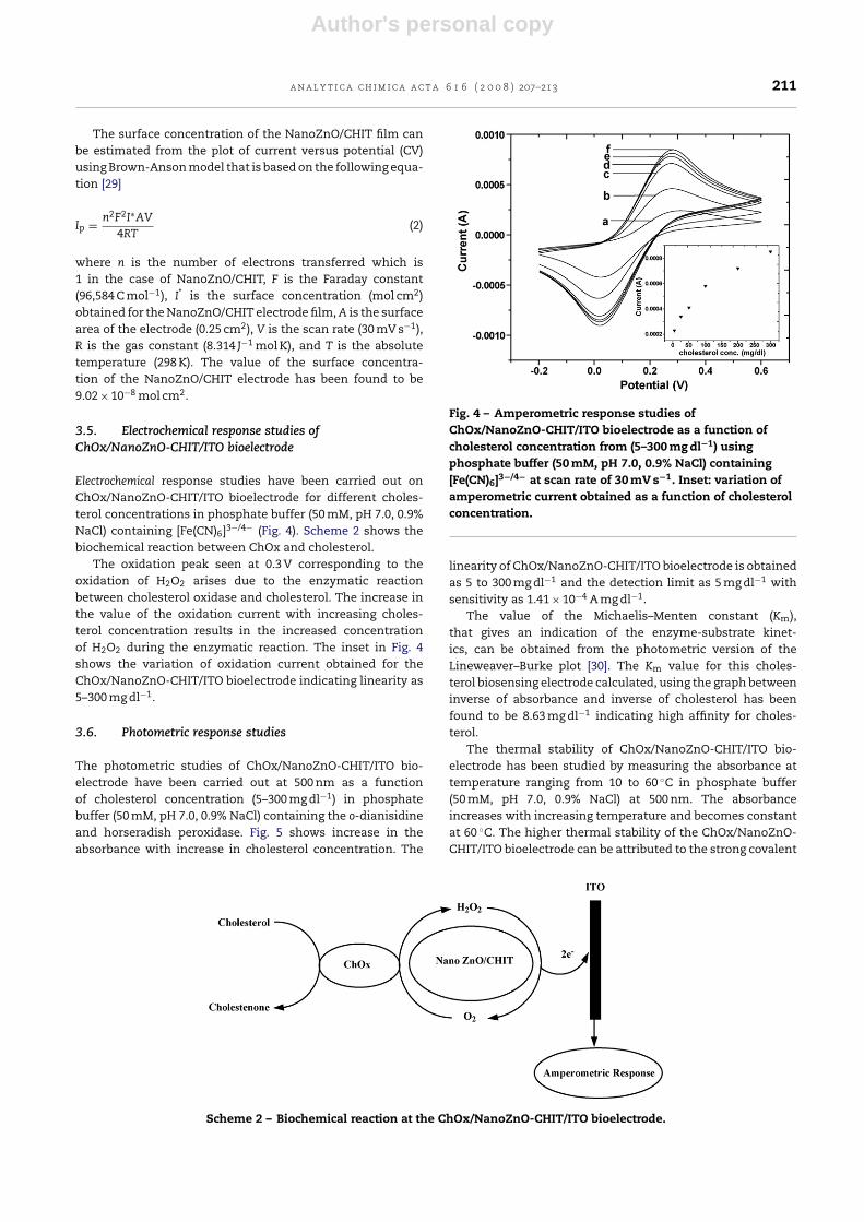
Electrochemical response studies have been carried out onChOx/NanoZnO-CHIT/ITO bioelectrode for different choles-terol concentrations in phosphate buffer (50 mM, pH 7.0, 0.9%NaCl) containing [Fe(CN)6]3−/4− (Fig. 4). Scheme 2 shows thebiochemical reaction between ChOx and cholesterol.
The oxidation peak seen at 0.3 V corresponding to theoxidation of H2O2 arises due to the enzymatic reactionbetween cholesterol oxidase and cholesterol. The increase inthe value of the oxidation current with increasing choles-terol concentration results in the increased concentrationof H2O2 during the enzymatic reaction. The inset in Fig. 4shows the variation of oxidation current obtained for theChOx/NanoZnO-CHIT/ITO bioelectrode indicating linearity as5–300 mg dl−1.
3.6. Photometric response studies
The photometric studies of ChOx/NanoZnO-CHIT/ITO bio-electrode have been carried out at 500 nm as a functionof cholesterol concentration (5–300 mg dl−1) in phosphatebuffer (50 mM, pH 7.0, 0.9% NaCl) containing the o-dianisidineand horseradish peroxidase. Fig. 5 shows increase in theabsorbance with increase in cholesterol concentration. The
Fig. 4 – Amperometric response studies ofChOx/NanoZnO-CHIT/ITO bioelectrode as a function ofcholesterol concentration from (5–300 mg dl−1) usingphosphate buffer (50 mM, pH 7.0, 0.9% NaCl) containing[Fe(CN)6]3−/4− at scan rate of 30 mV s−1. Inset: variation ofamperometric current obtained as a function of cholesterolconcentration.
linearity of ChOx/NanoZnO-CHIT/ITO bioelectrode is obtainedas 5 to 300 mg dl−1 and the detection limit as 5 mg dl−1 withsensitivity as 1.41 × 10⁻⁴ A mg dl−1.
The value of the Michaelis–Menten constant (Km),that gives an indication of the enzyme-substrate kinet-ics, can be obtained from the photometric version of theLineweaver–Burke plot [30]. The Km value for this choles-terol biosensing electrode calculated, using the graph betweeninverse of absorbance and inverse of cholesterol has beenfound to be 8.63 mg dl−1 indicating high affinity for choles-terol.
The thermal stability of ChOx/NanoZnO-CHIT/ITO bio-electrode has been studied by measuring the absorbance attemperature ranging from 10 to 60 ◦C in phosphate buffer(50 mM, pH 7.0, 0.9% NaCl) at 500 nm. The absorbanceincreases with increasing temperature and becomes constantat 60 ◦C. The higher thermal stability of the ChOx/NanoZnO-CHIT/ITO bioelectrode can be attributed to the strong covalent
Scheme 2 – Biochemical reaction at the ChOx/NanoZnO-CHIT/ITO bioelectrode.

Author's personal copy
212 a n a l y t i c a c h i m i c a a c t a 6 1 6 ( 2 0 0 8 ) 207–213
Table 1 – Effect of interferents on to ChOx/NanoZnO-CHIT/ITO bioelectrode in phosphate buffer (50 mM, pH 7.0, 0.9% NaCl)
Substrate/interferent
Cholesterol Cholesterol + glucose Cholesterol + ascorbic acid Cholesterol + uric acid Cholesterol + urea
Absorbance 0.012 0.012 0.013 0.011 0.014Error 0 −0.001 +0.001 −0.002
Table 2 – Determination of free cholesterol concentration in blood serum samples
Samples (mg dl−1) Free cholesterol (mg dl−1) Average (mg dl−1) RSD (%) Total cholesterol concentration(auto analyzer)
1 48.3, 49.5, 50.0, 52.1 49.97 3.05 1652 64.5, 62.2, 61.5, 65.3 63.37 1.79 2153 69.2, 70.1, 72.3, 73.6 71.30 1.59 240
Table 3 – Summary of the various metal oxide nanoparticles and metal mixed biopolymer based cholesterol biosensorreported in the literature, along with their important parameters
S. No. Electrode Sensitivity Responsetime (s)
Linear range Km values Shelf life Reference
1 ZnO nanoparticles – 15 25–400 mg dl−1 2.1 mM 75 days [13]2 Ni/K3Fe(CN)6/CNT – <20 0.005–3 mM – 1 month [31]3 ChOx/NanoPt/CNT – <20 4.0 × 10⁻⁶–1.0 × 10⁻⁴ mol l−1 – 6 weeks [32]4 ChOx/CS/MWCNT 1.55 �A mM−1 <20 4.0 × 10⁻⁶ to 7.0 × 10⁻⁴ 0.24 mM 50 days [33]5 ChOx/NanoZnO-CHIT 1.41 × 10⁻⁴ A mg dl−1 15 05–300 mg dl−1 8.63 mg dl−1 85 days Present work
binding of ChOx with NanoZnO-CHIT/ITO bioelectrode. Thevalue of the activation energy calculated from the Arrheniusplot has been obtained as 17.4 kJ mol−1.
The effect of interferents (ascorbic acid, uric acid, glucose,lactic acid and urea) on the cholesterol measurements hasbeen investigated by taking the solution containing (1:1) ratioof cholesterol (100 mg dl−1) and interferents such as glucose(5 mM), ascorbic acid (0.05 mM), uric acid (0.1 mM), lactic acid(0.5 mM) and urea (1 mM). The results (Table 1) indicate negli-gible effect of these interferents on the photometric responseof ChOx/NanoZnO-CHIT/ITO electrode.
Fig. 5 – Photometric response of ChOx/NanoZnO-CHIT/ITObioelectrode as a function of cholesterol concentration(5–300 mg dl−1) in PBS.
The response time of the ChOx/NanoZnO-CHIT/ITO bio-electrode has been determined as 15 s. The shelf-life ofthe ChOx/NanoZnO-CHIT/ITO bioelectrode measured after aninterval of 6 days has been estimated as 85 days. The decreasein the value of the amperomteric current has been foundto be about 25% up to about 8 weeks where after the cur-rent decreases sharply resulting in about 75% loss in about12 weeks (data not shown).
3.7. Response to serum samples
Attempts have been made to estimate free cholesterol(30%) using ChOx/NanoZnO-CHIT/ITO bioelectrode in serumsamples obtained from a clinic located in Delhi. Duringthese experiments, the ChOx/NanoZnO-CHIT/ITO bioelec-trode dipped into the reaction mixture containing theperoxidase, o-dianisidine in PBS and absorbance have beenrecorded at 500 nm. It can be seen (Table 2) that the resultsare in reasonable agreement with those obtained using theChOx/NanoZnO-CHIT/ITO bioelectrode. Table 3 shows theresults of the present studies obtained using ChOx/NanoZnO-CHIT/ITO bioelectrode along with those reported in literature.
4. Conclusions
Nanocomposite thin film of ZnO nanoparticle and CHIThas been fabricated to immobilize ChOx via electrostaticinteraction for the estimation cholesterol. It is found that gran-ular porous morphology of NanoZnO-CHIT provides a betterbiocompatible environment for the enzyme. ChOx/NanoZnO-CHIT/ITO bioelectrode has been used for estimation ofcholesterol in solution as well as in blood serum samples

Author's personal copy
a n a l y t i c a c h i m i c a a c t a 6 1 6 ( 2 0 0 8 ) 207–213 213
by photometric and amperometric techniques, respectively.ChOx/NanoZnO-CHIT/ITO bioelectrode has linearity from 5 to300 mg dl−1 and shelf life of 85 days. Efforts should be made toutilize ChOx/NanoZnO-CHIT/ITO bioelectrode to detect othermetabolites such as urea, glucose, uric acid including toxi-cants etc.
Acknowledgements
We are grateful to Dr. Vikram Kumar, Director, National Phys-ical Laboratory, New Delhi, India for providing the laboratoryfacilities. Dr. Raju Khan is thankful to the Department of Sci-ence & Technology (DST), Govt. of India, for financial supportunder the Young Scientist Scheme project SR/FTP/CS-77/2007.Dr. Pratima R. Solanki, Dr. Anees Ahmad Ansari and Mr.Ajeet Kaushik are thankful to CSIR India for the award ofSenior Research Associate, Research Associateship and SeniorResearch Fellow, respectively. The authors thank Dr. K.N. Sood(NPL, New Delhi, India) for SEM measurements. We acknowl-edge the financial support received from the Department ofScience & Technology Govt. of India (DST/GAP-070932) andDepartment of Biotechnology Govt. of India (DBT/GAP070832).
Appendix A. Supplementary data
Supplementary data associated with this article can be found,in the online version, at doi:10.1016/j.aca.2008.04.010.
r e f e r e n c e s
[1] N.A. Rakow, K.S. Suslick, Nature (Lond.) 406 (2000) 710.[2] S. Singh, P.R. Solanki, M.K. Pandey, B.D. Malhotra, Sens.
Actuators 115 (2006) 534.[3] S.K. Arya, P.R. Solanki, S.P. Singh, K. Kaneto, M.K. Pandey, M.
Datta, B.D. Malhotra, Biosens. Bioelectron. 22 (2007) 2516.[4] C. Dhand, S.P. Singh, S.K. Arya, M. Datta, B.D. Malhotra, Anal.
Chim. Acta 602 (2007) 244.[5] J.H. Holtz, S.A. Asher, Nature 389 (1997) 829.[6] S. Brahim, D. Narinesingh, A. Guiseppi-Elie, Anal. Chim. Acta
448 (2001) 27.[7] N. Zhou, J. Wang, T. Chen, Z. Yu, G. Li, Anal. Chem. 78 (2006)
5227.
[8] H.L. Jiang, J.T. Kwon, Y.K. Kim, E.M. Kim, R. Arote, H.J. Jeong,J.W. Nah, Y.J. Choi, T. Akaike, M.H. Cho, C.S. Cho, Gene Ther.14 (2007) 1389.
[9] J.D. Liao, S.P. Lin, Y.T. Wu, Biomacromolecules 6 (1) (2005) 392.[10] P. Pandey, M. Datta, B.D. Malhotra, Anal. Lett. 41 (2008)
157–207.[11] H. Zhou, X. Gan, J. Wang, X. Zhu, G. Li, Anal. Chem. 77 (2005)
6102.[12] S.A. Kumar, S.M. Chen, Anal. Lett. 41 (2008) 141.[13] S.P. Singh, S.K. Arya, M.K. Pandey, B.D. Malhotra, S. Saha, K.
Sreenivas, V. Gupta, Appl. Phys. Lett. 91 (2007) 063901.[14] A. Wei, X.W. Sun, J.X. Wang, Y. Lei, X.P. Cai, C.M. Li, Z.L. Dong,
W. Huang, Appl. Phys. Lett. 99 (2006) 123902.[15] J.X. Wang, X.W. Sun, A. Wei, Y. Lei, X.P. Cai, C.M. Li, Z.L. Dong,
Appl. Phys. Lett. 88 (2006) 233106.[16] G. Zhao, J.J. Xu, H.Y. Chen, Anal. Biochem. 350 (2006) 145.[17] X. Zhu, I. Yuri, X. Gan, I. Suzuki, G. Li, Biosens. Bioelectron.
22 (2007) 1600.[18] Y.F. Li, Z.M. Liu, Y.L. Liu, Y.H. Yang, G.L. Shen, R.Q. Yu, Anal.
Biochem. 349 (2006) 33.[19] F. Zhang, X. Wang, S. Ai, Z. Sun, Q. Wan, Z. Zhub, Y. Xian, L.
Jin, K. Yamamoto, Anal. Chim. Acta 519 (2004) 155.[20] Y.Q. Miao, S.W. Tan, Analyst 125 (2000) 1591.[21] K.J. Feng, Y.H. Yang, Z.J. Wang, J.H. Jiang, G.L. Shen, R.Q. Yu,
Talanta 70 (2006) 561.[22] Z. Wang, Y. Yang, Z. Li, J. Gong, G. Shen, R. Yu, Talanta 69
(2006) 686.[23] Y.L. Liu, Y.H. Yang, H.F. Yang, Z.M. Liu, G.L. Shen, R.Q. Yu, J.
Inorg. Biochem. 99 (2005) 2046.[24] S.F. Wang, L. Shen, W.D. Zhang, Y.J. Tong,
Biomacromolecules 6 (6) (2005) 3067.[25] S.P. Singh, O.P. Perez, M.S. Tomar, A.P. Polomino, A.R.
Mendoza, NSTI Nanotechnol. 2 (2005) 1.[26] A. Kumar, R. Malhotra, B.D. Malhotra, S.K. Grover, Anal.
Chim. Acta 414 (2000) 43.[27] L. Baoqiang, J. Dechang, Z. Yu, H. Qiaoling, C. Wei, J. Magn.
Magn. Mater. 300 (2006) 223.[28] H.L. Zhang, X.Z. Zou, G.S. Lai, D.Y. Han, F. Wang,
Electroanalysis 19 (2007) 1869.[29] A.J. Bard, L.R. Faulkner, Electrochemical Methods:
Fundamentals and Applications, second ed., Wiley, NewYork, 2000.
[30] R.A. Kamin, G.S. Wilson, Anal. Chem. 52 (1980) 1198.[31] M. Yang, Y. Yang, F. Qu, Y. Lu, G. Shen, Anal. Chim. Acta 571
(2006) 211.[32] S. Qiaocui, P. Tuzhi, Z. Yunu, C.F. Yang, Electroanalysis 17
(2005) 857.[33] X. Tan, M. Li, P. Cai, L. Luo, X. Zou, Anal. Biochem 337 (2005)
111.