Original Contribution Real-time monitoring of superoxide accumulation and antioxidant activity in a...
10
Original Contribution Real-time monitoring of superoxide accumulation and antioxidant activity in a brain slice model using an electrochemical cytochrome c biosensor Mallikarjunarao Ganesana a , Joseph S. Erlichman b , Silvana Andreescu a,n a Department of Chemistry and Biomolecular Science, Clarkson University, Potsdam, NY 13699, USA. b Department of Biology, St. Lawrence University, Canton, NY 13617, USA. article info Article history: Received 18 August 2012 Received in revised form 5 October 2012 Accepted 5 October 2012 Available online 17 October 2012 Keywords: Superoxide mobility Cerebral ischemia Electrochemical Cytochrome c microbiosensor Cerium oxide nanoparticles Nanoceria Free radicals abstract The overproduction of reactive oxygen species and the resulting damage are central to the pathology of many diseases. The study of the temporal and spatial accumulation of reactive oxygen species has been limited because of the lack of specific probes and techniques capable of continuous measurement. We demonstrate the use of a miniaturized electrochemical cytochrome c (Cyt c) biosensor for real-time measurements and quantitative assessment of superoxide production and inactivation by natural and engineered antioxidants in acutely prepared brain slices from mice. Under control conditions, super- oxide radicals produced from the hippocampal region of the brain in 400-mm-thick sections were well within the range of detection of the electrode. Exposure of the slices to ischemic conditions increased the superoxide production twofold and measurements from the slices were stable over a 3- to 4-h period. The stilbene derivative and anion channel inhibitor 4,4 0 -diisothiocyano-2,2 0 -disulfonic stilbene markedly reduced the extracellular superoxide signal under control conditions, suggesting that a transmembrane flux of superoxide into the extracellular space may occur as part of normal redox signaling. The specificity of the electrode for superoxide released by cells in the hippocampus was verified by the exogenous addition of superoxide dismutase (SOD), which decreased the superoxide signal in a dose-dependent manner. Similar results were seen with the addition of the SOD mimetic cerium oxide nanoparticles (nanoceria), in that the superoxide anion radical scavenging activity of nanoceria with an average diameter of 15 nm was equivalent to 527 U of SOD for each 1 mg/ml of nanoceria added. This study demonstrates the potential of electrochemical biosensors for studying real- time dynamics of reactive oxygen species in a biological model and the utility of these measurements in defining the relative contribution of superoxide to oxidative injury. & 2012 Elsevier Inc. All rights reserved. Introduction Reactive oxygen and nitrogen species (ROS and RNS), including superoxide, nitric oxide, hydrogen peroxide (H 2 O 2 ), and hydroxyl and peroxynitrite radicals, are potent oxidizing and nitrating agents that are produced under a variety of physiological and pathophysiological conditions. Physiological levels of these spe- cies appear to be involved in myriad of physiological processes. For example, endogenously generated oxidants act as second messengers, transcriptional regulators, and modulators of ion channels and enzyme activity [1,2]. The principle source of superoxide production under physiological conditions is thought to be the mitochondria, in which the rate of superoxide produc- tion in vitro is estimated to be 0.15–2% of the total cellular oxygen consumption [3,4]. Other potential sources of superoxide production include xanthine oxidases (XODs) and the nicotina- mide adenine dinucleotide phosphate (NADPH) family of oxi- dases. Of these, the latter seems to be unique in that superoxide production is thought to play an important role in normal cellular redox signaling [5,6]. The activity of both XOD and NADPH oxidases can be markedly increased after tissue injury or disease states [7,8]. Overproduction of ROS and RNS has been associated with development of a wide variety of neurodegenerative diseases as a result of their high chemical reactivity and potential for inducing oxidative damage to proteins, cells, and tissues [9–11]. Methods for monitoring ROS and RNS levels in intact, living tissues is a critical first step in unraveling their physiological roles in both healthy and disease states. Unfortunately, continuous in situ monitoring of these species in biological systems has been very challenging because of their high reactivity, low concentrations, and short half-lives. Moreover, the study of kinetics in cell-free systems is difficult because of the many interrelated coupled redox reactions that change dynamically over time. Contents lists available at SciVerse ScienceDirect journal homepage: www.elsevier.com/locate/freeradbiomed Free Radical Biology and Medicine 0891-5849/$ - see front matter & 2012 Elsevier Inc. All rights reserved. http://dx.doi.org/10.1016/j.freeradbiomed.2012.10.540 n Corresponding author. E-mail address: [email protected] (S. Andreescu). Free Radical Biology and Medicine 53 (2012) 2240–2249
Transcript of Original Contribution Real-time monitoring of superoxide accumulation and antioxidant activity in a...
Free Radical Biology and Medicine 53 (2012) 2240–2249
Contents lists available at SciVerse ScienceDirect
Free Radical Biology and Medicine
0891-58
http://d
n Corr
E-m
journal homepage: www.elsevier.com/locate/freeradbiomed
Original Contribution
Real-time monitoring of superoxide accumulation and antioxidant activityin a brain slice model using an electrochemical cytochrome c biosensor
Mallikarjunarao Ganesana a, Joseph S. Erlichman b, Silvana Andreescu a,n
a Department of Chemistry and Biomolecular Science, Clarkson University, Potsdam, NY 13699, USA.b Department of Biology, St. Lawrence University, Canton, NY 13617, USA.
a r t i c l e i n f o
Article history:
Received 18 August 2012
Received in revised form
5 October 2012
Accepted 5 October 2012Available online 17 October 2012
Keywords:
Superoxide mobility
Cerebral ischemia
Electrochemical
Cytochrome c microbiosensor
Cerium oxide nanoparticles
Nanoceria
Free radicals
49/$ - see front matter & 2012 Elsevier Inc. A
x.doi.org/10.1016/j.freeradbiomed.2012.10.54
esponding author.
ail address: [email protected] (S. Andre
a b s t r a c t
The overproduction of reactive oxygen species and the resulting damage are central to the pathology of
many diseases. The study of the temporal and spatial accumulation of reactive oxygen species has been
limited because of the lack of specific probes and techniques capable of continuous measurement. We
demonstrate the use of a miniaturized electrochemical cytochrome c (Cyt c) biosensor for real-time
measurements and quantitative assessment of superoxide production and inactivation by natural and
engineered antioxidants in acutely prepared brain slices from mice. Under control conditions, super-
oxide radicals produced from the hippocampal region of the brain in 400-mm-thick sections were well
within the range of detection of the electrode. Exposure of the slices to ischemic conditions increased
the superoxide production twofold and measurements from the slices were stable over a 3- to 4-h
period. The stilbene derivative and anion channel inhibitor 4,40-diisothiocyano-2,20-disulfonic stilbene
markedly reduced the extracellular superoxide signal under control conditions, suggesting that a
transmembrane flux of superoxide into the extracellular space may occur as part of normal redox
signaling. The specificity of the electrode for superoxide released by cells in the hippocampus was
verified by the exogenous addition of superoxide dismutase (SOD), which decreased the superoxide
signal in a dose-dependent manner. Similar results were seen with the addition of the SOD mimetic
cerium oxide nanoparticles (nanoceria), in that the superoxide anion radical scavenging activity of
nanoceria with an average diameter of 15 nm was equivalent to 527 U of SOD for each 1 mg/ml of
nanoceria added. This study demonstrates the potential of electrochemical biosensors for studying real-
time dynamics of reactive oxygen species in a biological model and the utility of these measurements in
defining the relative contribution of superoxide to oxidative injury.
& 2012 Elsevier Inc. All rights reserved.
Introduction
Reactive oxygen and nitrogen species (ROS and RNS), includingsuperoxide, nitric oxide, hydrogen peroxide (H2O2), and hydroxyland peroxynitrite radicals, are potent oxidizing and nitratingagents that are produced under a variety of physiological andpathophysiological conditions. Physiological levels of these spe-cies appear to be involved in myriad of physiological processes.For example, endogenously generated oxidants act as secondmessengers, transcriptional regulators, and modulators of ionchannels and enzyme activity [1,2]. The principle source ofsuperoxide production under physiological conditions is thoughtto be the mitochondria, in which the rate of superoxide produc-tion in vitro is estimated to be 0.15–2% of the total cellularoxygen consumption [3,4]. Other potential sources of superoxide
ll rights reserved.
0
escu).
production include xanthine oxidases (XODs) and the nicotina-mide adenine dinucleotide phosphate (NADPH) family of oxi-dases. Of these, the latter seems to be unique in that superoxideproduction is thought to play an important role in normal cellularredox signaling [5,6]. The activity of both XOD and NADPHoxidases can be markedly increased after tissue injury or diseasestates [7,8].
Overproduction of ROS and RNS has been associated withdevelopment of a wide variety of neurodegenerative diseases as aresult of their high chemical reactivity and potential for inducingoxidative damage to proteins, cells, and tissues [9–11]. Methodsfor monitoring ROS and RNS levels in intact, living tissues is acritical first step in unraveling their physiological roles in bothhealthy and disease states. Unfortunately, continuous in situmonitoring of these species in biological systems has been verychallenging because of their high reactivity, low concentrations,and short half-lives. Moreover, the study of kinetics in cell-freesystems is difficult because of the many interrelated coupledredox reactions that change dynamically over time.
M. Ganesana et al. / Free Radical Biology and Medicine 53 (2012) 2240–2249 2241
Cerebral ischemia is a leading cause of death and long-termdisability. During ischemia, blood flow to the tissue is inadequateto sustain the metabolism of the tissue, resulting in a progressivedecline in mitochondrial function and uncoupling of the electrontransport chain [12]. Consequently, many of the biological cas-cades involved in ischemic cell death have been related withoverproduction of ROS, including superoxide, which contributesto oxidative damage [13–16]. Moreover, XOD and NADPH oxi-dases have been implicated as the principle contributors ofsuperoxide generation leading to tissue damage after ischemia–reperfusion injury [17,18]. In support of this hypothesis, inhibi-tion of NADPH oxidase decreases ROS levels and preservesintegrity of the blood–brain barrier and neuronal function [17].
Although many biochemical processes contribute to the oxi-dative load generated by superoxide under normal and patholo-gical conditions, the damaging effects of superoxide are thoughtto be restricted to the cells generating this free radical. Thetranscellular movement of superoxide across biological mem-branes is not thought to contribute to any appreciable extentexcept in erythrocytes [19]. Unlike H2O2, which freely diffusesacross membranes, superoxide is relatively impermeative owingto its low water solubility in the charged state. Although theneutral, protonated form of superoxide (pKa 4.9) could traversebiological membranes, its low intracellular concentration pro-vides little driving force for diffusion into adjacent cellularcompartments. Moreover, the identification and localization ofthe superoxide dismutase (SOD) family of isozymes in themitochondria (SOD 2), the cytosol (SOD 1), and more recentlythe extracellular matrix (SOD 3) suggest that vectorially producedsuperoxide resides and reacts within defined cellular compart-ments. This notion has been challenged by the findings thatsuperoxide may cross mitochondrial and plasma membranethrough voltage-dependent anion channels (VDACs). Biochemicalevidence for VDAC distribution in the plasma membrane arosefrom the finding that VDAC1 is present in caveolae, a specializeddomain of the plasma membrane involved in endocytosis [20].Multiple functions have been ascribed to VDACs, including purinenucleotide transport (ATP and ADP), anion-channel-like activity,and transmembrane redox regulation arising from VDACs’ NADHreductase activity [21–24].
Evidence that superoxide may selectively cross through anionchannels was originally proposed by Lynch and Fridovich in XOD-loaded lipid vesicles [19] and later by Mao and Poznansky [25] inerythrocyte ghost membranes and in human amniotic cells [26].Han et al. [27] showed that �55% of the mitochondrial-generatedsuperoxide exited across the outer mitochondrial membranethrough 4,40-diisothiocyano-2,20-disulfonic stilbene (DIDS)-sensitive anion channels in endosomes isolated from isolatedmitochondria from the heart. More recently, a DIDS-sensitivesuperoxide flux was reported across the plasma membranes ofboth epithelial-derived endosomes [28] and endothelial cells.Despite the mounting evidence for the existence of superoxide-permeable channels, little is known regarding the mobility of thisfree radical and the extent to which this superoxide contributes tothe extracellular oxidant load. Understanding this relationshipcan potentially reshape our views on how the transcellularmovement of free radicals can influence oxidative damage inadjacent cells and tissues.
In general, measurement of ROS in living organisms has been asignificant analytical challenge. Most ROS are highly reactive andshort lived and therefore difficult to detect in complex biologicalmatrices. Additionally, ROS often are produced and/or neutralizedin subcellular compartments, which requires detection methodsdirected to specific subcellular localization. There are fewmethods that measure superoxide anion directly (i.e., electronparamagnetic resonance) and most techniques utilize indirect
absorbance or fluorescence measurements [29] or oxidationproducts, most of which are relatively nonspecific and havelimited temporal or spatial resolution. Thus the goals of thisstudy were threefold. First, using an electrochemical cytochromec (Cyt c) biosensor we wished to quantitatively monitor, in realtime, superoxide levels in living brain tissue. Second, using anin vitro ischemic brain slice model, we wished to demonstrateproof of concept of electrode specificity using several superoxidescavengers, including superoxide dismutase and cerium dioxidenanoparticles. Last, we wished to evaluate the role of VDACs inthe transmembrane flux of superoxide in brain slices duringcontrol and ischemic conditions.
Materials and methods
Reagents and stock solutions
XOD from bovine milk (EC 1.17.3.2), Cyt c from horse heart, SOD,hypoxanthine (HX), 11-mercapto-1-undecanol (MU), 3-mercapto-1-propionic acid (MPA), 1-ethyl-3-(3-dimethylaminopropyl) carbo-diimide (EDC), N-hydroxysuccinimide (NHS), and nanoceria (ceriumoxide (CeO2) nanoparticles with 15-nm average diameter deter-mined by field emission scanning electron microscopy and dynamiclight scattering) were purchased from Sigma (St Louis, MO, USA) andused as received. Sodium phosphate (monobasic), sodium hydroxide,sodium phosphate (dibasic, anhydrous), potassium chloride, EDTA,and ethyl alcohol were purchased from Fisher Scientific (Springfield,NJ, USA). Sulfuric acid (95.4%) was purchased from J.T. Baker(Phillipsburg, NJ, USA). DIDS was purchased from Sigma–Aldrich,dissolved in dimethyl sulfoxide and used at a final concentration of500 mM. All reagents were of analytical grade and were used withoutfurther purification. All solutions were prepared using distilled,deionized water (Millipore, Billerica, MA, USA; Direct-Q system) witha resistivity of 18.2 MO cm.
Instrumentation
Cyclic voltammetry (CV) and amperometric experiments werecarried out with a CHI electrochemical analyzer (CH Instruments,Austin, TX, USA). All experiments were carried out using a three-electrode system with a conventional cell equipped with a Ag/AgCl electrode (Ag/AgCl/3 M NaCl) as reference electrode, aplatinum wire (BAS; MW-1032) as counterelectrode, and a Cyt c
functionalized gold wire microelectrode with a protruding tip1.5 mm long and with a diameter of 0.25 mm.
Fabrication of the Cyt c biosensor
Gold wires with a diameter of 0.5 mm were cleaned electro-chemically in 0.1 M H2SO4 by cycling the potential between 0 andþ1.4 V at a scan rate of 0.1 V s�1 until the characteristic cyclicvoltammogram for gold was obtained. The cleaned Au wireelectrodes were then rinsed with water and ethanol. An electro-deposited layer of gold nanoparticles was formed by applying apotential of �0.2 V for 60 s to the electrode immersed in a HAuCl4
solution at a concentration of 0.01 M. Immediately after thegold deposition, the electrodes were thoroughly washed withwater and ethanol and incubated for 96 h at þ4 1C in an ethanolicsolution containing a mixture of carboxyl- and hydroxyl-terminated thiols (1.25 mM MPA and 3.75 mM MU). The thiol-modified electrodes were rinsed with ethanol and water toremove any unattached thiol molecules. To facilitate Cyt c
immobilization, the carboxyl groups of the surface thiolswere first activated with EDC and NHS. This was performed byincubating the electrodes in an aqueous solution containing
M. Ganesana et al. / Free Radical Biology and Medicine 53 (2012) 2240–22492242
200 mM EDC and 50 mM NHS for 30 min. Cyt c was covalentlyimmobilized by incubating the thiol-modified gold wire electrodein a Cyt c solution at a concentration of 5�10�6 M for 2 h. Finallythe Cyt c-modified electrodes were thoroughly rinsed with 0.1 Msodium phosphate buffer (PBS) solution to remove any unat-tached or weakly adsorbed Cyt c. The electrodes were stored atþ4 1C in a 0.1 M PBS solution containing 100 mM EDTA at pH7.5 until use.
Electrochemical monitoring of the superoxide radical in mouse brain
slices using the biosensor
CD-1 mice of either sex were used for these experiments. Allanimals used in this study were housed in the St. LawrenceUniversity’s vivarium, fed ad libitum, and kept in a normal light/dark cycle. All procedures were approved by the St. LawrenceUniversity Animal Care and Use Committee and performed inaccordance with the National Institutes of Health Guide for the Care
and Use of Laboratory Animals. Adult (2–4 months of age) mice,which have a more mature, fully developed central nervous system[30] and retain greater regional connectivity and physiologicalinteractions [31], were sacrificed via rapid decapitation; their brainswere quickly removed and placed in a chilled, choline-based slicingsolution containing (in mM), 24 choline bicarbonate, 135 cholinechloride, 1 kynurenic acid, 0.5 CaCl2, 1.4 Na2PO4, 5 glucose, 1 KCl, 20MgCl2, 5 mM ascorbic acid (315 mOsm) [32]. Transverse hippocam-pal slices 400 mm thick were cut along a rostral-to-caudal axis (�1.2to �2.8 mm bregma) using a Leica VT1200 Vibratome (LeicaMicrosystems, Wetzlar, Germany) and allowed to recover for 1 hin control artificial cerebral spinal fluid containing (in mM) 124NaCl, 3 KCl, 2.4 CaCl2, 1.3 MgSO4, 1.24 KH2PO4, 26 NaHCO3, 5 glucoseand bubbled with 5% CO2, 95% O2 gas (pH 7.4, 300 mOsm). Theculture medium contained 50% minimum essential medium(Hyclone Scientific, Logan, UT, USA), 25% horse serum, 25% Hanks’balanced salt solution (supplemented with 28 mM glucose, 20 mMHepes, and 4 mM NaHCO3), 50 U/ml penicillin, and 50 mg/ml strep-tomycin, pH 7.2 [30]. Solution osmolarity was measured using avapor pressure osmometer and corrected to 295 mOsm (Wescor,Logan, UT, USA). Hippocampal slices were placed in a culture dishand maintained in a humidified incubator (Galaxy 14S; EppendorfNew Brunswick, Cambridge, UK) at 37 1C with 5% CO2 during theexperiments.
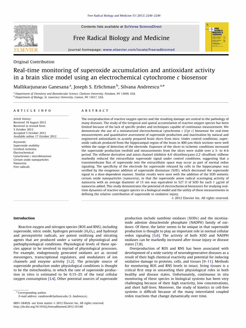
Fig. 1. Experimental setup for real-time monitoring of superoxide in brain slices. The r
wire electrode through the self-assembly of mixed-thiol monolayers and the redox pro
Amperometric measurements of enzymatically generated super-oxide were performed in a conventional electrochemical cell, underconstant stirring in air-saturated 0.1 M PBS with 100 mM EDTA at pH7.5 or in culture medium, at an applied potential of þ0.15 V. Thecurrent–time amperometric curves used to calibrate the Cyt c
biosensor were generated upon addition of various XOD concentra-tions (ranging from 2.5 to 80 mU/ml) to the air-saturated PBS orculture medium containing 100 mM HX. All electrochemical experi-ments were performed at 37 1C.
Electrochemical measurements for endogenously producedsuperoxide in hippocampal brain slices were performed by pla-cing the recording electrode midline along the dorsal–ventral axisof the slice. The sections used for the recording were located�2.06 mm from Bregma and included the hippocampus, dentategyrus, thalamus, and portions of the hypothalamus. All theelectrochemical recordings of superoxide in brain slices with thethree-electrode system were performed in the incubator. Undercontrol conditions, the O2 and CO2 concentrations (�17% O2/5%CO2) reached steady state in approximately 30 min after the doorwas closed. Control recordings under normoxic and normocapnicconditions lasted approximately 180 min, at which time, theresponse of the electrode to accumulated superoxide had reacheda steady state. Electrochemical experiments with brain slices forsuperoxide monitoring under ischemic conditions were carriedout in the same incubator at 37 1C by switching gas conditions to84% N2, 15% CO2, and 1% O2. In experiments that required theaddition of nanoceria or the addition of DIDS, these materialswere delivered directly to the culture well plate in the incubatorremotely with a syringe actuator connected by polyethylenetubing.
Results
Biosensor calibration and characterization
Cyt c immobilization and electrochemical characterization
The detection principle of the superoxide anion radical isbased on the redox reaction between the Cyt c, immobilized ontothe surface of a gold wire electrode, and the generated superoxide[11,33]. A new combination of self-assembled monolayer ofmixed thiols of MPA and MU was used in this work to covalentlyimmobilize Cyt c onto the gold wire electrodes, modified withgold nanoparticles, through thiol chemistry. The immobilized Cyt
ight side shows a schematic representation of the Cyt c immobilized onto the gold
cess of Cyt c in the presence of superoxide.

Fig. 2. Typical amperometric current–time response of the Cyt c biosensor upon
addition of various concentrations of XOD in the presence of 100 mM HX.
M. Ganesana et al. / Free Radical Biology and Medicine 53 (2012) 2240–2249 2243
c is reduced by superoxide and the reduced Cyt c is regeneratedelectrochemically at the electrode surface. The concentration ofsuperoxide was quantified by constant potential amperometrywith the electrode poised at the potential of 0.15 V vs Ag/AgCl,which corresponds to the oxidation potential of Cyt c. Theelectrogenerated current is proportional to the concentration ofthe superoxide at the electrode surface. A schematic diagram ofthe reaction process at the gold electrode is shown in Fig. 1. Theimmobilization of the Cyt c onto the gold electrode surface wasconfirmed by measuring the oxidation and reduction peaks of Cytc using CV. The peak current (Ip) for both the cathodic and theanodic currents were linearly proportional to scan rate, suggest-ing that the electrode reaction was typical of a surface-controlledprocess (Supplementary Material S1). Surface coverage (G) of theelectrode with Cyt c, calculated using the Laviron equation [34,35]by integrating the reduction peak, shows a surface concentrationof 3.17�10�11 mol cm�2 redox active Cyt c. The low surfacecoverage indicates the electrochemical behavior of a thin layer inwhich the redox process at the electrode surface is surfaceconfined and diffusionless.
Measurements were performed in culture medium at an applied potential of
0.15 V. The current difference between the baseline and the maximum value
reached at the plateau at 70 s was used to build the calibration curve of the
biosensor. The decrease in the current after reaching the plateau could be due to
the disproportionation of the enzymatically produced superoxide radicals into
H2O2.
Biosensor calibration
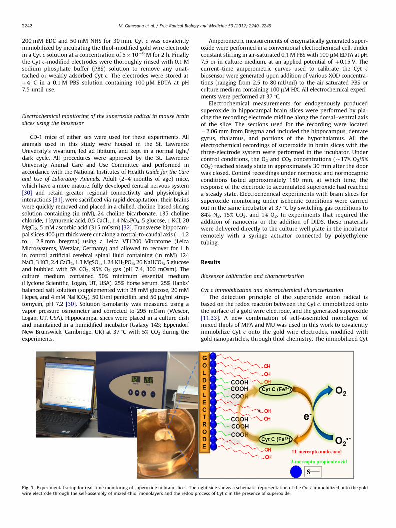
Biosensor calibration and optimization were carried out instandard solutions of superoxide generated by the HX/XODsystem. XOD catalyzes the oxidation of HX in the presence ofmolecular oxygen, to uric acid and H2O2, with the production ofsuperoxide as an intermediate in this reaction [36]. The calibra-tion curve obtained with the enzymatically generated superoxidewas used to quantify the superoxide levels produced in brainslices. Because superoxide can undergo spontaneous dismutationto H2O2, the steady-state sensor signal was quantified by takinginto account both generation and dismutation of the enzymati-cally generated superoxide [37–41]. Biosensor calibration wasperformed using an optimum HX concentration of 100 mM.The amperometric responses of the Cyt c biosensor generatedupon addition of variable amounts of XOD ranging from 2.5 to80 mU/ml to the reaction cell containing HX were measured andcorrelated with the theoretically estimated superoxide concen-tration. The calibration curve of the biosensor in culture mediumshowed in Fig. 2 demonstrates the dependence of the sensorsignal as a function of XOD activity as well as the theoreticallyestimated steady-state superoxide concentration. The sensorsignal was proportional to the square root of the XOD concentra-tion for enzymatic activities up to 80 mU/ml (Fig. 3). There waslittle difference between the biosensor responses to superoxidegenerated in PBS and in the culture medium. The linear rangesfor the detection of superoxide from the calibration curves were0–1.23 and 0–1.32 mM, with sensitivities of 11.78 � 102 and14.45 � 102 A M�1 m�2, in PBS and culture medium, respec-tively. The sensitivity of the Cyt c electrode is superior tothat reported in literature with other sensor configurations,including a mixed long-chain thiol-based Cyt c electrode (2.76 �102 A M�1 m�2) [41] and a mixed short-chain thiol Cyt c electrode(0.56 � 102 A M�1 m�2) [37].
The sensor response to superoxide was observed in less than1 s after XOD injection. A steady-state limiting current value wasreached in 4–5 s. The detection limit of the biosensor, calculatedaccording to the 3ss/R
0 criteria (R0 is the slope of the linearcalibration curve and ss is the standard deviation of the ampero-metric signal of the blank solution), were 4.1 and 2.3 nM in PBSand culture medium, respectively. The biosensor demonstratedgood functionality and high sensitivity in the culture medium.The enhanced signal may be due to a higher stability of thesuperoxide radical in the culture medium, whereas in PBS super-oxide tends to decompose spontaneously at faster rates into H2O2
[42]. The biosensors were stable up to 7 days with no change inresponse when stored in PBS at þ4 1C. Cyclic voltammogramsupon repeated scans showed stable redox peaks for 200 con-secutive cycles, which demonstrates the stability of the biosensor(Supplementary Material S2).
Biosensor selectivity
Electrochemically active uric acid is formed as a by-product ofthe HX/XOD superoxide generation system used to calibrate thebiosensor. Because of the short half-life of the superoxide radical,we studied the response selectivity against uric acid and thespecificity of the sensor toward the superoxide radical. In theoptimized configuration of mixed thiols, the biosensor showed noresponse when concentrations of uric acid of up to 50 mM wereadded into the medium (Supplementary Material S3). In addition,the sensor showed no response to H2O2, which can be explainedby the low applied potential of 0.15 V. To further confirm that thesignal is solely associated with the produced superoxide radicals,SOD was added to inactivate the superoxide radicals. When aconcentration of 10 U/ml SOD was added to a solution containing1.04 mM superoxide (theoretical concentration), the electroche-mical response was suppressed entirely until it reached thebaseline signal, indicating that the superoxide radicals generatedwere completely inactivated by SOD (Fig. 4). The results of thesestudies demonstrate that the signal is specific to superoxideradicals, with no interference from the electrochemically activereaction by-products. The observed selectivity indicates that themodification of the electrode surface with the layer of denselypacked thiols is effective at blocking diffusion of interferingspecies to the electrochemically active surface [41]. The thiollayer also reduces protein adsorption, ensuring good functionalityof the sensor in the culture medium.
Real-time measurement of superoxide in hippocampal brain slice
using the Cyt c biosensor
The biosensor allows continuous monitoring of superoxideradicals in the extracellular matrix with high temporal resolution,high sensitivity, and high selectivity. This was achieved by placing
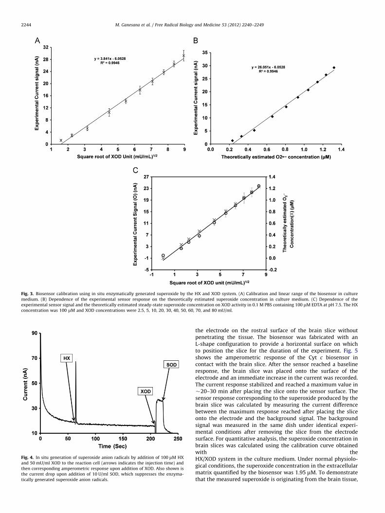
Fig. 3. Biosensor calibration using in situ enzymatically generated superoxide by the HX and XOD system. (A) Calibration and linear range of the biosensor in culture
medium. (B) Dependence of the experimental sensor response on the theoretically estimated superoxide concentration in culture medium. (C) Dependence of the
experimental sensor signal and the theoretically estimated steady-state superoxide concentration on XOD activity in 0.1 M PBS containing 100 mM EDTA at pH 7.5. The HX
concentration was 100 mM and XOD concentrations were 2.5, 5, 10, 20, 30, 40, 50, 60, 70, and 80 mU/ml.
Fig. 4. In situ generation of superoxide anion radicals by addition of 100 mM HX
and 50 mU/ml XOD to the reaction cell (arrows indicates the injection time) and
then corresponding amperometric response upon addition of XOD. Also shown is
the current drop upon addition of 10 U/ml SOD, which suppresses the enzyma-
tically generated superoxide anion radicals.
M. Ganesana et al. / Free Radical Biology and Medicine 53 (2012) 2240–22492244
the electrode on the rostral surface of the brain slice withoutpenetrating the tissue. The biosensor was fabricated with anL-shape configuration to provide a horizontal surface on whichto position the slice for the duration of the experiment. Fig. 5shows the amperometric response of the Cyt c biosensor incontact with the brain slice. After the sensor reached a baselineresponse, the brain slice was placed onto the surface of theelectrode and an immediate increase in the current was recorded.The current response stabilized and reached a maximum value in�20–30 min after placing the slice onto the sensor surface. Thesensor response corresponding to the superoxide produced by thebrain slice was calculated by measuring the current differencebetween the maximum response reached after placing the sliceonto the electrode and the background signal. The backgroundsignal was measured in the same dish under identical experi-mental conditions after removing the slice from the electrodesurface. For quantitative analysis, the superoxide concentration inbrain slices was calculated using the calibration curve obtainedwith theHX/XOD system in the culture medium. Under normal physiolo-gical conditions, the superoxide concentration in the extracellularmatrix quantified by the biosensor was 1.95 mM. To demonstratethat the measured superoxide is originating from the brain tissue,

M. Ganesana et al. / Free Radical Biology and Medicine 53 (2012) 2240–2249 2245
a control experiment was conducted under the same experimen-tal conditions but by placing the brain slice in the recording dish�1 cm away from the electrochemical active surface. When theelectrode was not in direct contact with the brain tissue, thesensor did not show any amperometric response (Fig. 5; inset).This indicates that only superoxide levels that are generatedwhen the biological tissue is in direct contact with the electrodeare measured.
To further demonstrate the specificity of the signal toward thesuperoxide radicals originating from the extracellular space of thebrain slice, we used the exogenous addition of SOD to facilitatethe conversion of superoxide to hydrogen peroxide (Fig. 6). Eachsequential addition of 500 U/ml SOD in the reaction cell decreasedthe amperometric current by approximately 14%, which suggeststhat the superoxide radicals were inactivated by the added SOD.Three sequential additions of 500 U/ml SOD reduced the electrodesignal near to baseline levels, suggesting that the exogenously
Fig. 5. Real-time amperometric response of the Cyt c biosensor in contact with the
hippocampal brain slice. The increase in the current indicates the release of
extracellular superoxide radicals from the brain slice under normal physiological
conditions. The inset shows a control experiment with the brain slice placed in the
measurement cell but far away from the electrode surface.
Fig. 6. Specificity of the biosensor response toward superoxide released by the
hippocampal brain slice under normal physiological conditions. With each addi-
tion of 500 U/ml SOD there is a decrease in the amperometric response, indicating
specific inactivation of the produced superoxide radicals. The arrows indicate the
times of SOD addition and the exposure of the brain to/from the electrode surface.
added SOD resulted in the dismutation of the superoxide pro-duced. The residual current in Fig. 6 can be attributed to thecapacitive current associated with the brain slice. After the slicewas removed from the electrode surface, the current reached abackground value that is slightly higher than the initial baselinecurrent recorded at the beginning of the experiment. This findingis probably the result of several factors, including nonspecificadsorption of proteins, the presence of residual tissue detritus onthe electrode surface, or mechanical damage of the Cyt c layerproduced when placing and removing the brain slice to/from theelectrode surface. To account for these effects, the superoxideconcentration was estimated using the residual current measuredafter removing the slice from the electrode surface as baselinevalue.
Evaluation of transmembrane flux of superoxide in brain slices
through VDACs
To examine the potential role of the transmembrane flux ofsuperoxide into the extracellular space, DIDS (0.5 mM final concen-tration for each addition; 2 mM total) was added to the culturemedium during the recordings. Previous studies have shown thatsuperoxide can penetrate a variety of biological membranes throughthe VDACs, which are inhibited with DIDS [27,28]. The mechanismby which DIDS inhibits anion channels is not known, although theinhibition seems to occur in the extracellular space and is reversiblein some systems. Under control conditions, the addition of DIDS tothe culture medium resulted in a decrease in the superoxide signal(Fig. 7) with each subsequent addition. Because it was not possibleto stir the solution during the experiment while recording, it is likelythe DIDS concentration was not uniform throughout the solution.In separate experiments, the addition of DIDS alone had no effect onsensor output.
Real-time continuous monitoring of superoxide dynamics during
ischemia
Immediately after the transition to hypoxic/hypercapnic gasmixtures in the incubator, superoxide levels began to increaseand reached a steady state after �180 min (Fig. 8). Compared tothe normoxic superoxide levels, peak superoxide levels increasedapproximately twofold during ischemia. Based on our calculationsthis resulted in an average increase in superoxide from 1.95 to4.1 mM. In contrast to our findings during normoxia, the
Fig. 7. Effects of four consecutive additions of DIDS (0.5 mM final concentration in
the reaction cell) on the amperometric response of the Cyt c biosensor. The arrows
indicate the times of DIDS addition.

Fig. 8. Typical current–time response of Cyt c biosensor showing continuous
monitoring of superoxide in mouse hippocampal brain slice exposed to simulated
ischemic conditions. The arrow indicates the time when the brain slice was
switched to ischemic insult by exposing the slice to hypoglycemic, acidic, and
hypoxic conditions (glucose was lowered to 2 mM and the cell was bubbled with
15% CO2 and 1% oxygen).
Fig. 9. Effects of nanoceria (successive additions of 1 mg/ml to the reaction cell)
on the superoxide level quantified by the Cyt c biosensor under (A) physiological
and (B) simulated ischemic conditions.
M. Ganesana et al. / Free Radical Biology and Medicine 53 (2012) 2240–22492246
sequential addition of DIDS had no effect on the extracellularlevels of superoxide.
Assessing antioxidant activity of cerium oxide nanoparticles in
ischemic brain slices
In this study, we used the Cyt c biosensor to quantitativelyassess the time-course inactivation of extracellular superoxide bynanoceria. Previous work from our lab showed that nanoceria iscapable of scavenging a variety of free radicals, including nitricoxide, superoxide, and peroxynitrite, in brain slices [32] and thecatalytic activity of nanoceria reduces superoxide to H2O2. Theaddition of nanoceria with an average particle size of 15 nminduced an immediate decrease in the superoxide signal undercontrol conditions, confirming the neutralizing activity of thesenanoparticles against superoxide (Fig. 9). Consecutive addition of1 mg/ml nanoceria decreased the superoxide levels by 14.574.5%(n¼5) per addition. After two consecutive nanoparticle additions,the superoxide levels remained stable for the remainder of theexperiment. In contrast, the addition of the same amount ofnanoceria during ischemia decreased the superoxide signal by8.172.2% (n¼3) per addition. Moreover, after two sequentialnanoparticle additions, the superoxide signal decreased progres-sively toward baseline for the remainder of the experiment.Supplementary Material S4 shows the decrease in the extracel-lular superoxide concentration for both SOD and nanoceria. Basedon these findings, a 1 mg/ml nanoceria concentration induced adecrease in the superoxide level under normoxic conditionsequivalent to that of 527 SOD activity units, as calculated fromthe amperometric responses of the biosensor in brain slices.
Discussion
This study demonstrates the potential of electrochemical micro-biosensors for studying the release, accumulation, and mobility ofthe superoxide radical, and potentially of other oxidative stressmarkers, in brain tissue with high spatial and temporal resolutionand can facilitate our fundamental understanding of the neurophy-siology of oxidative-stress-related diseases.
Current methods for monitoring the superoxide anion radicalin hippocampal brain slices include colorimetric and fluorescenceassays that measure intracellular superoxide [43]. A drawbackof these methods is the difficulty of obtaining a continuous,quantitative, real-time measurement of superoxide productionin biological tissue. Most often researchers interested in assessingoxidative damage rely on downstream markers such as lipid andprotein oxidation [44–46]. Because of their small size (micro-meter to submicrometer diameter) electrochemical sensors canprovide real-time measurements of ROS and RNS species [47]. Themicrobiosensor used in this work consists of a gold wire electrodemodified with mixed layers of thiols and immobilized Cyt c as abiorecognition element [33]. Electrochemical sensors for thedetection of superoxide have been reported in the literature[48] but these have seldom been used to measure real-timeproduction of superoxide in intact biological tissues. Most studieshave relied on standard solutions of enzymatically producedsuperoxide, with few examples using cell cultures. Rarely havethe studies demonstrated the applicability of similar devices tomeasure extracellular superoxide in biological tissues [49–51].There are no reports on the use of electrochemical microbiosen-sors for studying superoxide mobility across membranes andtheir application for studying oxidant and antioxidant mechan-isms of natural or synthetic antioxidants in the brain. Our abilityto measure superoxide in such a small volume of tissue in this
M. Ganesana et al. / Free Radical Biology and Medicine 53 (2012) 2240–2249 2247
study suggests that these electrodes should be equally sensitivein vivo.
To explore the potential of this technology, we examined thenature of the superoxide signal in the extracellular space of thebrain slice. There is increasing evidence that free radicals play acentral role in redox signaling both in health and in disease [9].Madesh et al. [52] first suggested that extracellular superoxideproduced by plasmalemmal NADPH oxidase or superoxide cross-ing the plasma membrane through a VDAC-like channel modifiedcell surface proteins to mediate cell signaling involved in pro-grammed cell death. Recent proteomic studies have confirmedthe presence of VDACs in the plasma membrane of human cells[53–55], including cultured septal and hippocampal neuronalcells and in membrane preparations from human brain cortex[56]. Although preliminary, this study is the first report of DIDSeliminating the superoxide signal in intact brain tissue. The DIDS-sensitive accumulation of superoxide under control conditionssuggests that the transmembrane flux of superoxide occurredthrough a VDAC-like channel and was not simply an artifact ofcompromised cell membranes associated with the sectioningprocess. The VDAC is a highly conserved protein in terms of bothits structural and its functional features across species [57]. It hasbeen hypothesized that VDACs perform a variety of functions,including cellular ATP release, maxi-anion channel, volume con-trol, a component of the GABAA receptor, plasma membraneNADH oxidoreductase, and modulation of programmed cell death.Our observation that extracellular superoxide concentrationswere unaffected by DIDS during ischemia suggests that VDACsmay either become insensitive to the effects of DIDS or closeduring the ischemic period. Consistent with the latter notion,previous studies have shown that NADH levels increase duringthe period of ischemia [58–60] and elevated cytosolic NADHconcentrations promote the closure of VDACs and apoptoticsignals [53,61,62]. The fact that extracellular superoxide is ele-vated during the ischemic period even with closure of VDACssuggests that there are alternative pathways for transmembraneflux of superoxide or there may be activation of plasmalemmalNADPH oxidase. Additional experiments will be needed to explorethese questions but this study highlights the importance of thistechnology in the study of the biological effects of free radicals.
Previous work from our lab [63–65] and others [66–75] hasdemonstrated the ability of ceria to neutralize a variety of biologi-cally relevant free radicals. The 3þ/4þ valence of nanoceria allowsthe particles to participate in reversible redox reactions similar toendogenous redox enzymes in cells. Given its high redox potential(1.55 V), ceria can accept electrons from hydroxyl, superoxide, andperoxynitrite in the þ4 state and donate electrons to H2O2 in theþ3 state. Our data acquired with the electrochemical Cyt c micro-sensor show that the catalytic activity for ceria is remarkably highwith respect to superoxide dismutase; 1 mg of ceria was equivalentto �527 U of SOD in neutralizing superoxide. The progressivedecrease in superoxide accumulation after repeated administrationof ceria during ischemia is probably due to the cumulative uptakeand intracellular scavenging of superoxide by the nanoparticles. Wehave previously shown that particles of nanoceria are endocytosedwithin 1 h and they localize to mitochondria, neurofilaments, andlipid/myelin membranes [63]. Given its regenerative, catalyticnature, nanoceria has become attractive as a potential pharmacolo-gical agent and potent antioxidant [66–74]. This being said it hasbecome increasingly clear that the contradictory biological effects ofnanoceria (see Yokel and others) [76–79] reported in the literaturestem from differences in the physical (i.e., size, z potential) andchemical characteristics (presence of dopants or stabilizers) of theparticles. Modest changes in the synthetic identity of the particlescan confer very different biological outcomes. We have recentlyexplored the therapeutic potential of nanoceria in a murine model of
multiple sclerosis [80] using both custom-synthesized and commer-cially available nanoceria. We found that the biological effectsand deposition of the particles in the brain were dependent onmultiple physical/chemical characteristics. Exploring the factors thatcontribute to the biological action of nanoceria will be an importantarea of research, as the therapeutic potential of these compoundswill be developed in the future.
Conclusions
This work demonstrates the ability of electrochemical biosensorsto study real-time dynamics of reactive oxygen species in a bio-logical model. The electrochemical measurements provided evi-dence of transmembrane mobility and extracellular superoxiderelease under normal physiological and simulated ischemic condi-tions in a brain slice model of ischemia. Pharmacological manipula-tions of VDACs, which facilitate transport of mitochondrialsuperoxide to the extracellular space, demonstrate the mobility ofsuperoxide measured electrochemically. The dynamics of super-oxide overproduction during ischemic brain injury was quantifiedover time and the specificity of signal was validated using SOD as amodel endogenous antioxidant. The potential of this technology forstudying the time course of inactivation and providing quantitativeassessment of the superoxide scavenging capacity by antioxidantcompounds has been demonstrated with an emerging engineeredantioxidant, nanoceria, which holds potential for treatment andtherapy of oxidative diseases. In summary, this study demonstratesthe potential of electrochemical sensors for providing quantitativeassessment of real-time changes in extracellular ROS and studyingfundamental mechanisms to understand ROS-mediated processesunder normal physiological and pathophysiological conditionsinvolved in disease progression and therapy.
Acknowledgment
This work was supported by NSF 0954919, NIH R21NS078738-01,and USAR W911NF-11-1-0304 to S.A.
Appendix A. Supporting information
Supplementary data associated with this article can be foundin the online version at http://dx.doi.org/10.1016/j.freeradbiomed.2012.10.540.
References
[1] Winterbourn, C. C.; Hampton, M. B. Thiol chemistry and specificity in redoxsignaling. Free Radic. Biol. Med. 45:549–561; 2008.
[2] Paulsen, C. E.; Carroll, K. S. Orchestrating redox signaling networks throughregulatory cysteine switches. ACS Chem. Biol. 5:47–62; 2010.
[3] Robinson, B. H. The role of manganese superoxide dismutase in health anddisease. J. Inherit. Metab. Dis. 21:598–603; 1998.
[4] Dickinson, B. C.; Peltier, J.; Stone, D.; Schaffer, D. V.; Chang, C. J. Nox2 redoxsignaling maintains essential cell populations in the brain. Nat. Chem. Biol.7:106–112; 2011.
[5] Daiber, A.; Frein, D.; Namgaladze, D.; Ullrich, V. Oxidation and nitrosation inthe nitrogen monoxide/superoxide system. J. Biol. Chem. 277:11882–11888;2002.
[6] Daiber, A.; Bachschmid, M. Enzyme inhibition by peroxynitrite-mediatedtyrosine nitration and thiol oxidation. Curr. Enzyme Inhibit 3:103–117; 2007.
[7] Kuroda, J.; Ago, T.; Matsushima, S.; Zhai, P. Y.; Schneider, M. D.; Sadoshima, J.NADPH oxidase 4 (Nox4) is a major source of oxidative stress in the failingheart. Proc. Natl. Acad. Sci. USA 107:15565–15570; 2010.
[8] Forstermann, U. Nitric oxide and oxidative stress in vascular disease. PflugersArch. Eur. J. Physiol 459:923–939; 2010.
[9] Zweier, J. L.; Talukder, M. A. H. The role of oxidants and free radicals inreperfusion injury. Cardiovasc. Res. 70:181–190; 2006.
M. Ganesana et al. / Free Radical Biology and Medicine 53 (2012) 2240–22492248
[10] Salvemini, D.; Cuzzocrea, S. Oxidative stress in septic shock and disseminatedintravascular coagulation. Free Radic. Biol. Med. 33:1173–1185; 2002.
[11] Arnold, S.; Feng, Z. Q.; Kakiuchi, T.; Knoll, W.; Niki, K. Investigation ofthe electrode reaction of cytochrome c through mixed self-assembledmonolayers of alkanethiols on gold(111) surfaces. J. Electroanal. Chem.438:91–97; 1997.
[12] Siemionow, M.; Arslan, E. Ischemia/reperfusion injury: a review in relation tofree tissue transfers. Microsurgery 24:468–475; 2004.
[13] Chrissobolis, S.; Faraci, F. M. The role of oxidative stress and NADPH oxidasein cerebrovascular disease. Trends Mol. Med. 14:495–502; 2008.
[14] Matsuda, S.; Umeda, M.; Uchida, H.; Kato, H.; Araki, T. Alterations of oxidativestress markers and apoptosis markers in the striatum after transient focalcerebral ischemia in rats. J. Neural Transm. 116:395–404; 2009.
[15] Oliver, C. N.; Starke-Reed, P. E.; Stadtman, E. R.; Liu, G. J.; Carney, J. M.; Floyd,R. A. Oxidative damage to brain proteins, loss of glutamine synthetaseactivity, and production of free radicals during ischemia/reperfusion-inducedinjury to gerbil brain. Proc. Natl. Acad. Sci. USA 87:5144–5147; 1990.
[16] Floyd, R. A.; Carney, J. M. Free radical damage to protein and DNA:mechanisms involved and relevant observations on brain undergoing oxida-tive stress. Ann. Neurol. 32(Suppl):S22–S27; 1992.
[17] Chen, H.; Song, Y. S.; Chan, P. H. Inhibition of NADPH oxidase is neuroprotectiveafter ischemia–reperfusion. J. Cereb. Blood Flow Metab. 29:1262–1272; 2009.
[18] Ono, T.; Tsuruta, R.; Fujita, M.; Aki, H. S.; Kutsuna, S.; Kawamura, Y.;Wakatsuki, J.; Aoki, T.; Kobayashi, C.; Kasaoka, S.; Maruyama, I.; Yuasa, M.;Maekawa, T. Xanthine oxidase is one of the major sources of superoxideanion radicals in blood after reperfusion in rats with forebrain ischemia/reperfusion. Brain Res. 1305:158–167; 2009.
[19] Lynch, R. E.; Fridovich, I. Permeation of the erythrocyte stroma by superoxideradical. J. Biol. Chem. 253:4697–4699; 1978.
[20] Lisanti, M. P.; Scherer, P. E.; Tang, Z.; Sargiacomo, M. Caveolae, caveolin andcaveolin-rich membrane domains: a signalling hypothesis. Trends Cell Biol.4:231–235; 1994.
[21] Baker, M. A.; Lane, D. J. R.; Ly, J. D.; De Pinto, V. Lawen, A. VDAC1 is atransplasma membrane NADH-ferricyanide reductase. J. Biol. Chem.279:4811–4819; 2004.
[22] Darbandi-Tonkabon, R.; Hastings, W. R.; Zeng, C. M.; Akk, G.; Manion, B. D.;Bracamontes, J. R.; Steinbach, J. H.; Mennerick, S. J.; Covey, D. F.; Evers, A. S.Photoaffinity labeling with a neuroactive steroid analogue—6-AZI-pregna-nolone labels voltage-dependent anion channel-1 in rat brain. J. Biol. Chem.278:13196–13206; 2003.
[23] Blatz, A. L.; Magleby, K. L. Single voltage-dependent chloride-selectivechannels of large conductance in cultured rat muscle. Biophys. J.43:237–241; 1983.
[24] Bahamonde, M. I.; Fernandez-Fernandez, J. M.; Guix, F. X.; Vazquez, E.;Valverde, M. A. Plasma membrane voltage-dependent anion channel med-iates antiestrogen-activated maxi Cl� currents in C1300 neuroblastoma cells.J. Biol. Chem. 278:33284–33289; 2003.
[25] Mao, G. D.; Poznansky, M. J. Electron spin resonance study on the perme-ability of superoxide radicals in lipid bilayers and biological membranes.FEBS Lett 305:233–236; 1992.
[26] Ikebuchi, Y.; Masumoto, N.; Tasaka, K.; Koike, K.; Kasahara, K.; Miyake, A.;Tanizawa, O. Superoxide anion increases intracellular pH, intracellular freecalcium, and arachidonate release in human amnion cells. J. Biol. Chem.266:13233–13237; 1991.
[27] Han, D.; Antunes, F.; Canali, R.; Rettori, D.; Cadenas, E. Voltage-dependentanion channels control the release of the superoxide anion from mitochon-dria to cytosol. J. Biol. Chem. 278:5557–5563; 2003.
[28] Mumbengegwi, D. R.; Li, Q.; Li, C.; Bear, C. E.; Engelhardt, J. F. Evidence for asuperoxide permeability pathway in endosomal membranes. Mol. Cell. Biol.28:3700–3712; 2008.
[29] Kalyanaraman, B.; Darley-Usmar, V.; Davies, K. J. A.; Dennery, P. A.; Forman,H. J.; Grisham, M. B.; Mann, G. E.; Moore, K.; Roberts, L. J.; Ischiropoulos, H.Measuring reactive oxygen and nitrogen species with fluorescent probes:challenges and limitations. Free Radic. Biol. Med. 52:1–6; 2012.
[30] Njagi, J.; Erlichman, J. S.; Aston, J. W.; Leiter, J. C.; Andreescu, S. A sensitiveelectrochemical sensor based on chitosan and electropolymerized Meldolablue for monitoring NO in brain slices. Sens. Actuators B 143:673–680; 2010.
[31] Sullivan, B. L.; Leu, D.; Taylor, D. M.; Fahlman, C. S.; Bickler, P. E. Isofluraneprevents delayed cell death in an organotypic slice culture model of cerebralischemia. Anesthesiology 96:189–195; 2002.
[32] Erlichman, J. S.; Hewitt, A.; Damon, T. L.; Hart, M.; Kurascz, J.; Li, A.; Leiter, J.C. Inhibition of monocarboxylate transporter 2 in the retrotrapezoid nucleusin rats: a test of the astrocyte–neuron lactate-shuttle hypothesis. J. Neurosci.28:4888–4896; 2008.
[33] Sato, Y.; Mizutani, F. Electrochemical responses of cytochrome c on a goldelectrode modified with mixed monolayers of 3-mercaptopropionic acid andn-alkanethiol. J. Electroanal. Chem 438:99–104; 1997.
[34] Laviron, E. Adsorption, autoinhibition and autocatalysis in polarography andin linear potential sweep voltammetry. J. Electroanal. Chem. InterfacialElectrochem 52:355–393; 1974.
[35] Laviron, E. General expression of the linear potential sweep voltammogramin the case of diffusionless electrochemical systems. J. Electroanal. Chem.Interfacial Electrochem 101:19–28; 1979.
[36] Fujita, M.; Tsuruta, R.; Kasaoka, S.; Fujimoto, K.; Tanaka, R.; Oda, Y.; Nanba,M.; Igarashi, M.; Yuasa, M.; Yoshikawa, T.; Maekawa, T. In vivo real-time
measurement of superoxide anion radical with a novel electrochemicalsensor. Free Radic. Biol. Med. 47:1039–1048; 2009.
[37] Tammeveski, K.; Tenno, T. T.; Mashirin, A. A.; Hillhouse, E. W.; Manning, P.;McNeil, C. J. Superoxide electrode based on covalently immobilized cyto-chrome c: modelling studies. Free Radic. Biol. Med. 25:973–978; 1998.
[38] McCord, J. M.; Fridovich, I. The reduction of cytochrome c by milk xanthineoxidase. J. Biol. Chem. 243:5753–5760; 1968.
[39] Behar, D.; Czapski, G.; Rabani, J.; Dorfman, L. M.; Schwarz, H. A. Aciddissociation constant and decay kinetics of the perhydroxyl radical. J. Phys.Chem. 74:3209–3213; 1970.
[40] Chen, X. J.; West, A. C.; Cropek, D. M.; Banta, S. Detection of the superoxideradical anion using various alkanethiol monolayers and immobilized cyto-chrome c. Anal. Chem. 80:9622–9629; 2008.
[41] Ge, B.; Lisdat, F. Superoxide sensor based on cytochrome c immobilized on mixed-thiol SAM with a new calibration method. Anal. Chim. Acta 454:53–64; 2002.
[42] Gobi, K. V.; Mizutani, F. Efficient mediatorless superoxide sensors usingcytochrome c-modified electrodes: surface nano-organization for selectivityand controlled peroxidase activity. J. Electroanal. Chem 484:172–181; 2000.
[43] Bindokas, V. P.; Jordan, J.; Lee, C. C.; Miller, R. J. Superoxide production in rathippocampal neurons: selective imaging with hydroethidine. J. Neurosci.16:1324–1336; 1996.
[44] Berlett, B. S.; Stadtman, E. R. Protein oxidation in aging, disease, and oxidativestress. J. Biol. Chem. 272:20313–20316; 1997.
[45] Schopfer, F. J.; Batthyany, C.; Baker, P. R.; Bonacci, G.; Cole, M. P.; Rudolph, V.;Groeger, A. L.; Rudolph, T. K.; Nadtochiy, S.; Brookes, P. S.; Freeman, B. A.Detection and quantification of protein adduction by electrophilic fatty acids:mitochondrial generation of fatty acid nitroalkene derivatives. Free Radic.Biol. Med. 46:1250–1259; 2009.
[46] Kochanek, P. M.; Berger, R. P.; Bayir, H.; Wagner, A. K.; Jenkins, L. W.; Clark, R.S. Biomarkers of primary and evolving damage in traumatic and ischemicbrain injury: diagnosis, prognosis, probing mechanisms, and therapeuticdecision making. Curr. Opin. Crit. Care 14:135–141; 2008.
[47] Wilson, G. S.; Johnson, M. A. In-vivo electrochemistry: what can we learnabout living systems? Chem. Rev. 108:2462–2481; 2008.
[48] Mesaros, S.; Vankova, Z.; Grunfeld, S.; Mesarosova, A.; Malinski, T. Prepara-tion and optimization of superoxide microbiosensor. Anal. Chim. Acta358:27–33; 1998.
[49] Buttemeyer, R.; Philipp, A. W.; Mall, J. W.; Ge, B. X.; Scheller, F. W.; Lisdat, F.In vivo measurement of oxygen-derived free radicals during reperfusioninjury. Microsurgery 22:108–113; 2002.
[50] Fabian, R. H.; Dewitt, D. S.; Kent, T. A. In-vivo detection of superoxide anionproduction by the brain using a cytochrome-c electrode. J. Cereb. Blood FlowMetab. 15:242–247; 1995.
[51] Scheller, W.; Jin, W.; Ehrentreich-Forster, E.; Ge, B.; Lisdat, F.; Buttemeier, R.;Wollenberger, U.; Scheller, F. W. Cytochrome c based superoxide sensor forin vivo application. Electroanalysis 11:703–706; 1999.
[52] Madesh, M.; Hawkins, B. J.; Milovanova, T.; Bhanumathy, C. D.; Joseph, S. K.;Ramachandrarao, S. P.; Sharma, K.; Kurosaki, T.; Fisher, A. B. Selective role forsuperoxide in InsP3 receptor-mediated mitochondrial dysfunction andendothelial apoptosis. J. Cell Biol. 170:1079–1090; 2005.
[53] Lisanti, M. P.; Scherer, P. E.; Vidugiriene, J.; Tang, Z. L.; Hermanowskivosatka,A.; Tu, Y. H.; Cook, R. F.; Sargiacomo, M. Characterization of caveolin-richmembrane domains isolated from an endothelial-rich source—implicationsfor human-disease. J. Cell Biol. 126:111–126; 1994.
[54] Stockwin, L. H.; Blonder, J.; Bumke, M. A.; Lucas, D. A.; Chan, K. C.; Conrads, T.P.; Issaq, H. J.; Veenstra, T. D.; Newton, D. L.; Rybak, S. M. Proteomic analysisof plasma membrane from hypoxia-adapted malignant melanoma. J. Pro-teome Res. 5:2996–3007; 2006.
[55] Schindler, J.; Lewandrowski, U.; Sickmann, A.; Friauf, E.; Nothwang, H. G.Proteomic analysis of brain plasma membranes isolated by affinity two-phase partitioning. Mol. Cell. Proteomics 5:390–400; 2006.
[56] Ramirez, C. M.; Gonzalez, M.; Diaz, M.; Alonso, R.; Ferrer, I.; Santpere, G.;Puig, B.; Meyer, G.; Marin, R. VDAC and ERalpha interaction in caveolae fromhuman cortex is altered in Alzheimer’s disease. Mol. Cell. Neurosci.42:172–183; 2009.
[57] Shoshan-Barmatz, V.; Israelson, A.; Brdiczka, D.; Sheu, S. S. The voltage-dependent anion channel (VDAC): function in intracellular signalling, cell lifeand cell death. Curr. Pharm. Des 12:2249–2270; 2006.
[58] Perez-Pinzon, M. A.; Mumford, P. L.; Rosenthal, M.; Sick, T. J. Antioxidants,mitochondrial hyperoxidation and electrical recovery after anoxia in hippo-campal slices. Brain Res. 754:163–170; 1997.
[59] Perez-Pinzon, M. A.; Mumford, P. L.; Carranza, V.; Sick, T. J. Calcium influxfrom the extracellular space promotes NADH hyperoxidation and electricaldysfunction after anoxia in hippocampal slices. J. Cereb. Blood Flow Metab.18:215–221; 1998.
[60] Foster, K. A.; Margraf, R. R.; Turner, D. A. NADH hyperoxidation correlateswith enhanced susceptibility of aged rats to hypoxia. Neurobiol. Aging29:598–613; 2008.
[61] Rostovtseva, T. K.; Bezrukov, S. M. VDAC regulation: role of cytosolic proteinsand mitochondrial lipids. J. Bioenerg. Biomembr. 40:163–170; 2008.
[62] Zizi, M.; Forte, M.; Blachly-Dyson, E.; Colombini, M. NADH regulates thegating of VDAC, the mitochondrial outer membrane channel. J. Biol. Chem.269:1614–1616; 1994.
[63] Estevez, A. Y.; Pritchard, S.; Harper, K.; Aston, J. W.; Lynch, A.; Lucky, J. J.;Ludington, J. S.; Chatani, P.; Mosenthal, W. P.; Leiter, J. C.; Andreescu, S.;Erlichman, J. S. Neuroprotective mechanisms of cerium oxide nanoparticles

M. Ganesana et al. / Free Radical Biology and Medicine 53 (2012) 2240–2249 2249
in a mouse hippocampal brain slice model of ischemia. Free Radic. Biol. Med.51:1155–1163; 2011.
[64] Estevez, A.; Erlichman, J. S. Cerium oxide nanoparticles for the treatment ofneurological oxidative stress diseases. In Oxidative Stress: Diagnostics, Pre-vention, and Ther. London: Oxford Univ. Press; 2012:255-288.
[65] Andreescu, S.; Ornatska, M.; Erlichman, J. S.; Estevez, A.; Leiter, J. C.Biomedical applications of metal oxide nanoparticles. In: Matijavic, E., editor.Fine Particles in Medicine and Pharmacy. New York: Springer; 2011.
[66] Chen, J.; Patil, S.; Seal, S.; McGinnis, J. F. Rare earth nanoparticles preventretinal degeneration induced by intracellular peroxides. Nat. Nanotechnol1:142–150; 2006.
[67] Das, M.; Patil, S.; Bhargava, N.; Kang, J. F.; Riedel, L. M.; Seal, S.; Hickman, J. J.Auto-catalytic ceria nanoparticles offer neuroprotection to adult rat spinalcord neurons. Biomaterials 28:1918–1925; 2007.
[68] D’Angelo, B.; Santucci, S.; Benedetti, E. Di Loreto, S.; Phani, R. A.; Falone, S.;Amicarelli, F.; Ceru, M. P.; Cimini, A. Cerium oxide nanoparticles triggerneuronal survival in a human Alzheimer disease model by modulating BDNFpathway. Curr. Nanosci. 5:167–176; 2009.
[69] Hirst, S. M.; Karakoti, A.; Singh, S.; Self, W.; Tyler, R.; Seal, S.; Reilly, C. M. Bio-distribution and in vivo antioxidant effects of cerium oxide nanoparticles inmice. Environ. Toxicol. ; 2011. in press.
[70] Hirst, S. M.; Karakoti, A. S.; Tyler, R. D.; Sriranganathan, N.; Seal, S.; Reilly, C.M. Anti-inflammatory properties of cerium oxide nanoparticles. Small5:2848–2856; 2009.
[71] Celardo, I.; Pedersen, J. Z.; Traversa, E.; Ghibelli, L. Pharmacological potentialof cerium oxide nanoparticles. Nanoscale 3:1411–1420; 2011.
[72] Colon, J.; Hsieh, N.; Ferguson, A.; Kupelian, P.; Seal, S.; Jenkins, D. W.; Baker,C. H. Cerium oxide nanoparticles protect gastrointestinal epithelium fromradiation-induced damage by reduction of reactive oxygen species and
upregulation of superoxide dismutase 2. Nanomed.-Nanotechnol 6:698–705;2010.
[73] Niu, J.; Azfer, A.; Rogers, L. M.; Wang, X.; Kolattukudy, P. E. Cardioprotectiveeffects of cerium oxide nanoparticles in a transgenic murine model ofcardiomyopathy. Cardiovasc. Res. 73:549–559; 2007.
[74] Tarnuzzer, R. W.; Colon, J.; Patil, S.; Seal, S. Vacancy engineered ceriananostructures for protection from radiation-induced cellular damage. NanoLett. 5:2573–2577; 2005.
[75] Niu, J. L.; Wang, K. K.; Kolattukudy, P. E. Cerium oxide nanoparticles inhibitoxidative stress and nuclear factor-kappa B activation in H9c2 cardiomyo-cytes exposed to cigarette smoke extract. J. Pharmacol. Exp. Ther. 338:53–61;2011.
[76] Hardas, S. S.; Butterfield, D. A. Sultana, R.; Tseng, M. T.; Dan, M.; Florence, R.L.; Unrine, J. M.; Graham, U. M.; Wu, P.; Grulke, E. A.; Yokel, R. A. Braindistribution and toxicological evaluation of a systemically delivered engi-neered nanoscale ceria. Toxicol. Sci. 116:562–576; 2010.
[77] Yokel, R. A. Florence, R. L.; Unrine, J. M.; Tseng, M. T.; Graham, U. M.; Wu, P.;Grulke, E. A.; Sultana, R.; Hardas, S. S.; Butterfield, D. A. Biodistribution andoxidative stress effects of a systemically-introduced commercial ceria engi-neered nanomaterial. Nanotoxicology 3:234–248; 2009.
[78] Park, E. J.; Choi, J.; Park, Y. K.; Park, K. Oxidative stress induced by ceriumoxide nanoparticles in cultured BEAS-2B cells. Toxicology 245:90–100; 2008.
[79] Auffan, M.; Rose, J.; Orsiere, T.; De Meo, M.; Thill, A.; Zeyons, O.; Proux, O.;Masion, A.; Chaurand, P.; Spalla, O.; Botta, A.; Wiesner, M. R.; Bottero, J. Y.CeO2 nanoparticles induce DNA damage towards human dermal fibroblastsin vitro. Nanotoxicology 3:U115–161; 2009.
[80] DeCoteau, W. E.; Estevez, A. Y.; Leo-Nyquist, S.; Heckman, K.; Reed, K.; Erlich-man, J. S. Ceria nanoparticles reduce disease severity in a mouse model ofmultiple sclerosis. Abstract presented at TechConnect World. Boston ; 2011.