Evaluation of the compounds commonly known as superoxide ...
21
HAL Id: hal-03187067 https://hal.archives-ouvertes.fr/hal-03187067 Submitted on 31 Mar 2021 HAL is a multi-disciplinary open access archive for the deposit and dissemination of sci- entific research documents, whether they are pub- lished or not. The documents may come from teaching and research institutions in France or abroad, or from public or private research centers. L’archive ouverte pluridisciplinaire HAL, est destinée au dépôt et à la diffusion de documents scientifiques de niveau recherche, publiés ou non, émanant des établissements d’enseignement et de recherche français ou étrangers, des laboratoires publics ou privés. Evaluation of the compounds commonly known as superoxide dismutase and catalase mimics in cellular models Amandine Vincent, Marion Thauvin, Elodie Quévrain, Emilie Mathieu, Sarah Layani-Moreno Layani, Philippe Seksik, Ines Batinic-Haberle, Sophie Vriz, Clotilde Policar, Nicolas Delsuc To cite this version: Amandine Vincent, Marion Thauvin, Elodie Quévrain, Emilie Mathieu, Sarah Layani-Moreno Layani, et al.. Evaluation of the compounds commonly known as superoxide dismutase and catalase mimics in cellular models. Journal of Inorganic Biochemistry, Elsevier, 2021, 219, pp.111431. 10.1016/j.jinorgbio.2021.111431. hal-03187067
-
Upload
khangminh22 -
Category
Documents
-
view
0 -
download
0
Transcript of Evaluation of the compounds commonly known as superoxide ...

HAL Id: hal-03187067https://hal.archives-ouvertes.fr/hal-03187067
Submitted on 31 Mar 2021
HAL is a multi-disciplinary open accessarchive for the deposit and dissemination of sci-entific research documents, whether they are pub-lished or not. The documents may come fromteaching and research institutions in France orabroad, or from public or private research centers.
L’archive ouverte pluridisciplinaire HAL, estdestinée au dépôt et à la diffusion de documentsscientifiques de niveau recherche, publiés ou non,émanant des établissements d’enseignement et derecherche français ou étrangers, des laboratoirespublics ou privés.
Evaluation of the compounds commonly known assuperoxide dismutase and catalase mimics in cellular
modelsAmandine Vincent, Marion Thauvin, Elodie Quévrain, Emilie Mathieu, Sarah
Layani-Moreno Layani, Philippe Seksik, Ines Batinic-Haberle, Sophie Vriz,Clotilde Policar, Nicolas Delsuc
To cite this version:Amandine Vincent, Marion Thauvin, Elodie Quévrain, Emilie Mathieu, Sarah Layani-Moreno Layani,et al.. Evaluation of the compounds commonly known as superoxide dismutase and catalasemimics in cellular models. Journal of Inorganic Biochemistry, Elsevier, 2021, 219, pp.111431.�10.1016/j.jinorgbio.2021.111431�. �hal-03187067�

1
Evaluation of the compounds commonly known as superoxide dismutase
and catalase mimics in cellular models
Amandine Vincent,
[a] Marion Thauvin,
[b],[c] Elodie Quévrain,
[a] Emilie Mathieu,
[a] Sarah
Layani,[d]
Philippe Seksik,[d]
Ines Batinic-Haberle,[e]
Sophie Vriz,[b],[f]
Clotilde Policar[a]*
and
Nicolas Delsuc[a]*
[a] Laboratoire des biomolécules, LBM, Département de chimie, Ecole Normale Supérieure, PSL
University, Sorbonne Université, CNRS, 75005 Paris, France
E-mail: [email protected]
[b] Centre Interdisciplinaire de Recherche en Biologie (CIRB), CNRS UMR7241/INSERM U1050/Collège
de France, 75231 Paris, Cedex 05, France
[c] Sorbonne Université, 4 place Jussieu, 75005, Paris, France
[d] Centre de Recherche Saint Antoine, INSERM, UMRS 938, Metabolism-Inflammation Department, 184
rue du Faubourg Saint-Antoine, 75012 Paris France
[e] Department of Radiation Oncology, Duke University School of Medicine, Durham, NC 27710
[f] Université de Paris, Faculty of Science - 75006 Paris, France
Abstract
Oxidative stress that results from an imbalance between the concentrations of reactive species (RS)
and antioxidant defenses is associated with many pathologies. Superoxide dismutase (SOD), catalase
(CAT), and glutathione peroxidase are among the key enzymes that maintain the low nanomolar
physiological concentrations of superoxide and hydrogen peroxide. The increase in the levels of these
species and their progeny could have deleterious effects. In this context, chemists have developed
SOD and CAT mimics to supplement them when cells are overwhelmed with oxidative stress.
However, the beneficial activity of such molecules in cells depends not only on their intrinsic catalytic
activities but also on their stability in biological context, their cell penetration and their cellular
localization. We have employed cellular assays to characterize several compounds that possess SOD
and CAT activities and have been frequently used in cellular and animal models. We used cellular
assays that address SOD and CAT activities of the compounds. Finally, we determined the effect of
compounds on the suppression of the inflammation in HT29-MD2 cells challenged by
lipopolysaccharide. When the assay requires penetration inside cells, the SOD mimics Mn(III) meso-
tetrakis(N-(2′-nbutoxyethyl)pyridinium-2-yl)porphyrin (MnTnBuOE-2-PyP5+
) and Mn(II)
dichloro[(4aR,13aR,17aR,21aR)-1,2,3,4,4a,5, 6,12,13,13a,14,15,16,17,17a,18,19,20,21,21a-
eicosahydro-11,7-nitrilo-7Hdibenzo[b,h][1,4,7,10] tetra--azacycloheptadecine-
κN5,κN13,κN18,κN21,κN22] (Imisopasem manganese, M40403, CG4419) were found efficacious at
10 µM, while Mn(II) chloro N-(phenolato)-N,N’-bis[2-(N-methyl-imidazolyl)methyl]-ethane-1,2-
diamine (Mn1) requires an incubation at 100 µM. This study thus demonstrates that MnTnBuOE-2-
PyP5+
, M40403 and Mn1 were efficacious in suppressing inflammatory response in HT29-MD2 cells
and such action appears to be related to their ability to enter the cells and modulate reactive oxygen
species (ROS) levels.

2
Introduction
Oxidative stress is associated with many physiological pathologies such as diabetes, inflammatory
bowel diseases, cancers, and neurodegenerative diseases [1]. It results from an imbalance between the
concentration of reactive species (RS) responsible for deleterious damages, and antioxidant defenses
[2]. Among naturally occurring antioxidants, the metalloenzymes superoxide dismutases (SOD),
catalase (CAT) and glutathione peroxidases (GPxs) play crucial roles in redox homeostasis. SODs
catalyze the dismutation of the superoxide anion O2—
into H2O2 and O2, CAT catalyzes H2O2
dismutation into H2O and O2 and GPxs catalyze the reduction of H2O2 and others hydroperoxides into
H2O or alcohols. These enzymes are thus an extraordinary source of inspiration for the design of low
molecular weight complexes possessing antioxidant properties that could be used as catalytic drugs to
supplement these enzymes under pathological situations [3]. Complexes mimicking SOD have
attracted much interest since superoxide is the first reactive species in the O2 reduction cascade
produced as byproduct during respiration in living aerobic organisms [4–9].
Among the metals used to develop SOD mimics, manganese is the most studied. If released,
manganese does not induce Fenton chemistry which would have otherwise led to increased oxidative
stress [6,10]. Indeed, to date, a huge diversity of manganese complexes has been reported for their
ability to react catalytically with superoxide. They involved ligands such as salen derivatives [11–13],
cyclic polyamine [14–19], tri- or dipodal nitrogen-centered ligands [20–24], 1,2-ethanediamine-
centered ligands [25–27], desferrioxamine derivatives [15,28,29], polyaminocarboxylato- [30–32] or
polycarboxylato ligands [33], peptides [34–36], porphyrins [37–42], phthalocyanines [43], texaphyrins
[44,45], corroles [46–48] or biliverdin and its derivatives [49]. Some of these complexes have thus far
been assayed on cellular [49–55] and in vivo models [9,53,56–65]. Interestingly, Mn cyclic polyamine
Mn(II) dichloro[(4aR,13aR,17aR,21aR)-1,2,3,4,4a,5,6,12,13,13a,14,15,16,17,17a,18,19,20,21,21a-
eicosahydro-11,7-nitrilo-7Hdibenzo[b,h] [1,4,7,10] tetraazacycloheptadecine-κN5,κN13,κN18,κN21,
κN22] (Imisopasem manganese, M40403, CG4419) and Mn porphyrins (Mn(III) meso-tetrakis(N-
ethylpyridinium-2-yl)porphyrin called MnTE-2-PyP5+
and Mn(III) meso-tetrakis(N-(2′-
nbutoxyethyl)pyridinium-2-yl)porphyrin (MnTnBuOE-2-PyP5+
) called MnTnBuOE-2-PyP5+
have
advanced to several clinical trials [66].
The efficiency of the complexes was linked to their intrinsic SOD activity but also to parameters
such as cellular uptake, localization inside cells and cellular fragments and their stability. It has been
shown that SOD mimics can also exhibit catalase activity and their dual activities have been recently
reviewed by Batinic-Haberle et al. and Signorella et al. [5,66–69]. We aimed here to undergo
comparative study of several compounds which are frequently tested in cellular and animal models
where they showed efficacy ultimately reducing oxidative stress. Small molecules such as those used
here do not have protein tertiary structure that would allow specificity for certain reactive species, and
thus react with variety of reactive species, both oxidizing or reducing them [7,5,68,66]. In particular,
studies on Mn porphyrins have demonstrated that these compounds upregulate MnSOD, catalase,
glutaredoxins (Grx), peroxiredoxins (Prxs) and other antioxidants via oxidizing cysteine of Kelch-like
enoyl-coenzyme A hydratase-associated protein 1 (Keap1) of Nuclear factor erythroid 2-related factor
2 (Nrf2) transcription factor [68,66,70]. In that way, rather than acting as SOD and catalase mimics,
they indirectly affect levels of O2—
and H2O2.
We used here two assays that are specific for O2—
and H2O2 to see if we can correlate the SOD and
CAT like activities of those compounds to their efficacy. For these assays, we have intentionally set
up experiments lasting less than 1h to focus on the mimics reactivity and to avoid upregulation of
antioxidant enzymes. Then, we looked at their efficacy in suppressing the inflammation in HT29-MD2

3
cells submitted to bacterial lipopolysaccharide (LPS) challenge. The three cellular assays have been
combined with inductively coupled plasma mass spectrometry (ICP-MS) quantification of the Mn
center of compounds to get insights into their cell penetration.
1. Experimental
1.1. Reagents and instruments
LPS (Escherichia coli O55:B5), bovine CuZnSOD, Nicotinamide adenine dinucleotide phosphate
(NADH), ferricytochrome c and pyruvic acid were purchased from Sigma Aldrich (Saint-Quentin
Fallavier, France). Interleukine-8 (IL-8) detection by enzyme-linked immunosorbent assay (ELISA)
was performed using a kit (Duoset) provided by R&D Systems (Minneapolis, Minnesota, USA).
Bicinchoninic acid assay (BCA) and bovine serum albumin (BSA) were from Uptima-Interchim
(Montluçon, France). Detection Enhanced Chemiluminescence (ECL) system and nitrocellulose
membranes were from Amersham Biosciences (Piscataway, New Jersey, USA). Dulbecco’s modified
Eagle medium (DMEM), DMEM without NaHCO3 (10 g.L-1
) and blasticidin, were from Invitrogen
(Thermo Fisher Scientific, Waltham, Massachusetts, USA). Fetal bovine serum (FBS) was from GE
Healthcare Life Sciences (South Logan, Utah, USA). (4-(2-hydroxyethyl)-1-piperazineethanesulfonic
acid (HEPES) buffer solution (1M), Ethylenediaminetetraacetic acid (EDTA) solution (0.5 M), and
Dulbecco’s phosphate Buffered Saline (10X, DPBS) were from Gibco (Thermo Fisher Scientific,
Waltham, Massachusetts, USA). The protease inhibitor cocktail was from Roche Diagnostics (Meylan,
France). Mn Porphyrins were obtained from I. Batinic-Haberle and prepared as detailed in: Rajic Z. et
al. for MnTnBuOE-2-PyP [71], Reboucas J.S.R. et al. for Manganese(III) 5,10,15,20-tetrakis(4-
benzoic acid)porphyrin (MnTBAP) [72], Batinic-Haberle I. et al. for MnTE-2-PyP [73,74]. 2,2,6,6-
Tetramethyl-4-[[5-(triphenylphosphonio)pentyl]oxy]-1-piperidinyloxy bromide (Mito-Tempol) was
purchased from Sigma Aldrich, the enantiomer CG4419 of M40403 from Astatech and Mn(III) 2,2′-
[1,2-Ethanediylbis(nitrilomethylidyne)]bis[6-methoxy-phenol) (EUK134) from Clinisciences. Mn1
ligand (N-(hydrobenzyl)-N,N’-bis[2-(N-methyl-imidazolyl)methyl]-ethane-1,2-diamine) and complex
were synthesized according to previously described protocols [75]. UV-visible spectra were recorded
on a Varian Cary 300 Bio spectrophotometer and ICP-MS analysis were performed on an Agilent
7700 X.
1.2. Methods
1.2.1. Stock solutions
Mito-Tempol, M40403 and EUK-134. 5 mM stock solutions in H2O were prepared and the
compounds were then added to reach the expected incubation concentrations.
Mn1. A 10 mM stock solution of the 1:1 complex was prepared in HEPES (50 mM, pH 7.5). The
complex was then added to reach the expected incubation concentrations.
Mn Porphyrins. stock solutions in water were purchased and the compounds were directly added to
reach the expected incubation concentrations. [MnTE-2-PyP5+
] = 4.5 mM, [MnTnBuOE-2-PyP5+
] =
5.23 mM and [MnTBAP3-
] = 4.47 mM.
1.2.2. Assay on macrophages
Cell culture. Murine macrophage RAW 264.7 (American Type Culture Collection) cell line was
cultured at 37°C under a 5% CO2 atmosphere in Dulbecco’s modified Eagle’s medium (DMEM)
containing 1.0 g. L-1
D-glucose and sodium pyruvate (Invitrogen). The medium was supplemented
with 5% fetal bovine serum (Invitrogen) and 20 µg. mL-1
gentamicin (Sigma).
Viability assay. Cell growth and viability of murine macrophages (RAW 264.7) was assessed by the
mitochondrial-dependent reduction of 3-(4,5-dimethylthiazol-2-yl)-2,5-diphenyltetrazolium bromide
(MTT) to formazan as previously reported [55].

4
Superoxide measurement. Ferricytochrome c reduction was used to assess the superoxide ion
production in RAW 264.7 cells. Cells were stimulated with culture medium containing 1 U mL-1
Interferon gamma (IFN-) and 1 ng mL-1
LPS for 24 h at 37°C (except for the negative control). They
were then incubated 1h at 37°C in a home-made medium with 100 μM ferricytochrome c (Sigma
Aldrich, prepared without using trichloroacid (TCA)), with or without antioxidant and with 800 nM
phorbol 12-myristate 13-acetate (PMA). A home-made medium is necessary to get rid of the colored
pH indicator present in DMEM that absorbs in the same range of wavelength as ferricytochrome c.
The medium is composed of 14.6 mM glucose, 358 mM NaCl, 12.7 mM KCl, 3.1 mM KH2PO4, 6.1
mM MgSO4, 3.1 mM CaCl2, 13 mM NaHCO3 and 53 mM HEPES buffer. The absorbance of
supernatants was measured at 550 nm, where ferrocytochrome c displays a peak. Contribution of
ferricytochrome c at 100 µM was subtracted from the absorbance of each sample. This protocol leads
to an integration over 1h of the production of superoxide in the extracellular medium. Note that this
assay was shown to provide a positive result with antioxidants not able to enter cells, such as purified
SOD in the extracellular medium [55], with its overall negative charge, as found again here in Figure
2. For that reason, it was labeled “extracellular activity assay” [55], but it most probably provide an
activity measurement composite between extracellular and the amount that penetrate in cells during
the 1-h incubation.
1.2.3. Assay on HeLa HyPer cells
Cell culture. Stable cell line HeLa HyPer1 was prepared using the HeLa Flp-In cell line, which was
kindly provided by Stephen Taylor [76], and cultured at 37°C under a 5% CO2 atmosphere in DMEM
containing 4.5 g.L-1
D-glucose (Invitrogen) supplemented with 10% of heat-inactivated FBS
(Invitrogen). HyPer1 expression in this stable cell line was controlled by doxycycline, added 24 h after
seeding. The cells were cultured for an additional 24 h before being processed for analysis. 1h before
imaging, the medium was replaced by DMEM without NaHCO3 containing 1.0 g.L-1
D-glucose and 30
mM HEPES (pH 7.5). This cell is of interest in this context as they endogenously produce H2O2 a way
to evaluate the catalase activity of antioxidants.
Pharmacological treatments. Cells were incubated with or without 100 µM of antioxidant during 1
hr at 37°C under a 5% CO2 atmosphere.
Imaging. Imaging was performed with a CSU-W1 Yokogawa spinning disk coupled to a Zeiss Axio
Observer Z1 inverted microscope equipped with a sCMOS Hamamatsu camera and a 63× objective
(63×/1.4 oil WD: 0.17 mm) oil objective. DPSS 100 mW 405 nm and 150 mW 491 nm lasers and a
525/50 bandpass emission filter were used.
H2O2 levels quantification and statistical analysis. Images were processed with the Fiji software, to
obtain the HyPer1 ratio of the emission at 530 nm (491/530)/(405/530). HyPer ratio was then
measured for several cells, and normalized to the ratio value of the control condition. Data were
analyzed using Kalaeidagraph and expressed as the mean ± standard error of the mean (SEM).
Statistical significance was calculated using an ordinary one-way ANOVA followed by Tukey’s
multiple comparison test. Note that this assay was shown to provide a positive result with antioxidants
not able to enter cells, such as purified catalase in the extracellular medium [77]. Indeed, H2O2 is able
to cross membranes and its consumption in the extracellular medium shifts the equilibrium and, in
fine, consumes also H2O2 inside cells, where it is measured by the Hyper probe. In this set-up, with a
short incubation, the measure is most probably dominated by the extracellular effect, although a
contribution of the amount of antioxidants that may have penetrated in cells in 1h is not excluded.
1.2.4. Assay on HT29-MD2 cells
Cell culture. HT29 human colon adenocarcinoma were obtained from the European Collection of
Authenticated Cell Cultures (ECACC, Wiltshire, UK) and stably transfected to over-express MD2 as

5
previously described.[78] Cells were cultured in DMEM supplemented with 10% of heat-inactivated
FBS, and 0.1% of blasticidin (10 µg mL-1
) at 37 °C in a 5% CO2/air atmosphere.
Cytotoxicity assay. Cytotoxicity of tested compounds and controls, with LPS, was assessed using
lactate dehydrogenase (LDH) release assay, by following the release of the cytosolic lactate
dehydrogenase (LDH) into the supernatant, indicating membrane damages [79]. Cytotoxicity was
considered when LDH release was more than 10%.
Concentration of LDH in supernatant: 800 µL of a pyruvate/NADH solution (see below) was added
into a 1 mL plastic cuvette, as well as 200 µL of supernatant, and the decrease in absorbance at 340
nm was immediately monitored for 1 minute. The slope is proportional to LDH concentration in
supernatant.
Concentration of LDH in cell lysate: 800 µL of a pyruvate/NADH solution (see below) was added into
a 1 mL plastic cuvette, as well as 190 µL of 0.1 M PBS and 10 µL of cell lysate, and the decrease in
absorbance at 340 nm was immediately measured for 1 minute. The slope is proportional to LDH
concentration in cell lysate.
The percentage of LDH released in the supernatant was calculated as follows:
Solution of pyruvate/NADH: 4.1 mg of pyruvic acid (0.62 mM), and 7.7 mg of NADH (0.18 mM) in
60 mL of 0.1 M PBS (pH 7.4).
Cell activation with LPS and incubation with the compounds. HT29-MD2 cells were seeded in 12
or 24 well-plates at 200 000 cells/well to reach 90% confluence after 3 or 4 days. Cells were incubated
with media only, or tested compounds at the desired concentration for 6 h in DMEM supplemented
with 10% of heat-inactivated FBS and with LPS (0.1 µg/mL). Supernatants were collected, and stored
at -20 °C before ELISA and LDH assay.
Determination of proteins concentrations in cell lysates. In order to normalize the IL-8
concentrations measured, protein content of each cell lysate was measured. They were determined
using BCA protein assay reagents and BSA as standard according to the manufacturer's instructions.
Briefly, a solution (98% BCA, 2% CuSO4) was added to the protein solution in 96-wells plate. After
30 min at 37°C, the absorbance was monitored at 560 nm in a SpectraMax M5e microplate reader
from Molecular Devices. Absorbance was linked to protein mass thanks to a calibration curve with
BSA.
IL8 quantification. Levels of the pro-inflammatory cytokine IL8 produced by cells were determined
in cell supernatants using a commercially available ELISA kit according to the instructions of the
manufacturer. IL8 levels were normalized by the protein content determined in the corresponding cell
lysates.
1.2.5. Quantification of manganese in cell lysates by ICP-MS
Cells were cultured in a 75 cm2 plastic flask to reach 90% confluency. They were incubated with
medium only, or tested compounds at the desired concentration for one or twenty-four hours, at 37 °C.
Cells were then washed twice with 0.9% NaCl. 700 µL of HEPES 0.1 M was then added to scratch
cells. For HT29-MD2 cells, 50 µL of the cell suspension was added to an aqueous solution of HNO3
2% (950 µL). The resulting solution was heated at 90°C overnight to ensure decoordination of Mn
from the porphyrins (Figure S5a, SI). The solution was diluted with a solution of HNO3 2% (1 mL),
filtrated on 0.2 µm sized filters to get rid of cellular debris before analysis. A calibration curve was
established using anhydrous MnCl2. A range of concentration going from zero to 100 ppb was
generally used for calibration.

6
For RAW cells, the same above-mentioned procedure was used but using 100 µL of the cell
suspension in 0.9 mL of an aqueous solution of HNO3 2%.
1.2.6. Quantification of porphyrins in HT29-MD2 cell lysates by UV-vis spectrometry
Cells were cultured in a 75 cm2 flask to reach 90% confluency. They were incubated with medium
only, or tested compounds at the desired concentration for one or twenty-four hours, at 37 °C. Cells
were then washed twice with 0.9% NaCl. 700 µL of HEPES 0.1 M was then added to scratch cells.
Cells were lysed by sonication. UV-vis spectra of the resulting solutions were recorded and the
spectrum of a solution of non-incubated cell was subtracted to get rid of the cell absorption. The
absorbance of the Soret band was compared with those of solutions (5 µM) of the pure porphyrins in
water. To compare the concentrations found by UV-vis with the concentrations measured by ICP-MS,
the concentrations in µM were converted into ppb considering the dilution used for samples
preparation in ICP-MS experiments (dilution 40).
2. Results and discussion
A nitroxide radical (Mito-TEMPOL) and several MnSOD mimics (Figure 1) belonging to the
above-mentioned SOD mimics families, covering a large range of SOD activity and/or catalase
activity were selected (Table 1). Mito-TEMPOL was included for comparison, since it does not
involve a metal cation to perform the redox catalysis, does not exhibit catalase activity but shows a
weak SOD activity only in acidic region [8,67]. The catalysis of superoxide dismutation occurs via
redox couple RNO/RNO
+ [80]. In addition, Mito-TEMPOL, possesses a triphenylphosphonium group
to increase its accumulation into mitochondria where reactive oxygen species (ROS) are mainly
produced [81]. Among the Mn porphyrins studied, two positively charged and differently lipophilic
(MnTE-2-PyP5+
and MnTnBuOE-2-PyP5+
) and one negatively charged (MnTBAP3-
) have been
selected. The positively charged Mn porphyrins were shown to enter the cell, where they prefer
nucleus and mitochondria being driven there by anionic phosphate groups and mitochondrial negative
potential [5,70,82,83]. It is expected that the negatively charged porphyrin will be less reactive
towards the negatively charged superoxide and less cell penetrant.
Figure 1. Structure of the selected antioxidants studied in this article.

7
SOD activity CAT activity
kMcCF (106 M
-1s
-1) kcat
(10
6 M
-1s
-1) kcatH2O2 (M
-1s
-1)
f references
Mito-TEMPOL 0.0297a none [84]
Mn1 7.0b 6.2
c SF 2.3
g [75],[85],[55]
M40403 3.55b 1.9
d SF 8.2 [7],[86],[67]
EUK-134 0.602b 13.0
h [52],[67],[87]
MnTE-2-PyP5+
57b
54 PR 63.3 [73],[67]
MnTnBuOE-2-PyP5+
68b
88.5 [5],[67]
MnTBAP3-
0.00145b
noned SF 5.8 [86],[72],[67]
MnCl2 1.3b 1.9
b PR none [49],[88],[86]
Mn(ClO4)2 1.3b [23,89]
CuZnSOD 692-1995b 2000
e SF [86],[90],[91],[92]
MnSOD ca. 3000 PR [93]
Catalase from bovine liver 1.5 106 [67]
Table 1. Antioxidants intrinsic dismutation constants. kMcCF refers to the rate constant calculated from the
McCord and Fridovich assay. a Ferricytochrome c, phosphate buffer (50 mM, pH 8).
b Ferricytochrome c,
phosphate buffer (50 mM, pH 7.8). c HEPES (60 mM, pH 7.4).
d Phosphate buffer (50 mM, pH 7.4).
e Phosphate
buffer (50 mM, pH 7.8). f Unless specified, catalytic constants were measured by polarography using a Clark-
type electrode in tris(hydroxymethyl)aminomethane (Tris) buffer (50 mM, pH 7.8). g HEPES (100 mM, pH 7.4).
h Value found for EUK-8 instead of EUK-134 that differs only by the two methoxy substituents. SF means
stopped-flow and PR pulse radiolysis.
While not reported as not being a catalytic SOD mimic [94], MnTBAP3-
can be interesting as it
reacts with peroxynitrite and has reportedly shown efficacy in vitro and in vivo [8,95,96]. We refer
herein as intrinsic activities to the kinetics of the reaction with superoxide or hydrogen peroxide
outside of any cellular context, whether it is catalytic or not, typically reported in Table 1 [6].
In order to compare the antioxidants selected for their SOD activity in cells, we have first used an
assay involving murine macrophages RAW 264.7 [55]. On this cell line, the production of RS
including superoxide can be induced upon stimulation with bacterial lipopolysaccharide (LPS) and
interferon (IFN-). The release of these RS in the surrounding medium is then triggered using
phorbol 12-myristate 13-acetate (PMA) [54,55,97]. Quantification of superoxide concentration is
made possible by using ferricytochrome c as superoxide UV-vis marker. As ferricytochrome c is not
able to penetrate cells, only the superoxide released extracellularly is measured. CuZnSOD was
incubated at 100 U/mL and all of the other antioxidants were incubated, in presence of
ferricytochrome c, for 1h at 5 µM. At this concentration, no toxicity was observed (MTT assay, Figure
S1, SI). It is worth mentioning that 1h-inccubation may be too short to lead to antioxidant proteins
upregulation [98]. In presence of superoxide, the ferricytochrome c is reduced into ferrocytochrome c.
A SOD mimic competes with ferricytochrome c for reaction with superoxide and leads to a lowering
of the amount in ferrocytochrome c. The ferrocytochrome c concentrations measured in the cell
supernatants were compared and the concentration found after activation and in absence of any
antioxidant was set at 100%. Of note, even if the superoxide concentration is measured outside the
cells, an antioxidant that enters cells and decreases superoxide concentration inside cells would also
result in lowering ferricytochrome c reduction [55]. As shown in Figure 2, three main groups
emerged: very efficient catalysts (% ferricytochrome c < 20 % activated), efficient catalysts (20 % <
% ferricytochrome c < 80 % activated), and inactive catalysts (% ferricytochrome c > 80 % activated).
In the first group, the recombinant CuZnSOD is very efficient at reducing the extracellular superoxide

8
concentration and was even able to result in superoxide levels lower than that obtained for non-
activated cells. In the second group, Mn1, M40403, EUK-134 and the two positively charged
porphyrins MnTE-2-PyP5+
and MnTnBuOE-2-PyP5+
were able to decrease the extracellular
superoxide concentrations to levels close to the non-activated situation. With exception of EUK-134,
such data reflect their similar SOD-like activities. In the third group, Mito-TEMPOL and MnTBAP3-
were found inactive in the tested conditions.
Figure 2. Evaluation of antioxidants on murine macrophages RAW 264.7. Reported values are related to the
values observed for activated cells and arbitrarily set at 100%. Macrophages were incubated with IFN- (1 U
mL-1
) and LPS (1 ng mL-1
) for 24 h at 37°C in DMEM followed by an incubation with ferricytochrome c (100
µM), PMA (800 nM) and antioxidants (100 µM) at 37°C for 1h in a homemade medium (see experimental
section). Data are mean ± SEM of three independent experiments with (**) p < 0.01 and (*) p < 0.05 versus
activated cells.
As expected, the obtained results are in line with the lack of their SOD activities reported in Table
1 (kMcCF or kcat). Both compounds have SOD-like activities lower than the rate for O2—
self-
dismutation of ~5 x 10⁵ M
-1 s
-1 and are thus not true catalytic SOD mimics [7].
In a second assay, we wanted to compare the antioxidants according to their catalase activity. We
have used HeLa cells expressing intracellularly the ratiometric fluorescent sensor of H2O2 called
HyPer [99]. HyPer is a circularly permuted yellow fluorescent protein (cpYFP) emitting at 530 nm
integrated into the regulatory domain of the bacterial H2O2 sensing protein OxyR (OxyR-RD). Upon
oxidation of HyPer thiols by H2O2, disulfide bridges formation induces a modification of the spectral
properties of the YFP. This leads to a ratiometric modification of the excitation spectrum of HyPer
that possesses two excitation maxima at 420 and 488 nm. Thus, by measuring the normalized ratio of
the intensity I(491/530)/I(405/530), it is possible to monitor specifically H2O2 concentration in cells. It is
worth mentioning that since H2O2 is able to passively diffuse across cytoplasmic membrane, the
observed effect of the antioxidants may be composite: it may be due to dismutation of H2O2 inside or
outside the cells. For instance, incubation of HeLa HyPer with the recombinant heme catalase, has
shown to reduce H2O2 concentration inside cells whereas it does not cross cells membrane [77].
HeLa HyPer cells were incubated with the antioxidants for 1h at 100 µM in HEPES buffer and
imaged at 530 nm by fluorescence microscopy upon excitation at two wavelengths 405 and 491 nm
respectively (Figures S2 and S3) [100]. This incubation time is long enough to detect an effect on
H2O2 concentration and short enough to exclude a possible feedback of the cell expressing anti-
oxidant enzymes [98]. Hence, this assay is meant to evidence a direct effect of the compounds to
reduce the level of H2O2. The ratios of the intensity I(491/530)/I(405/530) normalized against the control
(NFR for normalized fluorescence ratio) was determined for several cells (typically 50-150 cells) and

9
were reported in the Figure 3. In this assay, cells were not stressed, and, consequently, the effects
reported are those on basal H2O2 levels.
As shown in Figure 3, again, three groups of antioxidants were observed: efficacious catalyst (0.5
< NFR < 0.75), moderately efficacious catalysts (0.75 < NFR < 1) and prooxidant compounds (NFR >
1). In the first group, the two more efficacious CAT mimics from aqueous chemistry studies were
MnTE-2-PyP5+
(kcatH2O2 = 63.3 M-1
s-1
) [67] and MnTnBuOE-2-PyP5+
(kcatH2O2 = 88.5 M-1
s-1
) [67] and
were indeed found with NFR of 0.68 and 0.60 respectively. The effect of MnTnBuOE-2-Pyp5+
may be
higher as it distributes to a higher level to cells than does MnTE-2-PyP5+
[68]. In the second group,
Mn1 and EUK-134 reduced similarly the H2O2 concentration in HeLa cells which correlates with their
similar moderate intrinsic CAT activities (2.3 and 13 M-1
s-1
respectively) [55,67,87]. Surprisingly,
Mito-TEMPOL was found active in this assay whereas no CAT activity is reported for this compound.
In contrast, although M40403 exhibits a CAT activity (8.2 M-1
s-1
) similar to Mn1 and EUK-134 in
aqueous studies, in cells, M40403 induced increased levels of H2O2 [67]. In eukaryotic organisms, the
steady state concentration of superoxide and H2O2 were estimated to be 10-10
and 50 10-10
M
respectively, attesting the higher tolerance of eukaryotic cells for H2O2 than for O2—
[101]. Owing to
this ratio, it is very unlikely that by catalyzing superoxide dismutation the antioxidants lead to a
measurable increase of H2O2 concentration and thus to an increased fluorescence ratio. The increased
level of H2O2 observed with M40403 may instead be due to a low stability of the complexes which
may partly dissociate and lead to manganese release as reported recently [102,103]; there may be a
ligand chemistry or toxicity we are still not aware of and would require further studies. Increased
levels of H2O2 were also observed after incubation with the redox silent analogue of Mn1 called Zn1
and the manganese salt MnCl2.The negatively charged porphyrin MnTBAP3-
did not show a
significant effect on H2O2 concentration as it has no CAT-like activity. Overall, as H2O2 is mainly
produced at the plasma membrane and can easily diffuse, and as the results are in line with the
catalytic constants measured in test tubes with the positively charged porphyrins being the most
efficacious [104], these results suggest that the measured activities are mainly due to the portion of
antioxidants that remain outside the cells.
Figure 3. H2O2 levels measured in HyPer HeLa cells. The cells were incubated 1h at 37°C in HEPES (30 mM
pH 7.5) under 5% CO2 without (control) or with antioxidants at 100 µM. Fluorescence images were recorded
after excitation at 405 nm and 491 nm and the ratio of the emission at 530 nm I(491/530)/I(405/530) was measured for
several cells. The ratio was arbitrarily set at 1 for control cells i.e. cells incubated with HEPES only. Data are

10
mean ± SEM of several cells (from 48 to 163 cells) with **** p < 0.0001, *** p < 0.001, ** p < 0.01 and * p <
0.05 vs. control cells. ns means non-significant.
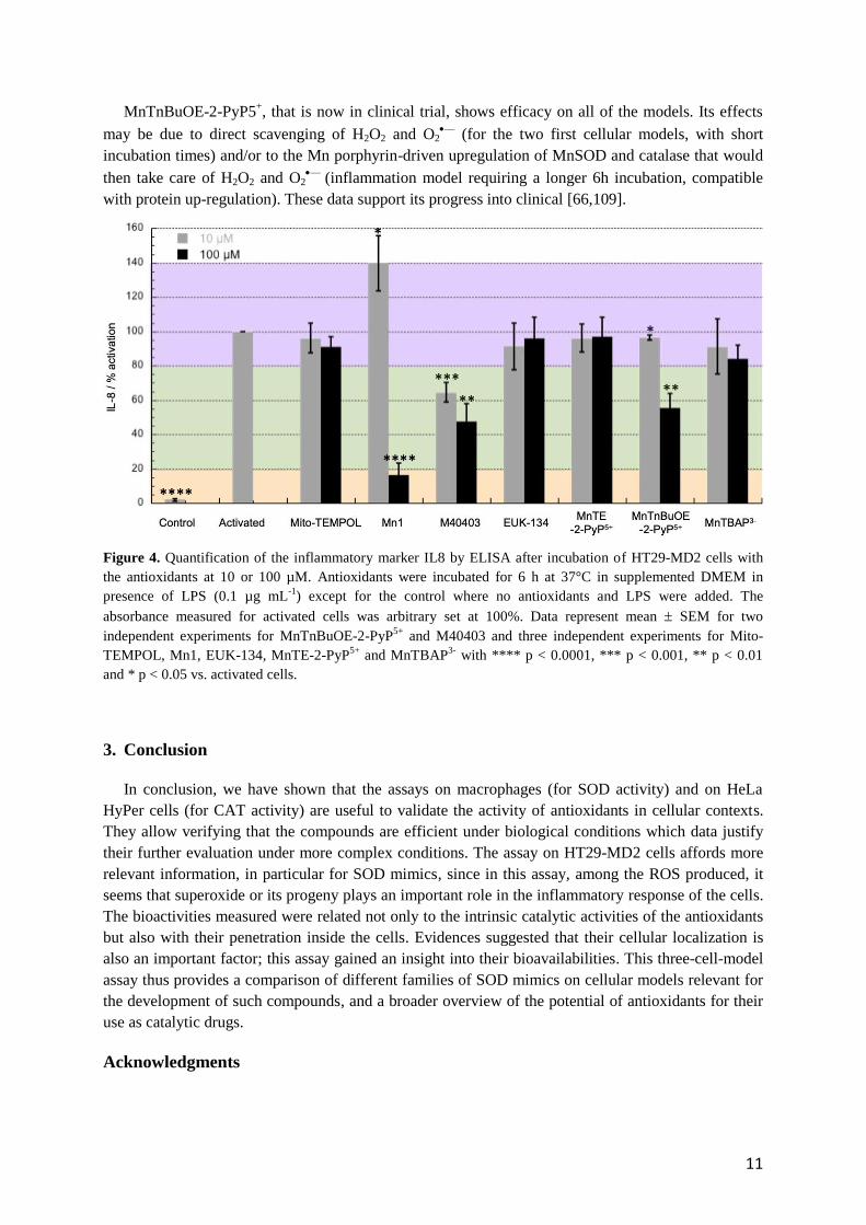
To go further, we have assayed the selected antioxidants on HT29-MD2 cells. These intestine
epithelial cells have been stably transfected to overexpress MD2 protein that promotes an enhanced
inflammatory response in presence of bacterial lipopolysaccharide [78]. Besides, it has been shown
that such inflammatory reaction is mediated by oxidative stress [105,106]. HT29-MD2 cells were thus
incubated in presence of LPS and the antioxidants at non-toxic concentrations: 10 and 100 µM (Figure
S4, SI) [9,103,107]. In this assay, 6h incubations were required to observe a significant inflammatory
response. Consequently, the observed anti-inflammatory effect of the antioxidants may be related to
either their direct scavenging of O2—
or H2O2, or mediated metabolic processes that would result in
reducing levels of those species and oxidative stress in general. The concentration of an inflammatory
marker, the interleukin 8 (IL8) has been measured by ELISA to compare the effects of the
antioxidants. As expected, upon LPS challenge, the IL8 concentration of non-incubated cells increased
significantly. Thus, the decreased level of IL8 in the presence of an antioxidant was indicative of anti-
inflammatory activity (Figure 4). After incubation of the antioxidants at 10 µM, only M40403, and
MnTnBuOE-2-PyP5+
to a lesser extent, showed a moderate anti-inflammatory effect. The other
compounds were found inactive or even pro-inflammatory in the case of Mn1. Taking into account
Mn1 dissociation constant in solution that has been estimated to be 6.5 ± 1.7 10-7
M [85], at a
concentration of 10 µM, it is expected that at least 23% of the ligand is not coordinated to Mn and this
value may increase in a competitive ligand biological environment. Because the ligand is toxic for the
cells [9], the pro-inflammatory response observed may be due to the toxicity of the non-coordinated
ligand. Note that in this model, MnCl2 was previously assayed and found to be not active or pro-
inflammatory [9,103,107]. After incubation at a higher concentration (100 µM), as expected, M40403
and MnTnBuOE-2-PyP5+
were found more active than at 10 µM. Mn1 also exhibited high anti-
inflammatory effect as already reported [9]. Surprisingly, MnTE-2-PyP5+
did not lead to a significant
decrease of IL8 level whereas it possesses similar catalytic constants for superoxide and H2O2
dismutation as MnTnBuOE-2-PyP5+
[68].
The bioavailability of cationic Mn-porphyrins in cellular fragments, cells and tissue has been
comprehensively studied and reported [70]. In this cellular assay, we observed that both the more
hydrophilic MnTE-2-PyP5+
and MnTnBuOE-2-PyP5+
penetrate similarly, as shown by quantification
of the Mn content by ICP-MS in cell lysates incubated with these porphyrins that lead to similar
values (Figure S6, SI). A similar trend was observed when the porphyrins were quantified in cell
lysates using their specific UV-Vis signature (Figure S5 b-c and Figure S6, SI). To note, very
surprisingly, quantification experiments showed that the negatively charged MnTBAP3-
was more
internalized than the positively charged porphyrins (Figure S5 b-c and Figure S6, SI), which was not
expected. It should however be mentioned that MnTBAP3-
was suggested to display an intracellular
and intramitochondrial bio-activity [108]. Although it penetrates in this model, no activity was
observed for this negatively charged porphyrin. Interestingly too, this study suggests that M40403 and
EUK134 penetrate into cells and confirmed the previous results reported for Mn1 [9,103,107]. The
different activities observed here may be related to different cellular localizations. In this assay,
antioxidants that are poor SOD mimics (MITO-TEMPOL, EUK-134, MnTBAP3-
) are not efficient
whatever their catalase-like activity, suggesting that superoxide and/or its progeny may be the main
reactive species that had to be controlled in this model and played an important role in the
inflammatory responses. While inferior at lower 10 µM concentration, due to its low metal-ligand
stability and/or inertia, at 100 µM, though, Mn1 showed the best anti-inflammatory activity in this
cellular model, likely due to its high SOD activity and high bioavailability.
11
MnTnBuOE-2-PyP5+, that is now in clinical trial, shows efficacy on all of the models. Its effects
may be due to direct scavenging of H2O2 and O2—
(for the two first cellular models, with short
incubation times) and/or to the Mn porphyrin-driven upregulation of MnSOD and catalase that would
then take care of H2O2 and O2—
(inflammation model requiring a longer 6h incubation, compatible
with protein up-regulation). These data support its progress into clinical [66,109].
Figure 4. Quantification of the inflammatory marker IL8 by ELISA after incubation of HT29-MD2 cells with
the antioxidants at 10 or 100 µM. Antioxidants were incubated for 6 h at 37°C in supplemented DMEM in
presence of LPS (0.1 µg mL-1
) except for the control where no antioxidants and LPS were added. The
absorbance measured for activated cells was arbitrary set at 100%. Data represent mean SEM for two
independent experiments for MnTnBuOE-2-PyP5+
and M40403 and three independent experiments for Mito-
TEMPOL, Mn1, EUK-134, MnTE-2-PyP5+
and MnTBAP3-
with **** p < 0.0001, *** p < 0.001, ** p < 0.01
and * p < 0.05 vs. activated cells.
3. Conclusion
In conclusion, we have shown that the assays on macrophages (for SOD activity) and on HeLa
HyPer cells (for CAT activity) are useful to validate the activity of antioxidants in cellular contexts.
They allow verifying that the compounds are efficient under biological conditions which data justify
their further evaluation under more complex conditions. The assay on HT29-MD2 cells affords more
relevant information, in particular for SOD mimics, since in this assay, among the ROS produced, it
seems that superoxide or its progeny plays an important role in the inflammatory response of the cells.
The bioactivities measured were related not only to the intrinsic catalytic activities of the antioxidants
but also with their penetration inside the cells. Evidences suggested that their cellular localization is
also an important factor; this assay gained an insight into their bioavailabilities. This three-cell-model
assay thus provides a comparison of different families of SOD mimics on cellular models relevant for
the development of such compounds, and a broader overview of the potential of antioxidants for their
use as catalytic drugs.
Acknowledgments

12
We thank Ecole Normale Supérieure Paris Saclay for A. Vincent’s PhD fellowship, TGE RENARD
(FR 3443, CNRS) and UMR8601 and S. Lajnef for access to the EPR spectrometer for the EPR
experiments. We also thank B. Goetz and P. Legeay for help with ICP-MS. ANR (ANR-15-CE07-
0027), Association François Aupetit and the Fondation pour la Recherche Biomédicale (project
DIE20151234413) are acknowledged for financial supports.
References
[1] B. Halliwell, J.M.C. Gutteridge, Free radicals in biology and medicine, Fifth, Oxford
University Press, New York, NY, 2015.
[2] R.L. Auten, J.M. Davis, Oxygen toxicity and reactive oxygen species: the devil is in the
details, Pediatr. Res. 66 (2009) 121–127.
https://doi.org/10.1203/PDR.0b013e3181a9eafb.
[3] J.J. Soldevila-Barreda, P.J. Sadler, Approaches to the design of catalytic metallodrugs,
Current Opinion in Chemical Biology. 25 (2015) 172–183.
https://doi.org/10.1016/j.cbpa.2015.01.024.
[4] O. Iranzo, Manganese complexes displaying superoxide dismutase activity: A balance
between different factors, Bioorganic Chemistry. 39 (2011) 73–87.
https://doi.org/10.1016/j.bioorg.2011.02.001.
[5] I. Batinic-Haberle, A. Tovmasyan, E.R.H. Roberts, Z. Vujaskovic, K.W. Leong, I.
Spasojevic, SOD Therapeutics: Latest Insights into Their Structure-Activity
Relationships and Impact on the Cellular Redox-Based Signaling Pathways, Antioxid
Redox Signal. 20 (2014) 2372–2415. https://doi.org/10.1089/ars.2012.5147.
[6] C. Policar, Mimicking SODs, Why and How: Bio-Inspired Manganese Complexes as
SOD Mimics, in: Redox-Active Therapeutics, Springer Science, New York, NY, 2016:
pp. 125–164.
[7] I. Batinić-Haberle, J.S. Rebouças, I. Spasojević, Superoxide Dismutase Mimics:
Chemistry, Pharmacology, and Therapeutic Potential, Antioxid Redox Signal. 13
(2010) 877–918. https://doi.org/10.1089/ars.2009.2876.
[8] S. Miriyala, I. Spasojevic, A. Tovmasyan, D. Salvemini, Z. Vujaskovic, D. St. Clair, I.
Batinic-Haberle, Manganese superoxide dismutase, MnSOD and its mimics,
Biochimica et Biophysica Acta (BBA) - Molecular Basis of Disease. 1822 (2012) 794–
814. https://doi.org/10.1016/j.bbadis.2011.12.002.
[9] E. Mathieu, A.-S. Bernard, N. Delsuc, E. Quévrain, G. Gazzah, B. Lai, F. Chain, P.
Langella, M. Bachelet, J. Masliah, P. Seksik, C. Policar, A Cell-Penetrant Manganese
Superoxide Dismutase (MnSOD) Mimic Is Able To Complement MnSOD and Exerts
an Antiinflammatory Effect on Cellular and Animal Models of Inflammatory Bowel
Diseases, Inorganic Chemistry. 56 (2017) 2545–2555.
https://doi.org/10.1021/acs.inorgchem.6b02695.
[10] J.G. Charrier, C. Anastasio, On dithiothreitol (DTT) as a measure of oxidative potential
for ambient particles: evidence for the importance of soluble transition metals, Atmos
Chem Phys. 12 (2012) 11317–11350. https://doi.org/10.5194/acpd-12-11317-2012.
[11] M. Baudry, S. Etienne, A. Bruce, M. Palucki, E. Jacobsen, B. Malfroy, Salen-
manganese complexes are superoxide dismutase-mimics, Biochem. Biophys. Res.
Commun. 192 (1993) 964–968. https://doi.org/10.1006/bbrc.1993.1509.
[12] A. Puglisi, G. Tabb , G. Vecchio, Bioconjugates of cyclodextrins of manganese salen-
type ligand with superoxide dismutase activity, Journal of Inorganic Biochemistry. 98
(2004) 969–976. https://doi.org/10.1016/j.jinorgbio.2004.02.012.
13
[13] W. Park, D. Lim, Effect of the oligo(ethylene glycol) group on the antioxidant activity
of manganese salen complexes, Bioorganic & Medicinal Chemistry Letters. 19 (2009)
614–617. https://doi.org/10.1016/j.bmcl.2008.12.063.
[14] D. Salvemini, C. Muscoli, D.P. Riley, S. Cuzzocrea, Superoxide Dismutase Mimetics,
Pulmonary Pharmacology & Therapeutics. 15 (2002) 439–447.
https://doi.org/10.1006/pupt.2002.0374.
[15] J.D. Rush, Z. Maskos, W.H. Koppenol, The superoxide dismutase activities of two
higher-valent manganese complexes, MnIV desferrioxamine and MnIII cyclam,
Archives of Biochemistry and Biophysics. 289 (1991) 97–102.
https://doi.org/10.1016/0003-9861(91)90447-Q.
[16] D.P. Riley, R.H. Weiss, Manganese macrocyclic ligand complexes as mimics of
superoxide dismutase, J. Am. Chem. Soc. 116 (1994) 387–388.
https://doi.org/10.1021/ja00080a051.
[17] D.P. Riley, S.L. Henke, P.J. Lennon, K. Aston, Computer-Aided Design (CAD) of
Synzymes: Use of Molecular Mechanics (MM) for the Rational Design of Superoxide
Dismutase Mimics, Inorg. Chem. 38 (1999) 1908–1917.
https://doi.org/10.1021/ic981319b.
[18] D. Salvemini, Z.-Q. Wang, J.L. Zweier, A. Samouilov, H. Macarthur, T.P. Misko, M.G.
Currie, S. Cuzzocrea, J.A. Sikorski, D.P. Riley, A Nonpeptidyl Mimic of Superoxide
Dismutase with Therapeutic Activity in Rats, Science. 286 (1999) 304–306.
https://doi.org/10.1126/science.286.5438.304.
[19] A. Dees, A. Zahl, R. Puchta, N.J.R. van Eikema Hommes, F.W. Heinemann, I.
Ivanović-Burmazović, Water Exchange on Seven-Coordinate Mn(II) Complexes with
Macrocyclic Pentadentate Ligands: Insight in the Mechanism of Mn(II) SOD
Mimetics, Inorg. Chem. 46 (2007) 2459–2470. https://doi.org/10.1021/ic061852o.
[20] N. Kitajima, M. Osawa, N. Tamura, Y. Morooka, T. Hirano, M. Hirobe, T. Nagano,
Monomeric (benzoato)manganese(II) complexes as manganese superoxide dismutase
mimics, Inorg. Chem. 32 (1993) 1879–1880. https://doi.org/10.1021/ic00062a001.
[21] A. Deroche, I. Morgenstern-Badarau, M. Cesario, J. Guilhem, B. Keita, L. Nadjo, C.
Houée-Levin, A Seven-Coordinate Manganese(II) Complex Formed with a Single
Tripodal Heptadentate Ligand as a New Superoxide Scavenger, J. Am. Chem. Soc. 118
(1996) 4567–4573. https://doi.org/10.1021/ja952508l.
[22] K. Yamato, I. Miyahara, A. Ichimura, K. Hirotsu, Y. Kojima, H. Sakurai, D. Shiomi, K.
Sato, T. Takui, Superoxide Dismutase Mimetic Complex of Mn(II) / N,N-Bis(2-
pyridylmethyl)-(S)-Histidine, Chem. Lett. 28 (1999) 295–296.
https://doi.org/10.1246/cl.1999.295.
[23] C. Policar, S. Durot, F. Lambert, M. Cesario, F. Ramiandrasoa, I.
Morgenstern‐ Badarau, New MnII Complexes with an N/O Coordination Sphere from
Tripodal N-Centered Ligands − Characterization from Solid State to Solution and
Reaction with Superoxide in Non-Aqueous and Aqueous Media, European Journal of
Inorganic Chemistry. 2001 (2001) 1807–1818. https://doi.org/10.1002/1099-
0682(200107)2001:7<1807::AID-EJIC1807>3.0.CO;2-Y.
[24] E.A. Lewis, H.H. Khodr, R.C. Hider, J.R.L. Smith, P.H. Walton, A manganese
superoxide dismutase mimic based on cis,cis-1,3,5-triaminocyclohexane, Dalton Trans.
(2004) 187–188. https://doi.org/10.1039/B314955B.
[25] Z.-R. Liao, X.-F. Zheng, B.-S. Luo, L.-R. Shen, D.-F. Li, H.-L. Liu, W. Zhao,
Synthesis, characterization and SOD-like activities of manganese-containing
complexes with N,N,N′,N′-tetrakis(2′-benzimidazolyl methyl)-1,2-ethanediamine
(EDTB), Polyhedron. 20 (2001) 2813–2821. https://doi.org/10.1016/S0277-
5387(01)00891-9.

14
[26] S. Groni, G. Blain, R. Guillot, C. Policar, E. Anxolabéhère-Mallart, Reactivity of MnII
with Superoxide. Evidence for a [MnIIIOO]+ Unit by Low-Temperature
Spectroscopies, Inorg. Chem. 46 (2007) 1951–1953.
https://doi.org/10.1021/ic062063+.
[27] F. Cisnetti, G. Pelosi, C. Policar, Synthesis and superoxide dismutase-like activity of
new manganese(III) complexes based on tridentate N2O ligands derived from
histamine, Inorganica Chimica Acta. 360 (2007) 557–562.
https://doi.org/10.1016/j.ica.2006.07.112.
[28] J.W. Beyer, I. Fridovich, Characterization of a superoxide dismutase mimic prepared
from desferrioxamine and MnO2., Arch Biochem Biophys. 271 (1989) 149–156.
https://doi.org/10.1016/0003-9861(89)90265-8.
[29] K.M. Faulkner, R.D. Stevens, I. Fridovich, Characterization of Mn(III) Complexes of
Linear and Cyclic Desferrioxamines as Mimics of Superoxide Dismutase Activity,
Archives of Biochemistry and Biophysics. 310 (1994) 341–346.
https://doi.org/10.1006/abbi.1994.1176.
[30] J. Lati, D. Meyerstein, Oxidation of first-row bivalent transition-metal complexes
containing ethylenediaminetetra-acetate and nitrilotriacetate ligands by free radicals: a
pulse-radiolysis study, J. Chem. Soc., Dalton Trans. (1978) 1105–1118.
https://doi.org/10.1039/DT9780001105.
[31] J. Stein, J.P. Fackler, G.J. McClune, J.A. Fee, L.T. Chan, Superoxide and
manganese(III). Reactions of manganese-EDTA and manganese-CyDTA complexes
with molecular oxygen. X-ray structure of potassium manganese-EDTA.2 water, Inorg.
Chem. 18 (1979) 3511–3519. https://doi.org/10.1021/ic50202a044.
[32] W.H. Koppenol, F. Levine, T.L. Hatmaker, J. Epp, J.D. Rush, Catalysis of superoxide
dismutation by manganese aminopolycarboxylate complexes, Archives of
Biochemistry and Biophysics. 251 (1986) 594–599. https://doi.org/10.1016/0003-
9861(86)90368-1.
[33] F.S. Archibald, I. Fridovich, The scavenging of superoxide radical by manganous
complexes: In vitro, Archives of Biochemistry and Biophysics. 214 (1982) 452–463.
https://doi.org/10.1016/0003-9861(82)90049-2.
[34] M.A. Bailey, M.J. Ingram, D.P. Naughton, A novel anti-oxidant and anti-cancer
strategy: a peptoid anti-inflammatory drug conjugate with SOD mimic activity,
Biochemical and Biophysical Research Communications. 317 (2004) 1155–1158.
https://doi.org/10.1016/j.bbrc.2004.03.162.
[35] A.E.O. Fisher, D.P. Naughton, Metal ion chelating peptides with superoxide dismutase
activity, Biomedicine & Pharmacotherapy. 59 (2005) 158–162.
https://doi.org/10.1016/j.biopha.2005.03.008.
[36] T. Piacham, C. Isarankura-Na-Ayudhya, C. Nantasenamat, S. Yainoy, L. Ye, L. Bülow,
V. Prachayasittikul, Metalloantibiotic Mn(II)–bacitracin complex mimicking
manganese superoxide dismutase, Biochemical and Biophysical Research
Communications. 341 (2006) 925–930. https://doi.org/10.1016/j.bbrc.2006.01.045.
[37] J.S. Valentine, A.E. Quinn, Reaction of superoxide with the manganese(III)
tetraphenylporphine cation, Inorg. Chem. 15 (1976) 1997–1999.
https://doi.org/10.1021/ic50162a058.
[38] K.M. Faulkner, S.I. Liochev, I. Fridovich, Stable Mn(III) porphyrins mimic superoxide
dismutase in vitro and substitute for it in vivo., J. Biol. Chem. 269 (1994) 23471–
23476.
[39] I. Batinić-Haberle, S.I. Liochev, I. Spasojević, I. Fridovich, A Potent Superoxide
Dismutase Mimic: Manganese β-Octabromo-meso-tetrakis-(N-methylpyridinium- 4-yl)
Porphyrin, Archives of Biochemistry and Biophysics. 343 (1997) 225–233.

15
https://doi.org/10.1006/abbi.1997.0157.
[40] I. Batinic-Haberle, L. Benov, I. Spasojevic, I. Fridovich, The Ortho Effect Makes
Manganese(III) Meso -Tetrakis( N -Methylpyridinium-2-yl)Porphyrin a Powerful and
Potentially Useful Superoxide Dismutase Mimic, Journal of Biological Chemistry. 273
(1998) 24521–24528. https://doi.org/10.1074/jbc.273.38.24521.
[41] P.J.F. Gauuan, M.P. Trova, L. Gregor-Boros, S.B. Bocckino, J.D. Crapo, B.J. Day,
Superoxide dismutase mimetics: synthesis and structure–activity relationship study of
MnTBAP analogues, Bioorganic & Medicinal Chemistry. 10 (2002) 3013–3021.
https://doi.org/10.1016/S0968-0896(02)00153-0.
[42] I. Batinić-Haberle, I. Spasojević, R.D. Stevens, B. Bondurant, A. Okado-Matsumoto, I.
Fridovich, Ž. Vujašković, M.W. Dewhirst, New PEG-ylated Mn( III ) porphyrins
approaching catalytic activity of SOD enzyme, Dalton Trans. (2006) 617–624.
https://doi.org/10.1039/B513761F.
[43] F. Matemadombo, M. Durmus, V. Escriou, S. Griveau, D. Scherman, F. Bedioui, T.
Nyokong, Evaluation of the Performance of Manganese Phthalocyanines as Superoxide
Dismutase Mimics, CAC. 5 (2009) 330–338.
https://doi.org/10.2174/157341109789077731.
[44] J.T. Brewster, G.D. Thiabaud, P. Harvey, H. Zafar, J.F. Reuther, S. Dell’Acqua, R.M.
Johnson, H.D. Root, P. Metola, A. Jasanoff, L. Casella, J.L. Sessler,
Metallotexaphyrins as MRI-Active Catalytic Antioxidants for Neurodegenerative
Disease: A Study on Alzheimer’s Disease, Chem. 6 (2020) 703–724.
https://doi.org/10.1016/j.chempr.2019.12.016.
[45] J.F. Arambula, C. Preihs, D. Borthwick, D. Magda, J.L. Sessler, Texaphyrins: Tumor
Localizing Redox Active Expanded Porphyrins, Anticancer Agents Med Chem. 11
(2011) 222–232.
[46] L.M.F. Gomes, A. Mahammed, K.E. Prosser, J.R. Smith, M.A. Silverman, C.J.
Walsby, Z. Gross, T. Storr, A catalytic antioxidant for limiting amyloid-beta peptide
aggregation and reactive oxygen species generation, Chem. Sci. (2018).
https://doi.org/10.1039/C8SC04660C.
[47] Z. Okun, L. Kupershmidt, M.B.H.Y. and Z. Gross, Cellular Uptake and Organ
Accumulation of Amphipolar Metallocorroles with Cytoprotective and Cytotoxic
Properties, Anti-Cancer Agents in Medicinal Chemistry. 11 (2011) 380–384.
http://www.eurekaselect.com/74149/article (accessed March 26, 2020).
[48] A. Haber, Z. Gross, Catalytic antioxidant therapy by metallodrugs: lessons from
metallocorroles, Chem. Commun. 51 (2015) 5812–5827.
https://doi.org/10.1039/C4CC08715A.
[49] I. Spasojević, I. Batinić-Haberle, R.D. Stevens, P. Hambright, A.N. Thorpe, J.
Grodkowski, P. Neta, I. Fridovich, Manganese(III) Biliverdin IX Dimethyl Ester: A
Powerful Catalytic Scavenger of Superoxide Employing the Mn(III)/Mn(IV) Redox
Couple, Inorganic Chemistry. 40 (2001) 726–739. https://doi.org/10.1021/ic0004986.
[50] M.N. Patel, Metalloporphyrins improve the survival of Sod2-deficient neurons, Aging
Cell. 2 (2003) 219–222. https://doi.org/10.1046/j.1474-9728.2003.00055.x.
[51] W. Munroe, C. Kingsley, A. Durazo, E. Butler Gralla, J.A. Imlay, C. Srinivasan, J.
Selverstone Valentine, Only one of a wide assortment of manganese-containing SOD
mimicking compounds rescues the slow aerobic growth phenotypes of both Escherichia
coli and Saccharomyces cerevisiae strains lacking superoxide dismutase enzymes,
Journal of Inorganic Biochemistry. 101 (2007) 1875–1882.
https://doi.org/10.1016/j.jinorgbio.2007.07.008.
[52] J.M. Pollard, J.S. Reboucas, A. Durazo, I. Kos, F. Fike, M. Panni, E.B. Gralla, J.S.
Valentine, I. Batinic-Haberle, R.A. Gatti, Radioprotective effects of manganese-

16
containing superoxide dismutase mimics on ataxia–telangiectasia cells, Free Radical
Biology and Medicine. 47 (2009) 250–260.
https://doi.org/10.1016/j.freeradbiomed.2009.04.018.
[53] J. Alexandre, C. Nicco, C. Chéreau, A. Laurent, B. Weill, F. Goldwasser, F. Batteux,
Improvement of the Therapeutic Index of Anticancer Drugs by the Superoxide
Dismutase Mimic Mangafodipir, J Natl Cancer Inst. 98 (2006) 236–244.
https://doi.org/10.1093/jnci/djj049.
[54] M.R. Filipović, A.C.W. Koh, S. Arbault, V. Niketić, A. Debus, U. Schleicher, C.
Bogdan, M. Guille, F. Lemaître, C. Amatore, I. Ivanović‐ Burmazović, Striking
Inflammation from Both Sides: Manganese(II) Pentaazamacrocyclic SOD Mimics Act
Also as Nitric Oxide Dismutases: A Single-Cell Study, Angewandte Chemie
International Edition. 49 (n.d.) 4228–4232. https://doi.org/10.1002/anie.200905936.
[55] A.-S. Bernard, C. Giroud, H.Y.V. Ching, A. Meunier, V. Ambike, C. Amatore, M.G.
Collignon, F. Lemaître, C. Policar, Evaluation of the anti-oxidant properties of a SOD-
mimic Mn-complex in activated macrophages, Dalton Trans. 41 (2012) 6399–6403.
https://doi.org/10.1039/C2DT12479C.
[56] B. Gauter-Fleckenstein, K. Fleckenstein, K. Owzar, C. Jian, I. Batinic-Haberle, Z.
Vujaskovic, Comparison of two Mn porphyrin-based mimics of superoxide dismutase
(SOD) in pulmonary radioprotection, Free Radic Biol Med. 44 (2008) 982–989.
https://doi.org/10.1016/j.freeradbiomed.2007.10.058.
[57] B. Gauter-Fleckenstein, K. Fleckenstein, K. Owzar, C. Jiang, J.S. Rebouças, I. Batinic-
Haberle, Z. Vujaskovic, Early and late administration of MnTE-2-PyP5+ in mitigation
and treatment of radiation-induced lung damage, Free Radical Biology and Medicine.
48 (2010) 1034–1043. https://doi.org/10.1016/j.freeradbiomed.2010.01.020.
[58] S.R. Doctrow, A. Lopez, A.M. Schock, N.E. Duncan, M.M. Jourdan, E.B. Olasz, J.E.
Moulder, B.L. Fish, M. Mäder, J. Lazar, Z. Lazarova, A Synthetic Superoxide
Dismutase/Catalase Mimetic EUK-207 Mitigates Radiation Dermatitis and Promotes
Wound Healing in Irradiated Rat Skin, Journal of Investigative Dermatology. 133
(2013) 1088–1096. https://doi.org/10.1038/jid.2012.410.
[59] S.R. Doctrow, K. Huffman, C.B. Marcus, G. Tocco, E. Malfroy, C.A. Adinolfi, H.
Kruk, K. Baker, N. Lazarowych, J. Mascarenhas, B. Malfroy, Salen−Manganese
Complexes as Catalytic Scavengers of Hydrogen Peroxide and Cytoprotective Agents:
Structure−Activity Relationship Studies, Journal of Medicinal Chemistry. 45 (2002)
4549–4558. https://doi.org/10.1021/jm020207y.
[60] H. Sheng, J.J. Enghild, R. Bowler, M. Patel, I. Batinić-Haberle, C.L. Calvi, B.J. Day,
R.D. Pearlstein, J.D. Crapo, D.S. Warner, Effects of metalloporphyrin catalytic
antioxidants in experimental brain ischemia, Free Radical Biology and Medicine. 33
(2002) 947–961. https://doi.org/10.1016/S0891-5849(02)00979-6.
[61] E. Masini, S. Cuzzocrea, E. Mazzon, C. Marzocca, P.F. Mannaioni, D. Salvemini,
Protective effects of M40403, a selective superoxide dismutase mimetic, in myocardial
ischaemia and reperfusion injury in vivo, British Journal of Pharmacology. 136 (2002)
905–917. https://doi.org/10.1038/sj.bjp.0704774.
[62] Y. Rong, S.R. Doctrow, G. Tocco, M. Baudry, EUK-134, a synthetic superoxide
dismutase and catalase mimetic, prevents oxidative stress and attenuates kainate-
induced neuropathology, PNAS. 96 (1999) 9897–9902.
https://doi.org/10.1073/pnas.96.17.9897.
[63] M.C. McDonald, R. d’Emmanuele di Villa Bianca, N.S. Wayman, A. Pinto, M.A.
Sharpe, S. Cuzzocrea, P.K. Chatterjee, C. Thiemermann, A superoxide dismutase
mimetic with catalase activity (EUK-8) reduces the organ injury in endotoxic shock,
European Journal of Pharmacology. 466 (2003) 181–189.

17
https://doi.org/10.1016/S0014-2999(03)01538-3.
[64] E. Masini, D. Bani, A. Vannacci, S. Pierpaoli, P.F. Mannaioni, S.A.A. Comhair, W.
Xu, C. Muscoli, S.C. Erzurum, D. Salvemini, Reduction of antigen-induced respiratory
abnormalities and airway inflammation in sensitized guinea pigs by a superoxide
dismutase mimetic, Free Radical Biology and Medicine. 39 (2005) 520–531.
https://doi.org/10.1016/j.freeradbiomed.2005.04.006.
[65] M.-A. Guillaumot, O. Cerles, H.C. Bertrand, E. Benoit, C. Nicco, S. Chouzenoux, A.
Schmitt, F. Batteux, C. Policar, R. Coriat, Oxaliplatin-induced neuropathy: the
preventive effect of a new super-oxide dismutase modulator, Oncotarget. 10 (2019)
6418–6431. https://doi.org/10.18632/oncotarget.27248.
[66] I. Batinic-Haberle, M.E. Tome, Thiol regulation by Mn porphyrins, commonly known
as SOD mimics, Redox Biology. 25 (2019) 101139.
https://doi.org/10.1016/j.redox.2019.101139.
[67] A. Tovmasyan, C.G.C. Maia, T. Weitner, S. Carballal, R.S. Sampaio, D. Lieb, R.
Ghazaryan, I. Ivanovic-Burmazovic, G. Ferrer-Sueta, R. Radi, J.S. Reboucas, I.
Spasojevic, L. Benov, I. Batinic-Haberle, A comprehensive evaluation of catalase-like
activity of different classes of redox-active therapeutics, Free Radical Biology and
Medicine. 86 (2015) 308–321. https://doi.org/10.1016/j.freeradbiomed.2015.05.018.
[68] A. Tovmasyan, J.C. Bueno-Janice, M.C. Jaramillo, R.S. Sampaio, J.S. Reboucas, N.
Kyui, L. Benov, B. Deng, T.-T. Huang, M.E. Tome, I. Spasojevic, I. Batinic-Haberle,
Radiation-Mediated Tumor Growth Inhibition Is Significantly Enhanced with Redox-
Active Compounds That Cycle with Ascorbate, Antioxidants & Redox Signaling. 29
(2018) 1196–1214. https://doi.org/10.1089/ars.2017.7218.
[69] S. Signorella, C. Palopoli, G. Ledesma, Rationally designed mimics of antioxidant
manganoenzymes: Role of structural features in the quest for catalysts with catalase
and superoxide dismutase activity, Coordination Chemistry Reviews. 365 (2018) 75–
102. https://doi.org/10.1016/j.ccr.2018.03.005.
[70] I. Batinic-Haberle, A. Tovmasyan, I. Spasojevic, Mn Porphyrin-Based Redox-Active
Drugs: Differential Effects as Cancer Therapeutics and Protectors of Normal Tissue
Against Oxidative Injury, Antioxidants & Redox Signaling. 29 (2018) 1691–1724.
https://doi.org/10.1089/ars.2017.7453.
[71] Z. Rajic, A. Tovmasyan, I. Spasojevic, H. Sheng, M. Lu, A.M. Li, E.B. Gralla, D.S.
Warner, L. Benov, I. Batinic-Haberle, A new SOD mimic, Mn(III) ortho N-
butoxyethylpyridylporphyrin, combines superb potency and lipophilicity with low
toxicity, Free Radical Biology and Medicine. 52 (2012) 1828–1834.
https://doi.org/10.1016/j.freeradbiomed.2012.02.006.
[72] J.S. Rebouças, I. Spasojević, I. Batinić-Haberle, Pure manganese(III) 5,10,15,20-
tetrakis(4-benzoic acid)porphyrin (MnTBAP) is not a superoxide dismutase mimic in
aqueous systems: a case of structure–activity relationship as a watchdog mechanism in
experimental therapeutics and biology, J Biol Inorg Chem. 13 (2008) 289–302.
https://doi.org/10.1007/s00775-007-0324-9.
[73] I. Batinić-Haberle, I. Spasojević, P. Hambright, L. Benov, A.L. Crumbliss, I. Fridovich,
Relationship among Redox Potentials, Proton Dissociation Constants of Pyrrolic
Nitrogens, and in Vivo and in Vitro Superoxide Dismutating Activities of
Manganese(III) and Iron(III) Water-Soluble Porphyrins, Inorganic Chemistry. 38
(1999) 4011–4022. https://doi.org/10.1021/ic990118k.
[74] I. Batinić-Haberle, I. Spasojević, R. D. Stevens, P. Hambright, I. Fridovich,
Manganese( iii ) meso -tetrakis( ortho - N -alkylpyridyl)porphyrins. Synthesis,
characterization, and catalysis of O 2 ˙ − dismutation, Journal of the Chemical Society,
Dalton Transactions. 0 (2002) 2689–2696. https://doi.org/10.1039/B201057G.

18
[75] F. Cisnetti, A.-S. Lefèvre, R. Guillot, F. Lambert, G. Blain, E. Anxolabéhère-Mallart,
C. Policar, A New Pentadentate Ligand Forms Both a Di- and a Mononuclear MnII
Complex: Electrochemical, Spectroscopic and Superoxide Dismutase Activity Studies,
European Journal of Inorganic Chemistry. 2007 (2007) 4472–4480.
https://doi.org/10.1002/ejic.200601236.
[76] A. Tighe, O. Staples, S. Taylor, Mps1 kinase activity restrains anaphase during an
unperturbed mitosis and targets Mad2 to kinetochores, J Cell Biol. 181 (2008) 893–
901. https://doi.org/10.1083/jcb.200712028.
[77] I. Amblard, M. Thauvin, C. Rampon, I. Queguiner, V.V. Pak, V. Belousov, A.
Prochiantz, M. Volovitch, A. Joliot, S. Vriz, H 2 O 2 and Engrailed 2 paracrine activity
synergize to shape the zebrafish optic tectum, Communications Biology. 3 (2020) 1–9.
https://doi.org/10.1038/s42003-020-01268-7.
[78] C. Lenoir, C. Sapin, A.H. Broquet, A.M. Jouniaux, S. Bardin, I. Gasnereau, G. Thomas,
P. Seksik, G. Trugnan, J. Masliah, M. Bachelet, MD-2 controls bacterial
lipopolysaccharide hyporesponsiveness in human intestinal epithelial cells., Life Sci.
82 (2008) 519–528. https://doi.org/10.1016/j.lfs.2007.12.007.
[79] V. Grondin, P. Seksik, S. Dumont, G. Thomas, G. Trugnan, J.F. Fléjou, J. Masliah, D.
Wendum, M. Bachelet, Regulation of colon cancer cell proliferation and migration by
MD-2 activity, Innate Immun. 17 (2011) 414–422.
https://doi.org/10.1177/1753425910375583.
[80] S. Goldstein, G. Merenyi, A. Russo, A. Samuni, The Role of Oxoammonium Cation in
the SOD-Mimic Activity of Cyclic Nitroxides, J. Am. Chem. Soc. 125 (2003) 789–795.
https://doi.org/10.1021/ja028190w.
[81] Y.N. Antonenko, A.V. Avetisyan, L.E. Bakeeva, B.V. Chernyak, V.A. Chertkov, L.V.
Domnina, O.Yu. Ivanova, D.S. Izyumov, L.S. Khailova, S.S. Klishin, G.A.
Korshunova, K.G. Lyamzaev, M.S. Muntyan, O.K. Nepryakhina, A.A. Pashkovskaya,
O.Yu. Pletjushkina, A.V. Pustovidko, V.A. Roginsky, T.I. Rokitskaya, E.K. Ruuge,
V.B. Saprunova, I.I. Severina, R.A. Simonyan, I.V. Skulachev, M.V. Skulachev, N.V.
Sumbatyan, I.V. Sviryaeva, V.N. Tashlitsky, J.M. Vassiliev, M.Yu. Vyssokikh, L.S.
Yaguzhinsky, A.A. Zamyatnin, V.P. Skulachev, Mitochondria-targeted plastoquinone
derivatives as tools to interrupt execution of the aging program. 1. Cationic
plastoquinone derivatives: Synthesis and in vitro studies, Biochemistry Moscow. 73
(2008) 1273–1287. https://doi.org/10.1134/S0006297908120018.
[82] I. Spasojević, Y. Chen, T.J. Noel, Y. Yu, M.P. Cole, L. Zhang, Y. Zhao, D.K. St. Clair,
I. Batinić-Haberle, Mn porphyrin-based superoxide dismutase (SOD) mimic, MnIIITE-
2-PyP5+, targets mouse heart mitochondria, Free Radical Biology and Medicine. 42
(2007) 1193–1200. https://doi.org/10.1016/j.freeradbiomed.2007.01.019.
[83] R.A.J. Smith, C.M. Porteous, A.M. Gane, M.P. Murphy, Delivery of bioactive
molecules to mitochondria in vivo, PNAS. 100 (2003) 5407–5412.
https://doi.org/10.1073/pnas.0931245100.
[84] J. Trnka, F.H. Blaikie, A. Logan, R.A.J. Smith, M.P. Murphy, Antioxidant properties of
MitoTEMPOL and its hydroxylamine, Free Radical Research. 43 (2009) 4–12.
https://doi.org/10.1080/10715760802582183.
[85] H.Y.V. Ching, I. Kenkel, N. Delsuc, E. Mathieu, I. Ivanović-Burmazović, C. Policar,
Bioinspired superoxide-dismutase mimics: The effects of functionalization with
cationic polyarginine peptides, Journal of Inorganic Biochemistry. 160 (2016) 172–
179. https://doi.org/10.1016/j.jinorgbio.2016.01.025.
[86] F.C. Friedel, D. Lieb, I. Ivanović-Burmazović, Comparative studies on manganese-
based SOD mimetics, including the phosphate effect, by using global spectral analysis,
Journal of Inorganic Biochemistry. 109 (2012) 26–32.

19
https://doi.org/10.1016/j.jinorgbio.2011.12.008.
[87] S. Melov, S.R. Doctrow, J.A. Schneider, J. Haberson, M. Patel, P.E. Coskun, K.
Huffman, D.C. Wallace, B. Malfroy, Lifespan Extension and Rescue of Spongiform
Encephalopathy in Superoxide Dismutase 2 Nullizygous Mice Treated with Superoxide
Dismutase–Catalase Mimetics, J. Neurosci. 21 (2001) 8348–8353.
https://doi.org/10.1523/JNEUROSCI.21-21-08348.2001.
[88] K. Barnese, E.B. Gralla, D.E. Cabelli, J. Selverstone Valentine, Manganous Phosphate
Acts as a Superoxide Dismutase, Journal of the American Chemical Society. 130
(2008) 4604–4606. https://doi.org/10.1021/ja710162n.
[89] S. Durot, C. Policar, F. Cisnetti, F. Lambert, J.-P. Renault, G. Pelosi, G. Blain, H.
Korri-Youssoufi, J.-P. Mahy, Series of Mn Complexes Based onN-Centered Ligands
and Superoxide - Reactivity in an Anhydrous Medium and SOD-Like Activity in an
Aqueous Medium Correlated to MnII/MnIII Redox Potentials, European Journal of
Inorganic Chemistry. 2005 (2005) 3513–3523. https://doi.org/10.1002/ejic.200400835.
[90] S. Goldstein, I. Fridovich, G. Czapski, Kinetic properties of Cu,Zn-superoxide
dismutase as a function of metal content—Order restored, Free Radical Biology and
Medicine. 41 (2006) 937–941. https://doi.org/10.1016/j.freeradbiomed.2006.05.026.
[91] C.K. Vance, A.-F. Miller, A Simple Proposal That Can Explain the Inactivity of Metal-
Substituted Superoxide Dismutases, J. Am. Chem. Soc. 120 (1998) 461–467.
https://doi.org/10.1021/ja972060j.
[92] L.M. Ellerby, D.E. Cabelli, J.A. Graden, J.S. Valentine, Copper−Zinc Superoxide
Dismutase: Why Not pH-Dependent?, J. Am. Chem. Soc. 118 (1996) 6556–6561.
https://doi.org/10.1021/ja953845x.
[93] I.A. Abreu, D.E. Cabelli, Superoxide dismutases—a review of the metal-associated
mechanistic variations, Biochimica et Biophysica Acta (BBA) - Proteins and
Proteomics. 1804 (2010) 263–274. https://doi.org/10.1016/j.bbapap.2009.11.005.
[94] J.S. Rebouças, I. Spasojević, I. Batinić-Haberle, Pure manganese(III) 5,10,15,20-
tetrakis(4-benzoic acid)porphyrin (MnTBAP) is not a superoxide dismutase mimic in
aqueous systems: a case of structure–activity relationship as a watchdog mechanism in
experimental therapeutics and biology, J Biol Inorg Chem. 13 (2008) 289–302.
https://doi.org/10.1007/s00775-007-0324-9.
[95] I. Batinić-Haberle, S. Cuzzocrea, J.S. Rebouças, G. Ferrer-Sueta, E. Mazzon, R. Di
Paola, R. Radi, I. Spasojević, L. Benov, D. Salvemini, Pure MnTBAP selectively
scavenges peroxynitrite over superoxide: Comparison of pure and commercial
MnTBAP samples to MnTE-2-PyP in two models of oxidative stress injury, an SOD-
specific Escherichia coli model and carrageenan-induced pleurisy, Free Radical
Biology and Medicine. 46 (2009) 192–201.
https://doi.org/10.1016/j.freeradbiomed.2008.09.042.
[96] S. Da Rocha, J. Bigot, F. Onodi, J. Cosette, G. Corre, J. Poupiot, D. Fenard, B. Gjata,
A. Galy, T.M.A. Neildez-Nguyen, Temporary Reduction of Membrane CD4 with the
Antioxidant MnTBAP Is Sufficient to Prevent Immune Responses Induced by Gene
Transfer, Molecular Therapy - Methods & Clinical Development. 14 (2019) 285–299.
https://doi.org/10.1016/j.omtm.2019.06.011.
[97] C. Amatore, S. Arbault, M. Guille, F. Lemaître, Electrochemical Monitoring of Single
Cell Secretion: Vesicular Exocytosis and Oxidative Stress, Chem. Rev. 108 (2008)
2585–2621. https://doi.org/10.1021/cr068062g.
[98] C.A. Harris, K.S. Derbin, B. Hunte-McDonough, M.R. Krauss, K.T. Chen, D.M.
Smith, L.B. Epstein, Manganese superoxide dismutase is induced by IFN-gamma in
multiple cell types. Synergistic induction by IFN-gamma and tumor necrosis factor or
IL-1., The Journal of Immunology. 147 (1991) 149–154.

20
[99] V.V. Belousov, A.F. Fradkov, K.A. Lukyanov, D.B. Staroverov, K.S. Shakhbazov,
A.V. Terskikh, S. Lukyanov, Genetically encoded fluorescent indicator for intracellular
hydrogen peroxide, Nat Methods. 3 (2006) 281–286. https://doi.org/10.1038/nmeth866.
[100] N.M. Mishina, K.N. Markvicheva, D.S. Bilan, M.E. Matlashov, M.V. Shirmanova, D.
Liebl, C. Schultz, S. Lukyanov, V.V. Belousov, Chapter Three - Visualization of
Intracellular Hydrogen Peroxide with HyPer, a Genetically Encoded Fluorescent Probe,
in: E. Cadenas, L. Packer (Eds.), Methods in Enzymology, Academic Press, 2013: pp.
45–59. https://doi.org/10.1016/B978-0-12-405883-5.00003-X.
[101] E. Cadenas, K.J.A. Davies, Guest Editors: Enrique Cadenas and Kelvin J. A. Davies,
Oxidative Stress. (n.d.) 9.
[102] C.M. Weekley, I. Kenkel, R. Lippert, S. Wei, D. Lieb, T. Cranwell, J.L. Wedding, A.S.
Zillmann, R. Rohr, M.R. Filipovic, I. Ivanović-Burmazović, H.H. Harris, Cellular Fates
of Manganese(II) Pentaazamacrocyclic Superoxide Dismutase (SOD) Mimetics:
Fluorescently Labeled MnSOD Mimetics, X-ray Absorption Spectroscopy, and X-ray
Fluorescence Microscopy Studies, Inorg. Chem. 56 (2017) 6076–6093.
https://doi.org/10.1021/acs.inorgchem.6b03073.
[103] E. Mathieu, A.-S. Bernard, E. Quévrain, M. Zoumpoulaki, S. Iriart, C. Lung-Soong, B.
Lai, K. Medjoubi, L. Henry, S. Nagarajan, F. Poyer, A. Scheitler, I. Ivanović-
Burmazović, S. Marco, A. Somogyi, P. Seksik, N. Delsuc, C. Policar, Intracellular
location matters: rationalization of the anti-inflammatory activity of a manganese(II)
superoxide dismutase mimic complex, Chem. Commun. 56 (2020) 7885–7888.
https://doi.org/10.1039/D0CC03398G.
[104] C.C. Winterbourn, Biological Production, Detection, and Fate of Hydrogen Peroxide,
Antioxidants & Redox Signaling. 29 (2018) 541–551.
https://doi.org/10.1089/ars.2017.7425.
[105] J.F. Valentine, H.S. Nick, Acute-phase induction of manganese superoxide dismutase
in intestinal epithelial cell lines, Gastroenterology. 103 (1992) 905–912.
https://doi.org/10.1016/0016-5085(92)90024-S.
[106] M.C. Denis, A. Furtos, S. Dudonné, A. Montoudis, C. Garofalo, Y. Desjardins, E.
Delvin, E. Levy, Apple Peel Polyphenols and Their Beneficial Actions on Oxidative
Stress and Inflammation, PLOS ONE. 8 (2013) e53725.
https://doi.org/10.1371/journal.pone.0053725.
[107] E. Mathieu, A.-S. Bernard, H.Y.V. Ching, A. Somogyi, K. Medjoubi, J.R. Fores, H.C.
Bertrand, A. Vincent, S. Trépout, J.-L. Guerquin-Kern, A. Scheitler, I. Ivanović-
Burmazović, P. Seksik, N. Delsuc, C. Policar, Anti-inflammatory activity of superoxide
dismutase mimics functionalized with cell-penetrating peptides, Dalton Trans. 49
(2020) 2323–2330. https://doi.org/10.1039/C9DT04619D.
[108] Q.-Y. Li, C. Pedersen, B.J. Day, M. Patel, Dependence of excitotoxic
neurodegeneration on mitochondrial aconitase inactivation, Journal of Neurochemistry.
78 (2001) 746–755. https://doi.org/10.1046/j.1471-4159.2001.00457.x.
[109] Y. Zhao, D.W. Carroll, Y. You, L. Chaiswing, R. Wen, I. Batinic-Haberle, S. Bondada,
Y. Liang, D.K. St. Clair, A novel redox regulator, MnTnBuOE-2-PyP5+, enhances
normal hematopoietic stem/progenitor cell function, Redox Biology. 12 (2017) 129–
138. https://doi.org/10.1016/j.redox.2017.02.005.