Rover-Based Visual Target Tracking Validation and Mission Infusion
Skeletal Muscle Vascular Control During Exercise: Impact of Nitrite Infusion During Nitric Oxide...
9
Page Proof Instructions and Queries Journal Title: JCP Article Number: 599061 No. Query Please confirm that all author information, including names, affiliations, sequence, and contact details, is correct. Please review the entire document for typographical errors, mathematical errors, and any other necessary corrections; check headings, tables, and figures. Please confirm that the Funding and Conflict of Interest statements are accurate. Please confirm you have reviewed this proof to your satisfaction and understand this is your final opportunity for review prior to publication. AQ: 1 Please provide degree(s) for all the authors listed. AQ: 2 Please provide expansion for L-NAME. AQ: 3 Please provide complete details for ‘‘Delp & Duan’’ to be included in the reference list.
-
Upload
independent -
Category
Documents
-
view
1 -
download
0
Transcript of Skeletal Muscle Vascular Control During Exercise: Impact of Nitrite Infusion During Nitric Oxide...

Page Proof Instructions and Queries
Journal Title: JCP
Article Number: 599061
No. Query
Please confirm that all author information, including names, affiliations, sequence, and contact details, is correct.
Please review the entire document for typographical errors, mathematical errors, and any other necessary
corrections; check headings, tables, and figures.
Please confirm that the Funding and Conflict of Interest statements are accurate.
Please confirm you have reviewed this proof to your satisfaction and understand this is your final opportunity for
review prior to publication.
AQ: 1 Please provide degree(s) for all the authors listed.
AQ: 2 Please provide expansion for L-NAME.
AQ: 3 Please provide complete details for ‘‘Delp & Duan’’ to be included in the reference list.

Original Article
Skeletal Muscle Vascular ControlDuring Exercise: Impact of NitriteInfusion During Nitric Oxide SynthaseInhibition in Healthy Rats
Scott K. Ferguson1, Angela A. Glean2, Clark T. Holdsworth1,Jennifer L. WrightAQ1
1, Alex J. Fees1, Trenton D. Colburn2, Thomas Stabler3,Jason D. Allen3, Andrew M. Jones4, Timothy I. Musch1,2, and David C. Poole1,2
AbstractThe nitric oxide synthase (NOS)-independent pathway of nitric oxide (NO) production in which nitrite (NO2
�) is reduced toNO may have therapeutic applications for those with cardiovascular diseases in which the NOS pathway is downregulated. Wetested the hypothesis that NO2
� infusion would reduce mean arterial pressure (MAP) and increase skeletal muscle blood flow(BF) and vascular conductance (VC) during exercise in the face of NOS blockade via L-NAMEAQ2 . Following infusion of L-NAME(10 mg kg�1: L-NAME), male Sprague-Dawley rats (3-6 months, n ¼ 8) exercised without (L-NAME) and after infusion ofsodium NO2
� (7 mg kg�1; L-NAME þNO2�). The MAP and hind limb skeletal muscle BF (radiolabeled microsphere infusions)
were measured during submaximal treadmill running (20 m min�1, 5% grade). Across group comparisons were made witha published control data set (n ¼ 11). Relative to L-NAME, NO2
� infusion significantly reduced MAP (P < .03). The lower MAPin L-NAMEþNO2
� was not different from healthy control animals (control: 137 + 3 L-NAME: 157 + 7, L-NAME þ NO2�:
136 + 5 mm Hg). Also, NO2� infusion significantly increased VC when compared to L-NAME (P < .03), ultimately negating any
significant differences from control animals (control: 0.78 + 0.05, L-NAME: 0.57 + 0.03, L-NAME þ NO2�; 0.69 + 0.04 mL
min�1 100 g�1 mm Hg�1) with no apparent fiber-type preferential effect. Overall, hind limb BF was decreased significantly byL-NAME; however, in L-NAME þ NO2
�, BF improved to a level not significantly different from healthy controls (control:108 + 8, L-NAME: 88 + 3, L-NAME þ NO2
�: 94 + 6 mL min�1 100 g�1, P ¼ .38 L-NAME vs L-NAME þ NO2�). Individuals
with diseases that impair NOS activity, and thus vascular function, may benefit from a NO2�-based therapy in which NO
bioavailability is elevated in an NOS-independent manner.
Keywordsnitric oxide, nitrate, blood flow
Introduction
The cardiovascular response to exercise is characterized by a
multitude of neural, humoral, and mechanical components
serving to elevate cardiac output and redistribute blood flow
(BF), and thus O2 delivery (QO2), to contracting myocytes.
Of the humoral regulators, the ubiquitous signaling molecule
nitric oxide (NO) plays a fundamental role in the hyperemic
response to exercise, and, as a result, its bioavailability is
key to elicit the changes in QO2 necessary to meet the rap-
idly rising O2 demand (oxygen uptake [VO2]) of the skeletal
muscle (reviewed by Joyner et al1). Indeed, disease states
hallmarked by reduced NO bioavailability (ie, chronic heart
failure, CHF, reviewed by Poole et al2) demonstrate a robust
disruption in spatial and temporal skeletal muscle QO2,
resulting in perturbed metabolic function and compromised
exercise tolerance.
Nitric oxide is synthesized endogenously in a reaction
catalyzed by the NO synthase (NOS) family of enzymes or the
1-step reduction of nitrite (NO2�) to NO, the latter being an
1 Department of Anatomy and Physiology, College of Veterinary Medicine,
Kansas State University, Manhattan, KS, USA2 Department of Kinesiology, Kansas State University, Manhattan, KS, USA3 Institute of Sport Exercise and Active Living, Victoria University, Melbourne,
Victoria, Australia4 Sport and Health Sciences, University of Exeter, St Luke’s Campus, Exeter,
United Kingdom
Manuscript submitted: March 05, 2015; accepted: June 17, 2015.
Corresponding Author:
Scott K. Ferguson, Department of Anatomy and Physiology, College of
Veterinary Medicine, Kansas State University, Manhattan, KS 66506, USA.
Email: [email protected]
Journal of CardiovascularPharmacology and Therapeutics1-8ª The Author(s) 2015Reprints and permission:sagepub.com/journalsPermissions.navDOI: 10.1177/1074248415599061cpt.sagepub.com

NOS-independent pathway (reviewed by Lundberg and Weitz-
berg3). Recent evidence from murine models suggests that the
bioactivity of NO2�may be upregulated via ingestion of nitrate
(NO3�)-rich food stuffs (ie, beetroot juice), thus likely elevat-
ing NO bioavailability (following the reduction of NO3� to
NO2� and finally NO) resulting in improved skeletal muscle
vascular, metabolic,4-6 and contractile7 function. These results
extend to humans as several laboratories have demonstrated
ergogenic effects of dietary NO3� supplementation in
healthy8-13 and diseased14-17 populations. Interestingly, while
these studies employ a dietary means of increasing endogenous
[NO2�], vasoactivity of the directly infused anion is evident in
humans18-21 and animals,22-25 suggesting that bolus delivery
may afford an expedited method of augmenting vascular and
metabolic control in vivo.
Bearing in mind the beneficial impacts of dietary NO3�
supplementation on exercise performance, and the vascular
effects of NO2� infusion highlighted earlier, it is logical to
consider that direct infusion with NO2� may also impact ske-
letal muscle vascular control during exercise. Furthermore,
when considering that NO2� reduction to NO is potentiated
in low PO2 and/or pH environments,18 bioactivity of NO2�
may be further facilitated (or relied upon) when NOS func-
tion is reduced or completely abolished and O2 transport is
impaired (as is the case in many pathological conditions).
If direct NO2� infusion augments exercising skeletal muscle
vascular function independent of NOS, NO2� therapy could
emerge as an attractive means of restoring NO bioavailability
in various cardiovascular diseases in which NOS function is
compromised.
Despite these prospects, there are no investigations into the
effects of NO2� infusion on exercising skeletal muscle vascular
control under conditions of NOS blockade. Therefore, the pur-
pose of this investigation was to determine the impacts of
NO2� infusion on skeletal muscle vascular control during exer-
cise in rats with NOS blockade elicited via L-NAME. We
tested the hypothesis that, relative to the L-NAME condition,
treatment with NO2� would restore exercising mean arterial
pressure (MAP) and total exercising hind limb skeletal muscle
BF and vascular conductance (VC) to values observed in
healthy young adult rats (with intact NOS function).
Methods
Ethical Approval
All procedures employed in this investigation were approved
by the Institutional Animal Care and Use Committee of Kan-
sas State University and were conducted under the guidelines
established by The Journal of Physiology.26 A total of 16
young adult male Sprague-Dawley rats (*3 months of age,
Charles River Laboratories, Wilmington, Massachusetts)
were maintained at accredited animal facilities at Kansas
State University on a 12:12-hour light–dark cycle with food
and water provided ad libitum. All rats were familiarized with
running on a custom-built motor-driven treadmill for 5 min
day�1 at a speed of 20 m min�1 up a 5% grade for *5 days.
In an effort to minimize the unnecessary use of additional
animals, control BF, VC, blood gas, [lactate], and plasma
[NO2�]/[NO3
�] values reported herein represent animals
from recently published work (n ¼ 11,27) and followed the
same experimental procedures as detailed subsequently.
Surgical Instrumentation
On the day of the experiment, rats were anesthetized initially
with a 5% isoflurane-O2 mixture and maintained subsequently
on 3% isoflurane/O2 mixture. A catheter (PE-10 connected to
PE-50, Intra-Medic polyethylene tubing, Clay Adams Brand,
Becton, Dickinson and Company, Sparks, Maryland) was
placed in the ascending aorta via the right carotid artery. A sec-
ond catheter was surgically placed in the caudal (tail) artery as
described previously.28 Both catheters were tunneled subcuta-
neously through the dorsal aspect of the cervical region and
exteriorized via a puncture wound in the skin. The incisions
were closed, anesthesia was terminated, and the rats were given
a minimum of 60 minutes to recover.29
L-NAME Infusion
Rats were then placed on the treadmill and, following a
*5-minute resting period, NG-nitro-L arginine methyl ester
(10 mg kg�1, L-NAME; n¼ 8, Sigma Chemical, St Louis, Mis-
souri) was administered to each rat via the caudal artery cathe-
ter to inhibit NOS. This dose has been used extensively in our
laboratory and has demonstrated inhibition of NOS via attenua-
tion of acetylcholine-induced reductions in MAP.30,31
Exercise Protocol and Measurement of Hind LimbSkeletal Muscle BF
Following L-NAME infusion, the caudal artery catheter was
connected to a 1-mL syringe chambered in a Harvard infu-
sion/withdrawal pump (model 907, Cambridge, Massachusetts)
and the carotid artery catheter was connected to a pressure
transducer (Gould Statham P23ID, Valley View, OH, USA)
maintained at the same height as the animal. Approximately,
3 minutes post-L-NAME infusion, exercise was initiated and
treadmill speed was increased progressively over a *30-second
period to a speed of 20 m min�1 (5% grade,*60% VO2max32).
The rats continued to exercise for another 2.5 minutes until a
total time of 3 minutes was reached. At 3 minutes, the Harvard
pump was activated and withdrawal was initiated at a rate of
0.25 mL min�1. Simultaneously, heart rate (HR) and MAP
were measured and recorded. The carotid artery catheter was
then disconnected from the pressure transducer and 0.5-0.6
� 106 15-mm diameter radiolabeled microspheres (57Co or85Sr in random order; Perkin Elmer, Waltham, Massachusetts)
were infused into the aortic arch for determination of regional
BF (L-NAME condition). Following the microsphere infusion,
*0.3 mL of blood was sampled from the carotid artery catheter
for the determination of blood [lactate] (Nova Stat Profile M,
2 Journal of Cardiovascular Pharmacology and Therapeutics
Nova Biomedical, Waltham, Massachusetts) and exercise was
terminated.
NO2� Infusion
Following a 30-minute recovery period, a bolus infusion of
sodium NO2� (7 mg kg�1 body mass, L-NAME þ NO2
�;
n ¼ 8, Sigma Chemical, St Louis, Missouri) was administered
to each rat via the caudal artery catheter. The exercise and
microsphere infusion protocols (radio-labeled differently from
the first) were then repeated (condition L-NAME þ NO2�).
Blood Sampling and Measurement of Plasma [NO3�]
and [NO2�]
Immediately following microsphere infusion but prior to the
termination of exercise, a *0.3-mL blood sample was drawn
from the carotid artery catheter for determination of blood
pH, PO2, and % O2 saturation (Nova Stat Profile M, Nova Bio-
medical,). For plasma [NO3�] and [NO2
�], following the ter-
mination of exercise *0.8 mL of blood was drawn into
heparinized tubes and rapidly centrifuged at 5000g at 4�C for
6 minutes. Plasma was then extracted and frozen immediately
at�80�C for later analysis via chemiluminescence as described
previously.4,5,27,33
Determination of BF and VC
Rats were euthanized via pentobarbital sodium overdose (�50
mg kg�1). The thorax of each rat was opened and accurate pla-
cement of the carotid artery catheter was confirmed before the
internal organs and 28 individual muscles and muscle parts of
the hind limb were excised.
Radioactivity of each tissue was determined with a gamma
scintillation counter (Packard Auto Gamma Spectrometer,
model 5230, Downers Grove, Illinois). Tissue BF was then
calculated using the reference sample method28 and expressed
as mL min�1 100g�1. VC was then calculated by normalizing
BF to MAP and expressed as mL� min�1 100g�1 mm Hg�1.
Statistical Analysis
Results were compared among (control vs L-NAME and
control vs L-NAME þ NO2�) and within (L-NAME vs
L-NAME þ NO2�) groups using a priori unpaired and paired
1-tail Student t tests, respectively, corrected for multiple
comparisons. Values are expressed as mean + standard error
of the mean.
Results
Mean Arterial Pressure, HR, Plasma [NO3�]
and [NO2�], and Blood Gases
Relative to control, post NO2� infusion plasma [NO2
�] (con-
trol: 0.17 + 0.2, L-NAME þ NO2�: 306.8 + 38.7 mmol/L,
P < .01) and [NO3�] (control: 17.8 + 1, L-NAME þ NO2
�:
152.5 + 35 mmol/L, P < .01) were significantly elevated. Rela-
tive to control, MAP was significantly higher in the L-NAME
condition (Figure 1, P < .03). Following NO2� infusion, MAP
was reduced significantly when compared to the L-NAME con-
dition (P < .03). Exercising MAP was not different between
control and L-NAMEþNO2� groups (P¼ .36). Relative to the
control and L-NAME þ NO2� conditions, exercising HR was
significantly lower in the L-NAME condition (control: 528 +12, L-NAME: 493 + 37, L-NAME þ NO2
�: 520 + 33 beats
min�1, P < .01).
There were no differences in arterial PO2, PCO2, or % O2
saturation during exercise. Arterial blood [lactate] during exer-
cise was greater following NO2� infusion (3.8 + 0.5 mmol/L)
compared to control (2.7 + 0.4 mmol/L) and L-NAME only
(2.1 + 0.3 mmol/L) conditions, (P < .016).
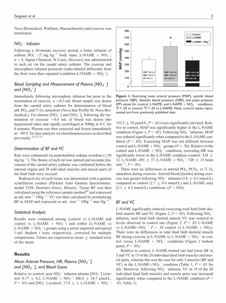
BF and VC
L-NAME significantly reduced exercising total hind limb ske-
letal muscle BF and VC (Figure 2, P < .03). Following NO2�
infusion, total hind limb skeletal muscle VC was restored to
levels observed in control rats (Figure 2, P < .03 L-NAME
vs L-NAMEþNO2�, P > .10 control vs L-NAME þ NO2).
There were no differences in total hind limb skeletal muscle
BF during exercise in L-NAME vs L-NAME þ NO2� or con-
trol versus L-NAME þ NO2� conditions (Figure 2 bottom
panel, P > .03).
Relative to control, L-NAME-treated rats had lower BF in
5 and VC in 15 of the 28 individual hind limb muscles and mus-
cle parts, whereas this was the case for only 3 muscles (BF and
VC) in the L-NAMEþNO2� condition (Table 1, P < .03 for
all). Moreover, following NO2� infusion, VC in 19 of the 28
individual hind limb muscles and muscle parts was increased
significantly when compared to the L-NAME condition (P <
.03, Table 1).
MA
P (m
mHg
)
0
110
120
130
140
150
160
170
180ControlL-NAME
L-NAME + NO2–*
SBP: 164 ± 4DBP: 124 ± 2PP: 40 ± 2
SBP: 155 ± 5#DBP: 126 ± 5#PP: 29 ± 3#
SBP: 189 ± 9*DBP: 140 ± 6*PP: 49 ± 4*
Figure 1. Exercising mean arterial pressure (MAP), systolic bloodpressure (SBP), diastolic blood pressure (DBP), and pulse pressure(PP) values for control, L-NAME, and L-NAME þ NO2
� conditions.aP < .03 vs control, bP < .03 vs L-NAME. Note: control values repre-sented are from previously published data.
Ferguson et al 3

Relative to control, BF and VC were lower in the adrenals
and pancreas while VC was lower in the kidneys, stomach,
and small intestine in rats treated with L-NAME (P < .03,
Table 2). Following NO2� infusion, renal and adrenal BF
and VC were lower when compared to control animals, while
renal and adrenal BF was reduced when compared to
L-NAME (P < .03, Table 2).
Discussion
The principal original finding of this investigation is that, in
the face of NOS blockade, NO2� infusion restored exercising
MAP and hind limb skeletal muscle VC to levels observed in
young adult healthy rats with intact NOS function. While
NO2� infusion did not increase BF when compared to the
L-NAME condition, it did abolish the lower BF induced by
L-NAME. Elevations in VC and reductions in MAP could
serve to reduce afterload and thus reduce the work of the
heart during exercise. These results demonstrate that NO2�
may serve as a powerful modulator of vascular control in
vivo, independent of NOS function and thus may hold
promising therapeutic potential, particularly in diseases with
impaired NOS function and chronically elevated MAP.
Effects of Inorganic NO2� Infusion on Skeletal Muscle
BF and VC and MAP
An abundance of research has focused on defining the
vasoactive/cardioprotective roles of NO2� with many studies
suggesting that the reduction of NO2� to NO compliments
the well-understood NOS pathway of NO production, partic-
ularly when NOS function becomes uncoupled or otherwise
impaired (reviewed by34,35). The vascular responses to NO2�
infusion presented herein support this notion. Similar to what
has been reported previously in our laboratory,36,37 infusion
with the comprehensive NOS blocker L-NAME increased
MAP *15% and decreased skeletal muscle VC *26% dur-
ing exercise. Consistent with our hypothesis, infusion with
NO2� (7 mg kg�1) restored MAP and VC to levels similar
to those observed in healthy control animals. One potential
explanation for these effects of NO2� could be the lower
PO2/pH environment present within the skeletal muscle fol-
lowing NOS inhibition.33 Such environments facilitate (or
uninhibit) NO2� reduction to NO in vivo,18,38 which may
allow local NO2� to support the blood–myocyte Po2 gradient
(via "QO2 and microvasculature PO2, PO2mv) that, when com-
promised, leads to tissue hypoxia and exacerbates intracellu-
lar perturbations.39
One striking aspect of this investigation, in which acute
NO2� infusion was employed, was that the augmented skele-
tal muscle VC was observed in muscles and muscle parts that
span the full spectrum of fast- and slow-twitch fiber types
(Table 1). This is in contrast to investigations using short-
term dietary NO3� supplementation as a means of increasing
circulating [NO2�]. Specifically, there is a fiber-type prefer-
ential effect of dietary NO3� supplementation as rats given
NO3�-rich beetroot juice for 5 days exhibited elevated skele-
tal muscle BF and VC exclusively in muscles and muscle por-
tions comprising � 66% type IIb þ d/x muscle fibres.27
Moreover, beetroot juice elevates PO2mv during muscle con-
tractions in the gastrocnemius (fast-twitch) but not soleus
(slow-twitch) muscles.33 The substantial array of muscles and
muscle portions exhibiting a vasoactive response to NO2�
infusion herein suggests that the fiber-type preferential effects
observed following dietary NO3� supplementation may be
conferred via changes in protein expression that require a lon-
ger period of elevated NO2� exposure to manifest. This idea is
supported by evidence from Hernandez et al7 in which the
improvements in fast-twitch skeletal muscle force production
evoked by NO3� supplementation were attributed to eleva-
tions in calcium handling proteins (ie, calsequestrin 1 and the
dihydropyridine receptor), which were present following mul-
tiple days of dietary NO3� supplementation.
Additionally, the discrepancies in the vascular responses to
NO3� versus NO2
� treatment could be related to the relative
impacts of NOS inhibition in fast- versus slow-twitch muscles.
Skeletal muscles comprised predominantly of slow-twitch
VC (m
l. min
–1. 1
00g–1
. mm
Hg–1
)
0.0
0.4
0.5
0.6
0.7
0.8
0.9
1.0ControlL-NAME L-NAME+NO2
–
*
#
BF
(ml. m
in–1. 1
00g–1
)
0
45
60
75
90
105
120
135
*
Figure 2. Total hindlimb skeletal muscle blood flow (BF) and vascularconductance (VC) for control, L-NAME, and L-NAME þ NO2
� con-ditions in rats during submaximal locomotory exercise. aP < .03 vscontrol, bP < .03 vs L-NAME. Note: control values represented arefrom previously published data.
4 Journal of Cardiovascular Pharmacology and Therapeutics

Table 1. Effects NO2� Infusion (7 mg kg�1) on Exercising Hindlimb Skeletal Muscle BF (mL min�1 100g�1) and VC (mL min�1 100g�1 mm Hg�1)
in Rats With NOS Blockade (L-NAME).a
BF VC
Control L-NAME L-NAMEþNO2� Control L-NAME L-NAMEþNO2
�
Ankle extensorsSoleus (9%) 295 + 42 242 + 71 285 + 36 2.14 + 0.30 1.56 + 0.17 2.06 + 0.23b
Plantaris (80%) 207 + 15 144 + 8c 173 + 15 1.50 + 0.10 0.93 + 0.06c 1.27 + 0.08b
Gastrocnemius, red (14%) 452 + 44 333 + 59 362 + 65 3.27 + 0.30 2.18 + 0.02c 2.63 + 0.44b
Gastrocnemius, white (100%) 42 + 7 26 + 3 37 + 4 0.30 + 0.05 0.17 + 0.02c 0.27 + 0.03b
Gastrocnemius, mixed (91%) 149 + 12 120 + 5 141 + 8 1.08 + 0.08 0.77 + 0.04c 1.04 + 0.04b
Tibialis posterior (73%) 118 + 17 81 + 12 91 + 13 0.85 + 0.12 0.51 + 0.07c 0.66 + 0.09b
Flexor digitorum longus (68%) 99 + 14 60 + 7c 69 + 9 0.71 + 0.09 0.38 + 0.04c 0.51 + 0.06b
Flexor halicus longus (71%) 75 + 10 68 + 8 99 + 14b 0.54 + 0.06 0.44 + 0.06 0.74 + 0.11b
Ankle flexorsTibialis anterior, red (63%) 343 + 35 209 + 10c 219 + 20* 2.48 + 0.23 1.36 + 0.10c 1.62 + 0.14c
Tibialis anterior, white (80%) 119 + 14 83 + 6c 89 + 12 0.86 + 0.09 0.54 + 0.05c 0.66 + 0.09b
Extensor digitorum longus (76%) 54 + 7 75 + 20 77 + 17 0.39 + 0.05 0.50 + 0.14 0.57 + 0.13b
Peroneals (67%) 128 + 11 72 + 14c 91 + 13c 0.93 + 0.08 0.46 + 0.09c 0.67 + 0.09b,c
Knee extensorsVastus intermedius (4%) 359 + 39 257 + 25 302 + 39 2.60 + 0.27 1.66 + 0.17c 2.20 + 0.25b
Vastus medialis (82%) 114 + 18 137 + 13 144 + 14 0.82 + 0.12 0.89 + 0.08 1.06 + 0.08b
Vastus lateralis, red (35%) 388 + 43 310 + 35 281 + 25 2.82 + 0.29 2.02 + 0.26 2.08 + 0.52Vastus lateralis, white (100%) 33 + 5 26 + 8 31 + 7 0.24 + 0.03 0.16 + 0.04 0.23 + 0.04b
Vastus lateralis, mixed (89%) 167 + 21 123 + 12 127 + 13 1.22 + 0.14 0.81 + 0.09c 0.94 + 0.09b
Rectus femoris, red (66%) 224 + 33 181 + 15 204 + 17 1.62 + 0.23 1.17 + 0.10 1.50 + 0.11b
Rectus femoris, white (100%) 101 + 13 81 + 7 91 + 8 0.73 + 0.09 0.52 + 0.05 0.67 + 0.06b
Knee flexorsBiceps femoris anterior (100%) 50 + 8 33 + 4 36 + 4 0.36 + 0.05 0.21 + 0.03c 0.27 + 0.03b
Biceps femoris posterior (92%) 79 + 8 65 + 3 71 + 5 0.58 + 0.06 0.42 + 0.02* 0.53 + 0.04b
Semitendinosus (83%) 56 + 6 34 + 3c 37 + 4c 0.40 + 0.04 0.22 + 0.02c 0.28 + 0.03c
Semimembranosus, red (72%) 119 + 14 86 + 7 83 + 14 0.87 + 0.09 0.56 + 0.05c 0.62 + 0.11Semimembranosus, white (100%) 33 + 6 38 + 7 40 + 11 0.24 + 0.04 0.25 + 0.05 0.30 + 0.09
Thigh AdductorsAdductor longus (5%) 315 + 38 263 + 26 231 + 31b 2.28 + 0.26 1.71 + 0.21 1.68 + 0.22Adductor magnus & brevis (89%) 83 + 8 80 + 7 80 + 9 0.60 + 0.05 0.52 + 0.05 0.60 + 0.06Gracilis (77%) 42 + 4 37 + 4 34 + 5 0.30 + 0.03 0.24 + 0.02 0.26 + 0.04Pectineus (69%) 54 + 8 40 + 6 46 + 11 0.39 + 0.06 0.25 + 0.03 0.34 + 0.08
Abbreviations: BF, blood flow; NOS, nitric oxide synthase; SEM, standard error of the mean;VC, vascular conductance.aData are mean + SEM. Values in parentheses indicate % type IIb þ d/x according to Delp & Duan. Control: n ¼ 11, L-NAME: n ¼ 8, L-NAME þ NO2
�: n ¼ 8.AQ3bP < .03 vs L-NAME.cP < .03 vs control.
Table 2. Effects of NO2� Infusion (7 mg kg�1) on Exercising BF (mL min�1 100g�1) and VC (mL �min�1 100g�1 mm Hg�1) in the Kidneys and
Organs of the Splanchnic Region.a
BF VC
Control L-NAME L-NAME þ NO2 Control L-NAME L-NAME þ NO2
Kidney 421 + 42 338 + 28 267 + 31b,c 3.05 + 0.28 2.22 + 0.25b 1.96 + 0.22b
Stomach 67 + 13 38 + 3 35 + 4 0.49 + 0.10 0.25 + 0.02b 0.25 + 0.03Adrenals 353 + 72 128 + 17* 100 + 66b 2.87 + 0.44 0.85 + 0.14b 0.72 + 0.15b
Spleen 61 + 14 102 + 21 48 + 7c 0.44 + 0.10 0.68 + 0.16 0.35 + 0.06Pancreas 110 + 15 72 + 8b 93 + 22 0.80 + 0.11 0.47 + 0.06b 0.67 + 0.15Small intestine 240 + 27 177 + 24 211 + 26 1.74 + 0.18 1.17 + 0.19b 1.55 + 0.17Large intestine 127 + 16 123 + 20 140 + 42 0.92 + 0.10 0.82 + 0.15 1.01 + 0.28Liverd 16 + 4 15 + 2 13 + 3 0.12 + 0.02 0.10 + 0.01 0.09 + 0.02
Abbreviations: BF, blood flow; SEM, standard error of the mean; VC, vascular conductance.aData are mean + SEM. Control: n ¼ 11, L-NAME: n ¼ 8, L-NAME þ NO2
�: n ¼ 8.bP < .03 vs control.cP < .03 vs L-NAME.dindicates arterial, not portal, BF and VC.
Ferguson et al 5

fibers demonstrate the greatest deficits in BF and VC following
L-NAME infusion36 likely due to a greater expression of
endothelial NOS (eNOS) within these tissues.40 These slow-
twitch muscles may exhibit much greater BF and VO2than their
fast-twitch counterparts both at rest and during exercise
(*100% greater for both BF and VO241). Consequently, NOS
inhibition may have crippled O2 delivery in these muscles suf-
ficiently enough to produce an environment ripe for NO2�
bioactivation (ie, very low PO2 and pH). This effect could place
more emphasis on NO2�, as the primary source of NO in these
specific tissues when vascular function is impaired as it is in
many disease states.42 In this regard, the spatial changes in
VC seen following NO2� infusion herein may mimic closely
what would be observed in individuals with diseases that com-
promise NOS function. However, these questions require fur-
ther investigation using specific models of vascular disease.
Clinical and Therapeutic implications
In healthy individuals, eNOS is the primary endogenous source
for NO2� and NO.43 Endothelial dysfunction becomes evident
early on in many diseases, including CHF (reviewed by Poole
et al2) and peripheral artery disease (reviewed by Brevetti
et al44) and thus likely limits vascular and metabolic function
via attenuated NO production from both NOS-dependent and
-independent pathways.43,45 As evidenced by Hirai et al,46,47
reduced NO from NOS dramatically impairs the matching
of skeletal muscle QO2 to VO2 such that superfusion of
L-NAME in the contracting rat spinotrapezius muscle trans-
forms the healthy PO2mv profile into one resembling CHF.46
In this regard, the blockade of NOS induced by L-NAME infu-
sion performed in the present investigation presents a challenge
that mimics the consequences of CHF and potentially other dis-
eases. Therefore, from the present findings, a therapy in which
systemic [NO2�] is elevated (via endogenous or exogenous
sources) may provide beneficial vascular responses indepen-
dent of NOS function. Even small improvements in vascular
function may enhance metabolic control during dynamic exer-
cise, potentially improving adherence to rehabilitation pro-
grams35 which in-and-of themselves would upregulate eNOS
function and endogenous NO2� production.
Experimental Considerations and Potential limitations
A surprising result of the present investigation was the rise in
exercising blood [lactate] following NO2� infusion (*41%
and 81% greater vs control and L-NAME, respectively). Lower
levels of NO may act as a useful brake on mitochondrial activ-
ity via competitive binding to complex IV of the respiratory
chain.48 In contrast, high concentrations of NO have been asso-
ciated with adverse effects on cell respiration via nitrosylation
of mitochondrial electron chain complexes, specifically com-
plex I.49 In addition, NO works to inhibit complex IV (cyto-
chrome oxidase), thereby reducing cellular O2 consumption.
Both of these effects may prove beneficial in certain environ-
ments or situations when O2 delivery becomes reduced as
reductions in tissue ½Errordot�VO2 work to extend the PO2 gra-
dient across a larger tissue area, effectively sharing the avail-
able O2.50 However, in the current study, it is possible that
the rate of NO2� reduction to NO became high enough to over-
whelm mitochondrial respiration, thus leading to impaired oxi-
dative metabolism and an increased reliance on glycolytic
means of ATP production. In addition, while the current dose
of NO2�-raised plasma [NO3
�] to levels very similar to what
has been reported following dietary NO3� supplementation in
humans9,14 and animals5,27 the plasma [NO2�] were much
greater than that achieved via NO3� supplementation and thus
may have contributed to the aforementioned effect on metabo-
lism. In this regard, a comprehensive dose–response relation-
ship will need to be determined before NO2� can be used as
an effective therapeutic.
Furthermore, considering that NOS was acutely inhibited in
the present investigation, the impacts of NO2� infusion may
differ when administered to specific models of vascular dis-
eases that have been developed chronically, as this would more
closely mimic specific etiologies. Additionally, due to the rel-
atively long half-life and bioactivity of L-NAME metabolites
(*20 hours in rats51) the experimental design was limited to
a fixed sequence and therefore, an ordering effect cannot be
ruled out. Future investigations in which NO2� is employed
in healthy control animals would also provide further insight
into the bioactivity of NO2� in animals with intact NOS func-
tion and could shed light on how a NO2�-based intervention
may impact healthy cardiovascular function.
Conclusion
These data highlight the potential for NO2� to act indepen-
dently of NOS and improve skeletal muscle vascular control
during exercise. Considering the multiple cardiovascular dis-
eases that impair NOS function, therapies that increase [NO2�]
may result in improved skeletal muscle vascular control during
exercise. However, the NO2� induced changes in blood [lac-
tate] seen during exercise herein suggests that the reduction
of NO3� to NO2
�, accomplished via facultative anaerobes in
the mouth following dietary NO3� consumption, may provide
the controlled release of NO2� needed to elicit the most bene-
ficial vascular and metabolic changes during exercise. It is
anticipated that future investigations into the vascular impacts
of both NO2�- and NO3
�-based therapies will provide crucial
insight into the potential benefits, and limitations, of both
interventions.
Acknowledgments
The authors would like to thank Ms K. Sue Hageman for her excellent
technical assistance.
Author Contributions
SKF, CTH, AMJ, TIM, and DCP contributed to conception and design
of the experiments.
SKF, AAG, CTH, JLW, AJF, TDC, TS, JDA, AMJ, TIM, and DCP
contributed to collection, analysis, and interpretation of data. SKF,
6 Journal of Cardiovascular Pharmacology and Therapeutics

CTH, TDC, JDA, AMJ, TIM, and DCP contributed to drafting the arti-
cle and revising it critically for important intellectual content. All the
authors have approved the final version of the article.
Declaration of Conflicting Interests
The author(s) declared no potential conflicts of interest with respect to
the research, authorship, and/or publication of this article.
Funding
The author(s) disclosed receipt of the following financial support for
the research, authorship, and/or publication of this article: These
experiments were funded by a Kansas State University SMILE award
to TIM, and American Heart Association Midwest Affiliate
(10GRNT4350011) and NIH (HL-108328) awards to DCP.
References
1. Joyner MJ, Tschakovsky ME. Nitric oxide and physiologic vaso-
dilation in human limbs: where do we go from here? Can J Appl
Physiol. 2003;28(3):475-490.
2. Poole DC, Hirai DM, Copp SW, Musch TI. Muscle oxygen trans-
port and utilization in heart failure: implications for exercise
(in)tolerance. Am J Physiol Heart Circ Physiol. 2012;302(5):
H1050-H1063.
3. Lundberg JO, Weitzberg E. NO generation from inorganic nitrate
and nitrite: role in physiology, nutrition and therapeutics. Arch
Pharm Res. 2009;32(8):1119-1126.
4. Ferguson SK, Hirai DM, Copp SW, et al. Effects of nitrate supple-
mentation via beetroot juice on contracting rat skeletal muscle
microvascular oxygen pressure dynamics. Respir Physiol Neuro-
biol. 2013;187(3):250-255.
5. Ferguson SK, Hirai DM, Copp SW, et al. Dose dependent effects
of nitrate supplementation on cardiovascular control and micro-
vascular oxygenation dynamics in healthy rats. Nitric Oxide.
2014;39:51-58.
6. Larsen FJ, Schiffer TA, Borniquel S, et al. Dietary inorganic
nitrate improves mitochondrial efficiency in humans. Cell Metab.
2011;13(2):149-159.
7. Hernandez A, Schiffer TA, Ivarsson N, et al. Dietary nitrate
increases tetanic [Ca2þ]i and contractile force in mouse fast-
twitch muscle. J Physiol. 2012;590(pt 15):3575-3583.
8. Bailey SJ, Fulford J, Vanhatalo A, et al. Dietary nitrate supple-
mentation enhances muscle contractile efficiency during knee-
extensor exercise in humans. J Appl Physiol. 2010;109(1):
135-148.
9. Bailey SJ, Winyard P, Vanhatalo A, et al. Dietary nitrate supple-
mentation reduces the O2 cost of low-intensity exercise and
enhances tolerance to high-intensity exercise in humans. J Appl
Physiol. 2009;107(4):1144-1155.
10. Muggeridge DJ, Howe CC, Spendiff O, Pedlar C, James PE,
Easton C. The effects of a single dose of concentrated beetroot
juice on performance in trained flatwater kayakers. Int J Sport
Nutr Exerc Metab. 2013;23(5):498-506.
11. Vanhatalo A, Bailey SJ, Blackwell JR, et al. Acute and chronic
effects of dietary nitrate supplementation on blood pressure and
the physiological responses to moderate-intensity and incremental
exercise. Am J Physiol Regul Integr Comp Physiol. 2010;299(4):
R1121-R1131.
12. Vanhatalo A, Fulford J, Bailey SJ, Blackwell JR, Winyard PG,
Jones AM. Dietary nitrate reduces muscle metabolic perturbation
and improves exercise tolerance in hypoxia. J Physiol. 2011;
589(pt 22):5517-5528.
13. Wylie LJ, Mohr M, Krustrup P, et al. Dietary nitrate supplemen-
tation improves team sport-specific intense intermittent exercise
performance. Eur J Appl Physiol. 2013;113(7):1673-1684.
14. Kenjale AA, Ham KL, Stabler T, et al. Dietary nitrate supplemen-
tation enhances exercise performance in peripheral arterial dis-
ease. J Appl Physiol. 2011;110(6):1582-1591.
15. Allen JD, Stabler T, Kenjale A, et al. Plasma nitrite flux pre-
dicts exercise performance in peripheral arterial disease after
3 months of exercise training. Free Radic Biol Med. 2010;
49(6):1138-1144.
16. Berry MJ, Justus NW, Hauser JI, et al. Dietary nitrate supplemen-
tation improves exercise performance and decreases blood pres-
sure in COPD patients. Nitric Oxide. 2015;48:22-30.
17. Zamani P, Rawat D, Shiva-Kumar P, et al. The effect of inorganic
nitrate on exercise capacity in heart failure with preserved ejec-
tion fraction. Circulation. 2015;131(4):371-380; discussion 380.
18. Cosby K, Partovi KS, Crawford JH, et al. Nitrite reduction to
nitric oxide by deoxyhemoglobin vasodilates the human circula-
tion. Nat Med. 2003;9(12):1498-1505.
19. Ingram TE, Pinder AG, Bailey DM, Fraser AG, James PE.
Low-dose sodium nitrite vasodilates hypoxic human pulmonary
vasculature by a means that is not dependent on a simultaneous
elevation in plasma nitrite. Am J Physiol Heart Circ Physiol.
2010;298(2):H331-H339.
20. Maher AR, Arif S, Madhani M, et al. Impact of chronic conges-
tive heart failure on pharmacokinetics and vasomotor effects of
infused nitrite. Br J Pharmacol. 2013;169(3):659-670.
21. Pluta RM, Oldfield EH, Bakhtian KD, et al. Safety and feasibility
of long-term intravenous sodium nitrite infusion in healthy volun-
teers. PloS One. 2011;6(1):e14504.
22. Alzawahra WF, Talukder MA, Liu X, Samouilov A, Zweier JL.
Heme proteins mediate the conversion of nitrite to nitric oxide
in the vascular wall. Am J Physiol Heart Circ Physiol. 2008;
295(2):H499-H508.
23. Ghosh SM, Kapil V, Fuentes-Calvo I, et al. Enhanced vasodilator
activity of nitrite in hypertension: critical role for erythrocytic
xanthine oxidoreductase and translational potential. Hyperten-
sion. 2013;61(5):1091-1102.
24. Pinheiro LC, Montenegro MF, Amaral JH, Ferreira GC, Oliveira
AM, Tanus-Santos JE. Increase in gastric pH reduces hypotensive
effect of oral sodium nitrite in rats. Free Radic Biol Med. 2012;
53(4):701-709.
25. Sindler AL, Fleenor BS, Calvert JW, et al. Nitrite supplementa-
tion reverses vascular endothelial dysfunction and large elastic
artery stiffness with aging. Aging Cell. 2011;10(3):429-437.
26. Drummond GB. Reporting ethical matters in the Journal of Phy-
siology: standards and advice. J Physiol. 2009;587(pt 4):713-719.
27. Ferguson SK, Hirai DM, Copp SW, et al. Impact of dietary nitrate
supplementation via beetroot juice on exercising muscle vascular
control in rats. J Physiol. 2013;591(pt 2):547-557.
Ferguson et al 7

28. Musch TI, Terrell JA. Skeletal muscle blood flow abnormalities
in rats with a chronic myocardial infarction: rest and exercise.
Am J Physiol. 1992;262(2 pt 2):H411-H419.
29. Flaim SF, Nellis SH, Toggart EJ, Drexler H, Kanda K, Newman
ED. Multiple simultaneous determinations of hemodynamics and
flow distribution in conscious rat. J Pharmacol Meth. 1984;11(1):
1-39.
30. Copp SW, Hirai DM, Hageman KS, Poole DC, Musch TI. Nitric
oxide synthase inhibition during treadmill exercise reveals fiber-
type specific vascular control in the rat hindlimb. Am J Physiol
Regul Integr Comp Physiol. 2010;298(2):R478-R485.
31. Hirai DM, Copp SW, Hageman KS, Poole DC, Musch TI. Aging
alters the contribution of nitric oxide to regional muscle hemody-
namic control at rest and during exercise in rats. J Appl Physiol.
2011;111(4):989-998.
32. Musch TI, Bruno A, Bradford GE, Vayonis A, Moore RL. Mea-
surements of metabolic rate in rats: a comparison of techniques.
J Appl Physiol. 1988;65(2):964-970.
33. Ferguson SK, Holdsworth CT, Wright JL, et al. Microvascular
oxygen pressures in muscles comprised of different fiber types:
Impact of dietary nitrate supplementation. Nitric Oxide. 2015;
48:38-43.
34. Bailey JC, Feelisch M, Horowitz JD, Frenneaux MP, Madhani M.
Pharmacology and therapeutic role of inorganic nitrite and nitrate
in vasodilatation. Pharmacol Ther. 2014;144(3):303-320.
35. Allen JD, Giordano T, Kevil CG. Nitrite and nitric oxide metabo-
lism in peripheral artery disease. Nitric Oxide. 2012;26(4):
217-222.
36. Hirai T, Visneski MD, Kearns KJ, Zelis R, Musch TI. Effects of
NO synthase inhibition on the muscular blood flow response to
treadmill exercise in rats. J Appl Physiol. 1994;77(3):1288-1293.
37. Musch TI, McAllister RM, Symons JD, et al. Effects of nitric
oxide synthase inhibition on vascular conductance during high
speed treadmill exercise in rats. Exp Physiol. 2001;86(6):
749-757.
38. Feelisch M, Fernandez BO, Bryan NS, et al. Tissue processing
of nitrite in hypoxia: an intricate interplay of nitric oxide-
generating and -scavenging systems. J Biol Chem. 2008;
283(49):33927-33934.
39. Arthur PG, Hogan MC, Bebout DE, Wagner PD, Hochachka PW.
Modeling the effects of hypoxia on ATP turnover in exercising
muscle. J Appl Physiol. 1992;73(2):737-742.
40. Woodman CR, Schrage WG, Rush JW, et al. Hindlimb unweight-
ing decreases endothelium-dependent dilation and eNOS expres-
sion in soleus not gastrocnemius. J Appl Physiol. 2001;91(3):
1091-1098.
41. Behnke BJ, McDonough P, Padilla DJ, Musch TI, Poole DC.
Oxygen exchange profile in rat muscles of contrasting fibre types.
J Physiol. 2003;549(pt 2):597-605.
42. Behnke BJ, Delp MD, McDonough P, Spier SA, Poole DC,
Musch TI. Effects of chronic heart failure on microvascular oxy-
gen exchange dynamics in muscles of contrasting fiber type. Car-
diovasc Res. 2004;61(2):325-332.
43. Kleinbongard P, Dejam A, Lauer T, et al. Plasma nitrite concen-
trations reflect the degree of endothelial dysfunction in humans.
Free Radic Biol Med. 2006;40(2):295-302.
44. Brevetti G, Silvestro A, Schiano V, Chiariello M. Endothelial
dysfunction and cardiovascular risk prediction in peripheral arter-
ial disease: additive value of flow-mediated dilation to ankle-
brachial pressure index. Circulation. 2003;108(17):2093-2098.
45. Kleinbongard P, Dejam A, Lauer T, et al. Plasma nitrite reflects
constitutive nitric oxide synthase activity in mammals. Free
Radic Biol Med. 2003;35(7):790-796.
46. Ferreira LF, Hageman KS, Hahn SA, et al. Muscle microvascular
oxygenation in chronic heart failure: role of nitric oxide availabil-
ity. Acta Physiol. 2006;188(1):3-13.
47. Ferreira LF, Padilla DJ, Williams J, Hageman KS, Musch TI,
Poole DC. Effects of altered nitric oxide availability on rat muscle
microvascular oxygenation during contractions. Acta physiol.
2006;186(3):223-232.
48. Erusalimsky JD, Moncada S. Nitric oxide and mitochondrial sig-
naling: from physiology to pathophysiology. Arterioscler Thromb
Vasc Biol. 2007;27(12):2524-2531.
49. Clementi E, Brown GC, Feelisch M, Moncada S. Persistent
inhibition of cell respiration by nitric oxide: crucial role of
S-nitrosylation of mitochondrial complex I and protective
action of glutathione. Proc Natl Acad Sci U S A. 1998;
95(13):7631-7636.
50. Thomas DD, Liu X, Kantrow SP, Lancaster JR Jr The biological
lifetime of nitric oxide: implications for the perivascular dynamics
of NO and O2. Proc Natl Acad Sci U S A. 2001;98(1):355-360.
51. Vitecek J, Lojek A, Valacchi G, Kubala L. Arginine-based inhibi-
tors of nitric oxide synthase: therapeutic potential and challenges.
Mediators Inflamm. 2012;2012:318087.
8 Journal of Cardiovascular Pharmacology and Therapeutics