
Muller cells are living optical fibers in the vertebrate retina
6
Mu ¨ ller cells are living optical fibers in the vertebrate retina Kristian Franze*, Jens Grosche* † , Serguei N. Skatchkov ‡ , Stefan Schinkinger § , Christian Foja ¶ , Detlev Schild , Ortrud Uckermann*, Kort Travis § , Andreas Reichenbach* , **, and Jochen Guck §†† *Paul Flechsig Institute of Brain Research, Universita ¨ t Leipzig, Jahnallee 59, 04109 Leipzig, Germany; † Interdisciplinary Center of Clinical Research, Inselstrasse 22, 04103 Leipzig, Germany; ‡ Center for Molecular Biology and Neuroscience, Department of Biochemistry, School of Medicine, Universidad Central de Caribe, Bayamon, Puerto Rico 00960; § Division of Soft Matter Physics, Department of Physics, Universita ¨ t Leipzig, Linne ´ strasse 5, 04103 Leipzig, Germany; ¶ Department of Ophthalmology and Eye Clinic, Universita ¨ t Leipzig, 04103 Leipzig,Germany; Deutsche Forschungsgemeinschaft Molecular Physiology of the Brain Research Center and Department of Neurophysiology and Cellular Biophysics, Universita ¨ t Go ¨ ttingen, 37073 Go ¨ ttingen, Germany; and †† Department of Physics, University of Cambridge, JJ Thomson Avenue, Cambridge CB3 0HE, United Kingdom Edited by Luke Lee, University of California, Berkeley, CA, and accepted by the Editorial Board March 27, 2007 (received for review December 15, 2006) Although biological cells are mostly transparent, they are phase objects that differ in shape and refractive index. Any image that is projected through layers of randomly oriented cells will normally be distorted by refraction, reflection, and scattering. Counterintu- itively, the retina of the vertebrate eye is inverted with respect to its optical function and light must pass through several tissue layers before reaching the light-detecting photoreceptor cells. Here we report on the specific optical properties of glial cells present in the retina, which might contribute to optimize this apparently unfavorable situation. We investigated intact retinal tissue and individual Mu ¨ ller cells, which are radial glial cells spanning the entire retinal thickness. Mu ¨ ller cells have an extended funnel shape, a higher refractive index than their surrounding tissue, and are oriented along the direction of light propagation. Transmission and reflection confocal microscopy of retinal tissue in vitro and in vivo showed that these cells provide a low-scattering passage for light from the retinal surface to the photoreceptor cells. Using a modified dual-beam laser trap we could also dem- onstrate that individual Mu ¨ ller cells act as optical fibers. Further- more, their parallel array in the retina is reminiscent of fiberoptic plates used for low-distortion image transfer. Thus, Mu ¨ ller cells seem to mediate the image transfer through the vertebrate retina with minimal distortion and low loss. This finding elucidates a fundamental feature of the inverted retina as an optical system and ascribes a new function to glial cells. fiberoptic plate glial cells refractive index light guides optical trap B iological cells and tissues are usually fairly transparent due to the lack of strong intrinsic chromophores in the visible part of the spectrum and especially in the near-infrared. This transparency is exploited, for example, in multiphoton micros- copy, where this low absorption of excitation light leads to relatively large penetration depths. Although such biological objects do not modulate the amplitude of a passing electromag- netic wave, they impart a phase shift due to refractive index variations. This property was recognized by Zernike and used for the contrast enhancement of individual cells in phase-contrast microscopy (1). However, when light passes through multiple layers of cells, as in tissues, images rapidly deteriorate due to scattering events caused by optical and geometrical inhomoge- neities with length scales on the order of the wavelength of visible light (2). Consequently, nature has implemented ingenious solutions in the properties and the arrangement of structures and cell assemblies that light has to pass for normal physiological func- tioning. The lens body in vertebrate eyes, for instance, consists of elongated fiber cells. These cells do not only display a very regular oval or hexagonal cross-section, a smooth surface, and a regular distribution, they even lose most of their organelles during differentiation, including the cell nucleus (3). In the vertebrate retina, the inner and outer segments of photoreceptor cells are considered natural optical fibers, supported by their highly specialized shape and optical properties (4). Other natural optical fibers occur in deep-sea glass sponges or in the compound eye of insects, whose biomimetic copies have even found their way into technical components (5, 6). What these examples have in common is a relatively regular geometry of the light-guiding structures and, in the case of living cells, a sophisticated spe- cialization for this very function. Considering these facts, it seems surprising that the retina in the vertebrate eye is inverted and that images projected onto the retina have to pass several layers of randomly oriented and irregularly shaped cells with intrinsic scatterers before they reach the light-detecting photoreceptor cells (7, 8). This situation seems to be ‘‘equivalent to placing a thin diffusing screen directly over the film in your camera’’ (9). However, this ‘‘screen’’ contains a regular pattern of cells, which are arranged in parallel to each other and span the entire thickness of the retina (150 m). These cells, Mu ¨ller cells, are radial glial cells in the inner vertebrate retina, which have a cylindrical, fiber-like shape (their original name was ‘‘radial fibers of Mu ¨ller’’) (10). They fulfill a wide range of physiological functions to support the functioning and survival of retinal neurons (11). For this purpose Mu ¨ller cells are, unlike the natural optical fibers mentioned above, endowed with many complex side branches, which ensheath neuronal compartments, such as synapses (12). On the other hand, they putatively occupy a strategic position in the path of light through the retina from the vitreous, where light enters the tissue, to the outer limiting membrane, where the inner segments of the photoreceptor cells receive the incident light. Therefore, it is intriguing to investigate whether they could play a role in the transfer of light through the inner retina. Results As a first step to characterize the retina as a phase object, we investigated freshly dissected guinea pig eyes by using modified transmission microscopy (Fig. 1 a and b). Physiological illumi- nation was simulated by insertion of an optical fiber as a light source into the eye cup. Images were obtained by scanning a plane close to the outer plexiform layer, corresponding to the Author contributions: K.F. and J. Grosche contributed equally to this work; K.F., S.N.S., S.S., D.S., K.T., A.R., and J. Guck designed research; K.F., J. Grosche, S.N.S., S.S., and O.U. performed research; C.F. contributed new reagents/analytic tools; K.F., J. Grosche, S.N.S., and K.T. analyzed data; and K.F., D.S., A.R., and J. Guck wrote the paper. The authors declare no conflict of interest. This article is a PNAS Direct Submission. L.L. is a guest editor invited by the Editorial Board. **To whom correspondence should be addressed. E-mail: [email protected]. This article contains supporting information online at www.pnas.org/cgi/content/full/ 0611180104/DC1. © 2007 by The National Academy of Sciences of the USA www.pnas.orgcgidoi10.1073pnas.0611180104 PNAS May 15, 2007 vol. 104 no. 20 8287– 8292 BIOPHYSICS
-
Upload
independent -
Category
Documents
-
view
5 -
download
0
Transcript of Muller cells are living optical fibers in the vertebrate retina

Muller cells are living optical fibersin the vertebrate retinaKristian Franze*, Jens Grosche*†, Serguei N. Skatchkov‡, Stefan Schinkinger§, Christian Foja¶, Detlev Schild�,Ortrud Uckermann*, Kort Travis§, Andreas Reichenbach*,**, and Jochen Guck§††
*Paul Flechsig Institute of Brain Research, Universitat Leipzig, Jahnallee 59, 04109 Leipzig, Germany; †Interdisciplinary Center of Clinical Research,Inselstrasse 22, 04103 Leipzig, Germany; ‡Center for Molecular Biology and Neuroscience, Department of Biochemistry, School of Medicine,Universidad Central de Caribe, Bayamon, Puerto Rico 00960; §Division of Soft Matter Physics, Department of Physics, Universitat Leipzig,Linnestrasse 5, 04103 Leipzig, Germany; ¶Department of Ophthalmology and Eye Clinic, Universitat Leipzig, 04103 Leipzig,Germany;�Deutsche Forschungsgemeinschaft Molecular Physiology of the Brain Research Center and Department of Neurophysiologyand Cellular Biophysics, Universitat Gottingen, 37073 Gottingen, Germany; and ††Department of Physics, Universityof Cambridge, JJ Thomson Avenue, Cambridge CB3 0HE, United Kingdom
Edited by Luke Lee, University of California, Berkeley, CA, and accepted by the Editorial Board March 27, 2007 (received for review December 15, 2006)
Although biological cells are mostly transparent, they are phaseobjects that differ in shape and refractive index. Any image that isprojected through layers of randomly oriented cells will normallybe distorted by refraction, reflection, and scattering. Counterintu-itively, the retina of the vertebrate eye is inverted with respect toits optical function and light must pass through several tissuelayers before reaching the light-detecting photoreceptor cells.Here we report on the specific optical properties of glial cellspresent in the retina, which might contribute to optimize thisapparently unfavorable situation. We investigated intact retinaltissue and individual Muller cells, which are radial glial cellsspanning the entire retinal thickness. Muller cells have an extendedfunnel shape, a higher refractive index than their surroundingtissue, and are oriented along the direction of light propagation.Transmission and reflection confocal microscopy of retinal tissue invitro and in vivo showed that these cells provide a low-scatteringpassage for light from the retinal surface to the photoreceptorcells. Using a modified dual-beam laser trap we could also dem-onstrate that individual Muller cells act as optical fibers. Further-more, their parallel array in the retina is reminiscent of fiberopticplates used for low-distortion image transfer. Thus, Muller cellsseem to mediate the image transfer through the vertebrate retinawith minimal distortion and low loss. This finding elucidates afundamental feature of the inverted retina as an optical systemand ascribes a new function to glial cells.
fiberoptic plate � glial cells � refractive index � light guides � optical trap
B iological cells and tissues are usually fairly transparent dueto the lack of strong intrinsic chromophores in the visible
part of the spectrum and especially in the near-infrared. Thistransparency is exploited, for example, in multiphoton micros-copy, where this low absorption of excitation light leads torelatively large penetration depths. Although such biologicalobjects do not modulate the amplitude of a passing electromag-netic wave, they impart a phase shift due to refractive indexvariations. This property was recognized by Zernike and used forthe contrast enhancement of individual cells in phase-contrastmicroscopy (1). However, when light passes through multiplelayers of cells, as in tissues, images rapidly deteriorate due toscattering events caused by optical and geometrical inhomoge-neities with length scales on the order of the wavelength of visiblelight (2).
Consequently, nature has implemented ingenious solutions inthe properties and the arrangement of structures and cellassemblies that light has to pass for normal physiological func-tioning. The lens body in vertebrate eyes, for instance, consistsof elongated fiber cells. These cells do not only display a veryregular oval or hexagonal cross-section, a smooth surface, and aregular distribution, they even lose most of their organellesduring differentiation, including the cell nucleus (3). In the
vertebrate retina, the inner and outer segments of photoreceptorcells are considered natural optical fibers, supported by theirhighly specialized shape and optical properties (4). Other naturaloptical fibers occur in deep-sea glass sponges or in the compoundeye of insects, whose biomimetic copies have even found theirway into technical components (5, 6). What these examples havein common is a relatively regular geometry of the light-guidingstructures and, in the case of living cells, a sophisticated spe-cialization for this very function.
Considering these facts, it seems surprising that the retina inthe vertebrate eye is inverted and that images projected onto theretina have to pass several layers of randomly oriented andirregularly shaped cells with intrinsic scatterers before they reachthe light-detecting photoreceptor cells (7, 8). This situationseems to be ‘‘equivalent to placing a thin diffusing screen directlyover the film in your camera’’ (9). However, this ‘‘screen’’contains a regular pattern of cells, which are arranged in parallelto each other and span the entire thickness of the retina (�150�m). These cells, Muller cells, are radial glial cells in the innervertebrate retina, which have a cylindrical, fiber-like shape (theiroriginal name was ‘‘radial fibers of Muller’’) (10). They fulfill awide range of physiological functions to support the functioningand survival of retinal neurons (11). For this purpose Muller cellsare, unlike the natural optical fibers mentioned above, endowedwith many complex side branches, which ensheath neuronalcompartments, such as synapses (12). On the other hand, theyputatively occupy a strategic position in the path of light throughthe retina from the vitreous, where light enters the tissue, to theouter limiting membrane, where the inner segments of thephotoreceptor cells receive the incident light. Therefore, it isintriguing to investigate whether they could play a role in thetransfer of light through the inner retina.
ResultsAs a first step to characterize the retina as a phase object, weinvestigated freshly dissected guinea pig eyes by using modifiedtransmission microscopy (Fig. 1 a and b). Physiological illumi-nation was simulated by insertion of an optical fiber as a lightsource into the eye cup. Images were obtained by scanning aplane close to the outer plexiform layer, corresponding to the
Author contributions: K.F. and J. Grosche contributed equally to this work; K.F., S.N.S., S.S.,D.S., K.T., A.R., and J. Guck designed research; K.F., J. Grosche, S.N.S., S.S., and O.U.performed research; C.F. contributed new reagents/analytic tools; K.F., J. Grosche, S.N.S.,and K.T. analyzed data; and K.F., D.S., A.R., and J. Guck wrote the paper.
The authors declare no conflict of interest.
This article is a PNAS Direct Submission. L.L. is a guest editor invited by the Editorial Board.
**To whom correspondence should be addressed. E-mail: [email protected].
This article contains supporting information online at www.pnas.org/cgi/content/full/0611180104/DC1.
© 2007 by The National Academy of Sciences of the USA
www.pnas.org�cgi�doi�10.1073�pnas.0611180104 PNAS � May 15, 2007 � vol. 104 � no. 20 � 8287–8292
BIO
PHYS
ICS

end of the ‘‘prephotoreceptor’’ light path. Remarkably, theseimages showed a high degree of inhomogeneity, revealing analmost regular pattern of bright spots alternating with areas oflower transmittance (Fig. 1b). This pattern showed that someretinal structures relayed light better than others.
Interpreting the dark areas in Fig. 1b as areas of higherscattering, laser scanning measurements in reflection mode (Fig.1 c and d) should approximately yield the negative of the aboveimage. Following this hypothesis, we took series of 50–60consecutive optical sections from flat-mounted retinae. Thevitreous body did not reflect any light, consistent with its lack ofphase variations. In contrast, the almost uniform reflectancethroughout much of the retinal thickness was interrupted by afairly regular pattern of dark, less reflective spots (Fig. 1d). Thespots had diameters of 2–3 �m and were spaced �5–6 �m apart,corresponding well to diameter and spacing of the bright spotsin Fig. 1b [see also supporting information (SI) Fig. 5]. The samereflection patterns were also observed in retinae of rabbits (datanot shown) and humans (SI Fig. 6). To show that the observedphenomenon is relevant in physiological conditions, these ex-periments were successfully repeated with retinae of livingguinea pigs in situ (SI Fig. 7d).
Importantly, reconstruction along the z axis (Fig. 2a and SIFigs. 6 and 7) showed that the dark spots were contiguous inadjacent horizontal sections and formed tubes that correspondedto distinct optical pathways. At the innermost retinal layer,closest to the vitreous body, these tubes widened to funnel-likestructures, which together formed a 15-�m-thick continuouslow-reflecting zone only interrupted by axon bundles (Fig. 2a).
The amount of back-scattering from the retina has previouslybeen measured to be 1–5% of the incident light (7, 8). Becausebiological tissues are typically strongly forward-scattering (13),the total amount of scattering in the retina is most likely at leasta factor of 2 larger. The distribution of this scattering within theretina is shown in Fig. 2a. Significant back-scattering occurred in
all retinal layers proximal to the photoreceptors with the excep-tion of the tubes. The main locations of light scattering are bothplexiform layers and the axon bundles (Fig. 2b), which containnumerous light-scattering objects with sizes on the order of thewavelength of visible light (14–16) such as ‘‘synaptosomes,’’bundles of neurofilaments, and neurotubules. In combination,our transmission and reflection measurements demonstrate thepresence of tubular structures in the retina that transmit signif-icantly more light than their surrounding tissue.
The observed spatial pattern of these tubes corresponded wellto the spacing and diameters of the columnar Muller cells (Fig.2b) (17, 18). Furthermore, the funnel-like structures observed inreflection-mode were reminiscent of the densely packed cob-blestone pattern of the Muller cell endfeet at the inner retinalsurface (19). Indeed, the tubular structures could be unambig-uously identified as Muller cells. They were capable of selectiveuptake of vital dyes (Fig. 2 b–g) (20, 21) and could be counter-stained with an antibody directed against vimentin (Fig. 2 f andg). In the retina, vimentin is a protein specific to Muller cells (17,22). Hence, it is the Muller cells that provide a passage for lightthrough the retina to the photoreceptor cells. These data,together with their cylindrical geometry, suggested a mechanismof light transport similar to optical fibers.
In classical optical fibers, light is confined in the transversedirection by an elevated refractive index of the core comparedwith its cladding. Thus, we analyzed the refractive indices ofenzymatically dissociated vital retinal cells by using quantitativephase microscopy (Fig. 3) (23, 24). The somata of various retinalneurons (ganglion, amacrine, and bipolar cells) displayed similarrefractive indices (n � 1.358 � 0.005; mean � SD) (Fig. 3a) closeto earlier estimates for the total retina (25–27). In contrast, themean refractive index of Muller cell stalks was significantlyhigher (n � 1.380 � 0.021) (Fig. 3a). Toward the so-calledendfoot, the funnel-shaped termination of the Muller cell facingthe vitreous body with n � 1.335 (26), the refractive index
Fig. 1. Light transmission and reflection in the inner retina. (a) Experimental design to study light transmission through the inner retina. (Inset) Light emanatingfrom a multimode optical fiber inserted into a freshly dissected eye simulates physiological illumination of the retina. The eye is opened at the posterior side,and all outer structures, including photoreceptor cells, are surgically removed. The laser light (� � 543 nm) that is transmitted through the inner retina (NFL, nervefiber layer; IPL, inner plexiform layer; INL, inner nuclear layer) is captured at the end of the prephotoreceptor light path with a confocal microscope. ONL, outernuclear layer; ROS, photoreceptor outer segments. (b) Confocal transmission image of a living unstained retina. The brighter the signal, the more light is relayedto the corresponding area of the tissue. (c) Light reflection in the inner retina. Laser light is delivered via the microscope objective of an upright confocalmicroscope, and light scattered back from inner retinal layers is detected. (d) Confocal reflection image at the level of the IPL. The brighter the signal, the morelight is reflected by the corresponding area. (Scale bar, 10 �m; also applies to b.)
8288 � www.pnas.org�cgi�doi�10.1073�pnas.0611180104 Franze et al.

decreased to n � 1.359 � 0.003. Such a local decrease of therefractive index could serve to minimize reflection at theinterface between vitreous and retina. Similar results wereconsistently found in Muller cells from four different vertebratespecies (SI Table 1).
Both the observed differences between the refractive indicesof Muller cells and their surroundings as well as the fiber-like cell
shape are reminiscent of the basic requirements of optical fibers.However, Muller cells display a complex morphology (Fig. 3),and their radius is comparable to the wavelength of light so thatthe typical total-internal-reflection model of light guidance is notapplicable. In a waveguide, light propagates in certain patterns,or modes, determined by boundary conditions following elec-tromagnetic theory (28). Light guidance only occurs if propa-gating modes exist. The key parameter most widely used inoptical engineering to evaluate the presence of propagatingmodes is the waveguide characteristic frequency, or V parameter,
V ��d�
�n12 � n2
2,
where � is the free-space wavelength of the visible light, d is thediameter of the waveguide, and n1 and n2 are the refractiveindices of the waveguide and the surrounding material, respec-tively (28, 29). For a conservative estimate, it is sufficient tocalculate V at the longest visible wavelength (700 nm) and thesmallest diameter, which occurs at the inner process (d � 2.8 �m)(21). The largest possible value for the extracellular refractiveindex is that of the adjacent neurons with n2 � 1.358. Thecalculated V � 2.6–2.9 for the different parts along the Mullercell (Fig. 3b) is sufficiently high to allow low-loss propagation ofa few modes in the structure even at 700 nm (28). At awavelength of 500 nm, the V parameter increases to V � 3.6–4.0.Although the refractive index and diameter of the Muller cellsboth change along their length, the V parameter and, thus, thelight-guiding capability stay nearly constant (Fig. 3b). In contrastto the smooth cylindrical shape of artificial or other biologicaloptical fibers (6, 30, 31), each cell possesses complex side-branching processes important to its interactions with neurons(12). Their inclusion through an ‘‘effective’’ refractive indexgradient actually increases the V parameter of the Muller cell(32). Consequently, despite their complex morphology, Mullercells could thus function as waveguides for visible light.
a e
f
g
c
d
b
Fig. 2. Structures of low reflection are Muller cells. (a) Z-line reconstruction of reflection images of a living retina. The main scattering elements (bright) arethe axon bundles and both plexiform layers. Low-reflecting tubular structures span the entire retina. (b) Living retinal slice preparation, visualizing Muller cellswith the vital dye CellTracker orange (green) and synaptic elements in both plexiform layers (IPL and OPL) with the activity-dependent dye FM1–43 (red) (20).The levels of the inner and outer plexiform layers (IPL and OPL, respectively) and nerve fiber layer (NFL) are the same as in a. The asterisks indicate axon bundlesin the NFL. (c and d) Overlay of light detected in reflection mode (purple) and the green fluorescence of the vital dye CellTracker green. (c) Z-line reconstructionof a confocal image stack. (d) Oblique optical section at the level of the red horizontal line in c. The dye-filled irregularly shaped Muller cell somata of the innernuclear layer (INL) are visible in the left upper part. The central area shows Muller cell cross-sections in the IPL. In the lower right part, the Muller cell endfeetare visible, which enclose the ganglion cell somata in the ganglion cell layer (GCL). The lack of merging of the two colors, which would result in white areas,demonstrates that the dye filled exclusively those structures that showed low light reflection. (e–g) Confocal image at the IPL of a retinal whole mount fixedin 4% paraformaldehyde after exposure to the green vital dye and immunocytochemical labeling of vimentin (red), which in the retina is specific to Muller cells(17, 22). (e) Fluorescence of the vital dye. ( f) Vimentin immunofluorescence. (g) Overlay of e and f. Colocalization of the red and green dyes results in yellowlabeling. The observed complete colocalization means that the vital dye-filled and the immunoreactive cells are identical and thus identifies the low-reflectingtubular structures as Muller cells. [Scale bars: b, 10 �m (also applies to a); c–g, 25 �m.]
Fig. 3. Muller cell shape, refractive properties, and light-guiding capability.(a) Nomarski differential interference contrast microscopy image of a disso-ciated guinea pig Muller cell with several adherent photoreceptor cells,including their outer segments (ROS) and a dissociated retinal neuron (bipolarcell) to the left. The refractive indices of the different cell sections are given.(b) Schematic illustration of a Muller cell in situ. The lighter the coloring of theMuller cell, the lower the refractive index. Typical diameters and the calcu-lated V parameters for 700 nm (red) and 500 nm (blue) are indicated at theendfoot, the inner process, and the outer process. Although diameters andrefractive indices change along the cell, its light-guiding capability remainsfairly constant. (Scale bar, 25 �m.)
Franze et al. PNAS � May 15, 2007 � vol. 104 � no. 20 � 8289
BIO
PHYS
ICS
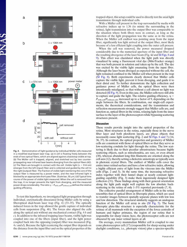
To test this hypothesis, we investigated light propagation throughindividual, enzymatically dissociated living Muller cells by using afiberoptical dual-beam laser trap (Fig. 4) (33–35). The opticallyinduced forces in the trap allowed the gentle capture of individualcells from suspension (Fig. 4b). The forces also aligned the cellsalong the optical axis without any mechanical contact (Fig. 4 b andc). In addition to the infrared trapping laser beams, visible light wascoupled into one of the fibers (input fiber), and the light powercoupled back into the opposing (output) fiber was measured (Fig.4 c and d). Because the light reentering the output fiber depends onthe distance from the input fiber and on the optical properties of the
trapped object, this setup could be used to directly test the axial lighttransmission through individual cells.
With a Muller cell present in the trap surrounded by media withrefractive indices up to 1.36 (to mimic the surrounding in theretina), light transmission into the output fiber was comparable tothe situation where both fibers were in contact, as long as thedirection of the light propagation was the same as in the retina.When the Muller cell endfoot was pointing away from the inputfiber, significantly less light arrived at the output fiber, most likelybecause of a less efficient light coupling into the outer cell process.
When the cell was removed, the power measured droppedconsiderably due to the numerical aperture of the input fiber andthe resulting divergence of the laser beam (Fig. 4d and SI Figs. 8 and9). This effect was elucidated when the light path was directlyvisualized by using a fluorescent vital dye (MitoTracker orange)that was both present in solution and taken up by the cell. The dyewas excited by the visible light emanating from the input fiber.Although the laser beam diverged as expected without the cell, thelight remained confined to the Muller cell when present in the trap(SI Fig. 8). Both experiments clearly showed that Muller cellscapture the visible light, prevent it from diverging, and guide it totheir distal end. To further demonstrate the light collection andguidance power of Muller cells, the optical fibers were thenintentionally misaligned, so that without a cell almost no light wasdetected (SI Fig. 9). Even in this case, the Muller cells were still ableto capture and guide the light. The relative guiding efficiency, � �Pwith�cell/Pwithout�cell, increased up to a factor of 9, depending on theangle between the fibers. In combination, our single-cell experi-ments, the theoretical considerations, and the transmission andreflection measurements strongly suggest that Muller cells are, andfunction as, optical fibers in the retina, relaying light from the innersurface to the layer of the photoreceptors while bypassing scatteringstructures present.
DiscussionThese results provide insight into the optical properties of theretina. Most structures in the retina, especially those in the nervefiber layer and both plexiform layers, are phase objects thatnecessarily cause light scattering (Fig. 2 and SI Figs. 6 and 7) (14,36, 37). In contrast, the optical properties and geometry of Mullercells are consistent with those of optical fibers so that they serve aslow-scattering conduits for light through the retina. The low scat-tering is likely due to their peculiar ultrastructure because highlyscattering objects, such as mitochondria, are rare, or even absent(38), whereas abundant long thin filaments are oriented along thecell axis (12), thereby setting a dielectric anisotropy as typically seenin photonic crystal fibers. The endfeet of Muller cells cover theentire inner retinal surface and have a low refractive index, allowinga highly efficient entry of light from the vitreous into the Mullercells (Figs. 2 and 3). At the same time, the increasing refractiveindex together with their funnel shape at nearly constant light-guiding capability (Fig. 3) make them ingeniously designed lightcollectors (31). These findings along with their general orientationalong the light path might well explain the low absolute back-scattering in the retina of only 1–5% reported previously (7, 8).
The collective parallel arrangement of Muller cells in the retinaresembles that of optical fibers in fiberoptic plates, which are usedto transfer images between spatially separate planes with low lossand low distortion. The structural similarity suggests an analogousfunction of the Muller cell array in situ (SI Fig. 5). The basicfiberoptic plate-like structure is especially characteristic for theretinae of all mammals with the exception of the fovea centralis ofhumans and higher primates, the region of our retina that isresponsible for sharp vision; here, the photoreceptor cells are notobscured by any inner retinal layers at all.
On average, every mammalian Muller cell is coupled to onecone photoreceptor cell (17) (responsible for sharp seeing underdaylight conditions, i.e., photopic vision) plus a species-specific
Fig. 4. Demonstration of light guidance by individual Muller cells measuredin a modified dual-beam laser trap. (a) A cell is floating freely between theends of two optical fibers, which are aligned against a backstop visible at top.(b) The Muller cell is trapped, aligned, and stretched out by two counter-propagating near-infrared laser beams diverging from the optical fibers (42).(c) The fibers are brought in contact with the cell. Visible light (� � 514 nm)emerges from the left (input) fiber and is collected and guided by the cell tothe right (output) fiber. The fraction of visible light reentering the core of theoutput fiber is measured by a power meter, and the near-infrared light isblocked by an appropriate short-pass filter. (Scale bar, 50 �m.) (d) Typical timecourse of the power of visible light measured. When the cell is removed fromthe trap, it no longer prevents the light from diverging, and the measuredpower drops considerably. The ratio � � Pwith�cell/Pwithout�cell defines the relativeguiding efficiency.
8290 � www.pnas.org�cgi�doi�10.1073�pnas.0611180104 Franze et al.

number of rod photoreceptor cells (17) (�10 in both man andguinea pig), serving low light level (scotopic) vision. Thus, in thecase of photopic vision, the parallel array of Muller cells maypreserve the initial image resolution by guiding the light directlyto their respective cone photoreceptor cell, minimizing imagedistortion. This array might also serve to improve image contrastby increasing the signal-to-noise ratio (39). In scotopic vision,Muller cells could reduce loss of intensity by minimizing lightreflection, particularly at the inner retinal surface. In summary,Muller cells in the retina assume the role of optical fibers andreliably transfer light with low scattering from the retinal surfaceto the photoreceptor cell layer. At the same time, their funnel-shape leaves �80% of the retinal volume for other cells and theneuronal connectivity (SI Fig. 5) and might thus spatially de-couple light transport from neuronal signal processing. Thefunction of glial cells that we describe here explains a funda-mental feature of the inverted retina as an optical system.
Materials and MethodsExperimental Animals. Adult guinea pigs of either sex (300–500 g)were used throughout the study if not mentioned otherwise.Animals were deeply anesthetized and killed by an overdose of2 g/kg urethane administered i.p.
All experiments were carried out in accordance with appli-cable German laws of animal protection and with the Associa-tion for Research in Vision and Ophthalmology Statement forthe Use of Animals in Ophthalmic and Vision Research. Theprotocol of this study was approved by the local ethical com-mittee and adhered to the tenets of the Declaration of Helsinkifor experiments involving human tissue.
Confocal Microscopy. Transmission measurements. Freshly dissectedguinea pig eyes were gently opened at opposite areas. Cornea andlens were removed for insertion of a multimode optical fiber. Sclera,choroid, pigment epithelium, and the photoreceptor layers werelocally cut away to allow direct optical access to the end of theprephotoreceptor light path. The transmitted light emanating fromthe optical fiber was captured through the objective (�40, N.A. �0.75, water immersion) of a confocal microscope (LSM 510 Meta;Zeiss, Oberkochen, Germany).Reflection measurements. Freshly isolated retinal whole-mounts ofseveral species, including man, were placed on a confocal micro-scope with the inner surface pointing toward the objective (�40,N.A. � 0.75, water immersion) and observed in reflection mode.In vivo measurements. Animals were anesthetized by i.m. applica-tion of 50 mg/kg ketamine (Ratiopharm, Ulm, Germany) and 5mg/kg xylazine (BayerVital, Leverkusen, Germany). Eyes weregently opened in situ from rostral, cornea and lens were removed,animals were placed on a confocal microscope (LSM 510 Meta),and the objective (�20, N.A. � 0.5, water immersion) wasinserted into the eye. Images were taken in reflection mode.
Cell Isolation. Fresh retinal pieces were incubated in Ca2�- andMg2�-free PBS containing 0.03–0.1 mg/ml Papain (Boehringer,Mannheim, Germany) for 30 min at 37°C. After washing with PBScontaining 200 units/ml DNase I (Sigma, Deisenhofen, Germany),the tissue pieces were gently triturated by a wide-pore pipette toobtain suspensions of isolated cells (40, 41). The supernatant wascollected, and PBS was replaced by a physiological salt solution(PSS; 136 mM NaCl/3 mM KCl/1 mM MgCl2/2 mM CaCl2/10 mMHepes/10 mM D-glucose). The pH of the solution was adjusted to
7.4 by using 1 M Tris. In the case of frog retinae, NaCl was reducedfrom 136 to 115 to maintain physiological osmolarity.
Refractometry. Cells were isolated from retinae of humans (clin-ical samples), cats, guinea pigs, and frogs (Rana pipiens) asdescribed above. The cells were then immersed in a chamberfilled with PSS on the stage of an upright phase microscope(MBIN-4; LOMO, St. Petersburg, Russia) and allowed to settleto its bottom. Computer-aided phase microscopy was used toobtain quantitative information about the refractive index dis-tribution within the cells as described in refs. 23 and 24. The useof a water-immersion lens (�40, N.A. � 0.65) yielded adiffraction-limited resolution of 0.4 �m. Imaging light wasfiltered by a monochromatic band-pass interference filter (� �550 � 5 nm), polarized, and used to measure differences inrefractive index between a cellular compartment and the sur-rounding PSS solution with known refractive index. Definedsamples of other salt solutions as well as rod outer segments weremeasured as controls. The latter yielded refractive indices of1.407 � 0.009 (frog) and 1.409 � 0.025 (guinea pig), which areclose to an average 1.41 published previously (31).
Modified Dual-Beam Laser Trap Experiments. Individual acutelyisolated guinea pig Muller cells were trapped and aligned in adual-beam laser trap (trapping power 0.1 W in each beam) aspreviously described (42). The output of a near-infrared fiberlaser (� � 1,064 nm; YLD-10-1064; IPG Photonics, Burbach,Germany) was fed into two single-mode fibers (PureMode HI1060; Corning, Berlin, Germany), which were aligned against abackstop opposing each other on the stage of an invertedmicroscope (DMIL; Leica, Wetzlar, Germany). An additionallaser with a wavelength in the visible range (argon ion laser; � �514 nm) was coupled into one of the fibers, and a power meter(LM2; Coherent Deutschland, Dieburg, Germany) measuredthe intensity of visible light that coupled back into the opposingfiber. A short-pass filter was used to exclude the infraredtrapping light from detection.
In a series of experiments, the refractive index n of the solutionwas increased to that of the natural surrounding of Muller cells(n � 1.36) (25–27) by adding BSA (P-0834; Sigma). The refrac-tive index of that solution was determined with a refractometer(Abbe-Refraktometer AR 4; A. Kruss Optronic, Hamburg,Germany). In a further set of experiments, the fibers’ backstopwas modified in a way such that the fibers were intentionallymisaligned by an angle of �2–3°.
We thank Dr. L. Cvetkova and Mrs. N. Mozhaiskaja for technical help; Drs.H. Wolburg and A. Lammel for contributions to preliminary stages of theexperiments; Drs. V. Govardovski, M. J. Eaton, and R. Novakovski forcritical discussions of earlier versions of the manuscript; Drs. J. Kas and P.Wiedemann for their continued support and critical discussions; Mr. M.Weinacht at Edmund Optics (Karlsruhe, Germany) for the generousprovision of a fiberoptic plate; and the library of the Universitat Leipzig formaking microfiches available for this project. This work was supported bythe Interdisziplinares Zentrum fur Klinische Forschung Leipzig (Faculty ofMedicine, Universitat Leipzig) (A.R., K.F., J. Grosche, K.T., and O.U.), theSachsisches Staatsministerium fur Wissenschaft und Kunst (A.R.), Deut-sche Forschungsgemeinschaft Research Training School ‘‘InterNeuro’’Grant GRK 1097, and individual grants to A.R. (RE 849/10-2) and D.S.[DFG-CMPB (FZT 103)]. S.N.S. was supported by National Institutes ofHealth Grants RCMI-G12RR03035 and MBRS-SO6-GM50695, NationalInstitute of Neurological Disorders and Stroke and National Center forResearch Resources Grant SNRP-NS39408, and National Institute ofNeurological Disorders and Stroke/Center for Neurological Studies GrantsS11-NS48201 and AABREP-20-RR-16470.
1. Zernike F (1955) Science 121:345–349.2. Tuchin V (2000) Tissue Optics (SPIE Press, Bellingham WA).3. Maisel H (1985) The Ocular Lens: Structure, Function, and Pathology (Dekker, New
York).4. Snyder AW, Menzel R (1975) Photoreceptor Optics (Springer, Berlin).
5. Lee LP, Szema R (2005) Science 310:1148–1150.6. Sundar VC, Yablon AD, Grazul JL, Ilan M, Aizenberg J (2003) Nature 424:899–900.7. Hammer M, Roggan A, Schweitzer D, Muller G (1995) Phys Med Biol
40:963–978.8. Vos JJ, Bouman MA (1964) J Opt Soc Am 54:95–100.
Franze et al. PNAS � May 15, 2007 � vol. 104 � no. 20 � 8291
BIO
PHYS
ICS

9. Goldsmith TH (1990) Q Rev Biol 65:285–287.10. Schultze M (1866) Zur Anatomie und Physiologie der Retina (Cohen & Sohn,
Bonn, Germany).11. Bringmann A, Pannicke T, Grosche J, Francke M, Wiedemann P, Skatchkov
SN, Osborne NN, Reichenbach A (2006) Prog Retin Eye Res 25:397–424.12. Reichenbach A, Schneider H, Leibnitz L, Reichelt W, Schaaf P, Schumann R
(1989) Anat Embryol 180:71–79.13. Cheong WF, Prahl SA, Welch AJ (1990) IEEE J Quantum Electron 26:2166–
2185.14. Knighton RW, Jacobson SG, Kemp CM (1989) Invest Ophthalmol Vis Sci
30:2392–2402.15. Boehm G (1940) Acta Ophthalmol 18:143–169.16. de Oliveira Castro G, Martins-Ferreira H, Gardino PF (1985) Ann Acad Brasil
Ciencias 57:95–103.17. Reichenbach A, Robinson SR (1995) Prog Retin Eye Res 15:139–171.18. Reichenbach A, Grimm D, Mozhaiskaja N, Distler C (1995) J Hirnforsch
36:305–311.19. Ashton N, Tripathi R (1972) Exp Eye Res 14:49–52.20. Miller RF, Fagerson MH, Staff NP, Wolfe R, Doerr T, Gottesman J, Sikora
MA, Schuneman R (2001) J Comp Neurol 437:129–155.21. Uckermann O, Iandiev I, Francke M, Franze K, Grosche J, Wolf S, Kohen L,
Wiedemann P, Reichenbach A, Bringmann A (2004) Glia 45:59–66.22. Shaw G, Weber K (1984) Eur J Cell Biol 33:95–104.23. Tychinsky VP, Masalov IN, Pankov VL, Ublinsky DV (1989) Opt Commun
74:37–40.24. Beuthan J, Minet O, Helfmann J, Herrig M, Muller G (1996) Phys Med Biol
41:369–382.25. Chen E (1993) Ophthalmic Res 25:65–68.
26. Valentin G (1879) Arch Physiol 19:78–105.27. Nordenson JW (1934) Acta Ophthalmol (Copenhagen) 12:171–175.28. Snyder AW, Love JD (1983) Optical Waveguide Theory (Chapman & Hall, New
York).29. Nielsen MD, Mortensen NA, Folkenberg JR, Bjarklev A (2003) Opt Lett
28:2309–2311.30. Tobey FL, Jr, Enoch JM, Scandrett JH (1975) Invest Ophthalmol 14:7–23.31. Winston R (1981) The Visual Receptor as a Light Collector (Springer, New
York).32. Sanyal S, Sarkar S (2002) Opt Eng 41:2290–2295.33. Ashkin A (1970) Phys Rev Lett 24:156–159.34. Constable A, Kim J, Mervis J, Zarinetchi F, Prentiss M (1993) Opt Lett
18:1867–1869.35. Guck J, Ananthakrishnan R, Moon TJ, Cunningham CC, Kas J (2000) Phys Rev
Lett 84:5451–5454.36. Martins-Ferreira H, de Castro GO (1966) J Neurophysiol 29:715–726.37. Toth CA, Narayan DG, Boppart SA, Hee MR, Fujimoto JG, Birngruber R,
Cain CP, DiCarlo CD, Roach WP (1997) Arch Ophthalmol 115:1425–1428.38. Reichenbach A (1989) J Hirnforsch 30:513–516.39. Barten PGJ (1999) Contrast Sensitivity of the Human Eye and Its Effects on
Image Quality (SPIE, Bellingham, WA).40. Reichenbach A, Birkenmeyer G (1984) Z Mikrosk Anat Forsch 98:789–792.41. Francke M, Weick M, Pannicke T, Uckermann O, Grosche J, Goczalik I,
Milenkovic I, Uhlmann S, Faude F, Wiedemann P, et al. (2002) InvestOphthalmol Vis Sci 43:870–881.
42. Lu YB, Franze K, Seifert G, Steinhauser C, Kirchhoff F, Wolburg H, Guck J,Janmey P, Wei EQ, Kas J, Reichenbach A (2006) Proc Natl Acad Sci USA103:17759–17764.
8292 � www.pnas.org�cgi�doi�10.1073�pnas.0611180104 Franze et al.