Experimental autoimmune encephalomyelitis repressed by microglial paralysis
Upload
independentCategory
view
1download
0
Neurobiology of Disease 71 (2014) 220–233
Contents lists available at ScienceDirect
Neurobiology of Disease
j ourna l homepage: www.e lsev ie r .com/ locate /ynbd i
Acute treatment with valproic acid and L-thyroxine ameliorates clinicalsigns of experimental autoimmune encephalomyelitis and preventsbrain pathology in DA rats
Gonçalo Castelo-Branco a,b,⁎, Pernilla Stridh c,1, André Ortlieb Guerreiro-Cacais c,1, Milena Z. Adzemovic c,d,Ana Mendanha Falcão a, Monica Marta c,e, Rasmus Berglund c, Alan Gillett c, Kedir Hussen Hamza a,Hans Lassmann d, Ola Hermanson b, Maja Jagodic c,⁎⁎a Laboratory of Molecular Neurobiology, Department of Medical Biochemistry and Biophysics, Karolinska Institutet, Stockholm, Swedenb Department of Neuroscience, Karolinska Institutet, Stockholm, Swedenc Department of Clinical Neuroscience, Center for Molecular Medicine, Karolinska Institutet, Stockholm, Swedend Center for Brain Research, Vienna, Austriae Neuroscience, Blizard Institute, Queen Mary University London, London, UK
Abbreviations:MS, multiple sclerosis; EAE, experimenelitis; HDACis, histone deacetylase inhibitors; VPA, valprT4, L-thyroxine; OL, oligodendrocyte; NSC, neural stem celsor; MOG, myelin oligodendrocyte glycoprotein; MBP, mAgouti; p.i., post immunization.⁎ Correspondence to: G. Castelo-Branco, Laborator
Department of Medical Biochemistry and Biophysics, KStockholm, Sweden. Fax: +46 8 34 19 60.⁎⁎ Correspondence to: M. Jagodic, Center for MoleculUniversity Hospital, SE-171 76 Stockholm, Sweden. Fax: +
E-mail addresses: [email protected] (G. Cas(M. Jagodic).
Available online on ScienceDirect (www.sciencedir1 Equal contribution.
http://dx.doi.org/10.1016/j.nbd.2014.08.0190969-9961/© 2014 The Authors. Published by Elsevier Inc
a b s t r a c t
a r t i c l e i n f oArticle history:Received 12 February 2014Revised 30 June 2014Accepted 11 August 2014Available online 19 August 2014
Keywords:Multiple sclerosisExperimental autoimmune encephalomyelitisEpigeneticsHistone deacetylasesThyroid hormoneNeuroinflammationOligodendrocyte precursorMyelinImmune systemT cells
Multiple sclerosis (MS) is themost common chronic inflammatory demyelinating disease of the central nervoussystem (CNS) in young adults. Chronic treatments with histone deacetylase inhibitors (HDACis) have been re-ported to ameliorate experimental autoimmune encephalomyelitis (EAE), a rodentmodel ofMS, by targeting im-mune responses. We have recently shown that the HDAC inhibition/knockdown in the presence of thyroidhormone (T3) can also promote oligodendrocyte (OL) differentiation and expression of myelin genes in neuralstem cells (NSCs) and oligodendrocyte precursors (OPCs). In this study, we found that treatment with anHDACi, valproic acid (VPA), and T3, alone or in combination, directly affects encephalitogenic CD4+ T cells.VPA, but not T3, compromised their proliferation, while both molecules reduced the frequency of IL-17-producing cells. Transfer of T3, VPA and VPA/T3 treated encephalitogenic CD4+ T cells into naïve rats inducedless severe EAE, indicating that the effects of thesemolecules are persistent and do not require theirmaintenanceafter the initial stimuli. Thus, we investigated the effect of acute treatment with VPA and L-thyroxine (T4), a pre-cursor of T3, on myelin oligodendrocyte glycoprotein-induced EAE in Dark Agouti rats, a close mimic of MS. Wefound that a brief treatment after disease onset led to sustained amelioration of EAE and prevention of inflamma-tory demyelination in the CNS accompanied with a higher expression of myelin-related genes in the brain.Furthermore, the treatment modulated immune responses, reduced the number of CD4+ T cells and affectedthe Th1 differentiation program in the brain. Our data indicate that an acute treatment with VPA and T4 afterthe onset of EAE can produce persistent clinically relevant therapeutic effects by limiting the pathogenic immunereactions while promoting myelin gene expression.
© 2014 The Authors. Published by Elsevier Inc. This is an open access article under the CC BY license(http://creativecommons.org/licenses/by/3.0/).
tal autoimmune encephalomy-oic acid; T3, thyroid hormone;l; OPC, oligodendrocyte precur-yelin basic protein; DA, Dark
y of Molecular Neurobiology,arolinska Institutet, SE-17177
ar Medicine, L8:04 Karolinska46 8 517 755 62.
telo-Branco), [email protected]
ect.com).
. This is an open access article under
Introduction
Multiple sclerosis (MS) is the most common chronic inflammatorydemyelinating neurodegenerative disease of the central nervous system(CNS) and the leading cause of non-traumatic neurological disability inyoung adults. The etiology of MS involves interplay between environ-mental factors and multiple susceptibility genes (IMSGC, 2011).Myelin-specific T cells are found with an increased frequency and activ-ity inMS patients (Olsson et al., 1990). Moreover, anMS-like disease canbe induced in rodents with the transfer of CD4+ T cells reactive againstmyelin antigens (Goverman, 2009). Infiltration of autoreactive cells trig-gers a cascade of immunological reactions that targetmyelin sheaths andmyelin-producing oligodendrocytes, and eventually cause permanent
the CC BY license (http://creativecommons.org/licenses/by/3.0/).
221G. Castelo-Branco et al. / Neurobiology of Disease 71 (2014) 220–233
neuronal loss. The majority of MS patients initially experience a relaps-ing–remitting disease course characterized by recurrent episodes of neu-rological deficits, considered to be clinical manifestations of acuteinflammatory demyelination, followed by periods of remission. MS ther-apies act via immunosuppressive or immunomodulatory mechanismsand are effective only in the relapsing–remitting stage. The permanentneuronal loss that starts early and characterizes the progressive stageof MS remains untreatable. Besides the need for more specific effectson the immune system, future therapeutic strategiesmust also target re-generation of oligodendrocytes and neurons (Deshmukh et al., 2013;Fancy et al., 2011).
To this aim, histone deacetylase inhibitors (HDACis) are a possibletreatment for MS. Inhibition of histone deacetylases has the ability toprevent or treat many inflammatory disease models in rodents (deZoeten et al., 2010; Glauben et al., 2006; Leoni et al., 2005; Lin et al.,2007; Mishra et al., 2003; Nishida et al., 2004; Saouaf et al., 2009;Zhang et al., 2010). Several HDACis are already in clinical use: valproicacid (VPA) for CNS disorders like epilepsy, migraine and psychosis(Chiu et al., 2013; Gerstner et al., 2008), and vorinostat and romidepsinfor cutaneous T cell lymphoma (Campas-Moya, 2009; Mann et al.,2007). These HDACis act on epigenetic mechanisms by interfering withthe function of histone deacetylases, which remove the acetyl groupsfrom lysine residues in histones leading to the formation of a transcrip-tionally inactive chromatin at a majority of regulatory promoters. Inthe immune system, HDAC inhibition affects antigen presentation, sig-naling, proinflammatory mediator production, and expression of MHCII and co-stimulatory molecules on APCs (Kramer et al., 2009;Sebastian et al., 2008; Song et al., 2011). Reduction of IL-2 production, an-ergy and apoptosis of T cells are induced by HDACis, which also affectproliferation and cytokine production in Th1 cells in vitro (Brogdonet al., 2007; Dagtas et al., 2009; Edens et al., 2006; Moreira et al., 2003).In addition, HDACis can promote the frequency and activity of regulatorycells such as Foxp3+ Tregs (de Zoeten et al., 2010; Lucas et al., 2009;Saouaf et al., 2009; Tao et al., 2007) and IL-10-producing suppressivemy-eloid (Villagra et al., 2009) and Tr1 cells (Lee et al., 2012).
Experimental autoimmune encephalomyelitis (EAE) has beenwidelyused to study pathogenic mechanisms shared with MS and to developtherapies and biomarkers (Steinman and Zamvil, 2006). Chronic HDACitreatment has been reported to prevent clinical signs of EAE in rats(Zhang et al., 2012) and mice (Camelo et al., 2005; Ge et al., 2013; Lvet al., 2012). HDAC inhibition targets the immune system resulting inthe reduction of IFN-γ-producing Th1 and IL-17-producing Th17CD4+ T helper lymphocytes in the periphery (Ge et al., 2013; Lv et al.,2012) and CNS (Lv et al., 2012) in murine EAE. HDACis also inducechanges in T helper cytokine expression in rat EAE (Zhang et al., 2012).While HDACi effects are significant when aggressive daily preventivetreatments are given (Ge et al., 2013; Lv et al., 2012; Zhang et al.,2012), treatments after the onset of disease, as would occur in MS, dem-onstrate onlymild amelioration in Lewis rats (Zhang et al., 2012) and stillrequire continuous daily HDACi use for efficacy in mice (Lv et al., 2012).
HDACs can be evicted from specific loci at the chromatin by thyroidhormone (T3) treatment. T3 can bind to specific HDAC-bound nuclear re-ceptors, inducing allosteric modifications and HDAC release (Perissi et al.,2010). Interestingly, an acute therapeutic treatment with L-thyroxine(T4), which is converted in target tissues to the active compound T3,leads to mild EAE amelioration in Lewis rats, but not in Dark Agouti(DA) rats (Fernandez et al., 2004). T3 appears to target directly the CNSinstead of the immune system, with increased expression of markers foroligodendrocyte precursor cells (OPCs) and accelerated remyelination(Fernandez et al., 2004). Remyelination can be induced in demyelinationmodels in rodents (including EAE) and in MS by recruitment of adultOPCs of the CNS and/or neural stem cells (NSCs) from the subventricularzone (SVZ) of the brain, followed by differentiation into OLs andmyelination (Fancy et al., 2011; Nait-Oumesmar et al., 2007; Tepavcevicet al., 2011; Zawadzka et al., 2010). SVZ NSCs and OPCs are recruited tothe sites of lesions in MS, where they differentiate and promote
remyelination in early stages of disease. However, OPCs eventually failto remyelinate leading to disability progression. Interestingly,we have re-cently shown that exposure of embryonic NSCs to HDAC inhibitors in theabsence of T3 leads to neuronal differentiation, while they promote in-creased OL differentiation and expression ofmyelin genes in the presenceof T3 (Castelo-Branco et al., 2014). In addition, knockdown of HDAC2 inthe presence of T3 in NSCs and OPCs also leads to spontaneous OL differ-entiation (Castelo-Branco et al., 2014). Thus, HDAC inhibition and T3 cansynergize for terminal oligodendrocyte differentiation in the CNS.
In this study, we investigated the effects of combinatorial HDAC in-hibition and thyroid hormone treatment on the immune system andCNS in the context of EAE. We observed that treatment with VPA, T3and VPA/T3 directly affects encephalitogenic CD4+ T cells, which,upon transfer, induced less severe EAE compared to untreated T cells.Our results indicate that the mechanisms by which T3 and VPA affectencephalitogenic CD4+ T cells and the immune system in EAE are dis-tinct and subsist even in the absence of the original stimuli. These find-ings, together with our previous results showing synergy between VPAand T3 on oligodendrocyte differentiation (Castelo-Branco et al., 2014),prompted us to investigate whether acute co-treatment with VPA andT4 at a critical windowwhen T cells andOPC/NSCs are present/recruitedto lesions sites attenuated disease progression. Strikingly, an acute threealternate day treatmentwith VPA in combinationwith T4 initiated afterthe clinical onset of EAE in DA rats significantly ameliorates clinicalsigns, with persistent and sustained effects. Acute treatment with VPAand T4 at the onset of EAE: 1) induced higher expression of myelingenes; 2) modulated the CD4+ T cell response at the periphery and inthe CNS; and 3) prevented spread of inflammatory demyelination tothe brain. Taken together, these results suggest that combined acutetreatment with HDACis and T3 (or its precursor T4) can be an alterna-tive therapeutic approach for MS to chronic HDACi treatment, targetingboth inflammation and remyelination and thereby ameliorating clinicalsymptoms.
Materials and methods
Experimental animals
Inbred DA/Kini rats are from the local colony at the animal facility atKarolinska Hospital (Stockholm, Sweden), originally obtained from theZentralinstitut für Versuchstierzucht (Hanover, Germany), or fromHarlan Laboratories (Blackthorn, UK). Animals were kept in apathogen-free and climate-controlled environment in polystyrenecages containing aspen wood shavings with free access to standard ro-dent chow and water with regulated 12-hour light/dark cycles. All ex-periments were performed in accordance with the ethical permitapproved by Stockholms norra djurförsöksetiska nämnd (NorthStockholm animal ethics committee).
Induction of passive and active EAE
Passive EAE was induced by transfer of myelin basic protein (MBP)-specific T cell lines. For generation of T cell lines animals were injecteds.c. in the tail base with a 200 μl inoculum containing 100 μggpMBP63–88 peptide (EZBiolab, IN, USA) emulsified 1:1 with Freund'sadjuvant containing 200 μg Mycobacterium tuberculosis (strain H37RA; Difco Laboratories, Detroit, MI). Single-cell suspension was pre-pared from inguinal lymph nodes 10 days post immunization (p.i.)and cellswere cultured three days inDMEM(Sigma-Aldrich) containing1% normal rat serum and 20 μg/ml gpMBP63–88 peptide and irradiatedthymocytes, followed by expansion with IL-2 containing supernatantfrom MLA cell cultures for five days after which T cells were separatedusing Ficoll (GE Healthcare Sciences) density gradient. The IL-2 expan-sion and gpMBP63–88 restimulation were repeated one more cycle be-fore transfer. After separation on the Ficoll density gradient, cells were
222 G. Castelo-Branco et al. / Neurobiology of Disease 71 (2014) 220–233
resuspended in saline and 1 ml containing 10 × 106 T cells was injectedi.v. into 8–10 week old age-matched naïve rats.
Active EAE was induced with recombinant myelin oligodendrocyteglycoprotein (MOG), amino acids 1–125 from the N terminus, whichwas expressed in Escherichia coli andpurified to homogeneity by chelatechromatography. Active EAE induced by MOG closely resembles MS,while MBP-induced EAE is a monophasic inflammatory disease, thusthe choice of MOG for our experiments. The purified protein, dissolvedin 6M urea, was dialyzed against PBS to obtain a physiological prepara-tion. Age-matched rats were anesthetized with isoflurane (Forane,Abbott Laboratories, Chicago, IL, USA) and injected s.c. in the tail basewith a 200 μl inoculum containing rMOG in PBS, emulsified 1:1 with in-complete Freund's adjuvant (Sigma-Aldrich, St. Louis, MO, US).
Rats were monitored daily for weight loss and clinical signs of EAE asfollows: 0= no detectable clinical signs, 1 = tail weakness- or paralysis,2 = hind limb hemi- or paraparesis, 3 = hind limb paralysis and 4 =tetraplegy ormoribund. The following disease parameters were assessedfor each animal: onset of EAE (the first day with clinical disease manifes-tation), maximum EAE score (the highest clinical score observed duringEAE), cumulative EAE score (the sumof daily clinical scores) and durationof EAE (the number of days with manifested disease).
Acute VPA/T4 treatment of active EAE
Acute treatment of active EAEwas initiated after the onset of diseasei.e. when the majority of animals displayed clinical signs of EAE orweight loss. Animals were randomized into the treatment group thatreceived 200 mg/kg VPA (Sigma, P4543) i.p. three times daily and0.2 mg/animal L-thyroxin (Sigma, 89430) s.c. once daily (Fig. 3E).These treatments were given 2 additional times, every second day (ina total of three alternate treatment days, spread over a period of fivedays, Fig. 3E). The doses of VPA and T4 are in the range, or even cumu-latively lower, than the doses reported in other similar studies wherethe effects of these compounds were studied in rodent EAE models(Fernandez et al., 2004; Lv et al., 2012; Zhang et al., 2012). The vehicletreated group received injectionswith the carrier (saline/PBS). Four dif-ferent experiments were performed with either only male rats (Fig. 3A,treated with VPA/T4 (n = 8) and vehicle (n = 6)) or female rats (Figs.3B, C and D, treated with VPA/T4 (n= 11, 15 and 15, respectively) andvehicle (n = 10, 15 and 15, respectively)). The treatment with VPA/T4was initiated at the onset (Figs. 3A and C) or three–four days after theonset of EAE (Figs. 3B and D).
VPA/T3 treatment of MBP-specific T cell lines
In the last restimulation/expansion cycle with gpMBP63–88/IL-2, Tcell lines were divided into the VPA/T3 treatment and control groups.The treatment group received 1 mM VPA and/or 340 ng/ml T3 for fivedays prior to injection into naïve rats, with VPA/T3 replacement after48 h and addition of VPA only 24 h prior to the injection. The controlgroup was kept in the corresponding medium. The doses of VPA, T3and VPA/T3 in vitro used are well established in the literature and canlead to chromatin and phenotypical changes in T cells (Fig. 1C), neuralstem cells and OPCs (Castelo-Branco et al., 2014). For qRT-PCR analysis(Supplementary Fig. 2, n = 2 per group), treatments were done at time0 for all three collection time points (6 h, 48 h and 96 h), and addition-ally added at 48 h for the 96 h collection time point.
Isolation of splenocytes and cells from the CNS
Animals were perfused with PBS containing Heparin (2500 IU/l)under isoflurane anesthesia. The spleens and lymph nodes were extract-ed and placed in DMEM (Gibco-BRL, Grand Island, NY, USA) enrichedwith 5% FCS, 1% L-glutamine, 1% penicillin–streptomycin, 1% pyruvicacid (all from Life Technologies, Paisley, Scotland) and 50 μM 2-Mercaptoethanol (Gibco-BRL). Spleens and lymph nodes were
mechanically separated. Splenocytes were subjected to erythrocytelysis using 0.84% NH4Cl pH 7.2–7.4 (Sigma-Aldrich). For cytokine mea-surements, 0.5 × 10⁶ cells from lymph nodes were plated per well in96-well U-bottom plates and stimulated 48 h with Concanavalin A(2.5 μg/ml).
Brains and spinal cords were extracted and placed separately in20 ml of a 50% Percoll solution containing 50 U/ml DNAse I (Roche Ap-plied Science). The tissues were dissociated using glass homogenizers,underlaid with 63% Percoll solution, and finally overlaid with a 30%Percoll solution. Samples were spun at 1000 g for 30min at 8 °C, myelinwas discarded and the whole intermediate layer containing glial cellsand leukocytes (approximately 30 ml) was collected, further dilutedin HBSS and spun at 600 g for 15 min. The cell pellet was resuspendedin PBS and divided into 3 even fractions, two used for flow cytometryand one for qPCR. The Percoll gradient was generated by dissolvingPercoll to 90% in 10 × HBSS (both from Sigma-Aldrich, Schnelldorf,Germany), and further diluting it to 30%, 50% or 63% in 1 × HBSS.
Flow cytometry analysis
For the assessment of major cell populations, splenocytes werestained for surface CD161, γδ TCR, CD4, CD3, CD8a and CD45RA (allfrom BD Biosciences), while cells of the oligodendrocyte lineage werestained for O4 (Miltenyi Biotec), followed by fixation/permeabilizationwith the transcription factor staining buffer set from eBioscience and in-tracellular staining for the proliferation marker Ki67 (BD Biosciences)and Foxp3 (eBioscience). Gene expression analysis on O4 enrichedcells (by magnetic sorting, using the same antibody used by FACS)from the brain and spinal cord of healthy rats indicates that these cellsare highly enriched in oligodendrocyte genes such as Plp, Mbp andCNPase, compared to the negative fraction (data not shown). For the as-sessment of cytokine production, lymph node cells, CNS-derived cellsand MBP-specific in vitro expanded T cells were stimulated with PMA(50 ng/ml), ionomycin (1 μg/ml) and Golgi Plug (1 μl/ml) in completemedium for 4 h at 37 °C followed by surface staining with CD3 andCD4 for lymph nodes and CD11b and CD4 for CNS cells. After fixation/permeabilization as described above, cells were stained with antibodiesto IFN-γ and Ki67 (both from BD Biosciences) as well as IL-17A andFoxp3 (both from eBioscience). All surface stainings were done in PBScontaining LIVE/DEAD® fixable far-red dead cell exclusion dye (LifeTechnologies). Cells were acquired in a Gallios flow cytometer and ana-lyzed with the Kaluza software (both from Beckman Coulter). For thespleens and lymph nodes, a minimum of 105 events per organ wereacquired, while 5 × 10⁴ cells were acquired forMBP-specific in vitro ex-panded T cells. Brain and spinal cord samples were carefully handledthroughout the experiment and resuspended in equal volumes, follow-ed by acquisition by fixed time in the flow cytometer, allowing for anapproximate quantification of cell numbers infiltrating the givenorgan and allowing for comparison betweendifferent treatment groups.
Western blot
For immunoblot analysis, cell pellets were resuspended in 2×Laemmli buffer, boiled for 5 min at 95 °C and sonicated for 5 min athigh power 30 sec on/30 sec off cycles to shear genomic DNA. Proteinswere separated by SDS-PAGE, transferred to PVDF membranes pre-wetin methanol (GE Healthcare) using wet transfer and incubated inblocking solution (5% milk in TBS containing 0.1% Tween) for 1 h atroom temperature. Membranes were incubated with primary antibodyat 4 °C overnight and appropriate HRP-conjugated secondary antibodyfor 2 h at room temperature. Membranes were then incubated forenhanced chemiluminescence (GE Healthcare) and proteins were visu-alized on a ChemiDoc™ XRS imaging system (Bio-Rad). Primary anti-bodies, diluted in blocking solution were used against acetyl-histoneH3 (Lys9) (α-H3K9ac, Cell Signaling, #9671 at 1:1000 dilution) andGAPDH (α-GAPDH, Cell Signaling, #5174 at 1:1000 dilution).
37 20
5
55 10
3
42 17
4
67 9
3IL-17
62 3555 36
Foxp3
Ki6
70
10203040506070
0
5
10
15
20
25
0
2
4
6
8
0
5
10
15
20
25
Ki6
7+
IL-1
7+
IFN
-γ-IL
-17+
IFN
-γ+I
L-17
+
**** ******* ****
* ******
T3VEHICLE VPA VPA/T3
VEHICLE T3 VPA VPA/T3UNTREATED
α-H3K9ac
α-GAPDH
A
B
C
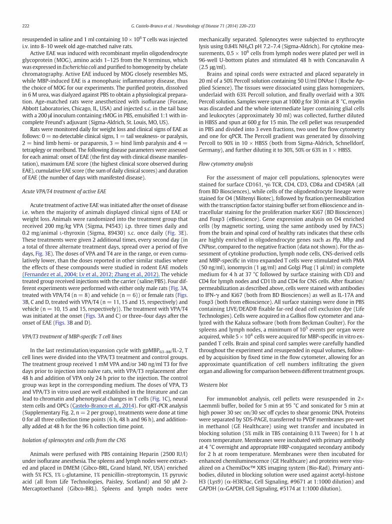
Fig. 1. Treatment ofMBP-specific T cell lines with VPA and VPA/T3 increases histone acetylation and reduces T cell proliferation and frequency of IL-17-producing cells. (A, B) Flow cytom-etry analysis of proliferationmarker Ki67 and Th1/Th17 cytokines, IL-17 and IFN-γ in T3, VPA, VPA/T3 and vehicle treated T cells (n= 3 per group) prior to injection to naïve DA recipients(Fig. 2). (C) Western blot analysis of lysine acetylation on histone H3 in untreated cells and T3, VPA, VPA/T3 and vehicle treated T cells 3 h after the second treatment. Representative ex-periments are shown. Error bars represent SEM on technical replicates with 5 × 10⁴ cells acquired per sample. Differences in cell frequencies were calculated with 1-way ANOVA withKruskal–Wallis test for multiple comparisons (p b 0.05 = *, p b 0.01 = **, p b 0.001 = ***).
223G. Castelo-Branco et al. / Neurobiology of Disease 71 (2014) 220–233
RNA, cDNA preparation and quantitative RT-PCR
RNAwas purified using anRNeasyMini kit ormiRNeasyMicro kit forCNS samples (Qiagen, Hilden, Germany), according to themanufacturerprotocols, including DNase I treatment. cDNA was subsequently pre-pared with the iScript kit (Bio-Rad, Hercules, USA) or High CapacitycDNA Reverse Transcription Kit (Life Technologies) for CNS samples.Quantitative real-time PCR was performed using a BioRad CFX384Touch real-time PCR system with a three-step PCR protocol (95 °C for3 min followed by 40 cycles of 95 °C for 10 s, 60 °C for 30 s and 72 °Cfor 30 s followed by melt curve analysis), using SYBR Green as thefluorophore (Bio-Rad). Cycle of threshold (Ct), efficiencies and meltcurves were analyzed in CFX Manager software (Bio-Rad) and relativeexpression was calculated in relation to the mean of housekeepinggenes, hypoxanthine phosphoribosyltransferase (Hprt) and ubiquitinC, using 2−ΔΔCt. For Fig. 4 and Supplementary Fig. 2, qPCR was per-formed in 7900HT Fast Real-Time PCR System (Applied Biosystems),with a 2 step qPCR protocol (95 °C for 20 s followed by 40 cycles of95 °C for 1 s, 60 °C for 20 s and 95 °C for 15 s followed by melt curveanalysis) and the FAST SYBR GreenMaster mix. Standard curve methodwas used for analysis. Samples were normalized by the geometricalmean of the housekeeping gene values (Hprt, and Tbp for Fig. 4, ubiqui-tin C, beta-actin and Hprt for Supplementary Fig. 2). The following
primers were used: Il7A_fwd CTC AGA CTA CCT CAA CCG TTC C andIl7A_rev GTG CCT CCC AGA TCA CAG AAG; IFNγ_fwd AAA GAC AACCAG GCC ATC AGC and IFNγ_rev TGG CGA TGC TCA TGA ATG C;Gata3_fwd CAC GAT CCA GCA CAG AAG GC and Gata3_rev GGT CTCCGT TAG CGT TCC TC; Il10_fwd GAC GCT GTC ATC GAT TTC TCC andIl10_rev CAG TAG ATG CCG GGT GGT TC; Hprt_fwd CTC ATG GAC TGATTA TGG ACA and Hprt_rev GCA GGT CAG CAA AGA ACT TAT; Tbp_fwdGGG GAG CTG TGA TGT GAA GT and Tbp_rev CCA GGA AAT AAT TCTGGC TCA; Ubc_fwd AAG GTC AAA CAG GAA GAT ACT CG and Ubc_revCTA AGA CAC CTC CCC ATG AAA C; Sox8_fwd AGA CCC TGG GCA AGCTGT and Sox8_rev GGG TGG TCC TTC TTG TGC T; Cnp_fwd AAA TTCTGT GAC TAC GGG AAG G and Cnp_rev GCC GTA AGA TCT CCT CACCA; Mbp_fwd GCT TCT TTA GCG GTG ACA GG and Mbp_rev CCT TGTACA TGT GGC ACA GC; Plp_fwd GCT AGG ACA TCC CGA CAA G andPlp_rev CAA ACA CCA GGA GCC ATA CA; Mag_fwd AAC CAG TAT GGCCAG AGA GC and Mag_rev GTT CCG GGT TGG ATT TTA CC; Mog_fwdGCC GTG GAG TTG AAA GTA GAA G and Mog_rev AGT TTT CCT CTCAGT CTG TGC. Additional primer sequences are available upon request.
Histopathological analyses
Animals were perfused via the left heart ventricle with PBS followedby 4% paraformaldehyde. Paraformaldehyde-fixed 3–5 mm thick
224 G. Castelo-Branco et al. / Neurobiology of Disease 71 (2014) 220–233
paraffin embedded sections of the brain and spinal cord were dewaxedin xylol, rehydrated and then stained with H&E and Luxol Fast Blue(Klüver, KL) to assess tissue inflammation and demyelination, respec-tively. The inflammatory index (I.I.) and demyelination score (DM)were determined from the number and size of demyelinating lesionsin each animal on at least ten complete spinal cord cross-sections aspreviously described (Storch et al., 1998).
Statistical analyses
p-Values for daily mean clinical EAE scores between the groups werecalculated with Wilcoxon matched pairs rank test in the Rcmdr packageof R software (R version 2.9.2 and Rcmdr version 1.5-4). p-Values for dif-ferences in linear regression clinical EAE slopes were calculated withANCOVA in GraphPad Prism software (San Diego, CA). Demyelinationscores, cell numbers and percentages and expression levels between thegroupswere testedusing1-wayANOVAwithKruskal–Wallis test formul-tiple comparisons, two-tailed unpaired t-test and Mann–Whitney testand differences in frequency of occurrence of demyelinating lesionswere tested using Fisher's exact test in GraphPad Prism software.
Results
Treatment with VPA, T3 and VPA/T3 affects encephalitogenic CD4+ T cellsin vitro
HDAC inhibition has been shown to modulate encephalitogenicCD4+ T cells (Ge et al., 2013; Lv et al., 2012; Zhang et al., 2012), whilelittle is known about the effects of thyroid hormone on these cells. There-fore, we treatedMBP63–88-specific T cell lines, which express HDACs andT3 receptor alpha and beta (data not shown), with VPA and T3 for fivedays in vitro. VPA and VPA/T3 strongly reduced T cell proliferation andfrequency of total IL-17-producing cells, Th17 cells and IFN-γ/IL-17 dou-ble positive cells (Figs. 1A, B), while VPA/T3 leads to an increase inIFNg+IL-17− cells (Supplementary Fig. 1). T3 alone had no effect on Tcell proliferation but showed small but significant effect on frequencyof IL-17-producing cells (Figs. 1A, B). Western blot analysis demonstrat-ed increased levels of lysine 9 acetylation of histone H3 in VPA and VPA/T3 treated T cells, but not in T3 treated cells (Fig. 1C). Nevertheless, T3potentiates the effects of HDAC inhibition on H3 acetylation (Fig. 1C).
00.51.01.52.02.53.0
2 4 6 8 10
VEHICLET3VPAVPA/T3
EA
E s
core
*
T3VEHICLE
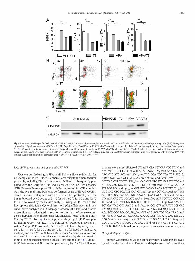
Fig. 2. Treatment of MBP-specific T cell lines with VPA, T3 and VPA/T3 changes their potency tocells i.v. treatedwith T3 (n=8), VPA (n=7), VPA/T3 (n=8) or vehicle (n=8) for five days. EMann–Whitney test (p b 0.05 = *).
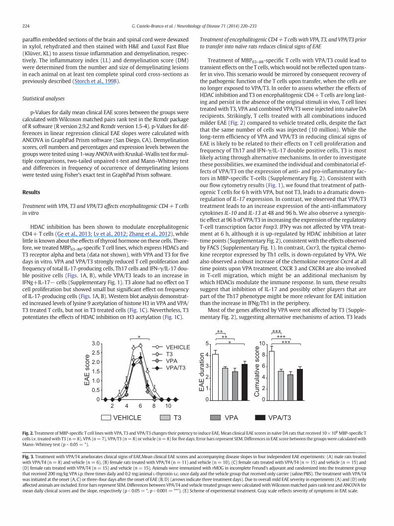
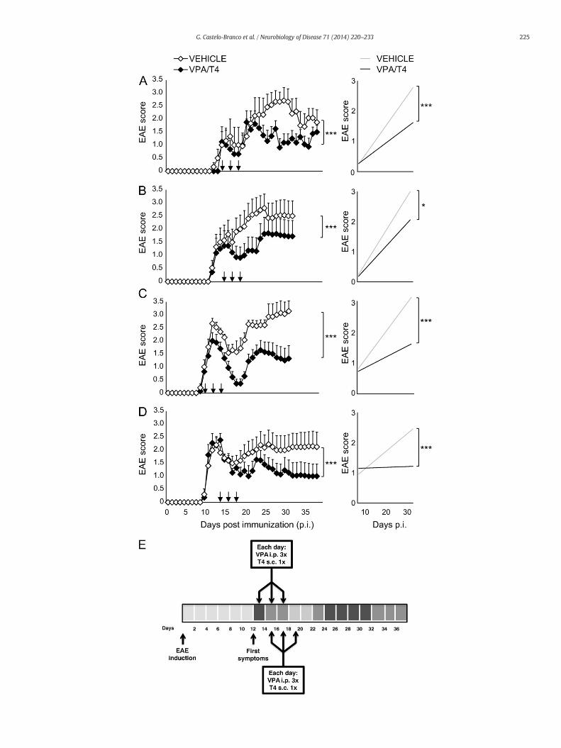
Fig. 3. Treatment with VPA/T4 ameliorates clinical signs of EAE.Mean clinical EAE scores and awith VPA/T4 (n= 8) and vehicle (n= 6), (B) female rats treated with VPA/T4 (n= 11) and v(D) female rats treated with VPA/T4 (n= 15) and vehicle (n= 15). Animals were immunizethat received 200mg/kg VPA i.p. three times daily and 0.2 mg/animal L-thyroxin s.c. once dailywas initiated at the onset (A, C) or three–four days after the onset of EAE (B, D) (arrows indicataffected animals are included. Error bars represent SEM. Differences betweenVPA/T4 and vehicmean daily clinical scores and the slope, respectively (p b 0.05= *, p b 0.001= ***). (E) Sche
Treatment of encephalitogenic CD4+T cells with VPA, T3, and VPA/T3 priorto transfer into naïve rats reduces clinical signs of EAE
Treatment of MBP63–88-specific T cells with VPA/T3 could lead totransient effects on the T cells,whichwould not be reflected upon trans-fer in vivo. This scenario would be mirrored by consequent recovery ofthe pathogenic function of the T cells upon transfer, when the cells areno longer exposed to VPA/T3. In order to assess whether the effects ofHDAC inhibition and T3 on encephalitogenic CD4+ T cells are long last-ing and persist in the absence of the original stimuli in vivo, T cell linestreated with T3, VPA and combined VPA/T3 were injected into naïve DArecipients. Strikingly, T cells treated with all combinations inducedmilder EAE (Fig. 2) compared to vehicle treated cells, despite the factthat the same number of cells was injected (10 million). While thelong-term efficiency of VPA and VPA/T3 in reducing clinical signs ofEAE is likely to be related to their effects on T cell proliferation andfrequency of Th17 and IFN-γ/IL-17 double positive cells, T3 is mostlikely acting through alternative mechanisms. In order to investigatethese possibilities, we examined the individual and combinatorial ef-fects of VPA/T3 on the expression of anti- and pro-inflammatory fac-tors in MBP-specific T-cells (Supplementary Fig. 2). Consistent withour flow cytometry results (Fig. 1), we found that treatment of path-ogenic T cells for 6 h with VPA, but not T3, leads to a dramatic down-regulation of IL-17 expression. In contrast, we observed that VPA/T3treatment leads to an increase expression of the anti-inflammatorycytokines IL-10 and IL-13 at 48 and 96 h. We also observe a synergis-tic effect at 96 h of VPA/T3 in increasing the expression of the regulatoryT-cell transcription factor Foxp3. IFNγ was not affected by VPA treat-ment at 6 h, although it is up-regulated by HDAC inhibition at latertime points (Supplementary Fig. 2), consistentwith the effects observedby FACS (Supplementary Fig. 1). In contrast, Cxcr3, the typical chemo-kine receptor expressed by Th1 cells, is down-regulated by VPA. Wealso observed a robust increase of the chemokine receptor Cxcr4 at alltime points upon VPA treatment. CXCR 3 and CXCR4 are also involvedin T-cell migration, which might be an additional mechanism bywhich HDACis modulate the immune response. In sum, these resultssuggest that inhibition of IL-17 and possibly other players that arepart of the Th17 phenotype might be more relevant for EAE initiationthan the increase in IFNg/Th1 in the periphery.
Most of the genes affected by VPA were not affected by T3 (Supple-mentary Fig. 2), suggesting alternative mechanisms of action. T3 leads
0
1
2
3
4
5
0
2
4
6
8
10
EA
E d
urat
ion
Cum
ulat
ive
scor
e
****
****
******
VPA VPA/T3
induce EAE.Mean clinical EAE scores in naïve DA rats that received 10 × 106MBP-specific Trror bars represent SEM. Differences in EAE score between the groupswere calculatedwith
ccompanying disease slopes in four independent EAE experiments: (A) male rats treatedehicle (n= 10), (C) female rats treated with VPA/T4 (n= 15) and vehicle (n= 15) andd with rMOG in incomplete Freund's adjuvant and randomized into the treatment groupand the vehicle group that received only carrier (saline/PBS). The treatment with VPA/T4e three treatment days). Due to overall mild EAE severity in experiments (A) and (D) onlyle treated groupswere calculatedwithWilcoxonmatched pairs rank test and ANCOVA forme of experimental treatment. Gray scale reflects severity of symptoms in EAE scale.
225G. Castelo-Branco et al. / Neurobiology of Disease 71 (2014) 220–233
C
0.00
0.05
0.10
0.15
0.20
0.0
0.5
1.0
1.5
2.0
2.5
0
1
2
3
4
5
0
1
2
3
4
5
0
10
20
30
40
0
1
2
3
4
5
6
0
10
20
30
0.00
0.02
0.04
0.06
0.08
0.10
0.0
0.2
0.4
0.6
0.8
1.0
0.0
0.4
0.8
1.2
1.6*
0.00
0.02
0.04
0.06
0.08
Sox
8 ex
pres
sion
0.00
0.01
0.02
0.03
0.04
0.05
Sox
8 ex
pres
sion
CN
Pas
e ex
pres
sion
CN
Pas
e ex
pres
sion
Mbp
exp
ress
ion
Mbp
exp
ress
ion
Plp
exp
ress
ion
Plp
exp
ress
ion
Mag
exp
ress
ion
Mag
exp
ress
ion
Mog
exp
ress
ion
Mog
exp
ress
ion
* * ** ** **
spin
al c
ord
brai
n
VPA/T4VEHICLE
A B
CD
45
CD11b O4
Ki6
7
A B
C
Gated on A
D
E
Gated on DC
D45
O4 0
5
10
15
20
% o
f O4+
Ki6
7+
0
10
20
30
0
1
2
0
5
10
15
brai
nsp
inal
cor
d
% o
f O4+
Ki6
7+
% o
f tot
al%
of t
otal
O4+Ki67+ O4+Ki67+
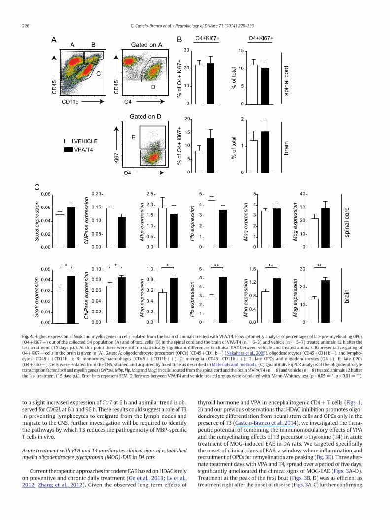
Fig. 4. Higher expression of Sox8 and myelin genes in cells isolated from the brain of animals treated with VPA/T4. Flow cytometry analysis of percentages of late pre-myelinating OPCs(O4+Ki67+) out of the collected O4 population (A) and of total cells (B) in the spinal cord and the brain of VPA/T4 (n = 6–8) and vehicle (n = 5–7) treated animals 12 h after thelast treatment (15 days p.i.). At this point there were still no statistically significant differences in clinical EAE between vehicle and treated animals. Representative gating ofO4+Ki67+ cells in the brain is given in (A). Gates: A: oligodendrocyte precursors (OPCs) (CD45+CD11b−) (Nakahara et al., 2005), oligodendrocytes (CD45+CD11b−), and lympho-cytes (CD45++CD11b−); B: monocytes/macrophages (CD45++CD11b++); C: microglia (CD45+CD11b++); D: late OPCs and oligodendrocytes (O4+); E: late OPCs(O4+Ki67+). Cells were isolated from the CNS, stained and acquired by fixed time as described in Materials and methods. (C) Quantitative qPCR analysis of the oligodendrocytetranscription factor Sox8 andmyelin genes (CNPase,Mbp, Plp,Mag andMog) in cells isolated fromthe spinal cord and the brain of VPA/T4 (n=8) and vehicle (n=8) treated animals 12h afterthe last treatment (15 days p.i.). Error bars represent SEM. Differences between VPA/T4 and vehicle treated groups were calculated with Mann–Whitney test (p b 0.05 = *, p b 0.01 = **).
226 G. Castelo-Branco et al. / Neurobiology of Disease 71 (2014) 220–233
to a slight increased expression of Ccr7 at 6 h and a similar trend is ob-served for CD62L at 6 h and 96 h. These results could suggest a role of T3in preventing lymphocytes to emigrate from the lymph nodes andmigrate to the CNS. Further investigation will be required to identifythe pathways by which T3 reduces the pathogenicity of MBP-specificT cells in vivo.
Acute treatment with VPA and T4 ameliorates clinical signs of establishedmyelin oligodendrocyte glycoprotein (MOG)-EAE in DA rats
Current therapeutic approaches for rodent EAE based onHDACis relyon preventive and chronic daily treatment (Ge et al., 2013; Lv et al.,2012; Zhang et al., 2012). Given the observed long-term effects of
thyroid hormone and VPA in encephalitogenic CD4+ T cells (Figs. 1,2) and our previous observations that HDAC inhibition promotes oligo-dendrocyte differentiation from neural stem cells and OPCs only in thepresence of T3 (Castelo-Branco et al., 2014), we investigated the thera-peutic potential of combining the immunomodulatory effects of VPAand the remyelinating effects of T3 precursor L-thyroxine (T4) in acutetreatment of MOG-induced EAE in DA rats. We targeted specificallythe onset of clinical signs of EAE, a window where inflammation andrecruitment of OPCs for remyelination are peaking (Fig. 3E). Three alter-nate treatment days with VPA and T4, spread over a period of five days,significantly ameliorated the clinical signs of MOG-EAE (Figs. 3A–D).Treatment at the peak of the first bout (Figs. 3B, D) was as efficient astreatment right after the onset of disease (Figs. 3A, C) further confirming
227G. Castelo-Branco et al. / Neurobiology of Disease 71 (2014) 220–233
the therapeutic potential of combined VPA/T4. Moreover, this shorttreatment induced a prolonged and persistent reduction of severity ofEAE, evenwhen a secondflare of the disease occurs (Figs. 3B, C). This ef-fect was independent of the initial disease severity as the treatmentshowed desired results in mild (Figs. 3A, D) as well as severe forms ofEAE (Figs. 3B, C), or of gender (Fig. 3A, male rats treated with VPA/T4(n = 8) and vehicle (n = 6); Figs. 3B, C and D, female rats treatedwith VPA/T4 (n = 11, 15 and 15, respectively) and vehicle (n = 10,15 and 15)).
Higher expression of myelin genes in the brain, but not spinal cord, upontreatment of EAE with VPA and T4
To investigate if the clinical effect of VPA and T4 is accompanied byan effect onNSC/OPCdifferentiation or lineage progression,mononucle-ar cells were isolated from the spinal cord and the brain 12 h after thelast treatment (5 days after the initial treatment). The isolation protocolwith Percoll involves the removal of myelin and thus of many of the as-sociated oligodendrocytes, while preserving immune cells, OPCs andpre-myelinating cells (expressing Mbp, CNPase and Plp, see Materialand methods) (Colello and Sato-Bigbee, 2001). We observed by flowcytometry a trend for an increase in the % of late pre-myelinatingOPCs (O4+Ki67+) out of the collected O4 population in the brain,but not in the spinal cord (Fig. 4A, gate D). There was a similar trendfor an increase in the % of late pre-myelinating OPCs out of total cells(Fig. 4B). Concomitantly, we observed by qPCR higher expression ofSox8, a transcription factor involved in terminal oligodendrocyte differ-entiation (Stolt et al., 2004) and a direct HDAC target (Castelo-Brancoet al., 2014) in the brains of treated animals (Fig. 4C) compared to un-treated animals. Likewise, all major myelin genes, CNPase, Mbp, Plp,Mag and Mog, displayed significantly higher expression in the brain oftreated animals (Fig. 4C). These results suggest that one of the mecha-nisms by which treatment with VPA and the T3 precursor might be act-ing in the brain is by promoting oligodendrocyte lineage progression.
CD4+ Foxp3-
0
200
400
600
800
0
500
1000
1500
2000
*
IFN-γ+IL-17-
0
50
100
150
200
0
50
100
150
200 *
1
2
3
4
5
10
15
20
020406080
100
020406080
100
0
5
10
15
20 p=0.06
01020304050 **
Cel
l num
ber
Cel
l num
ber
Cel
l num
ber
% o
f tot
al
% o
f CD
4+ F
oxp3
-
Cel
l num
ber
Cel
l num
ber
Cel
l num
ber
A
B
IL-17
CD
4
CD
4
SSC Foxp3
% o
f tot
al
% o
f CD
4+ F
oxp3
-
A B
Gated on A Ga
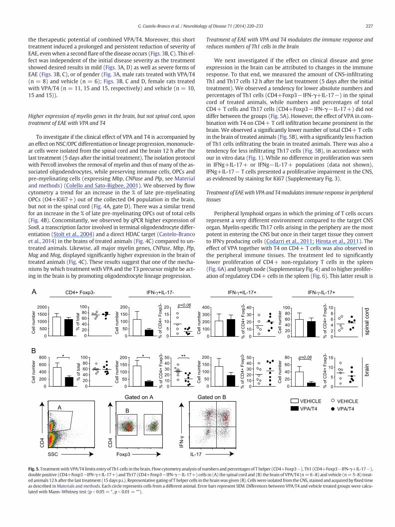
Fig. 5. Treatmentwith VPA/T4 limits entry of Th1 cells in the brain. Flow cytometry analysis of nudouble positive (CD4+Foxp3−IFN-γ+IL-17+) and Th17 (CD4+Foxp3−IFN-γ−IL-17+) celled animals 12 h after the last treatment (15 days p.i.). Representative gating of T helper cells in thas described inMaterials and methods. Each circle represents cells from a different animal. Errolated with Mann–Whitney test (p b 0.05 = *, p b 0.01 = **).
Treatment of EAE with VPA and T4 modulates the immune response andreduces numbers of Th1 cells in the brain
We next investigated if the effect on clinical disease and geneexpression in the brain can be attributed to changes in the immuneresponse. To that end, we measured the amount of CNS-infiltratingTh1 and Th17 cells 12 h after the last treatment (5 days after the initialtreatment). We observed a tendency for lower absolute numbers andpercentages of Th1 cells (CD4+Foxp3− IFN-γ+IL-17−) in the spinalcord of treated animals, while numbers and percentages of totalCD4+ T cells and Th17 cells (CD4+Foxp3− IFN-γ− IL-17+) did notdiffer between the groups (Fig. 5A). However, the effect of VPA in com-bination with T4 on CD4+ T cell infiltration became prominent in thebrain. We observed a significantly lower number of total CD4+ T cellsin the brain of treated animals (Fig. 5B), with a significantly less fractionof Th1 cells infiltrating the brain in treated animals. There was also atendency for less infiltrating Th17 cells (Fig. 5B), in accordance withour in vitro data (Fig. 1). While no difference in proliferation was seenin IFNg+IL-17+ or IFNg− IL-17+ populations (data not shown),IFNg+IL-17− T cells presented a proliferative impairment in the CNS,as evidenced by staining for Ki67 (Supplementary Fig. 3).
Treatment of EAEwith VPA and T4modulates immune response in peripheraltissues
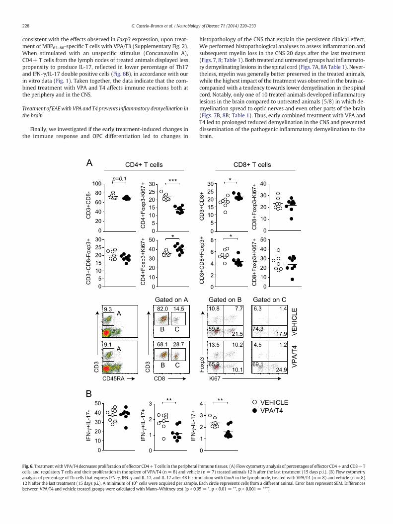
Peripheral lymphoid organs in which the priming of T cells occursrepresent a very different environment compared to the target CNSorgan. Myelin-specific Th17 cells arising in the periphery are the mostpotent in entering the CNS but once in their target tissue they convertto IFNγ producing cells (Codarri et al., 2011; Hirota et al., 2011). Theeffect of VPA together with T4 on CD4+ T cells was also observed inthe peripheral immune tissues. The treatment led to significantlylower proliferation of CD4+ non-regulatory T cells in the spleen(Fig. 6A) and lymph node (Supplementary Fig. 4) and to higher prolifer-ation of regulatory CD4+ cells in the spleen (Fig. 6). This latter result is
020406080
100
0
20
40
60
80 p=0.06
0
00
00
00
00
0
0
0
0
0
Cel
l num
ber
Cel
l num
ber
VPA/T4VEHICLE
VPA/T4VEHICLE
spin
al c
ord
brai
n
% o
f CD
4+ F
oxp3
-%
of C
D4+
Fox
p3-
% o
f CD
4+ F
oxp3
-%
of C
D4+
Fox
p3-
IFN-γ+IL-17+ IFN-γ-IL-17+
0
10
20
30
40
01020304050
02468
10
0
5
10
15
ted on B
mbers and percentages of T helper (CD4+Foxp3−), Th1 (CD4+Foxp3−IFN-γ+IL-17−),s in (A) the spinal cord and (B) the brain of VPA/T4 (n=6–8) and vehicle (n= 5–8) treat-e brainwas given (B). Cellswere isolated from the CNS, stained and acquired byfixed timer bars represent SEM. Differences between VPA/T4 and vehicle treated groups were calcu-
228 G. Castelo-Branco et al. / Neurobiology of Disease 71 (2014) 220–233
consistent with the effects observed in Foxp3 expression, upon treat-ment of MBP63–88-specific T cells with VPA/T3 (Supplementary Fig. 2).When stimulated with an unspecific stimulus (Concanavalin A),CD4+ T cells from the lymph nodes of treated animals displayed lesspropensity to produce IL-17, reflected in lower percentage of Th17and IFN-γ/IL-17 double positive cells (Fig. 6B), in accordance with ourin vitro data (Fig. 1). Taken together, the data indicate that the com-bined treatment with VPA and T4 affects immune reactions both atthe periphery and in the CNS.
Treatment of EAEwith VPA and T4 prevents inflammatory demyelination inthe brain
Finally, we investigated if the early treatment-induced changes inthe immune response and OPC differentiation led to changes in
*
p=0.1
CD
3+C
D8-
CD
3+C
D8-
Foxp
3+
***
CD
4+Fo
xp3+
Ki6
7+C
D4+
Foxp
3-K
i67+
A
B**
IFN
-γ+I
L-17
-
IFN
-γ+I
L-17
+
IFN
-γ-IL
-17+
0
20
40
60
80
100
05
1015202530
05
1015202530
0
10
20
30
40
50
0
10
20
30
40
50
0
1
2
3
CD4+ T cells
CD8
CD
3
82.0 14.5
28.768.1
CD
3
CD45RA
9.3
9.1
Gated on A
A
A
B C
B C
Fig. 6. Treatmentwith VPA/T4 decreases proliferation of effector CD4+T cells in the peripheralcells, and regulatory T cells and their proliferation in the spleen of VPA/T4 (n = 8) and vehiclanalysis of percentage of Th cells that express IFN-γ, IFN-γ and IL-17, and IL-17 after 48 h stim12 h after the last treatment (15 days p.i.). A minimum of 105 cells were acquired per sample.between VPA/T4 and vehicle treated groups were calculated with Mann–Whitney test (p b 0.0
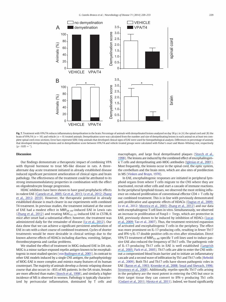
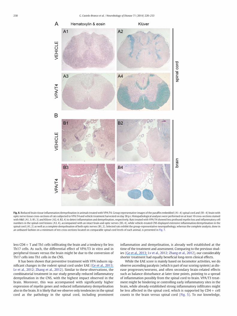
histopathology of the CNS that explain the persistent clinical effect.We performed histopathological analyses to assess inflammation andsubsequent myelin loss in the CNS 20 days after the last treatment(Figs. 7, 8; Table 1). Both treated and untreated groups had inflammato-ry demyelinating lesions in the spinal cord (Figs. 7A, 8A Table 1). Never-theless, myelin was generally better preserved in the treated animals,while the highest impact of the treatmentwas observed in the brain ac-companied with a tendency towards lower demyelination in the spinalcord. Notably, only one of 10 treated animals developed inflammatorylesions in the brain compared to untreated animals (5/8) in which de-myelination spread to optic nerves and even other parts of the brain(Figs. 7B, 8B; Table 1). Thus, early combined treatment with VPA andT4 led to prolonged reduced demyelination in the CNS and preventeddissemination of the pathogenic inflammatory demyelination to thebrain.
*
*
CD
3+C
D8+
CD
3+C
D8+
Foxp
3+
CD
8+Fo
xp3+
Ki6
7+C
D8+
Foxp
3-K
i67+
VPA/T4VEHICLE**
05
1015202530
0
2
4
6
8
0
10
20
30
40
0
10
20
30
40
50
0
1
2
3
4
CD8+ T cells
10.8 7.7
21.5 59.8
13.5 10.2
10.165.9
6.3 1.4
17.974.3
4.5 1.2
24.969.1
Ki67
Foxp
3
VE
HIC
LEV
PA
/T4
Gated on B Gated on C
immune tissues. (A) Flow cytometry analysis of percentages of effector CD4+and CD8+Te (n = 7) treated animals 12 h after the last treatment (15 days p.i.). (B) Flow cytometryulation with ConA in the lymph node, treated with VPA/T4 (n = 8) and vehicle (n = 8)Each circle represents cells from a different animal. Error bars represent SEM. Differences5 = *, p b 0.01 = **, p b 0.001 = ***).
0
0.5
1.0
1.5
2.0
2.5
3.0
3.5
VPA/T4VEHICLE
0
20
40
60
80
100
0
20
40
60
80
100
demyelinationno demyelination
**
Dem
yelin
atio
n sc
ore
Dem
yelin
atio
n sc
ore
Ani
mal
s (%
)A
nim
als
(%)
A
B
VPA/T4VEHICLE
spin
al c
ord
brai
n
VPA/T4VEHICLE
0
0.5
1.0
1.5
2.0
2.5
3.0
3.5
Fig. 7. Treatmentwith VPA/T4 reduces inflammatory demyelination in the brain. Percentage of animalswith demyelinated lesions analyzed on day 38 p.i. in (A) the spinal cord and (B) thebrain of VPA/T4 (n= 10) and vehicle (n= 8) treated animals. Demyelination score was calculated from the number and size of demyelinating lesions in each animal on at least ten com-plete spinal cord cross-sections. Error bars represent SEM. Only animals that developed clinical signs of EAEwere used for histopathological analysis. Differences in percentage of animalsthat developed demyelinating lesions and in demyelination score between VPA/T4 and vehicle treated groups were calculated with Fisher's exact and Mann–Whitney test, respectively(p b 0.05 = *).
229G. Castelo-Branco et al. / Neurobiology of Disease 71 (2014) 220–233
Discussion
Our findings demonstrate a therapeutic impact of combining VPAwith thyroid hormone to treat MS-like disease in rats. A three-alternate day acute treatment initiated in already established diseaseinduced significant persistent amelioration of clinical signs and brainpathology. The effectiveness of the treatment could be attributed to itsstrong immunomodulatory properties in combination with the effecton oligodendrocyte lineage progression.
HDAC inhibitors have been shown to have good prophylactic effectsin rodent EAE (Camelo et al., 2005; Ge et al., 2013; Lv et al., 2012; Zhanget al., 2012, 2010). However, the therapeutic potential in alreadyestablished disease is much clearer in our experiments with combinedT4 treatment. In previous studies, the treatment initiated at the onsetof EAE had a modest effect in MBP68–84-induced EAE in Lewis rats(Zhang et al., 2012) and treating MOG35–55-induced EAE in C57BL/6mice after onset had a substantial effect; however, the treatment wasadministered daily for the remainder of disease (Lv et al., 2012). Ourdata show that we can achieve a significant persistent amelioration ofEAE in rats with a short course of combined treatment. Cycles of shortertreatments would be more desirable in clinical settings due to theknown adverse effects of HDACis including diarrhea, vomiting, fatigue,thrombocytopenia and cardiac problems.
We studied the effect of treatment in MOG-induced EAE in DA rats.MOG is aminor surface-exposedmyelin antigen known to be encephali-togenic inmost studied species (von Budingen et al., 2001). In contrast toother EAE models induced by a single CNS antigen, the pathophysiologyof MOG-EAE is more complex and mimics many features of its humancounterpart. Themajority of animals develop a chronic relapsing diseasecourse that also occurs in ~85% of MS patients. In the DA strain, femalesare more affected thanmales (Storch et al., 1998), and similarly a higherincidence of MS is observed in women. Pathology is typically character-ized by perivascular inflammation, dominated by T cells and
macrophages, and large focal demyelinated plaques (Storch et al.,1998). The lesions are inducedby the combined effect of encephalitogen-ic T cells and demyelinating anti-MOG antibodies (Iglesias et al., 2001).Most frequently, the lesions occur in the spinal cord, the optic system,the cerebellum and the brain stem, which are also sites of predilectionin MS (Vinken and Bruyn, 1970).
In EAE, encephalitogenic responses are initiated in peripheral lym-phoid organs from where T cells migrate to the CNS where they arereactivated, recruit other cells and start a cascade of immune reactions.In the peripheral lymphoid tissues, we observed themost striking influ-ence on reduced proliferation of conventional effector CD4+ T cells byour combined treatment. This is in line with previously demonstratedanti-proliferative and apoptotic effects of HDACis (Dagtas et al., 2009;Lv et al., 2012; Moreira et al., 2003; Zhang et al., 2012) and our datawith encephalitogenic T cell lines in vitro. Simultaneously, we observedan increase in proliferation of Foxp3+ Tregs, which are protective inEAE, previously shown to be induced by inhibition of HDACs (Lucaset al., 2009; Tao et al., 2007). Thus, the treatment restricted expansionof activated and encephalitogenic T cells in the periphery. The effectwas more prominent on IL-17-producing cells, resulting in fewer Th17and IFN-γ/IL-17 double positive cells ex-vivo after stimulation. DirectVPA/T3 treatment of MBP63–88-specific T cell lines used to induce pas-sive EAE also reduced the frequency of Th17 cells. The pathogenic roleof IL-17-producing Th17 cells in EAE is well established (Langrishet al., 2005; Park et al., 2005). Th17 cells are able to enter the CNS with-out a compromised blood brain barrier and to initiate an inflammatorycascade and a secondwave of infiltration by Th1 and Th17 cells (Reboldiet al., 2009). Both Th1 and Th17 cells have shown pathogenic roles inEAE (Baron et al., 1993; Kroenke et al., 2008; Segal and Shevach, 1996;Stromnes et al., 2008). Additionally, myelin-specific Th17 cells arisingin the periphery are the most potent in entering the CNS but once intheir target tissue they can convert to IFN-γ producing Th1 cells(Codarri et al., 2011; Hirota et al., 2011). Indeed, we found significantly
A1 A2
A3 A4
B1 B2
B3 B4
Fig. 8.Reduced brain tissue inflammation/demyelination in animals treatedwith VPA/T4. Group representative images of the paraffin embedded (A1–4) spinal cord and (B1–4) brainwithoptic nerve tissue cross-sections of rats subjected to VPA/T4 and vehicle treatment harvested on day 38p.i. Histopathological analyseswere performed on at least 10 cross-sections stainedwithH&E (A1, 3; B1, 3) andKlüver (A2, 4; B2, 4) to detect inflammation anddemyelination, respectively. Rats treatedwith VPA/T4 showed less profoundmyelin loss and inflammatory cellnumbers in the spinal cord lesions (A3, 4), accompanied with an intact brain and optic nerves (B3, 4), while vehicle-treated CNS displayed extensive inflammation/demyelination in thespinal cord (A1, 2) aswell as a complete demyelination of both optic nerves (B1, 2). Selected rats exhibit the group-representative neuropathology, whereas the complete analysis, done inan unbiased fashion on a minimum of ten cross-sections located on comparable spinal cord levels of each animal, is presented in Fig. 7.
230 G. Castelo-Branco et al. / Neurobiology of Disease 71 (2014) 220–233
less CD4+ T and Th1 cells infiltrating the brain and a tendency for lessTh17 cells. As such, the differential effect of VPA/T3 in vitro and inperipheral tissues versus the brain might be due to the conversion ofTh17 cells into Th1 cells in the CNS.
It has been shown that preventive treatment with VPA induces sig-nificant changes in the rodent spinal cord under EAE (Ge et al., 2013;Lv et al., 2012; Zhang et al., 2012). Similar to these observations, thecombinatorial treatment in our study generally reduced inflammatorydemyelination in the CNS, with the highest impact observed in thebrain. Moreover, this was accompanied with significantly higherexpression of myelin genes and reduced inflammatory demyelinationalso in the brain. It is likely thatwe observe only tendencies in the spinalcord as the pathology in the spinal cord, including prominent
inflammation and demyelination, is already well established at thetime of the treatment and assessment. Comparing to the previous stud-ies (Ge et al., 2013; Lv et al., 2012; Zhang et al., 2012), our considerablyshorter treatment had equally beneficial long-term clinical effects.
While the EAE score is mainly based on locomotor activities, we doobserve ascending paralysis (which is part of our scoring system) as dis-ease progresses/worsens, and often secondary brain-related effectssuch as balance disturbance at later time points, pointing to a spreadof inflammation possibly from the spinal cord to brain. VPA/T3 treat-ment might be hindering or controlling early inflammatory sites in thebrain, while already established strong inflammatory infiltrates mightbe less affected in the spinal cord, which is supported by CD4+ cellcounts in the brain versus spinal cord (Fig. 5). To our knowledge,
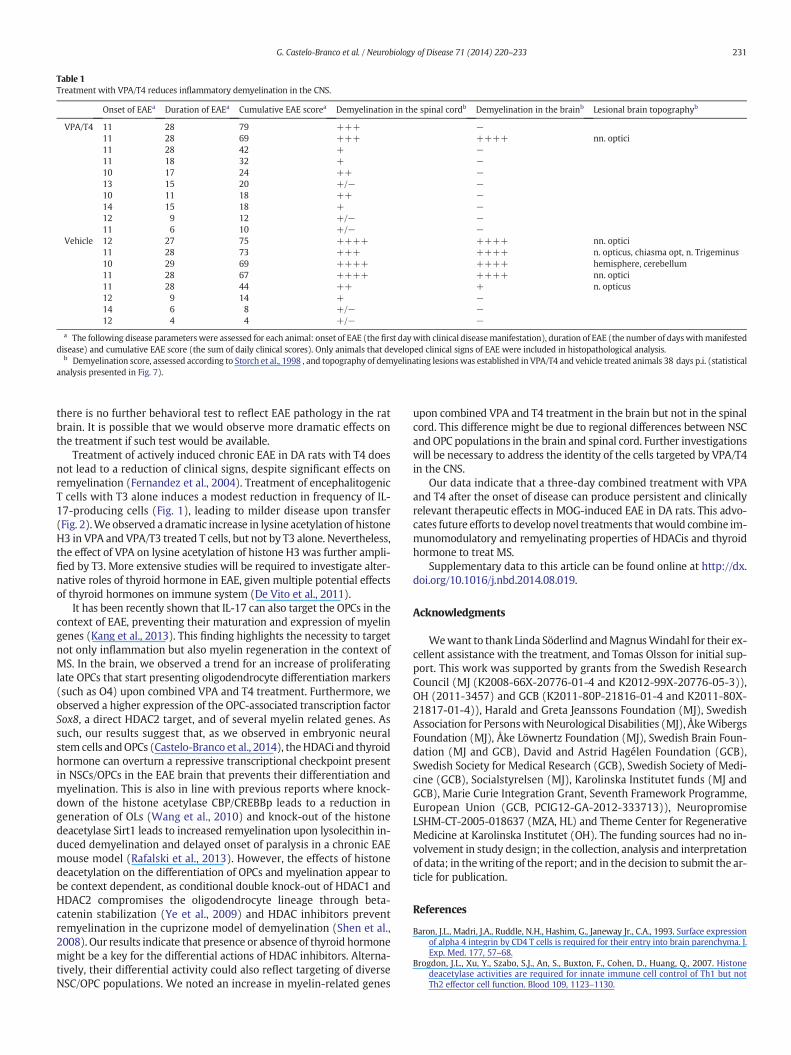
Table 1Treatment with VPA/T4 reduces inflammatory demyelination in the CNS.
Onset of EAEa Duration of EAEa Cumulative EAE scorea Demyelination in the spinal cordb Demyelination in the brainb Lesional brain topographyb
VPA/T4 11 28 79 +++ −11 28 69 +++ ++++ nn. optici11 28 42 + −11 18 32 + −10 17 24 ++ −13 15 20 +/− −10 11 18 ++ −14 15 18 + −12 9 12 +/− −11 6 10 +/− −
Vehicle 12 27 75 ++++ ++++ nn. optici11 28 73 +++ ++++ n. opticus, chiasma opt, n. Trigeminus10 29 69 ++++ ++++ hemisphere, cerebellum11 28 67 ++++ ++++ nn. optici11 28 44 ++ + n. opticus12 9 14 + −14 6 8 +/− −12 4 4 +/− −
a The following disease parameterswere assessed for each animal: onset of EAE (the first daywith clinical diseasemanifestation), duration of EAE (the number of dayswithmanifesteddisease) and cumulative EAE score (the sum of daily clinical scores). Only animals that developed clinical signs of EAE were included in histopathological analysis.
b Demyelination score, assessed according to Storch et al., 1998 , and topography of demyelinating lesionswas established in VPA/T4 and vehicle treated animals 38 days p.i. (statisticalanalysis presented in Fig. 7).
231G. Castelo-Branco et al. / Neurobiology of Disease 71 (2014) 220–233
there is no further behavioral test to reflect EAE pathology in the ratbrain. It is possible that we would observe more dramatic effects onthe treatment if such test would be available.
Treatment of actively induced chronic EAE in DA rats with T4 doesnot lead to a reduction of clinical signs, despite significant effects onremyelination (Fernandez et al., 2004). Treatment of encephalitogenicT cells with T3 alone induces a modest reduction in frequency of IL-17-producing cells (Fig. 1), leading to milder disease upon transfer(Fig. 2).We observed a dramatic increase in lysine acetylation of histoneH3 in VPA and VPA/T3 treated T cells, but not by T3 alone. Nevertheless,the effect of VPA on lysine acetylation of histone H3 was further ampli-fied by T3. More extensive studies will be required to investigate alter-native roles of thyroid hormone in EAE, given multiple potential effectsof thyroid hormones on immune system (De Vito et al., 2011).
It has been recently shown that IL-17 can also target the OPCs in thecontext of EAE, preventing their maturation and expression of myelingenes (Kang et al., 2013). This finding highlights the necessity to targetnot only inflammation but also myelin regeneration in the context ofMS. In the brain, we observed a trend for an increase of proliferatinglate OPCs that start presenting oligodendrocyte differentiation markers(such as O4) upon combined VPA and T4 treatment. Furthermore, weobserved a higher expression of the OPC-associated transcription factorSox8, a direct HDAC2 target, and of several myelin related genes. Assuch, our results suggest that, as we observed in embryonic neuralstem cells andOPCs (Castelo-Branco et al., 2014), theHDACi and thyroidhormone can overturn a repressive transcriptional checkpoint presentin NSCs/OPCs in the EAE brain that prevents their differentiation andmyelination. This is also in line with previous reports where knock-down of the histone acetylase CBP/CREBBp leads to a reduction ingeneration of OLs (Wang et al., 2010) and knock-out of the histonedeacetylase Sirt1 leads to increased remyelination upon lysolecithin in-duced demyelination and delayed onset of paralysis in a chronic EAEmouse model (Rafalski et al., 2013). However, the effects of histonedeacetylation on the differentiation of OPCs and myelination appear tobe context dependent, as conditional double knock-out of HDAC1 andHDAC2 compromises the oligodendrocyte lineage through beta-catenin stabilization (Ye et al., 2009) and HDAC inhibitors preventremyelination in the cuprizone model of demyelination (Shen et al.,2008). Our results indicate that presence or absence of thyroid hormonemight be a key for the differential actions of HDAC inhibitors. Alterna-tively, their differential activity could also reflect targeting of diverseNSC/OPC populations. We noted an increase in myelin-related genes
upon combined VPA and T4 treatment in the brain but not in the spinalcord. This difference might be due to regional differences between NSCand OPC populations in the brain and spinal cord. Further investigationswill be necessary to address the identity of the cells targeted by VPA/T4in the CNS.
Our data indicate that a three-day combined treatment with VPAand T4 after the onset of disease can produce persistent and clinicallyrelevant therapeutic effects in MOG-induced EAE in DA rats. This advo-cates future efforts to develop novel treatments thatwould combine im-munomodulatory and remyelinating properties of HDACis and thyroidhormone to treat MS.
Supplementary data to this article can be found online at http://dx.doi.org/10.1016/j.nbd.2014.08.019.
Acknowledgments
Wewant to thank Linda Söderlind andMagnusWindahl for their ex-cellent assistance with the treatment, and Tomas Olsson for initial sup-port. This work was supported by grants from the Swedish ResearchCouncil (MJ (K2008-66X-20776-01-4 and K2012-99X-20776-05-3)),OH (2011-3457) and GCB (K2011-80P-21816-01-4 and K2011-80X-21817-01-4)), Harald and Greta Jeanssons Foundation (MJ), SwedishAssociation for PersonswithNeurological Disabilities (MJ), ÅkeWibergsFoundation (MJ), Åke Löwnertz Foundation (MJ), Swedish Brain Foun-dation (MJ and GCB), David and Astrid Hagélen Foundation (GCB),Swedish Society for Medical Research (GCB), Swedish Society of Medi-cine (GCB), Socialstyrelsen (MJ), Karolinska Institutet funds (MJ andGCB), Marie Curie Integration Grant, Seventh Framework Programme,European Union (GCB, PCIG12-GA-2012-333713)), NeuropromiseLSHM-CT-2005-018637 (MZA, HL) and Theme Center for RegenerativeMedicine at Karolinska Institutet (OH). The funding sources had no in-volvement in study design; in the collection, analysis and interpretationof data; in thewriting of the report; and in the decision to submit the ar-ticle for publication.
References
Baron, J.L., Madri, J.A., Ruddle, N.H., Hashim, G., Janeway Jr., C.A., 1993. Surface expressionof alpha 4 integrin by CD4 T cells is required for their entry into brain parenchyma. J.Exp. Med. 177, 57–68.
Brogdon, J.L., Xu, Y., Szabo, S.J., An, S., Buxton, F., Cohen, D., Huang, Q., 2007. Histonedeacetylase activities are required for innate immune cell control of Th1 but notTh2 effector cell function. Blood 109, 1123–1130.
232 G. Castelo-Branco et al. / Neurobiology of Disease 71 (2014) 220–233
Camelo, S., Iglesias, A.H., Hwang, D., Due, B., Ryu, H., Smith, K., Gray, S.G., Imitola, J., Duran,G., Assaf, B., et al., 2005. Transcriptional therapy with the histone deacetylase inhibi-tor trichostatin A ameliorates experimental autoimmune encephalomyelitis. J.Neuroimmunol. 164, 10–21.
Campas-Moya, C., 2009. Romidepsin for the treatment of cutaneous T-cell lymphoma.Drugs Today 45, 787–795.
Castelo-Branco, G., Lilja, T., Wallenborg, K., Falcao, A.M., Marques, S.C., Gracias, A., Solum,D., Paap, R., Walfridsson, J., Teixeira, A.I., et al., 2014. Neural stem cell differentiation isdictated by distinct actions of nuclear receptor corepressors and histone deacetylases.Stem Cell Reports 3, 1–14.
Chiu, C.T., Wang, Z., Hunsberger, J.G., Chuang, D.M., 2013. Therapeutic potential of moodstabilizers lithium and valproic acid: beyond bipolar disorder. Pharmacol. Rev. 65,105–142.
Codarri, L., Gyulveszi, G., Tosevski, V., Hesske, L., Fontana, A., Magnenat, L., Suter, T.,Becher, B., 2011. RORgammat drives production of the cytokine GM-CSF in helper Tcells, which is essential for the effector phase of autoimmune neuroinflammation.Nat. Immunol. 12, 560–567.
Colello, R.J., Sato-Bigbee, C., 2001. Purification of oligodendrocytes and their progenitorsusing immunomagnetic separation and Percoll gradient centrifugation. Curr. Protoc.Neurosci. 3, 12–14 (editorial board, Jacqueline N Crawley [et al] Chapter 1–3,Unit 3.12).
Dagtas, A.S., Edens, R.E., Gilbert, K.M., 2009. Histone deacetylase inhibitor usesp21(Cip1) to maintain anergy in CD4+ T cells. Int. Immunopharmacol. 9,1289–1297.
De Vito, P., Incerpi, S., Pedersen, J.Z., Luly, P., Davis, F.B., Davis, P.J., 2011. Thyroid hormonesas modulators of immune activities at the cellular level. Thyroid 21, 879–890.
de Zoeten, E.F., Wang, L., Sai, H., Dillmann, W.H., Hancock, W.W., 2010. Inhibition ofHDAC9 increases T regulatory cell function and prevents colitis in mice. Gastroenter-ology 138, 583–594.
Deshmukh, V.A., Tardif, V., Lyssiotis, C.A., Green, C.C., Kerman, B., Kim, H.J., Padmanabhan,K., Swoboda, J.G., Ahmad, I., Kondo, T., et al., 2013. A regenerative approach to thetreatment of multiple sclerosis. Nature 502, 327–332.
Edens, R.E., Dagtas, S., Gilbert, K.M., 2006. Histone deacetylase inhibitors induce antigenspecific anergy in lymphocytes: a comparative study. Int. Immunopharmacol. 6,1673–1681.
Fancy, S.P., Chan, J.R., Baranzini, S.E., Franklin, R.J., Rowitch, D.H., 2011. Myelin regenera-tion: a recapitulation of development? Annu. Rev. Neurosci. 34, 21–43.
Fernandez, M., Giuliani, A., Pirondi, S., D'Intino, G., Giardino, L., Aloe, L., Levi-Montalcini, R., Calza, L., 2004. Thyroid hormone administration enhancesremyelination in chronic demyelinating inflammatory disease. Proc. Natl. Acad.Sci. U. S. A. 101, 16363–16368.
Ge, Z., Da, Y., Xue, Z., Zhang, K., Zhuang, H., Peng, M., Li, Y., Li, W., Simard, A., Hao, J., et al.,2013. Vorinostat, a histone deacetylase inhibitor, suppresses dendritic cell functionand ameliorates experimental autoimmune encephalomyelitis. Exp. Neurol. 241,56–66.
Gerstner, T., Bell, N., Konig, S., 2008. Oral valproic acid for epilepsy—long-term experiencein therapy and side effects. Expert Opin. Pharmacother. 9, 285–292.
Glauben, R., Batra, A., Fedke, I., Zeitz, M., Lehr, H.A., Leoni, F., Mascagni, P., Fantuzzi, G.,Dinarello, C.A., Siegmund, B., 2006. Histone hyperacetylation is associated with ame-lioration of experimental colitis in mice. J. Immunol. 176, 5015–5022.
Goverman, J., 2009. Autoimmune T cell responses in the central nervous system. Nat. Rev.Immunol. 9, 393–407.
Hirota, K., Duarte, J.H., Veldhoen, M., Hornsby, E., Li, Y., Cua, D.J., Ahlfors, H., Wilhelm, C.,Tolaini, M., Menzel, U., et al., 2011. Fate mapping of IL-17-producing T cells in inflam-matory responses. Nat. Immunol. 12, 255–263.
Iglesias, A., Bauer, J., Litzenburger, T., Schubart, A., Linington, C., 2001. T- and B-cell re-sponses to myelin oligodendrocyte glycoprotein in experimental autoimmune en-cephalomyelitis and multiple sclerosis. Glia 36, 220–234.
IMSGC, 2011. Genetic risk and a primary role for cell-mediated immune mechanisms inmultiple sclerosis. Nature 476, 214–219.
Kang, Z., Wang, C., Zepp, J., Wu, L., Sun, K., Zhao, J., Chandrasekharan, U., Dicorleto, P.E.,Trapp, B.D., Ransohoff, R.M., et al., 2013. Act1 mediates IL-17-induced EAE pathogen-esis selectively in NG2+ glial cells. Nat. Neurosci. 16, 1401–1408.
Kramer, O.H., Knauer, S.K., Greiner, G., Jandt, E., Reichardt, S., Guhrs, K.H., Stauber, R.H.,Bohmer, F.D., Heinzel, T., 2009. A phosphorylation–acetylation switch regulatesSTAT1 signaling. Genes Dev. 23, 223–235.
Kroenke, M.A., Carlson, T.J., Andjelkovic, A.V., Segal, B.M., 2008. IL-12- and IL-23-modulated T cells induce distinct types of EAE based on histology, CNS chemokineprofile, and response to cytokine inhibition. J. Exp. Med. 205, 1535–1541.
Langrish, C.L., Chen, Y., Blumenschein, W.M., Mattson, J., Basham, B., Sedgwick, J.D.,McClanahan, T., Kastelein, R.A., Cua, D.J., 2005. IL-23 drives a pathogenic T cell popu-lation that induces autoimmune inflammation. J. Exp. Med. 201, 233–240.
Lee, C.G., Kwon, H.K., Sahoo, A., Hwang, W., So, J.S., Hwang, J.S., Chae, C.S., Kim, G.C., Kim, J.E., So, H.S., et al., 2012. Interaction of Ets-1 with HDAC1 represses IL-10 expression inTh1 cells. J. Immunol. 188, 2244–2253.
Leoni, F., Fossati, G., Lewis, E.C., Lee, J.K., Porro, G., Pagani, P., Modena, D., Moras, M.L.,Pozzi, P., Reznikov, L.L., et al., 2005. The histone deacetylase inhibitor ITF2357 reducesproduction of pro-inflammatory cytokines in vitro and systemic inflammationin vivo. Mol. Med. 11, 1–15.
Lin, H.S., Hu, C.Y., Chan, H.Y., Liew, Y.Y., Huang, H.P., Lepescheux, L., Bastianelli, E., Baron, R., Rawadi, G., Clement-Lacroix, P., 2007. Anti-rheumatic activities of histonedeacetylase (HDAC) inhibitors in vivo in collagen-induced arthritis in rodents. Br. J.Pharmacol. 150, 862–872.
Lucas, J.L., Mirshahpanah, P., Haas-Stapleton, E., Asadullah, K., Zollner, T.M., Numerof, R.P.,2009. Induction of Foxp3+ regulatory T cells with histone deacetylase inhibitors.Cell. Immunol. 257, 97–104.
Lv, J., Du, C., Wei, W., Wu, Z., Zhao, G., Li, Z., Xie, X., 2012. The antiepileptic drug valproicacid restores T cell homeostasis and ameliorates pathogenesis of experimental auto-immune encephalomyelitis. J. Biol. Chem. 287, 28656–28665.
Mann, B.S., Johnson, J.R., Cohen, M.H., Justice, R., Pazdur, R., 2007. FDA approval summary:vorinostat for treatment of advanced primary cutaneous T-cell lymphoma. Oncologist12, 1247–1252.
Mishra, N., Reilly, C.M., Brown, D.R., Ruiz, P., Gilkeson, G.S., 2003. Histone deacetylase in-hibitors modulate renal disease in the MRL-lpr/lpr mouse. J. Clin. Invest. 111,539–552.
Moreira, J.M., Scheipers, P., Sorensen, P., 2003. The histone deacetylase inhibitorTrichostatin A modulates CD4+ T cell responses. BMC Cancer 3, 30.
Nait-Oumesmar, B., Picard-Riera, N., Kerninon, C., Decker, L., Seilhean, D., Hoglinger, G.U.,Hirsch, E.C., Reynolds, R., Baron-Van Evercooren, A., 2007. Activation of thesubventricular zone in multiple sclerosis: evidence for early glial progenitors. Proc.Natl. Acad. Sci. U. S. A. 104, 4694–4699.
Nakahara, J., Seiwa, C., Tan-Takeuchi, K., Gotoh, M., Kishihara, K., Ogawa,M., Asou, H., Aiso,S., 2005. Involvement of CD45 in central nervous system myelination. Neurosci. Lett.379, 116–121.
Nishida, K., Komiyama, T., Miyazawa, S., Shen, Z.N., Furumatsu, T., Doi, H., Yoshida, A.,Yamana, J., Yamamura, M., Ninomiya, Y., et al., 2004. Histone deacetylase inhibitorsuppression of autoantibody-mediated arthritis in mice via regulation of p16INK4aand p21(WAF1/Cip1) expression. Arthritis Rheum. 50, 3365–3376.
Olsson, T., Zhi, W.W., Hojeberg, B., Kostulas, V., Jiang, Y.P., Anderson, G., Ekre, H.P., Link, H.,1990. Autoreactive T lymphocytes in multiple sclerosis determined by antigen-induced secretion of interferon-gamma. J. Clin. Invest. 86, 981–985.
Park, H., Li, Z., Yang, X.O., Chang, S.H., Nurieva, R., Wang, Y.H., Wang, Y., Hood, L., Zhu, Z.,Tian, Q., et al., 2005. A distinct lineage of CD4 T cells regulates tissue inflammation byproducing interleukin 17. Nat. Immunol. 6, 1133–1141.
Perissi, V., Jepsen, K., Glass, C.K., Rosenfeld, M.G., 2010. Deconstructing repression: evolvingmodels of co-repressor action. Nat. Rev. Genet. 11, 109–123.
Rafalski, V.A., Ho, P.P., Brett, J.O., Ucar, D., Dugas, J.C., Pollina, E.A., Chow, L.M., Ibrahim, A.,Baker, S.J., Barres, B.A., et al., 2013. Expansion of oligodendrocyte progenitor cells fol-lowing SIRT1 inactivation in the adult brain. Nat. Cell Biol. 15, 614–624.
Reboldi, A., Coisne, C., Baumjohann, D., Benvenuto, F., Bottinelli, D., Lira, S., Uccelli, A.,Lanzavecchia, A., Engelhardt, B., Sallusto, F., 2009. C-C chemokine receptor 6-regulated entry of TH-17 cells into the CNS through the choroid plexus is requiredfor the initiation of EAE. Nat. Immunol. 10, 514–523.
Saouaf, S.J., Li, B., Zhang, G., Shen, Y., Furuuchi, N., Hancock, W.W., Greene, M.I., 2009.Deacetylase inhibition increases regulatory T cell function and decreases incidenceand severity of collagen-induced arthritis. Exp. Mol. Pathol. 87, 99–104.
Sebastian, C., Serra, M., Yeramian, A., Serrat, N., Lloberas, J., Celada, A., 2008. Deacetylaseactivity is required for STAT5-dependent GM-CSF functional activity in macrophagesand differentiation to dendritic cells. J. Immunol. 180, 5898–5906.
Segal, B.M., Shevach, E.M., 1996. IL-12 unmasks latent autoimmune disease in resistantmice. J. Exp. Med. 184, 771–775.
Shen, S., Sandoval, J., Swiss, V.A., Li, J., Dupree, J., Franklin, R.J., Casaccia-Bonnefil, P., 2008.Age-dependent epigenetic control of differentiation inhibitors is critical forremyelination efficiency. Nat. Neurosci. 11, 1024–1034.
Song, W., Tai, Y.T., Tian, Z., Hideshima, T., Chauhan, D., Nanjappa, P., Exley, M.A., Anderson,K.C., Munshi, N.C., 2011. HDAC inhibition by LBH589 affects the phenotype and func-tion of human myeloid dendritic cells. Leukemia 25, 161–168.
Steinman, L., Zamvil, S.S., 2006. How to successfully apply animal studies in experi-mental allergic encephalomyelitis to research on multiple sclerosis. Ann. Neurol.60, 12–21.
Stolt, C.C., Lommes, P., Friedrich, R.P., Wegner, M., 2004. Transcription factors Sox8 andSox10 perform non-equivalent roles during oligodendrocyte development despitefunctional redundancy. Development 131, 2349–2358.
Storch, M.K., Stefferl, A., Brehm, U., Weissert, R., Wallstrom, E., Kerschensteiner, M.,Olsson, T., Linington, C., Lassmann, H., 1998. Autoimmunity to myelin oligodendro-cyte glycoprotein in rats mimics the spectrum of multiple sclerosis pathology. BrainPathol. 8, 681–694.
Stromnes, I.M., Cerretti, L.M., Liggitt, D., Harris, R.A., Goverman, J.M., 2008. Differential reg-ulation of central nervous system autoimmunity by TH1 and TH17 cells. Nat. Med. 14,337–342.
Tao, R., de Zoeten, E.F., Ozkaynak, E., Chen, C., Wang, L., Porrett, P.M., Li, B., Turka, L.A.,Olson, E.N., Greene, M.I., et al., 2007. Deacetylase inhibition promotes the generationand function of regulatory T cells. Nat. Med. 13, 1299–1307.
Tepavcevic, V., Lazarini, F., Alfaro-Cervello, C., Kerninon, C., Yoshikawa, K., Garcia-Verdugo, J.M., Lledo, P.M., Nait-Oumesmar, B., Baron-Van Evercooren, A., 2011.Inflammation-induced subventricular zone dysfunction leads to olfactory deficitsin a targeted mouse model of multiple sclerosis. J. Clin. Invest. 121, 4722–4734.
Villagra, A., Cheng, F., Wang, H.W., Suarez, I., Glozak, M., Maurin, M., Nguyen, D.,Wright, K.L., Atadja, P.W., Bhalla, K., et al., 2009. The histone deacetylaseHDAC11 regulates the expression of interleukin 10 and immune tolerance. Nat.Immunol. 10, 92–100.
Vinken, P.I., Bruyn, G.W., 1970. The neuropathology of multiple sclerosis. Handbook ofClinical Neurology. Elsevier, New York, pp. 217–309.
von Budingen, H.C., Tanuma, N., Villoslada, P., Ouallet, J.C., Hauser, S.L., Genain, C.P., 2001.Immune responses against the myelin/oligodendrocyte glycoprotein in experimentalautoimmune demyelination. J. Clin. Immunol. 21, 155–170.
Wang, J., Weaver, I.C., Gauthier-Fisher, A., Wang, H., He, L., Yeomans, J., Wondisford, F.,Kaplan, D.R., Miller, F.D., 2010. CBP histone acetyltransferase activity regulates em-bryonic neural differentiation in the normal and Rubinstein–Taybi syndrome brain.Dev. Cell 18, 114–125.
Ye, F., Chen, Y., Hoang, T., Montgomery, R.L., Zhao, X.H., Bu, H., Hu, T., Taketo, M.M.,van Es, J.H., Clevers, H., et al., 2009. HDAC1 and HDAC2 regulate oligodendrocyte

233G. Castelo-Branco et al. / Neurobiology of Disease 71 (2014) 220–233
differentiation by disrupting the beta-catenin-TCF interaction. Nat. Neurosci. 12,829–838.
Zawadzka, M., Rivers, L.E., Fancy, S.P., Zhao, C., Tripathi, R., Jamen, F., Young, K.,Goncharevich, A., Pohl, H., Rizzi, M., et al., 2010. CNS-resident glial progenitor/stemcells produce Schwann cells as well as oligodendrocytes during repair of CNS demy-elination. Cell Stem Cell 6, 578–590.
Zhang, Z.Y., Zhang, Z., Schluesener, H.J., 2010. MS-275, an histone deacetylase inhibitor,reduces the inflammatory reaction in rat experimental autoimmune neuritis. Neuro-science 169, 370–377.
Zhang, Z., Zhang, Z.Y., Wu, Y., Schluesener, H.J., 2012. Valproic acid ameliorates inflamma-tion in experimental autoimmune encephalomyelitis rats. Neuroscience 221,140–150.
Copyright © 2022 FDOKUMEN