Preliminary brain-targeting studies on intranasal mucoadhesive microemulsions of sumatriptan
Upload
independentCategory
view
0download
0
Intranasal delivery of central nervous system-retargeted human
mesenchymal stromal cells prolongs treatment efficacy ofexperimental autoimmune encephalomyelitis
Moa Fransson,1,* Elena Piras,2,*
Hao Wang,1 Joachim Burman,1,3
Ida Duprez,1 Robert A. Harris,4
Katarina LeBlanc,5,6 Peetra U.
Magnusson,1 Eva Brittebo2 and
Angelica S. I. Loskog1
1Department of Immunology, Genetics and
Pathology, Science for Life Laboratory, Upp-
sala University, Uppsala, 2Department of
Pharmaceutical Biosciences, Uppsala Univer-
sity, Uppsala, 3Department of Neuroscience,
Uppsala University and University Hospital,
Uppsala, 4Department of Clinical Neuro-
sciences, Karolinska Institutet, Applied Immu-
nology, Centre for Molecular Medicine,
Karolinska University Hospital, Stockholm,5Division of Clinical Immunology, Karolinska
Institutet, Stockholm, and 6Haematology Cen-
tre, Karolinska University Hospital, Huddinge,
Sweden
doi:10.1111/imm.12275
Received 12 December 2013; revised 17
February 2014; accepted 17 February 2014.
*These authors contributed equally to this
work.
Correspondence: Moa Fransson, Department
of Immunology, Genetics and Pathology,
Uppsala University, Rudbeck Laboratory
C11 entrance floor, Dag Hammarskjoldsv
20, 751 85 Uppsala, Sweden.
Email: [email protected]
Senior author: Angelica Loskog,
email: [email protected]
Summary
Treatment with mesenchymal stromal cells (MSCs) is currently of interest
for a number of diseases including multiple sclerosis. MSCs are known to
target inflamed tissues, but in a therapeutic setting their systemic admin-
istration will lead to few cells reaching the brain. We hypothesized that
MSCs may target the brain upon intranasal administration and persist in
central nervous system (CNS) tissue if expressing a CNS-targeting recep-
tor. To demonstrate proof of concept, MSCs were genetically engineered
to express a myelin oligodendrocyte glycoprotein-specific receptor. Engi-
neered MSCs retained their immunosuppressive capacity, infiltrated into
the brain upon intranasal cell administration, and were able to signifi-
cantly reduce disease symptoms of experimental autoimmune encephalo-
myelitis (EAE). Mice treated with CNS-targeting MSCs were resistant to
further EAE induction whereas non-targeted MSCs did not give such per-
sistent effects. Histological analysis revealed increased brain restoration in
engineered MSC-treated mice. In conclusion, MSCs can be genetically
engineered to target the brain and prolong therapeutic efficacy in an EAE
model.
Keywords: chimeric antigen receptor; experimental autoimmune encepha-
litis; gene engineering; mesenchymal stromal cells; multiple sclerosis.
Introduction
Mesenchymal stromal cells (MSCs) are a heterogeneous
population of stromal cells residing in most connective
tissues, bone marrow, adipose tissue, umbilical cord,
blood and perivascular tissues. These stem cells can dif-
ferentiate into cells of the mesenchymal lineage, such as
bone, cartilage, adipose tissue, tendon and muscle.1,2
MSCs can suppress both innate and adaptive immune
reactions.3–5 They have been shown to modulate the
function of monocyte-derived dendritic cells via down-
regulation of antigen presentation and co-stimulation,6,7
inhibit proliferation, cytotoxicity and cytokine produc-
tion of natural killer cells and induce cell cycle arrest in
T cells.8,9 Also, MSCs inhibit B-cell proliferation, differ-
entiation and constitutive expression of chemokine
receptors10 and promote the generation of regulatory
T cells.11 Several studies have highlighted the ability of
MSCs to contribute to central nervous system (CNS)
repair in experimental studies of multiple sclerosis,
stroke,12–15 trauma16 and Parkinson’s disease.17 Migra-
tion of MSCs to the CNS also occurs in human recipi-
ents of bone marrow transplants and it has been
demonstrated that systemically infused therapeutic MSCs
ª 2014 John Wiley & Sons Ltd, Immunology, 142, 431–441 431
IMMUNOLOGY OR IG INAL ART ICLE

can migrate to and suppress the ongoing inflammation
in the CNS.18
There is considerable interest in cell-based therapies for
inflammatory diseases, especially upon MSCs because of
their excellence in suppressing inflammation. MSCs are
used in the clinic with great success to prevent graft-ver-
sus-host disease19 and could potentially be used in the
clinic for several other purposes: to repair damaged
tissues and to promote haematopoietic engraftment fol-
lowing autologous and allogeneic stem cell transplanta-
tion. Multiple clinical trials to evaluate their safety,
feasibility and efficacy are currently ongoing, as previously
reviewed.20 MSC therapy has been reported to reduce
clinical symptoms in an animal model of mulitple sclero-
sis, but because of poor access into the brain, high cell
numbers are required to achieve a therapeutic effect.21
MSCs have been injected intrathecally into patients with
multiple sclerosis, demonstrating the feasibility of MSC
therapy. However, clinical materials are generally still too
scarce to allow meaningful therapeutic conclusions.22–24
MSCs traffic to inflamed tissues, but it remains unclear
for how long the transplanted cells survive in vivo and if
they reside in or leave the site of action post-inflamma-
tion.
Experimental autoimmune encephalomyelitis (EAE) is
an immune-mediated disease model with pathological
similarities to human multiple sclerosis, including CNS
inflammation, myelin loss, axonal damage and neurologi-
cal disability. In EAE, immune cells, particularly activated
T cells, migrate from the periphery across the blood–brain barrier to the CNS parenchyma where they exert
pathogenic effects. MSCs have previously been used in
the EAE model with promising results. Barhum et al.25
demonstrated that human MSCs could be detected in the
mouse brain 1 month following intracerebroventricular
injection, indicating that the xenogeneic response is
diminished against these cells. Other groups have
reported that peripheral delivery of either human25,26 or
murine1 MSCs into mice with EAE reduces disease sever-
ity. Systemic infusion has been the predominant mode of
administration of MSCs, but the cells become entrapped
in the afferent vessels of the lung and are degraded
therein.27,28 Intraperitoneal (i.p.) or intravenous infusion
of MSCs only results in a minor influx of MSCs into
inflamed areas and they do not reside long enough for a
continuous effect.29
To increase homing efficiency and persistence of MSCs
in the CNS we hypothesized that MSCs can be genetically
engineered to target the CNS and suppress local inflam-
mation upon administration. Efficient CNS-targeting of
MSCs may improve their retention within the CNS. Resi-
dent MSCs may be able to better suppress ongoing
inflammation as well as preventing the formation of new
lesions. An interesting option to further increase the
transfer of MSCs into the brain and to minimize the
number of cells required is the use of intranasal (i.n.) cell
delivery. In the present study we investigated the treat-
ment efficacy of genetically engineered MSCs targeting
myelin oligodendrocyte glycoprotein (MOG) via a single
i.n. or i.p. cell administration in an EAE model.
Materials and methods
Isolation and expansion of adult human MSCs andHUVECs
Mesenchymal stromal cells were isolated and expanded
from bone marrow as previously described5 following
approval by the Ethics Committee at Huddinge Univer-
sity Hospital and have further been approved for use in
our studies (Dnr 2013/144). The cells were cultured in
MSC medium consisting of Dulbecco’s modified Eagle’s
medium–low glucose, supplemented with 10% heat-inac-
tivated fetal bovine serum (from PAA, Pasching, Austria)
and 1% antibiotic solution. Cells were classified as MSCs
based on plastic adherence, differentiation to bone and
fat, and expression of surface markers (CD166, CD105,
CD44, CD29, SH-3, SH-4 and negative for CD34 and
CD45).5 MSCs in passage five to eight from three donors
were used in this study. Human umbilical vein endothe-
lial cells (HUVECs; PromoCell, Heidelberg, Germany)
were cultured in endothelial cell growth medium (Promo-
Cell) on 1% gelatine-coated tissue-culture-treated plastic.
The HUVECs were cultured in passages 4–12.
Chimeric antigen receptor construct for MOG targeting
The CARaMOG vector was constructed as follows: a sin-
gle-chain variable fragment (scFv) was cloned from the
8.18C5 hybridoma30 producing anti-rat MOG antibodies.
The MOG scFv was inserted into a conventional chimeric
antigen receptor (CAR),31 which yields stable cell surface
expression of the targeting device. The final CAR con-
struct was inserted into the lentiviral vector pRRL-CMV
(a kind gift from R Houeben, Leiden University Medical
Centre, the Netherlands). Lentiviruses (Lenti-CARaMOG,
Lenti-Mock, Lenti-GFP, Lenti-MOG) were produced by
co-transfecting 293FT cells with pLP1, pLP2 and pLP/
VSVG (Invitrogen, Paisley, UK). Virus supernatants were
harvested on days 2 and 3 and concentrated by ultracen-
trifugation. Viral supernatants were added to 5 9 104
MSCs in 100 ll RPMI-1640 medium supplemented with
1% sodium pyruvate, 1% non-essential amino acids, 10%
fetal bovine serum, 1% penicillin/streptomycin (all from
Invitrogen) and 8 lg/ml Polybrene (Sigma-Aldrich Inc.,
St Louis, MO). Cells were incubated for 4 hr at 37°, 5%CO2 followed by addition of 300 ll of medium (as
above) and the following day medium was replaced. Cells
were cultured for 7 days. Transduction efficiency was
analysed 3–6 days post-transduction. Transduced cells
ª 2014 John Wiley & Sons Ltd, Immunology, 142, 431–441432
M. Fransson et al.

were incubated for 10 min at 4° with FITC-conjugated
monoclonal antibody specific for the j chain in the scFv
(BD Biosciences, San Diego, CA). Cells were washed with
PBS and resuspended in 1% paraformaldehyde in PBS.
Samples were analysed for surface expression of CAR or
GFP using FACScanto (BD Biosciences).
Antibody production and purification
Hybridoma cell line 8-18C528 was cultured in RPMI-1640
medium supplemented with 10% fetal calf serum. Anti-
bodies were purified using protein affinity column chro-
matography (HiTrap MabSelect; GE Healthcare, Little
Chalfont, UK) following addition of 0�5 M trisodium cit-
rate (Sigma, St Louis, MO) to the clarified supernatant.
The column was washed with 500 mM sodium citrate pH
8�5 and the antibody fraction was eluted with 0�1 M
glycine (Sigma) at pH 2�7. The eluate was neutralized
using Tris–HCl (Sigma) at pH 8 and concentrated using
a JumboSep ultrafiltration device and 10 000 molecular
weight cut-off filter (Pall Gellman, WWR International,
Stockholm, Sweden). Specificity was confirmed through
Western blotting analyses of whole mouse myelin and
recombinant MOG.
Suppression assay
For in vitro suppression, 3 9 104 CARaMOG or mock-
transduced MSCs were irradiated at 25 Gy and mixed in
different ratios with aCD3/interleukin-2-stimulated
splenocytes in a total volume of 200 ll/well RPMI-1640
medium supplemented with 1% sodium pyruvate, 1%
non-essential amino acids, 10% fetal bovine serum and
1% penicillin/streptomycin. Cells were seeded in 96-well
round-bottom tissue-culture-treated plates and incubated
for 48 hr after which 1 lCi of [3H]thymidine (PerkinEl-
mer, Waltham, MA) was added per well and cells were
incubated for an additional 8 hr before analysis using a
b-counter (Perkin Elmer Life Science, Turku, Finland). In
some experiments, as indicated in the figures, murine
macrophages (2�5 9 104) or MOG+ cells (2�5 9 104)
were added to cultures with CARaMOG-transduced
MSCs at a 1 : 1 ratio. Activated macrophages were
obtained via plastic adherence of monocytes from spleen
(see Supporting information, Fig. S1). MOG+ cells were
generated via lentiviral gene transfer of murine MOG to
293T cells. MOG expression was confirmed using immu-
nohistochemistry with aMOG antibodies from clone
8.18C5.
EAE induction and cell therapy
Female C57BL/6 (6 weeks) mice were purchased from Ta-
conic M&B (Ry, Denmark). Mice were housed in the
Department of Animal Resources facility at Uppsala
University and used at 6–8 weeks of age. All studies were
approved by the regional animal ethics committee in Upp-
sala (Dnr: C28/10). EAE was induced by immunization
with 200 lg MOG35–55 peptides emulsified in complete
Freunds’ adjuvant (Difco Laboratories, Detroit, MI) con-
taining 5 mg/ml Mycobacterium tuberculosis subcutane-
ously located near the limbs. Pertussis toxin (100 ng i.p.;
Sigma Aldrich) was given at the time of immunization
and a second dose 2 days later. Disease severity was moni-
tored according to the following scale: 0, no disease; 1,
flaccid tail; 2, hind limb weakness; 3, hind limb paralysis;
4, fore limb weakness; 5, moribund. When mice exhibited
a mean score of 3 (2 weeks post-immunization) they were
treated with cell therapy. A low dose of cells (1 9 104
CARaMOG or mock-transduced MSCs dispersed in 10 llPBS) or vehicle were administered i.n. in 5 ll PBS using a
plastic catheter connected to a pipette (polyethylene tube;
BD, Franklin Lakes, NJ) inserted for 3 mm in both nasal
nostrils of groups of mice during a short anaesthesia
(0�05–0�1 mg ketamine–xylazine mixture/10 g body-
weight; ketamine 50 mg/ml, Pfizer AB, Sweden; xylazine
20 mg/ml, Bayer AG Animal Health, Business group,
Wuppertal, Germany). For i.p. cell therapy, a low dose
(1 9 104 dispersed in 100 ll PBS) of CARaMOG or
mock-transduced MSCs or vehicle were injected into
groups of mice. Mice were killed with gaseous CO2 and
brains were excised and fixed in ice-cold 4% phosphate-
buffered formaldehyde (pH 7�4) for paraffin embedding
or were frozen by immersion in isopentane (with dry-ice)
for cryosectioning. Brains, embedded in low-melting-point
paraffin after a graded series of alcohol dehydration and
xylene treatment, were sectioned in the sagittal plane
(4 lm), mounted on gelatine-coated glass and used for
immunohistochemistry. In addition, to exclude the possi-
bility that treatment with human cells per se could reduce
EAE symptoms, groups of six EAE mice were given a uni-
lateral i.n. injection as above with 1 9 104 CARaMOG or
mock-transduced human HUVEC cells or PBS and the
disease severity was monitored for 15 days.
Tissue localization of human engineered MSCs in naivemice
The GFP/CARaMOG-engineered MSCs, 5 9 104 cells dis-
persed in 5 ll PBS, were unilaterally instilled in the right
nostril as described above in healthy naive mice, which
were killed 24 hr post-instillation. Horizontal cryosections
(10 lm) of the brain showing the olfactory bulb, cere-
brum and cerebellum were mounted onto plusglass, air-
dried and stored at �80°. Tissue sections at this level wereselected following brief staining with toluidine blue. Cryo-
sections washed in cold PBS were quenched with 0�3%H2O2 in methanol for 30 min, and then blocked with
2�5% normal horse serum for 1 hr. Immunofluorescence
studies were performed using a primary antibody for GFP
ª 2014 John Wiley & Sons Ltd, Immunology, 142, 431–441 433
CNS-targeted mesenchymal stromal cells in EAE

(1 : 300) ab390 (Abcam, Cambridge Science Park, Cam-
bridge, UK) overnight at 4°. Fluorescence double immu-
nohistochemistry was performed using primary antibodies
against GFP (1 : 100) and HuNu (Mab 1281-567 1 : 100)
and secondary antibody (Alexa-Fluor 488 anti-rabbit).
Specificity controls for immunostaining included sections
stained in the absence of primary antibody and staining,
using the full protocol, of brain sections from mice that
did not receive MSC therapy. For detection of DNA/
nuclei, sections were overlain with Vectashield Mounting
Medium with (DAPI) 40,60-diamidino-2-phen-
ylindoledihydrochloride (Vector Laboratories, Burlingame,
CA). Sections were assessed using fluorescence microscopy
for GFP (green), HuNu (red) and DAPI nuclear staining
(blue). Immunofluorescence images were captured using a
Leica DM RBE fluorescence microscope. Confocal images
were captured using a Zeiss 510 Meta confocal microscope
and the software ZEN light version (Carl Zeiss, MicroIma-
gining GmBh, Jena, Germany). Apotome images were cap-
tured with ZEISS AXIOIMAGER and AXIOVISION software (Carl
Zeiss, MicroImagining GmBh).
Immunohistochemistry for nerve damage and repair
Deparaffined and rehydrated sagittal sections from mouse
brains were rinsed with PBS-Tween before incubation in
peroxidase-blocking reagent (EnVision; DakoCytomation,
Glostrup, Denmark). Sections were analysed using a Leica
FDC320 microsystems microscope. Myelin basic protein
(MBP) and glial fibrillary acidic protein (GFAP) were
localized using the avidin–biotin complex method and
with DAB as chromogen. Deparaffined and rehydrated
sagittal sections were rinsed with PBS and PBS-Tween.
For GFAP antigen demasking was performed in a mi-
crowawe oven with 10 mM sodium citrate buffer. Endoge-
nous peroxidase activity was blocked with 1–3% H2O2 in
PBS-Tween incubation and non-specific background
staining was blocked with 4% BSA in PBS. The sections
were incubated overnight with the primary antibodies
(MBP 1 : 200 Abcam; GFAP 1 : 400 Millipore, Billerica,
MA). A biotinylated secondary antibody was used fol-
lowed by avidin–biotin complex (both from Vector Labo-
ratories). Immunoreactions were visualized with DAB
(Sigma-Aldrich). Sections were counterstained with hae-
matoxylin for 5 min followed by a tap water rinse.
Finally, the tissue sections were rinsed gradually through
a graded alcohol series and finally in xylene, and
mounted immediately after with Pertex (Histolab, G€ote-
borg, Sweden). The tissue sections were analysed using an
Olympus microscope and images were captured using a
digital camera (Nikon Dxm 1200F; Nikon corporation,
Tokyo, Japan) and NIKON ACT-1 version 2.62 (Nikon
corporation) software. All tissue sections used for analysis
were processed in parallel using the same reagent concen-
tration and incubation times. In addition, the procedure
was repeated on three separate occasions. The results were
analysed in a blinded mode scoring the level of staining
as weak, moderate or strong. Digital images were col-
lected at the same time using an identical setting with
respect to image exposure time and image compensation
setting. Images were processed in ADOBE PHOTOSHOP and
ILLUSTRATOR CS4.
Statistics
Significant differences between groups were calculated
using GRAPHPAD Software version 6.0 (La Jolla, CA).
Results
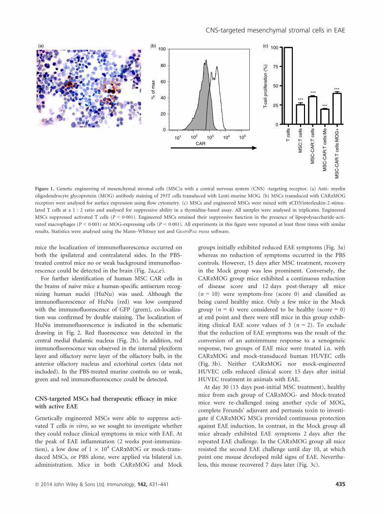
Engineered MSCs retain their ability to suppress Tcells in vitro
We constructed an scFv antibody domain from a hybrid-
oma producing anti-rat MOG antibody that cross-reacted
with murine MOG as evident by staining of murine
MOG-transduced cells (Fig. 1a). The MOG scFv construct
was introduced into a murine conventional CAR and the
CARaMOG construct was inserted into a lentiviral vector
system. Human MSCs were engineered by transducing
MSCs with the CARaMOG lentiviral vectors. Transduc-
tion efficiency ranged from 60% to 89% measured with a
j-chain-specific antibody and exemplified in Fig. 1(b)
depicting flow cytometric immunostaining. Engineered
MSCs were equally good as unmodified MSCs in sup-
pressing proliferation of polyclonally stimulated T cells
(P < 0�001) at a 1 : 2 ratio (Fig. 1c). Furthermore, when
exposing engineered MSCs to MOG+ cells or activated
macrophages (for phenotype expression see Supporting
information, Fig. S1) they were still able to suppress
T-cell proliferation (P < 0�001).
Intranasal administration effectively delivers MSCs tothe brain
Mesenchymal stromal cells co-expressing GFP and CARa-MOG were used to evaluate in vivo targeting following i.n.
cell delivery in naive mice after 24 hr. The overall localiza-
tion of GFP immunofluorescence is depicted in the upper
schematic drawing in Fig. 2. GFP-fluorescence was mainly
localized in clusters of cells in the internal plexiform layer
of the olfactory bulb and anterior olfactory nucleus
(Fig. 2b), ectorhinal cortex (Fig. 2d) and in the Purkinje
cell layer of the cerebellum (Fig. 2f). In addition, signals
were observed in the central medial genic nucleus and
ventral orbital cortex, lateral septal nucleus and central
medial thalamic nucleus, respectively (data not included).
Immunofluorescence was only observed in the soma and
was preferentially present in the perinuclear part.
Although a unilateral dose of MSC was given in naive
ª 2014 John Wiley & Sons Ltd, Immunology, 142, 431–441434
M. Fransson et al.
mice the localization of immunofluorescence occurred on
both the ipsilateral and contralateral sides. In the PBS-
treated control mice no or weak background immunofluo-
rescence could be detected in the brain (Fig. 2a,c,e).
For further identification of human MSC CAR cells in
the brains of naive mice a human-specific antiserum recog-
nizing human nuclei (HuNu) was used. Although the
immunofluorescence of HuNu (red) was low compared
with the immunofluorescence of GFP (green), co-localiza-
tion was confirmed by double staining. The localization of
HuNu immunofluorescence is indicated in the schematic
drawing in Fig. 2. Red fluorescence was detected in the
central medial thalamic nucleus (Fig. 2h). In addition, red
immunofluorescence was observed in the internal plexiform
layer and olfactory nerve layer of the olfactory bulb, in the
anterior olfactory nucleus and ectorhinal cortex (data not
included). In the PBS-treated murine controls no or weak,
green and red immunofluorescence could be detected.
CNS-targeted MSCs had therapeutic efficacy in micewith active EAE
Genetically engineered MSCs were able to suppress acti-
vated T cells in vitro, so we sought to investigate whether
they could reduce clinical symptoms in mice with EAE. At
the peak of EAE inflammation (2 weeks post-immuniza-
tion), a low dose of 1 9 104 CARaMOG or mock-trans-
duced MSCs, or PBS alone, were applied via bilateral i.n.
administration. Mice in both CARaMOG and Mock
groups initially exhibited reduced EAE symptoms (Fig. 3a)
whereas no reduction of symptoms occurred in the PBS
controls. However, 15 days after MSC treatment, recovery
in the Mock group was less prominent. Conversely, the
CARaMOG group mice exhibited a continuous reduction
of disease score and 12 days post-therapy all mice
(n = 10) were symptom-free (score 0) and classified as
being cured healthy mice. Only a few mice in the Mock
group (n = 4) were considered to be healthy (score = 0)
at end point and there were still mice in this group exhib-
iting clinical EAE score values of 3 (n = 2). To exclude
that the reduction of EAE symptoms was the result of the
conversion of an autoimmune response to a xenogeneic
response, two groups of EAE mice were treated i.n. with
CARaMOG and mock-transduced human HUVEC cells
(Fig. 3b). Neither CARaMOG nor mock-engineered
HUVEC cells reduced clinical score 15 days after initial
HUVEC treatment in animals with EAE.
At day 30 (15 days post-initial MSC treatment), healthy
mice from each group of CARaMOG- and Mock-treated
mice were re-challenged using another cycle of MOG,
complete Freunds’ adjuvant and pertussis toxin to investi-
gate if CARaMOG MSCs provided continuous protection
against EAE induction. In contrast, in the Mock group all
mice already exhibited EAE symptoms 2 days after the
repeated EAE challenge. In the CARaMOG group all mice
resisted the second EAE challenge until day 10, at which
point one mouse developed mild signs of EAE. Neverthe-
less, this mouse recovered 7 days later (Fig. 3c).
100 100
50
75
25
0
T c
ells
MS
C:T
cel
ls
MS
C-C
AR
:T c
ells
MS
C-C
AR
:T c
ells
:Mφ
MS
C-C
AR
:T c
ells
:MO
G+
T-ce
ll pr
olife
ratio
n (%
)80
60
40% o
f max
20
0
101 102 103
CAR104 105
***
******
***
(a) (b) (c)
Figure 1. Genetic engineering of mesenchymal stromal cells (MSC)s with a central nervous system (CNS) -targeting receptor. (a) Anti- myelin
oligodendrocyte glycoprotein (MOG) antibody staining of 293T cells transduced with Lenti-murine MOG. (b) MSCs transduced with CARaMOG
receptors were analysed for surface expression using flow cytometry. (c) MSCs and engineered MSCs were mixed with aCD3/interleukin-2-stimu-
lated T cells at a 1 : 2 ratio and analysed for suppressive ability in a thymidine-based assay. All samples were analysed in triplicates. Engineered
MSCs suppressed activated T cells (P < 0�001). Engineered MSCs retained their suppressive function in the presence of lipopolysaccharide-acti-
vated macrophages (P < 0�001) or MOG-expressing cells (P < 0�001). All experiments in this figure were repeated at least three times with similar
results. Statistics were analysed using the Mann–Whitney test and GRAPHPAD PRISM software.
ª 2014 John Wiley & Sons Ltd, Immunology, 142, 431–441 435
CNS-targeted mesenchymal stromal cells in EAE

CNS-directed MSCs modulate biomarkers in activeEAE
Upon immunohistochemical examination using markers
for reactive astrogliosis (GFAP) and myelination (MBP),
axonal recovery was confirmed in mice in the CARa-MOG group 15 days following i.n. administration of
MSC. Reactive astrogliosis was evaluated in the corpus
callosum (Fig. 4a–c), brainstem (Fig. 4d–f), cerebellum
(Fig. 4g–i), olfactory bulb (see Supporting information,
Fig. S2a–c) and hippocampus (Fig. S2d–f). An increased
GFAP staining was detected in MSC CARaMOG- or
Mock-treated EAE mice but not in PBS-treated EAE
mice. The level of staining was higher in CARaMOG-
treated EAE mice compared with Mock-treated EAE
mice in the cerebellum, brainstem and corpus callosum,
whereas the level of staining was similar in the hippo-
campus and olfactory bulb of CARaMOG-treated and
Mock-treated EAE mice.
Expression of MBP was evaluated in the hippocampus
(Fig. 4j–l), brainstem (Fig. 4m–o), cerebellum (Fig. 4p–r),olfactory bulb (Fig. S2g–i) and corpus callosum (Fig. S2j–l). The degree of demyelination, as indicated by the loss
of MBP staining, was greater in PBS-treated EAE mice
compared with CARaMOG-treated mice in all areas
except in the cerebellum and brainstem, where staining
intensity was similar.
PBS MSC CAR
PBS MSC CAR GFP HuNuAB
G
CDEF
HHuNuGFP
(a) (b)
(c)
(e)
(g)
(f)
(h)
(d)
Figure 2. Brain localization of central nervous
system (CNS) -targeted mesenchymal stromal
cells (MSC)s in naive mice following intranasal
delivery. Human MSCs were instilled via a
unilateral intransal (i.n.) administration and
the distribution of green fluorescent protein
(GFP) or human nuclei (HuNu) immunofluo-
rescence in horizontal cryosections of the brain
of naive mice was studied 24 hr after delivery.
The schematic depicts a selective GFP and
HuNu immunofluorescence (indicated by
green and red spots) in various brain regions.
Confocal microscopy reveals that GFP immu-
nofluorescence (green) is present in the inter-
nal plexiform layer of the olfactory bulb (b)
ectorhinal cortex (d) and Purkinje cells in the
cerebellum (f) in MSC CARaMOG-treated
naive mice. The corresponding areas in PBS-
treated naive mice are (a) ,(c,) and (e), respec-
tively. Cell nuclei (blue) are stained with DAPI.
Original magnifications 109 (a–f) and 409 (g,
h). Immunofluorescence microscopy reveals
that GFP fluorescence (green) and HuNu fluo-
rescence (red) are both present in the ectorhi-
nal cortex.
ª 2014 John Wiley & Sons Ltd, Immunology, 142, 431–441436
M. Fransson et al.

The MBP staining in the brain of MSC Mock-treated
EAE mice was lower compared with in MSC CAR-treated
and PBS-treated mice. Damage to axons was evaluated in
the cerebellum and brainstem.
CNS-infiltrating T cells have decreased interferon-cresponses but produce interleukin-17
T cells recovered from the spleens and brains of MSC
CARaMOG- and Mock-treated EAE mice were restimu-
lated in vitro to establish T-cell activation status. T cells
were stimulated with a mix of MHC I- and MHC
II-binding MOG peptides and evaluated by flow cytome-
try for secretion of interleukin-17 and interferon-c(Fig. 5a,b). Brain T cells from EAE mice treated with
either CARaMOG or Mock MSCs exhibited a low secre-
tion of interferon-c from both CD4+ and CD8+ T cells
whereas interleukin-17 was produced by the CD4+ popu-
lation. T cells isolated from the spleens of both experi-
mental groups responded to polyclonal stimuli, indicating
that peripheral T cells were not anergized and that the
suppressive effects of MSCs are restricted to the brain.
Brain biopsies of EAE mice were further analysed for
T helper type 1 cytokines (interferon-c and interleukin-
12) using real time PCR. While PBS-treated mice had
higher levels of both interferon-c and interleukin-12 tran-
scripts, EAE mice treated with either CARaMOG or
Mock MSCs had only low levels of these cytokines,
although these differences did not reach statistical signifi-
cance (Fig. 5c,d).
CNS-targeted MSCs, but not Mock MSCs, efficientlytreat EAE upon i.p. delivery
To investigate if the targeted MSCs had a therapeutic
effect following systemic administration, a low dose of
cells (1 9 104 CARaMOG or Mock-transduced MSCs)
was infused i.p. in EAE mice. CARaMOG MSCs were
able to reduce EAE symptoms whereas the symptoms in
Mock MSCs were comparable to those in PBS-treated
EAE mice (Fig. 6).
Discussion
In the current study we have demonstrated how MSCs
can be genetically engineered and targeted to the CNS.
4·0 Diseaseprogression PBS
MSC CARMSC Mock
MSC CAR
MSC Mock
3·5
3·0
2·5
2·0
1·5
0·5
1·0Mea
n E
AE
sco
re
0·00 4 8 12 15 17 19 21 23 25 27 29
CFA
No. of days
Treatment10 000 cells (in)+PT
4·0 Diseaseprogression
PBSHUVEC CAR
HUVEC Mock3·5
3·0
2·5
2·0
1·5
0·5
1·0Mea
n E
AE
sco
re
0·00 10 15 17 19 21 23 25 27
CFA
No. of days
Treatment10 000 cells (in)+PT
4·0
3·5
3·0
2·5
2·0
1·5
0·5
1·0Mea
n E
AE
sco
re
0·00 2 4 6 8 10 12 14 16 18 20
CFA
No. of days
+PT
*
**
*
(a)
(b)
(c)
Figure 3. Efficacy of central nervous system (CNS) -targeted mesen-
chymal stromal cells (MSC)s in experimental autoimmune encepha-
lomyelitis (EAE) mice. (a) Ten EAE mice in three groups were given
a low dose (1 9 104) of engineered CARaMOG or Mock MSCs or
PBS alone by intranasal (i.n.) administration at the peak of EAE
inflammation (15 days post-EAE immunization). Ten days after i.n.
MSC treatment all mice in the CARaMOG group were symptom-
free (**P < 0�01). At end-point (15 days after i.n. MSC treatment)
mice in the mock-treated group still exhibited EAE symptoms
(P < 0�05). The experiment was repeated three times with similar
results. (b) To exclude that treatment efficacy was due to a xenoge-
neic response of the human MSC, mice were treated with human
HUVEC cells equipped with the CARaMOG targeting receptor. Six
mice in three groups were given a low dose (1 9 104) of engineered
CARaMOG or Mock HUVECs, or PBS alone by i.n. delivery at the
peak of EAE inflammation (15 days post-EAE immunization). (c)
Cured EAE mice from each treatment group of i.n. MSCs (n = 6)
were given a second EAE immunization (as described previously).
MSC CARaMOG-treated mice were able to resist EAE inflammation
longer than MSC-mock-treated mice (*P < 0�05). Statistics were anal-ysed using the Mann–Whitney U-test and GRAPHPAD PRISM software.
ª 2014 John Wiley & Sons Ltd, Immunology, 142, 431–441 437
CNS-targeted mesenchymal stromal cells in EAE

Engineered MSCs potently reduce disease symptoms in an
EAE model followed by i.n. delivery. Furthermore, mice
that recovered from EAE symptoms by CARaMOG MSC
treatment were resistant to subsequent MOG antigen chal-
lenge while recovered mice that received non-targeted
MSCs rapidly developed EAE after being re-challenged.
Chimeric antigen receptors are currently used to retar-
get cytotoxic T cells to tumour antigens, creating
tumour-reactive T cells for cell therapy of cancer.32 We
constructed a similar CAR to generate MOG-targeting
MSCs and demonstrated proof-of-principle of their func-
tion. The MOG targeting CAR has previously been suc-
cessfully used to retarget T regulatory cells to the brain in
the EAE model.33 Genetic engineering of MSCs did not
affect their immunosuppressive capacity in our in vitro
model of inflammation where engineered MSCs were
equally good in suppressing activated T cells and macro-
phages as unmodified MSCs.
Due to immune modulatory differences in the murine
and human MSC population,34 human MSC were used in
the current study. Zhang et al.15 have previously demon-
strated that intravenous administration of human MSC
improved the clinical course of proteolipid protein-
induced EAE while human MSCs have been shown to be
rejected in a murine model of islet transplantation.35
These discrepancies may be due to disease settings or
because the brain is an immune privileged site.
Recent data from early clinical trials treating multiple
sclerosis patients with MSCs have revealed poor infiltra-
tion of cells into the CNS following either intravenous or
intrathecal injections.24 In an attempt to attain therapeu-
tically effective numbers of MSC in the brain we delivered
engineered cells via the nasal mucosa. Intranasal delivery
has shown potential for transplantation of cells into the
brain and may be a means of reducing the cell doses
required for therapeutic efficacy while at the same time
decreasing systemic exposure.36–39 We demonstrated that
it was possible to treat EAE mice with a single, low dose
of engineered MSCs and yet still achieve therapeutic effi-
cacy when the cells were delivered i.n. The low numbers
of cells used in our i.n. protocol are equivalent to a dose
of 0�5 9 105 MSC/kg in humans, a number that could
easily be obtained using current clinical production pro-
tocols of human MSCs.
PBS
GFAP
GFAP
GFAP
MBP
MBP
MBP
MSC Mock MSC CAR
(a) (b) (c)
(d) (e) (f)
(g) (h) (i)
(j) (k) (l)
(m) (n) (o)
(p) (q) (r)
Figure 4. Effects of intranasally delivered CNS-
targeted mesenchymal stromal cells (MSCs) on
axonal damage and tissue recovery in experi-
mental autoimmune encephalomyelitis (EAE)
mice. Immunohistochemical staining for glial
acidic fibrillary protein (GFAP) in corpus
callosum (a–c), brainstem (d–f) and cerebel-
lum (g–i) in brain sections from PBS-, MSC
Mock- and MSC CARaMOG-treated EAE mice
(15 days after i.n. treatment). In MSC CARa-MOG-treated EAE mice there is strong staining
in all areas (c,f,i). In MSC Mock-treated mice
there is strong staining in all areas (b,e,h). In
PBS-treated EAE mice there is weak staining
(a,d,g). Immunohistochemical staining for
myelin basic protein (MBP) in hippocampus
(j–l), brainstem (m–o) and cerebellum (p–r) in
brain sections from PBS-treated, MSC Mock-
treated and MSC CARaMOG-treated EAE
mice. In MSC CARaMOG-treated EAE mice
there is strong staining of all areas (l,o,r). In
MSC Mock-treated mice there is weak staining
in all areas (k,n,q). In PBS-treated EAE mice
there is moderate staining in hippocampus (j)
and strong staining in brainstem and cerebel-
lum (m,p). Original magnification 109.
ª 2014 John Wiley & Sons Ltd, Immunology, 142, 431–441438
M. Fransson et al.

Upon i.p. delivery, CARaMOG MSCs were able to
decrease disease symptoms at the same low cell doses
used in the i.n. study protocol while the Mock MSCs did
not. It is possible that a number of cells were rejected
because of xenogeneic responses to the i.p. injected cells
but as a result of the expression of the CARaMOG recep-
tor, sufficient numbers of cells might have been retained
in the CNS to exert a therapeutic effect. Reduced clinical
disease symptoms correlated with reduced damage to
axons were exemplified by immunohistochemical analyses
of MBP.
As it has been demonstrated that human MSCs
improve the clinical course of EAE in mice15,40 it was
expected that mice treated with both CARaMOG MSCs
and non-targeted MSCs would show reduced symptoms
of EAE. However, mice that recovered from EAE symp-
toms by CARaMOG MSC treatment were resistant to
subsequent MOG antigen challenge while recovered mice
that received non-targeted MSCs rapidly developed EAE
following re-challenge. This may be due to the ability of
targeted MSCs to reside in the brain post-inflammation
while non-targeted cells may migrate to other sites. It was
not possible to detect GFP-positive cells 15 days after
MSC treatment. The difference in treatment efficacy
between targeted and non-targeted MSCs might be due to
a biological reprogramming effect such as induction of
T regulatory cells at site. However, this issue remains to
be investigated through in vivo tracking of cells and bio-
logical markers, such as cytokines, over time.
Previous experimental studies using i.n. administration
of stem cells have indicated that their delivery into the
brain is relatively low.37,38 However, brain sections of
naive mice treated with GFP-labelled CARaMOG MSCs
suggest a localization of GFP immunofluorescence in var-
ious brain areas (such as the olfactory bulb) following i.n.
administration in naive mice after 24 hr. To confirm the
presence of human MSCs at these sites, additional immu-
nohistochemical studies were performed using an anti-
body specific for human nuclei.41 We revealed weak
HuNu staining in the same areas but both the number of
cells and the staining intensity were markedly lower than
that observed with GFP immunofluorescence. The
increased GFP immunofluorescence could be a result of
the MSCs’ capacity to transfer material and interact with
other cells. Previous reports suggest a fusion of bone-
marrow-derived cells with various epithelial cell types42–44
while others have demonstrated that MSCs can transfer
genetic material to neighbouring cells via microvesicles.45
20
15
10
5
0Res
pond
ing
T c
ells
(%
) 20
15
10
5
0
25
20
15
10
5
0IL12
p40
mR
NA
cop
y nu
mbe
r
Res
pond
ing
T c
ells
(%
)
0·25
0·20
0·15
0·10
0·05
0·00
PB
S
MS
C C
AR
MS
C M
ock
PB
S
MS
C C
AR
MS
C M
ock
IFN
g m
RN
A c
opy
num
ber
CD8+ IF
Ng+
CD4+ IF
Ng+
CD4+ IL
17+
CD8+ IF
Ng+
CD4+ IF
Ng+
CD4+ IL
17+
MSC CARMSC Mock MSC CAR
MSC Mock
Brain Spleen(a) (b)
(c) (d)
Figure 5. Immunological evaluation of intranasal mesenchymal stro-
mal cell (MSC) treatment efficacy. T cells from brain (a, n = 3) and
spleen (b, n = 3) from MSC CARaMOG and MSC Mock-treated
experimental autoimmune encephalomyelitisEAE mice were isolated
using a MACS T-cell separation kit, stimulated for 24 hr and
analysed for cytokine production [interleukin-17 (IL-17) and inter-
feron-c (IFN-c)]. T cells isolated from brain were stimulated with
myelin oligodendrocyte glycoprotein (MOG) peptides (MOG37–46,
MOG35–55) whereas T cells isolated from the spleen were stimulated
with aCD3/IL-2. In the brain CD4+ T cells responded by secreting
IL17 to a higher extent in the MSC Mock-treated mice compared
with MSC CARaMOG-treated mice; however, the difference was not
significant. Effector cytokines (c: IFN-c, d: IL-12) were analysed by
quantitative PCR from brain biopsies obtained from MSC CARa-MOG-treated and MSC Mock-treated mice (15 days post-MSC treat-
ment).
3·5 Diseaseprogression3·0
2·5
2·0
1·5
1·0
0·5
0·00 10 15 17 19 21 23 25 27 29
PBS
MSC CARMSC Mock
CFA+PT
Treatment10 000 cells (ip)
Mea
n E
AE
sco
re
** *
Figure 6. Efficacy of systemic delivery of central nervous system
(CNS) -targeted mesenchymal stromal cells (MSC)s in experimental
autoimmune encephalomyelitis (EAE) mice. Ten mice in three
groups were given a low dose (1 9 104) of CARaMOG or mock
MSCs or PBS alone by intraperitoneal (i.p.) infusion at peak of EAE
inflammation (15 days post-EAE immunization). Ten days post-
MSC treatment all mice in the MSC CARaMOG group were cured
(**P < 0�01). At end-point (15 days post-treatment) 7/10 mice in
the mock-treated group still exhibited EAE symptoms (*P < 0�05).Statistics were analysed using the Mann–Whitney U-test and GRAPH-
PAD PRISM software. The challenge experiment was repeated twice
with similar results.
ª 2014 John Wiley & Sons Ltd, Immunology, 142, 431–441 439
CNS-targeted mesenchymal stromal cells in EAE

Considerably, this might be a reason that GFP immuno-
fluorescence could be detected to a markedly higher
extent than human nuclear events (HuNu). Further stud-
ies are therefore needed to characterize the migration,
identity and long-term survival of engineered MSCs deliv-
ered via the i.n. route into the brain.
The olfactory pathways, located in the posterior nasal
cavity, may provide a port of entry for drugs, metals and
environmental pollutants into the brain because the olfac-
tory neurons have dendrites projecting into the nasal
mucus and axons projecting into the olfactory bulb.46,47
Therapeutic MSCs deposited locally on the olfactory epi-
thelium may therefore circumvent the blood–brain barrier
and pass the cribriform plate of the ethmoid bone to the
olfactory bulb in extracellular channels made up of olfac-
tory ensheathing cells. However, in our studies, GFP- and
HuNu-positive immmunofluorescence staining were
observed after 24 hr on both the ipsilateral and contralat-
eral sides following a unilateral application of engineered
MSCs, suggesting that a migration of MSCs to the brain
may not be confined to the olfactory pathways or that
there is migration within the brain following an initial
olfactory migration. In addition, migration of MSCs from
the nasal mucosa into the general blood circulation can-
not be excluded because the nasal mucosa is highly vascu-
larized.48,49
A potential risk of MSC treatment is their ability to
suppress anti-tumour surveillance that could result in
tumour progression and metastasis in the same manner
as that evident in patients subjected to life-long immuno-
suppressive drugs post-transplantation. A further risk
associated with MSC expansion in vitro is subsequent in
vivo differentiation into malignant cells as observed upon
in vivo administration in rodents.50 Nonetheless, there is
general agreement that bone-marrow-derived human
MSCs can safely be expanded in vitro with limited risk of
malignant transformation.51,52 By inserting an inducible
suicide gene in trans with CARaMOG the engineered
MSCs would provide a safer option than regular MSCs.
In conclusion, we have developed CNS-targeting MSCs
that efficiently reduce EAE symptoms in mice following
i.n. delivery. MSC-derived proteins were detected in
selected areas of the brain upon i.n. cell delivery and fur-
thermore, we have revealed that targeting of the MSCs
results in sustained treatment efficacy because engineered
MSC-treated EAE mice cannot be provoked to show new
EAE symptoms after re-challenge. Targeted MSCs may
therefore be an interesting therapeutic option for multiple
sclerosis as well as for other organ-specific autoimmune
conditions.
Acknowledgements
The authors thank Dr Lu at Pittsburgh University for
teaching us the EAE model and Mrs Raili Engdahl and
Berith Nilsson for technical assistance during animal
experiments and viral vector production, respectively.
This study was supported by grants to Dr Loskog from
the Swedish Research Council and the Medical Faculty at
Uppsala University, to Dr Fransson, from the G€oransson-
Sandviken fund.
Disclosures
The authors declare no conflict of interest except from
Dr Loskog who is the CEO of Lokon Pharma AB, scien-
tific advisor of NEXTTOBE AB and has a royalty agree-
ment with Alligator Biosciences AB. None of these have
an economical relation/conflict with the results presented
herein.
References
1 Bernado ME, Pagliara D, Locatelli F. Mesenchymal stromal cell therapy: a revolution in
regenerative medicine? Bone Marrow Transplant 2012; 47:164–71.
2 Pittenger MF, Mackay AM, Beck SC et al. Multilineage potential of adult human mes-
enchymal stem cells. Science 1999; 284:143–7.
3 Keating A. Mesenchymal stromal cells. Curr Opin Hematol 2006; 13:419–25.
4 Uccelli A, Moretta L, Pistoia V. Immunoregulatory function of mesenchymal stem cells.
Eur J Immunol 2006; 36:2566–73.
5 Le Blanc K, Tammik L, Sundberg B, Haynesworth SE, Ringden O. Mesenchymal
stem cells inhibit and stimulate mixed lymphocyte cultures and mitogenic responses
independently of the major histocompatibility complex. Scand J Immunol 2003;
57:11–20.
6 Aggarwal S, Pittenger MF. Human mesenchymal stem cells modulate allogeneic
immune cell responses. Blood 2005; 105:1815–22.
7 Beyth S, Borovsky Z, Mevorach D et al. Human mesenchymal stem cells alter antigen-
presenting cell maturation and induce T-cell unresponsiveness. Blood 2005; 105:2214–9.
8 Bartholomew A, Sturgeon C, Siatskas M et al. Mesenchymal stem cells suppress lym-
phocyte proliferation in vitro and prolong skin graft survival in vivo. Exp Hematol 2002;
30:42–8.
9 Rasmusson I, Ringden O, Sundberg B, Le Blanc K. Mesenchymal stem cells inhibit lym-
phocyte proliferation by mitogens and alloantigens by different mechanisms. Exp Cell
Res 2005; 305:33–41.
10 Augello A, Tasso R, Negrini SM et al. Bone marrow mesenchymal progenitor cells inhi-
bit lymphocyte proliferation by activation of the programmed death 1 pathway. Eur J
Immunol 2005; 35:1482–90.
11 Prevosto C, Zancolli M, Canevali P, Zocchi MR, Poggi A. Generation of CD4+ or
CD8+ regulatory T cells upon mesenchymal stem cell–lymphocyte interaction. Haema-
tologica 2007; 92:881–8.
12 Chen J, Li Y, Wang L, Lu M, Zhang X, Chopp M. Therapeutic benefit of intracerebral
transplantation of bone marrow stromal cells after cerebral ischemia in rats. J Neurol
Sci 2001; 189:49–57.
13 Li Y, Chen J, Chen XG et al. Human marrow stromal cell therapy for stroke in rat:
neurotrophins and functional recovery. Neurology 2002; 59:514–23.
14 Gerdoni E, Gallo B, Casazza S et al. Mesenchymal stem cells efficiently modulate patho-
genic immune response in experimental autoimmune encephalomyelitits. Ann Neurol
2007; 61:219–27.
15 Zhang J, Li Y, Chen J et al. Human bone marrow stromal cell treatments improves
neurological functional recovery in EAE mice. Exp Neurol 2005; 195:16–26.
16 Chopp M, Zhang XH, Li Y et al. Spinal cord injury in rat: treatment with bone marrow
stromal cell transplantation. NeuroReport 2000; 11:3001–5.
17 Schwarz EJ, Alexander GM, Prockop DJ, Azizi SA. Multipotential marrow stromal cells
transduced to produce L-DOPA: engraftment in a rat model of Parkinson disease. Hum
Gene Ther 1999; 10:2539–49.
18 Uccelli A, Pistoia V, Moretta L. Mesenchymal stem cells: a new strategy for immuno-
suppression? Trends Immunol 2007; 28:219–26.
19 Le Blanc K, Rasmusson I, Sundberg B et al. Treatment of severe acute graft-versus-host
disease with third party haploidentical mesenchymal stem cells. Lancet 2004; 363:1439–
41.
20 Tolar J, Le Blanc K, Keating A, Blazar BR. Concise review: hitting the right spot with
mesenchymal stromal cells. Stem Cells 2010; 28:1446–55.
ª 2014 John Wiley & Sons Ltd, Immunology, 142, 431–441440
M. Fransson et al.

21 Kassis I, Grigoriadis N, Gowda-Kurkalli B et al. Neuroprotection and immunomodula-
tion with mesenchymal stem cells in chronic experimental autoimmune encephalomy-
elitis. Arch Neurol 2008; 65:753–61.
22 Mohyeddin Bonab M, Yazdanbakhsh S, Lotfi J et al. Does mesenchymal stem cell ther-
apy help multiple sclerosis patients? Report of a pilot study. Iran J Immunol 2007;
4:50–7.
23 Yamout B, Hourani R, Salti H et al. Bone marrow mesenchymal stem cell transplanta-
tion in patients with multiple sclerosis: a pilot study. J Neuroimmunol 2010; 227:185–9.
24 Karussis D, Karageorgiou C, Vaknin-Dembinsky A et al. Safety and immunological
effects of mesenchymal stem cell transplantation in patients with multiple sclerosis and
amyotrophic lateral sclerosis. Arch Neurol 2010; 67:1187–94.
25 Barhum Y, Gai-Castro S, Bahat-Stromza M, Barzilay R, Melamed E, Offen D. Intracere-
broventricular transplantation of human mesenchymal stem cells induced to secrete
neurotrophic factors attenuates clinical symptoms in a mouse model of multiple sclero-
sis. J Mol Neurosci 2010; 41:129–37.
26 Bai L, Lennon DP, Eaton V et al. Human bone marrow-derived mesenchymal stem
cells induce Th2-polarized immune response and promote endogenous repair in animal
models of multiple sclerosis. Glia 2009; 57:1192–203.
27 Uccelli A, Prockop DJ. Why should mesenchymal stem cells (MSCs) cure autoimmune
diseases? Curr Opin Immunol 2010; 22:768–74.
28 Wang Y, Xu F, Zhang C et al. High MR sensitive fluorescent magnetite nanocluster for
stem cell tracking in ischemic mouse brain. Nanomedicine 2011; 7:1009–19.
29 Gordon D, Pavlovska G, Glover CP, Uney JB, Wraith D, Scolding NJ. Human mesen-
chymal stem cells abrogate experimental allergic encephalomyelitis after intraperitoneal
injection, and with sparse CNS infiltration. Neurosci Lett 2008; 448:71–3.
30 Breithaupt C, Schubart A, Zander H, Skerra A, Huber R, Linington C, Jacob U. Struc-
tural insights into the antigenicity of myelin oligodendrocyte glycoprotein. Proc Natl
Acad Sci USA 2003; 100:9446–51.
31 Pule M, Finney H, Lawson A. Artificial T-cell receptors. Cytotherapy 2003; 5:211–26.
32 Park TS, Rosenberg SA, Morgan RA. Treating cancer with genetically engineered T cells.
Trends Biotechnol 2011; 29:550–7.
33 Fransson M, Piras E, Burman J et al. CAR/FoxP3-engineered T regulatory cells target
the CNS and suppress EAE upon intranasal delivery. J Neuroinflammation 2012; 9:112.
34 Meisel R, Brockers S, Heseler K et al.. Human but not murine multipotent mesenchy-
mal stromal cells exhibit antimicrobial effector function mediated by indoleamine 2,3-
dioxygenase. Leukemia 2011; 25:648–54.
35 Johansson U, Rasmusson I, Niclou SP et al. Formation of composite endothelial cell–
mesenchymal stem cell islets: a novel approach to promote islet revasularization. Diabe-
tes 2008; 57:2393–401.
36 Jiang Y, Zhu J, Xu G, Liu X. Intranasal delivery of stem cells to the brain. Expert Opin
Drug Deliv 2011; 8:623–32.
37 Danielyan L, Sch€afer R, von Ameln-Mayerhofer A et al. Intranasal delivery of cells to
the brain. Eur J Cell Biol 2009; 88:315–24.
38 van Velthoven CT, Kavelaars A, van Bel F, Heijnen CJ. Nasal administration of stem
cells: a promising novel route to treat neonatal ischemic brain damage. Pediatr Res
2010; 68:419–22.
39 Danielyan L, Sch€afer R, von Ameln-Mayerhofer A et al. Therputic efficacy of intrana-
sally delivered mesenchymal stromal cells in a rat model of Parkinson’s disease. Rejuve-
nation Res 2011; 1:3–16.
40 Hou Y, Ryo CH, Park KY, Kim SM, Jeong CH, Jeun SS. Effective combination of
human bone marrow mesenchymal stem cells and minocycline in experimental autoim-
mune encephalomyelitis mice. Stem Cell Res 2013; 4:77.
41 Englund U, Bj€orklund A, Wictorin K. Migration patterns and phenotypic differentiation
of long-term expanded human neural progenitor cells after transplantation into the
adult rat brain. Brain Res Dev Brain Res 2002; 134:123–41.
42 Nern C, Wolff I, Macas J et al. Fusion of hematopoietic cells with Purkinje neurons
does not lead to stable heterokaryon formation under noninvasive conditions. J Neuro-
sci 2009; 29:3799–807.
43 Alvarez-Dolado M, Pardal R, Garcia-Verdugo JM et al. Fusion of bone-marrow-derived
cells with Purkinje neurons, cardiomyocytes and hepatocytes. Nature 2003; 425:968–73.
44 Kemp K, Gordon D, Wraith DC et al. Fusion between human mesenchymal stem cells
and rodent cerebellar Purkinje cells. Neuropathol Appl Neurobiol 2010; 37:166–78.
45 Iglesias D, El-Kares R, Taranta A et al. Stem cell microvesicles transfer cystinosin to
human cystinotic cells and reduce cystine accumulation in vitro. PLoS ONE 2012; 7:
e42840.
46 Westin U, Piras E, Jansson B et al. Transfer of morphine along the olfactory pathway
to the central nervous system after nasal administration to rodents. Eur J Pharm Sci
2005; 24:565–73.
47 Illum L. Transport of drugs from the nasal cavity to the central nervous system. Eur
J Pharm Sci 2000; 11:1–18.
48 Matsushita T, Kibayashi T, Katayama T et al. Mesenchymal stem cells transmigrate
across brain microvascular endothelial cell monolayers through transiently formed
inter-endothelial gaps. Neurosci Lett 2011; 502:41–5.
49 Kopen GC, Prockop DJ, Phinney DG. Marrow stromal cells migrate throughout fore-
brain and cerebellum, and they differentiate into astrocytes after injection into neonatal
mouse brains. Proc Natl Acad Sci USA 1999; 96:10711–6.
50 Tolar J, Nauta AJ, Osborn MJ et al. Sarcoma derived from cultured mesenchymal stem
cells. Stem Cells 2007; 25:371–9.
51 Bernardo ME, Zaffaroni N, Novara F et al. Human bone marrow derived mesenchymal
stem cells do not undergo transformation after long-term in vitro culture and do not
exhibit telomere maintenance mechanisms. Cancer Res 2007; 67:9142–9.
52 Pockop DJ, Brenner M, Fibbe WE et al. Defining the risks of mesenchymal stromal cell
therapy. Cytotherapy 2010; 12:576–8.
Supporting Information
Additional Supporting Information may be found in the
online version of this article:
Figure S1. Phenotype of lipopolysaccharide-activated
macrophages.
Figure S2. Glial fibrillary acidic protein (GFAP) and
myelin basic protein (MBP) staining of the olfactory bulb
and hippocampus.
ª 2014 John Wiley & Sons Ltd, Immunology, 142, 431–441 441
CNS-targeted mesenchymal stromal cells in EAE
Copyright © 2022 FDOKUMEN