Glial reactions and degeneration of myelinated processes in spinal cord gray matter in chronic...
11
GLIAL REACTIONS AND DEGENERATION OF MYELINATED PROCESSES IN SPINAL CORD GRAY MATTER IN CHRONIC EXPERIMENTAL AUTOIMMUNE ENCEPHALOMYELITIS J. WU, M. OHLSSON, E. A. WARNER, K. K. LOO, T. X. HOANG, R. R. VOSKUHL AND L. A. HAVTON* Department of Neurology, David Geffen School of Medicine at Univer- sity of California, Los Angeles, Charles Young Drive South, Los An- geles, CA 90095, USA Abstract—Multiple sclerosis and experimental autoimmune en- cephalomyelitis (EAE) result in inflammatory white matter le- sions in the CNS. However, information is sparse with regard to the effects of autoimmune demyelinating disease on gray mat- ter regions. Therefore, we studied the late effects of chronic EAE in C57BL/6 mice on the spinal cord gray matter using immunohistochemistry. Here, EAE induced marked astrocytic, microglial, and macrophage activation in the ventral horn gray matter, without any motoneuron loss. Activated caspase-3 was also increased in the ventral horn gray matter. Furthermore, activated poly (ADP-ribose) polymerase (PARP), another apo- ptotic marker, co-localized with myelin basic protein (MBP) of oligodendrocyte processes, but not with the oligodendroglial cell body marker, adenomatous polyposis coli gene clone CC1 (APC-CC1), or with neurofilament marker (RT-97) or synapto- physin of axonal arbors. However, there was no associated increase in the number of terminal deoxynucleotidyl transferase (TdT) mediated-dUTP nick end labeling positive nuclei in the spinal cord gray matter of EAE mice. In addition, co-localization of MBP and the low-affinity neurotrophin receptor, p75, was demonstrated, further supporting the notion of apoptotic oligo- dendrocyte process degeneration in the gray matter of EAE mice. © 2008 IBRO. Published by Elsevier Ltd. All rights reserved. Key words: multiple sclerosis, inflammation, mouse, degen- eration, demyelination. Multiple sclerosis (MS) is an immune-mediated disorder, which affects the CNS with inflammatory lesions and demyelination ( Lucchinetti et al., 1998 ). In addition to the focal destruction of myelin in the white matter tracts of the brain and spinal cord, MS lesions may also exhibit transected axons ( Trapp et al., 1998 ). The latter findings are of particular clinical importance, as neurological dis- ability has been correlated with axonal loss in the spinal cord of chronic MS patients ( Bjartmar et al., 2000 ). Interestingly, in experimental autoimmune encephalomyelitis (EAE), an animal model for MS, a similar axonal loss in spinal cord white matter is correlated with permanent neurological disability ( Wujek et al., 2002 ). Therefore, from a pathological perspective, the axonal transections encountered in MS and EAE may result in partial or complete neurological discon- nection syndromes, which are similar to those encountered following traumatic brain and spinal cord injuries. In recent years, an emerging literature from the neu- roimaging field has demonstrated pathologic changes remote from the inflammatory white matter lesions of MS and EAE. For instance, human imaging studies have demonstrated gray matter atrophy in patients with MS (Chard et al., 2002; Dalton et al., 2004 ). Gray matter atrophy has been similarly documented in the cerebellar cortical gray matter of mice with EAE ( MacKenzie-Gra- ham et al., 2006 ). However, pathological effects within gray matter structures remain unclear. The goal of this study was to investigate long-term effects of EAE on spinal cord gray matter beyond the sites of inflammatory white matter lesions. During the chronic phase of EAE in C57BL/6 mice, we investigated late glial and inflammatory changes in the ventral horn gray matter as well as in the dorsal corticospinal tract. In addition, the ventral horn gray matter was studied for signs of neuronal and myelin degeneration. We report that EAE induces a marked astrocytic, microglial, and macrophage activation in the ventral horn gray matter in the absence of motoneu- ron death, but in the presence of apoptotic degeneration of oligodendrocyte processes. This degeneration of oligoden- drocyte processes was associated with the expression of the low affinity neurotrophin receptor, p75. EXPERIMENTAL PROCEDURES Animal procedures All procedures were performed according to the standards estab- lished by the National Institutes of Health (NIH) Guide for the care and use of Laboratory Animals (NIH publications No. 80-23, revised 1996) and were approved by the Chancellor’s Animal Research Committee at UCLA. All efforts were made to minimize the number of animals used and their suffering. Two-month-old male C57BL/6 mice (Jackson Laboratory, Bar Harbor, ME, USA) were used for the study (n14). To induce active EAE, rodent myelin oligodendrocyte glycoprotein (MOG) peptide, amino acids 35–55 (200 g/mouse), mixed with mycobacterium tuberculosis (200 g/mouse) in 0.2 ml complete Freund’s adjuvant (CFA) was administered by s.c. flank injection on day 0 and day 7, and the pertussis toxin (500 ng/mouse in 200 l PBS) was administered *Corresponding author. Tel: 1-310-206-5757; fax: 1-310-206-5353. E-mail address: [email protected] (L. A. Havton). Abbreviations: AIF1, allograft inflammatory factor; APC-CC1, adeno- matous polyposis coli gene clone CC1; CFA, complete Freund’s ad- juvant; ChAT, choline acetyl transferase; DAB, diaminobenzidine; EAE, experimental autoimmune encephalomyelitis; GFAP, glial fibril- lary acidic protein; IR, immunoreactivity; MBP, myelin basic protein; MOG, myelin oligodendrocyte glycoprotein; MS, multiple sclerosis; NIH, National Institutes of Health; PARP, poly (ADP-ribose) polymer- ase; ROI, region of interest; RT-97, neurofilament marker; TUNEL, terminal deoxynucleotidyl transferase (TdT) mediated-dUTP nick end labeling. Neuroscience 156 (2008) 586 –596 0306-4522/08 © 2008 IBRO. Published by Elsevier Ltd. All rights reserved. doi:10.1016/j.neuroscience.2008.07.037 586
-
Upload
independent -
Category
Documents
-
view
1 -
download
0
Transcript of Glial reactions and degeneration of myelinated processes in spinal cord gray matter in chronic...
GPE
JT
Dsg
AcsttEimmaapoc(pi(soddmr
Ke
Mwdtotaa
*EAmjElMNatl
Neuroscience 156 (2008) 586–596
0d
LIAL REACTIONS AND DEGENERATION OF MYELINATEDROCESSES IN SPINAL CORD GRAY MATTER IN CHRONIC
XPERIMENTAL AUTOIMMUNE ENCEPHALOMYELITIScI(cdpEnf
rrad(achg
eopaavamirodt
A
AlarRtmwm3(a
. WU, M. OHLSSON, E. A. WARNER, K. K. LOO,
. X. HOANG, R. R. VOSKUHL AND L. A. HAVTON*
epartment of Neurology, David Geffen School of Medicine at Univer-ity of California, Los Angeles, Charles Young Drive South, Los An-eles, CA 90095, USA
bstract—Multiple sclerosis and experimental autoimmune en-ephalomyelitis (EAE) result in inflammatory white matter le-ions in the CNS. However, information is sparse with regard tohe effects of autoimmune demyelinating disease on gray mat-er regions. Therefore, we studied the late effects of chronicAE in C57BL/6 mice on the spinal cord gray matter using
mmunohistochemistry. Here, EAE induced marked astrocytic,icroglial, and macrophage activation in the ventral horn grayatter, without any motoneuron loss. Activated caspase-3 was
lso increased in the ventral horn gray matter. Furthermore,ctivated poly (ADP-ribose) polymerase (PARP), another apo-totic marker, co-localized with myelin basic protein (MBP) ofligodendrocyte processes, but not with the oligodendroglialell body marker, adenomatous polyposis coli gene clone CC1APC-CC1), or with neurofilament marker (RT-97) or synapto-hysin of axonal arbors. However, there was no associated
ncrease in the number of terminal deoxynucleotidyl transferaseTdT) mediated-dUTP nick end labeling positive nuclei in thepinal cord gray matter of EAE mice. In addition, co-localizationf MBP and the low-affinity neurotrophin receptor, p75, wasemonstrated, further supporting the notion of apoptotic oligo-endrocyte process degeneration in the gray matter of EAEice. © 2008 IBRO. Published by Elsevier Ltd. All rights
eserved.
ey words: multiple sclerosis, inflammation, mouse, degen-ration, demyelination.
ultiple sclerosis (MS) is an immune-mediated disorder,hich affects the CNS with inflammatory lesions andemyelination (Lucchinetti et al., 1998). In addition to
he focal destruction of myelin in the white matter tractsf the brain and spinal cord, MS lesions may also exhibitransected axons (Trapp et al., 1998). The latter findingsre of particular clinical importance, as neurological dis-bility has been correlated with axonal loss in the spinal
Corresponding author. Tel: �1-310-206-5757; fax: �1-310-206-5353.-mail address: [email protected] (L. A. Havton).bbreviations: AIF1, allograft inflammatory factor; APC-CC1, adeno-atous polyposis coli gene clone CC1; CFA, complete Freund’s ad-
uvant; ChAT, choline acetyl transferase; DAB, diaminobenzidine;AE, experimental autoimmune encephalomyelitis; GFAP, glial fibril-
ary acidic protein; IR, immunoreactivity; MBP, myelin basic protein;OG, myelin oligodendrocyte glycoprotein; MS, multiple sclerosis;IH, National Institutes of Health; PARP, poly (ADP-ribose) polymer-se; ROI, region of interest; RT-97, neurofilament marker; TUNEL,
perminal deoxynucleotidyl transferase (TdT) mediated-dUTP nick endabeling.
306-4522/08 © 2008 IBRO. Published by Elsevier Ltd. All rights reserved.oi:10.1016/j.neuroscience.2008.07.037
586
ord of chronic MS patients (Bjartmar et al., 2000).nterestingly, in experimental autoimmune encephalomyelitisEAE), an animal model for MS, a similar axonal loss in spinalord white matter is correlated with permanent neurologicalisability (Wujek et al., 2002). Therefore, from a pathologicalerspective, the axonal transections encountered in MS andAE may result in partial or complete neurological discon-ection syndromes, which are similar to those encountered
ollowing traumatic brain and spinal cord injuries.In recent years, an emerging literature from the neu-
oimaging field has demonstrated pathologic changesemote from the inflammatory white matter lesions of MSnd EAE. For instance, human imaging studies haveemonstrated gray matter atrophy in patients with MS Chard et al., 2002; Dalton et al., 2004). Gray mattertrophy has been similarly documented in the cerebellarortical gray matter of mice with EAE (MacKenzie-Gra-am et al., 2006). However, pathological effects withinray matter structures remain unclear.
The goal of this study was to investigate long-termffects of EAE on spinal cord gray matter beyond the sitesf inflammatory white matter lesions. During the chronichase of EAE in C57BL/6 mice, we investigated late glialnd inflammatory changes in the ventral horn gray matters well as in the dorsal corticospinal tract. In addition, theentral horn gray matter was studied for signs of neuronalnd myelin degeneration. We report that EAE induces aarked astrocytic, microglial, and macrophage activation
n the ventral horn gray matter in the absence of motoneu-on death, but in the presence of apoptotic degeneration ofligodendrocyte processes. This degeneration of oligoden-rocyte processes was associated with the expression ofhe low affinity neurotrophin receptor, p75.
EXPERIMENTAL PROCEDURES
nimal procedures
ll procedures were performed according to the standards estab-ished by the National Institutes of Health (NIH) Guide for the carend use of Laboratory Animals (NIH publications No. 80-23,evised 1996) and were approved by the Chancellor’s Animalesearch Committee at UCLA. All efforts were made to minimize
he number of animals used and their suffering. Two-month-oldale C57BL/6 mice (Jackson Laboratory, Bar Harbor, ME, USA)ere used for the study (n�14). To induce active EAE, rodentyelin oligodendrocyte glycoprotein (MOG) peptide, amino acids5–55 (200 �g/mouse), mixed with mycobacterium tuberculosis200 �g/mouse) in 0.2 ml complete Freund’s adjuvant (CFA) wasdministered by s.c. flank injection on day 0 and day 7, and the
ertussis toxin (500 ng/mouse in 200 �l PBS) was administered
oodfilp
S
Abs7mbsp(m(mi
H
SwtwaP
I
FtqwaNb4isrbLfmsn5
toiRWoUsLcI
Tm
Acwatcawcm
L
Fi(imCrscafmrttwL
S
Acas
T
A
AACCCGMpPRS
J. Wu et al. / Neuroscience 156 (2008) 586–596 587
n day 0 and day 2 (Hjelmstrom et al., 1998). Controls consistedf gender, age and strain matched mice. The mice were observedaily, and neurological deficits were ranked and recorded on ave-point scale as previously described: 0, unaffected; 1, tailimpness; 2, failure to resist on attempt to roll over; 3, partialaralysis; 4, complete paralysis; 5, moribund (Suen et al., 1997).
pinal cord tissue fixation and processing
ll mice were deeply anesthetized (sodium pentobarbital, Ab-ott Laboratories, North Chicago, IL, USA) and perfused tran-cardially with phosphate-buffered 4% paraformaldehyde (pH.4) at 42 days post-immunization. The spinal cord was re-oved and post-fixed at room temperature with phosphate-uffered 4% paraformaldehyde (pH 7.4) for 1–2 h. Then, thepinal cord was immersed in 30% sucrose at 4 °C overnightrior to being embedded in O.C.T cryostat mounting compoundSakura, Torrence, CA, USA). The L4 and L5 spinal cord seg-ents were cryosectioned serially in the transverse plane
14 �m thickness). Subsequently, the spinal cord sections wereounted on glass slides for histological and immunohistochem-
cal studies.
istology
pinal cord sections of the L4 and L5 segments were stainedith 0.1% Cresyl Violet in 1% acetic acid for 15 min at room
emperature. After a brief rinse with distilled water, the sectionsere dehydrated through increasing concentrations of ethanolnd coverslipped in Permount (Fisher Scientific, Pittsburgh,A, USA).
mmunohistochemistry
or light stable immunohistochemistry, L4/L5 spinal cord sec-ions on slides were first treated with 0.3% H2O2 for 10 min touench endogenous peroxidase activity. Then, the sectionsere blocked with 5% BSA/0.3% Triton-X-100 in PBS (pH 7.4)t room temperature for 1 h followed by a brief rinse with PBS.ext, the sections were incubated with primary antibodies (Ta-le 1), which were diluted in the blocking buffer overnight at°C. After extensive washing in PBS, the tissue sections were
ncubated with biotinylated secondary antibodies of appropriatepecies (1:200 in PBS, Vector Laboratories, Burlingame, CA) atoom temperature for 1 h, followed by incubation with avidin-iotin complex (1:200 in PBS, Vectastain ABC Elite kit; Vectoraboratories Inc., Burlingame, CA, USA) at room temperatureor 1 h. Finally, immunoreactivity (IR) was visualized with dia-inobenzidine (DAB; Sigma, St. Louis, MO, USA) and the
ections were coverslipped in Permount. For fluorescent immu-ohistochemical staining, the tissue sections were blocked in% BSA/0.3% Triton-X-100 in PBS (pH 7.4) at room tempera-
able 1. Listing of primary antibodies used
ntibody Species and type Dilution
IF1 Goat polyclonal 1:100PC-CC1 Mouse monoclonal 1:500aspase3 Rabbit polyclonal 1:100D68 Rat monoclonal 1:50hAT Goat polyclonal 1:200FAP Rabbit polyclonal 1:1000BP Rat monoclonal 1:20075 Rabbit polyclonal 1:100ARP Rabbit polyclonal 1:100T97 Mouse monoclonal 1:2000
ynaptophysin Mouse monoclonal 1:2000ure for 1 h, followed by incubation with primary antibodiesvernight at 4 °C. After extensive washing, the sections were
ncubated with secondary antibodies conjugated to RhodamineedTM (1:100 in PBS; Jackson ImmunoResearch Laboratories,est Grove, PA, USA), or Alexa Fluor 488 (green) or 594 (red)
f appropriate species (1:500 in PBS; Invitrogen, Carlsbad, CA,SA) at room temperature for 1 h. The fluorescence labeledections were dried and coverslipped in Vectashield (Vectoraboratories Inc.). For colocalization studies, the sections wereoverslipped in Vectashield with DAPI (Vector Laboratoriesnc.).
erminal deoxynucleotidyl transferase (TdT)ediated-dUTP nick end labeling (TUNEL) staining
poptotic cells were detected with an in situ TUNEL kit (Chemi-on, Temecula, CA, USA). L4/L5 spinal cord sections on slidesere treated with pre-cooled ethanol:acetic acid (2:1) for 5 mint �20 °C for permeabilization. Then, the manufacturer’s pro-ocol was followed in labeling DNA fragments with digoxigenin-onjugated nucleotides and subsequently with anti-digoxigeninntibody that is conjugated to peroxidase. The apoptotic cellsere visualized by DAB (Sigma). The tissue sections wereounterstained with 0.5% (w:v) Methyl Green. The slides wereounted in Permount.
ight microscopy and quantitative analysis
our non-overlapping light stable or fluorescent microscopicmages of the L4-L5 ventral horn from all animals were capturedObjective lens 40�) with a Micropublisher five megapixel dig-tal camera (Q Imaging, Burnaby, BC) attached to a Nikon E600
icroscope (Nikon Inc., Melville, NY, USA) and analyzed using-imaging software (Compix Inc., Sewickley, PA, USA). Two
egions of interest (ROI) were selected for quantitative analy-is. One ROI was within the ventral horn gray matter, whichontains spinal motoneurons innervating hind limb muscles,nd the other ROI was within the ventral portion of the dorsaluniculus (Fig. 2). The quantitative data were presented asean labeled area as a percentage of the ROI. The motoneu-
ons from eight hemi-sections per mouse were counted usinghe Abercrombie method (Coggeshall and Lekan, 1996). Sec-ions labeled with fluorescent markers for colocalization studiesere photographed using a confocal microscope (TCP-SP;eica, Mannheim, Germany).
tatistical analysis
ll quantitative data were presented as mean�S.E.M. Statisti-al analysis was performed by using one-way analysis of vari-nce (ANOVA) with Tukey’s multiple comparison test (Sigma-tat 3.1, Systat Software, Inc., Point Richmond, CA, USA), and
Source Reference
Abcam #ab5076 (Ramprasad et al., 1996)Calbiochem #OP80 (McTigue et al., 2001)Promega #G748 (Guseva et al., 2002)Cell Science #HM1070 (Autieri and Agrawal, 1998)Chemicon #AB144P (Shiromani et al., 1987)Chemicon #AB5804 (Hammerle et al., 2003)Chemicon #MAB386 (Glynn et al., 1987)Promega #G323A (Chao and Hempstead, 1995)Cell signaling #9544 (Oliver et al., 1998)Hybridoma bank, NICHD (Young and Black, 2004)
Chemicon #MAB5258 (Masliah et al., 2001)
Pd
Co
Mdssprcm
rppmswddqcas
A
I(bmctcsm1pg(cw
F(a
Fmas
J. Wu et al. / Neuroscience 156 (2008) 586–596588
�0.05 was regarded as reflecting a statistically significantifference between samples.
RESULTS
linical and general pathological featuresf EAE mice
ice of the EAE group demonstrated onset of clinicalisease at 18 days after EAE induction (Fig. 1). Clinicaligns peaked at 27 days after induction and remainedtable until the termination of experiments at 42 daysost-induction (clinical score�2.3�0.3, n�8) (Fig. 1). Thiselatively milder form of EAE was induced so that miceould be followed for weeks chronically without reaching aoribund state. In contrast, mice of the control series
ig. 1. EAE induced neurological deficits in male C57BL/6 micen�8). The mice were observed daily for clinical signs and scored onscale of 0–5 from 42 days after EAE induction.
ig. 2. EAE induces inflammatory infiltrates in the spinal cord of EAEatter of the lumbar segment (L4/L5) of the EAE mouse. The arrow po
and box b) in (A) indicate the areas chosen for quantitative analysis of activahown at higher magnification in (C) for dorsal column and (D) for ventral horn
emained neurologically intact for the duration of the studyeriod (n�6). The Cresyl Violet staining demonstratedatchy cellular infiltrates in the L4 and L5 spinal cord whiteatter of EAE mice (Fig. 2) but not in mice of the control
eries. These cell accumulations were often associatedith vascular structures and were consistent with classicescription of white matter inflammation in EAE. Yet, theorsal columns, including our selected region of interest foruantitative morphological studies, were devoid of suchellular infiltrates at these segmental levels in all studiednimals. Furthermore, the spinal cord gray matter demon-trated an absence of any local cellular infiltrates.
stroglial reactions
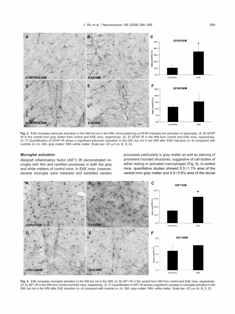
mmunohistochemistry for glial fibrillary acidic proteinGFAP) detected normal and reactive astroglia in the lum-ar spinal cord of both control and EAE mice. In controlice, numerous astrocytic GFAP immunoreactive pro-
esses were detected throughout the gray and white mat-ers (Fig. 3). In contrast, the gray matter of the L4/L5 spinalord segments of EAE mice demonstrated astrocytes withwollen processes and increased GFAP IR. In controlice, 10.6�2.5% area of the ventral horn gray matter and1.4�1.4% area of the dorsal column white matter ex-ressed GFAP IR (n�4). In EAE mice, the ventral hornray matter demonstrated a markedly increased GFAP IR35.1�4.4%, n�4, P�0.05), whereas no significant in-rease was observed in GFAP IR in the dorsal columnhite matter (15.5�3.2%, n�4).
) Cresyl Violet staining shows the inflammatory infiltrates in the whiteion in (A) is shown at higher magnification in (B). The two boxes (box
mice. (Ainted reg
tion of the inflammatory markers. These two boxed areas in (A) areafter Cresyl Violet staining. Scale bar�250 �m (A); 50 �m (B–D).
M
Acas
ppemv
FI(c m (A, B,
F(G
J. Wu et al. / Neuroscience 156 (2008) 586–596 589
icroglial activation
llograft inflammatory factor (AIF1) IR demonstrated mi-roglia with thin and ramified processes in both the graynd white matters of control mice. In EAE mice, however,everal microglia were retracted and exhibited swollen
ig. 3. EAE increases astrocyte activation in the GM but not in the WMR in the ventral horn gray matter from control and EAE mice, respeC, F) Quantification of GFAP IR shows a significant astrocytic activaontrols (n�4). GM�gray matter; WM�white matter. Scale bar�25 �
ig. 4. EAE increases microglial activation in the GM but not in the WM
D, E) AIF1 IR in the WM from control and EAE mice, respectively. (C, F) QuantificaM, but not in the WM after EAE induction (n�4) compared with controls (n�4).rocesses particularly in gray matter as well as staining ofrominent rounded structures, suggestive of cell bodies ofither resting or activated macrophages (Fig. 4). In controlice, quantitative studies showed 5.3�1.1% area of the
entral horn gray matter and 4.9�0.8% area of the dorsal
ostaining of GFAP indicates the activation of astrocytes. (A, B) GFAP, E) GFAP IR in the WM from control and EAE mice, respectively.
e GM, but not in the WM after EAE induction (n�4) compared withD, E).
IF1 IR in the ventral horn GM from control and EAE mice, respectively.
. Immunctively. (Dtion in th
. (A, B) A
tion of AIF1 IR shows a significant increase in microglial activation in theGM�gray matter; WM�white matter. Scale bar�25 �m (A, B, D, E).
cmind
M
IipEdmmh
s1
Ah
Itcanmedimt
FEi 4) compab
Fh(
J. Wu et al. / Neuroscience 156 (2008) 586–596590
olumn white matter expressing AIF1 IR (n�4). In EAEice, the ventral horn gray matter showed a markedly
ncreased AIF1 IR (32.2�1.5%, n�4, P�0.05), whereaso significant difference in AIF1 IR was detected in theorsal column white matter (6.3�0.5%, n�4).
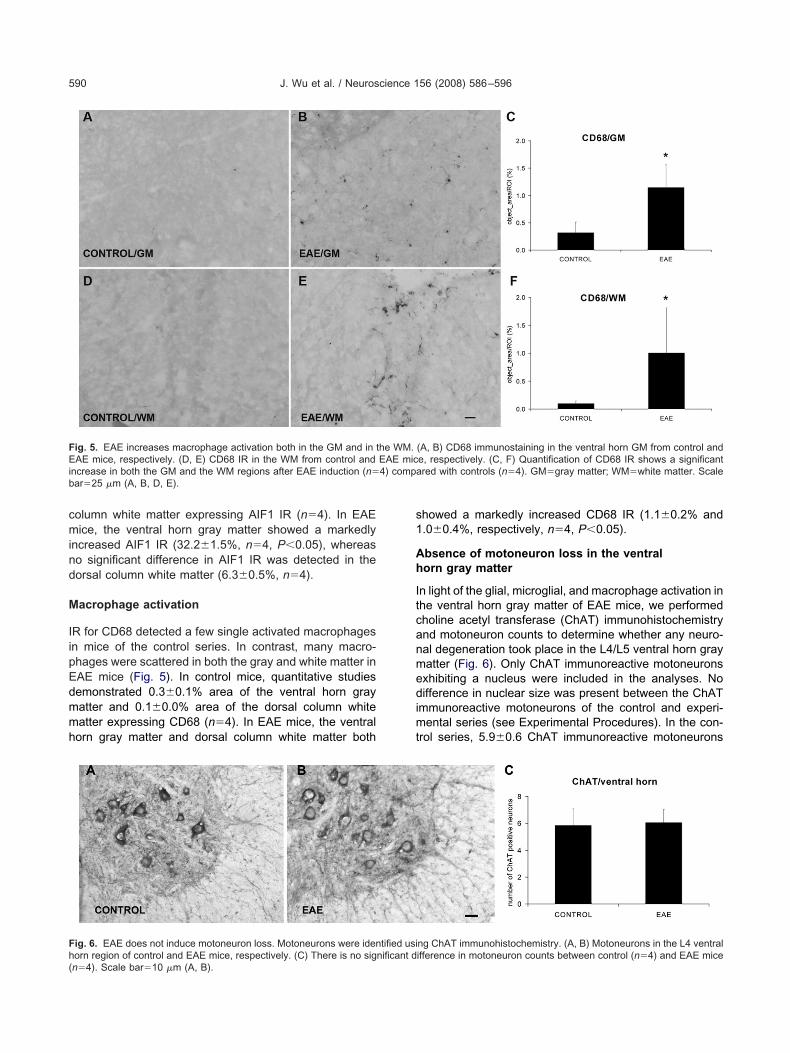
acrophage activation
R for CD68 detected a few single activated macrophagesn mice of the control series. In contrast, many macro-hages were scattered in both the gray and white matter inAE mice (Fig. 5). In control mice, quantitative studiesemonstrated 0.3�0.1% area of the ventral horn grayatter and 0.1�0.0% area of the dorsal column whiteatter expressing CD68 (n�4). In EAE mice, the ventralorn gray matter and dorsal column white matter both
ig. 5. EAE increases macrophage activation both in the GM and in tAE mice, respectively. (D, E) CD68 IR in the WM from control and
ncrease in both the GM and the WM regions after EAE induction (n�ar�25 �m (A, B, D, E).
ig. 6. EAE does not induce motoneuron loss. Motoneurons were iden
orn region of control and EAE mice, respectively. (C) There is no significant dn�4). Scale bar�10 �m (A, B).howed a markedly increased CD68 IR (1.1�0.2% and.0�0.4%, respectively, n�4, P�0.05).
bsence of motoneuron loss in the ventralorn gray matter
n light of the glial, microglial, and macrophage activation inhe ventral horn gray matter of EAE mice, we performedholine acetyl transferase (ChAT) immunohistochemistrynd motoneuron counts to determine whether any neuro-al degeneration took place in the L4/L5 ventral horn grayatter (Fig. 6). Only ChAT immunoreactive motoneuronsxhibiting a nucleus were included in the analyses. Noifference in nuclear size was present between the ChAT
mmunoreactive motoneurons of the control and experi-ental series (see Experimental Procedures). In the con-
rol series, 5.9�0.6 ChAT immunoreactive motoneurons
(A, B) CD68 immunostaining in the ventral horn GM from control ande, respectively. (C, F) Quantification of CD68 IR shows a significantred with controls (n�4). GM�gray matter; WM�white matter. Scale
ng ChAT immunohistochemistry. (A, B) Motoneurons in the L4 ventral
he WM.EAE mic
tified usi
ifference in motoneuron counts between control (n�4) and EAE mice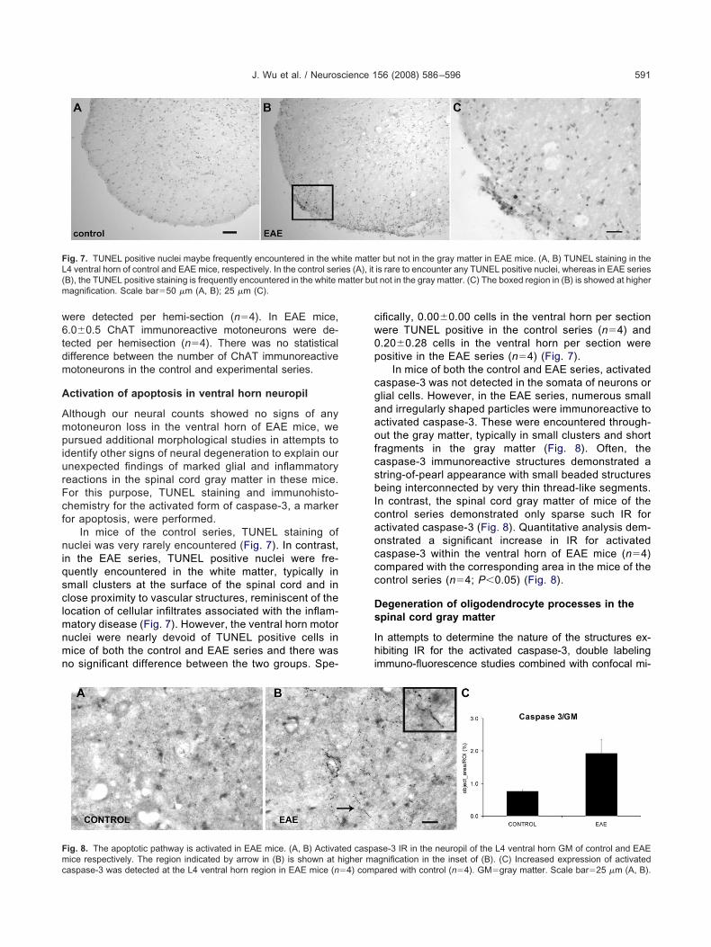
w6tdm
A
AmpiurFcf
niqsclmnmn
cw0p
cgaaofcsbIcaoccc
Ds
Ihi
FL(m
Fmc
J. Wu et al. / Neuroscience 156 (2008) 586–596 591
ere detected per hemi-section (n�4). In EAE mice,.0�0.5 ChAT immunoreactive motoneurons were de-ected per hemisection (n�4). There was no statisticalifference between the number of ChAT immunoreactiveotoneurons in the control and experimental series.
ctivation of apoptosis in ventral horn neuropil
lthough our neural counts showed no signs of anyotoneuron loss in the ventral horn of EAE mice, weursued additional morphological studies in attempts to
dentify other signs of neural degeneration to explain ournexpected findings of marked glial and inflammatoryeactions in the spinal cord gray matter in these mice.or this purpose, TUNEL staining and immunohisto-hemistry for the activated form of caspase-3, a markeror apoptosis, were performed.
In mice of the control series, TUNEL staining ofuclei was very rarely encountered (Fig. 7). In contrast,
n the EAE series, TUNEL positive nuclei were fre-uently encountered in the white matter, typically inmall clusters at the surface of the spinal cord and inlose proximity to vascular structures, reminiscent of theocation of cellular infiltrates associated with the inflam-
atory disease (Fig. 7). However, the ventral horn motoruclei were nearly devoid of TUNEL positive cells inice of both the control and EAE series and there waso significant difference between the two groups. Spe-
ig. 7. TUNEL positive nuclei maybe frequently encountered in the wh4 ventral horn of control and EAE mice, respectively. In the control serB), the TUNEL positive staining is frequently encountered in the white magnification. Scale bar�50 �m (A, B); 25 �m (C).
ig. 8. The apoptotic pathway is activated in EAE mice. (A, B) Activa
ice respectively. The region indicated by arrow in (B) is shown at higher maaspase-3 was detected at the L4 ventral horn region in EAE mice (n�4) comp
ifically, 0.00�0.00 cells in the ventral horn per sectionere TUNEL positive in the control series (n�4) and.20�0.28 cells in the ventral horn per section wereositive in the EAE series (n�4) (Fig. 7).
In mice of both the control and EAE series, activatedaspase-3 was not detected in the somata of neurons orlial cells. However, in the EAE series, numerous smallnd irregularly shaped particles were immunoreactive toctivated caspase-3. These were encountered through-ut the gray matter, typically in small clusters and short
ragments in the gray matter (Fig. 8). Often, theaspase-3 immunoreactive structures demonstrated atring-of-pearl appearance with small beaded structureseing interconnected by very thin thread-like segments.
n contrast, the spinal cord gray matter of mice of theontrol series demonstrated only sparse such IR forctivated caspase-3 (Fig. 8). Quantitative analysis dem-nstrated a significant increase in IR for activatedaspase-3 within the ventral horn of EAE mice (n�4)ompared with the corresponding area in the mice of theontrol series (n�4; P�0.05) (Fig. 8).
egeneration of oligodendrocyte processes in thepinal cord gray matter
n attempts to determine the nature of the structures ex-ibiting IR for the activated caspase-3, double labeling
mmuno-fluorescence studies combined with confocal mi-
r but not in the gray matter in EAE mice. (A, B) TUNEL staining in theis rare to encounter any TUNEL positive nuclei, whereas in EAE seriest not in the gray matter. (C) The boxed region in (B) is showed at higher
se-3 IR in the neuropil of the L4 ventral horn GM of control and EAE
ite matteies (A), it
atter bu
ted caspa
gnification in the inset of (B). (C) Increased expression of activatedared with control (n�4). GM�gray matter. Scale bar�25 �m (A, B).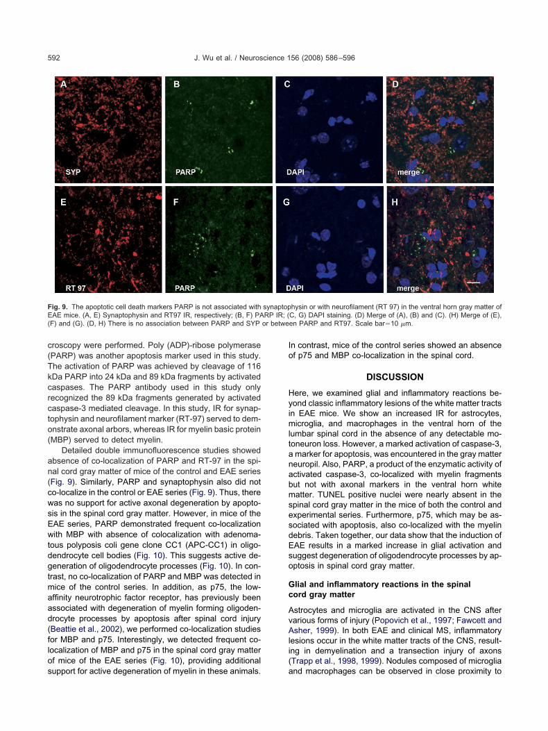
c(Tkcrcto(
an(cwsEwtdgtmaad(flos
Io
HyimltanabmsesdEso
Gc
AvAli(
FE RP IR; (( or betwe
J. Wu et al. / Neuroscience 156 (2008) 586–596592
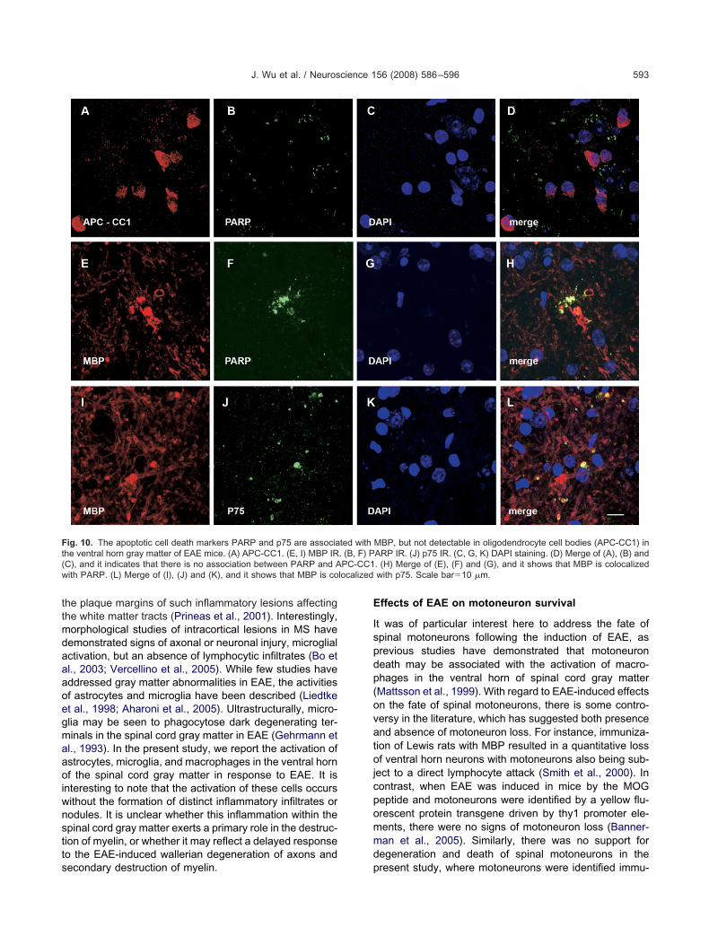
roscopy were performed. Poly (ADP)-ribose polymerasePARP) was another apoptosis marker used in this study.he activation of PARP was achieved by cleavage of 116Da PARP into 24 kDa and 89 kDa fragments by activatedaspases. The PARP antibody used in this study onlyecognized the 89 kDa fragments generated by activatedaspase-3 mediated cleavage. In this study, IR for synap-ophysin and neurofilament marker (RT-97) served to dem-nstrate axonal arbors, whereas IR for myelin basic proteinMBP) served to detect myelin.
Detailed double immunofluorescence studies showedbsence of co-localization of PARP and RT-97 in the spi-al cord gray matter of mice of the control and EAE seriesFig. 9). Similarly, PARP and synaptophysin also did noto-localize in the control or EAE series (Fig. 9). Thus, thereas no support for active axonal degeneration by apopto-is in the spinal cord gray matter. However, in mice of theAE series, PARP demonstrated frequent co-localizationith MBP with absence of colocalization with adenoma-
ous polyposis coli gene clone CC1 (APC-CC1) in oligo-endrocyte cell bodies (Fig. 10). This suggests active de-eneration of oligodendrocyte processes (Fig. 10). In con-
rast, no co-localization of PARP and MBP was detected inice of the control series. In addition, as p75, the low-ffinity neurotrophic factor receptor, has previously beenssociated with degeneration of myelin forming oligoden-rocyte processes by apoptosis after spinal cord injuryBeattie et al., 2002), we performed co-localization studiesor MBP and p75. Interestingly, we detected frequent co-ocalization of MBP and p75 in the spinal cord gray matterf mice of the EAE series (Fig. 10), providing additional
ig. 9. The apoptotic cell death markers PARP is not associated withAE mice. (A, E) Synaptophysin and RT97 IR, respectively; (B, F) PA
F) and (G). (D, H) There is no association between PARP and SYP
upport for active degeneration of myelin in these animals. a
n contrast, mice of the control series showed an absencef p75 and MBP co-localization in the spinal cord.
DISCUSSION
ere, we examined glial and inflammatory reactions be-ond classic inflammatory lesions of the white matter tracts
n EAE mice. We show an increased IR for astrocytes,icroglia, and macrophages in the ventral horn of the
umbar spinal cord in the absence of any detectable mo-oneuron loss. However, a marked activation of caspase-3,marker for apoptosis, was encountered in the gray mattereuropil. Also, PARP, a product of the enzymatic activity ofctivated caspase-3, co-localized with myelin fragmentsut not with axonal markers in the ventral horn whiteatter. TUNEL positive nuclei were nearly absent in the
pinal cord gray matter in the mice of both the control andxperimental series. Furthermore, p75, which may be as-ociated with apoptosis, also co-localized with the myelinebris. Taken together, our data show that the induction ofAE results in a marked increase in glial activation anduggest degeneration of oligodendrocyte processes by ap-ptosis in spinal cord gray matter.
lial and inflammatory reactions in the spinalord gray matter
strocytes and microglia are activated in the CNS afterarious forms of injury (Popovich et al., 1997; Fawcett andsher, 1999). In both EAE and clinical MS, inflammatory
esions occur in the white matter tracts of the CNS, result-ng in demyelination and a transection injury of axonsTrapp et al., 1998, 1999). Nodules composed of microglia
hysin or with neurofilament (RT 97) in the ventral horn gray matter ofC, G) DAPI staining. (D) Merge of (A), (B) and (C). (H) Merge of (E),en PARP and RT97. Scale bar�10 �m.
synaptop
nd macrophages can be observed in close proximity to
ttmdaaaoegmaaoiwnstts
E
Ispdp(ovatojcpommd
Ft( PC-CC1w localized
J. Wu et al. / Neuroscience 156 (2008) 586–596 593
he plaque margins of such inflammatory lesions affectinghe white matter tracts (Prineas et al., 2001). Interestingly,orphological studies of intracortical lesions in MS haveemonstrated signs of axonal or neuronal injury, microglialctivation, but an absence of lymphocytic infiltrates (Bo etl., 2003; Vercellino et al., 2005). While few studies haveddressed gray matter abnormalities in EAE, the activitiesf astrocytes and microglia have been described (Liedtket al., 1998; Aharoni et al., 2005). Ultrastructurally, micro-lia may be seen to phagocytose dark degenerating ter-inals in the spinal cord gray matter in EAE (Gehrmann etl., 1993). In the present study, we report the activation ofstrocytes, microglia, and macrophages in the ventral hornf the spinal cord gray matter in response to EAE. It is
nteresting to note that the activation of these cells occursithout the formation of distinct inflammatory infiltrates orodules. It is unclear whether this inflammation within thepinal cord gray matter exerts a primary role in the destruc-ion of myelin, or whether it may reflect a delayed responseo the EAE-induced wallerian degeneration of axons and
ig. 10. The apoptotic cell death markers PARP and p75 are associahe ventral horn gray matter of EAE mice. (A) APC-CC1. (E, I) MBP IRC), and it indicates that there is no association between PARP and Aith PARP. (L) Merge of (I), (J) and (K), and it shows that MBP is co
econdary destruction of myelin. p
ffects of EAE on motoneuron survival
t was of particular interest here to address the fate ofpinal motoneurons following the induction of EAE, asrevious studies have demonstrated that motoneuroneath may be associated with the activation of macro-hages in the ventral horn of spinal cord gray matterMattsson et al., 1999). With regard to EAE-induced effectsn the fate of spinal motoneurons, there is some contro-ersy in the literature, which has suggested both presencend absence of motoneuron loss. For instance, immuniza-ion of Lewis rats with MBP resulted in a quantitative lossf ventral horn neurons with motoneurons also being sub-
ect to a direct lymphocyte attack (Smith et al., 2000). Inontrast, when EAE was induced in mice by the MOGeptide and motoneurons were identified by a yellow flu-rescent protein transgene driven by thy1 promoter ele-ents, there were no signs of motoneuron loss (Banner-an et al., 2005). Similarly, there was no support foregeneration and death of spinal motoneurons in the
MBP, but not detectable in oligodendrocyte cell bodies (APC-CC1) inARP IR. (J) p75 IR. (C, G, K) DAPI staining. (D) Merge of (A), (B) and. (H) Merge of (E), (F) and (G), and it shows that MBP is colocalizedwith p75. Scale bar�10 �m.
ted with. (B, F) P
resent study, where motoneurons were identified immu-
nbot
Ww
TmwwddimdswbSirnctaacpt2fr2ctarlTale
Sc
AalscEoamcs(b
plcgmieeialmaptacc
ftodsonsnFdaflacstmfa
ANSdtNB
A
A
B
J. Wu et al. / Neuroscience 156 (2008) 586–596594
ohistochemically by the expression of ChAT. It is plausi-le that differences in experimental models and methodsf analysis may contribute to the above differences be-ween studies.
allerian degeneration of axons after lesions tohite matter tracts
raumatic injuries and inflammatory lesions of the whiteatter tracts of the brain and spinal cord may result inallerian degeneration of the distal axonal segment,hich has been separated from its parent cell body. Theistal portion of the axon and its terminal boutons un-ergo a series of characteristic morphological changes,
ncluding dense and filamentous degeneration, whichay be detected ultrastructurally within the first feways after the lesion (Ralston, 1990; Peters and Web-ter, 1991). More recent studies have demonstrated thatallerian degeneration of the distal axonal segment maye associated with apoptosis after injuries in the CNS.pecifically, a transection of the medial forebrain bundle
n the neonatal rat induces within hours after the injury aetrograde effect with apoptosis of the dopaminergiceurons in the substantia nigra but also activation ofaspases and detection of caspase cleavage products inheir degenerating axons distal to the lesion (El-Khodornd Burke, 2002). Similarly, a traumatic brain injury indult rats induces early activation of caspase-3 andleavage of �-amyloid precursor protein in the terminalrocesses of lesioned axons, detectable by immunohis-ochemistry within hours after the injury (Buki et al.,000; Stone et al., 2002). These expressions of markersor apoptosis subside over days and may be notablyeduced by 10 days after the axonal lesion (Stone et al.,002). Thus, both the ultrastructural and immunohisto-hemical studies suggest that wallerian degenerationakes place early, primarily within the first week, after thexonal lesion in the CNS. As it has been previouslyeported that axonal loss has already taken place at aate time point in chronic EAE mice (DeBoy et al., 2007;iwari-Woodruff et al., 2007), it is not surprising that ourxonal markers, RT-97 and synaptophysin, did not co-
ocalize with markers for apoptosis in our EAE micexamined at the similar late time point.
econdary degeneration of myelin in the spinalord gray matter
lthough we did not encounter any examples of activexonal degeneration by apoptosis at these relatively
ater time points examined during EAE, we did detectupport for apoptosis in association with oligodendro-yte processes but not oligodendrocyte cell bodies inAE mice. Specifically, we demonstrated co-localizationf cleaved PARP, a product of proteolytic cleavage byctivated caspase-3, and MBP in the spinal cord grayatter of EAE mice. However, apoptosis of oligodendro-
yte cell bodies in the spinal cord gray matter has beenuggested in earlier studies of EAE in the Lewis ratPender et al., 1991). Interestingly delayed associations
etween markers of apoptosis and oligodendroglia havereviously been encountered after traumatic spinal cordesions in both rodents and non-human primates andorrelate with lesion extension along fiber tracts under-oing wallerian degeneration (Crowe et al., 1997; Shu-an et al., 1997; Warden et al., 2001). Such injury-
nduced death of oligodendrocytes requires the pres-nce of p75, a low-affinity neurotrophin receptor (Beattiet al., 2002). In addition, all the apoptosis markers used
n this study are late markers of apoptosis, includingctivated caspase-3, PARP, and TUNEL staining. Fol-
owing CNS injury, these committed steps of apoptosisay be preceded by the cleavage of multiple proteinsnd activation of enzymatic reactions along differentathways, including the extrinsic and mitochondrial (in-
rinsic) pathways (Yakovlev and Faden, 2001; Stirling etl., 2005). The detailed features of the early steps ofaspase-dependent apoptosis pathways in the spinalord gray matter in EAE remain to be investigated.
One interesting finding in the present study was that IRor apoptotic markers p75 was demonstrated in associa-ion with degenerating myelin but not in the cell bodies ofligodendrocytes. Thus, our data suggest that oligoden-roglial cells may undergo apoptotic elimination of a sub-et of their myelin forming processes without death of theligodendrocyte itself. A similar process may take place ineurons during the normal development of the nervousystem, where select axon collateral branches are elimi-ated without loss of the parent neuron (Raff et al., 2002).urthermore, axotomized spinal motoneurons may un-ergo partial elimination of whole intramedullary motor-xon collateral trees without retrograde neuronal deathollowing a peripheral nerve transection (Havton and Kel-erth, 1990). We speculate that the secondary and delayedpoptosis of oligodendrocyte processes reported here mayontribute to chronic demyelination and dysfunction of thepinal cord in the settings of post-inflammatory myelopa-hies. In addition, our neuropathologic findings in the MSodel further support the notion of using neuroimaging
ocused on the gray matter to discern neurodegenerativespects of clinical disease.
cknowledgments—This study was supported by grants from theIH/NCRR (1 U54 RR021813) and National Multiple Sclerosisociety (CA 1028 and RG 3593). The RT97 monoclonal antibodyeveloped by Dr. John Wood was obtained from the Developmen-al Studies Hybridoma Bank developed under the auspices of theICHD and maintained by the University of Iowa, Department ofiological Sciences, Iowa City, IA 52242.
REFERENCES
haroni R, Arnon R, Eilam R (2005) Neurogenesis and neuroprotectioninduced by peripheral immunomodulatory treatment of experimentalautoimmune encephalomyelitis. J Neurosci 25:8217-8228.
utieri MV, Agrawal N (1998) IRT-1, a novel interferon-gamma-responsive transcript encoding a growth-suppressing basic leucinezipper protein. J Biol Chem 273:14731–14737.
annerman PG, Hahn A, Ramirez S, Morley M, Bonnemann C, Yu S,Zhang GX, Rostami A, Pleasure D (2005) Motor neuron pathologyin experimental autoimmune encephalomyelitis: studies in THY1-
YFP transgenic mice. Brain 128:1877–1886.
B
B
B
B
C
C
C
C
D
D
E
F
G
G
G
H
H
H
L
L
M
M
M
M
O
P
P
P
P
R
R
R
S
S
S
S
S
S
T
J. Wu et al. / Neuroscience 156 (2008) 586–596 595
eattie MS, Harrington AW, Lee R, Kim JY, Boyce SL, Longo FM,Bresnahan JC, Hempstead BL, Yoon SO (2002) ProNGF inducesp75-mediated death of oligodendrocytes following spinal cord in-jury. Neuron 36:375–386.
jartmar C, Kidd G, Mork S, Rudick R, Trapp BD (2000) Neurologicaldisability correlates with spinal cord axonal loss and reduced N-acetyl aspartate in chronic multiple sclerosis patients. Ann Neurol48:893–901.
o L, Vedeler CA, Nyland H, Trapp BD, Mork SJ (2003) Intracorticalmultiple sclerosis lesions are not associated with increased lym-phocyte infiltration. Mult Scler 9:323–331.
uki A, Okonkwo DO, Wang KK, Povlishock JT (2000) Cytochrome crelease and caspase activation in traumatic axonal injury. J Neu-rosci 20:2825–2834.
hao MV, Hempstead BL (1995) p75 And Trk: a two-receptor system.Trends Neurosci 18:321–326.
hard DT, Griffin CM, Parker GJ, Kapoor R, Thompson AJ, Miller DH(2002) Brain atrophy in clinically early relapsing-remitting multiplesclerosis. Brain 125:327–337.
oggeshall RE, Lekan HA (1996) Methods for determining numbers ofcells and synapses: a case for more uniform standards of review.J Comp Neurol 364:6–15.
rowe MJ, Bresnahan JC, Shuman SL, Masters JN, Beattie MS(1997) Apoptosis and delayed degeneration after spinal cord injuryin rats and monkeys. Nat Med 3:73–76.
alton CM, Chard DT, Davies GR, Miszkiel KA, Altmann DR, Fer-nando K, Plant GT, Thompson AJ, Miller DH (2004) Early devel-opment of multiple sclerosis is associated with progressive greymatter atrophy in patients presenting with clinically isolated syn-dromes. Brain 127:1101–1107. Epub 2004 Mar 1103.
eBoy CA, Zhang J, Dike S, Shats I, Jones M, Reich DS, Mori S,Nguyen T, Rothstein B, Miller RH, Griffin JT, Kerr DA, Calabresi PA(2007) High resolution diffusion tensor imaging of axonal damagein focal inflammatory and demyelinating lesions in rat spinal cord.Brain 130:2199–2210. Epub 2007 Jun 2198.
l-Khodor BF, Burke RE (2002) Medial forebrain bundle axotomyduring development induces apoptosis in dopamine neurons of thesubstantia nigra and activation of caspases in their degeneratingaxons. J Comp Neurol 452:65–79.
awcett JW, Asher RA (1999) The glial scar and central nervoussystem repair. Brain Res Bull 49:377–391.
ehrmann J, Gold R, Linington C, Lannes-Vieira J, Wekerle H,Kreutzberg GW (1993) Microglial involvement in experimental au-toimmune inflammation of the central and peripheral nervous sys-tem. Glia 7:50–59.
lynn P, Chantry A, Groome N, Cuzner ML (1987) Basic proteindissociating from myelin membranes at physiological ionic strengthand pH is cleaved into three major fragments. J Neurochem48:752–759.
useva NV, Taghiyev AF, Rokhlin OW, Cohen MB (2002) Contributionof death receptor and mitochondrial pathways to Fas-mediatedapoptosis in the prostatic carcinoma cell line PC3. Prostate 51:231–240.
ammerle B, Elizalde C, Galceran J, Becker W, Tejedor FJ, CarniceroA, Ceron J, Martinez S, Ferrus A (2003) The MNB/DYRK1A proteinkinase: neurobiological functions and Down syndrome implications.J Neural Transm Suppl 17:129–137.
avton L, Kellerth JO (1990) Elimination of intramedullary axon col-laterals of cat spinal alpha-motoneurons following peripheral nerveinjury. Exp Brain Res 79:65–74.
jelmstrom P, Juedes AE, Fjell J, Ruddle NH (1998) B-cell-deficientmice develop experimental allergic encephalomyelitis with demy-elination after myelin oligodendrocyte glycoprotein sensitization.J Immunol 161:4480–4483.
iedtke W, Edelmann W, Chiu FC, Kucherlapati R, Raine CS (1998)Experimental allergic encephalomylitis in mice lacking glial fibril-
lary acidic protein is characterized by a more severe clinicalcourse and an infiltrative central nervous system lesion. Am JPathol 152:251–259.
ucchinetti CF, Brueck W, Rodriguez M, Lassmann H (1998) Multi-ple sclerosis: lessons from neuropathology. Semin Neurol 18:337–349.
acKenzie-Graham A, Tinsley MR, Shah KP, Aguilar C, Strickland LV,Boline J, Martin M, Morales L, Shattuck DW, Jacobs RE, VoskuhlRR, Toga AW (2006) Cerebellar cortical atrophy in experimentalautoimmune encephalomyelitis. Neuroimage 32:1016–1023. Epub2006 Jun 1027.
asliah E, Rockenstein E, Veinbergs I, Sagara Y, Mallory M, Hashi-moto M, Mucke L (2001) Beta-amyloid peptides enhance alpha-synuclein accumulation and neuronal deficits in a transgenicmouse model linking Alzheimer’s disease and Parkinson’s dis-ease. Proc Natl Acad Sci U S A 98:12245–12250. Epub 12001 Sep12225.
attsson P, Meijer B, Svensson M (1999) Extensive neuronal celldeath following intracranial transection of the facial nerve in theadult rat. Brain Res Bull 49:333–341.
cTigue DM, Wei P, Stokes BT (2001) Proliferation of NG2-positivecells and altered oligodendrocyte numbers in the contused ratspinal cord. J Neurosci 21:3392–3400.
liver FJ, de la Rubia G, Rolli V, Ruiz-Ruiz MC, de Murcia G, MurciaJM (1998) Importance of poly(ADP-ribose) polymerase and itscleavage in apoptosis. Lesson from an uncleavable mutant. J BiolChem 273:33533–33539.
ender MP, Nguyen KB, McCombe PA, Kerr JF (1991) Apoptosis inthe nervous system in experimental allergic encephalomyelitis.J Neurol Sci 104:81–87.
eters APS, Webster HdeF (1991) The fine structure of the nervoussystem, 3rd edition. Oxford England: Oxford University Press.
opovich PG, Wei P, Stokes BT (1997) Cellular inflammatory re-sponse after spinal cord injury in Sprague-Dawley and Lewis rats.J Comp Neurol 377:443–464.
rineas JW, Kwon EE, Cho ES, Sharer LR, Barnett MH, Oleszak EL,Hoffman B, Morgan BP (2001) Immunopathology of secondary-progressive multiple sclerosis. Ann Neurol 50:646–657.
aff MC, Whitmore AV, Finn JT (2002) Axonal self-destruction andneurodegeneration. Science 296:868–871.
alston HJ 3rd (1990) Analysis of neuronal networks: a review oftechniques for labeling axonal projections. J Electron Microsc Tech15:322–331.
amprasad MP, Terpstra V, Kondratenko N, Quehenberger O, Stein-berg D (1996) Cell surface expression of mouse macrosialin andhuman CD68 and their role as macrophage receptors for oxidizedlow density lipoprotein. Proc Natl Acad Sci U S A 93:14833–14838.
hiromani PJ, Armstrong DM, Bruce G, Hersh LB, Groves PM, GillinJC (1987) Relation of pontine choline acetyltransferase immuno-reactive neurons with cells which increase discharge during REMsleep. Brain Res Bull 18:447–455.
human SL, Bresnahan JC, Beattie MS (1997) Apoptosis of microgliaand oligodendrocytes after spinal cord contusion in rats. J NeurosciRes 50:798–808.
mith T, Groom A, Zhu B, Turski L (2000) Autoimmune encephalo-myelitis ameliorated by AMPA antagonists. Nat Med 6:62–66.
tirling DP, Koochesfahani KM, Steeves JD, Tetzlaff W (2005) Mino-cycline as a neuroprotective agent. Neuroscientist 11:308–322.
tone JR, Okonkwo DO, Singleton RH, Mutlu LK, Helm GA, Pov-lishock JT (2002) Caspase-3-mediated cleavage of amyloid pre-cursor protein and formation of amyloid beta peptide in traumaticaxonal injury. J Neurotrauma 19:601–614.
uen WE, Bergman CM, Hjelmstrom P, Ruddle NH (1997) A criticalrole for lymphotoxin in experimental allergic encephalomyelitis.J Exp Med 186:1233–1240.
iwari-Woodruff S, Morales LB, Lee R, Voskuhl RR (2007) Differentialneuroprotective and antiinflammatory effects of estrogen receptor(ER)alpha and ERbeta ligand treatment. Proc Natl Acad Sci U S A
104:14813–14818. Epub 12007 Sep 14814.
T
T
V
W
W
Y
Y
J. Wu et al. / Neuroscience 156 (2008) 586–596596
rapp BD, Bo L, Mork S, Chang A (1999) Pathogenesis of tissue injuryin MS lesions. J Neuroimmunol 98:49–56.
rapp BD, Peterson J, Ransohoff RM, Rudick R, Mork S, Bo L (1998)Axonal transection in the lesions of multiple sclerosis. N EnglJ Med 338:278–285.
ercellino M, Plano F, Votta B, Mutani R, Giordana MT, Cavalla P(2005) Grey matter pathology in multiple sclerosis. J NeuropatholExp Neurol 64:1101–1107.
arden P, Bamber NI, Li H, Esposito A, Ahmad KA, Hsu CY, Xu XM
(2001) Delayed glial cell death following wallerian degeneration inwhite matter tracts after spinal cord dorsal column cordotomy inadult rats. Exp Neurol 168:213–224.
ujek JR, Bjartmar C, Richer E, Ransohoff RM, Yu M, Tuohy VK,Trapp BD (2002) Axon loss in the spinal cord determines perma-nent neurological disability in an animal model of multiple sclerosis.J Neuropathol Exp Neurol 61:23–32.
akovlev AG, Faden AI (2001) Caspase-dependent apoptotic path-ways in CNS injury. Mol Neurobiol 24:131–144.
oung HE, Black AC Jr (2004) Adult stem cells. Anat Rec A Discov Mol
Cell Evol Biol 276:75–102.(Accepted 15 July 2008)(Available online 26 July 2008)