Stable isotope-resolved metabolomic analysis of lithium effects on glial-neuronal metabolism and...
24
Stable isotope-resolved metabolomic analysis of lithium effects on glial-neuronal metabolism and interactions Teresa W.-M. Fan, Department of Chemistry, Center for Regulatory and Environmental Analytical Metabolomics, University of Louisville, Louisville, KY 40292, USA Department of Medicine, Structural Biology Program, James Graham Brown Cancer Center, University of Louisville, Louisville, KY 40202, USA Department of Chemistry, University of Louisville, 2210 S. Brook St, Rm 348 John W. Shumaker Research Building, Louisville, KY 40208, USA, [email protected] Peixiong Yuan, Biomarker Laboratory, Laboratory of Molecular Pathophysiology, Mood and Anxiety Disorder Program, National Institute of Mental Health, NIH, Bethesda, MD 20892, USA Andrew N. Lane, Department of Chemistry, Center for Regulatory and Environmental Analytical Metabolomics, University of Louisville, Louisville, KY 40292, USA Department of Medicine, Structural Biology Program, James Graham Brown Cancer Center, University of Louisville, Louisville, KY 40202, USA Richard M. Higashi, Department of Chemistry, Center for Regulatory and Environmental Analytical Metabolomics, University of Louisville, Louisville, KY 40292, USA Department of Medicine, Structural Biology Program, James Graham Brown Cancer Center, University of Louisville, Louisville, KY 40202, USA Yun Wang, Biomarker Laboratory, Laboratory of Molecular Pathophysiology, Mood and Anxiety Disorder Program, National Institute of Mental Health, NIH, Bethesda, MD 20892, USA Anahita B. Hamidi, Biomarker Laboratory, Laboratory of Molecular Pathophysiology, Mood and Anxiety Disorder Program, National Institute of Mental Health, NIH, Bethesda, MD 20892, USA Rulun Zhou, Biomarker Laboratory, Laboratory of Molecular Pathophysiology, Mood and Anxiety Disorder Program, National Institute of Mental Health, NIH, Bethesda, MD 20892, USA Xavier Guitart, Biomarker Laboratory, Laboratory of Molecular Pathophysiology, Mood and Anxiety Disorder Program, National Institute of Mental Health, NIH, Bethesda, MD 20892, USA © Springer Science+Business Media, LLC 2010 Correspondence to: Teresa W.-M. Fan; Rima Kaddurah-Daouk. T. W.-M. Fan and P. Yuan contributed equally to this work. Electronic supplementary material The online version of this article (doi:10.1007/s11306-010-0208-9) contains supplementary material, which is available to authorized users. NIH Public Access Author Manuscript Metabolomics. Author manuscript; available in PMC 2010 July 13. Published in final edited form as: Metabolomics. 2010 June 1; 6(2): 165–179. doi:10.1007/s11306-010-0208-9. NIH-PA Author Manuscript NIH-PA Author Manuscript NIH-PA Author Manuscript
-
Upload
independent -
Category
Documents
-
view
2 -
download
0
Transcript of Stable isotope-resolved metabolomic analysis of lithium effects on glial-neuronal metabolism and...

Stable isotope-resolved metabolomic analysis of lithium effectson glial-neuronal metabolism and interactions
Teresa W.-M. Fan,Department of Chemistry, Center for Regulatory and Environmental Analytical Metabolomics,University of Louisville, Louisville, KY 40292, USA
Department of Medicine, Structural Biology Program, James Graham Brown Cancer Center,University of Louisville, Louisville, KY 40202, USA
Department of Chemistry, University of Louisville, 2210 S. Brook St, Rm 348 John W. ShumakerResearch Building, Louisville, KY 40208, USA, [email protected]
Peixiong Yuan,Biomarker Laboratory, Laboratory of Molecular Pathophysiology, Mood and Anxiety DisorderProgram, National Institute of Mental Health, NIH, Bethesda, MD 20892, USA
Andrew N. Lane,Department of Chemistry, Center for Regulatory and Environmental Analytical Metabolomics,University of Louisville, Louisville, KY 40292, USA
Department of Medicine, Structural Biology Program, James Graham Brown Cancer Center,University of Louisville, Louisville, KY 40202, USA
Richard M. Higashi,Department of Chemistry, Center for Regulatory and Environmental Analytical Metabolomics,University of Louisville, Louisville, KY 40292, USA
Department of Medicine, Structural Biology Program, James Graham Brown Cancer Center,University of Louisville, Louisville, KY 40202, USA
Yun Wang,Biomarker Laboratory, Laboratory of Molecular Pathophysiology, Mood and Anxiety DisorderProgram, National Institute of Mental Health, NIH, Bethesda, MD 20892, USA
Anahita B. Hamidi,Biomarker Laboratory, Laboratory of Molecular Pathophysiology, Mood and Anxiety DisorderProgram, National Institute of Mental Health, NIH, Bethesda, MD 20892, USA
Rulun Zhou,Biomarker Laboratory, Laboratory of Molecular Pathophysiology, Mood and Anxiety DisorderProgram, National Institute of Mental Health, NIH, Bethesda, MD 20892, USA
Xavier Guitart,Biomarker Laboratory, Laboratory of Molecular Pathophysiology, Mood and Anxiety DisorderProgram, National Institute of Mental Health, NIH, Bethesda, MD 20892, USA
© Springer Science+Business Media, LLC 2010Correspondence to: Teresa W.-M. Fan; Rima Kaddurah-Daouk.T. W.-M. Fan and P. Yuan contributed equally to this work.Electronic supplementary material The online version of this article (doi:10.1007/s11306-010-0208-9) contains supplementarymaterial, which is available to authorized users.
NIH Public AccessAuthor ManuscriptMetabolomics. Author manuscript; available in PMC 2010 July 13.
Published in final edited form as:Metabolomics. 2010 June 1; 6(2): 165–179. doi:10.1007/s11306-010-0208-9.
NIH
-PA Author Manuscript
NIH
-PA Author Manuscript
NIH
-PA Author Manuscript

Guang Chen,Biomarker Laboratory, Laboratory of Molecular Pathophysiology, Mood and Anxiety DisorderProgram, National Institute of Mental Health, NIH, Bethesda, MD 20892, USA
Husseini K. Manji, andBiomarker Laboratory, Laboratory of Molecular Pathophysiology, Mood and Anxiety DisorderProgram, National Institute of Mental Health, NIH, Bethesda, MD 20892, USA
Johnson & Johnson, Titusville, NJ, USA
Rima Kaddurah-DaoukDepartment of Psychiatry, Duke University Medical Center, Box 3950, Durham, NC 27710, USA,[email protected]
AbstractDespite the long-established therapeutic efficacy of lithium in the treatment of bipolar disorder(BPD), its molecular mechanism of action remains elusive. Newly developed stable isotope-resolvedmetabolomics (SIRM) is a powerful approach that can be used to elucidate systematically how lithiumimpacts glial and neuronal metabolic pathways and activities, leading ultimately to deciphering itsmolecular mechanism of action. The effect of lithium on the metabolism of three different 13C-labeledprecursors ([U-13C]-glucose, 13C-3-lactate or 13C-2,3-alanine) was analyzed in cultured ratastrocytes and neurons by nuclear magnetic resonance (NMR) spectroscopy and gas chromatographymass spectrometry (GC-MS). Using [U-13C]-glucose, lithium was shown to enhance glycolyticactivity and part of the Krebs cycle activity in both astrocytes and neurons, particularly the anapleroticpyruvate carboxylation (PC). The PC pathway was previously thought to be active in astrocytes butabsent in neurons. Lithium also stimulated the extracellular release of 13C labeled-lactate, -alanine(Ala), -citrate, and -glutamine (Gln) by astrocytes. Interrogation of neuronal pathways using 13C-3-lactate or 13C-2,3-Ala as tracers indicated a high capacity of neurons to utilize lactate and Ala in theKrebs cycle, particularly in the production of labeled Asp and Glu via PC and normal cycle activity.Prolonged lithium treatment enhanced lactate metabolism via PC but inhibited lactate oxidation viathe normal Krebs cycle in neurons. Such lithium modulation of glycolytic, PC and Krebs cycleactivity in astrocytes and neurons as well as release of fuel substrates by astrocytes should helpreplenish Krebs cycle substrates for Glu synthesis while meeting neuronal demands for energy.Further investigations into the molecular regulation of these metabolic traits should provide newinsights into the pathophysiology of mood disorders and early diagnostic markers, as well as newtarget(s) for effective therapies.
KeywordsBipolar disorder; Lithium; 13C-labeled tracers; Astrocytes; Neurons; Pyruvate carboxylation; Glu/Gln cycling
1 IntroductionMood disorders are common, chronic, severe mental illnesses that affect the lives of millionsof individuals worldwide. Despite overwhelming evidence for a biological etiology andpathophysiology, such as alterations in neuronal plasticity (Manji et al. 2000; Schloesser et al.2008) and cellular resilience (Hunsberger et al. 2009), the underlying mechanisms of thedisorders remain largely unknown. Numerous methods can be used to identify putative noveltargets in mood disorders; one of the most popular strategies is to study the actions of effectivepsychotropic medications both in vivo and in vitro.
Fan et al. Page 2
Metabolomics. Author manuscript; available in PMC 2010 July 13.
NIH
-PA Author Manuscript
NIH
-PA Author Manuscript
NIH
-PA Author Manuscript

Although its mechanism of action remains poorly understood, lithium has long been the first-line gold standard for the treatment of bipolar disorder, and as an add-on medication in unipolarand treatment-resistant depression (Manji et al. 1999). Lithium’s remarkable efficacy sparkeda revolution that reshaped not only medical and scientific concept of mental illness, but alsopopular concepts as well. Indeed, the efforts to understand how a simple monovalent cationlike lithium could exert such profound beneficial effects have led investigators to examineseveral signaling pathways and networks involved in mood disorders (Gould and Manji2002; Manji et al. 1995).
During the past several years, lithium has been shown to have protective effects on neural cellsin vivo and in vitro (Gould and Manji 2002; Manji et al. 1995). Underlying mechanisms thathave been implicated include metabolic enzymes (Nordenberg et al. 1982), secondary signalpathways and down-stream molecular targets (Chen et al. 2000; Chen et al. 2003; Engel et al.2009; Manji et al. 1995). One of the most interesting targets is glycogen synthase kinase-3(GSK-3), which regulates more than 50 enzymes including the metabolism regulators glycogensynthase and pyruvate dehydrogenase (Hedgepeth et al. 1997; Hoshi et al. 1996). This workhas generated interest in the effects of lithium on glucose metabolic pathways. Notably, lithiumappears to stimulate glucose uptake and glycogen synthesis in cultured cells (Birch and Hughes1989; Chen et al. 1998) and in rat brain in vivo (Basselin et al. 2006; Plenge 1976, 1982). Inhuman subjects, a recent study using positron emission tomography (PET) showed that lithiumaltered relative regional cerebral glucose metabolism (rCMRglc) in some specific brain regions(Kohno et al. 2007). Furthermore, the possible indirect effects of lithium on pyruvatedehydrogenase (PDH) activity (Hedgepeth et al. 1997; Hoshi et al. 1996) implicates a possibleshift in the balance between glycolysis, lactate production and mitochondrial functionincluding the synthesis of glutamate, and concomitant anaplerotic reactions (Mason et al.2007; Serres et al. 2008).
Besides its effect on glucose metabolism, lithium also influences brain monoamine synthesis,such as 5-hy-droxytryptophan (5HT) synthesis (Berggren 1985) and glutamate metabolism(Marcus et al. 1986) (O’Donnell et al. 2003), which may also be related to their mechanismsof action in bipolar disorder. The mechanisms underlying lithium’s effects on thesebiochemical metabolic pathways need further elucidation.
Although brain energy metabolic pathways have been well studied, the different metabolicprofiles of astroglia and neurons, as well as details about neuron-glial cell interaction networkare still being extensively explored. In general, the astroglia are highly glycolytic, and secretesignificant amounts of lactate that is taken up and consumed by the neurons (Bouzier-Sore etal. 2006). The neuron consumes most of the glucose and lactate by mitochondrial oxidation toproduce energy (Pellerin et al. 2007; Serres et al. 2008a; Zwingmann and Leibfritz 2003;Zwingmann et al. 2001). Astrocytes have high levels of pyruvate carboxylase (Hertz et al.2007; Mason et al. 2007), which is one of the major anaplerotic pathways. However, the extentof anaplerosis in the neurons is largely unknown. Moreover, the astrocytes and neuronscooperate via the well-established glutamate-glutamine cycle (Hyder et al. 2006; Serres et al.2008; Xu and Shen 2006). The importance of “symbiotic” metabolism in brain is wellestablished, and it is plausible that neural suppressive agents such as lithium may affect thecritical cycling of these metabolites either at the synapses themselves or through the specificmetabolism of these compounds.
Metabolomics provides powerful tools for defining perturbations in metabolic pathways andnetworks in human disease and during treatment (Kaddurah-Daouk et al. 2008; Kristal et al.2007; Lindon et al. 2007). Metabolic signatures for CNS disorders could result in theidentification of biomarkers for disease, disease progression, and response to therapy, as wellas new insights into mechanisms of disease (Kaddurah-Daouk 2009; Kaddurah-Daouk et al.
Fan et al. Page 3
Metabolomics. Author manuscript; available in PMC 2010 July 13.
NIH
-PA Author Manuscript
NIH
-PA Author Manuscript
NIH
-PA Author Manuscript

2008; Kaddurah-Daouk et al. 2007; Paige et al. 2007; Rozen et al. 2005). Additionally,metabolomics provides detailed biochemical knowledge about drug mechanism of action,therapeutic potential and side effects (Kaddurah-Daouk et al. 2008). In the present study, wehave used the stable isotope resolved metabolomics (SIRM) approach (Fan et al. 2009) to mapeffects of lithium on the metabolism of isolated neurons and glial cells. This approach utilizedstable isotope tracers such as 13C labeled glucose in conjunction with NMR and massspectrometry to map biochemical transformation pathway(s) in neurons and glial cellsimpacted by lithium treatment. With SIRM technologies, multiple pathways can be probedsimultaneously, instead of one at a time as in the past. This not only greatly facilitates thediscovery of pathway(s) perturbed by lithium treatment but also enables elucidation ofmetabolic networks spanning different cellular compartments. These approaches could providenew insights into the pathophysiology of BPD and may be used to direct research intouncovering early diagnostic markers and novel therapeutic strategies.
2 Materials and methodsIsotopes: [U-13C]-glucose, [2,3-13C]-alanine and [3-13C]-lactate were purchased fromCambridge Isotope Laboratories (Andover, MA) and were used without further purification.
2.1 Primary cortical neuron cultureCultures of cortical neurons were prepared as previously described (Hao et al. 2004) with minormodifications. In brief, timed-pregnant (E-18) Sprague–Dawley rats (Charles River, Frederick,MD) were euthanized by CO2 inhalation, according to NIH-approved protocols for the careand use of lab animals. Embryos were removed, chilled on ice, and the cortex wasmicrodissected under sterile conditions. Cortical cells was dissociated in Ca2+- and Mg2+-freeHBSS containing 0.125% tyrosine for 15 min, triturated in Dulbecco’s Modified Eagle’sMedium (DMEM, Invitrogen, Carlsbad, CA) supplied with 10% FBS and antibiotic-antimyotics (Invitrogen), and plated in 150-mm dishes pre-coated with 5 µg/ml poly-D-lysine(Sigma, St. Louis, MO). Cells were grown at 37°C, 5% CO2 and 95% humidity, first in 10%FBS/DMEM, and one day later switched to serum-free Neurobasal medium (Invitrogen,Cat#21103. Alanine 2 mg/l; Lactate 0; Glutamate 0), plus B27 (Invitrogen). Cultures weregrown in serum-free medium for 10 days before being used for experiments, and the mediumwas changed every 3 days. These cultures yielded >95% neurons as identified byimmunocytochemistry with anti-NeuN antibody (Cat# MAB377; Millipore, Billerica, MA).
2.2 Astrocyte cultureAstrocytes were isolated from cerebral cortex of postnatal day 3 Sprague–Dawley rats aspreviously described (Zhang et al. 2002). Briefly, the brains were removed, washed in PBS,and placed in HBSS with antibiotic-antimyotics (Invitrogen). Tissues were dissected andtransferred to 15-ml tubes and triturated by mechanical dissociation with a 10-ml pipette,followed by passing through #19 needle once, #21 needle twice and finally into the #25 needleonce. After being triturated well, cells were cultured in 75-cm2 flasks (at the equivalent of twobrains per flask) and were maintained at 37°C in a humidified incubator maintained at 5%CO2.
2.3 Cell treatmentAfter the cultures were shifted to glucose-free medium (DMEM from Sigma. CustomizedNeurobasal media from Invitrogen) supplied with 0.2% 13C-glucose (10.75 mM), 2 mM 13C-alanine, or 5 mM 13C-lactate, the cells were treated with or without 1.0 mM LiCl (Sigma) for1, 2, 3 days. This is in the range of the therapeutic lithium concentration in patient blood, rodentblood and brain tissues (Jensen et al. 1996). The same concentrations of non-isotopic glucose,alanine or lactate (Sigma) were supplied to the other group for comparison. Cultured media
Fan et al. Page 4
Metabolomics. Author manuscript; available in PMC 2010 July 13.
NIH
-PA Author Manuscript
NIH
-PA Author Manuscript
NIH
-PA Author Manuscript

were filtered, and the cells were washed with PBS, pelletted and immediately frozen at −80°C.
2.4 Metabolite extraction and determinationThe cold pellet or medium was extracted twice with 10% cold trichloroacetic acid (TCA),followed by lyophilization as previously described (Fan et al. 2005; Fan et al. 2006). Glucosewas quantified by NMR using the 13C-1αH satellite signals centered at 5.22 ppm. This accountsfor 36% of the total glucose. The 13C and 12C lactate and alanine concentrations weredetermined by integration of the methyl peak and its satellites and normalized to theconcentration of the standard DSS. From this, the amount of glucose consumed (ΔGlc) and thefraction converted to lactate plus alanine (F) could be estimated, according to Eqs. 1 and 2(Lane and Fan 2007):
(1)
(2)
1 – F then represents the glucose carbon that enters other metabolites and macromolecules inthe cell mass or otherwise not accounted for.
Abundant media components (e.g. threonine and valine) were similarly integrated and theirconcentrations determined as a function of sampling time to assess the utilization of essentialamino acids. The concentrations of choline metabolites (choline, phosphocholine andglycerophosphocholine) were determined from the areas of the trimethyl ammonium resonancenear 3.22 ppm as described previously (Fan et al. 1993).
2.5 NMR spectroscopyNMR spectra were recorded at 14.1 T or 18.8 T on Varian Inova NMR spectrometer at 20°Cusing a 90° excitation pulse, an acquisition time of 2 s and a relaxation delay of 3 s. Underthese conditions, the protons are essentially full relaxed, as determined by independentmeasurements of T1. For identification and determining the positional enrichment with 13C weused 1D HSQC and 2D TOCSY (Fan et al. 2005; Fan et al. 2006; Lane and Fan 2007; Lane etal. 2008). 1D HSQC spectra were recorded at 14.1 T using an acquisition time of 0.15 s and arecycle time of 1.5 s. Garp decoupling was applied during the proton acquisition period.
Metabolites were assigned based on their 1H chemical shift, TOCSY connectivity patterns andcorrelation with 13C, as described previously (Fan and Lane 2008; Fan 1996). All metabolitesexcept choline and the nucleotides were quantified from GC-MS data (Fan et al. 1986; Laneet al. 2008). Positional 13C enrichment was determined from 1D or 2D proton NMR spectraas previously described (Lane and Fan 2007; Lane et al. 2008).
2.6 GC-MS analysisAfter NMR analysis samples were lyophilized and resus-pended in nanopure H20. An aliquotof the sample was mixed with 40% TCA to a final 10% TCA concentration. Fifty µl of 0.1mM noreleucine (Sigma) was also added as an internal standard. Samples were lyophilizedand silylated with 50 µl of acetonitrile:MTBSTFA (N-methyl-N-[tert-butyl-dimethylsilyl]trifluoroacetamide, Regis Chemical, Morton Grove, IL) (v/v 1:1) for 3 h under sonication,followed by overnight incubation. The derivatized extracts were centrifuged to remove
Fan et al. Page 5
Metabolomics. Author manuscript; available in PMC 2010 July 13.
NIH
-PA Author Manuscript
NIH
-PA Author Manuscript
NIH
-PA Author Manuscript

particulates before analysis using PolarisQ GC-ion trap MSn (ThermoFinnigan, Austin, TX)as previously described (Fan et al. 2005). The metabolites were identified and quantified usingXCalibur software (ThermoFinnigan).
2.7 ImmunoblottingImmunoblotting was done as previously described (Hao et al. 2004). The specific signals onthe blots were detected with ECL and visualized with Kodak X-ray film. Densitometricanalyses of PC and beta-actin immunoreactivities were conducted with a Kodak Image Station440CF.
RNA isolation and reverse transcription: Total RNA was isolated from neuronal cells usingTrizol reagent and PureLink RNA mini kit (Invitrogen) according to the manufacturer’sinstructions. Reverse transcription reactions were performed using SuperScript III first-strandcDNA synthesis system (Invitrogen) and carried out at 50°C for 45 min followed by reactioninactivation at 70°C for 15 min.
2.8 Quantitative Real-Time PCRThe primers for PC and beta-actin were synthesized by Invitrogen. The sequences of the PCprimers were as follows: forward primer 5′-GCGTGTTTGACTACAGTGA G-3′ and reverseprimer 5′-TCTTGACCTCCTTGAACTT G-3′. Quantitative PCR was performed with iQ5Real-Time PCR detection system (Bio-Rad Laboratories, Hercules, CA). The Real-Time PCRprogram included an enzyme activation step at 95°C (10 min) and 40 cycles of 95°C (30 s),60°C (30 s). Cycle threshold (Ct) values obtained were converted into mRNA copy numberusing standard plots of Ct versus log copy number. Data obtained from triplicate runs for targetcDNA amplification were averaged and expressed as % of control.
2.9 Statistical analysisGC-MS analysis was performed in triplicate and %RSD (relative standard deviation) ofanalysis was calculated for each reported metabolite. ANOVA was used for multiple groupcomparisons. Comparisons between two groups were done using Student’s t-test withappropriate corrections of multiple comparisons. P < 0.05 is considered significantly different.
3 Results and discussion3.1 Effects of lithium on metabolite uptake and release in astrocytes and neurons
Astrocytes and neurons have different demands for glucose metabolism, and differ in thespecific metabolites that are exchanged. We therefore measured the cellular uptake of glucoseand other metabolites from the bathing medium, and release of metabolites into the media ofcultured cells.
Over a period of three days, the cells consumed about one-third of the initial glucose, andconverted approximately 90% of all of the consumed glucose into secreted lactate (cf. Eq. 1;Fig. 1). In contrast to glucose consumption, the uptake of essential amino acids such as Thrand Val was very low over the 3-day period (data not shown). The high conversion of glucoseto lactate implies that very little of this glucose carbon was used for biosynthesis ormitochondrial oxidation, but rather for energy and presumably to supply the neurons withusable substrates. The rate of lactate release by the cells treated with lithium was initially fasterthan the control cells, while the glucose consumption rates were similar. This resulted in ahigher fraction of glucose to lactate conversion in the lithium-treated cells than the control,indicating that lithium influences the balance between lactic fermentation and other utilizationof glucose (see below).
Fan et al. Page 6
Metabolomics. Author manuscript; available in PMC 2010 July 13.
NIH
-PA Author Manuscript
NIH
-PA Author Manuscript
NIH
-PA Author Manuscript

The release of metabolites into the growth medium was also examined by GC-MS. 13C3-lactatewas by far the most abundant metabolite released by astrocytes, in common with manymammalian cells. However, unlike other mammalian cells we have analyzed (Fan et al.2005; Fan et al. 2008; Lane et al. 2009), astrocytes also released an appreciable amountof 13C-labeled citrate, Gln, and Ala into the medium (Fig. 2). Lithium enhanced the release ofthese labeled metabolites, particularly after 3 days of treatment. Given the “helper” status ofastrocytes for neurons, the astrocyte-released lactate has been postulated to serve as asupplementary fuel source for neurons in the so-called astrocyte-neuron lactate shuttlehypothesis (Bonvento et al. 2005; Bouzier-Sore et al. 2003; Bouzier-Sore et al. 2006). Lithiumstimulation of lactate release by astrocytes may help improve the energy status of neurons. Therelease of newly synthesized Gln is to be expected on the premise of Gln-Glu cycling betweenastrocytes and neurons (Zwingmann and Leibfritz 2003). However, the fate of released citrateand Ala is unknown.
The time course release of 13C-labeled metabolites by cultured neuronal cells and lithium’seffect on the release was analyzed by GC-MS. As with the astrocytes, 13C-lactate was by farthe most abundant metabolite released by neuronal cells while 13C-Glu and 13C-Gln were thedistant second, followed by Ala, succinate, and Asn (Fig. S1 and data not shown). Unlike theastrocytes (see above) lithium did not have a significant effect on the release of thesemetabolites, nor on the release of 13C-Ala and 13C-succinate (data not shown).
To understand the effects of lithium on pathways in greater detail, we next measured theintracellular fate of labeled precursors.
3.2 Effect of lithium on astrocyte metabolic pathwaysWe used [U-13C]-glucose to trace the changes in primary metabolic pathways in astrocytesinduced by lithium treatment. The 13C label incorporation into various metabolites wasanalyzed by a combination of 1-D and 2-D 1H TOCSY (Fig. S2) and 1H-13C-HSQC (Fig. S3)NMR experiments, which provide quantitative 13C positional enrichment data (Fan and Lane2008) as well as GC-MS which quantified the 13C enrichment in metabolites regardless of thelabeled position (13C mass isotopologues). Figure 3 illustrates 1-D 1H-13C HSQC analysis ofpolar extracts obtained from lithium and control-treated astrocytes. This 1-D version of the 2-D HSQC experiment detects protons that are directly attached to 13C-labeled carbons, therebyrevealing 13C positional isotopomers. Lithium treatment led to a large increase in lactate peakintensity (which is proportional to concentration) with 13C label at the C-3 position (13C-3-lactate), relative to the control treatment. A smaller lithium-induced increase in the peakintensity of 13C-3-Ala, 13C-1–5′-ribose-AXP, and 13C-1′-ribose-UXP was also evident.
The same polar extracts were analyzed by GC-MS, which both confirmed and complementedthe NMR analysis. Total absolute concentrations (in µ mol/g cell dry weight) of metabolitesand their 13C mass isotopologues as a function of time in response to lithium treatment weredetermined by GC-MS (Table S1, Supplementary Materials). Relative to control, lithium-treated cells accumulated more glycolytic metabolites (lactate, Ala, αglycerol-3-phosphate orGlyOH3P), Krebs cycle intermediates (succinate, citrate, and α-ketoglutarate or α-KG),essential amino acids (Ile, Thr), neurotransmitter amino acids (Gln but not Glu) and other non-essential amino acids (Asn, Asp, Pro, Gly) after three days (Fig. 4a; Table S1 in SupplementalMaterials). However, the time course of this accumulation varied among metabolites; somepersisted over the 3 day period (e.g. lactate, Ala, succinate, citrate, α-KG, Phe, and Pro),whereas others either plateaued (e.g. Asp) or declined (e.g. Glu) over the three days. Theconcentration of some of the 13C-labeled metabolites (e.g. 13C-lactate, 13C-Ala, 13C-succinate, 13C-citrate, 13C-αKG, 13C-Asp, triply labeled Asp or 13C3-Asp) exhibitedqualitatively similar time courses, while others differed (13C-Glu, 13C-Gly) (Fig. 4b). Inparticular, whereas the total Glu concentration decreased after three days, the 13C-labeled Glu
Fan et al. Page 7
Metabolomics. Author manuscript; available in PMC 2010 July 13.
NIH
-PA Author Manuscript
NIH
-PA Author Manuscript
NIH
-PA Author Manuscript

concentration increased in response to lithium treatment. Similarly, 13C3-Asp isotopologuecontinued to accumulate while the increase in total Asp concentration plateaued in astrocytesin response to lithium treatment (cf. Figs. 4a, b). Such divergent behavior of labeled metabolitesfrom that of the total illustrates the ability of tracers to discriminate the contribution of eachmetabolite derived from different pathways to the total metabolite pool.
A major fraction of the 13C-labeled lactate and Ala was uniformly labeled ([U-13C]-lactateor 13C3-lactate (Fig. S4) and 13C3-Ala (data not shown) as shown by the characteristic splittingpattern of 13C satellite peaks of lactate (Fan and Lane 2008;Lane et al. 2008) in the 1-D 1HNMR spectrum of astrocyte extracts. The production of 13C3-lactate and 13C3-Ala from[U-13C]-Glc implies active glycolysis and to a lesser extent the pentose phosphate pathway(PPP). The 13C labeling of Glu and glutathione (GSH) at the C-2, 3, and 4 positions (Fig. 3)and 13C-labeled citrate, α-KG, succinate, and citrate (Fig. 4b) were presumably derived from[U-13C]-Glc via the sequence of glycolysis and the Krebs cycle (Fan et al. 2009) while 13C5-ribose-AXP (Fig. 3) was synthesized from [U-13C]-Glc via PPP. Thus, lithium treatmentenhanced the glycolytic pathway over the 3-day treatment period, as evidenced by the time-dependent response of 13C-labeled lactate, Ala, and GlyOH3P. The stimulation of Krebs cycleafter prolonged lithium treatment was clear from the increased synthesis of the 13C-labeledKrebs cycle intermediates, citrate, succinate, αKG, Glu and Asp (Fan et al. 2009) after 3 days(Fig. 4b). There is also evidence for a lithium-induced enhancement in the anaplerotic inputinto the Krebs cycle via the pyruvate carboxylation reaction (PC), as indicated by a progressiveincrease in the synthesis of 13C3-Asp over 3-day period (Fig. 4b;Table S1), which appears tobe derived from 13C3-pyruvate (end product of glycolysis) plus CO2 (Fan et al. 2009). PC isknown to be active in astrocytes and contributes significantly to Glu synthesis (Hertz et al.2007). A further enhancement in PC by lithium may have important implications inmitochondrial energetics and Glu/Gln cycling. Such enhanced PC, together with anacceleration of PPP may underlie the increased synthesis of the 13C-ribose moiety of 5′-adeninenucleotides (5′AXP) (Fig. 3).
3.3 Effect of lithium on neuronal metabolic pathways3.3.1 Effects observed using [U-13C]-Glc as tracer—The same [U-13C]-Glc tracerapproach was applied to probe the metabolic effect of lithium on cultured neurons. The 13Cisotopomers of polar metabolites extracted from lithium-treated and control neurons wereprofiled by 1-D 1H-13C HSQC analysis (Fig. S5). As for astrocytes, all metabolites and 13C-isotopomers were identified by 2-D 1H TOCSY and 2-D 1H-13C HSQC analysis. Neuronalcells actively transformed [U-13C]-Glc into metabolites via glycolysis (lactate, Ala), Krebscycle (succinate, Glu, Asp, GSH, Gln), and PPP (ribose moiety of 5’AXP, 5’UTP). Based onthe peak intensity (proportional to 13C isotope concentration) of protons attached to 13C in Fig.S5, lithium induced a time-dependent enhancement in the synthesis of 13C labeled lactate atC-2,3 positions, 13C labeled Ala at C-3, 13C labeled Asp at C2,3, and 13C labeled ribose moietyof 5’AXP at C1–5, relative to the control. The LiCl-enhanced synthesis of 5’AXP-ribose wasalso evidenced from the 13C satellite peak intensity of the 1′-ribose moiety of 5’AXP in the1-D 1H NMR spectra of neuronal extracts (data not shown). By calibration against the internalstandard DSS, the unlabeled and 13C-labeled AXP concentrations in the pooled day 1–3neuronal extracts were, respectively, calculated to be 7.2 and 8.2 µ mol/g cell dry weight forthe control treatment and 9.4 and 10.2 µ mol/g cell dry weight for the LiCl treatment.
Quantitative GC-MS analysis of the same extracts corroborated the semi-quantitativeassessment of the 13C positional isotopomer levels by 1-D HSQC analysis. In addition, GC-MS provided absolute quantification of total metabolites and various 13C mass isotopologuesincluding the monoisotopic (all 12C species), particularly for metabolites that were difficult toresolve or too low in concentration to measure by NMR (cf. Table S2). Relative to the control,
Fan et al. Page 8
Metabolomics. Author manuscript; available in PMC 2010 July 13.
NIH
-PA Author Manuscript
NIH
-PA Author Manuscript
NIH
-PA Author Manuscript

lithium-enhanced accumulation of Ala reached a maximum after 2 days of treatment whilelactate accumulation peaked after 1 day and diminished thereafter (Fig. 5a). This trend differedfrom that observed for the astrocytes. Likewise, lithium-elicited accumulation of amino acidssuch as Ile, Pro, Asn, Gln, Asp, Thr, Phe, and Tyr differed from the corresponding changes inastrocytes. In particular, Glu remained significantly elevated in neuronal cells while it wasdepleted in astrocytes after 3 days of lithium treatment (cf. Figs. 4a, 5a). Interestingly, inneurons, lithium depleted the neurotransmitter precursor Trp over time (Fig. 5a) whereas theopposite was evident in lithium-treated astrocytes, relative to control cells (Fig. 4a). Succinateaccumulation (relative to the control) peaked in lithium-treated neuronal cells after 2 days.These succinate changes were much greater in extent than those observed in astrocytes. Incontrast, the effect of lithium on other Krebs cycle intermediates citrate, malate, and αKG wassmall.
These changes in total concentrations of metabolites did not provide reliable insights intoperturbation of specific pathways since each metabolite pool can be derived from multiplepathways, as indicated earlier. This is where detailed knowledge of the 13C-labeling pattern ofindividual metabolites is necessary (cf Table S2, Supplementary Materials). Based on thetotal 13C isotopomer concentrations, lactate and Glu were the main 13C sink metabolites forthe [U-13C]-Glc tracer in neuronal cells (Fig. 5b; Table S2). A significant amount of 13C labelfrom the tracer was also incorporated into Asp, Ala, and succinate. A substantial fraction ofthe 13C-lactate, 13C-Ala and 13C-glycerol-3-P pools were as the uniformly labeled species(i.e. 13C3-lactate, 13C3-Ala, and 13C3-glycerol-3-P) (Table S2), which indicates their synthesisfrom [U-13C]-Glc via glycolysis without metabolic scrambling. 13C-labeled lactate (13C2-and 13C3-lactate) can also be synthesized via the pentose phosphate pathway (PPP) but to amuch lesser extent.
For the Krebs cycle metabolites (succinate, fumarate, malate, citrate, Asp, and Glu), massisotopologues with 2, 3 and 4 13C atoms were all present and quantified by GC-MS (Table S2).The +2 isotopologue of these metabolites (i.e. 13C2-Asp, 13C2-citrate, 13C2-fumarate, 13C2-Glu, 13C2-malate, 13C2-succinate) were the highest in concentration, followed by the +3 and+4 isotopologues. The +2 isotopologue reflected the Krebs cycle activity after one turn (Fig.6a) while the +4 isotopologue of citrate (13C4-citrate) should be derived from two turns ofKrebs cycle activity (Fig. 6c). Three turns of Krebs cycle activity would generate 13C5-citratespecies (cf. Fig. 6d). However, since the concentration of 13C5-citrate was negligible (data notshown), the extent to which the neurons underwent 3 turns of Krebs cycle in this experimentmust have been minimal. It should also be noted that the concentration of 13C2-citrate washigher than that of 13C4-citrate, indicating that the majority of the neurons have progressedthrough only one turn of the Krebs cycle.
For one cycle turn, no 13C3-Asp should be produced, which was not as observed (Table S2;Fig. 5b). Thus, the production of an appreciable amount of 13C3-Asp isotopologue suggests asignificant contribution of pyruvate carboxylation (PC) to the synthesis of 13C3-Asp after onecycle turn (cf. Fig. 6b). Moreover, there was a quantifiable amount of +4 isotopologue speciesfor succinate, malate, and Asp (Table S2), which can be derived from the carboxylationof 13C3-pyruvate plus condensation with 13C2-acetyl CoA after one cycle turn (Fig. 6e) oralternatively by normal Krebs cycle activity after three turns (Fig. 6d). Since a large fractionof the neurons progressed through only one Krebs cycle turn, it is reasonable to conclude thatPC was the main route of production for 13C4-Asp. Therefore, the individual isotopologues ofGlu, Asp, malate, succinate, and citrate are valuable indicators for discriminating lithium’seffect between anaplerotic PC and normal Krebs cycle activity. Likewise, the +3 isotopologuesof lactate and Ala revealed how lithium impacts glycolysis.
Fan et al. Page 9
Metabolomics. Author manuscript; available in PMC 2010 July 13.
NIH
-PA Author Manuscript
NIH
-PA Author Manuscript
NIH
-PA Author Manuscript

For glycolysis, despite the diminishing increase in total lactate concentrations (Fig. 5a),lithium-treated cells showed a progressive increase in the level of 13C2- and 13C3-lactate,relative to the control (Fig. 5b). This is consistent with the enhancement of 13C2- and 13C3-Alalevels elicited by lithium. As rationalized above, 13C3-lactate and 13C3-Ala are derived from[U-13C]-Glc via glycolysis and increase in their concentrations therefore indicates enhancedglycolysis by lithium. On the other hand, the +2 isotopomers of lactate and Ala are scrambledproducts of non-oxidative branch of the PPP. The enhanced production of 13C2-lactateand 13C2-Ala by lithium could result from a stimulation of the PPP (Fig. 5b).
With regards to the Krebs cycle products, the relative level of +2 isotopomers of Glu and citratewas not significantly altered by lithium (Fig. 5b), which indicates that lithium had at most asmall effect on the 1st turn of the Krebs cycle through the Glu arm. However, a time-dependentdepletion of 13C4-citrate was elicited by lithium, relative to the control treatment. This resultcould be interpreted as lithium-attenuation of the 2nd Krebs cycle turn and/or enhancement indiverting the newly synthesized citrate to other metabolic pathways such as lipid biosynthesis.The time course effect of lithium on the +2 isotopomers of succinate, malate, and Asp wasstimulatory (Fig. 5b; Table S2), which suggests that the 1st turn of the Krebs cycle wasenhanced. This is not consistent with lithium’s effect on 13C2-Glu, which could be explainedby an increase in flux from oxaloacetate (OAA) to succinate (i.e. Krebs cycle in reverse, cf.Fig. 6a) and from OAA to Asp via transamination, while the forward flux of the Krebs cyclefrom OAA to Glu remains unaffected. The three enzymes catalyzing the conversion of OAAto succinate (malate dehydrogenase, fumarate hydratase, and succinate dehydrogenase (Hirstet al. 1996)) are freely reversible while the succinyl CoA synthetase (SCS) step is irreversible(cf. Fig. 6a), leading to the observed buildup of total and 13C-labeled succinate (Fig. 5a, b).Moreover, lithium enhanced the production of 13C3-Asp (Fig. 5b), which reflects the increasedanaplerotic pyruvate carboxylation, as reasoned above. Enhanced PC would in turn increasethe production of OAA, which can lead to increased flux from OAA to succinate and to Aspvia mass action.
Although PC is known to be active in astrocytes, it is thought to be absent in neurons (Hertzet al. 2007). However the labeled isotopic patterns in sentinel metabolites indicate a significantflux through PC in cultured neuronal cells. This is further supported by RT-PCR measurementsof PCB mRNA and Western Blot analysis of pyruvate carboxylase protein (Fig. S6,Supplementary Materials). This enzyme was present at significant levels in the primary neuronsobtained from the rat cortex both in terms of mRNA and protein. However, the effect of lithiumon pyruvate carboxylase was negative at the mRNA level and insignificant at the protein level.Since the absolute activity of PC depends also on the concentration of the allosteric activatoracetyl CoA (Jitrapakdee et al. 2008), it is possible that lithium altered the acetyl CoAconcentration, which can account for its stimulatory effect on PC.
Overall, lithium stimulated glycolysis, the anaplerotic part of the Krebs cycle and PPP in bothneurons and astrocytes. Major differences in the effect of lithium on the two cell types werethe time course and extent of the synthesis and release of labeled glycolytic and Krebs cyclemetabolites. Lithium stimulated the production of these metabolites in neuronal cells peakedearlier than in astro-cytes, except for 13C-Glu, which showed an opposite trend. Lithiumenhanced the release of 13C-lactate, Ala, citrate, and Gln by astrocytes but not by neurons.Such differential effects on astrocytes and neurons could help promote the ability of astrocytesto supply neurons fuel substrates for sustaining energy production and the Glu/Gln cyclebetween the two cell types.
3.3.2 Effects observed using 13C-3-lactate or 13C-2,3-Ala as hypothesis-driventracers—Since lithium enhanced the release of 13C-Ala and 13C-lactate from astrocytes, thequestion remains whether these metabolites are utilized by neurons and if so, in which pathways
Fan et al. Page 10
Metabolomics. Author manuscript; available in PMC 2010 July 13.
NIH
-PA Author Manuscript
NIH
-PA Author Manuscript
NIH
-PA Author Manuscript

they participate. To address this question, 13C-3-lactate (lactate with 13C labeled at the methylcarbon) and 13C-2,3-Ala (Ala with 13C labeled at the methyl and methine carbons) were usedas tracers to probe lithium’s effect on neuronal pathways. Figure S7 in Supplementary Materialsshows the 1-D 1H-13C HSQC spectral profile of neurons after 1 day of lithium treatment witheither 13C-3-lactate or 13C-2,3-Ala as the tracer.
After 1 day of culture in 13C-3-lactate, the 13C abundance of 3-lactate, 3-Ala, 3-Glu, 4-Glu, 4-Gln, 2,3-succinate, 3-Asp, 2-Glu, and 2-Asp was greatly enhanced over the natural abundance(nominally 1.11%) (Fig. S7A). This means that neurons not only took up labeled lactate fromthe medium, but also incorporated lactate carbon into Krebs cycle metabolites. The major sinksfor lactate metabolism were Glu, Asp, Ala, and succinate (cf. Table S3, SupplementaryMaterials). This is consistent with the view that lactate can be an energy source in neuronalcells (Bouzier-Sore et al. 2006). A similar case can be stated for neuronal cells grownin 13C-2,3-Ala, except that Glu was a less significant sink than that for lactate (Fig. S7B).Absolute quantification of the same cell extracts by GC-MS (Fig. 7; Table S3) agreed with andcomplemented the NMR analysis. The GC-MS analysis also showed that the intracellular levelsof labeled lactate and Asp increased from day 1 to day 3 while the labeled Glu had largelydisappeared by day 3 (cf. Fig. 7). The transformation of 13C-3-lactate into 13C-4-Glu, 13C-2or 3-succinate, 13C-2 or 3-Asp represents normal Krebs cycle activity while conversionof 13C-3-lactate to 13C-2-Glu, 13C-3-Asp, 13C-1- or 4-succinate (not observable in Fig. S7A)would arise from anaplerotic PC activity (Fig. S8B, Supplementary Materials).
Lithium treatment appeared to have two notable effects on 13C-3-lactate metabolism. FigureS7A illustrates the opposite effect of lithium on the peak intensity of protons attachedto 13C-4-Glu and 13C-2-Glu. The increase in 13C-2-Glu intensity indicates enhanced PCactivity while the decreased 13C-4-Glu intensity could arise from attenuation of the forwardKrebs cycle activity to the Glu step (cf. Fig. S8A). This opposing change in levels of 13C-positional isotopomers of Glu could not be revealed from the quantification of total 13C labeledGlu (13C-Glu in Fig. 7; Table S3) and the major isotopologues (13C1-Glu and 13C2-Glu in Fig.7; Table S3) by GC-MS. Here again, such an effect on neuronal pathways illustrates the needfor detailed knowledge of the 13C-positional isotopomers (in addition to 13C-massisotopologues) of Glu. The stimulation of PC activity by lithium is consistent with the dataacquired using [U-13C]-Glc as tracer described above.
The second significant effect of lithium was evident from the GC-MS analysis (Fig. 7). Lithiumreduced the depletion of 13C2-Glu by day 3, which could result from an inhibition of oxidationof 13C-lactate-derived Glu via the forward Krebs cycle and/or decreased utilization of labeledGlu. A major sink for Glu could be the synthesis of Gln and its release to the medium. Wefound a significant level of 13C-labeled Gln (cf. Fig. S9, Supplemental Materials) in bothcontrol and LiCl treatment media. However, lithium did not reduce the release of labeled Glnat day 3, which is therefore not a contributing factor to the reduced depletion ofintracellular 13C-Glu. In contrast, lithium significantly enhanced the production of 13C1-Aspisotopologue at day 3 (Fig. 7), which together with the increased production of 13C-3-Aspand 13C-2-Glu isotopomers (Fig. S7) could again be explained by enhanced carboxylation ofpyruvate derived from lactate (cf. Fig. S8B). This lactate-linked stimulation of PC andinhibition of Glu oxidation by lithium suggests that prolonged LiCl treatment and metabolismof extracellular lactate may lead more to Glu synthesis via PC than total oxidation for energeticpurpose.
In conclusion, the combined use of 13C-labeled tracers and 13C isotopomer-resolvedmetabolomic (SIRM) analysis by NMR and GC-MS for cultured neurons and astrocytesrevealed a number of consistent yet unexpected features of neuron and glial metabolism aswell as their interaction as summarized in Fig. 8. Of particular note was the presence of
Fan et al. Page 11
Metabolomics. Author manuscript; available in PMC 2010 July 13.
NIH
-PA Author Manuscript
NIH
-PA Author Manuscript
NIH
-PA Author Manuscript

anaplerotic pyrvuate carboxylation in neurons. Lithium enhanced glycolysis and PPP in bothcell types, as supported by the 13C-labeling patterns of lactate, Ala, and ribose moiety ofnucleotides. In addition, lithium altered functioning of the Krebs cycle and stimulated the PCpathway, as revealed by the detailed 13C-labeling patterns of the Krebs cycle metabolites inboth cell types. Moreover, lithium enhanced the release of fuel substrates by astrocytes, whichwere actively metabolized by neurons for energy and neurotransmitter production, asevidenced by the stimulated release of labeled lactate and Ala by astrocytes and theirmetabolism by neurons. Further investigations into the molecular regulation of these metabolictraits should provide new insights into the pathophysiology of mood disorders and earlydiagnostic markers, as well as new target(s) for effective therapies. Specifically, we plan toinvestigate the relative importance of PC and glutaminolysis in anaplerosis and release ofdifferent fuel substrates for sustaining the Glu/Gln cycle between neurons and astrocytes usingother tracers such as 13C-labeled Gln. We also plan to probe how LiCl may alter the interactionand molecular regulation of the two anaplerotic pathways. The information learned from thesecell studies should facilitate the design and interpretation of whole brain studies in modelanimals and human subjects.
Supplementary MaterialRefer to Web version on PubMed Central for supplementary material.
Abbreviations
BPD Bipolar disorder
PPP Pentose phosphate pathway
SIRM Stable isotope resolved metabolomics
TOCSY Total correlation spectroscopy
HSQC Heteronuclear single quantum coherence spectroscopy
[U-13C]-glucose Uniformly 13C labeled glucose
AcknowledgmentsNMR spectra were recorded at the JG Brown Cancer Center NMR facility, and mass spectra were obtained from theCenter for Regulatory and Environmental Analytical Metabolomics (CREAM) facility at the University of Louisville.Ioline Henter of NIMH provided invaluable editorial assistance. Financial support: The study was supported in partby NIH Grant Numbers P20RR018733 from the National Center for Research Resources, 1R01CA118434-01A2 (TF,ANL, RMH), 3R01CA118434-02S1 (TF, RMH), and R24GM078233 (RKD, TF) and National Science FoundationEPSCoR grant # EPS-0447479 (TF, ANL).
ReferencesBasselin M, Chang L, Rapoport SI. Chronic lithium chloride administration to rats elevates glucose
metabolism in wide areas of brain, while potentiating negative effects on metabolism of dopamineD2-like receptor stimulation. Psychopharmacology (Berlin) 2006;187:303–311. [PubMed: 16786332]
Berggren U. The effect of acute lithium administration on brain monoamine synthesis and the precursoramino acids tyrosine and tryptophan in brain and plasma in rats. Journal of Neural Transmission1985;61:175–181. [PubMed: 3989522]
Birch NJ, Hughes MS. Lithium and magnesium-dependent processes in glucose metabolism. Physiologie1989;26:25–29. [PubMed: 2502781]
Bittar PG, Charnay Y, Pellerin L, Bouras C, Magistretti PJ. Selective distribution of lactate dehydrogenaseisoen-zymes in neurons and astrocytes of human brain. Journal of Cerebral Blood Flow andMetabolism 1996;16:1079–1089. [PubMed: 8898679]
Fan et al. Page 12
Metabolomics. Author manuscript; available in PMC 2010 July 13.
NIH
-PA Author Manuscript
NIH
-PA Author Manuscript
NIH
-PA Author Manuscript

Bonvento G, Herard AS, Voutsinos-Porche B. The astrocyte-neuron lactate shuttle: a debated but stillvaluable hypothesis for brain imaging. Journal of Cerebral Blood Flow and Metabolism 2005;25:1394–1399. [PubMed: 15843788]
Bouzier-Sore AK, Serres S, Canioni P, Merle M. Lactate involvement in neuron-glia metabolicinteraction: C-13-NMR spectroscopy contribution. Biochimie 2003;85:841–848. [PubMed:14652173]
Bouzier-Sore AK, Voisin P, Bouchaud V, Bezancon E, Franconi JM, Pellerin L. Competition betweenglucose and lactate as oxidative energy substrates in both neurons and astrocytes: a comparative NMRstudy. European Journal of Neuroscience 2006;24:1687–1694. [PubMed: 17004932]
Chen G, Masana MI, Manji HK. Lithium regulates PKC-mediated intracellular cross-talk and geneexpression in the CNS in vivo. Bipolar Disorder 2000;2:217–236.
Chen X, McMahon EG, Gulve EA. Stimulatory effect of lithium on glucose transport in rat adipocytesis not mediated by elevation of IP1. American Journal of Physiology 1998;275:E272–E277. [PubMed:9688629]
Chen RW, Qin ZH, Ren M, Kanai H, Chalecka-Franaszek E, Leeds P, et al. Regulation of c-Jun N-terminal kinase, p38 kinase and AP-1 DNA binding in cultured brain neurons: roles in glutamateexcitotoxicity and lithium neuroprotection. Journal of Neurochemistry 2003;84:566–575. [PubMed:12558976]
Engel SR, Creson TK, Hao Y, Shen Y, Maeng S, Nekrasova T, et al. The extracellular signal-regulatedkinase pathway contributes to the control of behavioral excitement. Molecular Psychiatry2009;14:448–461. [PubMed: 18227838]
Fan TW-M. Metabolite profiling by one- and two-dimensional NMR analysis of complex mixtures.Progress in Nuclear Magnetic Resonance Spectroscopy 1996;28:161–219.
Fan TW-M, Bandura L, Higashi R, Lane A. Metabolomics-edited transcriptomics analysis of Seanticancer action in human lung cancer cells. Metabolomics Journal 2005;1:325–339.
Fan TW-M, Colmer TD, Lane AN, Higashi RM. Determination of metabolites by H-1-NMR and GC--analysis for organic osmolytes in crude tissue-extracts. Analytical Biochemistry 1993;214:260–271.[PubMed: 8250233]
Fan TW-M, Higashi RM, Lane AN. Integrating metabolomics and transcriptomics for probing Seanticancer mechanisms. Drug Metabolism Reviews 2006;38:707–732. [PubMed: 17145697]
Fan TW-M, Higashi RM, Lane AN, Jardetzky O. Combined use of H-1-NMR and Gc-Ms for metabolitemonitoring and invivo H-1-NMR assignments. Biochimica et Biophysica Acta 1986;882:154–167.[PubMed: 3011112]
Fan TW-M, Kucia M, Jankowski K, Higashi R, Ratajczak J, Ratajczak M, et al. Rhabdomyosarcomacells show an energy producing anabolic metabolic phenotype compared with primary myocytes.Molecular Cancer 2008;7:79. [PubMed: 18939998]
Fan TW-M, Lane AN. Structure-based profiling of metabolites and isotopomers by NMR. Progress inNMR Spectroscopy 2008;52:69–117.
Fan TW-M, Lane AN, Higashi RM, Farag MA, Gao H, Bousamra M, et al. Altered regulation of metabolicpathways in human lung cancer discerned by (13)C stable isotope-resolved metabolomics (SIRM).Molecular Cancer 2009;8:41. [PubMed: 19558692]
Gould TD, Manji HK. Signaling networks in the pathophysiology and treatment of mood disorders.Journal of Psychosomatic Research 2002;53:687–697. [PubMed: 12169343]
Haberg A, Qu H, Haraldseth O, Unsgard G, Sonnewald U. In vivo injection of [1–13C]glucose and [1,2–13C]acetate combined with ex vivo 13C nuclear magnetic resonance spectroscopy: a novelapproach to the study of middle cerebral artery occlusion in the rat. Journal of Cerebral Blood Flowand Metabolism 1998;18:1223–1232. [PubMed: 9809511]
Hao YL, Creson T, Zhang L, Li PP, Du F, Yuan PX, et al. Mood stabilizer valproate promotes ERKpathway-dependent cortical neuronal growth and neurogenesis. Journal of Neuroscience2004;24:6590–6599. [PubMed: 15269271]
Hedgepeth CM, Conrad LJ, Zhang J, Huang HC, Lee VM, Klein PS. Activation of the Wnt signalingpathway: a molecular mechanism for lithium action. Developmental Biology 1997;185:82–91.[PubMed: 9169052]
Fan et al. Page 13
Metabolomics. Author manuscript; available in PMC 2010 July 13.
NIH
-PA Author Manuscript
NIH
-PA Author Manuscript
NIH
-PA Author Manuscript

Hertz L, Peng L, Dienel GA. Energy metabolism in astrocytes: high rate of oxidative metabolism andspatiotemporal dependence on glycolysis/glycogenolysis. Journal of Cerebral Blood Flow andMetabolism 2007;27:219–249. [PubMed: 16835632]
Hirst J, Sucheta A, Ackrell BAC, Armstrong FA. Electrocatalytic voltammetry of succinatedehydrogenase: direct quantification of the catalytic properties of a complex electron-transportenzyme. Journal of the American Chemical Society 1996;118:5031–5038.
Hoshi M, Takashima A, Noguchi K, Murayama M, Sato M, Kondo S, et al. Regulation of mitochondrialpyruvate dehydrogenase activity by tau protein kinase I/glycogen synthase kinase 3beta in brain.Proceedings of the National Academy of Sciences of the United States of America 1996;93:2719–2723. [PubMed: 8610107]
Hunsberger JG, Austin DR, Chen G, Manji HK. Cellular mechanisms underlying affective resiliency:the role of glucocorticoid receptor- and mitochondrially-mediated plasticity. Brain Res. 2009
Hyder F, Patel AB, Gjedde A, Rothman DL, Behar KL, Shulman RG. Neuronal-glial glucose oxidationand glutamatergic--GABAergic function. Journal of Cerebral Blood Flow and Metabolism2006;26:865–877. [PubMed: 16407855]
Jensen HV, Aggernæs H, Plenge P, Mellerup ET, Stensgaard A, Thomsen C, et al. Twelve-hour brainlithium concentration in lithium maintenance treatment of manic-depressive disorder: daily versusalternate-day dosing schedule. Psychopharmacology 1996;124:275–278. [PubMed: 8740050]
Jitrapakdee S, St Maurice M, Rayment I, Cleland WW, Wallace JC, Attwood PV. Structure, mechanismand regulation of pyruvate carboxylase. Biochemical Journal 2008;413:369–387. [PubMed:18613815]
Kaddurah-Daouk R. Metabolomics: a global biochemical approach to the study of neuropsychiatricdisorders. Biological Psychiatry 2009;65:259.
Kaddurah-Daouk R, Kristal BS, Weinshilboum RM. Metabolomics: a global biochemical approach todrug response and disease. Annual Review of Pharmacology and Toxicology 2008;48:653–683.
Kaddurah-Daouk R, McEvoy J, Baillie RA, Lee D, Yao JK, Doraiswamy PM, et al. Metabolomic mappingof atypical antipsychotic effects in schizophrenia. Molecular Psychiatry 2007;12:934–945. [PubMed:17440431]
Kohno T, Shiga T, Toyomaki A, Kusumi I, Matsuyama T, Inoue T, et al. Effects of lithium on brainglucose metabolism in healthy men. Journal of Clinical Psychopharmacology 2007;27:698–702.[PubMed: 18004140]
Kristal, BS.; Kaddurah-Daouk, R.; Beal, MF.; Matson, WR. Handbook of neurochemistry and molecularneurobiology: brain energetics. Integration of molecular and cellular processes. New York: Springer;2007. Metabolomics: concept and potential neuroscience application; p. 889-912.
Lane AN, Fan TW-M. Quantification and identification of isotopomer distributions of metabolites incrude cell extracts using 1H TOCSY. Metabolomics 2007;3:79–86.
Lane AN, Fan TW-M, Higashi RM. Isotopomer-based metabolomic analysis by NMR and massspectrometry. Methods in Cell Biology 2008;84:541–588. [PubMed: 17964943]
Lane AN, Fan TW-M, Xie X, Moseley HN, Higashi RM. Stable isotope analysis of lipid biosynthesis byhigh resolution mass spectrometry and NMR. Analytica Chimica Acta 2009;651:201–208. [PubMed:19782812]
Lindon JC, Holmes E, Nicholson JK. Metabonomics in pharmaceutical R&D. FEBS Journal2007;274:1149–1151.
Magistretti PJ. Role of glutamate in neuron-glia metabolic coupling. American Journal of ClinicalNutrition 2009;90:875S–880S. [PubMed: 19571222]
Manji HK, Moore GJ, Chen G. Lithium at 50: have the neuroprotective effects of this unique cation beenoverlooked? Biological Psychiatry 1999;46:929–940. [PubMed: 10509176]
Manji HK, Moore GJ, Rajkowska G, Chen G. Neuroplasticity and cellular resilience in mood disorders.Molecular Psychiatry 2000;5:578–593. [PubMed: 11126389]
Manji HK, Potter WZ, Lenox RH. Signal transduction pathways. Molecular targets for lithium’s actions.Archives of General Psychiatry 1995;52:531–543. [PubMed: 7598629]
Marcus SR, Nadiger HA, Chandrakala MV, Rao TI, Sadasivudu B. Acute and short-term effects of lithiumon glutamate metabolism in rat brain. Biochemical Pharmacology 1986;35:365–369. [PubMed:2868724]
Fan et al. Page 14
Metabolomics. Author manuscript; available in PMC 2010 July 13.
NIH
-PA Author Manuscript
NIH
-PA Author Manuscript
NIH
-PA Author Manuscript

Mason GF, Petersen KF, de Graaf RA, Shulman GI, Rothman DL. Measurements of the anaplerotic ratein the human cerebral cortex using C-13 magnetic resonance spectroscopy and [1-C-13] and [2-C-13]glucose. Journal of Neurochemistry 2007;100:73–86. [PubMed: 17076763]
Nordenberg J, Kaplansky M, Beery E, Klein S, Beitner R. Effects of lithium on the activities ofphosphofructokinase and phosphoglucomutase and on glucose-1, 6-diphosphate levels in rat muscles,brain and liver. Biochemical Pharmacology 1982;31:1025–1031. [PubMed: 6211175]
O’Donnell T, Rotzinger S, Ulrich M, Hanstock CC, Nakashima TT, Silverstone PH. Effects of chroniclithium and sodium valproate on concentrations of brain amino acids. EuropeanNeuropsychopharmacology 2003;13:220–227. [PubMed: 12888180]
Paige LA, Mitchell MW, Krishnan KRR, Kaddurah-Daouk R, Steffens DC. A preliminary metabolomicanalysis of older adults with and without depression. International Journal of Geriatric Psychiatry2007;22:418–423. [PubMed: 17048218]
Patel AB, de Graaf RA, Mason GF, Kanamatsu T, Rothman DL, Shulman RG, et al. Glutamatergicneurotrans-mission and neuronal glucose oxidation are coupled during intense neuronal activation.Journal of Cerebral Blood Flow and Metabolism 2004;24:972–985. [PubMed: 15356418]
Pellerin L, Bouzier-Sore AK, Aubert A, Serres S, Merle M, Costalat R, et al. Activity-dependentregulation of energy metabolism by astrocytes: An update. Glia 2007;55:1251–1262. [PubMed:17659524]
Plenge P. Acute lithium effects on rat brain glucose metabolism--in vivo. InternationalPharmacopsychiatry 1976;11:84–92. [PubMed: 939673]
Plenge P. Lithium effects on rat brain glucose metabolism in vivo. Effects after administration of lithiumby various routes. Psychopharmacology (Berl) 1982;77:348–355. [PubMed: 6813896]
Rozen S, Cudkowicz ME, Bogdanov M, Matson WR, Kristal BS, Beecher C, et al. Metabolomic analysisand signatures in motor neuron disease. Metabolomics 2005;1:101–108. [PubMed: 18820733]
Schloesser RJ, Huang J, Klein PS, Manji HK. Cellular plasticity cascades in the pathophysiology andtreatment of bipolar disorder. Neuropsychopharmacology 2008;33:110–133. [PubMed: 17912251]
Serres S, Raffard G, Franconi JM, Merle M. Close coupling between astrocytic and neuronal metabolismsto fulfill anaplerotic and energy needs in the rat brain. Journal of Cerebral Blood Flow and Metabolism2008;28:712–724. [PubMed: 17940539]
Xu S, Shen J. In vivo dynamic turnover of cerebral C-13 isotopomers from [U-(13) C]glucose. Journalof Magnetic Resonance 2006;182:221–228. [PubMed: 16859940]
Zhang L, Li BS, Zhao WQ, Chang YH, Ma W, Dragan M, et al. Sex-related differences in MAPKsactivation in rat astrocytes: effects of estrogen on cell death. Molecular Brain Research 2002;103:1–11. [PubMed: 12106687]
Zwingmann C, Leibfritz D. Regulation of glial metabolism studied by C-13-NMR. NMR in Biomedicine2003;16:370–399. [PubMed: 14679501]
Zwingmann C, Richter-Landsberg C, Leibfritz D. C-13 isotopomer analysis of glucose and alaninemetabolism reveals cytosolic pyruvate compartmentation as part of energy metabolism in astrocytes.Glia 2001;34:200–212. [PubMed: 11329182]
Fan et al. Page 15
Metabolomics. Author manuscript; available in PMC 2010 July 13.
NIH
-PA Author Manuscript
NIH
-PA Author Manuscript
NIH
-PA Author Manuscript

Fig. 1.1H NMR analysis of glucose consumption and lactate release by astrocytes grown in [U-13C]-glucose. Panel a Time course of metabolite concentrations in the medium. Open symbols arecontrol, filled symbols are +1 mM lithium. Red: valine; Blue: 13C3 lactate, black: [U-13C]-glucose. Valine shows no significant consumption, whereas labeled glucose decreased whilelabeled lactate increased. Panel b Time course of 13C enrichment in lactate and conversion oflabeled glucose to lactate (Eqs. 1, 2). Filled squares: fraction of 13C3-lactate. Filled circles:fraction of 13C-glucose consumed by cells and secreted as 13C3-lactate. Red control, blue +1mM Li+
Fan et al. Page 16
Metabolomics. Author manuscript; available in PMC 2010 July 13.
NIH
-PA Author Manuscript
NIH
-PA Author Manuscript
NIH
-PA Author Manuscript
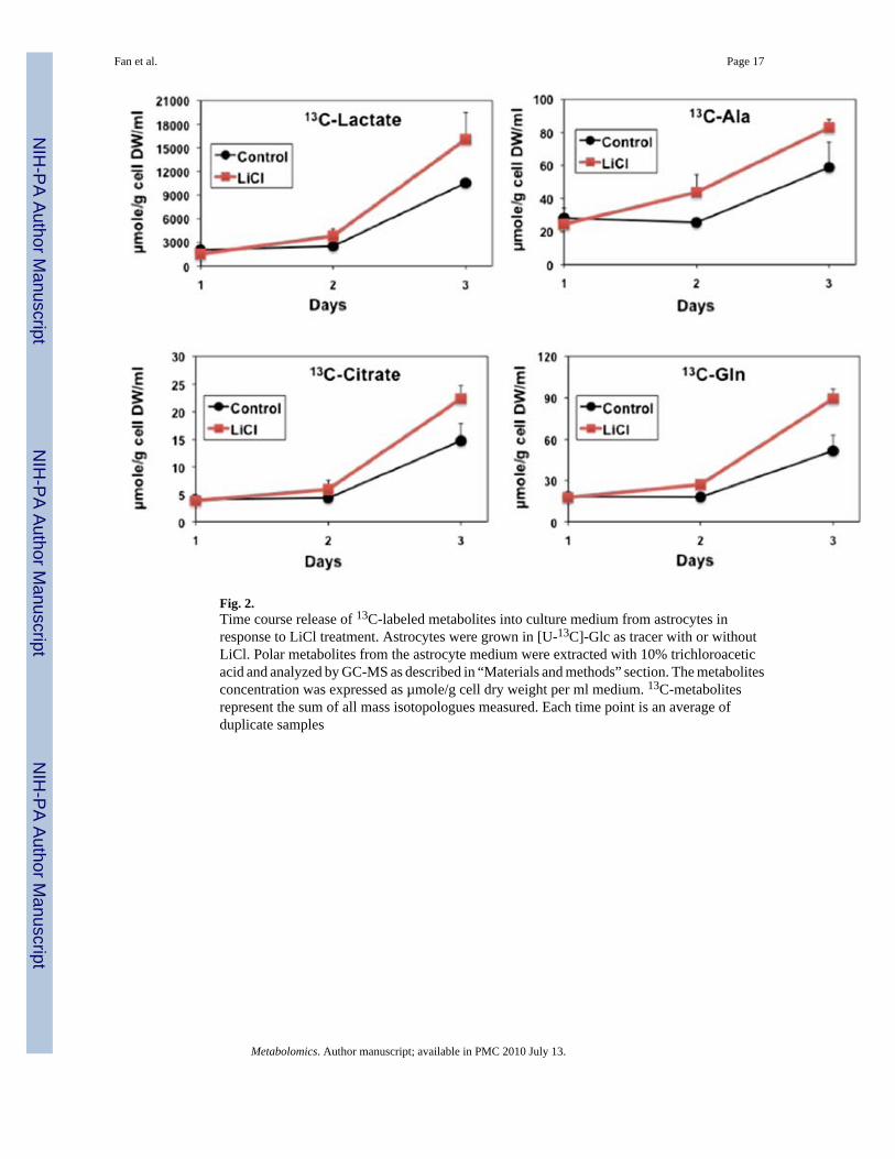
Fig. 2.Time course release of 13C-labeled metabolites into culture medium from astrocytes inresponse to LiCl treatment. Astrocytes were grown in [U-13C]-Glc as tracer with or withoutLiCl. Polar metabolites from the astrocyte medium were extracted with 10% trichloroaceticacid and analyzed by GC-MS as described in “Materials and methods” section. The metabolitesconcentration was expressed as µmole/g cell dry weight per ml medium. 13C-metabolitesrepresent the sum of all mass isotopologues measured. Each time point is an average ofduplicate samples
Fan et al. Page 17
Metabolomics. Author manuscript; available in PMC 2010 July 13.
NIH
-PA Author Manuscript
NIH
-PA Author Manuscript
NIH
-PA Author Manuscript

Fig. 3.1-D 1H-13C HSQC spectra of LiCl- versus control-treated astrocyte extracts. Astrocytes weregrown in [U-13C]-glucose (as tracer) with or without LiCl. Polar metabolites were extractedfrom treated astrocytes with 60% acetonitrile and spectra were acquired at 14.1 T as describedin “Materials and methods” section. Each 1H peak arose from protons directly attachedto 13C and the peak assignment denotes the 13C-carbon. Thus, the peak intensity reflects 13Cabundance of the attached carbon. Dashed lines highlight changes in peak intensity elicited byLiCl treatment
Fan et al. Page 18
Metabolomics. Author manuscript; available in PMC 2010 July 13.
NIH
-PA Author Manuscript
NIH
-PA Author Manuscript
NIH
-PA Author Manuscript

Fig. 4.LiCl-induced time course changes of selected metabolites and 13C-labeled metabolites incultured astrocytes grown in [U-13C]-glucose. The same polar extracts from Fig. 2. weresilylated and analyzed by GC-MS as described in “Materials and methods” section. Thedifference in metabolite concentrations between LiCl and control treatments for totalmetabolites (panel a) or sum of 13C-isotopologues (panel b) was plotted as a function oftreatment days. Each time point is an average of duplicate samples (see Table S1, SupplementalMaterials for standard error). Li-Control: metabolite concentrations of Li-treated cells minuscontrol; aKG: αKetoglu-tarate; GlyOH3P: αGlycerol-3-phosphate; 13C-metabolites: µmole/gdry cell mass of 13C-labeled metabolites; 13C3-Asp: triply 13C-labeled Asp
Fan et al. Page 19
Metabolomics. Author manuscript; available in PMC 2010 July 13.
NIH
-PA Author Manuscript
NIH
-PA Author Manuscript
NIH
-PA Author Manuscript

Fig. 5.LiCl-induced time course changes of selected metabolites and 13C-labeled metabolites incultured neurons grown in [U-13C]-glucose. The polar extracts of neuronal cells were preparedand analyzed by GC-MS as described in Fig. 3. Each time point is an average of duplicatesamples. Plots in panel a are as total metabolite concentration while plots in panel b are as thesum of all 13C-isotopologues (13C-metabolites) and selected individual 13C-isotopologues(13Cx-metabolites are mass isotopologues with x 13C-carbons)
Fan et al. Page 20
Metabolomics. Author manuscript; available in PMC 2010 July 13.
NIH
-PA Author Manuscript
NIH
-PA Author Manuscript
NIH
-PA Author Manuscript

Fig. 6.Expected 13C-positional isotopomer patterns of glycolytic and Krebs cycle metabolitessynthesized from [U-13C]-Glc by mammalian cells. Unlabeled carbons are in black circles,unscrambled 13C-carbons are in red circles, while scrambled 13C-carbons are in magenta,green, or orange circles. Reversible reactions are depicted by double-headed arrows whiletwo different pools of pyruvate are differentiated by black and blue letters. PC pyruvatecarboxylase, SCS Succinyl CoA synthetase. Panels a, c, d, respectively display the numberand position of 13C-carbons in metabolites after one, two, and three turns of normal Krebscycle activity. Panels b and e trace the number and position of 13C-carbons in metabolites ofKrebs cycle with input of anaplerotic pyruvate carboxylation
Fan et al. Page 21
Metabolomics. Author manuscript; available in PMC 2010 July 13.
NIH
-PA Author Manuscript
NIH
-PA Author Manuscript
NIH
-PA Author Manuscript

Fig. 7.GC-MS analysis of LiCl effect on 13C-isotopologue profile of selected neuronal metabolitesgrown in 13C-3-lactate. Neuronal cells were grown in 13C-lactate labeled at carbon 2 or 13C-Ala labeled at carbons 2 and 3 as tracer with or without LiCl. The polar extraction and GC-MS analysis were as described in Fig. 2. 13Cx-metabolites are mass isotopologues with x 13C-carbons, such as 13C1-Asp and 13C2-Asp are 13C-isotopologues with respectively, one andtwo 13C carbons. Each value is an average of duplicate samples
Fan et al. Page 22
Metabolomics. Author manuscript; available in PMC 2010 July 13.
NIH
-PA Author Manuscript
NIH
-PA Author Manuscript
NIH
-PA Author Manuscript

Fig. 8.Diagram of metabolic interactions between neurons and glia. This diagram summarizesneuronal and glial metabolism as well as their interactions revealed by SIRM and consistentwith the literature. Neurons and glia metabolize glucose via glycolysis and the Krebs cycle togenerate the neurotransmitter Glu and energy (ATP). During brain activation, Glu is releasedby neurons into the synaptic cleft, which is then transported to the surrounding glial endprocesses via glutamate transport 1 (GLT1). Once in the glia, Glu is converted to glutamine(Gln) by glutamine synthetase (GS). Gln is then released by the glia, transported into theneurons, and converted to Glu by phosphate-activated glutaminase (Glnase), which completesthe Glu/Gln cycle (Bittar et al. 1996; Haberg et al. 1998; Hyder et al. 2006; Magistretti,2009; Patel et al. 2004). Lithium stimulates glycolysis and lactate/Ala production as well asanaplerotic pyruvate carboxylation (PC) in the glia. Both lactate and Ala are ultimatelytransferred to neurons where they are used as oxidative substrates for ATP production and/orprecursor for Glu synthesis. The neuronal pathways from lactate to Glu involve conversion oflactate to pyruvate, followed by either the pyruvate dehydrogenase (PDH) or the PC pathwayin the Krebs cycle, the latter of which is enhanced by lithium. Red glucose depicts [U-13C]-glucose while green lactate and Ala represents 13C-3-lactate and 13C-2,3-Ala, respectively.Block arrows indicate transport processes. Italicized texts denote enzymes or pathways, redarrows in glia indicate glucose-associated pathways activated by lithium, and orange arrow
Fan et al. Page 23
Metabolomics. Author manuscript; available in PMC 2010 July 13.
NIH
-PA Author Manuscript
NIH
-PA Author Manuscript
NIH
-PA Author Manuscript

in neurons represents activation of PC linked to both lactate and glucose metabolism. Black,red, and green dots depict Glu in neurons, synaptic cleft, and glia, respectively
Fan et al. Page 24
Metabolomics. Author manuscript; available in PMC 2010 July 13.
NIH
-PA Author Manuscript
NIH
-PA Author Manuscript
NIH
-PA Author Manuscript