An isoform of ankyrin is localized at nodes of Ranvier in myelinated axons of central and peripheral...
12
An Isoform of Ankyrin Is Localized at Nodes of Ranvier in Myelinated Axons of Central and Peripheral Nerves Ekaterini Kordeli, Jonathan Davis, Bruce Trapp,¢ and Vann Bennett Ho~. rd Hughes Medical Institute, Department of Biochemistry, Duke University Medical Center, Durham, North Carolina 27710; and S Department of Neurology, Johns Hopkins University School of Medicine, Baltimore, Maryland 21205 Abstract. Two variants of ankyrin have been distin- guished in rat brain tissue using antibodies: a broadly distributed isoform (ankyrinB) that represents the ma- jor form of ankyrin in brain and another isoform with a restricted distribution (ankyrinR) that shares epitopes with erythrocyte ankyrin. The ankyrinR isoform was localized by immunofluorescence in cryosections of rat spinal cord gray matter and myelinated central and pe- ripheral nerves to: (a) perikarya and initial axonal seg- ments of neuron cells, (b) nodes of Ranvier of myelin- ated nerve with no detectable labeling in other areas of the myelinated axons, and (c) the axolemma of un- myelinated axons. Immunogold EM on ultrathin cryo- sections of myelinated nerve showed that ankyrinR was localized on the cytoplasmic face of the axolemma and was restricted to the nodal and, in some cases, para- nodal area. The major isoform of ankyrin in brain (ankyrinB) displayed a broad distribution on glial and neuronal cells of the gray matter and a mainly glial distribution in central myelinated axons with no significant labeling on the axolemma. These results show that (a) ankyrin isoforms display a differential distribution on glial and neuronal cells of the nervous tissue; (b) an isoform of ankyrin codistributes with the voltage-dependent sodium channel in both myelinated and unmyelinated nerve fibers. Ankyrin interacts in vitro with the voltage-dependent sodium channel (Srinivasan, Y., L. Elmer, J. Davis, V. Bennett, and K. Angelides. 1988. Nature (Lond.). 333:177-180). A specific interaction of an isoform of ankyrin with the sodium channel thus may play an important role in the morphogenesis and/or maintenance of the node of Ranvier. T HE node of Ranvier in the myelinated nerve fiber is a highly differentiated structure of the axonal plasma membrane (axolemma), where the myelin sheath is in- terrupted and saltatory conduction of the action potential takes place. Integral membrane proteins involved in salta- tory conduction, such as voltage-dependent sodium chan- nels (reviewed in references 11 and 44) and certain isoforms of the Na+/K÷ATPase (2), accumulate in high concentra- tions at the nodes and are present in very much lower density in the internodal regions of the axolemma underlying the my- elin sheath. Because axonal proteins are synthesized in the neuron cell body and subsequently transported in the axon, the node of Ranvier represents an interesting model for the study of the cellular mechanisms involved in the sorting, tar- geting, and local stabilization of integral proteins participat- ing in specialized membrane domains. Interactions between integral membrane proteins and cy- toskeletal components have been proposed to account for the formation and maintenance of the nodal membrane domain (45). A characteristic dense cytoplasmic coating underlines the nodal axolemma (33), indicating that a differentiated membrane skeleton is associated with the nodal integral pro- teins. Ankyrin is a peripheral membrane protein of potential relevance to the node of Ranvier, since it interacts in vitro with both integral membrane proteins known to be concen- trated at the nodes: the voltage-dependent sodium channel (38) and the kidney isoform(s) of the Na+/K÷ATPase (23, 28, 31). Ankyrin in erythrocytes links the anion channel (band 3) to a membrane skeleton composed of spectrin and actin (5, 26). Ankyrin is associated with the plasma mem- brane in many tissues where it is likely to also interconnect integral proteins with the spectrin-based membrane skeleton (16, 29). A major form of ankyrin in brain tissue has been isolated and characterized (12, 13). Brain ankyrin shares an- tigenic sites and a similar domain structure and binding properties with human erythrocyte ankyrin. Brain and eryth- rocyte ankyrin differ in relative affinities for membrane sites (14, 15) and for spectrin (9, 19), and are products of distinct genes (13; Otto E., T. McLaughlin, and V. Bennett, manu- script in preparation). Ankyrin is a diverse family of polypeptides and therefore is likely to be involved in local segregation of integral pro- teins through selective interactions between different an- kyrin isoforms and families of integral proteins. Two iso- forms of ankyrin have been reported in brain (30) together with two isoforms of spectrin (24, 34), and can be divided into two groups: those with a restricted localization and those with a broader localization in cells. Isoforms of an- © The Rockefeller University Press, 0021-9525/90/04/1341/12 $2.00 The Journal of Cell Biology, Volume 110, April 1990 1341-1352 [341 on November 15, 2014 jcb.rupress.org Downloaded from Published April 1, 1990
-
Upload
independent -
Category
Documents
-
view
0 -
download
0
Transcript of An isoform of ankyrin is localized at nodes of Ranvier in myelinated axons of central and peripheral...
An Isoform of Ankyrin Is Localized at Nodes of Ranvier in Myelinated Axons of Central and Peripheral Nerves Ekaterini Kordeli , J o n a t h a n Davis, Bruce Trapp,¢ and Vann Bennet t
Ho~. rd Hughes Medical Institute, Department of Biochemistry, Duke University Medical Center, Durham, North Carolina 27710; and S Department of Neurology, Johns Hopkins University School of Medicine, Baltimore, Maryland 21205
Abstract. Two variants of ankyrin have been distin- guished in rat brain tissue using antibodies: a broadly distributed isoform (ankyrinB) that represents the ma- jor form of ankyrin in brain and another isoform with a restricted distribution (ankyrinR) that shares epitopes with erythrocyte ankyrin. The ankyrinR isoform was localized by immunofluorescence in cryosections of rat spinal cord gray matter and myelinated central and pe- ripheral nerves to: (a) perikarya and initial axonal seg- ments of neuron cells, (b) nodes of Ranvier of myelin- ated nerve with no detectable labeling in other areas of the myelinated axons, and (c) the axolemma of un- myelinated axons. Immunogold EM on ultrathin cryo- sections of myelinated nerve showed that ankyrinR was localized on the cytoplasmic face of the axolemma and was restricted to the nodal and, in some cases, para- nodal area. The major isoform of ankyrin in brain
(ankyrinB) displayed a broad distribution on glial and neuronal cells of the gray matter and a mainly glial distribution in central myelinated axons with no significant labeling on the axolemma. These results show that (a) ankyrin isoforms display a differential distribution on glial and neuronal cells of the nervous tissue; (b) an isoform of ankyrin codistributes with the voltage-dependent sodium channel in both myelinated and unmyelinated nerve fibers. Ankyrin interacts in vitro with the voltage-dependent sodium channel (Srinivasan, Y., L. Elmer, J. Davis, V. Bennett, and K. Angelides. 1988. Nature (Lond.). 333:177-180). A specific interaction of an isoform of ankyrin with the sodium channel thus may play an important role in the morphogenesis and/or maintenance of the node of Ranvier.
T HE node of Ranvier in the myelinated nerve fiber is a highly differentiated structure of the axonal plasma membrane (axolemma), where the myelin sheath is in-
terrupted and saltatory conduction of the action potential takes place. Integral membrane proteins involved in salta- tory conduction, such as voltage-dependent sodium chan- nels (reviewed in references 11 and 44) and certain isoforms of the Na+/K÷ATPase (2), accumulate in high concentra- tions at the nodes and are present in very much lower density in the internodal regions of the axolemma underlying the my- elin sheath. Because axonal proteins are synthesized in the neuron cell body and subsequently transported in the axon, the node of Ranvier represents an interesting model for the study of the cellular mechanisms involved in the sorting, tar- geting, and local stabilization of integral proteins participat- ing in specialized membrane domains.
Interactions between integral membrane proteins and cy- toskeletal components have been proposed to account for the formation and maintenance of the nodal membrane domain (45). A characteristic dense cytoplasmic coating underlines the nodal axolemma (33), indicating that a differentiated membrane skeleton is associated with the nodal integral pro- teins. Ankyrin is a peripheral membrane protein of potential relevance to the node of Ranvier, since it interacts in vitro
with both integral membrane proteins known to be concen- trated at the nodes: the voltage-dependent sodium channel (38) and the kidney isoform(s) of the Na+/K÷ATPase (23, 28, 31). Ankyrin in erythrocytes links the anion channel (band 3) to a membrane skeleton composed of spectrin and actin (5, 26). Ankyrin is associated with the plasma mem- brane in many tissues where it is likely to also interconnect integral proteins with the spectrin-based membrane skeleton (16, 29). A major form of ankyrin in brain tissue has been isolated and characterized (12, 13). Brain ankyrin shares an- tigenic sites and a similar domain structure and binding properties with human erythrocyte ankyrin. Brain and eryth- rocyte ankyrin differ in relative affinities for membrane sites (14, 15) and for spectrin (9, 19), and are products of distinct genes (13; Otto E., T. McLaughlin, and V. Bennett, manu- script in preparation).
Ankyrin is a diverse family of polypeptides and therefore is likely to be involved in local segregation of integral pro- teins through selective interactions between different an- kyrin isoforms and families of integral proteins. Two iso- forms of ankyrin have been reported in brain (30) together with two isoforms of spectrin (24, 34), and can be divided into two groups: those with a restricted localization and those with a broader localization in cells. Isoforms of an-
© The Rockefeller University Press, 0021-9525/90/04/1341/12 $2.00 The Journal of Cell Biology, Volume 110, April 1990 1341-1352 [341
on Novem
ber 15, 2014jcb.rupress.org
Dow
nloaded from
Published April 1, 1990
kyrin and spectrin with a restricted distribution are localized in dendrites and neuronal cell bodies, and are expressed late in development. Isoforms of ankyrin and spectrin (fodrin) with a broader distribution occur along axons as well as cell bodies, and assemble on the plasma membrane early in de- velopment. Two isoforms ofankyrin have been also localized recently in kidney tissue: a restricted isoform immunologi- cally related to erythrocyte ankyrin that is present in basolat- eral domains of only two cell types, and an isoform with a broad distribution on the membranes of different cell types that cross-reacts preferentially with antibody against the ma- jor form of ankyrin isolated from brain (15).
We report here that brain tissue contains a restricted iso- form of ankyrin (ankyrinR) that is localized at specific, mainly neuronal sites including perikarya of neuron cells, initial segments of axons and the nodes of Ranvier of myelin- ated nerves, whereas the major isoform of brain ankyrin (ankyrinB) displays a broader distribution in both glial and neuronal cells of the central nervous system. A role of the restricted isoform of ankyrin in the formation and mainte- nance of the node of Ranvier is proposed.
Materials and Methods
Antibodies Affinity-purified polyclonal antibodies against human erythrocyte ankyrin (8) and human erythrocyte anion channel (reference 7, band 3) were de- scribed previously. Antisera against ankyrina, the major isoform of an- kyrin in brain tissue, were prepared using as an immunogen bovine brain ankyrin that was isolated in a final step of gel filtration in the presence of 1% SDS and combined with glutaraldehyde-activated rabbit serum albumin before immunization. Affinity-purified antibody against brain ankyrins was isolated from these antisera as described (13). Antibodies specific for ankyrinR, the erythroid isoform of ankyrin in brain tissue, were isolated by adsorption of the affinity-purified antibody against human erythrocyte an- kyrln with bovine brain ankyrin (ankyrins) coupled to ngarose, resulting in elimination of Ig cross-reacting with both forms of ankyrin. In some experi- ments, an affinity-purified antibody against the Mr ffi 90000 domain of hu- man erythrocyte ankyrin (6) was used, because of its high reactivity in im- munocytochemistry.
Procedures Brain tissue was obtained from rats perfused with 150 mM NaCI, 10 mM sodium phosphate pH 7.5, 5 mM NaEDTA and 2.5 mM diisopropylfluoro- phosphate (DFP, Sigma) at 4°C for 15 rain, to minimize proteolysis and to remove erythrocytes. The tissue was homogenized with a Polytron (Brink- mann Instruments, Westbury, NY) and the postnuclear membrane fraction (30000 g pellet) isolated as described (12). SDS-PAGE and immunoblot analysis was performed as described (12). Erythrocyte ghost membranes (4) and bovine brain ankyrin (13) were prepared as described. Rat brain ankyrin was partially purified by chromatographic steps of gel filtration, Mono Q ion-exchange chromatography and hydroxylapatite chromatography.
Light Microscopic Immunocytochemistry Adult male Sprngue-Dawley rats were perfused with either 1% NaNO3, pH 7.5 or PBS containing 5 mM NaEDTA, and then with 4% paraformaldehyde and 0.08 M phosphate buffer, pH 7.5. The spinal cord, ventral and dorsal roots, and sciatic nerve were removed and placed in fixative overnight. For 4-#m-thick cryostat sections, the tissue was cryoprotected with 0.7 M su- crose, frozen in liquid nitrogen-cooled isopentane, and cut at -20°C. Sec- tions were mounted on egg white-coated glass slides, air dried, and stored at -80°C. For l-;tm-thick cryosections, the tissue was cryoprotected with 2.3 M sucrose and 30% polyvinyl pyrrolidone. The tissue pieces were mounted on specimens stubs, frozen in liquid nitrogen, and cut at -80°C using an ultracryomicrotome, l-ttm-thick cryosections were mounted on polylysine-coated glass slides and rinsed in PBS. Sections were immuno- stained using antibodies at 1-5 #g/ml by either indirect immunofluorescence
or by the peroxidase-antiperoxidase procedure as described previously (42). After the immunostaining procedure, the sections prepared for immunofiu- orescence were mounted in 60% glycerol-PBS. Sections stained by the PAP technique were dehydrated and overlaid with coverslips. As a control, a 20- fold molar excess of heat-denatured (60°C, 25 min) antigenic proteins was added to the specific antibodies and allowed to interact for 1-3 h at 0°C, before incubation with sections.
Electron Microscopic lmmunocytochemistry 7-d-old Sprague-Dawley rats were perfused with a solution containing 4% paraformaldehyde, 2.5% glutaraldehyde, and 0.08 M phosphate buffer. The IA and L5 ventral root and dorsal root ganglion were dissected from the animal and placed in fixative overnight. Segments of this tissue (2-4 mm) were cryoprotected with 2.3 M sucrose and 30% polyvinyl pyrrolidone, placed on specimen stubs, and frozen in liquid nitrogen. Ultrathin cryosec- tions (100-120 nm thick) were cut on an ultracryomicrotome maintained at - I I0°C. The sections were transferred to Formvar- and carbon-coated grids and maintained on 2% gelatin in PBS for 2 h. Sections were immuno- stained by colloidal gold procedures, postfixed, and embedded in methyl- cellulose using previously described modifications (42) of standard proce- dares (37, 41).
Results
Two lsoforms of Ankyrin Are lmmunologically Distinguished in Rat Brain Several findings during isolation of brain ankyrin (13) sug- gested the possibility that brain tissue contains two forms of ankyrin: (a) a major, extractable form that is the principal component of purified brain ankyrin (Mr = 220,000 and 210000 in bovine brain or 220000 in rat brain); (b) a second population of ankyrin polypeptides that remain associated with brain membranes even after extensive extraction with a combination of 0.8 M KI and a detergent (13). The retained protein contains an additional polypeptide of Mr = 186,000 that is not present in the extracted fraction of ankyrin. The M, = 186000 polypeptide cross-reacts with antibody against erythrocyte ankyrin, but does not cross-react with antibody against bovine brain ankyrin (Fig. 1, lanes 2 in A and D, re- spectively). It is of interest that human erythrocyte ankyrin also includes a minor polypeptide of Mr = 186000 (refer- ence 19; band 2.2) in addition to the major polypeptide of Mr = 215,000 (band 2.1). These observations suggested the possibility that the Mr = 186,000 polypeptide associated with brain membranes represents an isoform of ankyrin closely related to erythrocyte ankyrin and that this polypep- tide may be analogous to band 2.2 of erythrocyte mem- branes.
In this study, the presence of an erythroid isoform of an- kyrin in brain has been demonstrated using an erythroid- specific antibody that does not react with the major form of brain ankyrin (Fig. 1). The erythroid-specific antibody was prepared by passage of afffinity-purified antibody against erythrocyte ankyrin through an affinity column containing the major form of brain ankyrin. Erythrocyte ankyrin anti- body before immunoadsorption with brain ankyrin (Fig. 1 A) cross-reacted with isolated rat brain ankyrin, which mi- grates as a single polypeptide of Mr = 220,000 (lane 3) and with two polypeptides in rat brain homogenates of Mr = 215,000-220,000 and 186,000 (lane 2). The antibody de- pleted by immunoadsorption against brain ankyrin (Fig. 1 B) no longer cross-reacted with isolated rat brain ankyrin (lanes 3, 3'), but still reacted with the two forms of ankyrin of Mr = 215,000 (band 2.1) and 186,000 (band 2.2) in human
The Journal of Cell Biology, Volume I I0, 1990 1342
on Novem
ber 15, 2014jcb.rupress.org
Dow
nloaded from
Published April 1, 1990
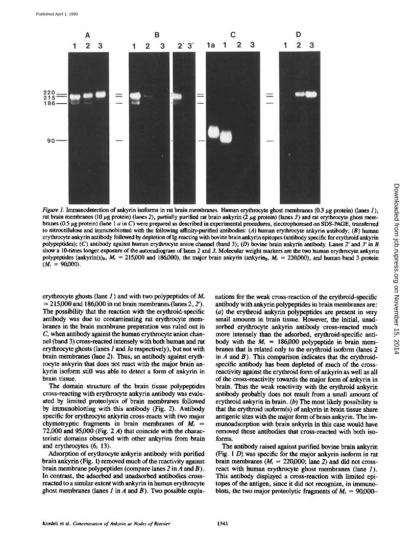
Figure 1. Immunodetection of ankyrin isoforms in rat brain membranes. Human erythrocyte ghost membranes (0.3/~g protein) (lanes I), rat brain membranes (10/zg protein) (lanes 2), partially purified rat brain ankyrin (2/zg protein) (lanes 3) and rat erythrocyte ghost mem- branes (0.5 ~g protein) (lane 1 a in C) were prepared as described in experimental procedures, electropboresed on SDS-PAGE, transferred to nitrocellulose and immunoblotted with the following afffinity-purified antibodies: (A) human erythrocyte ankyrin antibody; (B) human erythrocyte ankyrin antibody followed by depletion of Ig reacting with bovine brain ankyrin epitopes (antibody specific for erythroid ankyrin polypeptides); (C) antibody against human erythrocyte anion channel (band 3); (D) bovine brain ankyrin antibody. Lanes 2' and 3' in B show a 10-times longer exposure of the autoradiogram of lanes 2 and 3. Molecular weight markers are the two human erythrocyte ankyrin polypeptides (ankyrin(s)R, Mr = 215,000 and 186,000), the major brain ankyrin (ankyrina, M, = 220,000), and human band 3 protein (Mr = 90,000).
erythrocyte ghosts (lane 1 ) and with two polypeptides of Mr = 215,000 and 186,000 in rat brain membranes (lanes 2, 2'). The possibility that the reaction with the erythroid-specific antibody was due to contaminating rat erythrocyte mem- branes in the brain membrane preparation was ruled out in C, when antibody against the human erythrocyte anion chan- nel (band 3) cross-reacted intensely with both human and rat erythrocyte ghosts (lanes 1 and la respectively), but not with brain membranes (lane 2). Thus, an antibody against eryth- rocyte ankyrin that does not react with the major brain an- kyrin isoform still was able to detect a form of ankyrin in brain tissue.
The domain structure of the brain tissue polypeptides cross-reacting with erythrocyte ankyrin antibody was evalu- ated by limited proteolysis of brain membranes followed by immunoblotting with this antibody (Fig. 2). Antibody specific for erythrocyte ankyrin cross-reacts with two major chymotryptic fragments in brain membranes of Mr = 72,000 and 95,000 (Fig. 2 A) that coincide with the charac- teristic domains observed with other ankyrins from brain and erythrocytes (6, 13).
Adsorption of erythrocyte ankyrin antibody with purified brain ankyrin (Fig. 1) removed much of the reactivity against brain membrane polypeptides (compare lanes 2 in A and B). In contrast, the adsorbed and unadsorbed antibodies cross- reacted to a similar extent with ankyrin in human erythrocyte ghost membranes (lanes 1 in A and B). Two possible expla-
nations for the weak cross-reaction of the erythroid-specific antibody with ankyrin polypeptides in brain membranes are: (a) the erythroid ankyrin polypeptides are present in very small amounts in brain tissue. However, the initial, unad- sorbed erythrocyte ankyrin antibody cross-reacted much more intensely than the adsorbed, erythroid-specific anti- body with the Mr = 186,000 polypeptide in brain mem- branes that is related only to the erythroid isoform (lanes 2 in A and B). This comparison indicates that the erythroid- specific antibody has been depleted of much of the cross- reactivity against the erythroid form of ankyrin as well as all of the cross-reactivity towards the major form of ankyrin in brain. Thus the weak reactivity with the erythroid ankyrin antibody probably does not result from a small amount of erythroid ankyrin in brain. (b) The most likely possibility is that the erythroid isoform(s) of ankyrin in brain tissue share antigenic sites with the major form of brain ankyrin. The im- munoadsorption with brain ankyrin in this case would have removed those antibodies that cross-reacted with both iso- forms.
The antibody raised against purified bovine brain ankyrin (Fig. 1 D) was specific for the major ankyrin isoform in rat brain membranes (Mr = 220,000; lane 2) and did not cross- react with human erythrocyte ghost membranes (lane 1). This antibody displayed a cross-reaction with limited epi- topes of the antigen, since it did not recognize, in immuno- blots, the two major proteolytic fragments of Mr = 90,000-
Kordeli et al. Concentration of Ankyrin at Nodes of Ranvier 1343
on Novem
ber 15, 2014jcb.rupress.org
Dow
nloaded from
Published April 1, 1990
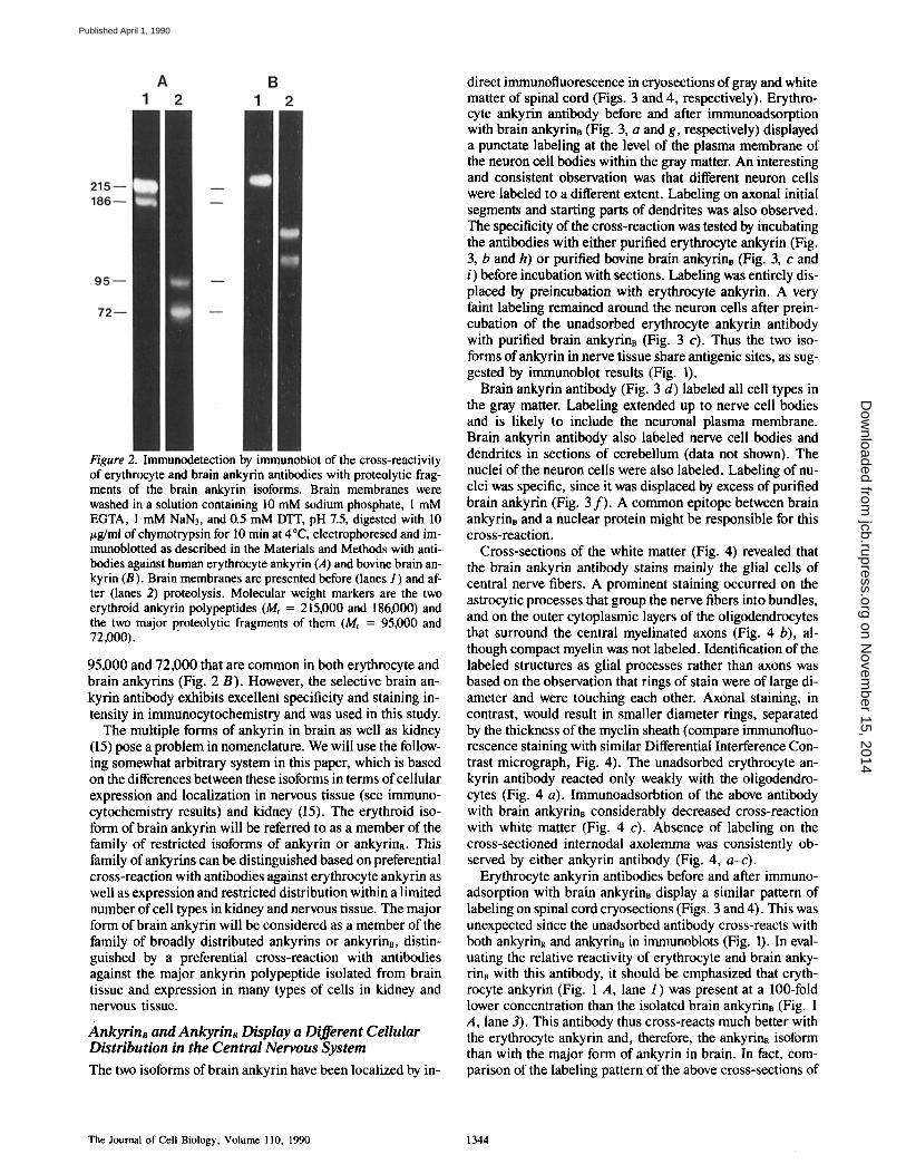
Figure 2. Immunodetection by immunoblot of the cross-reactivity of erythrocyte and brain ankyrin antibodies with proteolytic frag- ments of the brain ankyrin isoforms. Brain membranes were washed in a solution containing 10 mM sodium phosphate, 1 mM EGTA, 1 mM NAN3, and 0.5 mM DTT, pH 7.5, digested with 10 /~g/mi of chymotrypsin for 10 min at 4°C, electrophoresed and im- munoblotted as described in the Materials and Methods with anti- bodies against human erythrocyte ankyrin (A) and bovine brain an- kyrin (B). Brain membranes are presented before (lanes 1 ) and af- ter (lanes 2) proteolysis. Molecular weight markers are the two erythroid ankyrin polypeptides (Mr = 215,000 and 186,000) and the two major proteolytic fragments of them (Mr = 95,000 and 72,000).
95,000 and 72,000 that are common in both erythrocyte and brain ankyrins (Fig. 2 B). However, the selective brain an- kyrin antibody exhibits excellent specificity and staining in- tensity in immunocytochemistry and was used in this study.
The multiple forms of ankyrin in brain as well as kidney (15) pose a problem in nomenclature. We will use the follow- ing somewhat arbitrary system in this paper, which is based on the differences between these isoforms in terms of cellular expression and localization in nervous tissue (see immuno- cytochemistry results) and kidney (15). The erythroid iso- form of brain ankyrin will be referred to as a member of the family of restricted isoforms of ankyrin or ankyrinR. This family of ankyrins can be distinguished based on preferential cross-reaction with antibodies against erythrocyte ankyrin as well as expression and restricted distribution within a limited number of cell types in kidney and nervous tissue. The major form of brain ankyrin will be considered as a member of the family of broadly distributed ankyrins or ankyrina, distin- guished by a preferential cross-reaction with antibodies against the major ankyrin polypeptide isolated from brain tissue and expression in many types of cells in kidney and nervous tissue.
AnkyrinB and AnkyrinR Display a Different Cellular Distribution in the Central Nervous System' The two isoforms of brain ankyrin have been localized by in-
direct immunofluorescence in cryosections of gray and white matter of spinal cord (Figs. 3 and 4, respectively). Erythro- cyte ankyrin antibody before and after immunoadsorption with brain ankyrin~ (Fig. 3, a and g, respectively) displayed a punctate labeling at the level of the plasma membrane of the neuron cell bodies within the gray matter. An interesting and consistent observation was that different neuron cells were labeled to a different extent. Labeling on axonal initial segments and starting parts of dendrites was also observed. The specificity of the cross-reaction was tested by incubating the antibodies with either purified erythrocyte ankyrin (Fig. 3, b and h) or purified bovine brain ankyrinB (Fig. 3, c and i) before incubation with sections. Labeling was entirely dis- placed by preincubation with erythrocyte ankyrin. A very faint labeling remained around the neuron cells after prein- cubation of the unadsorbed erythrocyte ankyrin antibody with purified brain ankyrinB (Fig. 3 c). Thus the two iso- forms of ankyrin in nerve tissue share antigenic sites, as sug- gested by immunoblot results (Fig. 1).
Brain ankyrin antibody (Fig. 3 d) labeled all cell types in the gray matter. Labeling extended up to nerve cell bodies and is likely to include the neuronal plasma membrane. Brain ankyrin antibody also labeled nerve cell bodies and dendrites in sections of cerebellum (data not shown). The nuclei of the neuron cells were also labeled. Labeling of nu- clei was specific, since it was displaced by excess of purified brain ankyrin (Fig. 3 f ) . A common epitope between brain ankyrinB and a nuclear protein might be responsible for this cross-reaction.
Cross-sections of the white matter (Fig. 4) revealed that the brain ankyrin antibody stains mainly the glial cells of central nerve fibers. A prominent staining occurred on the astrocytic processes that group the nerve fibers into bundles, and on the outer cytoplasmic layers of the oligodendrocytes that surround the central myelinated axons (Fig. 4 b), al- though compact myelin was not labeled. Identification of the labeled structures as glial processes rather than axons was based on the observation that rings of stain were of large di- ameter and were touching each other. Axonal staining, in contrast, would result in smaller diameter rings, separated by the thickness of the myelin sheath (compare immunofluo- rescence staining with similar Differential Interference Con- trast micrograph, Fig. 4). The unadsorbed erythrocyte an- kyrin antibody reacted only weakly with the oligodendro- cytes (Fig. 4 a). Immunoadsorbtion of the above antibody with brain ankyrinB considerably decreased cross-reaction with white matter (Fig. 4 c). Absence of labeling on the cross-sectioned internodal axolemma was consistently ob- served by either ankyrin antibody (Fig. 4, a-c).
Erythrocyte ankyrin antibodies before and after immuno- adsorption with brain ankyrinB display a similar pattern of labeling on spinal cord cryosections (Figs. 3 and 4). This was unexpected since the unadsorbed antibody cross-reacts with both ankyrinR and ankyrinB in immunoblots (Fig. 1). In eval- uating the relative reactivity of erythrocyte and brain anky- rinB with this antibody, it should be emphasized that eryth- rocyte ankyrin (Fig. 1 A, lane 1) was present at a 100-fold lower concentration than the isolated brain ankyrinB (Fig. 1 A, lane 3). This antibody thus cross-reacts much better with the erythrocyte ankyrin and, therefore, the ankyrinR isoform than with the major form of ankyrin in brain. In fact, com- parison of the labeling pattern of the above cross-sections of
The Journal of Cell Biology, Volume 110, 1990 1344
on Novem
ber 15, 2014jcb.rupress.org
Dow
nloaded from
Published April 1, 1990
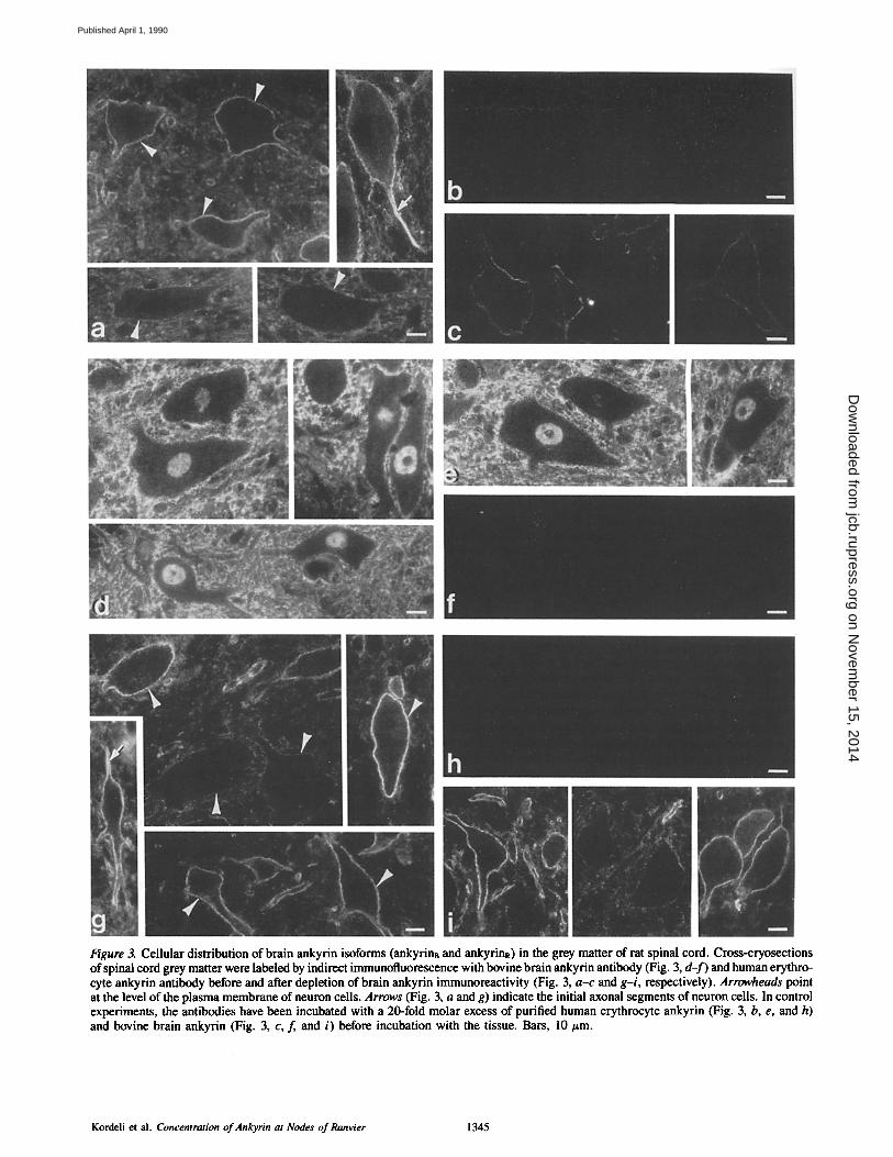
Figure 3. Cellular distribution of brain ankyrin isoforms (ankyrins and ankyrinR) in the grey matter of rat spinal cord. Cross-cryosections of spinal cord grey matter were labeled by indirect immunofluorescence with bovine brain ankyrin antibody (Fig. 3, d- f ) and human erythro- cyte ankyrin antibody before and after depletion of brain ankyrin immunoreactivity (Fig. 3, a-c and g-i, respectively). Arrowheads point at the level of the plasma membrane of neuron cells. Arrows (Fig. 3, a and g) indicate the initial axonal segments of neuron cells. In control experiments, the antibodies have been incubated with a 20-fold molar excess of purified human erythrocyte ankyrin (Fig. 3, b, e, and h) and bovine brain ankyrin (Fig. 3, c, f and i) before incubation with the tissue. Bars, l0 #m.
Kordeli et al. Concentration of Ankyrin at Nodes of Ranvier 1345
on Novem
ber 15, 2014jcb.rupress.org
Dow
nloaded from
Published April 1, 1990
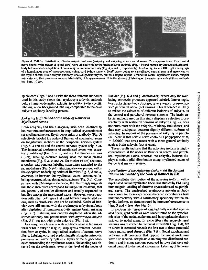
Figure 4. Cellular distribution of brain ankyrin isoforms (ankyrins and ankyrinR in rat central nerve. Cmss-cryoseetions of rat central nerve fibers (white matter of spinal cord) were labeled with bovine brain ankyrin antibody (Fig. 4 b) and human erythrocyte ankyrin anti- body before and after depletion of brain ankyrin immunoreactivity (Fig. 4, a and c, respectively). Inset in Fig. 4 c is a DIC light micrograph of a homologous area of cross-sectioned spinal cord (white matter). Small arrow points to a myelinated central axon and arrowhead to the myelin sheath. Brain ankyrin antibody labels oligodendrocytes, but not compact myelin, around the central myelinated axons. Subpial astrocytes and their processes are also labeled (Fig. 4 b, open arrows). Note the absence of labeling on the axolemma with all three antibod- ies. Bars, 10 #m.
spinal cord (Figs. 3 and 4) with the three different antibodies used in this study shows that erythrocyte ankyrin antibody before immunoadsorption exhibits, in addition to the specific labeling, a low background labeling comparable to the brain ankyrin antibody labeling pattern.
AnkyrinR Is Enriched at the Node of Ranvier in Myelinated Axons
Brain ankyrinR and brain ankyrine have been localized by indirect immunofluorescence in longitudinal cryosections of rat myelinated nerve. Erythrocyte ankyrin antibody (Fig. 5) selectively labeled the nodes of Ranvier of myelinated axons on longitudinal sections of the peripheral nervous system (Fig. 5, a and d) and the central nervous system (Fig. 5 c). The internodal axolemma of myelinated axons was essen- tially unlabeled (Fig. 5, a, c, and d). On thinner sections (1 #m), labeling occurred mainly near the nodal plasma membrane (Fig. 5, a, c, and e). On thicker (4/zm) sections, a weaker and punctate labeling sometimes extended to the paranodal area (Fig. 5,f-h). Staining also was present within the cytoplasm underlying nodes of Ranvier (Fig. 5, d and h, asterisk). In between the myelinated axons, continuous la- beling occurred along elongated structures (Fig. 5 a). Com- parison with EM images (see below, Fig. 8) strongly suggests that these structures correspond to unmyelinated axons, that are generally of smaller diameter and usually organized in bundles among the myelinated axons. However, cross-reac- tion with other cell types that exist between myelinated ax- ons, such as fibroblasts, can not be excluded. Nodes of Ran- vier were still stained with the erythrocyte ankyrin antibody after depletion of immunoreactivity against brain ankyrinB (Fig. 5 i). Labeling was entirely displaced when the ad- sorbed antibody was ~ preincubated with erythrocyte ankyrin (Fig. 5 j ) but not with brain ankyrinB (Fig. 5 k).
Ankyrins, which reacts with antibody against the major form of brain ankyrin (Fig. 6), displayed a different localiza- tion from ankyrinR in longitudinal sections of central nerve fibers. Labeling occurred predominantly along the astrocytic processes and outer cytoplasmic layers of the oligodendro- cytes surrounding the myelinated axons. No labeling was ob- served on the axolemma, even at the level of the nodes of
Ranvier (Fig. 6, d and g, arrowheads), where only the over- laying astrocytic processes appeared labeled. Interestingly, brain ankyrin antibody displayed a very weak cross-reaction with peripheral nerve (not shown). This difference is likely to reflect the existence of different isoforms of ankyrins in the central and peripheral nervous systems. The brain an- kyrin antibody used in this study displays a selective cross- reactivity with restricted domains of ankyrin (Fig. 2), does not cross-react with the ankyrins of kidney (not shown) and thus may distinguish between slightly different isoforms of ankyrins. In support of the presence of ankyrinB in periph- eral nerve is that sciatic nerve contains a polypeptide of Mr = 220,000 that cross-reacts with a more general antibody against brain ankyrin (not shown).
These results indicate that the ankyrin~ isoform is highly concentrated at the nodes of Ranvier in central and periph- eral myelinated axons, whereas the ankyrins isoform dis- plays a mainly glial distribution along myelinated axons of the central nervous system.
Localization of the Ankyrin~ lsoform on the Axonal Plasma Membrane of the Node of Ranvier by EM
The subcellular distribution of the ankyrinR isoform within myelinated and unmyelinated fibers was studied by EM using immunogold-labeling of ultrathin cryosections of rat periph- eral nerve. The unadsorbed erythrocyte ankyrin antibody was chosen for these experiments because it combines a high immunoreactivity with a satisfactory specificity for the an- kyrinR isoform, as demonstrated by immunofluorescence in Figs. 3 and 4 (see also Fig. 5).
In electron micrographs of longitudinally oriented myelin- ated fibers, gold particles were concentrated on the cytoplas- mic side of the nodal axolemma and in cytoplasmic sites re- stricted to nodal areas. In some fibers the subaxolemmal staining was restricted to the nodal axolemma (Fig. 7 A), but in others it extended beneath the first two to three paranodal loops and stopped abruptly (Fig. 7 B). Nodal axoplasm and Schwann cell processes that oppose the nodal axolemma were also labeled. Axoplasmic staining was distributed ran- domly and in some sections occurred in rows that were ori- ented parallel to the nodal axolemma. Labeling of Schwann
The Journal of Cell Biology, Volume 110, 1990 1346
on Novem
ber 15, 2014jcb.rupress.org
Dow
nloaded from
Published April 1, 1990
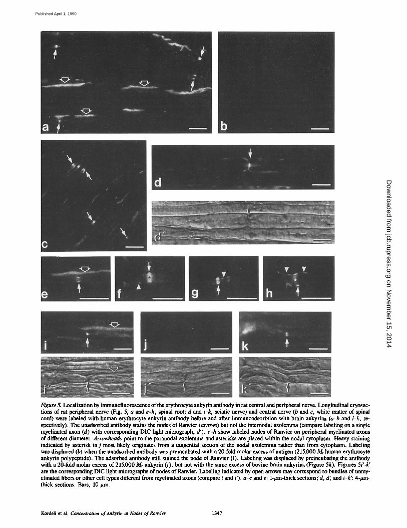
Figure 5. Localization by immunofluorescence of the erythrocyte ankyrin antibody in rat central and peripheral nerve. Longitudinal cryosec- tions of rat peripheral nerve (Fig. 5, a and e-h, spinal root; d and i-k, sciatic nerve) and central nerve (b and c, white matter of spinal cord) were labeled with human erythrocyte ankyrin antibody before and after immunoadsorbtion with brain ankyrins (a-h and i-k, re- spectively). The unadsorbed antibody stains the nodes of Ranvier (arrows) but not the intemodal axolemma (compare labeling on a single myelinated axon (d) with corresponding DIC light micrograph, d'). e-h show labeled nodes of Ranvier on peripheral myelinated axons of different diameter. Arrowheads point to the paranodal axolemma and asterisks are placed within the nodal cytoplasm. Heavy staining indicated by asterisk in f most likely originates from a tangential section of the nodal axolemma rather than from cytoplasm. Labeling was displaced (b) when the unadsorbed antibody was preincubated with a 20-fold molar excess of antigen (215,000 Mr human erythrocyte ankyrin polypeptide). The adsorbed antibody still stained the node of Ranvier (i). Labeling was displaced by preincubating the antibody with a 20-fold molar excess of 215,000 Mr ankyrin (j), but not with the same excess of bovine brain ankyrinB (Figure 5k). Figures 5i'-k' are the corresponding DIC light micrographs of nodes of Ranvier. Labeling indicated by open arrows may correspond to bundles of unmy- elinated fibers or other cell types different from myelinated axons (compare i and i'). a-c and e: l-#m-thick sections; d, d', and i-k': 4-#m- thick sections. Bars, 10 #m.
Kordeli et al. Concentration of Ankyrin at Nodes of Ranvier 1347
on Novem
ber 15, 2014jcb.rupress.org
Dow
nloaded from
Published April 1, 1990
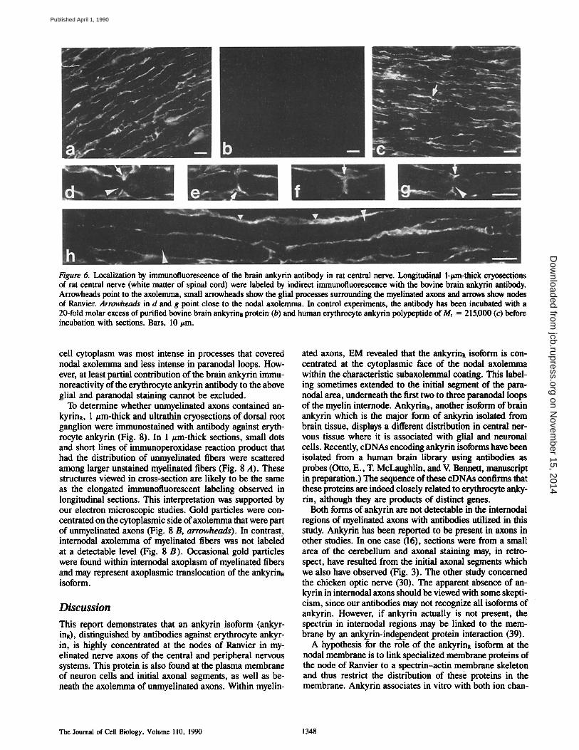
Figure 6. Localization by immunofluorescence of the brain ankyrin antibody in rat central nerve. Longitudinal l-/~m-thick cryosections of rat central nerve (white matter of spinal cord) were labeled by indirect immunofluorescence with the bovine brain ankyrin antibody. Arrowheads point to the axolemma, small arrowheads show the glial processes surrounding the myelinated axons and arrows show nodes of Ranvier. Arrowheads in d and g point close to the nodal axolemma. In control experiments, the antibody has been incubated with a 20-fold molar excess of purified bovine brain ankyrin8 protein (b) and human erythrocyte ankyrin polypeptide of Mr = 215,000 (c) before incubation with sections. Bars, 10/~m.
cell cytoplasm was most intense in processes that covered nodal axolemma and less intense in paranodal loops. How- ever, at least partial contribution of the brain ankyrin immu- noreactivity of the erythrocyte ankyrin antibody to the above glial and parunodal staining cannot be excluded.
To determine whether unmyelinated axons contained an- kyrinR, 1 t~m-thick and ultrathin cryosections of dorsal root ganglion were immunostained with antibody against eryth- rocyte ankyrin (Fig. 8). In 1 ttm-thick sections, small dots and short lines of immunoperoxidase reaction product that had the distribution of unmyelinated fibers were scattered among larger unstained myelinated fibers (Fig. 8 A). These structures viewed in cross-section are likely to be the same as the elongated immunofluorescent labeling observed in longitudinal sections. This interpretation was supported by our electron microscopic studies. Gold particles were con- centrated on the cytoplasmic side of axolemma that were part of unmyelinated axons (Fig. 8 B, arrowheads). In contrast_, internodal axolemma of myelinated fibers was not labeled at a detectable level (Fig. 8 B). Occasional gold particles were found within internodal axoplasm of myelinated fibers and may represent axoplasmic translocation of the ankyrinR isoform.
Discussion
This report demonstrates that an ankyrin isoform (ankyr- inR), distinguished by antibodies against erythrocyte ankyr- in, is highly concentrated at the nodes of Ranvier in my- elinated nerve axons of the central and peripheral nervous systems. This protein is also found at the plasma membrane of neuron cells and initial axonal segments, as well as be- neath the axolemma of unmyelinated axons. Within myelin-
ated axons, EM revealed that the ankyrinR isoform is con- centrated at the cytoplasmic face of the nodal axolemma within the characteristic subaxolemmal coating. This label- ing sometimes extended to the initial segment of the para- nodal area, underneath the first two to three paranodal loops of the myelin internode. AnkyrinB, another isoform of brain ankyrin which is the major form of ankyrin isolated from brain tissue, displays a different distribution in central ner- vous tissue where it is associated with glial and neuronal cells. Recently, cDNAs encoding ankyrin isoforms have been isolated from a human brain library using antibodies as probes (Otto, E., T. McLaughlin, and V. Bennett, manuscript in preparation.) The sequence of these cDNAs confirms that these proteins are indeed closely related to erythrocyte anky- rin, although they are products of distinct genes.
Both forms of ankyrin are not detectable in the internodal regions of myelinated axons with antibodies utilized in this study. Ankyrin has been reported to be present in axons in other studies. In one case (16), sections were from a small area of the cerebellum and axonal staining may, in retro- spect, have resulted from the initial axonal segments which we also have observed (Fig. 3). The other study concerned the chicken optic nerve (30). The apparent absence of an- kyrin in internodal axons should be viewed with some skepti- cism, since our antibodies may not recognize all isoforms of ankyrin. However, if ankyrin actually is not present, the spectrin in internodal regions may be linked to the mem- brane by an anl~rin-inde3_~_ ndent protein interaction (39).
A hypothesis for the role of the ankyrinR isoform at the nodal membrane is to link specialized membrane proteins of the node of Ranvier to a spectrin-actin membrane skeleton and thus restrict the distribution of these proteins in the membrane. Ankyrin associates in vitro with both ion chan-
The Journal of Cell Biology, Volume 110, 1990 1348
on Novem
ber 15, 2014jcb.rupress.org
Dow
nloaded from
Published April 1, 1990
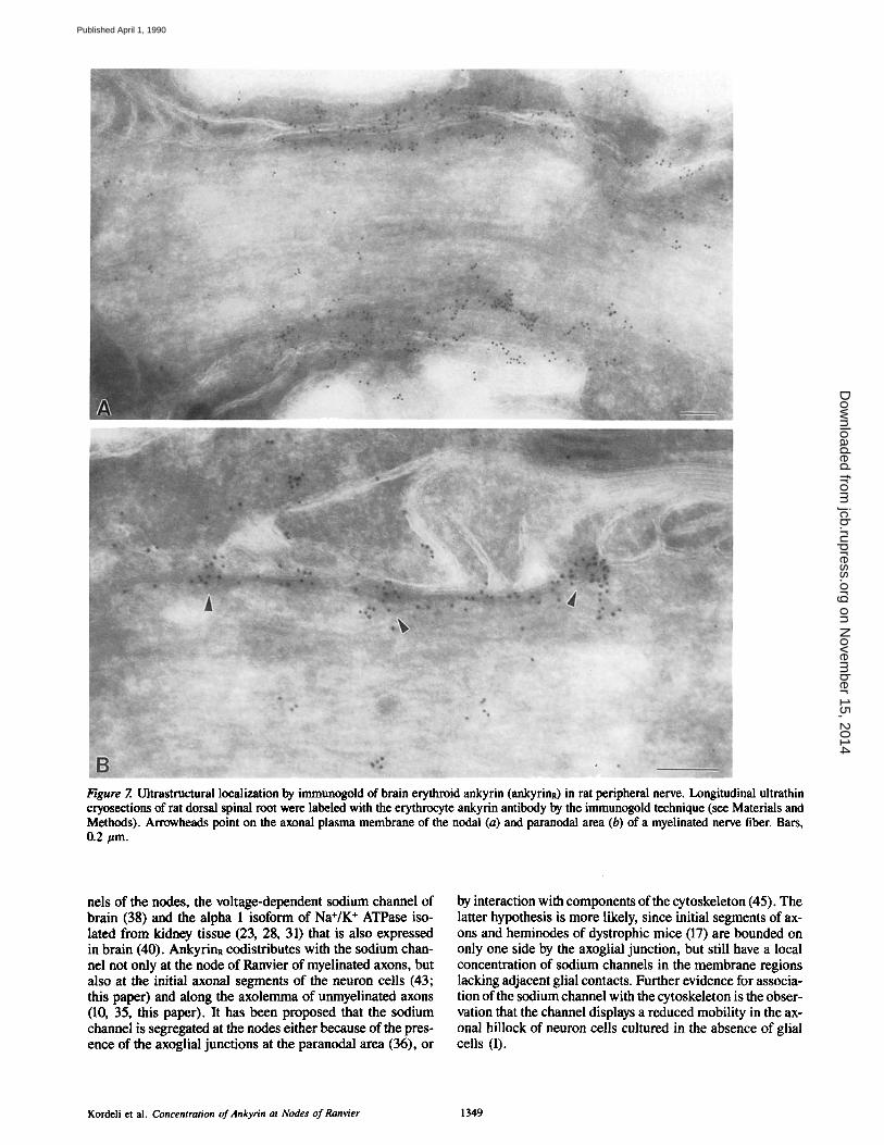
Figure 7. Ultrastructural localization by immunogold of brain erythroid ankyrin (ankyrinn) in rat peripheral nerve. Longitudinal ultrathin cryosections of rat dorsal spinal root were labeled with the erythrocyte ankyrin antibody by the immunogold technique (see Materials and Methods). Arrowheads point on the axonal plasma membrane of the nodal (a) and paranodal area (b) of a myelinated nerve fiber. Bars, 0.2 #m.
nels of the nodes, the voltage-dependent sodium channel of brain (38) and the alpha 1 isoform of Na÷/K + ATPase iso- lated from kidney tissue (23, 28, 31) that is also expressed in brain (40). AnkyrinR codistributes with the sodium chan- nel not only at the node of Ranvier of myelinated axons, but also at the initial axonal segments of the neuron cells (43; this paper) and along the axolemma of unmyelinated axons (10, 35, this paper). It has been proposed that the sodium channel is segregated at the nodes either because of the pres- ence of the axoglial junctions at the paranodal area (36), or
by interaction with components of the cytoskeleton (45). The latter hypothesis is more likely, since initial segments of ax- ons and heminodes of dystrophic mice (17) are bounded on only one side by the axoglial junction, but still have a local concentration of sodium channels in the membrane regions lacking adjacent glial contacts. Further evidence for associa- tion of the sodium channel with the cytoskeleton is the obser- vation that the channel displays a reduced mobility in the ax- onal hillock of neuron cells cultured in the absence of glial cells (1).
Kordeli et al. Concentration of Ankyrin at Nodes of Ranvier 1349
on Novem
ber 15, 2014jcb.rupress.org
Dow
nloaded from
Published April 1, 1990
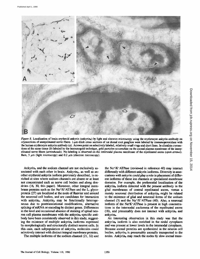
Figure 8. Localization of brain erythroid ankyrin (ankyrinR) by light and electron microscopy using the erythrocyte ankyrin antibody on cryosections of unmyelinated nerve fibers. 1-#m-thick cross sections of rat dorsal root ganglion were labeled by immunoperoxidase with the human erythrocyte ankyrin antibody (a). Arrows point on selectively labeled, relatively small rings and short lines. In ultrathin cryosec- tions of the same tissue (b) labeled by the immunogold technique, gold particles accumulate on the axonal plasma membrane of the unmy- elinated nerve fibers (arrowheads). No labeling is observed on the internodal plasma membrane of the myelinated axons (open arrows). Bars, 5 #m (light microscopy) and 0.2 #m (electron microscopy).
AnkyrinR and the sodium channel are not exclusively as- sociated with each other in brain. AnkyrinR, as well as an- other erythroid ankyrin isoform previously described, is en- riched at sites where sodium channels are absent or at least not concentrated such as nerve cell bodies and along den- drites (16, 30, this paper). Moreover, other integral mem- brane proteins such as the Na+/K+ATPase and the LI glyco- protein (27) are localized at the node of Ranvier and around the neuronal cell bodies, and are candidates for interaction with ankyrina. AnkyrinR may be functionally heteroge- neous due to posttranslational modifications, alternative splicing of mRNA or encoding by distinct genes. Differences in the level and occasional absence of staining of spinal neu- ron cell plasma membranes with the ankyrina-specific anti- body have been consistently observed in this study, suggest- ing the existence of multiple ankyrinR isoforms expressed by morphologically and structurally distinct neuron cells. In this case, each subpopulation of ankyrinR molecules could selectively interact with distinct integral membrane proteins.
The multiple isoforms of the sodium channel (21, 32) and
the Na+/K÷ATPase (reviewed in reference 40) may interact differently with different ankyrin isoforms. Diversity in asso- ciations with ankyrin could play a role in placement of differ- ent isoforms of these ion channels at specialized membrane domains. For example, the preferential localization of the ankyrinB isoform detected with the present antibody in the glial membranes of central myelinated axons, versus a mainly neuronal distribution of ankyrinR might be related to the existence of glial and neuronal forms of the sodium channel (3) and the Na+/K+ATPase (40). Also, a neuronal isoform of the Na+K+ATPase is present in high concentra- tions in the internodal axolemma of the myelinated axons (18), and presumably does not interact with ankyrinR and ankyrinB.
An interesting observation in this study was that the ankyrinR isoform is also enriched in the nodal cytoplasm and was present at lower levels in the internodal cytoplasm. Because axonal proteins are synthesized in the neuron cell bodies, ankyrinR is presumably axonally transported to the nodes. Ankyrina may reach the nodes by slow axonal trans-
The Journal of Cell Biology, Volume 110, 1990 1350
on Novem
ber 15, 2014jcb.rupress.org
Dow
nloaded from
Published April 1, 1990
port along with other soluble cytoplasmic proteins and may associate with the sodium channel (and other proteins) after they are inserted in the nodal membrane, providing stabiliza- tion. Cytoplasmic labeling then could represent a cytoplas- mic pool of free ankyrin in the nodal cytoplasm. Another possibility is that ankyrinR associates with the cytoplasmic surface of sodium channels immediately after biosynthesis. The two proteins would then be axonally transported in vesi- cles by a fast, kinesin-driven mechanism along microtubules to the nodes of Ranvier. In this case, cytoplasmic labeling would represent accumulation of protein-carrying vesicles at nodes and ankyrin would have the potential to target the so- dium channel to the specialized nodal axolemma. It is perti- nent in this regard that sodium channels have been observed to undergo fast axonal transport (25) and ankyrin can inter- act in vitro with tubulin (8, 13). It will be important for the understanding of the role of ankyrinR at the nodes to deter- mine the rate of axonal transport of this isoform.
The concentration of the ankyrinR isoform at the node of Ranvier raises the question of which isoform of spectrin is present at these sites: the generally expressed form of spec- trin (fodrin), or erythroid spectrin. Erythrocyte ankyrin binds preferentially in in vitro assays to erythrocyte spectrin rather than fodrin (9, 20). Based on the immunological rela- tionship of ankyrinR with erythrocyte ankyrin it might be anticipated that ankyrinR also would bind with higher affini- ty to erythroid spectrin, and that these proteins would be seg- regated with each other. However, axons are believed to con- tain fodrin rather than erythroid spectrin (24, 34). Fodrin has been observed to be concentrated at the nodes of Ranvier of isolated desheathed peripheral axons (22) and in para- nodal loops of myelinated fibers in rat peripheral nerve (42). It is conceivable that the ankyrin~ isoform is a chimeric an- kyrin with certain features in common with erythrocyte an- kyrin, but a binding preference for fodrin at the nodes of Ranvier.
In summary, local segregation of an apparently minor iso- form of ankyrin at the nodes of Ranvier provides indirect evi- dence suggesting a role for ankyrin in the formation and/or the maintenance of this axonal structure which displays a high degree of structural and functional differentiation. An experimental challenge in the future will be to resolve the activities of ankyrin at the node as well as at other specialized membrane domains.
Jodi Telander and Cindy Cootauco are gratefully acknowledged for help in preparing the manuscript and technical assistance.
This research was supported in part by grants from National Institutes of Health to V. Bennett (GM-33996 and AM-1198808) and to B. Trapp (NS-22849) and from the National Multiple Sclerosis Society to B. Trapp (JF2030 A-l). B. Trapp is a Harry Weaver Neuroscience Scholar of the National Multiple Sclerosis Society.
References
I. Angelides, K. J., L. W. Elmer, D. Loftus, and E. Elson. 1988. Distribution and lateral mobility of voltage-dependent sodium channels in neurons. J. Cell Biol. 106:1911-1925.
2. Ariyasu, R. G., J. A. Nichol, and M. H. Ellisman. 1985. Localization of sodium/potassium Adenosine Triphosphatase in multiple cell types of the routine nervous system with antibodies raised against the enzyme from kidney. J. Neurasci. 5:2581-2596.
3. Barres, B. A., L. L. Y. Chun, and D. P. Corey. 1989. Glial and neuronal forms of the voltage-dependent sodium channel: characteristics and cell- type distribution. Neuron. 2:1375-1388.
4. Bennett, V. 1983. Proteins involved in membrane-cytoskeleton association
in human erythrocytes: spectrin, ankyrin and band 3. Methods Enzymol. 96:313-324.
5. Bennett, V. 1985. The membrane skeleton of human erythrocytes and its implications for more complex cells. Annu. Rev. Biochem. 54:273-304.
6. Bennett, V., and P. Stenbuck. 1980. Human erythrocyte ankyrin purifica- tion and properties. J. Biol. Chem. 255:2540-2548.
7. Bennett, V., and P. Stenbuck. 1980. Association between ankyrin and the cytoplasmic domain of band 3 isolated from the human erythrocyte mem- brane. J. Biol. Chem. 255:6424-6432.
8. Bennett, V., and J. Davis. 1982. Immunoreactive forms of human erythro- cyte ankyrin are localized in mitotic structures in cultured cells and are associated with microtubules in brain. Cold Spring Harbor Symp. Quant. Biol. 46:647-657.
9. Bennett, V., J. Davis, and W. E. Fowler. 1982. Brain spectrin, a membrane-associated protein related in structure and function to erythro- cyte spectrin. Nature (Lond.). 299:126-131.
10. Black, J. A., R. E. Foster, and S. G. Waxman. 1981. Freeze-fracture ultra- structure of rat CNS and PNS nonmyelinated axolemma. J. Neurocytol. 19:981-993.
I 1. Catterall, W. A. 1984. The molecular basis of neuronal excitability. Sci- ence (Wash. DC). 223:653-661.
12. Davis, J. Q., and V. Bennett. 1984. Brain ankyrin: purification of a 72,000 Mr spectrin-binding domain. J. Biol. Chem. 259:1874- ! 88 I.
13. Davis, J. Q., and V. Bennett. 1984. Brain ankyrin: a membrane-associated protein with binding sites for spectrin, tubulin, and the cytoplasmic do- main of the erythrocyte anion channel. J. Biol. Chem. 259:13550-13559.
14. Davis, J. Q., and V. Bennett. 1986. Association of brain ankyrin with brain membranes and isolation of active proteolytic fragments of membrane- associa~xi ankyrin-binding protein(s). J. Biol. Chem. 261:16198-16206.
15. Davis, J., L. Davis, and V. Bennett. 1989. Diversity in membrane binding sites of ankyrins: brain ankyrin, erythrocyte ankyrin, and processed erythrocyte ankyrin associate with distinct sites in kidney microsomes. J. Biol. Chem. 264:6417-6426.
16. Drenckhahn, D., and V. Bennett. 1987. Polarized distribution of Mr 210,000 and 190,000 analogs of erythrocyte anky-rin along the plasma membrane of transporting epithelia, neurons and photoreceptors. Eur. J. Cell Biol. 43:479-486.
17. Ellisman, M. H. 1979. Molecular specializations of the axon membrane at nodes of Ranvier are not dependent upon myelination. J. Neurocytol. 8:719-735.
18. Fambrough, D. M., B. A. Wolitzky, and D. W. Pumplin. 1985. Develop- mental and regulatory aspects of the sodium- and potassium-ion- stimulated ATPase in avian nerve and muscle. In Regulation and Devel- opment of Membrane Transport Processes. J. S. Graves, editor. John Wiley & Sons, New York. 39:265-282.
19. Hall, T. G., and V. Bennett. 1987. Regulatory domains of erythrocyte an- kyrin. J. Biol. Chem. 262:10537-10545.
20. Howe, C. L., L. M. Sacramone, M. S. Mooseker, andJ. S. Morrow. 1985. Mechanisms of cytoskeletal regulation: Modulation of membrane affinity in avian brush border and erythrocyte spectrins. J. Cell Biol. 101 : 1379- 1385.
21. Kayano, T., M. Noda, V. Flockerzi, H. Takahashi, and S. Numa. 1988. Functional expression of cloned eDNA encoding sodium channel IIL FEBS (Fed. Eur. Biochem. Soc.) Lett. 228:195-200.
22. Koenig, E., and E. Repasky. 1985. A regional analysis of alpha-spectrin in the isolated Mauthner neuron and in isolated axons of the goldfish and rabbit. J. Neurosci. 5:705-714.
23. Koob, R., M. Zimmermann, W. Schoner, and D. Drenckhshn. 1987. Colo- calization and coprecipitation of ankyrin and Na +, K+-ATPase in kidney epithelial cells. Fur. J. Cell Biol. 45:230-237.
24. I oTarides, E., W. L. Nelson, and T. Kasamatsu. 1984. Segregation of two spectrin forms in the chicken optic system: a mechanism for establishing restricted membrane-cytoskeletal domains in neurons. Cell. 36:269-278.
25. Lombet, A., P. Laduron, C. Mourre, Y. Jacomet, and M. I.atzdunski. 1985. Axonal transport of the voltagu-dependent Na + channel protein identified by its tetrodotoxin binding site in rat sciatic nerves. Brain Res. 345:153-158.
26. Marchesi, V. T. 1985. Stabilizing infrastructure of cell membranes. Annu. Rev. Cell Biol. 1:531-561.
27. Mirsky, R., K. R. Jessea, M. Schachner, and C. Goridis. 1986. Distribu- tion of the adhesion molecules N-CAM and L~ on peripheral neurons and glia in adult rats. J. Neurocytol. 15:799-815.
28. Morrow, J. S., C. D. Cianci, T. Ardito, A. S. Mann, and M. Kashgarian. 1989. Ankyrin links fodrin to the alpha subunit of Na,K-ATPase in Madin-Darby canine kidney cells and in intact renal tubule cells. J. Cell Biol. 108:455--465.
29. Nelson, W. J., and E. Lazarides. 1984. Goblin (ankyrin) in striated muscle: Identification of the potential membrane receptor for erythroid spectrin in muscle cells. Proc. Natl. Acad. Sci. USA. 81:3292-3296.
30. Nelson, W. J., and E. ~ d e s . 1984. The patterns of expression of two ankyrin isoforms demonstrate distinct steps in the assembly of the mem- brane skeleton in neuronal morpbogenesis. Cell. 39:309-320.
31. Nelson, W. J., and P. J. Veshnock. 1987. Ankyrin binding to (Na + +K +) ATPase and implications for the organization of membrane domains in polarized cells. Nature (Lond.). 328:533-536.
Kordeli et al. Concentration of Ankyrin at Nodes of Ranvier 1351
on Novem
ber 15, 2014jcb.rupress.org
Dow
nloaded from
Published April 1, 1990
32. Noda, M., T. lkeda, T. Kayano, H. Suzuki, H. Takeshima, M. Kurasaki, H. Takahashi, and S. Numa. 1986. Existence of distinct sodium channel messenger RNAs in rat brain. Nature (Lond.). 320:188-192.
33. Peters, A. 1966. The node of Ranvier in the central nervous system. Q. J. Exp. Physiol. 51:229-236.
34. Rieder, B. M., I. S. Zagon, and S. R. Goodman. 1986. Brain spectrin (240/235) and brain spectrin (240/235E): two distinct speetrin subtypes with different locations within mammalian neural cells. J. Cell Biol. 102:2088-2097.
35. Ritchie, J. M., R. B. Rogart, and G. Strichartz. 1976. A new method for labeling saxitoxin and its binding to non-myelinated fibres of the rabbit vagus, lobster walking and garfish olfactory nerves. J. Physiol. (Lond.). 261:477--494.
36. Rosenbluth, J. 1976. Intramembranous panicle distribution at the node of Ranvier and adjacent axolemma in myelinated axons of the frog brain. J. Neurocytol. 5:731-745.
37. Slot, J. W., and H. J. Geuze. 1984. Gold markers for single and double immunolabeling of ultrathin cryosections. In lmmunolabeling for Elec- trnn Microscopy. J. M. Polak and I. M. Varndell, editors. Elsevier Sci- ence Publishers, New York. 129-142.
38. Srinivasan, Y., L. Elmer, J. Davis, V. Bennett, and K. Angelides. 1988.
Ankyrin and spectrin associate with voltage-dependent sodium channels in brain. Nature (Lond.). 333:177-180.
39. Steiner, J. P., H. T. Walke, Jr., and V. Bennett. 1989. Calcium/calmodulin inhibits direct binding of spectrin to synaptosomal membranes. J. Biol. Chem. 264:2783-2791.
40. Sweadner, K. J. 1989. Isozymes of the Na+/K÷-ATPase. Biochim. Bio- phys. Acta. 988:185-220.
41. Tokuyasu, K. T. 1986. Application of cryoultramierntomy to immunocy- tochemistry. J. Microscopy. 143:139-149.
42. Trapp, B. D., S. B. Andrews, A. Wong, M. O'Connell, and J. W. Griffin. 1989. Co-localization of the myelin-associated glycoprotein and the microfilament components, F-aetin and spectrin, in Schwann cells of my- elinated nerve fibers. J. Neurocytol. 18:47-60.
43. Waxman, S. G., and R. E. Foster. 1980. Ionic channel distribution and het- erngeneity of the axon membrane in myelinated fibers. Brain Res. Rev. 2:205-234.
44. Waxman, S. G., and J. M. Ritchie. 1985. Organization of ion channels in the myelinated nerve fiber. Science (Wash. DC). 228:1502-1507.
45. Wiley-Livingston, C. A., and M. H. Ellisman. 1980. Development of ax- onal membrane specializations defines nodes of Ranvier and precedes Schwann cell myelin elaboration. Dev. Biol. 79:334-355.
The Journal of Cell Biology, Volume 110, 1990 1352
on Novem
ber 15, 2014jcb.rupress.org
Dow
nloaded from
Published April 1, 1990