Electron tomographic analysis of cytoskeletal cross-bridges in the paranodal region of the node of...
21
Electron Tomographic Analysis of Cytoskeletal Cross-Bridges in the Paranodal Region of the Node of Ranvier in Peripheral Nerves Guy A. Perkins 1 , Gina E. Sosinsky 1,2 , Sassan Ghassemzadeh 1 , Alex Perez 1 , Ying Jones 1 , and Mark H. Ellisman 1,2 1 National Center for Microscopy and Imaging Research, Center for Research in Biological Systems, University of California, San Diego 92093-0608 2 Dept. of Neurosciences, University of California, San Diego 92093-0608 Abstract The node of Ranvier is a site for ionic conductances along myelinated nerves and governs the saltatory transmission of action potentials. Defects in the cross-bridging and spacing of the cytoskeleton is a prominent pathologic feature in diseases of the peripheral nerve. Electron tomography was used to examine cytoskeletal-cytoskeletal, membrane-cytoskeletal, and heterologous cell connections in the paranodal region of the node of Ranvier in peripheral nerves. Focal attachment of cytoskeletal filaments to each other and to the axolemma and paranodal membranes of the Schwann cell via narrow cross-bridges was visualized in both neuronal and glial cytoplasms. A subset of intermediate filaments associates with the cytoplasmic surfaces of supramolecular complexes of transmembrane structures that are presumed to include known and unknown junctional proteins. Mitochondria were linked to both microtubules and neurofilaments in the axoplasm and to neighboring smooth endoplasmic reticulum by narrow cross-bridges. Tubular cisternae in the glial cytoplasm were also linked to the paranodal glial cytoplasmic loop juxtanodal membrane by short cross-bridges. In the extracellular matrix between axon and Schwann cell, junctional bridges formed long cylinders inking the two membranes. Interactions between cytoskeleton, membranes, and extracellular matrix associations in the paranodal region is likely critical not only for scaffolding, but also for intracellular and extracellular communication. Keywords cross-bridge; cytoskeleton; electron tomography; freeze substitution; high-pressure freezing; microfilaments; microtubules; neurofilaments; node of Ranvier; paranodal junctional bridge; paranode 1. Introduction Node of Ranvier complexes are heterologous cellular specializations containing components from both neuron and glial cells. These nodes are found in both the central nervous (CNS) and peripheral nervous systems (PNS). The node of Ranvier aids the amplification and regeneration Contact Person: Guy Perkins, National Center for Microscopy and Imaging Research, University of California, San Diego, 9500 Gilman Dr., La Jolla, CA 92093-0608, 858-534-7968 voice, 858-534-7497 fax. Publisher's Disclaimer: This is a PDF file of an unedited manuscript that has been accepted for publication. As a service to our customers we are providing this early version of the manuscript. The manuscript will undergo copyediting, typesetting, and review of the resulting proof before it is published in its final citable form. Please note that during the production process errors may be discovered which could affect the content, and all legal disclaimers that apply to the journal pertain. NIH Public Access Author Manuscript J Struct Biol. Author manuscript; available in PMC 2009 March 1. Published in final edited form as: J Struct Biol. 2008 March ; 161(3): 469–480. doi:10.1016/j.jsb.2007.10.005. NIH-PA Author Manuscript NIH-PA Author Manuscript NIH-PA Author Manuscript
-
Upload
independent -
Category
Documents
-
view
1 -
download
0
Transcript of Electron tomographic analysis of cytoskeletal cross-bridges in the paranodal region of the node of...

Electron Tomographic Analysis of Cytoskeletal Cross-Bridges inthe Paranodal Region of the Node of Ranvier in Peripheral Nerves
Guy A. Perkins1, Gina E. Sosinsky1,2, Sassan Ghassemzadeh1, Alex Perez1, Ying Jones1,and Mark H. Ellisman1,2
1 National Center for Microscopy and Imaging Research, Center for Research in Biological Systems,University of California, San Diego 92093-0608
2 Dept. of Neurosciences, University of California, San Diego 92093-0608
AbstractThe node of Ranvier is a site for ionic conductances along myelinated nerves and governs the saltatorytransmission of action potentials. Defects in the cross-bridging and spacing of the cytoskeleton is aprominent pathologic feature in diseases of the peripheral nerve. Electron tomography was used toexamine cytoskeletal-cytoskeletal, membrane-cytoskeletal, and heterologous cell connections in theparanodal region of the node of Ranvier in peripheral nerves. Focal attachment of cytoskeletalfilaments to each other and to the axolemma and paranodal membranes of the Schwann cell vianarrow cross-bridges was visualized in both neuronal and glial cytoplasms. A subset of intermediatefilaments associates with the cytoplasmic surfaces of supramolecular complexes of transmembranestructures that are presumed to include known and unknown junctional proteins. Mitochondria werelinked to both microtubules and neurofilaments in the axoplasm and to neighboring smoothendoplasmic reticulum by narrow cross-bridges. Tubular cisternae in the glial cytoplasm were alsolinked to the paranodal glial cytoplasmic loop juxtanodal membrane by short cross-bridges. In theextracellular matrix between axon and Schwann cell, junctional bridges formed long cylinders inkingthe two membranes. Interactions between cytoskeleton, membranes, and extracellular matrixassociations in the paranodal region is likely critical not only for scaffolding, but also for intracellularand extracellular communication.
Keywordscross-bridge; cytoskeleton; electron tomography; freeze substitution; high-pressure freezing;microfilaments; microtubules; neurofilaments; node of Ranvier; paranodal junctional bridge;paranode
1. IntroductionNode of Ranvier complexes are heterologous cellular specializations containing componentsfrom both neuron and glial cells. These nodes are found in both the central nervous (CNS) andperipheral nervous systems (PNS). The node of Ranvier aids the amplification and regeneration
Contact Person: Guy Perkins, National Center for Microscopy and Imaging Research, University of California, San Diego, 9500 GilmanDr., La Jolla, CA 92093-0608, 858-534-7968 voice, 858-534-7497 fax.Publisher's Disclaimer: This is a PDF file of an unedited manuscript that has been accepted for publication. As a service to our customerswe are providing this early version of the manuscript. The manuscript will undergo copyediting, typesetting, and review of the resultingproof before it is published in its final citable form. Please note that during the production process errors may be discovered which couldaffect the content, and all legal disclaimers that apply to the journal pertain.
NIH Public AccessAuthor ManuscriptJ Struct Biol. Author manuscript; available in PMC 2009 March 1.
Published in final edited form as:J Struct Biol. 2008 March ; 161(3): 469–480. doi:10.1016/j.jsb.2007.10.005.
NIH
-PA Author Manuscript
NIH
-PA Author Manuscript
NIH
-PA Author Manuscript

of action potentials in axons by facilitating segregation of ion channels into discrete domainsand enabling the saltatory propagation of large ionic currents (Ellisman et al., 1982). Saltatoryconduction permits a rapid propagation of action potentials without an increase in axonal borewidth. The overall structure of the node of Ranvier appears to be highly developed in part tocontain large ionic fluxes to compartmentalized domains. The formation of the node of Ranvierrequires specific contacts between axons and the surrounding glial cells, generating specializedmembrane sub-domains with distinct compositions of ion channels (Bonnon et al., 2007;Sosinsky et al., 2005). Much of the node's complexity stems from it being a unique heterologoussystem, requiring close interaction between two or more distinct cell types.
The term “node of Ranvier complex” often refers not only to the nodal gap that contains nomyelin, but also to the paranodal regions with its uncompacted cytoplasm-containing terminalmyelin loops. The nodal gap is bracketed by the paranodes and defined as the region in whichthe cytoplasm-encompassing margins of the uncompacted myelin lamellae abut the axon. Theglial margin wraps around the paranodal axolemma in a helical fashion. Both intracellular andintercellular bridging or junctional molecules are abundant in the paranodal and juxtaparanodalregions (Ichimura and Ellisman, 1991). Macromolecular junctional complexes, form betweenthe axolemma to the spiral paranodal loops. Another role of these junctions is to act as barriersto separate the sodium and potassium channels into the nodal, paranodal, and juxtaparanodalmembranes where they are needed (Ellisman and Levinson, 1982; Mi et al., 1995; Mi et al.,1996; Mi et al., 1999; Rasband, 2004; Trimmer and Rhodes, 2004).
The three types of cytoskeletal filaments prevalent in the PNS are intermediate filaments (bothglial filaments and neurofilaments), microtubules, and microfilaments. Neurofilaments aretype IV intermediate filaments and are numerous in the axons of vertebrate neurons (Karp,2002). They measure between 9-11 nm in diameter when assembled. These filaments are oftenarranged as loosely packed bundles oriented parallel to the axon length and can be extensivelycross-linked (Nakagawa et al., 1995). The extent of neurofilament gene expression is correlatedto axonal bore diameter, which controls the longitudinal resistance of the fiber, influencing thespacing between nodes of Ranvier and thereby how fast electrical signals traverse the axon(Lee and Cleveland, 1996; Marszalek et al., 1996). Microtubules are the “highways” oftransport for materials moving rapidly within the axons of neurons (Li et al., 2004) and havea diameter of about 24 nm; they vary in length from several hundred nanometers to hundredsof micrometers in axons. Microfilaments, composed of F-actin, and found in the cytoplasm ofthe paranodal loops (Ichimura and Ellisman, 1991; Scherer, 1996; Trapp et al., 1989) measureabout 7 nm in diameter and are likely also involved with transport. In addition, all three filamenttypes seem to function as mechanical stabilizers of the cytoplasm to resist mechanical stress(Fuchs and Cleveland, 1998; Garcia and Cleveland, 2001).
Associated with the axon of the PNS, the glial Schwann cell provides myelin at the internode,lateral loops at the paranode, and lammelopodia (also called microvilli) that abut the axon atthe node (Ellisman and Porter, 1980; Ellisman et al., 1987). Between the loops of the paranodeand the axolemma in the extracellular matrix are linear arrays of heterogeneously sized particlesof 8-16 nm in diameter that bridge this space (Ellisman, 1979; Wiley and Ellisman, 1980;Wiley-Livingston and Ellisman, 1981); we name these paranodal junctional bridges (PJB).They have also been called “septate junctions” or “septate-like junctions” (Ichimura andEllisman, 1991; Sosinsky et al., 2005; Spiegel and Peles, 2002). These structures, which appearcontinuous, are comprised of heterologous macromolecules and appear to insert into both theglial and axonal plasma membranes.
The proteins in the PJB that link the loop membranes with the axolemma form a complex ofheterodimers consisting of neurofascin 155 and caspr/paranodin (Boyle et al., 2001; Poliak etal., 2001; Scherer and Arroyo, 2002; Tait et al., 2000). In a unique heterologous handshake,
Perkins et al. Page 2
J Struct Biol. Author manuscript; available in PMC 2009 March 1.
NIH
-PA Author Manuscript
NIH
-PA Author Manuscript
NIH
-PA Author Manuscript

the glial cell contributes neurofascin 155 and the neuronal cell provides caspr. Contactin,residing in the axonal membrane, brings the other proteins to the handshake. Deficiency ineither caspr, contactin, or neurofascin 155 produces a disorganization of the paranodal loopsand a reduction in axon conduction velocity (Bhat et al., 2001; Bonnon et al., 2007; Boyle etal., 2001; Sherman et al., 2005). At the nodal/paranodal region in the myelinated axons ofsciatic nerves of mice deficient in caspr, PJBs were missing and abnormal, swollenmitochondria were observed (Einheber et al., 2006). In contrast, mitochondria located in theaxoplasm outside this region appeared normal, suggesting that the PJB influencesmitochondrial function and perhaps transport in the axoplasm of the nodal/paranodal regionof myelinated axons of peripheral nerves. The distribution of neurofascin 155 in the glialmembrane is altered in one form of multiple sclerosis (MS), a demyelinating disease (Maieret al., 2007). This group discovered that efficient association of neurofascin 155 with lipid raftdomains is required for the maintenance of the paranodal axo-glial junction and without thisjunction, demyelination occurs in MS. The PJB complex was seen previously in images ofdeep-etched rotary-shadowed preparations and described as 16-nm “pegs” that line up inordered arrays (Ellisman, 1979; Ichimura and Ellisman, 1991; Wiley and Ellisman, 1980;Wiley-Livingston and Ellisman, 1981). In our reconstructions, the PJBs were only occasionallyobserved as discrete pegs, but more often formed a continuous rod of density. This appearanceconforms to the role described for PJBs as septate-like junctions that serve in a paracellularcapacity to block the diffusion of ions and channels between the paranodal and juxtaparanodalareas (Rios et al., 2003; Salzer, 2003).
This study extends the earlier work of Sosinsky and co-workers (Sosinsky et al., 2005), whoprovided a broad structural and modeling framework of the node from electron tomographystructure determination. Here, we examine in greater detail the cross-bridges connectingcytoskeleton, membranes, and adjacent neuronal and glial cells in the paranodal region of thePNS. This is the first report of electron tomography being used to characterize these cross-bridges. The filamentous or spanning nature of the bridges, their focal attachment, and theirassociation with membrane particles were visualized in three dimensions in tomographicreconstructions. We found that at the paranode, filaments extend from the linear array of PJBsinto both loops and axoplasm thus forming an apparent filamentous link of the cytoskeletonin the paranodal loop with that in the axon. Membrane-cytoskeleton cross-bridges, sometimesconnecting to the PJBs, were also found. The cross-bridges we have measured have thicknessesin the intermediate filament and microfilament range and also function to tether cytoplasmicorganelles such as mitochondria and tubular cisternae (TC) and other membranous organellessuch as autophagosomes and lysosomes to cytoskeleton or membrane. Combination of the 3Dinformation contained in tomographic volumes support the construction of a model that includefilaments with specialized orientations and attachments to axonal and glial components viashort cross-bridges.
2. Materials and MethodsConventional preservation with aldehyde-fixation of PNS samples from rat was accomplishedas described previously (Perkins et al., 2001). Conventional electron microscopy of thinsections (∼80 nm thick) was performed with a JEOL 1200 EX microscope operated at 80 kV.
2.1 High Pressure FreezingThe procedures for high pressure freezing of chemically fixed tissue followed by freezesubstitution follow that described in detail in Sosinsky et al., (2008). Rats were firstanaesthetized by intraperitoneal injection of Nembutal (35 mg per kg body weight), and wholeblood was flushed out with oxygenated rat Ringers solution at 37° C. They were then fixed byintracardial perfusion with an aldehyde mixture composed of 2% paraformaldehyde and 2.5%
Perkins et al. Page 3
J Struct Biol. Author manuscript; available in PMC 2009 March 1.
NIH
-PA Author Manuscript
NIH
-PA Author Manuscript
NIH
-PA Author Manuscript

glutaraldehyde in 0.15 M cacodylate buffer (pH 7.4) at 37° C. The spinal root was dissectedout, immersed in the same fixative and transferred to the high-pressure freezing (HPF) unit.These samples were trimmed and blotted (to remove excess water) before placing in the carrier.Samples were loaded into the carriers of the Bal-Tec HPM010 HPF unit (Bal-Tec,Liechtenstein) and operated according to manufacturers instructions. Carriers with a depth of0.2 mm were used with thin bundles of spinal root to provide uniform ultrastructuralpreservation. The inert liquid, hexadecene, was added to the carrier to fill in the well, excludingair from surrounding the sample.
2.2 Freeze SubstitutionAfter the samples were high-pressure frozen, they were freeze-substituted in dry acetonecontaining 0.1% tannic acid for 24 hrs at -90° C in a Leica EMAFS freeze substitution unit(FSU, Leica Microsystems, Bannockburn, IL), then in acetone containing 2% OsO4 and 0.1%of uranyl acetate for the remaining time of the procedure. Both tannic acid and uranyl acetatewere added not only to help stain the material but also to aid in membrane preservation. Duringthis procedure, the water in the sample is completely extracted and replaced by the fixative,i.e., freeze-substituted. The temperature for substitution was maintained at −90° C for 3 daysto ensure that the biological structures were cryo-immobilized during this substitution. Thetemperature was raised gradually at a rate of 2° C per hour to −60° C and held there for 8 hoursbecause OsO4 does not start to cross-link until around −70° C. The samples were then warmedto -20 °C at a rate of 5° C per hour and held at this temperature for 12 hours. The carriers werethen taken from the FSU, the tissue removed from the carriers and quickly washed severaltimes in dry acetone at room temperature (RT) to remove the OsO4 and uranyl acetate. Acetoneabove 70% can extract lipids at RT. Hence, the exposure time to it was minimized. The tissueswere then incubated in a 50:50 mixture of acetone:Durcupan for 4 hour at RT followed by100% Durcupan resin of 24 hrs infiltration with 3 changes. Resin polymerization wasaccomplished in a vacuum oven at 65-70 °C for 48 hours. The resin-embedded samples wereremoved from the oven and sections were cut using a Leica ultramicrotome. Sections werethen stained 30 min in 2% aqueous uranyl acetate, followed by 15 min in lead salts. Fiducialcues consisting of 15 and 20 nm colloidal gold particles were deposited on opposite sides ofthe section.
2.3 Electron TomographyTwo tomograms from HPF/FS samples (both single-tilt) and two tomograms fromconventionally prepared samples (1 single-tilt, 1 double-tilt) were analyzed. For eachreconstruction, a series of images at regular tilt increments was collected with a JEOL 4000EXintermediate-voltage electron microscope (JEOL USA, Peabody, MA) operated at 400 kV. Thespecimens were irradiated before initiating a tilt series in order to limit anisotropic specimenthinning during image collection. Tilt series were recorded on film at 20,000 timesmagnification with an angular increment of 2° from -60° to +60° about an axis perpendicularto the optical axis of the microscope using a computer-controlled goniometer to incrementaccurately the angular steps. For the double-tilt series, the grid was subsequently rotated 90°and a second tilt series was collected with the same angular increment and range. Theillumination was held to near parallel beam conditions and optical density maintained constantby varying the exposure time. The negatives were digitized with a Nikon CoolScan at 1800dpi producing images of size 4033 × 6010 pixels. The final pixel size in our reconstructionswas 1.41 nm. The IMOD package (Kremer et al., 1996) was used for rough alignment with thefine alignment and reconstruction performed using the TxBR package (Lawrence et al.,2006). One of the HPF/FS single-tilt series reconstructions (310 nm thickness) had a (global)resolution estimated by using the even/odd Fourier shell correlation (Cardone et al., 2005) andsoftware provided by Alasdair Steven (NIH) to be 8.4 nm at a threshold of 0.3; the other twosingle-tilt reconstructions analyzed likely had similar resolution estimates because they had
Perkins et al. Page 4
J Struct Biol. Author manuscript; available in PMC 2009 March 1.
NIH
-PA Author Manuscript
NIH
-PA Author Manuscript
NIH
-PA Author Manuscript

similar thicknesses and angular increment and range. The double-tilt series reconstruction (215nm thickness) had a resolution estimated to be 4.6 nm by the same criterion. Volumesegmentation was performed by manual tracing in the planes of highest resolution with theprogram Xvoxtrace (Perkins et al., 1997b). The reconstructions were visualized using Analyze(Mayo Foundation, Rochester, MN) or the surface-rendering graphics of Synu (Hessler et al.,1996). These programs allow one to step through slices of the reconstruction in any orientationand to track or model features of interest in three dimensions. Measurements of structuralfeatures were made using NIH ImageJ (Rasband, W.S., ImageJ, National Institutes of Health,Bethesda, Maryland, USA, http://rsb.info.nih.gov/ij/, 1997-2004.) Movies of surface-renderedvolumes and slices through the reconstructions were made using Amira (Mercury/TGS, SanDiego, CA). For the averages shown in Fig. 3, we had to first select for cross-bridges of eachtype that appeared straight, not kinked along their lengths. Subvolumes of similar dimensionwere cut out and aligned together in 3D using Analyze. After alignment, multiple subvolumeswere averaged using EMAN (Schmid and Booth, 2008; Tang et al., 2007) to increase the signal-to-noise ratio, enabling visualization of greater structural details.
3. ResultsElectron tomography coupled with preservation by high-pressure freezing and freeze-substitution has enabled additional resolution of structural details in the paranodal region. Thispreparation has an advantage in preserving the fine structure of fluid space occupied by anaqueous environment, since the water molecules are replaced by substitution fluid while theyare in the frozen state. Shrinkage and warping of membranes and cytoskeleton during thedehydration process are minimized.
3.1 Overall organization of the paranodeThe paranodal region is rich in juxtapositions of membrane-bounded structures of the Schwanncell cytoplasm and neuronal axoplasm and includes linkages of cytoskeletons, organelles, withnumerous examples of cross-bridges between them (Fig. 1). In the Schwann cell cytoplasmand the axoplasm there are extensive cytoskeletal networks and numerous organelles such asmitochondria, endoplasmic reticulum, autophagosomes, lysosomes, and tubular and cisternaethat are linked to the cytoskeleton, sometimes forming anatomosing networks. Themitochondria in the axoplasm appear different from those normally seen in tissues becausethey are in the condensed state (Hackenbrock, 1966), a structural configuration that is nowstarting to become more recognized in the axoplasm far from somas and terminals (Perkins etal., unpublished results). Condensed mitochondria have a dark matrix that appears like a longtwisted sausage in 3D and are thought to be indicative of greater ATP production. The cristajunction (Frey and Mannella, 2000;Mannella, 2006;Perkins et al., 1997a) may be either anarrow tube or an elongated opening (Renken et al., 2002).
The use of electron tomography revealed a variety of cross-bridges (Fig. 2), including thepresence of arrays of transcellular junctions, PJBs, traversing the paranodal glial-axonaljuxtaposition (Fig. 2a, a′). This juxtaposition is constituted by the terminal cytoplasmic loopsof the myelin forming Schwann cell, the extracellular matrix studded with septate junction-like structures that span the space between the glial and axonal membranes, and the axolemma.The PJBs not only span the cleft between cells, but also appear to insert into both glial andaxonal membranes (Fig. 2a, h, a′, h′). Tubular cisternae (TC) consist of tubules of smoothendoplasmic reticulum of various diameters and are closely associated with juxtapositionalmembranes in the glial loops. Filamentous cross-bridges of diameter similar to F-actin linkingthe TC to the loop membrane were revealed in tomographic volumes (Fig. 2b, c, b′, c′). Focalattachments of cytoskeletal filaments to the paranodal membranes were visualized in both
Perkins et al. Page 5
J Struct Biol. Author manuscript; available in PMC 2009 March 1.
NIH
-PA Author Manuscript
NIH
-PA Author Manuscript
NIH
-PA Author Manuscript

cytoplasms of the glial loops and axon (Fig. 2d, d′). A subset of these filaments attaches to thetransmembrane PJB (Fig. 2a′).
Organelles are also connected to the cytoskeleton and occasionally to each other in theaxoplasm. Mitochondria were seen connected by short tethers to neurofilaments (NF) (Fig. 2e,e′) or microtubules (MT) (Fig. 2f, f′). These tethers had a diameter similar to that ofneurofilaments. These filaments may be comprised of desmin or keratin (Toivola et al.,2005), which attach to mitochondria. Mitochondrial-ER cross-bridges were also observed (Fig.2g, g′). Multiple cross-bridges of the same diameter and similar lengths were found connectingthe two organelles. Microfilaments in the glial cytoplasm running more-or-less perpendicularto the filaments and microtubules in the axoplasm were found in the sub-volume closest to thejuxtapositional loop membrane (Fig. 2h, h′). These actin filaments did not appear bundled, nordid they appear to have cross-bridges with each other, other cytoskeleton, or membranes. Cross-bridges are also common between neurofilaments (Fig. 2i, i′) and between neurofilaments andmicrotubules (Fig. 2j, k, j′, k′). These cross-bridges have a diameter about the same asneurofilaments. We noticed that a small subset of neurofilaments appear to terminate directlyat the axolemma, including at the PJBs (data not shown).
Averages of tomographic subvolumes of cross-bridges enhanced the contrast of the sometimesfaint individual examples of such bridges (Fig. 3). These averages have an increased signal-to-noise ratio, enabling visualization of greater structural details. A general difference in cross-bridge diameter between the NF-NF, NF-MT, and mito-MT averages on one hand and the NF-Axl and TC-PNL averages on the other hand is noticeable. In addition, a variation in diameteralong the length of each bridge is evident, which might reflect variability in stain deposition.
3.1 Analysis of cytoskeletal elements in the paranodal regionFigure 4 is a 3D model that incorporates the structural information obtained in the presentstudy. The associations of the major cytoskeletal components are shown: neurofilaments, glialfilaments, microtubules, microfilaments, and cross-bridges of various sizes (Fig. 4a). Thecross-bridges that connect neurofilaments to each other or to microtubules are few and farbetween. We found only one or two such bridges per connecting cytoskeletal components (Fig.4b). This limited connectivity was also seen with the cross-bridging of neurofilaments to theaxolemma. The PJBs are seen to be rod-like structures (Fig. 4c) of uneven lengths (mean±stddev=45±34 nm; range:16-128 nm) and arranged in nearly parallel arrays separated byremarkably uniform spacing (HPF/FS samples: mean±std=19±4 nm; conventional samples:mean±std=21±.3 nm). The focal attachment of axoplasmic neurofilaments to the PJBs appearsto be complemented by a similar attachment of glial cytoskeletal filaments on the glial side.Occasionally, instead of being connected to the axolemma via cross-bridges, neurofilamentswould connect by terminating at this membrane, often at the site of a PJB (Fig. 4d). This site-specific link is suggestive of intercellular association between axon and glia. Tubular cisternae,possibly involved in regulating intracellular calcium levels, are linked to the loop membranealso by only a small number of cross-bridges (Fig. 4d). Mitochondria and ER were foundpositioned within a dense “jungle” of cytoskeleton (Fig. 4a), yet the connection of theseorganelles to the cytoskeleton was limited to either a single neurofilament or microtubule (Fig.4e) and moreover, a single cross-bridge. Intercellular cytoskeletal connections, not alwaysapparent in thin-section micrographs or in slices through the tomographic reconstruction, wererevealed after volume segmentation. An example is an axoplasmic cross-bridge connected toa glial cross-bridge through more than 40 nm of intervening PJB rod (Fig. 4f). Whereasneurofilaments and microtubules predominate the axoplasm, microfilaments are morepronounced in the loop cytoplasm (Fig. 4g, h). What role the microfilaments play is not impliedfrom ultrastructure revealed here as we observed no cross-bridges to each other, to other
Perkins et al. Page 6
J Struct Biol. Author manuscript; available in PMC 2009 March 1.
NIH
-PA Author Manuscript
NIH
-PA Author Manuscript
NIH
-PA Author Manuscript
cytoskeletal elements or any attachments to organelles or loop membranes (see supplementalmovie).
Measurements of diameters uncovered two distinct classes of cross-bridges (Fig. 5). The rangeand number of measurements are found in Table 1. Those bridges connecting neurofilaments,microtubules, and organelles have different cross-bridging diameters to the second class--thoseconnecting membranes: either filaments to the axolemma or between the TC and paranodalloop membrane. The two different classes are reflected in the probability of sampling from thesame population values, p, derived from the Student's t-test. For example, p=0.0014, 0.000038,0.00020, 1.2×10⁻¹⁰, and 3.1×10⁻⁶ for NF-NF vs. TC-PNL, NF-MT vs. TC-PNL, NF-NF vs.NF-Axl, NF-MT vs. NF-Axl, and mito-MT vs. NF-Axl, respectively. No other t-test betweencross-bridge types had p<0.01, which we used as the cutoff for statistical significance, includingcomparisons between the measured diameters from HPF/FS and conventional data. The lengthsshow similarity between NF-NF, NF-MT, and mito-MT cross-bridges (Fig. 5b). A notablelength dissimilarity exists between the TC-PNL cross-bridge and all the NF cross-bridges, i.e.,NF-NF, NF-MT, and NF-Axl cross-bridges (p<0.01 for all 3 comparisons) and is remarkablebecause the NF-Axl and TC-PNL mean diameters are similar. It may be that the links positionthe TC close to the PNL for the purpose of limited-diffusion calcium release or sequestration.The PJB diameter of 15±2 nm (mean±std dev; range 12-19 nm) for HPF/FS samples or 16±4nm (range 10-22 nm) for conventional samples is much greater than the fibrous cross-bridges(Fig. 5a) and emphasizes that PJBs are rods with an axis running lengthwise along thejuxtapositional membranes, not between the membranes. By plotting cross-bridge diameter vs.length, we found some indication that there exists a linear correlation between four of them:NF-NF, NF-MT, NF-Axl, and TC-PNL (Fig. 5c, 5d) and more noticeable with theconventionally prepared samples. In other words, as the diameter of the cross-bridge increased,it's length also tended to increase linearly. The one exception was the mito-MT cross-bridge,consistent with our observations that these are generally short-linkers. Because of the size ofmitochondria, a thicker cross-bridge might be required.
4. DiscussionHighly organized cytoskeletal networks appear to be one way of imposing rigidity andcompartmentalization, or alternatively, flexible spacing to an area of cellular specializationsuch as is found in the node of Ranvier. The filamentous cross-bridges, some thinner than 7nm, might serve as spacing struts that are easily pushed apart by organelles translocatingthrough the axoplasm or Schwann cell cytoplasm. We characterized the junctional bridge andcross-bridges that exist in the paranodal region, where they can be found, which componentsare cross-linked, the extent of cross-linking, and the structural features of these bridges.Analyzing tomographic volumes aided the understanding of the 3D relationship between cross-bridges and the components they link. We uncovered two classes of cross-bridges in theparanodal region. The cross-bridges of the first class have similar diameters to that ofneurofilaments—9-11 nm (Karp, 2002). Intermediate filaments are constructed from a familyof proteins, related by sequence, that number about 70 to date (Karp, 2002). Neurofilamentsare built from 3 proteins in the type IV family of such filaments. There are six family types todraw on for cross-bridges that would have the same range of diameter and yet the cross-bridgeconstituents may not be from these families; it simply is not known. Likewise, even thoughthe second cross-bridge class has filament diameter similar to that of microfilaments, it is notknown if the constituent is actin. The PJB is not a fibrous connection, appearing like a thickrod, and may function more as a tight junction.
Cytoskeleton-membrane-transcellular interactions at the paranode were described previously(Ichimura and Ellisman, 1991). Support for the integrated operation of the juxtaposed neuronaland glial cells during the propagation of nerve impulses abounds (Bonnon et al., 2007; Carley
Perkins et al. Page 7
J Struct Biol. Author manuscript; available in PMC 2009 March 1.
NIH
-PA Author Manuscript
NIH
-PA Author Manuscript
NIH
-PA Author Manuscript
and Raymond, 1987; Endres et al., 1986; Halter and Clark, 1993; Lev-Ram and Grinvald,1986; Lev-Ram and Grinvald, 1987; Lev-Ram and Ellisman, 1995; Murray and Steck, 1984;Rasband, 2006; Sosinsky et al., 2005; Wurtz and Ellisman, 1986). Equally compelling supportfor an axo-glial integration of function is provided by the disruption of the myelin structure ormaintenance of the glial cell in Charcot-Marie-Tooth disease, a set of human hereditaryneuropathies (Bergoffen et al., 1993; Martini, 2001), causing changes in axonal morphologyand nerve impulse conduction. Pathological conditions in peripheral neuropathies are alsofound when constituent proteins of the node of Ranvier malfunction (Bergoffen et al., 1993;Griffin et al., 1996; Rasband et al., 2003; Sima, 1993), are deficiently expressed (Bhat et al.,2001; Boyle et al., 2001; Sherman et al., 2005; Weber et al., 1999) or after axonal ischemicinjury (Waxman et al., 1992) and trauma (Maxwell et al., 1991).
4.1 Protein composition of the PJBAn additional observation is that cross-bridges to cytoskeleton in the cytoplasm and occasionalneurofilaments terminations were found connected to PJBs. These cytoskeletal componentsmight also serve to separate this septate-like junction with a nearly uniform spacing we reporthere. We found that similar filamentous cross-bridges exist in the glial cytoplasm and theaxoplasm originating from the PJB that might be part of an intercellular link of glial and axonalcytoskeletons. We propose that the elegant architecture of the transcellular cytoskeletalnetwork provides a mechanical link that stabilizes the two-cell spacing particularly for efficientaction potential conduction.
4.2 Cytoskeletal cross-bridges serve to maintain the structural integrity of the paranodeWe found three types of neurofilament cross-bridging. One type involved connecting to theaxolemma via thin fibrous extensions, usually no longer than 100 nm. A second type ofassociation was direct attachment of one end of the neurofilament to the axolemma. The thirdtype of bridge was a cross-linking filament, about the same diameter as neurofilamentsthemselves, between neurofilaments or neurofilaments and microtubules (Fig. 5). The cross-bridges connecting to the axolemma had a diameter consistent with actin. Actin filaments areoutnumbered by neurofilaments by about an order of magnitude in our tomographic volumes,consistent with the report that intermediate filaments can account for up to 85% of the totalprotein complement in fully differentiated cells (Fuchs and Weber, 1994). The cross-bridgingof neurofilaments provides flexible resilience to the axoplasm in response to external stressesby reversibly cross-linking these filaments to the other cytoskeletal filaments and attachmentsites at the axolemma (Fuchs and Cleveland, 1998). Part of the cross-bridging of neurofilamentsto other neurofilaments and microtubules is likely accomplished by the long carboxyl tails ofthe neurofilament proteins NF-M and NF-H that protrude up to 30 nm from the filamentbackbone. Cross-bridging might also be through plectin, BPAG1n, and related proteins (Fuchsand Cleveland, 1998).
The cross-bridges between membranes and cytoskeleton reported here have both similar anddissimilar linkers to those reported previously by Ichimura and Ellisman (1991) who used threedifferent methods for tissue preparation and three different electron microscopic techniques.Two cross-bridging types connecting neurofilaments with the glial-axonal junction werereported in the previous work. One is via 5-8 nm filaments equivalent to those we found thathave a mean diameter of about 7 nm and a range of 6-9 nm (Table I). The slight difference inrange of diameter could be due to the different contrasting mechanism—negative stain in thework of Ichimura and Ellisman and positive stain in our work. The other neurofilament cross-bridges previously reported were 3-5 nm short linkers. However, we did not observe theselinkers and it may be that these fine filaments are just below the resolution or detection limitin our tomographic volumes. Another cross-bridge reported in the two studies was foundconnecting the tubular cisternae with the paranodal loop membrane (TC-PNL). However, in
Perkins et al. Page 8
J Struct Biol. Author manuscript; available in PMC 2009 March 1.
NIH
-PA Author Manuscript
NIH
-PA Author Manuscript
NIH
-PA Author Manuscript
the previous work, 3-5 nm fine linking filaments were observed whereas we found that themeasured diameter was ∼5-9 nm. Could it be that the positive stain is adding thickness to thesefilaments not present in the earlier work because of the negative contrast? Support for thisinterpretation is seen in fig. 3 where the stain accumulation is not uniform along the fiberlengths, giving the appearance of thicker and thinner regions. Further, the grains of uranylacetate are about 1.5 nm across and so ∼3 nm to the thickness of our positively stained cross-bridges may be attributable to stain deposition.
Mutations that weaken or disrupt the cytoskeletal network are implicated in a number of humandisorders. For example, missense and splice-site mutations of tau, a protein that stabilizesmicrotubules, cause aberrant filamentous assembly that is involved with a variety of humancognitive diseases, including Alzheimer's disease, inherited frontotemporal dementia, andParkinsonism (Garcia and Cleveland, 2001). Disruption of cross-bridging and abnormalaccumulation of neurofilaments are pathologic for a series of motor neuron diseases, amongwhich are infantile spinal muscular atrophy, familial and sporadic amyotrophic lateral sclerosis(ALS), and hereditary sensory motor neuropathy (Lee and Cleveland, 1996; Xu et al., 1993).The mechanism of ALS disease is thought to stem from disorganization of neurofilamentconnectivity that would disrupt the transportation of cargo along the axon (Collard et al.,1995; Fuchs and Cleveland, 1998).
The cross-bridges that we have analyzed are central to the cyto-architecture and structuralintegrity of the paranode. The continued work to identify the proteins that constitute thesebridges, how they link with each other to form filaments or rods, and how they attach tomembranes and cytoskeleton is anticipated to enhance discovery of functional interactions.The combination of high-resolution 3D structures of the paranodal region with the localizationof specific macromolecules that populate this region is essential for subsequent modelingstudies (Sosinsky et al., 2005) and for a better understanding of the structural and molecularbases for diseases affecting the node and paranode (Bergoffen et al., 1993; Bhat et al., 2001;Boyle et al., 2001; Griffin et al., 1996; Martini, 2001; Rasband et al., 2003; Sherman et al.,2005; Sima, 1993; Weber et al., 1999). The cytoskeletal filaments with their various cross-bridges, including those that are transcellular, are assumed to play an important role inmaintaining the electrophysiological properties required for functioning of the node of Ranvierby establishing a mechanical link between the glial and neuronal cytoskeletons, therebystabilizing the relationship between these cells.
Supplementary MaterialRefer to Web version on PubMed Central for supplementary material.
AcknowledgementsArrowsmith was used for literature searches. We are grateful to Drs. Alasdair Steven and Michael Schmid for providingtheir tomography related software for our analysis. The reported work was supported by NIH grants NS14718 (MHE)NCRR P41 R004050 (MHE), LM007292 (GAP), and GM072881 (GES) and NSF grant MCB-0131425 (GES).
ReferencesBergoffen J, Scherer SS, Wang S, Scott MO, Bone LJ, Paul DL, Chen K, Lensch MW, Chance PF,
Fischbeck KH. Connexin mutations in X-linked Charcot-Marie-Tooth disease. Science1993;262:2039–42. [PubMed: 8266101]
Bhat MA, Rios JC, Lu Y, Garcia-Fresco GP, Ching W, St Martin M, Li J, Einheber S, Chesler M,Rosenbluth J, Salzer JL, Bellen HJ. Axon-glia interactions and the domain organization of myelinatedaxons requires neurexin IV/Caspr/Paranodin. Neuron 2001;30:369–83. [PubMed: 11395000]
Perkins et al. Page 9
J Struct Biol. Author manuscript; available in PMC 2009 March 1.
NIH
-PA Author Manuscript
NIH
-PA Author Manuscript
NIH
-PA Author Manuscript

Bonnon C, Bel C, Goutebroze L, Maigret B, Girault JA, Faivre-Sarrailh C. PGY repeats and N-glycansgovern the trafficking of paranodin and its selective association with contactin and neurofascin-155.Mol Biol Cell 2007;18:229–41. [PubMed: 17093057]
Boyle ME, Berglund EO, Murai KK, Weber L, Peles E, Ranscht B. Contactin orchestrates assembly ofthe septate-like junctions at the paranode in myelinated peripheral nerve. Neuron 2001;30:385–97.[PubMed: 11395001]
Cardone G, Grunewald K, Steven AC. A resolution criterion for electron tomography based on cross-validation. J Struct Biol 2005;151:117–29. [PubMed: 15964766]
Carley LR, Raymond SA. Comparison of the after-effects of impulse conduction on threshold at nodesof Ranvier along single frog sciatic axons. J Physiol 1987;386:503–27. [PubMed: 3500303]
Collard JF, Cote F, Julien JP. Defective axonal transport in a transgenic mouse model of amyotrophiclateral sclerosis. Nature 1995;375:61–4. [PubMed: 7536898]
Einheber S, Bhat MA, Salzer JL. Disrupted Axo-Glial Junctions Result in Accumulation of AbnormalMitochondria at Nodes of Ranvier. Neuron Glia Biol 2006;2:165–174. [PubMed: 17460780]
Ellisman MH. Molecular specializations of the axon membrane at nodes of Ranvier are not dependentupon myelination. J Neurocytol 1979;8:719–35. [PubMed: 541690]
Ellisman MH, Porter KR. Microtrabecular structure of the axoplasmic matrix: visualization of cross-linking structures and their distribution. J Cell Biol 1980;87:464–79. [PubMed: 6159361]
Ellisman MH, Levinson SR. Immunocytochemical localization of sodium channel distributions in theexcitable membranes of Electrophorus electricus. Proc Natl Acad Sci U S A 1982;79:6707–11.[PubMed: 6292913]
Ellisman MH, Palmer DE, Andre MP. Diagnostic levels of ultrasound may disrupt myelination. ExpNeurol 1987;98:78–92. [PubMed: 3308504]
Ellisman MH, Agnew WS, Miller JA, Levinson SR. Electron microscopic visualization of thetetrodotoxin-binding protein from Electrophorus electricus. Proc Natl Acad Sci U S A 1982;79:4461–5. [PubMed: 6289303]
Endres W, Grafe P, Bostock H, ten Bruggencate G. Changes in extracellular pH during electricalstimulation of isolated rat vagus nerve. Neurosci Lett 1986;64:201–5. [PubMed: 2421206]
Frey TG, Mannella CA. The internal structure of mitochondria. Trends Biochem Sci 2000;25:319–24.[PubMed: 10871882]
Fuchs E, Weber K. Intermediate filaments: structure, dynamics, function, and disease. Annu Rev Biochem1994;63:345–82. [PubMed: 7979242]
Fuchs E, Cleveland DW. A structural scaffolding of intermediate filaments in health and disease. Science1998;279:514–9. [PubMed: 9438837]
Garcia ML, Cleveland DW. Going new places using an old MAP: tau, microtubules and humanneurodegenerative disease. Curr Opin Cell Biol 2001;13:41–8. [PubMed: 11163132]
Griffin JW, Li CY, Macko C, Ho TW, Hsieh ST, Xue P, Wang FA, Cornblath DR, McKhann GM, AsburyAK. Early nodal changes in the acute motor axonal neuropathy pattern of the Guillain-Barresyndrome. J Neurocytol 1996;25:33–51. [PubMed: 8852937]
Hackenbrock CR. Ultrastructural bases for metabolically linked mechanical activity in mitochondria. I.Reversible ultrastructural changes with change in metabolic steady state in isolated livermitochondria. J Cell Biol 1966;30:269–97. [PubMed: 5968972]
Halter JA, Clark JW Jr. The influence of nodal constriction on conduction velocity in myelinated nervefibers. Neuroreport 1993;4:89–92. [PubMed: 8384020]
Hessler D, Young SJ, Ellisman MH. A flexible environment for the visualization of three-dimensionalbiological structures. J Struct Biol 1996;116:113–9. [PubMed: 8742732]
Ichimura T, Ellisman MH. Three-dimensional fine structure of cytoskeletal-membrane interactions atnodes of Ranvier. J Neurocytol 1991;20:667–81. [PubMed: 1719139]
Karp, G. Cell and Molecular Biology. 3rd. John Wiley; Hoboken, NJ: 2002.Kremer JR, Mastronarde DN, McIntosh JR. Computer visualization of three-dimensional image data
using IMOD. J Struct Biol 1996;116:71–6. [PubMed: 8742726]
Perkins et al. Page 10
J Struct Biol. Author manuscript; available in PMC 2009 March 1.
NIH
-PA Author Manuscript
NIH
-PA Author Manuscript
NIH
-PA Author Manuscript

Lawrence A, Bouwer JC, Perkins G, Ellisman MH. Transform-based backprojection for volumereconstruction of large format electron microscope tilt series. J Struct Biol 2006;154:144–67.[PubMed: 16542854]
Lee MK, Cleveland DW. Neuronal intermediate filaments. Annu Rev Neurosci 1996;19:187–217.[PubMed: 8833441]
Lev-Ram V, Grinvald A. Ca2+- and K+-dependent communication between central nervous systemmyelinated axons and oligodendrocytes revealed by voltage-sensitive dyes. Proc Natl Acad Sci U SA 1986;83:6651–5. [PubMed: 2428038]
Lev-Ram V, Grinvald A. Activity-dependent calcium transients in central nervous system myelinatedaxons revealed by the calcium indicator Fura-2. Biophys J 1987;52:571–6. [PubMed: 3676438]
Lev-Ram V, Ellisman MH. Axonal activation-induced calcium transients in myelinating Schwann cells,sources, and mechanisms. J Neurosci 1995;15:2628–37. [PubMed: 7722618]
Li Z, Okamoto K, Hayashi Y, Sheng M. The importance of dendritic mitochondria in the morphogenesisand plasticity of spines and synapses. Cell 2004;119:873–87. [PubMed: 15607982]
Maier O, Baron W, Hoekstra D. Reduced raft-association of NF155 in active MS-lesions is accompaniedby the disruption of the paranodal junction. Glia 2007;55:885–895. [PubMed: 17405145]
Mannella CA. Structure and dynamics of the mitochondrial inner membrane cristae. Biochim BiophysActa 2006;1763:542–8. [PubMed: 16730811]
Marszalek JR, Williamson TL, Lee MK, Xu Z, Hoffman PN, Becher MW, Crawford TO, Cleveland DW.Neurofilament subunit NF-H modulates axonal diameter by selectively slowing neurofilamenttransport. J Cell Biol 1996;135:711–24. [PubMed: 8909545]
Martini R. The effect of myelinating Schwann cells on axons. Muscle Nerve 2001;24:456–66. [PubMed:11268016]
Maxwell WL, Irvine A, Graham, Adams JH, Gennarelli TA, Tipperman R, Sturatis M. Focal axonalinjury: the early axonal response to stretch. J Neurocytol 1991;20:157–64. [PubMed: 1709964]
Mi H, Deerinck TJ, Ellisman MH, Schwarz TL. Differential distribution of closely related potassiumchannels in rat Schwann cells. J Neurosci 1995;15:3761–74. [PubMed: 7751944]
Mi H, Deerinck TJ, Jones M, Ellisman MH, Schwarz TL. Inwardly rectifying K+ channels that mayparticipate in K+ buffering are localized in microvilli of Schwann cells. J Neurosci 1996;16:2421–9. [PubMed: 8786419]
Mi H, Harris-Warrick RM, Deerinck TJ, Inman I, Ellisman MH, Schwarz TL. Identification andlocalization of Ca(2+)-activated K+ channels in rat sciatic nerve. Glia 1999;26:166–75. [PubMed:10384881]
Murray N, Steck AJ. Impulse conduction regulates myelin basic protein phosphorylation in rat opticnerve. J Neurochem 1984;43:243–8. [PubMed: 6202844]
Nakagawa T, Chen J, Zhang Z, Kanai Y, Hirokawa N. Two distinct functions of the carboxyl-terminaltail domain of NF-M upon neurofilament assembly: cross-bridge formation and longitudinalelongation of filaments. J Cell Biol 1995;129:411–29. [PubMed: 7721944]
Perkins G, Renken C, Martone ME, Young SJ, Ellisman M, Frey T. Electron tomography of neuronalmitochondria: three-dimensional structure and organization of cristae and membrane contacts. JStruct Biol 1997a;119:260–72. [PubMed: 9245766]
Perkins GA, Renken CW, Frey TG, Ellisman MH. Membrane architecture of mitochondria in neuronsof the central nervous system. J Neurosci Res 2001;66:857–65. [PubMed: 11746412]
Perkins GA, Renken CW, Song JY, Frey TG, Young SJ, Lamont S, Martone ME, Lindsey S, EllismanMH. Electron tomography of large, multicomponent biological structures. J Struct Biol 1997b;120:219–27. [PubMed: 9441927]
Poliak S, Gollan L, Salomon D, Berglund EO, Ohara R, Ranscht B, Peles E. Localization of Caspr2 inmyelinated nerves depends on axon-glia interactions and the generation of barriers along the axon.J Neurosci 2001;21:7568–75. [PubMed: 11567047]
Rasband MN. It's “juxta” potassium channel! J Neurosci Res 2004;76:749–57. [PubMed: 15160387]Rasband MN. Neuron-glia interactions at the node of Ranvier. Results Probl Cell Differ 2006;43:129–
49. [PubMed: 17068970]
Perkins et al. Page 11
J Struct Biol. Author manuscript; available in PMC 2009 March 1.
NIH
-PA Author Manuscript
NIH
-PA Author Manuscript
NIH
-PA Author Manuscript
Rasband MN, Taylor CM, Bansal R. Paranodal transverse bands are required for maintenance but notinitiation of Nav1.6 sodium channel clustering in CNS optic nerve axons. Glia 2003;44:173–82.[PubMed: 14515333]
Renken C, Siragusa G, Perkins G, Washington L, Nulton J, Salamon P, Frey TG. A thermodynamic modeldescribing the nature of the crista junction: a structural motif in the mitochondrion. J Struct Biol2002;138:137–44. [PubMed: 12160710]
Rios JC, Rubin M, St Martin M, Downey RT, Einheber S, Rosenbluth J, Levinson SR, Bhat M, SalzerJL. Paranodal interactions regulate expression of sodium channel subtypes and provide a diffusionbarrier for the node of Ranvier. J Neurosci 2003;23:7001–11. [PubMed: 12904461]
Salzer JL. Polarized domains of myelinated axons. Neuron 2003;40:297–318. [PubMed: 14556710]Scherer SS. Molecular specializations at nodes and paranodes in peripheral nerve. Microsc Res Tech
1996;34:452–61. [PubMed: 8837021]Scherer SS, Arroyo EJ. Recent progress on the molecular organization of myelinated axons. J Peripher
Nerv Syst 2002;7:1–12. [PubMed: 11939347]Sherman DL, Tait S, Melrose S, Johnson R, Zonta B, Court FA, Macklin WB, Meek S, Smith AJ, Cottrell
DF, Brophy PJ. Neurofascins are required to establish axonal domains for saltatory conduction.Neuron 2005;48:737–42. [PubMed: 16337912]
Schmid MF, Booth CR. Methods for aligning and foraveraging 3D volumes with missing data. J StructBiol. 2008in press
Sima AA. Diabetic neuropathy--the presence and future of a common but silent disorder. Mod Pathol1993;6:399–401. [PubMed: 8415582]
Sosinsky GE, Deerinck TJ, Greco R, Buitenhuys CH, Bartol TM, Ellisman MH. Development of a modelfor microphysiological simulations: small nodes of ranvier from peripheral nerves of micereconstructed by electron tomography. Neuroinformatics 2005;3:133–62. [PubMed: 15988042]
Sosinsky GE, Crum J, Jones YZ, Lanman J, Smarr B, Terada M, Martone ME, Deerinck TJ, Johnson JE,Ellisman MH. The combination of chemical fixation procedures with high pressure freezing andfreeze substitution preserves highly labile tissue ultrastructure for electron tomography. J Struct Biol.2007in press
Spiegel I, Peles E. Cellular junctions of myelinated nerves (Review). Mol Membr Biol 2002;19:95–101.[PubMed: 12126235]
Tait S, Gunn-Moore F, Collinson JM, Huang J, Lubetzki C, Pedraza L, Sherman DL, Colman DR, BrophyPJ. An oligodendrocyte cell adhesion molecule at the site of assembly of the paranodal axo-glialjunction. J Cell Biol 2000;150:657–66. [PubMed: 10931875]
Tang G, Peng L, Baldwin PR, Mann DS, Jiang W, Rees I, Ludtke SJ. EMAN2: an extensible imageprocessing suite for electron microscopy. J Struct Biol 2007;157:38–46. [PubMed: 16859925]
Toivola DM, Tao GZ, Habtezion A, Liao J, Omary MB. Cellular integrity plus: organelle-related andprotein-targeting functions of intermediate filaments. Trends Cell Biol 2005;15:608–17. [PubMed:16202602]
Trapp BD, Andrews SB, Wong A, O'Connell M, Griffin JW. Co-localization of the myelin-associatedglycoprotein and the microfilament components, F-actin and spectrin, in Schwann cells of myelinatednerve fibres. J Neurocytol 1989;18:47–60. [PubMed: 2468742]
Trimmer JS, Rhodes KJ. Localization of voltage-gated ion channels in mammalian brain. Annu RevPhysiol 2004;66:477–519. [PubMed: 14977411]
Waxman SG, Black JA, Stys PK, Ransom BR. Ultrastructural concomitants of anoxic injury and earlypost-anoxic recovery in rat optic nerve. Brain Res 1992;574:105–19. [PubMed: 1638387]
Weber P, Bartsch U, Rasband MN, Czaniera R, Lang Y, Bluethmann H, Margolis RU, Levinson SR,Shrager P, Montag D, Schachner M. Mice deficient for tenascin-R display alterations of theextracellular matrix and decreased axonal conduction velocities in the CNS. J Neurosci1999;19:4245–62. [PubMed: 10341229]
Wiley CA, Ellisman MH. Rows of dimeric-particles within the axolemma and juxtaposed particles withinglia, incorporated into a new model for the paranodal glial-axonal junction at the node of Ranvier. JCell Biol 1980;84:261–80. [PubMed: 7380883]
Perkins et al. Page 12
J Struct Biol. Author manuscript; available in PMC 2009 March 1.
NIH
-PA Author Manuscript
NIH
-PA Author Manuscript
NIH
-PA Author Manuscript

Wiley-Livingston CA, Ellisman MH. Myelination-dependent axonal membrane specializationsdemonstrated in insufficiently myelinated nerves of the dystrophic mouse. Brain Res 1981;224:55–67. [PubMed: 7284840]
Wurtz CC, Ellisman MH. Alterations in the ultrastructure of peripheral nodes of Ranvier associated withrepetitive action potential propagation. J Neurosci 1986;6:3133–43. [PubMed: 3490547]
Xu Z, Cork LC, Griffin JW, Cleveland DW. Involvement of neurofilaments in motor neuron disease. JCell Sci Suppl 1993;17:101–8. [PubMed: 8144684]
AbbreviationsHPF/FS
high-pressure freezing, freeze-substitution
PNS peripheral nervous system
CNS central nervous system
PJB paranodal junctional bridges
TC tubular cisternae
Perkins et al. Page 13
J Struct Biol. Author manuscript; available in PMC 2009 March 1.
NIH
-PA Author Manuscript
NIH
-PA Author Manuscript
NIH
-PA Author Manuscript
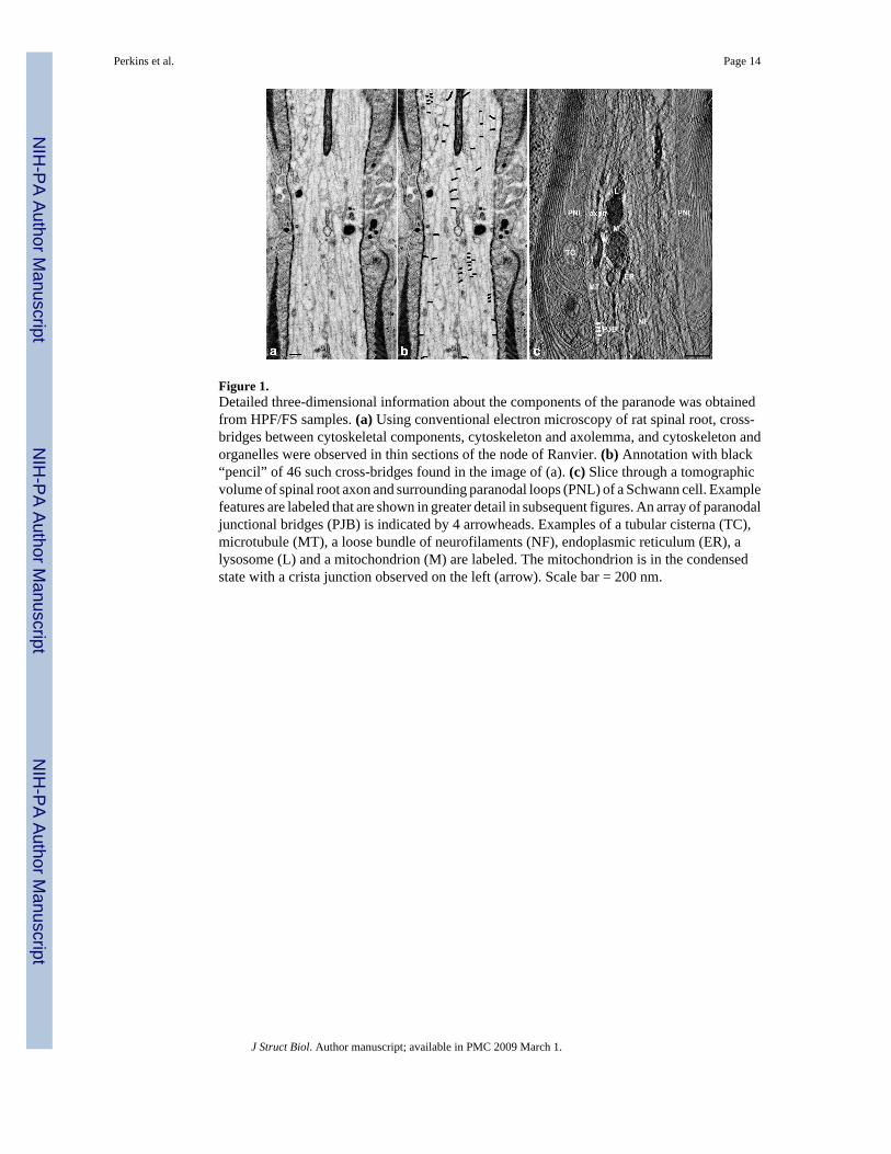
Figure 1.Detailed three-dimensional information about the components of the paranode was obtainedfrom HPF/FS samples. (a) Using conventional electron microscopy of rat spinal root, cross-bridges between cytoskeletal components, cytoskeleton and axolemma, and cytoskeleton andorganelles were observed in thin sections of the node of Ranvier. (b) Annotation with black“pencil” of 46 such cross-bridges found in the image of (a). (c) Slice through a tomographicvolume of spinal root axon and surrounding paranodal loops (PNL) of a Schwann cell. Examplefeatures are labeled that are shown in greater detail in subsequent figures. An array of paranodaljunctional bridges (PJB) is indicated by 4 arrowheads. Examples of a tubular cisterna (TC),microtubule (MT), a loose bundle of neurofilaments (NF), endoplasmic reticulum (ER), alysosome (L) and a mitochondrion (M) are labeled. The mitochondrion is in the condensedstate with a crista junction observed on the left (arrow). Scale bar = 200 nm.
Perkins et al. Page 14
J Struct Biol. Author manuscript; available in PMC 2009 March 1.
NIH
-PA Author Manuscript
NIH
-PA Author Manuscript
NIH
-PA Author Manuscript
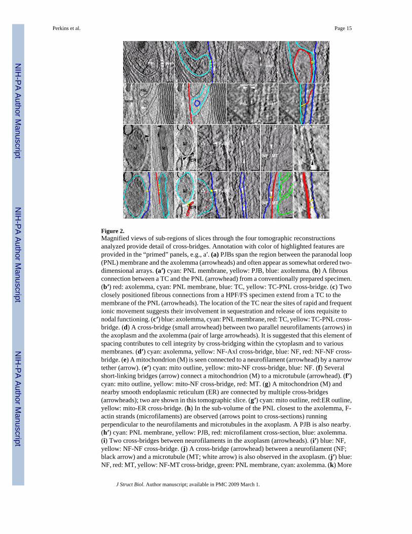
Figure 2.Magnified views of sub-regions of slices through the four tomographic reconstructionsanalyzed provide detail of cross-bridges. Annotation with color of highlighted features areprovided in the “primed” panels, e.g., a′. (a) PJBs span the region between the paranodal loop(PNL) membrane and the axolemma (arrowheads) and often appear as somewhat ordered two-dimensional arrays. (a′) cyan: PNL membrane, yellow: PJB, blue: axolemma. (b) A fibrousconnection between a TC and the PNL (arrowhead) from a conventionally prepared specimen.(b′) red: axolemma, cyan: PNL membrane, blue: TC, yellow: TC-PNL cross-bridge. (c) Twoclosely positioned fibrous connections from a HPF/FS specimen extend from a TC to themembrane of the PNL (arrowheads). The location of the TC near the sites of rapid and frequentionic movement suggests their involvement in sequestration and release of ions requisite tonodal functioning. (c′) blue: axolemma, cyan: PNL membrane, red: TC, yellow: TC-PNL cross-bridge. (d) A cross-bridge (small arrowhead) between two parallel neurofilaments (arrows) inthe axoplasm and the axolemma (pair of large arrowheads). It is suggested that this element ofspacing contributes to cell integrity by cross-bridging within the cytoplasm and to variousmembranes. (d′) cyan: axolemma, yellow: NF-Axl cross-bridge, blue: NF, red: NF-NF cross-bridge. (e) A mitochondrion (M) is seen connected to a neurofilament (arrowhead) by a narrowtether (arrow). (e′) cyan: mito outline, yellow: mito-NF cross-bridge, blue: NF. (f) Severalshort-linking bridges (arrow) connect a mitochondrion (M) to a microtubule (arrowhead). (f′)cyan: mito outline, yellow: mito-NF cross-bridge, red: MT. (g) A mitochondrion (M) andnearby smooth endoplasmic reticulum (ER) are connected by multiple cross-bridges(arrowheads); two are shown in this tomographic slice. (g′) cyan: mito outline, red:ER outline,yellow: mito-ER cross-bridge. (h) In the sub-volume of the PNL closest to the axolemma, F-actin strands (microfilaments) are observed (arrows point to cross-sections) runningperpendicular to the neurofilaments and microtubules in the axoplasm. A PJB is also nearby.(h′) cyan: PNL membrane, yellow: PJB, red: microfilament cross-section, blue: axolemma.(i) Two cross-bridges between neurofilaments in the axoplasm (arrowheads). (i′) blue: NF,yellow: NF-NF cross-bridge. (j) A cross-bridge (arrowhead) between a neurofilament (NF;black arrow) and a microtubule (MT; white arrow) is also observed in the axoplasm. (j′) blue:NF, red: MT, yellow: NF-MT cross-bridge, green: PNL membrane, cyan: axolemma. (k) More
Perkins et al. Page 15
J Struct Biol. Author manuscript; available in PMC 2009 March 1.
NIH
-PA Author Manuscript
NIH
-PA Author Manuscript
NIH
-PA Author Manuscript

pronounced examples of NF-MT cross-bridges (arrowheads) from a tomographic slice of aconventionally prepared sample. (k′) blue: NF, red: MT, yellow: NF-MT cross-bridge. Scalebar = 100 nm for all panels.
Perkins et al. Page 16
J Struct Biol. Author manuscript; available in PMC 2009 March 1.
NIH
-PA Author Manuscript
NIH
-PA Author Manuscript
NIH
-PA Author Manuscript

Figure 3.Comparison of cross-bridge structures by type after alignment and averaging of tomographicvolumes. Images from a central slice through each averaged subvolume of (a) NF-NF, (b) NF-MT, (c) mito-MT, (d) NF-Axl, and (e) TC-PNL types are shown. The variation in diameteralong the length of each bridge is evident as well as the general difference in cross-bridgediameter between the first three and last two types of bridge. The numbers of subvolumesaveraged together per cross-bridge type are 15, 17, 8, 11, and 15 for NF-NF, NF-MT, mito-MT, NF-Axl, and TC-PNL, respectively. Scale bar = 10 nm.
Perkins et al. Page 17
J Struct Biol. Author manuscript; available in PMC 2009 March 1.
NIH
-PA Author Manuscript
NIH
-PA Author Manuscript
NIH
-PA Author Manuscript

Figure 4.Segmentation of the volumes shown in Figs. 1-3 provides surface-rendered volumes used forvisualization and dissection of connections and cross-bridges. Cytoskeletal networks providea flexible scaffolding to impart structure to the cytoplasm and to resist stresses appliedexternally to the cell. (a) All segmented components are displayed. In the axoplasm, there were32 MTs (thicker orange threads), 158 NFs (thinner green threads), 8 mitochondria and othermembranous organelles (magenta) (outer membrane only shown), 5 fibrous connectionsbetween PJBs and NFs (magenta), and 5 fibrous extensions from PJBs into the axoplasm (red).In the paranodal loop region, there were 32 PNLs (alternating blue and cyan), 23 PJBs (yellow),9 fibrous extensions from PJBs into the PNL (red), 7 TCs (dark green), and 24 F-actin fibers(microfilaments) (cyan). Scale bar = 200 nm. (b) Cross-bridges connect neurofilaments (NF)to each other and to microtubules (MT) in the axoplasm although none were observed betweenMTs. Three examples of NF-NF and two examples of NF-MT cross-bridges are shown. Onlyone or two cross-bridges between individual cytoskeletal threads were found. Cross-bridgeswere also found between NFs and the axolemma (arrowhead). (c) PJBs form nearly equidistantrows that connect the PNL membrane (blue) with the axolemma (not shown). Six PJBs (yellow)are shown of various lengths (arrowhead points to an example). Cross-bridges between PJBsand NFs were found (magenta filaments), but not between PJBs and MTs. Filamentousextensions originating from PJBs were observed (red filaments) that had no obvious connection
Perkins et al. Page 18
J Struct Biol. Author manuscript; available in PMC 2009 March 1.
NIH
-PA Author Manuscript
NIH
-PA Author Manuscript
NIH
-PA Author Manuscript
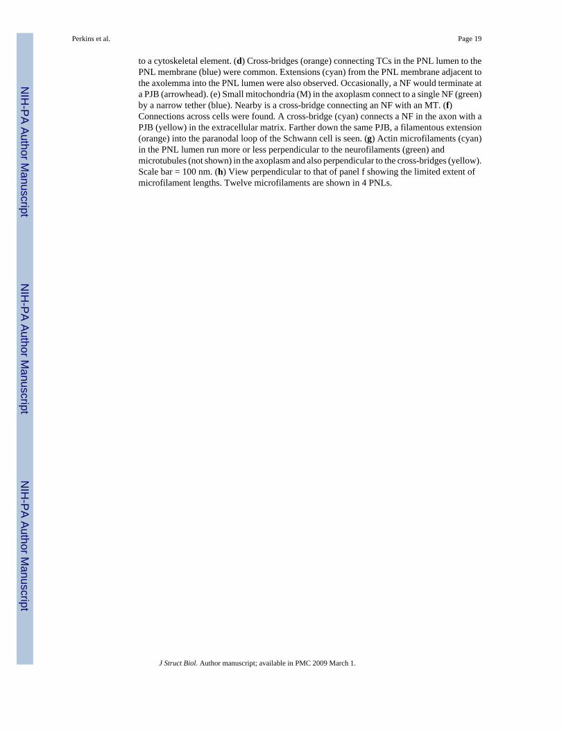
to a cytoskeletal element. (d) Cross-bridges (orange) connecting TCs in the PNL lumen to thePNL membrane (blue) were common. Extensions (cyan) from the PNL membrane adjacent tothe axolemma into the PNL lumen were also observed. Occasionally, a NF would terminate ata PJB (arrowhead). (e) Small mitochondria (M) in the axoplasm connect to a single NF (green)by a narrow tether (blue). Nearby is a cross-bridge connecting an NF with an MT. (f)Connections across cells were found. A cross-bridge (cyan) connects a NF in the axon with aPJB (yellow) in the extracellular matrix. Farther down the same PJB, a filamentous extension(orange) into the paranodal loop of the Schwann cell is seen. (g) Actin microfilaments (cyan)in the PNL lumen run more or less perpendicular to the neurofilaments (green) andmicrotubules (not shown) in the axoplasm and also perpendicular to the cross-bridges (yellow).Scale bar = 100 nm. (h) View perpendicular to that of panel f showing the limited extent ofmicrofilament lengths. Twelve microfilaments are shown in 4 PNLs.
Perkins et al. Page 19
J Struct Biol. Author manuscript; available in PMC 2009 March 1.
NIH
-PA Author Manuscript
NIH
-PA Author Manuscript
NIH
-PA Author Manuscript
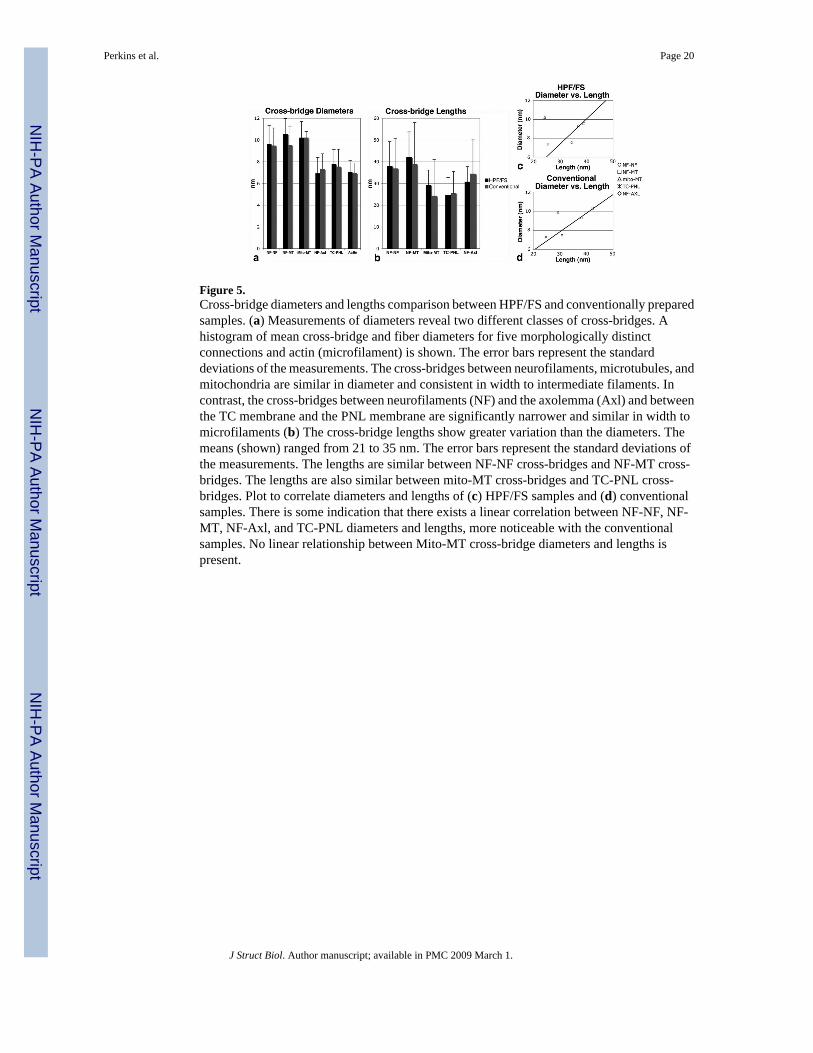
Figure 5.Cross-bridge diameters and lengths comparison between HPF/FS and conventionally preparedsamples. (a) Measurements of diameters reveal two different classes of cross-bridges. Ahistogram of mean cross-bridge and fiber diameters for five morphologically distinctconnections and actin (microfilament) is shown. The error bars represent the standarddeviations of the measurements. The cross-bridges between neurofilaments, microtubules, andmitochondria are similar in diameter and consistent in width to intermediate filaments. Incontrast, the cross-bridges between neurofilaments (NF) and the axolemma (Axl) and betweenthe TC membrane and the PNL membrane are significantly narrower and similar in width tomicrofilaments (b) The cross-bridge lengths show greater variation than the diameters. Themeans (shown) ranged from 21 to 35 nm. The error bars represent the standard deviations ofthe measurements. The lengths are similar between NF-NF cross-bridges and NF-MT cross-bridges. The lengths are also similar between mito-MT cross-bridges and TC-PNL cross-bridges. Plot to correlate diameters and lengths of (c) HPF/FS samples and (d) conventionalsamples. There is some indication that there exists a linear correlation between NF-NF, NF-MT, NF-Axl, and TC-PNL diameters and lengths, more noticeable with the conventionalsamples. No linear relationship between Mito-MT cross-bridge diameters and lengths ispresent.
Perkins et al. Page 20
J Struct Biol. Author manuscript; available in PMC 2009 March 1.
NIH
-PA Author Manuscript
NIH
-PA Author Manuscript
NIH
-PA Author Manuscript

NIH
-PA Author Manuscript
NIH
-PA Author Manuscript
NIH
-PA Author Manuscript
Perkins et al. Page 21Ta
ble
IR
ange
of C
ross
-Brid
ge D
iam
eter
s and
Len
gths
Cro
ss B
ridg
e T
ype
HPF
Dia
met
ers (
nm)
Num
ber
of M
easu
rem
ents
CA
F D
iam
eter
s (nm
)N
umbe
r of
Mea
sure
men
tsH
PF L
engt
hs (n
m)
CA
F L
engt
hs (n
m)
NF-
NF
7.4-
12.2
§40
7.6-
10.9
§34
21.8
-74.
1§18
.8-6
2.5§
NF-
MT
8.1-
12.4
267.
7-11
.523
23.9
-65.
819
.8-9
3.8
mito
-MT
8.1-
11.4
189.
9-10
.93
13.8
-51.
117
.6-3
5.8
TC-P
NL
5-8.
718
5.4-
9.2
219.
6-45
.78.
5-84
NF-
Axl
5.7-
9.4
365.
4-9.
112
21.6
-66.
516
.3-6
0.4
Act
in5.
5-8.
447
5.6-
8.2
17
§ Min
imum
-max
imum
mea
sure
d va
lues
HPF
= h
igh
pres
sure
free
zing
with
ald
ehyd
e fix
atio
n
CA
F =
conv
entio
nal a
ldeh
yde
fixat
ion
J Struct Biol. Author manuscript; available in PMC 2009 March 1.