A dynamic X-ray diffraction study of anaesthesia action. Changes in myelin structure and electrical...
13
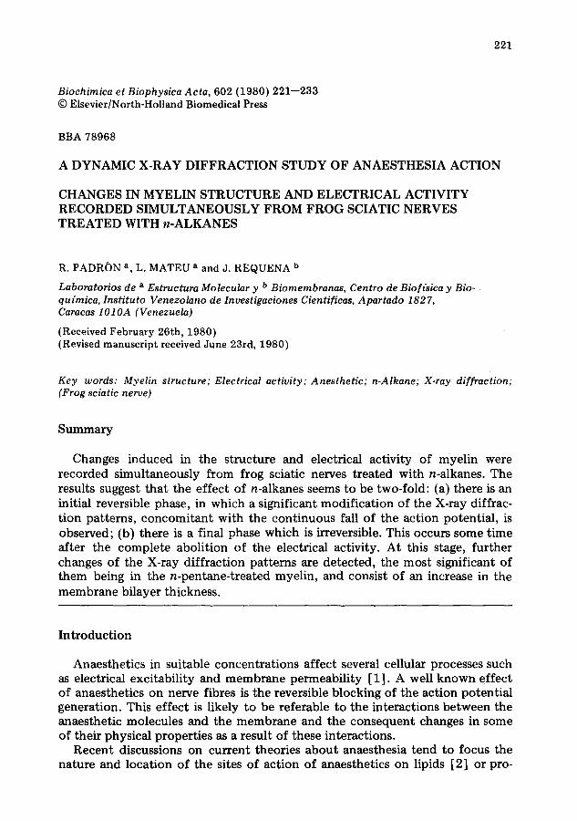
221 Biochimica et Biophysica Acta, 602 (1980) 221--233 © Elsevier/North-Holland Biomedical Press BBA 78968 A DYNAMIC X-RAY DIFFRACTION STUDY OF ANAESTHESIA ACTION CHANGES IN MYELIN STRUCTURE AND ELECTRICAL ACTIVITY RECORDED SIMULTANEOUSLY FROM FROG SCIATIC NERVES TREATED WITH n-ALKANES R. PADR()N a, L. MATEU a and J. REQUENA b Laboratorios de a Estructura Molecular y b Biomembranas, Centro de Biof(sica y Bio- qu(mica, Instituto Venezolano de Investigaciones Cientificas, Apartado 1827, Caracas 1010A (Venezuela) (Received February 26th, 1980) (Revised manuscript received June 23rd, 1980) Key words: Myelin structure; Electrical activity; Anesthetic; n-Alkane; X-ray diffraction; (Frog sciatic nerve) Summary Changes induced in the structure and electrical activity of myelin were recorded simultaneously from frog sciatic nerves treated with n-alkanes. The results suggest that the effect of n-alkanes seems to be two-fold: (a) there is an initial reversible phase, in which a significant modification of the X-ray diffrac- tion patterns, concomitant with the continuous fall of the action potential, is observed; (b) there is a final phase which is irreversible. This occurs some time after the complete abolition of the electrical activity. At this stage, further changes of the X-ray diffraction patterns are detected, the most significant of them being in the n-pentane-treated myelin, and consist of an increase in the membrane bilayer thickness. Introduction Anaesthetics in suitable concentrations affect several cellular processes such as electrical excitability and membrane permeability [1]. A well known effect of anaesthetics on nerve fibres is the reversible blocking of the action potential generation. This effect is likely to be referable to the interactions between the anaesthetic molecules and the membrane and the consequent changes in some of their physical properties as a result of these interactions. Recent discussions on current theories about anaesthesia tend to focus the nature and location of the sites of action of anaesthetics on lipids [2] or pro-
-
Upload
independent -
Category
Documents
-
view
3 -
download
0
Transcript of A dynamic X-ray diffraction study of anaesthesia action. Changes in myelin structure and electrical...
221
Biochimica et Biophysica Acta, 602 (1980) 221--233 © Elsevier/North-Holland Biomedical Press
BBA 78968
A DYNAMIC X-RAY DIFFRACTION STUDY OF ANAESTHESIA ACTION
CHANGES IN MYELIN STRUCTURE AND ELECTRICAL ACTIVITY RECORDED SIMULTANEOUSLY FROM FROG SCIATIC NERVES TREATED WITH n-ALKANES
R. PADR()N a, L. MATEU a and J. REQUENA b
Laboratorios de a Estructura Molecular y b Biomembranas, Centro de Biof(sica y Bio- qu(mica, Instituto Venezolano de Investigaciones Cientificas, Apartado 1827, Caracas 1010A (Venezuela)
(Received February 26th, 1980) (Revised manuscript received June 23rd, 1980)
Key words: Myelin structure; Electrical activity; Anesthetic; n-Alkane; X-ray diffraction; (Frog sciatic nerve)
Summary
Changes induced in the structure and electrical activity of myelin were recorded simultaneously from frog sciatic nerves treated with n-alkanes. The results suggest that the effect of n-alkanes seems to be two-fold: (a) there is an initial reversible phase, in which a significant modification of the X-ray diffrac- tion patterns, concomitant with the continuous fall of the action potential, is observed; (b) there is a final phase which is irreversible. This occurs some time after the complete abolition of the electrical activity. At this stage, further changes of the X-ray diffraction patterns are detected, the most significant of them being in the n-pentane-treated myelin, and consist of an increase in the membrane bilayer thickness.
Int roduct ion
Anaesthetics in suitable concentrations affect several cellular processes such as electrical excitability and membrane permeabili ty [1]. A well known effect of anaesthetics on nerve fibres is the reversible blocking of the action potential generation. This effect is likely to be referable to the interactions between the anaesthetic molecules and the membrane and the consequent changes in some of their physical properties as a result of these interactions.
Recent discussions on current theories about anaesthesia tend to focus the nature and location of the sites of action of anaesthetics on lipids [2] or pro-
222
teins [3,4], or on both protein and lipid molecules [1] of the excitable mem- brane. Interestingly, most of the proposed models share a common feature: the site of action of these molecules is a hydrophobic region of the membrane. On the other hand, while Seeman [ 1] postulated that anaesthetics and other nerve- blocking drugs adsorb to the membrane causing an expansion of the volume and/or the area of its hydrophobic domains -- a process which blocks the ionic channels responsible for action potential - - , Haydon et al. [2] proposed that during anaesthesia by n-alkanes there occurs a thickening of the lipid bilayer region of the membrane, also a process which blocks the excitable ionic chan- nels. On the other hand, Franks and Lieb [3] and Richards et al. [4] concluded that the site of action of general anaesthetics is the hydrophobic port ion of a membrane-bound protein.
The use of a physical technique such as X:ray diffraction could give further insight about the mechanism by which n-alkanes block the electrical activity of biological membranes. Unfortunately, this is not easy to perform since excit- able membranes are not naturally ordered. A reasonable alternative for obtain- ing useful structural information is the s tudy of this process on a membrane system such as the nerve myelin sheath, which, inexcitable though it is, is naturally ordered. In addition, it has been suggested that myelin could be passively involved in the saltatory phenomenon of nerve conduct ion [5,6]. Finally, after the extensive related work that has been carried out by several groups during the last three decades, the structure of myelin is well known at present (see review in Ref. 7). Indeed, Caspar and Kirschner [8], on the basis of a high-resolution X-ray diffraction study, have proposed a molecular model for myelin membranes. More recently, these structural concepts have been exhaust- ively revised and enriched by Nelander and Blaurock [9].
We have recently reported [10] a dynamic X-ray diffraction study on the effect of a hydrocarbon solution on the structure of myelin (at the low resolu- tion of 34 •). We found that the perfusion during 24 h of frog sciatic nerves with n-pentane-saturated Ringer solution caused a 6% increase in the thickness of the myelin membrane lattice, although such a change was hardly visible for the first 18 h. In the present communicat ion, a more detailed s tudy of the effect of several n-alkanes on the structure of myelin is carried out. The aim of these experiments was to determine the relationship existing between the sup- pression of the nerve impulse and the structural changes of myelin (membrane thickening) and its reversibility, at the much improved resolution of about 15 A. In order to do this, the nervous electrical activity and the X-ray diffrac- tion patterns were simultaneously recorded on the same frog sciatic nerve.
Materials and Methods
Freshly dissected frog (Rana pipiens) sciatic nerves were deprived of the epi- neurium (desheathed) and mounted on a specimen holder with a continuous-per- fusion system which permits frog Ringer solutions to flow around the nerve [ 11 ]. These n-alkane-saturated Ringer solutions were prepared by equilibrating normal frog Ringer solution with liquid alkane for 24 h with gently stirring. Great care was taken to avoid emulsions. The alkanes assayed were n-pentane, n-hexane and n-decane. These hydrocarbons (purest grade) were purchased
223
from BDH (Poole, U.K.), or Koch-Light (Colnbrook, U.K.) and used wi thout further purification.
The specimen holder was provided with two pairs of platinum wire elec- trodes for nerve stimulation and for the recording of the compound action potential. The circuitry was similar to that currently used in electrophysio- logical practice [13]. The compound action potential height was registered in a storage oscilloscope.
The small-angle X-ray diffraction techniques were those currently used in our laboratory [10--12,14,15] . The X-ray patterns were recorded with a linear position-sensitive detector [16] from a 1 cm long sciatic segment, which was located between the two pairs of electrodes. The patterns were stored in an on-line computer . This experimental arrangement facilitated the simultaneous recording of the X-ray diffraction patterns and the electrical activity of the nerve.
For control purposes, an X-ray diffraction pattern was accumulated during 4 h from a sciatic nerve in normal Ringer solution. At the end of this data collection, the perfusion was exchanged to the alkane-containing solution and the structural modifications induced by the alkane were sequentially followed by recording a series of 5-min counting spectra. The perfusion was continued until no further change in the patterns was observed. At this point, another 4-h counting spectrum was accumulated from the alkane-perfused sciatic nerve. This register was compared with the original pattern recorded from the same nerve before the alkane t reatment during an identical period of time.
The diffraction patterns were displayed on a cathode-ray tube and photo- graphed on a 35 mm film. Periodicity and intensity measurements were deter- mined from logarithmic plots of the computer-stored data. The error esti- mated for the repeat period was _+1%. The intensity of the reflections was mea- sured from peak areas after background subtraction. Electron-density profiles were calculated as previously described [10]. Different profiles were scaled by setting ZF2(h)/d = constant [17].
Results
The action of n-pentane, n-hexane and n-decane on the structure and electri- cal activity of myelin was investigated for several sciatic nerves. X-ray pattern modifications of different nerve specimens were entirely reproducible for a given alkane from one nerve preparation to the next. However, depending on nerve dimensions and the flow rate of perfusion, time-course variability was ob- served. In order to minimise these variations, the results reported herein will refer to sciatic nerves having similar thickness (approx. 1 mm), which were per- fused at an identical f low rate (5 ml/min).
Reversible effects o f n-alkanes on nerve impulse blockage and myelin structure As stated in Materials and Methods, 5-min counting X-ray diffraction
patterns were sequentially registered during the perfusion of the nerves wi th different alkane-containing solutions. The time courses of the action potential blockage and X-ray pattern modifications are shown in Fig. 1 for sciatic nerves treated with n-pentane, n-hexane and n-decane, toge the r with ~the results for a
2 2 4
IO0
z-~(%) 0 E
= . . . . . . " a
IO0
CAP(%) I 0 - b
d(~) 19o I .... ... .:O: " ° 180 ............ 0""
r6o
C
L J I i I L I 0 I0 20 30
t(hours)
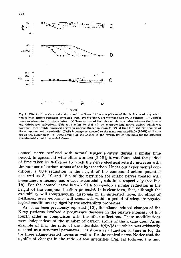
Fig. 1. E f f e c t o f t h e e l ec t r i ca l a c t i v i t y a n d the X - r a y d i f f r a c t i o n p a t t e r n o f t h e p e r f u s l o n o f f r o g sc ia t i c ne rves w i t h R i n g e r s o l u t i o n s s a t u r a t e d w i t h : (w) n - d e c a n e , (u) n - h e x a n e a n d (o) n - p e n t a n e . (o) C o n t r o l ne rve in a l k a n e - f r e e R i n g e r s o l u t i o n . (a) T ime cou r se o f t he re la t ive i n t e n s i t y r a t i o b e t w e e n the f o u r t h - a n d t h i r d - o r d e r r e f l e c t i o n s . Th i s r a t i o r e fe r s to t h a t o f t he c o r r e s p o n d i n g na t i ve p a t t e r n w h i c h was r e c o r d e d f r o m f r e sh ly d i s s e c t e d ne rves in n o r m a l R i n g e r s o l u t i o n ( 1 0 0 % a t t i m e 0 h ) . (b) T ime cou r se o f t h e c o m p o u n d a c t i o n p o t e n t i a l ( C A P ) b l o c k a g e as r e f e r r e d t o t he m a x i m u m a m p l i t u d e ( 1 0 0 % ) a t t h e on- se t o f t h e e x p e r i m e n t . (c) T ime c o u r s e o f t he c h a n g e in t he m y e l i n l a t t i ce t h i c k n e s s fo r the different e x p e r i m e n t a l c o n d i t i o n s s t a t e d a b o v e .
control nerve perfused with normal Ringer solution during a similar time period. In agreement with other workers [2,18], it was found that the period of time taken by n-alkanes to block the nerve electrical activity increases with the number of carbon atoms of the hydrocarbon. Under our experimental con- ditions, a 50% reduction in the height of the compound action potential occurred at 5, 10 and 15 h of the perfusion for sciatic nerves treated with n-pentane-, n-hexane- and n-decane-containing solutions, respectively (see Fig. lb) . For the control nerve it took 21 h to develop a similar reduction in the height of the compound action potential. It is clear then, that, although the excitability will spontaneously disappear in an untreated nerve, the effect of n-alkanes, even n-decane, will occur well within a period of adequate physio- logical conditions as judged by the excitability properties.
As it has been previously reported [10], the alkane-induced changes of the X-ray patterns involved a progressive decrease in the relative intensity of the fourth order in comparison with the other reflections. These modifications were independent of the number of carbon atoms of the alkane used. As an example of this, the ratio of the intensities I(4)//(3) -- which was arbitrarily selected as a structural parameter -- is shown as a function of time in Fig. l a for three alkane-treated nerves as well as for the control nerve. Notice that the significant changes in the ratio of the intensities {Fig. la) followed the time
225
course of the process that led to the suppression of the propagation action potential (Fig. lb) in nerves exposed to alkane-containing Ringer solution, while the control nerve did not show such a change. Fig. lc reveals that the myelin lattice thickness remained constant during the process in which the compound action potential was progressively reduced by alkane treatment. At this stage, neither the background nor the half-width peak intensity was significantly altered. It should be observed that prolonged exposure to alkane, specifically n-pentane, caused the already reported thickening of the myelin lattice [10], a process which will be shown below to be irreversible.
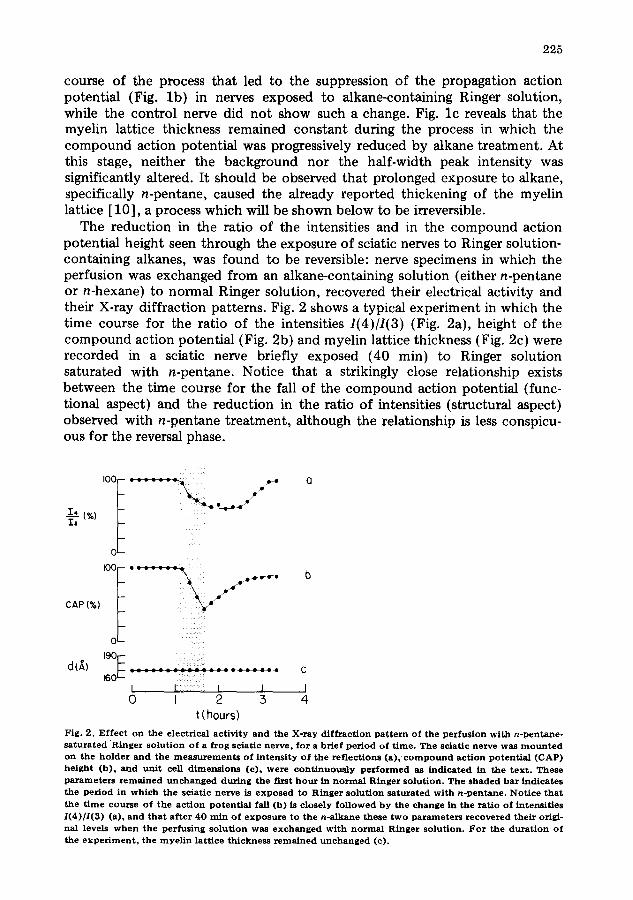
The reduction in the ratio of the intensities and in the compound action potential height seen through the exposure of sciatic nerves to Ringer solution- containing alkanes, was found to be reversible: nerve specimens in which the perfusion was exchanged from an alkane-containing solution (either n-pentane or n-hexane) to normal Ringer solution, recovered their electrical activity and their X-ray diffraction patterns. Fig. 2 shows a typical experiment in which the time course for the ratio of the intensities 1(4)/1(3) (Fig. 2a), height of the compound action potential (Fig. 2b) and myelin lattice thickness (Fig. 2c) were recorded in a sciatic nerve briefly exposed (40 min) to Ringer solution saturated with n-pentane. Notice that a strikingly close relationship exists between the time course for the fall of the compound action potential (func- tional aspect) and the reduction in the ratio of intensities (structural aspect) observed with n-pentane treatment, although the relationship is less conspicu- ous for the reversal phase.
I__~, (%) I,
CAP (%)
0
o~-
: : : : - ' : : : : : : C 160 L -
I F I J [ 0 I 2 5 4
t (hours)
Fig . 2 . E f f e c t o n t h e e l ec t r i ca l a c t i v i t y a n d t h e X - r a y d i f f r a c t i o n p a t t e r n o f t he p e r f u s i o n w i t h n - p e n t a n e - s a t u r a t e d R i n g e r s o l u t i o n o f a f r o g sc i a t i c n e r v e , f o r a b r i e f p e r i o d o f t i m e . T h e s c i a t i c n e r v e w a s m o u n t e d o n t h e h o l d e r a n d t h e m e a s u r e m e n t s o f i n t e n s i t y o f t h e r e f l e c t i o n s (a ) , c o m p o u n d a c t i o n p o t e n t i a l (CAP) h e i g h t ( b ) , a n d u n i t cell d i m e n s i o n s (c ) , w e r e c o n t i n u o u s l y p e r f o r m e d as i n d i c a t e d in t h e t e x t . T h e s e p a r a m e t e r s r e m a i n e d u n c h a n g e d d u r i n g t h e f i rs t h o u r in n o r m a l R i n g e r s o l u t i o n . T h e s h a d e d b a r i n d i c a t e s t h e p e r i o d in w h i c h t h e sc i a t i c ne rve is e x p o s e d t o R i n g e r s o l u t i o n s a t u r a t e d w i t h n - p e n t a n e . N o t i c e t h a t t h e t i m e c o u r s e o f t h e a c t i o n p o t e n t i a l fall (b ) is c lo se ly f o l l o w e d b y the c h a n g e in t h e r a t i o o f i n t e n s i t i e s 1(4)/1(3) (a) , a n d t h a t a f t e r 4 0 r a in o f e x p o s u r e t o t h e n - a l k a n e t h e s e t w o p a r a m e t e r s r e c o v e r e d t h e i r origi - na l levels w h e n t h e p e r f u s i n g s o l u t i o n w a s e x c h a n g e d w i t h n o r m a l R i n g e r s o l u t i o n . F o r t h e d u r a t i o n o f t h e e x p e r i m e n t , t h e m y e l i n l a t t i c e t h i c k n e s s r e m a i n e d u n c h a n g e d (c) .
226
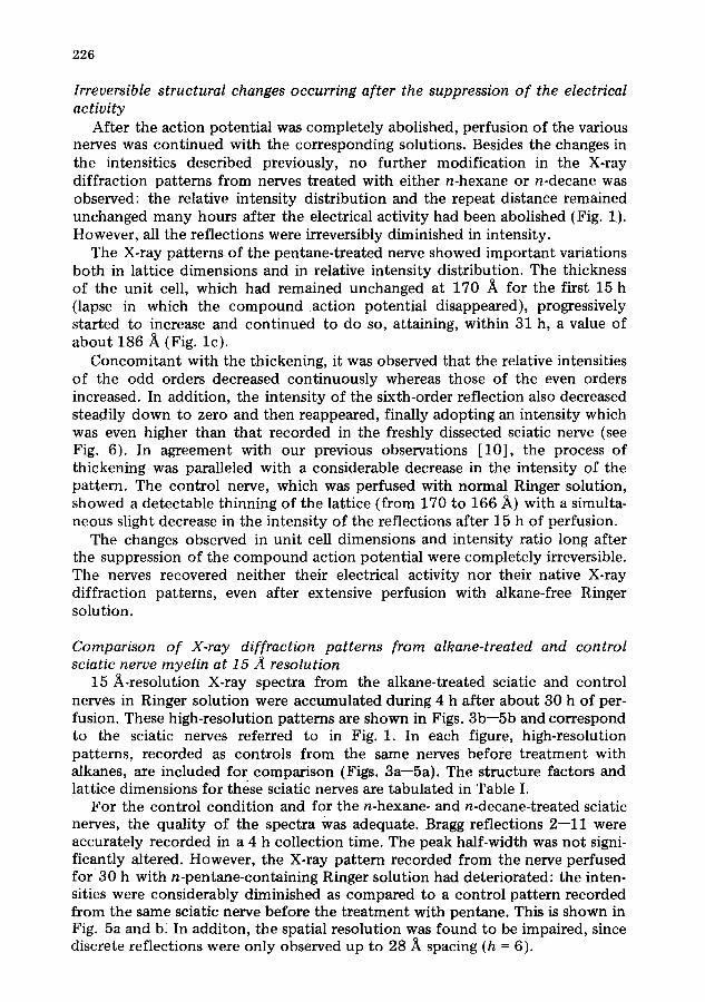
Irreversible structural changes occurring after the suppression o f the electrical activity
After the action potential was completely abolished, perfusion of the various nerves was continued with the corresponding solutions. Besides the changes in the intensities described previously, no further modification in the X-ray diffraction patterns from nerves treated with either n-hexane or n-decane was observed: the relative intensity distribution and the repeat distance remained unchanged many hours after the electrical activity had been abolished (Fig. 1). However, all the reflections were irreversibly diminished in intensity.
The X-ray patterns of the pentane-treated nerve showed important variations both in lattice dimensions and in relative intensity distribution. The thickness of the unit cell, which had remained unchanged at 170 /~ for the first 15 h (lapse in which the compound action potential disappeared), progressively started to increase and continued to do so, attaining, within 31 h, a value of about 186 .~ (Fig. lc) .
Concomitant with the thickening, it was observed that the relative intensities of the odd orders decreased continuously whereas those of the even orders increased. In addition, the intensity of the sixth-order reflection also decreased steadily down to zero and then reappeared, finally adopting an intensity which was even higher than that recorded in the freshly dissected sciatic nerve (see Fig. 6). In agreement with our previous observations [10], the process of th ickening was paralleled with a considerable decrease in the intensity of the pattern. The control nerve, which was perfused with normal Ringer solution, showed a detectable thinning of the lattice (from 170 to 166 ~) with a simulta- neous slight decrease in the intensity of the reflections after 15 h of perfusion.
The changes observed in unit cell dimensions and intensity ratio long after the suppression of the compound action potential were completely irreversible. The nerves recovered neither their electrical activity nor their native X-ray diffraction patterns, even after extensive perfusion with alkane-free Ringer solution.
Comparison o f X-ray diffraction patterns from alkane-treated and control sciatic nerve myelin at 15 .~ resolution
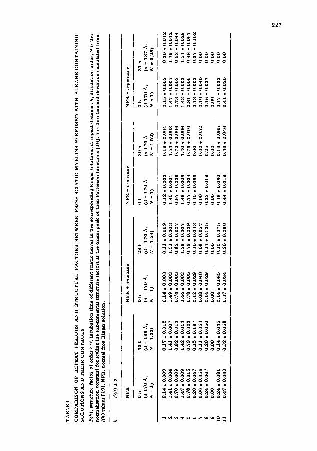
15 h-resolut ion X-ray spectra from the alkane-treated sciatic and control nerves in Ringer solution were accumulated during 4 h after about 30 h of per- fusion. These high-resolution patterns are shown in Figs. 3b--5b and correspond to the sciatic nerves referred to in Fig. 1. In each figure, high-resolution patterns, recorded as controls from the same nerves before t reatment with alkanes, are included for comparison (Figs. 3a--5a). The structure factors and lattice dimensions for these sciatic nerves are tabulated in Table I.
For the control condition and for the n-hexane- and n-decane-treated sciatic nerves, the quality of the spectra Was adequate. Bragg reflections 2--11 were accurately recorded in a 4 h collection time. The peak half-width was not signi- ficantly altered. However, the X-ray pattern recorded from the nerve perfused f o r 30 h with n-pentane-containing Ringer solution had deteriorated: the inten- sities were considerably diminished as compared to a control pattern recorded from the same sciatic nerve before the t reatment with pentane. This is shown in Fig. 5a and b, In additon, the spatial resolution was found to be impaired, since discrete reflections were only observed up to 28 .~ spacing (h = 6).
TA
BL
E I
CO
MP
AR
ISO
N
OF
R
EP
EA
T
PE
RIO
DS
A
ND
S
TR
UC
TU
RE
F
AC
TO
RS
B
ET
WE
EN
F
RO
G
SC
IAT
IC
MY
EL
INS
P
ER
FU
SE
D
WIT
H
AL
KA
NE
-CO
NT
AIN
ING
S
OL
UT
ION
S
AN
D T
HE
IR
CO
NT
RO
LS
F(h
),
stru
ctu
re
fac
tor
of
ord
er
h ;
t, i
nc
ub
ati
on
ti
me
of
dif
fere
nt
scia
tic
ne
rve
s in
th
e c
orr
esp
on
din
g R
ing
er
solu
tio
ns;
d,
rep
ea
t d
ista
nc
e;
h,
dif
fra
cti
on
ord
er;
N
is t
he
no
rma
lisa
tio
n
co
nst
an
t fo
r sc
alin
g t
he
ex
pe
rim
en
tal
stru
ctu
re
fac
tors
at
the
ori
gin
pe
ak
of
the
ir P
att
ers
on
fu
nc
tio
ns
[16
].
o is
th
e s
tan
da
rd
de
via
tio
n c
alc
ula
ted
fr
om
l(
h)
val
ues
[1
9].
NF
R,
no
rma
l fr
og
Rin
ge
r so
luti
on
.
h F
(h)
± o
NFR
NFR
+ n-decane
NFR
+ n-hexane
NFR
+ n-pentane
Oh
3
0h
O
h
28
h
Oh
3
0h
O
h
31
h
(d 1
70
A,
(d =
16
6 A
, (d
= 1
70
A,
(d =
17
0 A
, (d
= 1
70
A,
(d 1
70
A,
(d 1
70
A,
(d =
18
7 A
,
N
= I
) N
=
1.3
3)
N
= I
) N
=
1.5
4)
N
= I
) N
=
1.5
2)
N
= I
) N
=
3.2
3)
1 0
.14
±
0.0
09
0
.17
±
0.0
12
0
.14
±
0.0
03
0
.11
±
0.0
09
0
.12
±
0.0
03
0
.16
±
0.0
04
0
.15
±
0.0
02
0
.20
±
0.0
12
2 1
.41
±
0.0
04
1
.41
±
0.0
07
1
.49
±
0.0
03
1
.51
±
0.0
03
1
.45
±
0.0
01
1
.53
±
0.0
03
1
.47
±
0.0
01
1
.79
±
0.0
12
3 0
.70
± 0
.00
9
0.8
2
± 0
.01
3
0.7
4
± 0
.00
3
0.8
2
± 0
.00
7
0.6
7
± 0
.00
6
0.7
2
± 0
.00
6
0.7
3
± 0
.00
3
0.5
3
± 0
.04
4
4 1
.47
+
0.0
09
1
.42
± 0
.01
4
1.4
4
± 0
.00
3
1.3
9
± 0
.00
7
1.4
8
± 0
.00
3
1.4
0
± 0
.00
6
1.4
3
± 0
.00
3
1.5
1
± 0
.02
9
5 0
.78
±
0.0
15
0
.79
±
0.0
23
0
.78
±
0.0
05
0
.79
±
0.0
29
0
.77
±
0.0
04
0
.73
±
0.0
10
0
.81
±
0.0
05
0
.48
±
0.0
67
6 0
.20
±
0.0
47
0
.15
±
0.1
87
0
.12
±
0.0
29
0
.10
±
0.0
43
0
.15
±
0.0
63
0
.00
0
.13
±
0.0
02
0
.37
±
0.1
02
7
0.0
6
± 0
.05
6
0.1
1
± 0
.05
4
0.0
8
± 0
.04
0
0.0
8
+ 0
.05
7
0.0
0
0.0
0
± 0
.05
2
0.1
0
± 0
.04
0
0.0
0
8 0
.24
+
0.0
67
0
.20
+
0.0
50
0
.14
±
0.0
29
0
.17
±
0.1
25
0
.23
±
0.0
19
0
.25
0
.16
±
0.0
27
0
.00
9
0.0
0
0.0
0
0.0
0
0.0
0
0.0
0
0.0
0
0.0
0
0.0
0
10
0
.24
+
0.0
81
0
.14
±
0.0
45
0
.14
±
0.0
85
0
.10
±
0.0
75
0
.18
±
0.0
30
0
.16
±
0.0
65
0
.17
±
0.0
32
0
.00
1
1
0.4
7
+ 0
.06
0
0.3
2
± 0
.05
8
0.3
7
± 0
.03
4
0.3
0
± 0
.08
0
0.4
4
± 0
.01
9
0.4
6
± 0
.04
6
0.4
1
± 0
.02
0
0.0
0
bO
t~
228
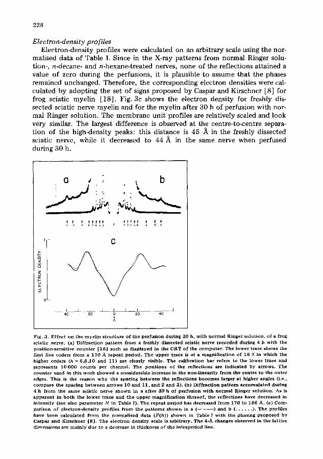
Electron-density profiles Electron-density profiles were calculated on an arbitrary scale using the nor-
malised data of Table I. Since in the X-ray patterns from normal Ringer solu- tion-, n-decane- and n-hexane-treated nerves, none of the reflections attained a value of zero during the perfusions, it is plausible to assume that the phases remained unchanged. Therefore, the corresponding electron densities were cal- culated by adopting the set of signs proposed by Caspar and Kirschner [8] for frog sciatic myelin [18] . Fig. 3c shows the electron density for freshly dis- sected sciatic nerve myelin and for the myelin after 30 h of perfusion with nor- mal Ringer solution. The membrane unit profiles are relatively scaled and look very similar. The largest difference is observed at the centre-to-centre separa- tion of the high-density peaks: this distance is 45 A in the freshly dissected sciatic nerve, while it decreased to 44 ,~ in the same nerve when perfused during 30 h.
a I : i' b
$ f' ~ f '~ f t ' ¢ +~ ' t ' I ' f ' ¢ f' {i {o e 6 5 4 3 2 h 23456 8 ro i{
'F
i ' E3
z I =o I -
,-J bJ
0 ~
C
L_ { { { { 1 L ~ { J { { 40 20 o 2 0 40
Fig. 3. E f fec t on the m y e l i n s t r uc tu r e of the pe r fus ion dur ing 30 h, w i th n o r m a l Ringer so lu t ion , of a f rog sciat ic nerve . (a) D i f f r ac t ion p a t t e r n f r o m a f reshly d issec ted sciat ic nerve r e c o r d e d dur ing 4 b w i th the posi t ion-sensi t ive c o u n t e r [16 ] such as d i sp layed in the CRT of the c o m p u t e r . The l o wer t race shows the first five orders f r o m a 170 A r e p e a t per iod . The u p p e r t r ace is a t a m a g n i f i c a t i o n of 16 X in wh ich the h igher o rders (h = 6 ,8 ,10 and 11) are clearly visible. The ca l ib ra t ion b a r refers to the l o wer t race and rep resen t s 10 000 coun t s per channe l . The pos i t ions of the re f lec t ions are ind ica ted by a r rows . Th e c o u n t e r used in this w o r k s h o w e d a cons iderab le ~ncrease in the non- l inear i ty f r o m the cen t re to the o u t e r edges. This is the r eason w h y the spacing b e t w e e n the re f lec t ions b e c o m e s larger a t h igher angles (i.e., c o m p a r e the spacing b e t w e e n a r rows 10 a nd 11, a nd 2 and 3). (b) D i f f r ac t ion p a t t e r n a c c u m u l a t e d dur ing 4 h f r o m the s ame sciatic nerve s h o w n in a a f t e r 30 h of pe r fus inn wi th n o r m a l Ringer so lu t ion . As is a p p a r e n t in b o t h t he l owe r t race and the u p p e r m a g n i f i c a t i o n t he r eo f , the re f lec t ions have decreased in in tens i ty (see also p a r a m e t e r N in Table I). The r epea t pe r iod has dec reased f r o m 170 to 166 A. (e) Com- par i son of e l ec t ron-dens i ty prof i les f r o m the pa t t e rn s s h o w n in a ( ) and b ( . . . . . . ). Th e prof i les have been ca l cu la t ed f r o m the n o r m a l i s e d d a t a (F(h)) s h o w n in Table I w i th the phas ing p r o p o s e d by Caspar and K i r s chne r [8 ] . The e l ec t ron dens i ty scale is a rb i t r a ry . Th e 4-A changes obse rved in the la t t ice d imens ions are m a i n l y due to a decrease in th ickness of the i n t r ap e r io d line.
229
0L
I ' I'"
45 s to i,
C
t I I I L I [ ] I L I 4 0 2O 0 2O 4O
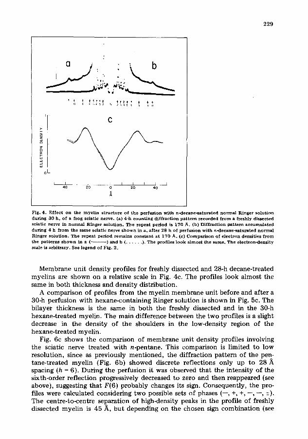
Fig. 4. E f f ec t on the m y e l i n s t r uc tu r e o f the perfusion wi th n-decane - sa tu ra t ed n o r m a l R inger solut ion dur ing 30 h , of a f rog sciatic nerve , (a) 4-h c o u n t i n g d i f f r ac t i on p a t t e r n r e c o r d e d f r o m a f reshly d issec ted sciat ic ne rve in n o r m a l R inger so lu t ion . The repeat period is 170 A. (13) Di f f r ac t ion p a t t e r n a c c u m u l a t e d dur ing 4 h f r o m the s ame sciat ic ne rve s h o w n in a, a f t e r 28 h of pe r fus ion wi th n -decane - sa tu ra t ed n o r m a l Ringer so lu t ion . T he r e p e a t pe r iod r e m a i n s c o n s t a n t a t 170 A. (c) C o m p a r i s o n of e l ec t ron densi t ies f r o m the p a t t e r n s s h o w n in a (.. ) and b ( . . . . . . ). The prof i les l ook a l m o s t the same . Th e e l ec t ron -dens i ty scale is a rb i t r a ry . See legend o f Fig. 2.
Membrane unit density profiles for freshly dissected and 28-h decane-treated myelins are shown on a relative scale in Fig. 4c. The profiles look almost the same in both thickness and density distribution.
A comparison of profiles from the myelin membrane unit before and after a 30-h perfusion with hexane-containing Ringer solution is shown in Fig. 5c. The bilayer thickness is the same in both the freshly dissected and in the 30-h hexane-treated myelin. The main difference between the two profiles is a slight decrease in the density of the shoulders in the low-density region of the hexane-treated myelin.
Fig. 6c shows the comparison of membrane unit density profiles involving the sciatic nerve treated with n-pentane. This comparison is limited to low resolution, since as previously mentioned, the diffraction pattern of the pen- tane-treated myelin (Fig. 6b) showed discrete reflections only up to 28/~ spacing (h = 6). During the perfusion it was observed that the intensity of the sixth-order reflection progressively decreased to zero and then reappeared (see above), suggesting that F(6) probably changes its sign. Consequently, the pro- files were calculated considering two possible sets of phases (--, +, +, , , -+}. The centre-to-centre separation of high-density peaks in the profile of freshly dissected myelin is 45/~, but depending on the chosen sign combination (see
230
• J , : b
w
i i0 8 6 5 4 5 2 h 2 3 4 5 8 io ii
C
L_ l I ~ l _ _ j I I l I i ] 4 0 2 0 0 2 0 4 0
1
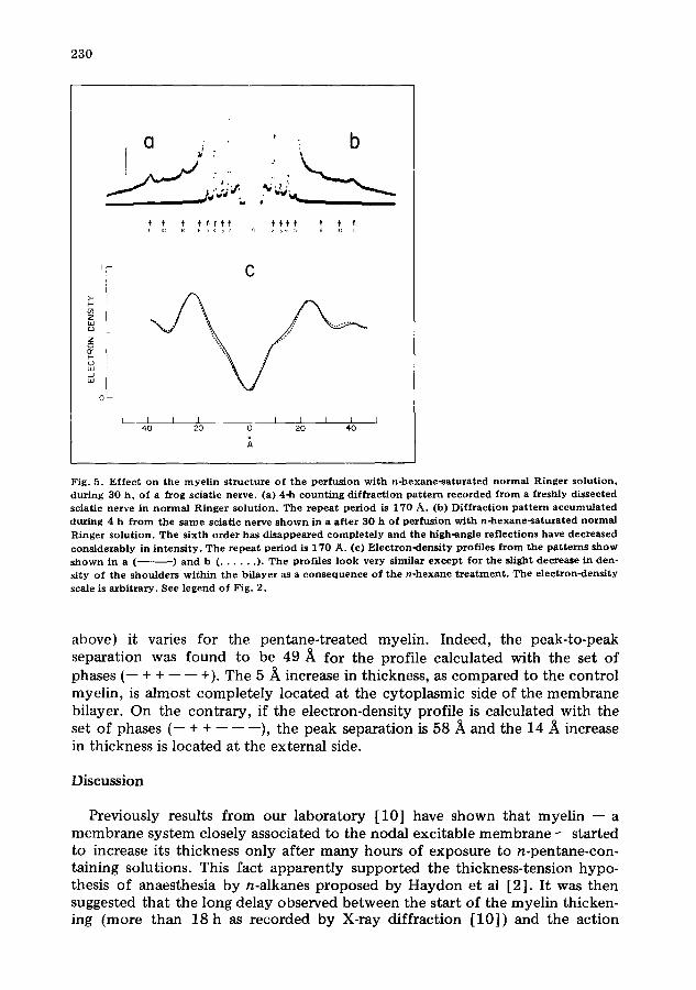
Fig. 5 . E f f e c t o n the m y e l i n s t r u c t u r e o f t h e p e r f u s i o n w i t h n - h e x a n e - s a t u r a t e d n o r m a l R i n g e r s o l u t i o n , d u r i n g 3 0 h , o f a f rog sc ia t i c ne rve . (a) 4-h c o u n t i n g d i f f r a c t i o n p a t t e r n r e c o r d e d f r o m a f r e sh ly d i s s ec t ed sc ia t i c ne rve in n o r m a l R i n g e r s o l u t i o n . The r e p e a t p e r i o d is 1 7 0 A . (b) D i f f r a c t i o n p a t t e r n a c c u m u l a t e d d u r i n g 4 h f r o m t h e s a m e sc ia t i c n e r v e s h o w n in a a f t e r 3 0 h o f p e r f u s i o n w i t h n - h e x a n e - s a t u ~ a t e d n o r m a l R i n g e r s o l u t i o n . T h e s i x t h o r d e r h a s d i s a p p e a r e d c o m p l e t e l y a n d t h e h igh -ang l e r e f l e c t i o n s have d e c r e a s e d c o n s i d e r a b l y in i n t e n s i t y . T h e r e p e a t p e r i o d is 1 7 0 A . (e) E l e c t r o n - d e n s i t y p ro f i l e s f r o m the p a t t e r n s s h o w s h o w n in a ( ) a n d b ( . . . . . . ). T h e p ro f i l e s l o o k ve ry s imi l a r e x c e p t f o r t h e s l ight d e c r e a s e in den - s i ty o f t h e s h o u l d e r s w i t h i n t h e b i l a y e r as a c o n s e q u e n c e o f t h e n - h e x a n e t r e a t m e n t . T h e e l e c t r o n - d e n s i t y scale is a r b i t r a r y . See l e g e n d o f F ig . 2 .
above) it varies for the pentane-treated myelin. Indeed, the peak-to-peak separation was found to be 49 i~ for the profile calculated with the set of phases (-- + + -- -- +). The 5/~ increase in thickness, as compared to the control myelin, is almost completely located at the cytoplasmic side of the membrane bilayer. On the contrary, if the electron-density profile is calculated with the set of phases (-- + + -- -- --), the peak separation is 58/~ and the 14 i~ increase in thickness is located at the external side.
Discussion
Previously results from our laboratory [10] have shown that myelin -- a membrane system closely associated to the nodal excitable membrane -- started to increase its thickness only after many hours of exposure to n-pentane-con- taining solutions. This fact apparently supported the thickness-tension hypo- thesis of anaesthesia by n-alkanes proposed by Haydon et al [2]. It was then suggested that the long delay observed between the start of the myelin thicken- ing (more than 18 h as recorded by X-ray diffraction [10]) and the action
231
a b
C
I . ...: : .:/ . /"
• .,. .../"
_ _ 1 _ I : _ ~ _ _ J _ [ J _ [ A_ [ 40 20 0 20 40
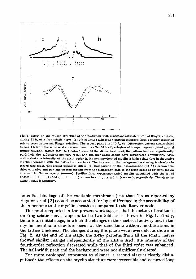
Fig. 6. Ef fec t on the m y e l i n s t r uc tu r e o f the pe r fus ion wi th n - p e n t a n e - s a t u r a t e d n o r m a l Ringer so lu t ion , du r ing 31 h , of a f rog sciatic nerve . (a) 4-h c o u n t i n g d i f f r ac t ion p a t t e r n r e c o r d e d f r o m a f reshly dissected sciat ic ne rve in n o r m a l R inger so lu t ion . The r e p e a t pe r iod is 170 A. (b) Di f f r ac t ion p a t t e r n a c c u m u l a t e d dur ing 4 h f r o m the s a m e sciat ic ne rve s h o w n in a a f t e r 31 h o f pe r fus ion wi th n - p e n t a n e - s a t u r a t e d n o r m a l Ringer so lu t ion . Not ice t ha t , as a c o n s e q u e n c e of the a lkane t r e a t m e n t , the p a t t e r n has b e e n s ignif icant ly mod i f i ed : the re f lec t ions axe very w e a k a nd the high-angle orders have d issapeared c o m p l e t e l y . Also, no t i ce t h a t the i n t e n s i t y o f the s ixth o rde r in the p e n t a n e - t r e a t e d m y e l i n is h igher t h a n tha t in the na t ive m y e l i n ( c o m p a r e w i th the p a t t e r n s h o w n in a). The increase in t h e , b a c k g r o u n d sca t t e r ing is c lear ly ob- served (see t ex t ) . T he r e p e a t pe r iod is 186 A. (c) C o m p a r i s o n of the low-reso lu t ion (34 A) e l ec t ron den- sities of na t ive and p e n t a n e - t r e a t e d m y e l i n f r o m the d i f f r ac t i on da t a to the s ix th o r d e r of pa t t e rn s s h o w n in a and b. Nat ive m y e l i n ( ). Profiles f r o m n - p e n t a n e - t r e a t e d m y e l i n ca lcu la ted w i th the set of phases (-- + + - - - - +) and ( - - + + - - - - - - ) s h o w n in ( . . . . . . ) an d in ( . . . . . ), r e spec t ive ly . Th e e lec t ron- dens i ty scale is a rb i t r a ry .
potential blockage of the excitable membrane (less than 1 h as reported by Haydon et al [2]) could be accounted for by a difference in the accessibility of the n-pentane to the myellin sheath as compared to the Ranvier node.
The results reported in the present work suggest that the action of n-alkanes on frog sciatic nerves appears to be two-fold, as is shown in Fig. 1. Firstly, there is an initial stage, in which the changes in the electrical activity and in the myelin membrane structure occur at the same time without modifications in the lattice thickness. The changes during this phase were reversible, as shown in Fig. 2. At the end of this stage, the X-ray patterns from all the sciatic nerves showed similar changes independently of the alkane used: the intensity of the fourth-order reflection decreased while that of the third order was enhanced. The half-width peak and the background were not significantly altered.
For more prolonged exposures to alkanes, a second stage is clearly distin- guished: the effects on the myelin structure were irreversible and occurred long
232
after the electrical activity was completely abolished. For instance, the nerve treated with n-hexane showed slight modifications within the bilayer structure. In agreement with other workers [2,18], it was found that the effects on the compound action potential observed during the t reatment with n-alkanes decreased with increasing chain length (compare the diffraction patterns shown in part b of Figs. 3--5). Indeed, the density profile of the nerve specimen treated with n-decane was very similar to that of the freshly dissected sciatic nerve (Fig. 3).
In agreement with our previous results regarding n-pentane [10], a 9% increase in the myelin membrane lattice is observed after 20 h of exposure to the alkane. However, the myelin thickness is associated with the irreversible stage since, for the first 15 h, no detectable thickening was observed.
The final X-ray diffraction of pentane-treated myelin showed a considerable deterioration as compared with t h e native pattern: the background increased considerably and the higher order reflections disappeared completely. This is shown in Fig. 6b. This effect suggests that some of the myelin become more disordered. However, in the results presented above, neither the lattice disorder in the freshly dissected myelin membrane nor the possible changes in this dis- order induced by alkane treatment have been considered as far as the analysis of the diffuse scatter is concerned [9]. This is certainly an important point which should be investigated using a two-dimensional position-sensitive detec- tor. Unfor tunately for us, though, such an instrument is not available at present.
The electron densities of Fig. 6c indicate that whichever the sign of the sixth-order reflection, the myelin membrane unit is thickened by the action of n-pentane. If we assume that the sign of F(6) remains positive during the experiment, then the increase in thickness is mainly located at the cytoplasmic side of the membrane. On the contrary, a larger change is detected at the exter- nal side when the profile is calculated with F(6) being negative. At present, the answer to this crystallographic problem is unknown. However, the fact that the sixth-order intensity cont inuously disappeared and reappeared during the per- fusion suggests that probably F (6) is negative.
In a previous report from our laboratory [10], it was suggested that the effect of n-pentane was faster on nerve conduct ion than on myelin structure.
This was inferred from morphological considerations such as the fact that the Ranvier node, where an action potential is generated, is more accessible to external solutions than myelin.
The results presented here and summarised in Figs. 1 and 2 disprove the no- tion that alkanes interact slowly and/or in a restricted fashion with myelin. Indeed, concomitant ly with the fall in the eletrical activity produced by the action of n-alkanes, there is a structural modification in myelin which is not an increase in the lattice dimensions.
The experiments have been performed in myelin, which although not being excitable, is a natural membrane system that may be used, in a first approxima- tion, as a model system for studying the structural effect of anaesthetics on biological membranes. If anaesthesia by diverse neutral molecules arises from a unique mechanism common to any cell membrane, it should be emphasised that if the excitability blockage is chosen as a measure of the uptake of n-pen-
233
tane by the axolemma at the node of Ranvier, the structural parameter - - ratio of the intensities I ( 4 ) / I ( 3 ) - - arbitrarily adopted here appears to fol low equally well the interaction of n-pentane with myelin. The fact that n-alkanes do not thicken the membranes is not a direct proof that the nodal excitable membranes behave in the same way. However, the structural modification detected as a change in the intensity of Bragg reflections calls for studies at higher resolu- tion. It is clear, though, that in order to obtain direct information about the molecular mechanisms of anaesthesia, these experiments have to performed on excitable membranes and using either general or local anaesthetic molecules (Padr6n, R. and Mateu, L., unpublished data).
Acknowledgements
The authors wish to thank Mr. J. Mora for building the specimen holder and Miss I. Otaegui for helpful secretarial assistance. This work was partially sup- ported by a grant ( $1-0353 to L.M.) from CONICIT.
References
1 Seeman, P. (1972) Pharmacol. Rev. 24, 583--655 2 Haydon, D.A., Hendry, B.M., Levinson, S.R. and Requena, J. (1977) Nature 268 ,356 - -358 3 Franks, N.P. and Lieb, W.R. (1978) Nature 274, 339--342 4 Richard, C.C., Martin, K., Gregory, S., Keightley, C.A., Hesketh, T.E., Smith, G.A., Warren, G.B. and
Metcalfe , J.C. (1978) Nature 276 ,775 - - 779 5 Huxley, A.F. and St~Lmpfli, R. (1948) J. Physiol. 108, 315--339 6 St~'mpfli, R. (1954) Physiol. Rev. 34, 101--112 7 Kirschner, D.A. (1977) in Myelin (Morell, P., ed.), pp. 51---89, Plenum Press, New York 8 Caspar, D.L.D. and Kirschner, D.A. (1971) Nat. New. Biol. 231, 46--52 9 Nelander, J.C. and Blaurock, A.E. (1978) J. Mol. Biol. 118, 497--532
10 Padr6n, R., Mateu, L. and Requena, J. (1979) Biochim. Biophys. Acta 552, 535--539 11 Padr6n0 R., Mateu, L. and Kirschner, D.A. (1979) Biophy$. J. 28 ,231-240 12 Padr6n, R. (1979) Ph. Sc. Thesis, Centro de Estudios Avanzados , IVIC, Caracas, Venezue la 13 Rud, J. (1961) Acta Physiol. Seand. 51, 7--171 14 Mateu, L. and Kirchhausen, T. (1980) Acta Cient. Venez. 30, 478--483 15 Mateu, I., Kirchhausen, T. and Camejo, G. (1978) Biochemistry 17, 1436--1440 16 Gabriel, A. and Dupont , Y. (1972) Rev. Sci. Instr. 43, 1600--1602 17 Blaurock, A.E. (1971) J. Mol. Biol. 56, 35--52 18 Mullins, L.J. (1971) in Handbook of Neurochemistry (Lajtha, A., ed.), Vol. 6, pp. 395--421, Plenum
Press, New York 19 Klug, H.P. and Alexander, L.E. (1974) X-Ray Diffract ion Procedures, pp. 360---364, John Wiley and
Sons, New York