Cystic fibrosis and the physiological responses to exercise
12
Cystic fibrosis and physiological responses to exercise Expert Rev. Respir. Med. 8(6), 751–762 (2014) Craig A Williams*, Zoe L Saynor, Owen W Tomlinson and Alan R Barker Children’s Health and Exercise Research Centre, Sport and Health Sciences, University of Exeter, St Luke’s Campus, Heavitree Road, Exeter, EX1 2Lu, UK *Author for correspondence: Tel.: +44 139 272 4890 Fax: +44 139 272 4726 [email protected] Cardiopulmonary exercise testing is underutilized within the clinical management of patients with cystic fibrosis. But within the last 5 years, there has been considerable interest in its implementation, which has included deliberations by the European Cystic Fibrosis Society about incorporating this method within the clinical assessment of patients. This review examines the current use of cardiopulmonary exercise testing in assessing the extent and cause(s) of exercise limitation from a pediatric perspective. Examples of the measured parameters and their interpretation are provided. Critical synthesis of recent work in the oxygen uptake (VO 2 ) kinetics response to and following exercise is also discussed, and although identified more as a research tool, its utilization advances researchers understanding of the cardiovascular, respiratory and muscular limitations to exercise tolerance. Finally, exercise and its application in therapeutic interventions are highlighted and a number of recommendations made about the utility of exercise prescription. KEYWORDS: cardiopulmonary exercise testing . inspiratory muscle training . Ivacaftor . oxygen uptake kinetics . pediatrics . skeletal muscle Cystic fibrosis (CF) is the most common inher- ited, life-shortening disease amongst the Cauca- sian population. In 1985, the genetic defect in CF (located on chromosome 7) was discovered, eventually leading to the gene defect identified by full-length gene sequencing in 1989 [1]. The defect is expressed as a disruption in the CF transmembrane conductance regulator protein (CFTR), which is found in the membranes of cells that line airways of the lungs, liver, pancreas, intestines, reproductive tract and skin. Abnormal CFTR function affects the ion transport neces- sary for the functioning of epithelial structure. In the lungs, abnormal thick and dry mucus ensues, resulting in a vicious cycle of bronchial airway obstruction, bacterial infection and inflamma- tion. The resulting obstructive syndrome causes progressive disability and as this cycle continues, lung tissue is progressively destroyed, with even- tual respiratory failure. Therefore, while this is a complex, multi-organ disease, lung disease accounts for more than 95% of the morbidity and mortality associated with CF. Although the natural history of CF is pro- gressive loss of lung function that leads to death, with early diagnosis and aggressive therapeutic intervention, survival into the third and fourth decades of life is now common. Indeed, a dra- matic increase has been observed in the median survival of UK patients from 10.6 years in 1966 to 29.4 years in 1992 [2]. In 2010, for the ~9385 patients registered in the UK, the pre- dicted life expectancy had risen to 43.5 years [3]. Despite this improvement, largely resulting from earlier diagnosis and enhancement in the pharmacological treatment, there remains no cure for CF. Therefore, clinicians now face the challenge of fostering a normal quality of life (QoL) for this aging patient population. Devel- oping and enhancing additional treatment and management strategies to facilitate this is, there- fore, a clinical priority. The initiative ‘Exercise is Medicine TM ’ by the American College of Sports Medicine is a timely reminder of the important role of exercise within medicine. Of course, this message is not new, but the challenge still remains to increase the utilization of exercise per se into the clinical management of chronic diseases. However, there remains sparse information on the physiological responses to exercise of children and adolescents with CF. Increased exercise (structured and informahealthcare.com 10.1586/17476348.2014.966693 Ó 2014 Informa UK Ltd ISSN 1747-6348 751 Review Expert Review of Respiratory Medicine Downloaded from informahealthcare.com by University of Exeter on 11/17/14 For personal use only.
Transcript of Cystic fibrosis and the physiological responses to exercise

Cystic fibrosis andphysiological responsesto exerciseExpert Rev. Respir. Med. 8(6), 751–762 (2014)
Craig A Williams*,Zoe L Saynor,Owen W Tomlinsonand Alan R BarkerChildren’s Health and Exercise Research
Centre, Sport and Health Sciences,
University of Exeter, St Luke’s Campus,
Heavitree Road, Exeter, EX1 2Lu, UK
*Author for correspondence:
Tel.: +44 139 272 4890
Fax: +44 139 272 4726
Cardiopulmonary exercise testing is underutilized within the clinical management of patientswith cystic fibrosis. But within the last 5 years, there has been considerable interest in itsimplementation, which has included deliberations by the European Cystic Fibrosis Societyabout incorporating this method within the clinical assessment of patients. This reviewexamines the current use of cardiopulmonary exercise testing in assessing the extent andcause(s) of exercise limitation from a pediatric perspective. Examples of the measuredparameters and their interpretation are provided. Critical synthesis of recent work in theoxygen uptake (VO2) kinetics response to and following exercise is also discussed, andalthough identified more as a research tool, its utilization advances researchers understandingof the cardiovascular, respiratory and muscular limitations to exercise tolerance. Finally,exercise and its application in therapeutic interventions are highlighted and a number ofrecommendations made about the utility of exercise prescription.
KEYWORDS: cardiopulmonary exercise testing . inspiratory muscle training . Ivacaftor . oxygen uptake kinetics. pediatrics . skeletal muscle
Cystic fibrosis (CF) is the most common inher-ited, life-shortening disease amongst the Cauca-sian population. In 1985, the genetic defect inCF (located on chromosome 7) was discovered,eventually leading to the gene defect identifiedby full-length gene sequencing in 1989 [1]. Thedefect is expressed as a disruption in the CFtransmembrane conductance regulator protein(CFTR), which is found in the membranes ofcells that line airways of the lungs, liver, pancreas,intestines, reproductive tract and skin. AbnormalCFTR function affects the ion transport neces-sary for the functioning of epithelial structure. Inthe lungs, abnormal thick and dry mucus ensues,resulting in a vicious cycle of bronchial airwayobstruction, bacterial infection and inflamma-tion. The resulting obstructive syndrome causesprogressive disability and as this cycle continues,lung tissue is progressively destroyed, with even-tual respiratory failure. Therefore, while this is acomplex, multi-organ disease, lung diseaseaccounts for more than 95% of the morbidityand mortality associated with CF.
Although the natural history of CF is pro-gressive loss of lung function that leads to death,with early diagnosis and aggressive therapeutic
intervention, survival into the third and fourthdecades of life is now common. Indeed, a dra-matic increase has been observed in the mediansurvival of UK patients from 10.6 years in1966 to 29.4 years in 1992 [2]. In 2010, for the~9385 patients registered in the UK, the pre-dicted life expectancy had risen to 43.5 years [3].Despite this improvement, largely resultingfrom earlier diagnosis and enhancement in thepharmacological treatment, there remains nocure for CF. Therefore, clinicians now face thechallenge of fostering a normal quality of life(QoL) for this aging patient population. Devel-oping and enhancing additional treatment andmanagement strategies to facilitate this is, there-fore, a clinical priority.
The initiative ‘Exercise is MedicineTM’ by theAmerican College of Sports Medicine is atimely reminder of the important role of exercisewithin medicine. Of course, this message is notnew, but the challenge still remains to increasethe utilization of exercise per se into the clinicalmanagement of chronic diseases. However, thereremains sparse information on the physiologicalresponses to exercise of children and adolescentswith CF. Increased exercise (structured and
informahealthcare.com 10.1586/17476348.2014.966693 � 2014 Informa UK Ltd ISSN 1747-6348 751
Review
Exp
ert R
evie
w o
f R
espi
rato
ry M
edic
ine
Dow
nloa
ded
from
info
rmah
ealth
care
.com
by
Uni
vers
ity o
f E
xete
r on
11/
17/1
4Fo
r pe
rson
al u
se o
nly.

organized with specific targeted fitness outcomes) and/or physicalactivity (any bodily movement resulting in energy expenditure) isbeneficial for healthy children and children with chronic dis-eases [4]. Bar-Or and Rowland’s [4] comment about children withchronic disease that ‘by prescribing exercise we are signalling tothe child that he or she can, and should, act like his or her healthypeers’ (p. 112) is, therefore, pertinent. For example, a recent studyreported an attenuated decline in lung function in patients withCF aged 7–17 years who increased their levels of physical activityover a 7-year period [5]. Therefore, while it is intuitive to promoteexercise in diseased children, the evidence base is currently sparsecompared to that of healthy children.
The utilization of exercise would include not only rehabilita-tive or training aspects for patients, but also a more regular andcomprehensive use of exercise stress testing compared to the cur-rent practice. Despite the acceptance by clinical and exercisephysiologists that cardiopulmonary exercise testing (CPET) is themost objective test to determine the limits and/or mechanisms ofexercise (in)tolerance, it remains significantly underutilized inclinical practice [6]. This is disappointing for several reasons.Firstly, a more standardized approach to CPET in all clinicalpatients (where applicable) would establish a normative databaseupon which a determination of what is a ‘normal’ or ‘abnormal’physiological response can be made. Second, establishing apatient’s ability to tolerate exercise and physiological responseprofile will enhance the precision of exercise prescription, whichis currently generalized, if at all utilized on an individual basis.This may be in the form of the prescription of exercise trainingintensities [7]. Lastly, routine CPET will allow for the prognosticvalue of the exercise parameters to be examined alongside morecommon clinical outcome measures, that is, lung function, QoL,hospital admissions, drug administration. As noted above, therehas been a marked increase in the median survival age of patientswith CF [8]. Therefore, the early publication of exercise testing inCF patients for prognostic purposes is largely redundant andurgently needs updation [9].
The recent promotion of CPET as a clinical outcome mea-sure by the European Cystic Fibrosis Society (ECFS) [10] andthe need for comprehensive and accurate outcomes to evaluatethe rapidly advancing pharmacological treatment of CF aretimely [11]. This is an exciting phase in the treatment and man-agement of this complex condition; however, it is importantthat exercise testing should not be sidelined. As recommendedby the British Thoracic Society and the Association of Char-tered Physiotherapists in Respiratory Care [12], exercise shouldbe an integral part of CF clinical care. The purpose of thisreview is to highlight the advances in the CPET method overrecent years and outline its current and future clinical applica-tions in pediatric CF patients.
Provision of CPET in CFCurrent provision of exercise testing in both clinical and researchassessments remains limited [6,13,14]. As outlined by Stevens et al.[6], exercise testing is underused (53% uptake) within CF clinicsacross the UK, despite the care teams recognizing its value [6].
Similar figures (44 and 63%, respectively) have been reported inCF centers in the US and Germany [13,14]. Furthermore, there isa notable lack of protocol standardization. Interestingly, how-ever, as highlighted by Stevens et al. [6], the limited applicationof CPET within CF care is in contrast to the high importancegiven to its clinical utility by healthcare providers.
High aerobic fitness in CF has shown to be of clinical impor-tance because of its positive association with patients’ QoL [15],prognosis [16–18] and risk of hospitalization [19]. CPET, incorpo-rating the measurement of pulmonary gas exchange, providesthe most precise measure of aerobic fitness in patients withmild-to-moderate CF, and invariably focuses on the determina-tion of maximal oxygen uptake (VO2max). Not only does com-prehensive CPET hold clinical utility, but also it enables thefactor(s) limiting patients’ exercise capacity (e.g., motivation,poor fitness and/or disease pathophysiology) to be determinedand is valuable for understanding the mechanism(s) by whichaerobic fitness is reduced and by which new treatment strategiesmight be influenced. Currently, there is no consensus regardingwhether aerobic fitness of CF patients is predominantly limitedby respiratory, cardiovascular and/or muscular factors [20–22], asdisease severity will be an important confounder.
Lung function and structural investigations and/or measuresof nutritional status are traditionally relied upon to measuredisease severity and progression in CF, but they cannot accu-rately predict exercise capacity and are often not sensitive tochange in mild-to-moderate disease. Appropriate exercise test-ing provides an integrated, objective assessment of cardiovascu-lar, respiratory, muscular and metabolic function of patients.This information, therefore, provides a more comprehensiveclinical assessment which can inform medication and therapystrategies and assist with pre-transplant stratification. Further-more, thorough exercise testing enables individualized exerciseprescription plans. Therefore, current clinical standards for CFmanagement recommend exercise testing on at least an annualbasis (CF Trust) [10].
Besides VO2max, additional submaximal gas exchange meas-urements, such as the VE/VCO2-slope and the oxygen uptakeefficiency slope (OUES), have yielded superior prognostic util-ity in other clinical populations [23,24] and, thus, warrant inves-tigation in CF. Furthermore, despite the wealth of knowledgeit provides, exercise testing as an outcome in therapeutic trialsremains in its infancy [25]. To objectively quantify physicalfunctional changes under different pharmacological treatments,CPET should be included within future, long-term research.
CPET protocolsAlthough CPET has been established for many decades, theECFS Exercise Working Group has only recently promotedCPET as the exercise testing method of choice for this patientgroup [10]. Moreover, the ECFS Clinical Trials Network Stand-ardisation Committee called for assessment of the validity,reproducibility and feasibility of the outcome measures utilizedin the assessment of CF patients and advocated research intothe most appropriate test for pediatric patients [26].
Review Williams, Saynor, Tomlinson & Barker
752 Expert Rev. Respir. Med. 8(6), (2014)
Exp
ert R
evie
w o
f R
espi
rato
ry M
edic
ine
Dow
nloa
ded
from
info
rmah
ealth
care
.com
by
Uni
vers
ity o
f E
xete
r on
11/
17/1
4Fo
r pe
rson
al u
se o
nly.

Consequently, there has been much debate regarding whatare the most appropriate testing protocols and guidelines toimplement for these patients. This uncertainty is likely to be afactor in the current poor clinical uptake of CPET [6]. Withregard to treadmill versus cycling exercise, the consensusappears to be cycle-based protocols. Not only do these preventfalls and facilitate measurements such as electrocardiographyduring exercise, but also they are more pediatric-friendly andcan enable testing of patients as young as 7 years of age [27,28].
The Godfrey protocol [29] consists of a 3-min warm-up fol-lowed by increases in work rate each minute until exhaustion.In children with chronic conditions, however, it has been sug-gested that a ramp incremental protocol, whereby exerciseintensity is increased linearly rather than each minute, is moreappropriate [30]. The consensus by exercise physiologists is thata linear increase in work rate is important to depict the pro-gressive response to exercise. Additional protocols, such as thesteep ramp test, have also recently been introduced [30]. Onekey consideration in the choice of testing protocol is how accu-rate is the derived aerobic fitness measurement.
Accuracy considerations
Obtaining a valid VO2max measurement is critical and a partic-ularly important issue in pediatric groups. VO2max representsthe integrated capacity of the pulmonary, cardiovascular andmuscular systems to transport and utilize oxygen during intenseexercise, and is traditionally identified by a VO2 plateau uponexhaustion despite an increasing work rate. However, youngpeople rarely exhibit this response [31,32] and the term VO2peak
is often used. Reliance has, therefore, traditionally fallen uponsecondary verification criteria, encompassing subjective indica-tors of effort (sweating, facial flushing and hyperpnea) andobjective physiological secondary criteria (heart rate, respiratoryexchange ratio and/or blood lactate concentration). But adher-ence to these criteria has been shown to drastically under mea-sure VO2max in patients with CF [27].
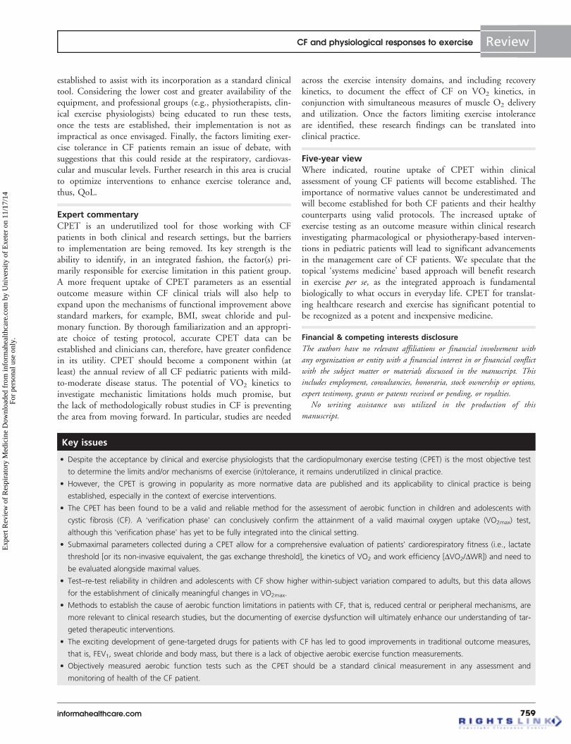
A procedure termed the ‘verification phase’, whereby CPETis followed by an individualized supramaximal ‘step’ test toexhaustion (Smax), can ensure that a valid VO2max is mea-sured. The verification phase is completed after 15 min recov-ery from the CPET test and requires the patient to cycle at110% of the peak power attained at the end of the CPET foras long as possible until the required cadence cannot be main-tained. If VO2 is lower or equal to the CPET oxygen uptakevalues, then the clinician can be assured it is a maximal effort.Although this requires another 20 min onto the length of aCPET, it is the only way (apart from asking the patient toreturn to the laboratory a few days later to repeat the CPET)that the VO2 values can be verified as a ‘true’ maximum.A recent study by Saynor et al. (FIGURE 1) demonstrated thatSmax can verify VO2max in most cases and, importantly, iden-tify those patients who have provided a submaximal effort [27].Not only can the combination of a traditional ramp incre-mental and Smax test permit measurement of a valid VO2max,but also this protocol can be safely and effectively
administered within a single visit, confirming that maximalCPET is safe for patients with mild-to-moderate CF [12,33].
Recommendations & considerations
While the importance of calibration of metabolic carts is widelyacknowledged with regard to the accuracy of CPET-deriveddata [34,35], thorough familiarization and the choice of testingprotocol are also vital. From a practical point of view, patients’familiarization with the expectations of the protocol and whatit feels like to maintain a steady cadence at various work ratesis important. This is very relevant when testing young and/ornervous patients. The choices of protocol and verification crite-ria are also essential to obtain a ‘true’ representation of patients’maximal aerobic fitness. However, at a time when the ECFShas recognized CPET as the exercise testing method of choicewhen assessing aerobic fitness in mild-to-moderate CF patients,only an Smax verification protocol, as outlined by Saynor et al.[27], can confirm a ‘true’ VO2max measurement obtained duringa progressive exercise test in young CF patients. Furthermore, akey benefit of ramp incremental exercise compared with ashorter steep ramp test is that ramp testing spans the range ofexercise intensities and, therefore, derives both important maxi-mal and submaximal fitness parameters [36].
These recommendations have significant implications for theassessment and interpretation of CPET in young CF patientsin clinical and research settings. To utilize VO2max in prognos-tic stratification and assessment of clinical or research interven-tions, it is essential that ‘true’ measurements are obtained.Accepting submaximal efforts will significantly distort the clini-cal interpretation of patients’ aerobic fitness. From a practicalviewpoint, Smax verification is straightforward to implement, asthe imposed power is calculated on an individual basis fromthe peak power output achieved during the ramp test and, clin-ically, may minimize the costs associated with re-tests when thevalidity of a test is questionable. While it is important thatthese new concepts and advances within exercise physiology areincorporated within clinical practice, the authors [27] did cau-tion that the safety of Smax exercise in older patients with moresevere CF has yet to be confirmed.
D G
F
EC
B
A
Figure 1. Schematic of the exercise test protocol. A: 3-minwarm up at 20 W. B: Incremental ramp exercise at a rate of10–30 W/min (individualized to patients’ anthropometric data).C: 5-min active recovery (unloaded pedaling). D: 10-min seatedrecovery off the cycle ergometer. E: 3-min warm-up at 20 W. F:Supramaximal confirmation bout of exercise to volitional exhaus-tion at 110% of the peak power output produced in the priorramp exercise test B. G: 3-min recovery (unloaded pedaling).
CF and physiological responses to exercise Review
informahealthcare.com 753
Exp
ert R
evie
w o
f R
espi
rato
ry M
edic
ine
Dow
nloa
ded
from
info
rmah
ealth
care
.com
by
Uni
vers
ity o
f E
xete
r on
11/
17/1
4Fo
r pe
rson
al u
se o
nly.

Interpretation of CPET dataSeveral excellent reviews, books and position stands have beenpublished regarding interpretation of CPET outcomes [7,34,37–40].A CPET provides a large number of important parameters,including but not limited to, gas exchange and metabolic data(VO2, carbon dioxide output [VCO2], gas exchange threshold[GET], minute ventilation [VE], ventilatory equivalents for O2
[VE/VO2] and CO2 [VE/VCO2], O2 pulse, arterial oxygen sat-uration [SaO2] via pulse oximetry, end-tidal O2 and CO2), inconjunction with test duration and work rate. These additionalparameters are often overlooked in favor of the final maximalvalues. However, submaximal data should assist with diagnosticand prognostic evaluations and need to be incorporated morefully in patient reports [7]. No one single parameter should beused exclusively; rather it is the integration of the exerciseresponses as a whole which add value when utilizing CPETcombined to other tests.
Parameters of aerobic function
Maximal oxygen uptake (VO2max) represents the gold standardmeasure of aerobic fitness and provides an indication of howwell patients’ lungs, pulmonary and cardio circulation (largeand small vessels), and muscles function in an integrated systemduring exercise. This allows researchers, clinicians and otherhealthcare professionals to assess the entire cardiopulmonarypathway from mouth to the exercising muscle in one simplemeasurement (i.e., the rate of oxygen flowing through the vari-ous subsystems). Unlike other chronic conditions, CF appreci-ably affects the gas exchange response to exercise.
VO2max is currently the principal outcome measure from aCPET, as it has been shown to be an independent predictor ofmortality in CF (14). However, a more comprehensive evalua-tion of patients’ cardiorespiratory fitness may be gained throughquantification of submaximal parameters of aerobic function(i.e., lactate threshold [or its non-invasive equivalent the GET],the kinetics of VO2 and work efficiency [DVO2/DWR]). Thesethree parameters combined with VO2max represent the keyparameters of aerobic function [41]. Despite suggestions that itmay be difficult to non-invasively identify the lactate threshold(via the GET and ventilatory threshold) in patients with chronicrespiratory disease and airflow limitation [42], it has been demon-strated that these parameters are reproducible using a cluster ofmeasures and two independent observers [23].
Ventilatory function is best examined by relating VO2 andVCO2 dynamics to VE [43], through the slope of ventilatoryequivalent for CO2 response (VE/VCO2-slope) and the OUES.The OUES is useful since it is, theoretically, resistant to earlytest termination and intra- and inter-observer variability [43].Although these parameters possess documented utility to iden-tify the presence and severity of ventilatory inefficiency of theheart/lung organs and/or response to intervention in heart fail-ure patients, their uptake within the assessment of respiratoryconditions has been scarce [44].
Systemically, the greater the VE required for a given amount ofgas exchange (VCO2), the less efficient is the cardiopulmonary
system. This has been documented as irregular in CF patients.Consideration of altered ventilatory equivalents for CO2 hasfeatured in heart failure CPET over recent years, however lessso in CF, but the reduced elimination of carbon dioxide as acentral mechanism may also reduce aerobic fitness. However,use of the OUES and VO2 gain have been advocated andreported [30,45], but their reliability and validity are still beingdebated [44]. From a practical point of view, outcome meas-ures which can assess patients’ function at submaximal intensi-ties, similar to activities of daily living, are also important.Furthermore, submaximal parameters may be especially usefulin the clinic environment when patients may be unwilling toprovide a maximal effort and/or are limited by ventilatorycapacity. In addition, the GET can improve independently ofany changes in VO2max [46,47] and is often used in the pre-scription of individualized exercise intensities within specificexercise intensity domains (i.e., at a %GET), as recently dem-onstrated by Stevens and colleagues [48]. Additional commonparameters of interest attained during CPET include peakpower output, SaO2, time to exhaustion, subjective ratings ofperceived exertion and dyspnea, which aid interpretation oflimiting factors as either more respiratory or muscular.
Indices of cardiac function and muscle oxygenation
In addition to the reported VO2max measurements, parameterssuch as O2 delivery and estimations of SV can be calculated. TheFick cardiac output (Q) equation can be used to estimate strokevolume, while SaO2 can also be estimated when examining oxy-gen delivery during exercise. There is growing support for the con-tribution from inadequate delivery of oxygen to the tissueresulting in suppressed aerobic fitness in CF patients [49]. It is,therefore, advantageous that measures of cardiac function and O2
delivery are included in the CPET of these patients, although thismight not be convenient and does require more technical support.
Key to exploring the effectiveness and adequacy of oxygendelivery to the exercising muscles during exercise is havingsome measurements of cardiac function and provision from thecardiorespiratory unit. Due to ethical constraints, non-invasivedevices such as thoracic bioelectrical impedance analysis havebeen validated in CF patients. Such tools provide non-invasiveestimations of stroke volume and cardiac output. It may well,depending on the equipment available, be important to investi-gate ventricular function, as this may result in impaired oxygendelivery during exercise in these patients [21].
Minimal important clinical changes
Interpreting data in relation to normative values and typicalerror enables researchers and clinicians to determine meaningfulchange. Reproducibility over time is critical when evaluatingthe efficacy of CF treatments (e.g., antimicrobials, mucolyticsand gene mutation targeted therapies) which may accrue overweeks or months, as well as monitoring exercise training inter-ventions. One study previously reported the reproducibility ofCPET in children with CF [50]; however, this study was limitedsince an intermittent sprint cycle test preceded the ramp test,
Review Williams, Saynor, Tomlinson & Barker
754 Expert Rev. Respir. Med. 8(6), (2014)
Exp
ert R
evie
w o
f R
espi
rato
ry M
edic
ine
Dow
nloa
ded
from
info
rmah
ealth
care
.com
by
Uni
vers
ity o
f E
xete
r on
11/
17/1
4Fo
r pe
rson
al u
se o
nly.

resulting in insufficient test durations (~4 min). A recentstudy [28] examined the reproducibility of CPET-derived maxi-mal and submaximal outcome measures using a valid protocolin young patients with mild-to-moderate CF. Using a solitarytraditional ramp test, the coefficients of variation of 6.9 [51]
and 8.5% [52] in VO2peak have been reported over 4 weeks inCF adults, demonstrating CPET is a reliable tool.
While the typical error reported in pediatric patients [28] iscomparable to earlier studies, the improved validity should beconsidered in future studies. While the compromised validityof traditional tests to ascertain VO2peak, for example, the God-frey protocol, has previously been documented [27,28], it has alsobeen demonstrated that there is a larger within-subject variationin VO2max over both the short term (13.5 vs 9.3%) andmedium term (15.2 vs 13.3%), compared with the combinedramp and Smax approach to testing. Determining the extent towhich changes in outcome measures relate to a given referencemeasure is essential to the utility of CPET. So also is the estab-lishment of national normative data, but currently limited nor-mative data exists [51]. Establishing robust normative data forboth CF patients and their healthy counterparts represents animportant next step.
CPET & limiting factors to exerciseDue to limited scientific evidence, it remains unclear what arethe relative contributions and interactions of the central(reduced oxygen delivery) and peripheral (altered muscle massand function) mechanisms that result in the reduced aerobicfitness of patients with CF compared to healthy controls.Although dysfunction at the skeletal muscle level have beenproposed [20,53], this may not be specific to CF per se and,rather, presents as a consequence of chronic respiratory sep-sis [54]. A review by Rand and Prasad [55] recently outlined anumber of factors which likely contribute to the reduced exer-cise performance in patients with CF, including lung function,nutrition, muscle (dys)function, genotype, habitual physicalactivity levels (and gender) and psychosocial influences. How-ever, little attention was focused on the possible contributionfrom altered oxygen delivery (i.e., hypoxemia, reduced strokevolume and cardiac output) during exercise.
Evidence to support contribution from an inadequate O2
delivery to reduced VO2max [54] and slow VO2 kinetics [56] inCF patients has been presented. These suggestions are sup-ported by previous reports demonstrating altered cardiac func-tion [18,21,57] and an inability to augment stroke volume duringexercise in this patient group [54]. Rosenthal and colleagues [54]
presented an important investigation with regard to the mecha-nistic bases of exercise limitation in CF.
The body’s upper limit for O2 utilization during exercise isdetermined by the maximal Q, arterial oxygen content, frac-tional distribution of O2 to the exercising muscles and the abil-ity of the skeletal muscle to extract this O2. Simultaneousmeasurements at both the central (cardiorespiratory unit) andperipheral (skeletal muscle) levels are essential to understandthe dynamic matching of O2 delivery-to-O2 utilization. Since
previous studies in CF have largely neglected to investigate thiscomplex interaction and have based their inferences on investi-gations of isolated organ systems [19–21], knowledge of inte-grated function is limited.
Other non-invasive techniques are available to provide fur-ther evidence on how CF disease pathophysiology alters theoxygen delivery-to-oxygen utilization relationship during exer-cise. Near-infrared spectroscopy is one such instrument,whereby the signal of muscle deoxygenated hemoglobin/myoglobin may be used to provide a non-invasive insight intomicrovascular O2 extraction dynamics at the muscle level.Using this device, Saynor and colleagues [58] have recentlyshown children with CF to have comparable muscle O2 extrac-tion dynamics compared to healthy controls, despite animpaired aerobic fitness. The latter was likely caused byimpairment in muscle O2 delivery, as evidenced through areduction in arterial blood O2 saturation.
The oxygen uptake kinetics response to exerciseAs mentioned above, the measurement of VO2max in patientswith CF has been associated with mortality [18], QoL [15] andthe risk of hospitalization [19]. However, patients with CF rarelyexercise at their maximal metabolic rate, meaning the measure-ment of VO2max lacks external validity with regard to the ‘real-world’ challenges faced by the O2 transport and utilizationpathways. Rather, activities of daily living require repeated tran-sitions to and from a range of submaximal metabolic rates.The adequacy of a patient to undertake these activities can becaptured in the VO2 kinetic response during exercise, whichreflects the integration of the pulmonary, cardiovascular andmuscular systems to meet the increasing and decreasing energydemands within the muscle. Dysfunction at any step of the O2
transport and utilization pathway, as typically found in disease,leads to a slowed VO2 kinetic response during exercise. Thisincreases the O2 deficit, the requirement for substrate-levelphosphorylation (e.g., muscle phosphocreatine breakdown,anaerobic glycolysis) and the accumulation of fatigue-inducingmetabolites (e.g., inorganic phosphate, hydrogen ions), whichimpairs exercise tolerance [59]. Consequently, the measurementof the VO2 kinetic response to and from exercise in patientswith CF provides valuable insight into the limiting factors ofoxidative phosphorylation and exercise tolerance.
Methodological considerations
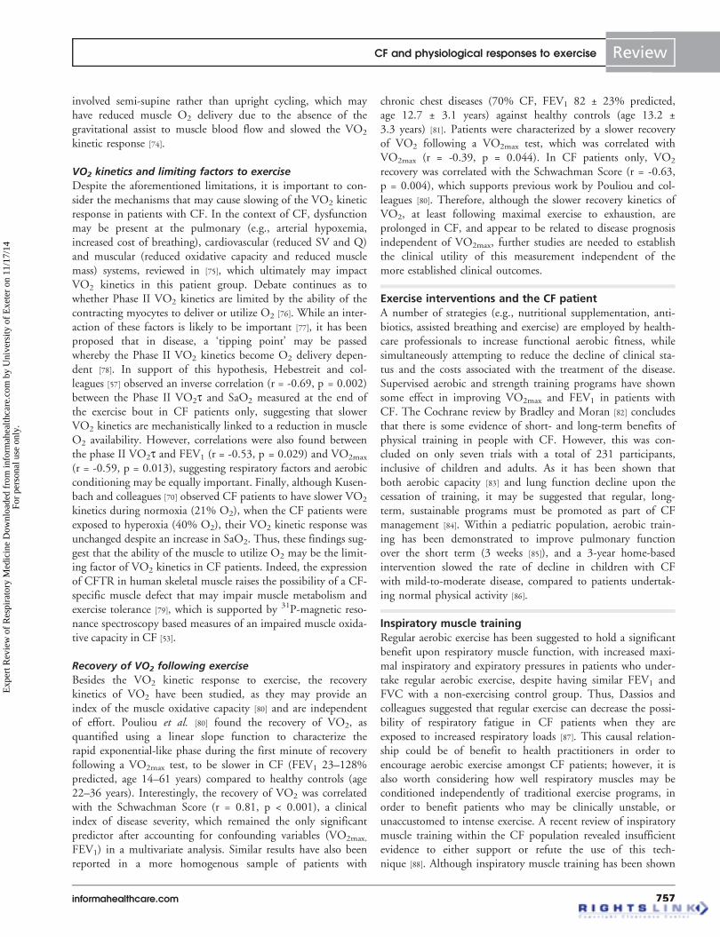
To understand how the VO2 kinetic response to exercise isaltered by CF, a brief overview of the different phases of theVO2 response to exercise with respect to exercise intensity isrequired, which is reviewed elsewhere (FIGURE 2) [59,60]. At theonset of constant work rate moderate-intensity exercise, that is,work rates below the GET, VO2 increases almost immediately(Phase I, termed cardiodynamic) due to a rise in Q and pulmo-nary blood flow [61]. Subsequently, the reduced mixed venousO2 content reaches the lungs and, with the increasing Qresponse, drives the exponential rise (Phase II) in VO2 towarda new steady state (Phase III). The kinetics of Phase II have
CF and physiological responses to exercise Review
informahealthcare.com 755
Exp
ert R
evie
w o
f R
espi
rato
ry M
edic
ine
Dow
nloa
ded
from
info
rmah
ealth
care
.com
by
Uni
vers
ity o
f E
xete
r on
11/
17/1
4Fo
r pe
rson
al u
se o
nly.

been shown to reflect the kinetics of muscle O2 consump-tion [62] and muscle phosphocreatine in humans [63]. The timeconstant (t, time to reach 63% of the response amplitude) ofthe Phase II response during upright cycling is ~20–30 s inhealthy children and young adults, and reaches a steady state(Phase III) with an O2 cost of exercise (D VO2/DW) of~10 ml/min/W [64,65]. For work rates above the GET, the VO2
response is characterized by an increase in the O2 cost of exer-cise that either attains an steady state (heavy-intensity exercise)or increases with time until VO2max is achieved (very heavyexercise) [66]. The heavy and very heavy exercise intensitydomains are demarcated by the critical power, which representsthe asymptote of the hyperbolic relationship between work rateand time to exhaustion [66,67]. The development of the so-calledVO2 slow component during supra GET work rates increasesthe O2 cost of exercise to ~12–14 ml/min/W (impaired effi-ciency) and reflects the fatigue processes occurring within thecontracting myocytes. The higher the work rate is above criticalpower, the lower the magnitude of the VO2 slow component,such that at work rates close to VO2max, the VO2 kinetic fol-lows a single-exponential function until exhaustion occurs withthe participant achieving VO2max [65].
The impact of exercise intensity on the VO2 kinetic responseto exercise requires careful consideration. For example, the pre-scription of a single absolute work rate (e.g., 150 W or 1.5 W/kgof body mass) is likely to render the CF patient exercising at ahigher percentage of his/her aerobic capacity compared to a
healthy control, given the well-documented reduced VO2max inthis group [68]. Equally, the prescription of a work rate relative toVO2max (e.g., 60% VO2max) is flawed, as although the GETappears to be preserved in CF, when normalized to VO2max
[56,68], there is a large variability in the position of the GET rela-tive to VO2max across CF patients and healthy controls. Conse-quently, the use of an absolute work rate or a work rate inrelation to VO2max will result in participants exercising across themoderate- or heavy-intensity domains, which will mask ourunderstanding of the effect of CF on the VO2 kinetic response.
Are VO2 kinetics altered in CF patients?
Unfortunately, our understanding of the effect of CF on theVO2 kinetic response to exercise is limited, as this has beenapplied more to clinical research than practice. In the first studyto address this topic, Braggion and colleagues [69] reported nodifferences in the VO2 kinetic response between CF patients(FEV1 77 ± 22% predicted, age 11.1–15.3 years) and age-matched controls (age 12.2–15.2 years) during 6 min ofsubmaximal exercise equivalent to 1.7 W/kg of body mass.However, it should be noted that in this study, only a singleexercise transition was employed to quantify the VO2 response,and the modeling procedure employed did not isolate thePhase II kinetic response, which is crucial to reflect muscle O2
consumption [62]. Furthermore, no consideration was given tostandardizing the exercise intensity domain within and betweenthe groups. In contrast, using pseudo-random binary sequenceexercise, slowed VO2 kinetics have been observed in CF patients(FEV1 42–88% predicted, age 13–31 years) compared to con-trols (age 9–29 years), although this analysis was also unable todifferentiate between Phase I and II of the kinetic response [70].
To extend this work, Hebestreit et al. [56] examined the VO2
kinetics in CF patients (FEV1 37–98% predicted, age 9.8–33.8 years) compared to healthy controls (age 9.9–30.8 years)during a two-stage protocol consisting of semi-supine cycling at20 W for 2 min and for further 3 min at a work rate calcu-lated as 1.4 W/kg for males and 1.3 W/kg for females. Thisprotocol was repeated 2–4 times to improve the signal-to-noiseratio of the VO2 kinetic response [71]. After analysis of theVO2 kinetic response, the authors found a slower Phase IIVO2t in CF compared to the control group (36.8 ± 13.6 vs26.4 ± 9.1 s), with no difference in the O2 cost of exercise(10.9 ± 1.8 vs 10.2 ± 1.6 ml/min/W). Although this study sug-gests CF patients are characterized by sluggish VO2 kineticsand thus an increased O2 deficit, a number of methodologicalissues confound interpretation of this study. Firstly, the workrate was not prescribed to a particular exercise intensitydomain, increasing the possibility that participants within andbetween groups were exercising across the moderate- or heavy-intensity domains. Second, two to four repetitions of the exer-cise protocol were undertaken on the same day with only10 min recovery. This raises the possibility that a ‘priming’effect may have occurred on the VO2 kinetic response [72],which is likely to be more influential on those with slowerVO2 kinetics (i.e., CF patients) [73]. Finally, the exercise
2.0
–6.0
1.5
1.0
0.5
0.0
VO
2 (L
.min
–1)
Time (s)
60 120 180 240 300 3600
I II III
Figure 2. Example VO2 kinetics profile in a pediatric patientwith cystic fibrosis during moderate (.) and very heavy (*)intensity cycling exercise. The vertical dotted lines representthe different phases (I, II and III) of the VO2 kinetic response.Notice that during moderate-intensity exercise, after Phase II(~120 s), the VO2 response has attained a steady state. In con-trast, for very heavy exercise, after Phase II (~120 s), a steady-state VO2 response is not achieved due to the emergence of theVO2 slow component which increases the O2 cost of exercisetoward VO2 max, which in this patient was 1.68 l/min.VO2: Oxygen uptake.
Review Williams, Saynor, Tomlinson & Barker
756 Expert Rev. Respir. Med. 8(6), (2014)
Exp
ert R
evie
w o
f R
espi
rato
ry M
edic
ine
Dow
nloa
ded
from
info
rmah
ealth
care
.com
by
Uni
vers
ity o
f E
xete
r on
11/
17/1
4Fo
r pe
rson
al u
se o
nly.
involved semi-supine rather than upright cycling, which mayhave reduced muscle O2 delivery due to the absence of thegravitational assist to muscle blood flow and slowed the VO2
kinetic response [74].
VO2 kinetics and limiting factors to exercise
Despite the aforementioned limitations, it is important to con-sider the mechanisms that may cause slowing of the VO2 kineticresponse in patients with CF. In the context of CF, dysfunctionmay be present at the pulmonary (e.g., arterial hypoxemia,increased cost of breathing), cardiovascular (reduced SV and Q)and muscular (reduced oxidative capacity and reduced musclemass) systems, reviewed in [75], which ultimately may impactVO2 kinetics in this patient group. Debate continues as towhether Phase II VO2 kinetics are limited by the ability of thecontracting myocytes to deliver or utilize O2 [76]. While an inter-action of these factors is likely to be important [77], it has beenproposed that in disease, a ‘tipping point’ may be passedwhereby the Phase II VO2 kinetics become O2 delivery depen-dent [78]. In support of this hypothesis, Hebestreit and col-leagues [57] observed an inverse correlation (r = -0.69, p = 0.002)between the Phase II VO2t and SaO2 measured at the end ofthe exercise bout in CF patients only, suggesting that slowerVO2 kinetics are mechanistically linked to a reduction in muscleO2 availability. However, correlations were also found betweenthe phase II VO2t and FEV1 (r = -0.53, p = 0.029) and VO2max
(r = -0.59, p = 0.013), suggesting respiratory factors and aerobicconditioning may be equally important. Finally, although Kusen-bach and colleagues [70] observed CF patients to have slower VO2
kinetics during normoxia (21% O2), when the CF patients wereexposed to hyperoxia (40% O2), their VO2 kinetic response wasunchanged despite an increase in SaO2. Thus, these findings sug-gest that the ability of the muscle to utilize O2 may be the limit-ing factor of VO2 kinetics in CF patients. Indeed, the expressionof CFTR in human skeletal muscle raises the possibility of a CF-specific muscle defect that may impair muscle metabolism andexercise tolerance [79], which is supported by 31P-magnetic reso-nance spectroscopy based measures of an impaired muscle oxida-tive capacity in CF [53].
Recovery of VO2 following exercise
Besides the VO2 kinetic response to exercise, the recoverykinetics of VO2 have been studied, as they may provide anindex of the muscle oxidative capacity [80] and are independentof effort. Pouliou et al. [80] found the recovery of VO2, asquantified using a linear slope function to characterize therapid exponential-like phase during the first minute of recoveryfollowing a VO2max test, to be slower in CF (FEV1 23–128%predicted, age 14–61 years) compared to healthy controls (age22–36 years). Interestingly, the recovery of VO2 was correlatedwith the Schwachman Score (r = 0.81, p < 0.001), a clinicalindex of disease severity, which remained the only significantpredictor after accounting for confounding variables (VO2max,
FEV1) in a multivariate analysis. Similar results have also beenreported in a more homogenous sample of patients with
chronic chest diseases (70% CF, FEV1 82 ± 23% predicted,age 12.7 ± 3.1 years) against healthy controls (age 13.2 ±3.3 years) [81]. Patients were characterized by a slower recoveryof VO2 following a VO2max test, which was correlated withVO2max (r = -0.39, p = 0.044). In CF patients only, VO2
recovery was correlated with the Schwachman Score (r = -0.63,p = 0.004), which supports previous work by Pouliou and col-leagues [80]. Therefore, although the slower recovery kinetics ofVO2, at least following maximal exercise to exhaustion, areprolonged in CF, and appear to be related to disease prognosisindependent of VO2max, further studies are needed to establishthe clinical utility of this measurement independent of themore established clinical outcomes.
Exercise interventions and the CF patientA number of strategies (e.g., nutritional supplementation, anti-biotics, assisted breathing and exercise) are employed by health-care professionals to increase functional aerobic fitness, whilesimultaneously attempting to reduce the decline of clinical sta-tus and the costs associated with the treatment of the disease.Supervised aerobic and strength training programs have shownsome effect in improving VO2max and FEV1 in patients withCF. The Cochrane review by Bradley and Moran [82] concludesthat there is some evidence of short- and long-term benefits ofphysical training in people with CF. However, this was con-cluded on only seven trials with a total of 231 participants,inclusive of children and adults. As it has been shown thatboth aerobic capacity [83] and lung function decline upon thecessation of training, it may be suggested that regular, long-term, sustainable programs must be promoted as part of CFmanagement [84]. Within a pediatric population, aerobic train-ing has been demonstrated to improve pulmonary functionover the short term (3 weeks [85]), and a 3-year home-basedintervention slowed the rate of decline in children with CFwith mild-to-moderate disease, compared to patients undertak-ing normal physical activity [86].
Inspiratory muscle trainingRegular aerobic exercise has been suggested to hold a significantbenefit upon respiratory muscle function, with increased maxi-mal inspiratory and expiratory pressures in patients who under-take regular aerobic exercise, despite having similar FEV1 andFVC with a non-exercising control group. Thus, Dassios andcolleagues suggested that regular exercise can decrease the possi-bility of respiratory fatigue in CF patients when they areexposed to increased respiratory loads [87]. This causal relation-ship could be of benefit to health practitioners in order toencourage aerobic exercise amongst CF patients; however, it isalso worth considering how well respiratory muscles may beconditioned independently of traditional exercise programs, inorder to benefit patients who may be clinically unstable, orunaccustomed to intense exercise. A recent review of inspiratorymuscle training within the CF population revealed insufficientevidence to either support or refute the use of this tech-nique [88]. Although inspiratory muscle training has been shown
CF and physiological responses to exercise Review
informahealthcare.com 757
Exp
ert R
evie
w o
f R
espi
rato
ry M
edic
ine
Dow
nloa
ded
from
info
rmah
ealth
care
.com
by
Uni
vers
ity o
f E
xete
r on
11/
17/1
4Fo
r pe
rson
al u
se o
nly.

to improve inspiratory muscle endurance in an adult CF group,no increase in FEV1, FVC or VO2max was observed [89]. How-ever, increases in lung volume and exercise performance havebeen observed in a pediatric CF group (7–14 years) [90]. Thisdiscrepancy observed between adult and pediatric CF patientsmay be due to the progressive and life-limiting nature of thedisease, with the beneficial pulmonary effects of inspiratorymuscle training being masked in adults (>18 years) as they caneffectively be classified as ‘middle age’ once they reach adult-hood, a point whereby irreversible lung damage may havealready occurred [88].
High-intensity interval trainingHigh-intensity interval training (HIIT) has been proposed as atime-efficient alternative to traditional programs and may be ofparticular benefit to unstable, or de-conditioned, patients dueto the intermittent nature of the exercise, providing patientswith regular breaks between exercise bouts [91]. A recent HIITstudy in adults with CF has shown this exercise modality (con-tinuous walking speed between 3 and 4 km/h lasting 16 min,5 times weekly, and comprising 10 intervals of 20 or 30 shigh-intensity bouts at 50% of maximal grade achieved duringthe steep ramp test, followed by 60 s active recovery phases at0% grade treadmill inclination) to be equally effective atimproving VO2max as a traditional aerobic exercise program,with benefits seen at both maximal (VO2max) and submaximalexercise intensities (ventilatory threshold) [92]. The efficacy ofHIIT has been further supported by a case study in an adoles-cent female with CF, whereby VO2peak improved by 18% fol-lowing a 6-week HIIT program. In addition, pulmonaryparameters were improved (FEV1: 49%; VE: 50%) when com-pared to baseline [93]. HIIT, therefore, warrants further investi-gation in pediatric CF patients, especially as the intermittentnature of the exercise closely mimics physical activity patternsof children and may subsequently promote sustainable engage-ment in physical activity.
Multimodal interventionsWhile it is informative to note what effects independent inter-vention strategies have upon pulmonary factors, it is imperativeto understand how these strategies combined might improveaerobic fitness in the CF child. Furthermore, multimodal inter-ventions have been identified to reduce the reliance on antibi-otics and associated healthcare costs, while simultaneouslyimproving the aerobic function and reducing the rate of declineof lung function [94].
Pharmacological interventionWhile exercise appears to be a highly beneficial mode for improv-ing pulmonary function and QoL, not all patients will be wellenough to undertake intense exercise. As a short-term measure,intravenous antibiotics (IVABs) can be utilized to improve theclinical status and restore exercise performance. In an adult popu-lation, FEV1 was restored to baseline within 7 days following theinitiation of a course of antibiotics [95]. Lung function has also
been shown to increase (FEV1: +9.5%) in a pediatric populationfollowing a 14-day course of IVABs. In addition to improve-ments in pulmonary function, exercise performance (steppingexercise) was also improved with significantly reduced heart rateand dyspnea, alongside increased SaO2 following a 3-min steptest [96]. However, to the best of the authors’ knowledge, noIVAB study has utilized CPET. While IVABs may provide anintermediary improvement in function, and patients report feel-ing well, whether lung function is sensitive to detect enough ofan improvement to send the patient home or stop treatment athome remains to be investigated.
To combat reliance on IVABs as a treatment option, long-term pharmacological treatments are continually being devel-oped, with the most promising of recent developments being anorally administered CFTR potentiator, VX-770, otherwiseknown as Ivacaftor, and most recently marketed under the nameKalydeco�. Ivacaftor has been shown to be effective in moder-ately ill patients (40–90% predicted FEV1) who are heterozy-gous for the G551D mutation, with a significant treatmenteffect identified in children (<18 years) through increases inpredicted FEV1, weight and QoL (CFQ-R [16,97]), alongsidereduction in pulmonary exacerbations [98]. Furthermore, admin-istration of Ivacaftor has revealed improvements in the pulmo-nary function of severely ill patients (<40% predicted FEV1)and young children aged 6–11 [99]. Exercise performance hasalso been shown to improve within 2 weeks, with distance inthe 6-min walking distance increasing by over 200% in the casestudy of a woman who was homozygous for the G551D muta-tion. The majority of this improvement was observed within2 weeks of the initiation of Ivacaftor therapy and continued toincrease for the following 50 weeks. This is in contrast to theplateau observed in functional capacity for heterozygous individ-uals [100]. However, the subjective nature of the 6-min walk testprovides little insight into the mechanistic explanations of theimprovement of aerobic function. Despite the promising resultsshown by the administration of Ivacaftor, no studies have yetdully assessed its impact upon the aerobic capacity of patientsthrough a rigorous exercise protocol. Furthermore, as only 5.6%of the UK population is identified as having the G551Dallele [101], the development of new treatments to target theDF508 mutation is essential. Assessment of aerobic functionshould be a key clinical outcome of these future trials.
ConclusionPrevious studies indicate the impact of aerobic fitness upon sur-vival rates in patients with CF [16,18], and therefore, it is appro-priate that valid and reliable exercise tests are utilized withinthis population to determine VO2max where possible. However,as the increased life expectancy of patients today is considerablygreater than that observed in the 1980s, this associationbetween prognosis and fitness needs urgent re-evaluation.Importantly, measures of VO2max should only be determinedvia valid and reproducible tests as it is possible that fitness lev-els of CF pediatric patients may have been underestimated [30].Currently, the cost–effectiveness data for CPET need to be
Review Williams, Saynor, Tomlinson & Barker
758 Expert Rev. Respir. Med. 8(6), (2014)
Exp
ert R
evie
w o
f R
espi
rato
ry M
edic
ine
Dow
nloa
ded
from
info
rmah
ealth
care
.com
by
Uni
vers
ity o
f E
xete
r on
11/
17/1
4Fo
r pe
rson
al u
se o
nly.
established to assist with its incorporation as a standard clinicaltool. Considering the lower cost and greater availability of theequipment, and professional groups (e.g., physiotherapists, clin-ical exercise physiologists) being educated to run these tests,once the tests are established, their implementation is not asimpractical as once envisaged. Finally, the factors limiting exer-cise tolerance in CF patients remain an issue of debate, withsuggestions that this could reside at the respiratory, cardiovas-cular and muscular levels. Further research in this area is crucialto optimize interventions to enhance exercise tolerance and,thus, QoL.
Expert commentaryCPET is an underutilized tool for those working with CFpatients in both clinical and research settings, but the barriersto implementation are being removed. Its key strength is theability to identify, in an integrated fashion, the factor(s) pri-marily responsible for exercise limitation in this patient group.A more frequent uptake of CPET parameters as an essentialoutcome measure within CF clinical trials will also help toexpand upon the mechanisms of functional improvement abovestandard markers, for example, BMI, sweat chloride and pul-monary function. By thorough familiarization and an appropri-ate choice of testing protocol, accurate CPET data can beestablished and clinicians can, therefore, have greater confidencein its utility. CPET should become a component within (atleast) the annual review of all CF pediatric patients with mild-to-moderate disease status. The potential of VO2 kinetics toinvestigate mechanistic limitations holds much promise, butthe lack of methodologically robust studies in CF is preventingthe area from moving forward. In particular, studies are needed
across the exercise intensity domains, and including recoverykinetics, to document the effect of CF on VO2 kinetics, inconjunction with simultaneous measures of muscle O2 deliveryand utilization. Once the factors limiting exercise intoleranceare identified, these research findings can be translated intoclinical practice.
Five-year viewWhere indicated, routine uptake of CPET within clinicalassessment of young CF patients will become established. Theimportance of normative values cannot be underestimated andwill become established for both CF patients and their healthycounterparts using valid protocols. The increased uptake ofexercise testing as an outcome measure within clinical researchinvestigating pharmacological or physiotherapy-based interven-tions in pediatric patients will lead to significant advancementsin the management care of CF patients. We speculate that thetopical ‘systems medicine’ based approach will benefit researchin exercise per se, as the integrated approach is fundamentalbiologically to what occurs in everyday life. CPET for translat-ing healthcare research and exercise has significant potential tobe recognized as a potent and inexpensive medicine.
Financial & competing interests disclosure
The authors have no relevant affiliations or financial involvement with
any organization or entity with a financial interest in or financial conflict
with the subject matter or materials discussed in the manuscript. This
includes employment, consultancies, honoraria, stock ownership or options,
expert testimony, grants or patents received or pending, or royalties.
No writing assistance was utilized in the production of this
manuscript.
Key issues
• Despite the acceptance by clinical and exercise physiologists that the cardiopulmonary exercise testing (CPET) is the most objective test
to determine the limits and/or mechanisms of exercise (in)tolerance, it remains underutilized in clinical practice.
• However, the CPET is growing in popularity as more normative data are published and its applicability to clinical practice is being
established, especially in the context of exercise interventions.
• The CPET has been found to be a valid and reliable method for the assessment of aerobic function in children and adolescents with
cystic fibrosis (CF). A ‘verification phase’ can conclusively confirm the attainment of a valid maximal oxygen uptake (VO2max) test,
although this ‘verification phase’ has yet to be fully integrated into the clinical setting.
• Submaximal parameters collected during a CPET allow for a comprehensive evaluation of patients’ cardiorespiratory fitness (i.e., lactate
threshold [or its non-invasive equivalent, the gas exchange threshold], the kinetics of VO2 and work efficiency [DVO2/DWR]) and need to
be evaluated alongside maximal values.
• Test–re-test reliability in children and adolescents with CF show higher within-subject variation compared to adults, but this data allows
for the establishment of clinically meaningful changes in VO2max.
• Methods to establish the cause of aerobic function limitations in patients with CF, that is, reduced central or peripheral mechanisms, are
more relevant to clinical research studies, but the documenting of exercise dysfunction will ultimately enhance our understanding of tar-
geted therapeutic interventions.
• The exciting development of gene-targeted drugs for patients with CF has led to good improvements in traditional outcome measures,
that is, FEV1, sweat chloride and body mass, but there is a lack of objective aerobic exercise function measurements.
• Objectively measured aerobic function tests such as the CPET should be a standard clinical measurement in any assessment and
monitoring of health of the CF patient.
CF and physiological responses to exercise Review
informahealthcare.com 759
Exp
ert R
evie
w o
f R
espi
rato
ry M
edic
ine
Dow
nloa
ded
from
info
rmah
ealth
care
.com
by
Uni
vers
ity o
f E
xete
r on
11/
17/1
4Fo
r pe
rson
al u
se o
nly.

References
Papers of special note have been highlighted as:. of interest.. of considerable interest
1. Collins FS. Cystic Fibrosis. Molecular
biology and therapeutic implications.
Science 1992;256:774-9
2. Orenstein DM, Higgins LW. Update on
the role of exercise in cystic fibrosis. Curr
Opin in Pulm Med 2005;111:519-23
3. Cystic Fibrosis Trust. UK Cystic Fibrosis
registry annual data report 2012. 2013.
Available from: www.cysticfibrosis.org.uk/
media/316760/Scientific%20Registry%
20Review%202012.pdf
4. Bar-Or O, Rowland TW. Pediatric Exercise
Medicine. From physiologic principles to
health care application. Human Kinetics;
Champaign, IL, USA: 2004
5. Schniederman JE, Wilkes DL, Atenafu EG,
et al. Longitudinal relationship between
physical activity and lung health in patients
with Cystic Fibrosis. Eur Resp J 2014;43:
817-23
6. Stevens D, Oades PJ, Armstrong N, et al.
A survey of exercise testing and training in
UK cystic fibrosis clinics. J Cyst Fibros
2010;9(5):302-6
7. Wasserman K, Hansen JE, Sue DY, et al.
Principles of exercise testing and
interpretation: pathophysiology and clinical
applications. Lippincott Williams &
Wilkins; MD, USA: 2005
.. Excellent and comprehensive coverage of
exercise physiology testing including
examples of clinical case studies.
8. Dodge JA, Lewis PA, Stanton M, et al.
Cystic Fibrosis mortality and survival in the
UK: 1947-2007. Eur Resp J 2007;29:522-6
9. Rosenthal M. Prognostication in cystic
fibrosis: another futile pastime? Arch Dis
Child 2013;99:2-3
10. Cystic Fibrosis Trust. Standards for clinical
care of children and adolescents with cystic
fibrosis in the UK. 2011. Available from:
www.cysticfibrosis.org.uk/media/82070/
CD_Standards_of_Care_Dec_11.pdf [Last
accessed 29 January 14]
11. Armstrong DK, Cunningham S, Davies JC,
Alton E. Gene therapy in cystic fibrosis.
Arch Dis Child doi: 10/1136/archdischild-
2012-302158
12. ATS/ACCP Joint statement on
cardiopulmonary exercise testing. Am J
Respir Crit Care Med 2003;167:211-77
13. Barker M, Hebestreit A, Gruber W, et al.
Exercise testing and training in German CF
centres. Pediatr Pulmonol 2004;37(4):351-5
14. Kaplan TA, ZeBranek JD, McKey RM Jr.
Use of exercise testing in the management
of cystic fibrosis: short communication
about a survey of cystic fibrosis referral
centres. Pediatr Pulmonol 1991;10(3):205-7
15. de Jong W, Kaptein AA, van der Schans CP,
et al. Quality of life in patients with cystic
fibrosis. Pediatr Pulmonol 1997;23(2):
95-100
16. Nixon PA, Orenstein DM, Kelsey SF,
Doershuk CF. The prognostic value of
exercise testing in patients with cystic
fibrosis. N Engl J Med 1992;327(25):
1785-8
17. Moorcroft AJ, Dodd ME, Webb AK.
Exercise testing and prognosis in adult cystic
fibrosis. Thorax 1997;52(3):291-3
18. Pianosi P, Leblanc J, Almudevar A. Peak
oxygen uptake and mortality in children
with cystic fibrosis. Thorax 2005;60(1):50-4
19. Perez M, Groeneveld IF, Santana-Sosa E,
et al. Aerobic fitness is associated with lower
risk of hospitalization in children with cystic
fibrosis. Pediatr Pulmonol 2014;49(7):641-9
20. Divangahi M, Balghi H, Danialou G, et al.
Lack of CFTR in skeletal muscle
predisposes to muscle wasting and
diaphragm muscle pump failure in cystic
fibrosis mice. PLoS Genet 2009;5(7):
e1000586
21. Ionescu AA, Ionescu A-A, Payne N, et al.
Subclinical right ventricular dysfunction in
cystic fibrosis. Am J Respir Crit Care Med
2001;163:1212-18
22. Lands LC, Heigenhauser GJ, Jones NL.
Cardiac output determination during
progressive in cystic fibrosis. Chest
1992;102:1118-23
23. Arena R, Guazzi M, Myers J, et al. The
relationship between minute ventilation and
oxygen consumption in heart failure:
comparing peak VE/VO2 and the oxygen
uptake efficiency slope. Int J Cardiol 2011;
154(3):384-5
24. Kasikcioglu E, Toker A, Tanju S, et al.
Oxygen uptake kinetics during exercise
testing and postoperative complications in
patients with lung cancer. Lung Cancer
2009;66(1):85-8
25. Bell SC, Morris NR. Exercise testing in
patients with cystic fibrosis: why and which?
J. Cyst. Fibrosis 2010;9(5):299-301
26. Bradley JM, Madge S, Morton AM, et al.
Cystic fibrosis research in allied health and
nursing professions. J Cyst Fibros 2012;
11(5):387-92
27. Saynor ZL, Barker AR, Oades PJ,
Williams CA. A protocol to determine valid
V˙O2max in young cystic fibrosis patients.
J Sci Med Sport 2013;16(6):539-44
28. Saynor ZL, Barker AR, Oades PJ,
Williams CA. Reproducibility of maximal
cardiopulmonary exercise testing for young
cystic fibrosis patients. J Cystic Fibros
Available from: http://dx.doi.org/10.1016/j.
jcf.2013.04.012
29. Godfrey S. Exercise testing in children:
applications in health and disease. W.B.
Saunders; Philadelphia, PA: 1974
30. Bongers B, van Brussel M, Hulzebos HJ,
Takken T. Paediatric exercise testing in
clinics and classrooms: a comparative review
of different assessments. OA Epidemiology
2013;1(2):14
31. Barker AR, Williams CA, Jones AM, et al.
Establishing maximal oxygen uptake in
young people during a ramp cycle test to
exhaustion. Br J Sports Med 2011;45(6):
498-503
32. Rowland TW. Does peak V˙O2 reflect
V˙O2max in children?: evidence from
supramaximal testing. Med Sci Sports Exerc
1993;25:689-93
33. Ruf K, Winkler B, Hebestreit A, et al. Risks
associated with exercise testing and sports
participation in cystic fibrosis. J Cyst Fibros
2010;9:339-45
34. Stringer WW. Cardiopulmonary exercise
testing: current applications. Expert Rev
Resp Med 2010;4(2):179-88
35. Porszasz J, Stringer WW, Casaburi R.
Equipment, measurements and quality
control in clinical exercise testing. In:
Ward SA, Palange P, editors. Clinical
Exercise Testing. European Respiratory
Society Task Force Project; 2007. 1-21
36. Radtke T, Stevens D, Benden C,
Williams CA. Clinical exercise testing in
children and adolescents with Cystic
fibrosis. Pediatr PhysTher 2009;21:275-81
37. Palange P, Ward SA, Carlsen K-H, et al.
Recommendations on the use of exercise
testing in clinical practice. Eur Respir J
2007;28:185-209
38. American College of Sports Medicine.
ACSM’s guidelines for exercise testing and
prescription. Lippincott Williams &
Wilkins; MD, USA: 2000
39. Albouaini K, Egred M, Alahmar A,
Wright D. Cardiopulmonary exercise testing
and its application. Heart 2007;93:1285-92
Review Williams, Saynor, Tomlinson & Barker
760 Expert Rev. Respir. Med. 8(6), (2014)
Exp
ert R
evie
w o
f R
espi
rato
ry M
edic
ine
Dow
nloa
ded
from
info
rmah
ealth
care
.com
by
Uni
vers
ity o
f E
xete
r on
11/
17/1
4Fo
r pe
rson
al u
se o
nly.

40. Elina R, Husain R, Lang C.
Cardiopulmonary exercise testing: utility in
research and patient care. JUMMEC
2003;8:9-22
41. Whipp BJ, Davis JA, Torres F,
Wasserman K. A test to determine
parameters of aerobic function during
exercise. J Appl Physiol 1981;50:217-21
42. Sexauer WP, Chang HK, Fiel SB. Utility of
the breathing reserve index at the anaerobic
threshold in determining ventilatory-limited
exercise in adults with cystic fibrosis. Chest
2003;124(4):1469-75
43. Akkerman M, van Brussel M, Hulzebos HJ,
et al. The oxygen uptake efficiency slope:
what do we know? J Cardiopulm Rehabil
Prev 2010;30(6):357-73
44. Bongers BC, Hulzebos EHJ, Arets BGM,
Takken T. Validity of the oxygen uptake
efficiency slope in children with cystic
fibrosis and mild-to-moderate airflow
obstruction. Pediatr Exerc Sci 2012;24:
129-41
45. Takken T, Terlingen HC, Helders PJM,
et al. Cardiopulmonary fitness and muscle
strength in patients with osteogensis
imperfecta type I. J Pediatr 2004;145:
813-18
46. Wasserman K. The anaerobic threshold
measurement to evaluate exercise
performance. Am Rev Respir Dis 1984;126:
S35-40
47. Casaburi R, Patessio A, Ioli F, et al.
Reductions in lactic acidosis and ventilation
as a result of exercise training in patients
with obstructive lung disease. Am Rev
Respir Dis 1991;143(1):9-18
48. Stevens D, Oades PJ, Armstrong N,
Williams CA. Exercise metabolism during
moderate-intensity exercise in children with
cystic fibrosis following heavy-intensity
exercise. Appl Physiol Nutr Metab 2011;
36(6):920-7
. Highlights the potential fatigue
differences in young cystic fibrosis (CF)
patients compared to healthy controls
after interval training type exercise.
49. Rosenthal M, Narang I, Edwards L, Bush A.
Non-invasive assessment of exercise
performance in children with cystic fibrosis
(CF) and non-cystic fibrosis bronchiectasis: is
there a CF specific muscle defect? Ped
Pulmonol 2009;44:222-30
50. Kent L, O’Neill B, Davidson G, et al. Cycle
ergometer tests in children with cystic
fibrosis: reliability and feasibility. Pediatr
Pulmonol 2012;47(12):1226-34
52. McKone EF, Barry SC, Fitzgerald MX,
Gallagher CG. Reproducibility of maximal
exercise ergometer testing in patients with
cystic fibrosis. Chest 1999;116(2):363-8
51. Bongers BC, Hulzebos HJ, van Brussel M,
Takken T. Pediatric norms for
cardiopulmonary exercise testing. Uitgeverij,
BoxPress; Hoertogenbosch: 2012. p. 1-11
53. Wells GD, Wilkes DL, Schneiderman JE,
et al. Skeletal muscle metabolism in cystic
fibrosis and primary ciliary dyskinesia.
Pediatr Res 2011;69(1):40-5
54. Rosenthal M, Narang I, Edwards L, Bush A.
Non-invasive assessment of exercise
performance in children with cystic fibrosis
(CF) and non-cystic fibrosis bronchiectasis: is
there a CF specific muscle defect? Ped
Pulmonol 2009;44:222-30
. Examines the evidence for specific defects
within muscle of CF patients.
55. Rand S, Prasad SA. Exercise as part of a
cystic fibrosis therapeutic routine. Expert
Rev Respir Med 2012;6(3):341-52
56. Hebestreit H, Hebestreit A, Trusen A,
Hughson RL. Oxygen uptake kinetics are
slowed in cystic fibrosis. Med Sci Sports
Exerc 2005;37(1):10-17
. Examines the oxygen uptake profile in
CF patients.
57. Benson LN, Newth CJ, DeSouza M, et al.
Radionucline assessment of right and left
ventricular function during bicycle exercise
in young patients with cystic fibrosis. Am
Rev Respir Dis 1984;130:987-92
58. Saynor ZL, Barker AR, Oades P,
Williams CA. Impaired aerobic function in
young Cystic Fibrosis patients during ramp
exercise. Med Sci Sports Exerc 2014. [Epub
ahead of print]
59. Rossiter HB. Exercise: kinetic considerations
for gas exchange. Compr Physiol 2011;1(1):
203-44
60. Armstrong N, Barker AR. Oxygen uptake
kinetics in children and adolescents:
a review. Pediatr Exerc Sci 2009;21(2):
130-47
61. Yoshida T, Yamamoto K, Udo M.
Relationship between cardiac output and
oxygen uptake at the onset of exercise. Eur J
Appl Physiol Occup Physiol 1993;66(2):
155-60
62. Krustrup P, Jones AM, Wilkerson D, et al.
Muscular and pulmonary O2 uptake
kinetics during moderate- and high-intensity
sub-maximal knee-extensor exercise in
humans. J Physiol 2009;587(Pt 8):1843-56
63. Barker AR, Welsman JR, Fulford J, et al.
Muscle phosphocreatine and pulmonary
oxygen uptake kinetics in children at the
onset and offset of moderate intensity
exercise. Eur J Appl Physiol 2008;102(6):
727-38
64. Breese BC, Barker AR, Armstrong N, et al.
The effect of baseline metabolic rate on
pulmonary O(2) uptake kinetics during very
heavy intensity exercise in boys and men.
Respir Physiol Neurobiol 2012;180(2-3):
223-9
65. Koga S, Shiojiri T, Shibasaki M, et al.
Kinetics of oxygen uptake during supine
and upright heavy exercise. J Appl Physiol
1999;87(1):253-60
66. Poole DC, Ward SA, Gardner GW,
Whipp BJ. Metabolic and respiratory profile
of the upper limit for prolonged exercise in
man. Ergonomics 1988;31(9):1265-79
67. Barker AR, Bond B, Toman C, et al.
Critical power in adolescents: physiological
bases and assessment using all-out exercise.
Eur J Appl Physiol 2011;112(4):1359-70
68. Thin AG, Linnane SJ, McKone EF, et al.
Use of the gas exchange threshold to
noninvasively determine the lactate
threshold in patients with cystic fibrosis.
Chest 2002;121(6):1761-70
69. Braggion C, Cornacchia M, Miano A, et al.
Exercise tolerance and effects of training in
young patients with cystic fibrosis and mild
airway obstruction. Pediatr Pulmonol 1989;
7(3):145-52
70. Kusenbach G, Wieching R, Barker M, et al.
Effects of hyperoxia on oxygen uptake
kinetics in cystic fibrosis patients as
determined by pseudo-random binary
sequence exercise. Eur J Appl Physiol
Occup Physiol 1999;79(2):192-6
71. Whipp BJ, Ward SA, Lamarra N, et al.
Parameters of ventilatory and gas exchange
dynamics during exercise. J Appl Physiol
1982;52(6):1506-13
72. Burnley M, Doust JH, Jones AM. Time
required for the restoration of normal heavy
exercise VO2 kinetics following prior heavy
exercise. J Appl Physiol 2006;101(5):1320-7
73. Gurd BJ, Scheurmann BW, Paterson DH,
Kowalchuk JM. Prior heavy-intensity
exercise speeds VO2 kinetics during
moderate-intensity exercise in young adults.
J Appl Physiol 2005;98(4):1371-8
74. Jones AM, Burnley M. Effect of exercise
modality on VO2 kinetics, in Oxygen
Uptake Kinetics in Sport, Exercise and
Medicine. In: Jones AM, Poole DC, editors.
Routledge; London and New York: 2005. p.
95-114
75. Almajed A, Lands LC. The evolution of
exercise capacity and its limiting factors in
CF and physiological responses to exercise Review
informahealthcare.com 761
Exp
ert R
evie
w o
f R
espi
rato
ry M
edic
ine
Dow
nloa
ded
from
info
rmah
ealth
care
.com
by
Uni
vers
ity o
f E
xete
r on
11/
17/1
4Fo
r pe
rson
al u
se o
nly.

cystic fibrosis. Paediatr Respir Rev 2012;
13(4):195-9
76. Poole DC, Barstow TJ, McDonough P,
Jones AM. Control of oxygen uptake during
exercise. Med Sci Sports Exerc 2008;40(3):
462-74
77. Tschakovsky ME, Hughson RL. Interaction
of factors determining oxygen uptake at the
onset of exercise. J Appl Physiol 1999;86(4):
1101-13
78. VO2 kinetics in different disease states, in
oxygen uptake kinetics in sport, exercise and
medicine. Poole DC, Kindig CA,
Behnke BJet al. editors. Routledge, London
and New York; 2005;p. 353-72
79. Lamhonwah AM, Bear CE, Huan LJ, et al.
Cystic fibrosis transmembrane conductance
regulator in human muscle: dysfunction
causes abnormal metabolic recovery in
exercise. Ann Neurol 2010;67(6):802-8
80. Pouliou E, Nanas S, Papamichalopoulos A,
et al. Prolonged oxygen kinetics during early
recovery from maximal exercise in adult
patients with cystic fibrosis. Chest 2001;
119(4):1073-8
81. Stevens D, Oades PJ, Armstrong N,
Williams CA. Early oxygen uptake recovery
following exercise testing in children with
chronic chest diseases. Pediatr Pulmonol
2009;44(5):480-8
82. Bradley JM, Moran F. Physical training for
cystic fibrosis. Cochrane Database Syst Rev
2008(1):CD002768
83. Sosa ES, Groeneveld IF, Gonzalez-Saiz L,
et al. Intrahospital weight and aerobic
training in children with Cystic Fibrosis:
a randomized controlled trial. Med Sci
Sports Exerc 2012;44(1):2-11
84. Kriemler S, Kieser S, Junge S, et al. Effect
of supervised training on FEV1 in cystic
fibrosis: a randomised control trial. J Cystic
Fibrosis 2013;12:714-20
85. Moeller A, Stampfli SF, Rueckert B, et al.
Effects of a short-term rehabilitation
program on airway inflammation in
children with cystic fibrosis. Pediatr
Pulmonol 2010;45:541-51
86. Schneiderman-Walker J, Pollock SL,
Corey M, et al. A randomised controlled
trial of a 3-year home exercise program in
cystic fibrosis. J Pediatr 2000;136(3):304-10
87. Dassios T, Katelari A, Doudounakis S,
Dimitrou G. Aerobic exercise and
respiratory muscle strength in patients with
cystic fibrosis. Respir Med 2013;107:684-90
88. Houston BW, Mills N, Solis-Moya A.
Inspiratory muscle training for cystic
fibrosis. Cochrane Database Syst Rev
2013;11:CD006112
89. De Jong W, van Aalderen WMC, Kraan J,
et al. Inspiratory muscle training in patients
with cystic fibrosis. Respir Med 2001;35:
31-6
90. Sawyer EH, Clanton TL. Improved
pulmonary function and exercise tolerance
with inspiratory muscle conditioning in
children with cystic fibrosis. Chest 1993;
104(5):1490-7
91. Gibala MJ, Little JP, MacDonald MJ,
Hawley JA. Physiological adaptations to
low-volume, high-intensity interval training
in health and disease. J Physiol 2012;
590(5):1077-84
. Interesting review of high-intensity
interval training as applied to health and
disease groups.
92. Gruber W, Orenstein DM, Braumann KM,
Beneke R. Interval exercise training in cystic
fibrosis – Effects on exercise capacity in
severely affected adults. J Cystic Fibros
2014;13:86-91
93. Hulzebos HJ, Snieder H, van der Net J,
et al. High-intensity interval training in an
adolescent with cystic fibrosis:
a physiological perspective. Physio. Theory
Prac 2011;27(3):231-7
94. Ledger SJ, Owen E, Prasad SA, et al.
A pilot outreach physiotherapy and dietetic
quality improvement initiative reduces IV
antibiotic requirements in children with
moderate-severe cystic fibrosis. J Cystic
Fibros 2013;12:766-72
95. Daniels TWV, Rogers GB, Stressmann FA,
et al. Impact of antibiotic treatment for
pulmonary exacerbations on bacterial
diversity in cystic fibrosis. J Cystic Fibros
2013;12:22-8
96. Pike SE, Prasad SA, Balfour-Lynn IM.
Effect of intravenous antibiotics on exercise
tolerance (3-Min Step Test) in cystic
fibrosis. Pediatr Pulmonol 2001;32:38-43
97. Quittner AL, Buu A, Messer MA, et al.
Development and validation of the Cystic
Fibrosis Questionnaire in the United States:
a health-related quality-of-life measure for
cystic fibrosis. Chest 2005;128:2347-54
98. Ramsey BW, Davies J, McElvaney NG,
et al. A CFTR potentiator in patients with
cystic fibrosis and the G551D mutation.
New Eng J Med 2011;365(18):1663-72
99. Hebestreit H, Sauer-Heilborn A, Fischer R,
et al. Effects of Ivacaftor on severely ill
patients with cystic fibrosis carrying a
G551D mutation. J Cystic Fibros 2013;12:
599-603
100. Davies JC, Wainwright CE, Canny GJ,
et al. Efficacy and safety of Ivacaftor in
patients aged 6 to 11 years with cystic
fibrosis with a G551D mutation. Am J
Respir Crit Care Med 2013;187(11):
1219-25
101. Harrison MJ, Murphy DM, Plant BJ.
Ivacaftor in a G551D homozygote with
cystic fibrosis. New Eng J Med 2013;369:
1280-2
Review Williams, Saynor, Tomlinson & Barker
762 Expert Rev. Respir. Med. 8(6), (2014)
Exp
ert R
evie
w o
f R
espi
rato
ry M
edic
ine
Dow
nloa
ded
from
info
rmah
ealth
care
.com
by
Uni
vers
ity o
f E
xete
r on
11/
17/1
4Fo
r pe
rson
al u
se o
nly.