Dissecting muscle and neuronal disorders in a Drosophila model of muscular dystrophy
13
Dissecting muscle and neuronal disorders in a Drosophila model of muscular dystrophy Halyna R Shcherbata 1 , Andriy S Yatsenko 1,2 , Larissa Patterson 1 , Vanita D Sood 1 , Uri Nudel 3 , David Yaffe 3 , David Baker 1 and Hannele Ruohola-Baker 1, * 1 Department of Biochemistry, Institute for Stem Cell and Regenerative Medicine, University of Washington, Seattle, WA, USA, 2 Ivan Franko National University in Lviv, Lviv, Ukraine and 3 Molecular Cell Biology, The Weizmann Institute of Science, Rehovot, Israel Perturbation in the Dystroglycan (Dg)–Dystrophin (Dys) complex results in muscular dystrophies and brain abnor- malities in human. Here we report that Drosophila is an excellent genetically tractable model to study muscular dystrophies and neuronal abnormalities caused by defects in this complex. Using a fluorescence polarization assay, we show a high conservation in Dg–Dys interaction between human and Drosophila. Genetic and RNAi-induced perturbations of Dg and Dys in Drosophila cause cell polarity and muscular dystrophy phenotypes: decreased mobility, age-dependent muscle degeneration and defective photoreceptor path-finding. Dg and Dys are required in targeting glial cells and neurons for correct neuronal migration. Importantly, we now report that Dg interacts with insulin receptor and Nck/Dock SH2/SH3-adaptor molecule in photoreceptor path-finding. This is the first demonstration of a genetic interaction between Dg and InR. The EMBO Journal (2007) 26, 481–493. doi:10.1038/ sj.emboj.7601503; Published online 11 January 2007 Subject Categories: development; molecular biology of disease Keywords: axon path-finding; Dystroglycan–Dystrophin complex; insulin receptor; muscular dystrophy; Nck/Dock Introduction The transmembrane protein Dystroglycan (Dg) is part of a complex that links the extracellular matrix (ECM) to cyto- skeletal actin via the cytoplasmic protein Dystrophin (Dys). The Dys contains an actin binding domain on its N-terminus and the Dg interacting WW þ EF hand-domain on its C-terminus (Hoffman et al, 1987; Koenig et al, 1987; Winder, 2001). These linkages are vital and disruption of any component or the interaction between them can cause muscular dystrophy and brain defects in humans (Campbell, 1995; Cohn and Campbell, 2000; Michele et al, 2002; Moore et al, 2002; Montanaro and Carbonetto, 2003; Cohn, 2005). Mutations in Dystrophin glycoprotein complex (DGC) in vertebrates lead to muscle degeneration as well as pheno- types in many other cell types (Durbeej and Campbell, 2002; Cohn, 2005). For example, several muscular dystrophies exhibit neuronal migration disorders (Muntoni et al, 2002; Qu and Smith, 2004), showing that Dg interactions are essential for normal neuron migration. However, the mechan- ism of action and regulation of this complex are not fully understood in any cell type. Multiple proteins interacting with Dg have been identified through biochemical assays resulting in the hypothesis that Dg is involved in regulation of the actin cytoskeleton, signal transduction and cell morphology (Yang et al, 1995; Sotgia et al, 2001; Spence et al, 2002, 2004a, b). It is now critical to analyze which of these biochemical interactions are required for Dg–Dys function and regulation and in which cell types do these interactions take place. Model organisms are essential for these functional studies and a few of such models exist and have been analyzed. For example, Dys is defective in Duchenne Muscular Dystrophy (DMD) patients as well as in mdx mice, the highly studied mouse model for DMD. However, in mdx mice, a compensating process limits muscular necrosis during most of the animal’s life (Durbeej and Campbell, 2002; Michele et al, 2002; Moore et al, 2002). In addition, Caenorhabditis elegans and zebrafish have recently been used to model muscular dystrophies (Gieseler et al, 2000; Parsons et al, 2002; Bassett and Currie, 2003). Dys is a 427 kDa rod-shaped protein that is defective in DMD. The huge gene encodes for three full-length dystrophin isoforms and four shorter, truncated products, controlled by different internal promoters. The complex structure of the gene is highly conserved during evolution. Similarly to the mammalian dystrophin gene, the fly gene encodes three full- length dystrophin-like products (DLPs) and three truncated products consisting of the C-terminal and cysteine-rich domains with various extensions into the spectrin-like repeats domain of DLP. Like the human gene products, the Drosophila gene products are expressed in a tissue-specific manner (Neuman et al, 2001, 2005; Figure 1A). We now report that Drosophila Dg and Dys mutants devel- op age-dependent muscle degeneration and mobility defects, indicating that this easy to genetically manipulate organism serves as a remarkably good model for muscular dystrophy. Using this model, we demonstrate that Dg–Dys complex is required in brain in the photoreceptor neurons and in the targeting glial cells for proper axon path-finding, suggesting that ECM-based process regulated both from neuronal and glial side contribute to axon migration. Furthermore, the loss- of-function-mutant analysis and genetic interactions suggest that Dg and Dys act in similar axon path-finding processes as Insulin Receptor (InR) and the adaptor protein Nck/Dock. Results Dg and Dys are both required for cellular polarity in Drosophila A gain of function screen for mutants defective in polarity in Drosophila oogenesis resulted in the finding of Drosophila Received: 7 June 2006; accepted: 22 November 2006; published online: 11 January 2007 *Corresponding author. Department of Biochemistry, University of Washington, Box 357350, Seattle, WA 98195, USA. Tel.: þ 1 206 543 1710; Fax: þ 1 206 685 1792; E-mail: [email protected] The EMBO Journal (2007) 26, 481–493 | & 2007 European Molecular Biology Organization | All Rights Reserved 0261-4189/07 www.embojournal.org & 2007 European Molecular Biology Organization The EMBO Journal VOL 26 | NO 2 | 2007 EMBO THE EMBO JOURNAL THE EMBO JOURNAL 481
-
Upload
independent -
Category
Documents
-
view
1 -
download
0
Transcript of Dissecting muscle and neuronal disorders in a Drosophila model of muscular dystrophy
Dissecting muscle and neuronal disorders ina Drosophila model of muscular dystrophy
Halyna R Shcherbata1, AndriyS Yatsenko1,2, Larissa Patterson1,Vanita D Sood1, Uri Nudel3, David Yaffe3,David Baker1 and Hannele Ruohola-Baker1,*1Department of Biochemistry, Institute for Stem Cell and RegenerativeMedicine, University of Washington, Seattle, WA, USA, 2Ivan FrankoNational University in Lviv, Lviv, Ukraine and 3Molecular Cell Biology,The Weizmann Institute of Science, Rehovot, Israel
Perturbation in the Dystroglycan (Dg)–Dystrophin (Dys)
complex results in muscular dystrophies and brain abnor-
malities in human. Here we report that Drosophila is an
excellent genetically tractable model to study muscular
dystrophies and neuronal abnormalities caused by defects
in this complex. Using a fluorescence polarization assay,
we show a high conservation in Dg–Dys interaction
between human and Drosophila. Genetic and RNAi-induced
perturbations of Dg and Dys in Drosophila cause cell
polarity and muscular dystrophy phenotypes: decreased
mobility, age-dependent muscle degeneration and defective
photoreceptor path-finding. Dg and Dys are required in
targeting glial cells and neurons for correct neuronal
migration. Importantly, we now report that Dg interacts
with insulin receptor and Nck/Dock SH2/SH3-adaptor
molecule in photoreceptor path-finding. This is the first
demonstration of a genetic interaction between Dg and InR.
The EMBO Journal (2007) 26, 481–493. doi:10.1038/
sj.emboj.7601503; Published online 11 January 2007
Subject Categories: development; molecular biology of disease
Keywords: axon path-finding; Dystroglycan–Dystrophin
complex; insulin receptor; muscular dystrophy; Nck/Dock
Introduction
The transmembrane protein Dystroglycan (Dg) is part of a
complex that links the extracellular matrix (ECM) to cyto-
skeletal actin via the cytoplasmic protein Dystrophin (Dys).
The Dys contains an actin binding domain on its N-terminus
and the Dg interacting WWþEF hand-domain on its
C-terminus (Hoffman et al, 1987; Koenig et al, 1987;
Winder, 2001). These linkages are vital and disruption of
any component or the interaction between them can cause
muscular dystrophy and brain defects in humans (Campbell,
1995; Cohn and Campbell, 2000; Michele et al, 2002; Moore
et al, 2002; Montanaro and Carbonetto, 2003; Cohn, 2005).
Mutations in Dystrophin glycoprotein complex (DGC) in
vertebrates lead to muscle degeneration as well as pheno-
types in many other cell types (Durbeej and Campbell, 2002;
Cohn, 2005). For example, several muscular dystrophies
exhibit neuronal migration disorders (Muntoni et al, 2002;
Qu and Smith, 2004), showing that Dg interactions are
essential for normal neuron migration. However, the mechan-
ism of action and regulation of this complex are not fully
understood in any cell type. Multiple proteins interacting with
Dg have been identified through biochemical assays resulting
in the hypothesis that Dg is involved in regulation of the actin
cytoskeleton, signal transduction and cell morphology (Yang
et al, 1995; Sotgia et al, 2001; Spence et al, 2002, 2004a, b).
It is now critical to analyze which of these biochemical
interactions are required for Dg–Dys function and regulation
and in which cell types do these interactions take place. Model
organisms are essential for these functional studies and a few
of such models exist and have been analyzed. For example, Dys
is defective in Duchenne Muscular Dystrophy (DMD) patients
as well as in mdx mice, the highly studied mouse model for
DMD. However, in mdx mice, a compensating process limits
muscular necrosis during most of the animal’s life (Durbeej
and Campbell, 2002; Michele et al, 2002; Moore et al, 2002). In
addition, Caenorhabditis elegans and zebrafish have recently
been used to model muscular dystrophies (Gieseler et al, 2000;
Parsons et al, 2002; Bassett and Currie, 2003).
Dys is a 427 kDa rod-shaped protein that is defective in
DMD. The huge gene encodes for three full-length dystrophin
isoforms and four shorter, truncated products, controlled by
different internal promoters. The complex structure of the
gene is highly conserved during evolution. Similarly to the
mammalian dystrophin gene, the fly gene encodes three full-
length dystrophin-like products (DLPs) and three truncated
products consisting of the C-terminal and cysteine-rich
domains with various extensions into the spectrin-like
repeats domain of DLP. Like the human gene products, the
Drosophila gene products are expressed in a tissue-specific
manner (Neuman et al, 2001, 2005; Figure 1A).
We now report that Drosophila Dg and Dys mutants devel-
op age-dependent muscle degeneration and mobility defects,
indicating that this easy to genetically manipulate organism
serves as a remarkably good model for muscular dystrophy.
Using this model, we demonstrate that Dg–Dys complex is
required in brain in the photoreceptor neurons and in the
targeting glial cells for proper axon path-finding, suggesting
that ECM-based process regulated both from neuronal and
glial side contribute to axon migration. Furthermore, the loss-
of-function-mutant analysis and genetic interactions suggest
that Dg and Dys act in similar axon path-finding processes as
Insulin Receptor (InR) and the adaptor protein Nck/Dock.
Results
Dg and Dys are both required for cellular polarity in
Drosophila
A gain of function screen for mutants defective in polarity in
Drosophila oogenesis resulted in the finding of DrosophilaReceived: 7 June 2006; accepted: 22 November 2006; publishedonline: 11 January 2007
*Corresponding author. Department of Biochemistry, University ofWashington, Box 357350, Seattle, WA 98195, USA.Tel.: þ 1 206 543 1710; Fax: þ 1 206 685 1792;E-mail: [email protected]
The EMBO Journal (2007) 26, 481–493 | & 2007 European Molecular Biology Organization | All Rights Reserved 0261-4189/07
www.embojournal.org
&2007 European Molecular Biology Organization The EMBO Journal VOL 26 | NO 2 | 2007
EMBO
THE
EMBOJOURNAL
THE
EMBOJOURNAL
481
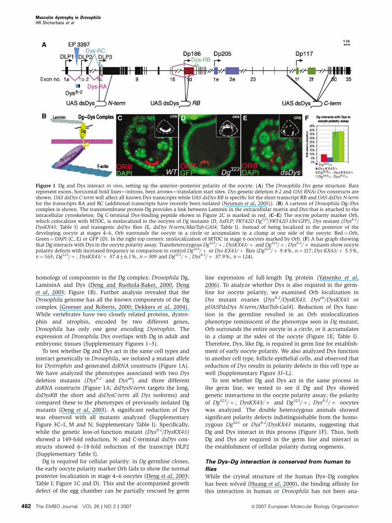
homologs of components in the Dg complex: Drosophila Dg,
LamininA and Dys (Deng and Ruohola-Baker, 2000; Deng
et al, 2003; Figure 1B). Further analysis revealed that the
Drosophila genome has all the known components of the Dg
complex (Greener and Roberts, 2000; Dekkers et al, 2004).
While vertebrates have two closely related proteins, dystro-
phin and utrophin, encoded by two different genes,
Drosophila has only one gene encoding Dystrophin. The
expression of Drosophila Dys overlaps with Dg in adult and
embryonic tissues (Supplementary Figures 1–3).
To test whether Dg and Dys act in the same cell types and
interact genetically in Drosophila, we isolated a mutant allele
for Dystrophin and generated dsRNA constructs (Figure 1A).
We have analyzed the phenotypes associated with two Dys
deletion mutants (Dys8-2 and Dyse6) and three different
dsRNA constructs (Figure 1A; dsDysN-term targets the long,
dsDysRB the short and dsDysC-term all Dys isoforms) and
compared these to the phenotypes of previously isolated Dg
mutants (Deng et al, 2003). A significant reduction of Dys
was observed with all mutants analyzed (Supplementary
Figure 3C–I, M and N; Supplementary Table I). Specifically,
while the genetic loss-of-function mutant (Dys8-2/DysKX43)
showed a 149-fold reduction, N- and C-terminal dsDys con-
structs showed 6–18-fold reduction of the transcript DLP2
(Supplementary Table I).
Dg is required for cellular polarity: in Dg germline clones,
the early oocyte polarity marker Orb fails to show the normal
posterior localization in stage 4–6 oocytes (Deng et al, 2003;
Table I; Figure 1C and D). This and the accompanied growth
defect of the egg chamber can be partially rescued by germ
line expression of full-length Dg protein (Yatsenko et al,
2006). To analyze whether Dys is also required in the germ-
line for oocyte polarity, we examined Orb localization in
Dys mutant ovaries (Dys8-2/DysKX43, Dyse6/DysKX43 or
pUASPdsDys N-term/MatTub-Gal4). Reduction of Dys func-
tion in the germline resulted in an Orb mislocalization
phenotype reminiscent of the phenotype seen in Dg mutant;
Orb surrounds the entire oocyte in a circle, or it accumulates
in a clump at the sides of the oocyte (Figure 1E; Table I).
Therefore, Dys, like Dg, is required in germ line for establish-
ment of early oocyte polarity. We also analyzed Dys function
in another cell type, follicle epithelial cells, and observed that
reduction of Dys results in polarity defects in this cell type as
well (Supplementary Figure 3J–L).
To test whether Dg and Dys act in the same process in
the germ line, we tested to see if Dg and Dys showed
genetic interactions in the oocyte polarity assay; the polarity
of Dg323/þ ; DysKX43/þ and Dg323/þ ; Dys8-2/þ oocytes
was analyzed. The double heterozygous animals showed
significant polarity defects indistinguishable from the homo-
zygous Dg323 or Dys8-2/DysKX43 mutants, suggesting that
Dg and Dys interact in this process (Figure 1F). Thus, both
Dg and Dys are required in the germ line and interact in
the establishment of cellular polarity during oogenesis.
The Dys–Dg interaction is conserved from human to
flies
While the crystal structure of the human Dys–Dg complex
has been solved (Huang et al, 2000), the binding affinity for
this interaction in human or Drosophila has not been ana-
Figure 1 Dg and Dys interact in vivo, setting up the anterior–posterior polarity of the oocyte. (A) The Drosophila Dys gene structure. Barsrepresent exons, horizontal bold lines—introns, bent arrows—translation start sites. Dys genetic deletion 8-2 and UAS RNAi Dys constructs areshown. UAS dsDys C-term will affect all known Dys transcripts while UAS dsDys RB is specific for the short transcript RB and UAS dsDys N-termfor the transcripts RA and RC (additional transcripts have recently been isolated (Neuman et al, 2005)). (B) A cartoon of Drosophila Dg–Dyscomplex is shown. The transmembrane protein Dg provides a link between Laminin in the extracellular matrix and Dys that is attached to theintracellular cytoskeleton. Dg C-terminal Dys-binding peptide shown in Figure 2C is marked in red. (C–E) The oocyte polarity marker Orb,which colocalizes with MTOC, is mislocalized in the oocytes of Dg mutants (D, hsFLP; FRT42D Dg323/FRT42D Ubi-GFP), Dys mutant (Dys8-2/DysKX43; Table I) and transgenic dsDys flies (E, dsDys N-term/MatTub-GAl4; Table I). Instead of being localized to the posterior of thedeveloping oocyte at stages 4–6, Orb surrounds the oocyte in a circle or accumulates in a clump at one side of the oocyte. Red¼Orb,Green¼DAPI (C, E) or GFP (D). In the right top corners: mislocalization of MTOC in stage 6 oocytes marked by Orb. (F) A bar graph showingthat Dg interacts with Dys in the oocyte polarity assay. Transheterozygous Dg323/þ ; DysKX43/þ and Dg323/þ ; Dys8-2/þ mutants show oocytepolarity defects with increased frequency in comparison to control Dg323/þ or Dys KX43/þ flies (Dg323/þ 9.4%, n¼ 117; Dys KX43/þ 5.5%,n¼ 163; Dg323/þ ; DysKX43/þ 37.476.1%, n¼ 309 and Dg323/þ ; Dys8-2/þ 37.9%, n¼ 124).
Muscular dystrophy in DrosophilaHR Shcherbata et al
The EMBO Journal VOL 26 | NO 2 | 2007 &2007 European Molecular Biology Organization482
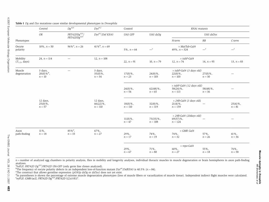
Table I Dg and Dys mutations cause similar developmental phenotypes in Drosophila
Control Dg323 Dys8-2 Control RNAi mutants
OR FRT42DDg323/FRT42DDg323
Dys8-2/Def KX43 UAS GFP UAS dsDg UAS dsDys
Phenotypes N-term RB C-term
Oocytepolarity
10%, n¼ 50 96%a, n¼ 26 41%b, n¼ 69 �MatTub-Gal45%, n¼ 64 —c 49%, n¼ 324 —c —c
Mobility(T1/2, days)
24, n¼ 114 — 12, n¼ 108 � tubP-Gal422, n¼ 91 10, n¼ 79 12, n¼ 74 14, n¼ 95 13, n¼ 83
Muscledegeneration
3 days,20(0)%d,n¼ 10
— 3 days,35(0)%,n¼ 34
� tubP-Gal4 (3 days old)17(0)%,n¼ 23
24(8)%,n¼ 103
22(0)%,n¼ 103
27(0)%,n¼ 30
—
� tubP-Gal4 (12 days old)24(0)%,n¼ 54
62(48)%,n¼ 65
58(24)%,n¼ 113
58(48)%,n¼ 36
—
12 days,25(0)%,n¼ 57
12 days,66(22)%,n¼ 110
� 24B-Gal4 (3 days old)18(0)%,n¼ 110
32(8)%,n¼ 119
21(4)%,n¼ 159
— 25(4)%,n¼ 81
� 24B-Gal4 (20days old)11(0)%,n¼ 47
73(33)%,n¼ 108
69(57)%,n¼ 124
— —
Axonpath-finding
11%,n¼ 18
85%e,n¼ 33
67%,n¼ 27
�GMR Gal429%,n¼ 17
74%,n¼ 19
74%,n¼ 32
57%,n¼ 26
61%,n¼ 36
� repo-Gal429%,n¼ 67
71%,n¼ 80
60%,n¼ 27
55%,n¼ 18
76%,n¼ 58
n¼number of analyzed egg chambers in polarity analysis, flies in mobility and longevity analyses, individual thoracic muscles in muscle degeneration or brain hemispheres in axon path-findinganalyses.ahsFLP; FRT42D Dg323/FRT42D Ubi-GFP (only germ line clones analyzed).bThe frequency of oocyte polarity defects in an independent loss-of-function mutant Dyse6/DefKX43 is 40.5% (n¼ 84).cThe construct that allows germline expression (pUASp dsDg or dsDys) does not yet exist.dIn parentheses is shown the percentage of extreme muscle degeneration phenotypes (loss of muscle fibers or vacuolization of muscle tissue). Independent indirect flight muscles were calculated.eeyFLP, GMR-lacZ; FRT42D Dg323/FRT42D l(2)cl-R111.
Muscular
dystrophyin
Drosophila
HR
Shc
herbataet
al
&2007
Euro
pean
Mole
cula
rB
iolo
gy
Org
aniza
tion
The
EM
BO
Journ
al
VO
L26
|N
O2
|2007
48
3
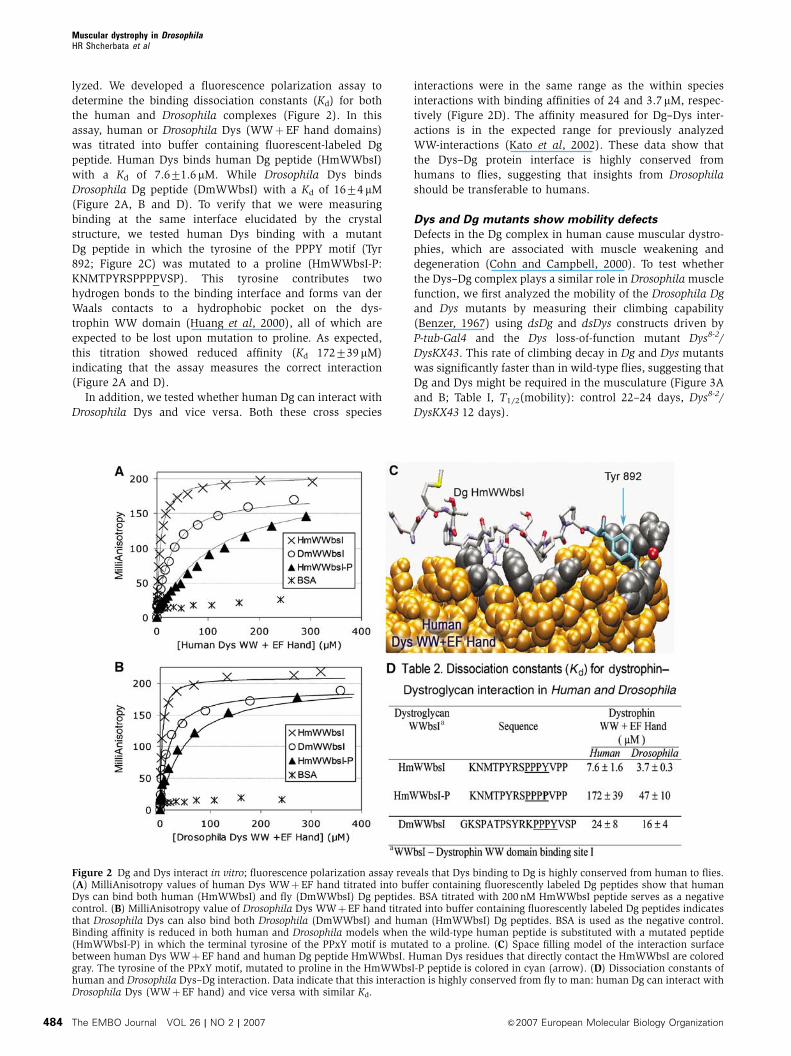
lyzed. We developed a fluorescence polarization assay to
determine the binding dissociation constants (Kd) for both
the human and Drosophila complexes (Figure 2). In this
assay, human or Drosophila Dys (WWþEF hand domains)
was titrated into buffer containing fluorescent-labeled Dg
peptide. Human Dys binds human Dg peptide (HmWWbsI)
with a Kd of 7.671.6 mM. While Drosophila Dys binds
Drosophila Dg peptide (DmWWbsI) with a Kd of 1674mM
(Figure 2A, B and D). To verify that we were measuring
binding at the same interface elucidated by the crystal
structure, we tested human Dys binding with a mutant
Dg peptide in which the tyrosine of the PPPY motif (Tyr
892; Figure 2C) was mutated to a proline (HmWWbsI-P:
KNMTPYRSPPPPVSP). This tyrosine contributes two
hydrogen bonds to the binding interface and forms van der
Waals contacts to a hydrophobic pocket on the dys-
trophin WW domain (Huang et al, 2000), all of which are
expected to be lost upon mutation to proline. As expected,
this titration showed reduced affinity (Kd 172739 mM)
indicating that the assay measures the correct interaction
(Figure 2A and D).
In addition, we tested whether human Dg can interact with
Drosophila Dys and vice versa. Both these cross species
interactions were in the same range as the within species
interactions with binding affinities of 24 and 3.7 mM, respec-
tively (Figure 2D). The affinity measured for Dg–Dys inter-
actions is in the expected range for previously analyzed
WW-interactions (Kato et al, 2002). These data show that
the Dys–Dg protein interface is highly conserved from
humans to flies, suggesting that insights from Drosophila
should be transferable to humans.
Dys and Dg mutants show mobility defects
Defects in the Dg complex in human cause muscular dystro-
phies, which are associated with muscle weakening and
degeneration (Cohn and Campbell, 2000). To test whether
the Dys–Dg complex plays a similar role in Drosophila muscle
function, we first analyzed the mobility of the Drosophila Dg
and Dys mutants by measuring their climbing capability
(Benzer, 1967) using dsDg and dsDys constructs driven by
P-tub-Gal4 and the Dys loss-of-function mutant Dys8-2/
DysKX43. This rate of climbing decay in Dg and Dys mutants
was significantly faster than in wild-type flies, suggesting that
Dg and Dys might be required in the musculature (Figure 3A
and B; Table I, T1/2(mobility): control 22–24 days, Dys8-2/
DysKX43 12 days).
Figure 2 Dg and Dys interact in vitro; fluorescence polarization assay reveals that Dys binding to Dg is highly conserved from human to flies.(A) MilliAnisotropy values of human Dys WWþEF hand titrated into buffer containing fluorescently labeled Dg peptides show that humanDys can bind both human (HmWWbsI) and fly (DmWWbsI) Dg peptides. BSA titrated with 200 nM HmWWbsI peptide serves as a negativecontrol. (B) MilliAnisotropy value of Drosophila Dys WWþEF hand titrated into buffer containing fluorescently labeled Dg peptides indicatesthat Drosophila Dys can also bind both Drosophila (DmWWbsI) and human (HmWWbsI) Dg peptides. BSA is used as the negative control.Binding affinity is reduced in both human and Drosophila models when the wild-type human peptide is substituted with a mutated peptide(HmWWbsI-P) in which the terminal tyrosine of the PPxY motif is mutated to a proline. (C) Space filling model of the interaction surfacebetween human Dys WWþEF hand and human Dg peptide HmWWbsI. Human Dys residues that directly contact the HmWWbsI are coloredgray. The tyrosine of the PPxY motif, mutated to proline in the HmWWbsI-P peptide is colored in cyan (arrow). (D) Dissociation constants ofhuman and Drosophila Dys–Dg interaction. Data indicate that this interaction is highly conserved from fly to man: human Dg can interact withDrosophila Dys (WWþEF hand) and vice versa with similar Kd.
Muscular dystrophy in DrosophilaHR Shcherbata et al
The EMBO Journal VOL 26 | NO 2 | 2007 &2007 European Molecular Biology Organization484
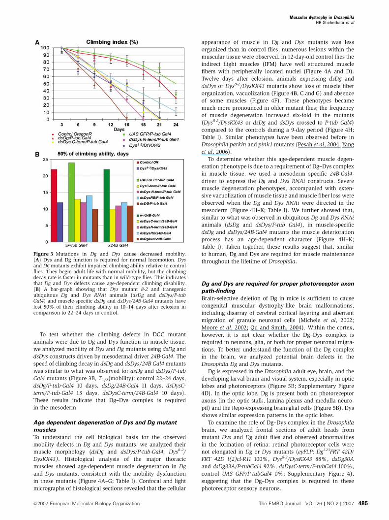
To test whether the climbing defects in DGC mutant
animals were due to Dg and Dys function in muscle tissue,
we analyzed mobility of Dys and Dg mutants using dsDg and
dsDys constructs driven by mesodermal driver 24B-Gal4. The
speed of climbing decay in dsDg and dsDys/24B Gal4 mutants
was similar to what was observed for dsDg and dsDys/P-tub
Gal4 mutants (Figure 3B, T1/2(mobility): control 22–24 days,
dsDg/P-tub-Gal4 10 days, dsDg/24B-Gal4 11 days, dsDysC-
term/P-tub-Gal4 13 days, dsDysC-term/24B-Gal4 10 days).
These results indicate that Dg–Dys complex is required
in the mesoderm.
Age dependent degeneration of Dys and Dg mutant
muscles
To understand the cell biological basis for the observed
mobility defects in Dg and Dys mutants, we analyzed their
muscle morphology (dsDg and dsDys/P-tub-Gal4, Dys8-2/
DysKX43). Histological analysis of the major thoracic
muscles showed age-dependent muscle degeneration in Dg
and Dys mutants, consistent with the mobility dysfunction
in these mutants (Figure 4A–G; Table I). Confocal and light
micrographs of histological sections revealed that the cellular
appearance of muscle in Dg and Dys mutants was less
organized than in control flies, numerous lesions within the
muscular tissue were observed. In 12-day-old control flies the
indirect flight muscles (IFM) have well structured muscle
fibers with peripherally located nuclei (Figure 4A and D).
Twelve days after eclosion, animals expressing dsDg and
dsDys or Dys8-2/DysKX43 mutants show loss of muscle fiber
organization, vacuolization (Figure 4B, C and G) and absence
of some muscles (Figure 4F). These phenotypes became
much more pronounced in older mutant flies; the frequency
of muscle degeneration increased six-fold in the mutants
(Dys8-2/DysKX43 or dsDg and dsDys crossed to P-tub Gal4)
compared to the controls during a 9-day period (Figure 4H;
Table I). Similar phenotypes have been observed before in
Drosophila parkin and pink1 mutants (Pesah et al, 2004; Yang
et al, 2006).
To determine whether this age-dependent muscle degen-
eration phenotype is due to a requirement of Dg–Dys complex
in muscle tissue, we used a mesoderm specific 24B-Gal4-
driver to express the Dg and Dys RNAi constructs. Severe
muscle degeneration phenotypes, accompanied with exten-
sive vacuolization of muscle tissue and muscle fiber loss were
observed when the Dg and Dys RNAi were directed in the
mesoderm (Figure 4H–K; Table I). We further showed that,
similar to what was observed in ubiquitous Dg and Dys RNAi
animals (dsDg and dsDys/P-tub Gal4), in muscle-specific
dsDg and dsDys/24B-Gal4 mutants the muscle deterioration
process has an age-dependent character (Figure 4H–K;
Table I). Taken together, these results suggest that, similar
to human, Dg and Dys are required for muscle maintenance
throughout the lifetime of Drosophila.
Dg and Dys are required for proper photoreceptor axon
path-finding
Brain-selective deletion of Dg in mice is sufficient to cause
congenital muscular dystrophy-like brain malformations,
including disarray of cerebral cortical layering and aberrant
migration of granule neuronal cells (Michele et al, 2002;
Moore et al, 2002; Qu and Smith, 2004). Within the cortex,
however, it is not clear whether the Dg–Dys complex is
required in neurons, glia, or both for proper neuronal migra-
tions. To better understand the function of the Dg complex
in the brain, we analyzed potential brain defects in the
Drosophila Dg and Dys mutants.
Dg is expressed in the Drosophila adult eye, brain, and the
developing larval brain and visual system, especially in optic
lobes and photoreceptors (Figure 5B; Supplementary Figure
4D). In the optic lobe, Dg is present both on photoreceptor
axons (in the optic stalk, lamina plexus and medulla neuro-
pil) and the Repo-expressing brain glial cells (Figure 5B). Dys
shows similar expression patterns in the optic lobes.
To examine the role of Dg–Dys complex in the Drosophila
brain, we analyzed frontal sections of adult heads from
mutant Dys and Dg adult flies and observed abnormalities
in the formation of retina: retinal photoreceptor cells were
not elongated in Dg or Dys mutants (eyFLP; Dg323FRT 42D/
FRT 42D l(2)cl-R11 100%, Dys8-2/DysKX43 88%, dsDg30A
and dsDg33A/P-tubGal4 92%, dsDysC-term/P-tubGal4 100%,
control UAS GFP/P-tubGal4 0%; Supplementary Figure 4),
suggesting that the Dg–Dys complex is required in these
photoreceptor sensory neurons.
Figure 3 Mutations in Dg and Dys cause decreased mobility.(A) Dys and Dg function is required for normal locomotion. Dysand Dg mutants exhibit impaired climbing ability relative to controlflies. They begin adult life with normal mobility, but the climbingdecay rate is faster in mutants than in wild-type flies. This indicatesthat Dg and Dys defects cause age-dependent climbing disability.(B) A bar-graph showing that Dys mutant 8-2 and transgenicubiquitous Dg and Dys RNAi animals (dsDg and dsDys/P-tubGal4) and muscle-specific dsDg and dsDys/24B-Gal4 mutants havelost 50% of their climbing ability in 10–14 days after eclosion incomparison to 22–24 days in control.
Muscular dystrophy in DrosophilaHR Shcherbata et al
&2007 European Molecular Biology Organization The EMBO Journal VOL 26 | NO 2 | 2007 485
The Drosophila compound eye consists of B800 ommati-
dia, each containing eight different photoreceptor sensory
neurons, R cell subtypes that project axons into one of two
optic ganglia layers in the brain during late larval develop-
ment. R1–R6 axons innervate the most superficial layer, the
lamina, generating a smooth lamina plexus, whereas R7 and
R8 project axons through the lamina into the deeper medulla
layer (Figure 5A and D) (Perez and Steller, 1996; Tessier-
Lavigne and Goodman, 1996; Clandinin and Zipursky, 2002;
Ruan et al, 2002). The patterning of the R-cell subtypes in eye
discs and the extension of their axons to the optic lobes of
the developing brains occur by late third instar larvae, while
the elongation of the retinal cell body takes place at pupal
stage (Izaddoost et al, 2002).
Figure 4 Dg and Dys mutants manifest age-dependent muscle degeneration. (A–C) Confocal analysis of histological transverse sections of IFMof 12 days old adult flies stained with a nuclear marker DAPI in red. (A) Control flies (UASGFP/P-tub-Gal4) show normal organization of theIFM and the muscle fibers are well structured with the nuclei located at the periphery (arrows). (B, C) Dg and Dys mutants (dsDg30A anddsDysN-term/P-tub-Gal4) show severe muscle degeneration: wasting and loss of muscle tissue, vacuolization (asterisks), the integrity ofsubsets of muscle cells is disrupted and the nuclei appear to be dispersed between fibers (arrows). (D–G, I–K) Light microscopy of histologicaltransverse sections of IFMs stained with H&E. (D) Control (UASGFP/P-tub-Gal4, 12 days after eclosion). (E–G) Dys and Dg mutants (dsDys N-term/P-tub-Gal4, dsDg30A/P-tub-Gal4) exhibit mainly normal muscle architecture at 3 days after eclosion, but at 12 days in most of the casesthe muscle degeneration progresses, the density of myofibrils per muscle decreases and some muscles are absent (arrowhead) or vacuolized(G, asterisk). (H) Bar graph represents increase in frequency of muscle fiber organization defects in 9 days in Dys8-2and Dg–Dys transgenicanimals (dsDg and dsDys/P-tub-Gal4) and in 17 days in flies with directed knockout of Dg and Dys in muscle (dsDg and dsDys/24B-Gal4), whichsuggest that muscle degeneration has an age-dependent character. Independent IFMs were calculated (Table I). (I–K) The mesoderm-specificRNAi-based reduction of Dg and Dys (dsDys C-term and dsDg30A/24B-Gal4) at 20 days after eclosion, but not at 3 days after eclosion (I) showobvious IFM muscle pathology: the loss of fiber density and vacuolization (asterisks).
Muscular dystrophy in DrosophilaHR Shcherbata et al
The EMBO Journal VOL 26 | NO 2 | 2007 &2007 European Molecular Biology Organization486
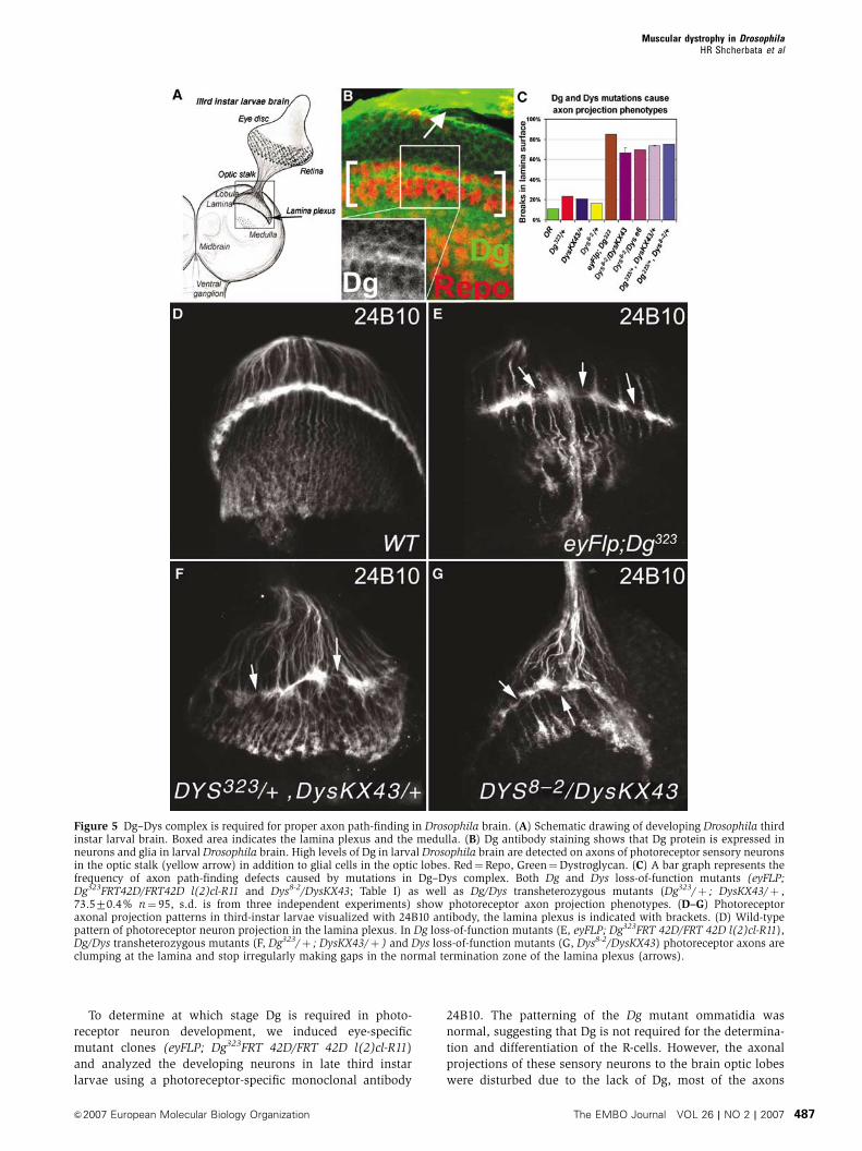
To determine at which stage Dg is required in photo-
receptor neuron development, we induced eye-specific
mutant clones (eyFLP; Dg323FRT 42D/FRT 42D l(2)cl-R11)
and analyzed the developing neurons in late third instar
larvae using a photoreceptor-specific monoclonal antibody
24B10. The patterning of the Dg mutant ommatidia was
normal, suggesting that Dg is not required for the determina-
tion and differentiation of the R-cells. However, the axonal
projections of these sensory neurons to the brain optic lobes
were disturbed due to the lack of Dg, most of the axons
Figure 5 Dg–Dys complex is required for proper axon path-finding in Drosophila brain. (A) Schematic drawing of developing Drosophila thirdinstar larval brain. Boxed area indicates the lamina plexus and the medulla. (B) Dg antibody staining shows that Dg protein is expressed inneurons and glia in larval Drosophila brain. High levels of Dg in larval Drosophila brain are detected on axons of photoreceptor sensory neuronsin the optic stalk (yellow arrow) in addition to glial cells in the optic lobes. Red¼Repo, Green¼Dystroglycan. (C) A bar graph represents thefrequency of axon path-finding defects caused by mutations in Dg–Dys complex. Both Dg and Dys loss-of-function mutants (eyFLP;Dg323FRT42D/FRT42D l(2)cl-R11 and Dys8-2/DysKX43; Table I) as well as Dg/Dys transheterozygous mutants (Dg323/þ ; DysKX43/þ ,73.570.4% n¼ 95, s.d. is from three independent experiments) show photoreceptor axon projection phenotypes. (D–G) Photoreceptoraxonal projection patterns in third-instar larvae visualized with 24B10 antibody, the lamina plexus is indicated with brackets. (D) Wild-typepattern of photoreceptor neuron projection in the lamina plexus. In Dg loss-of-function mutants (E, eyFLP; Dg323FRT 42D/FRT 42D l(2)cl-R11),Dg/Dys transheterozygous mutants (F, Dg323/þ ; DysKX43/þ ) and Dys loss-of-function mutants (G, Dys8-2/DysKX43) photoreceptor axons areclumping at the lamina and stop irregularly making gaps in the normal termination zone of the lamina plexus (arrows).
Muscular dystrophy in DrosophilaHR Shcherbata et al
&2007 European Molecular Biology Organization The EMBO Journal VOL 26 | NO 2 | 2007 487
migrate to the correct termination zone in lamina, but formed
abnormal patches in the lamina plexus. Similar axonal
problems were observed in Dys mutants. In the normal
wild-type brain the photoreceptor axons terminate in a
stereotypic fashion producing a fan-like structure in the
lamina plexus (Figure 5A, B and D). However, in 85% of
Dg loss-of-function (eyFLP; Dg323FRT 42D/FRT 42D l(2)cl-
R11) and 67–75% of Dys loss-of-function (Dys8-2/DfKX43
and Dyse6/Dys8-2) mutant third-instar larvae optic lobes the
lamina plexus is irregular; photoreceptor axons stop irregu-
larly making gaps in the normal termination zone of the
lamina plexus, deviate from the path and bundle aberrantly
(Figure 5C, E and G; Table I).
Importantly, Dg and Dys proteins interact in controlling the
photoreceptor axon path-finding since simultaneous reduc-
tion of the level of both genes (Dg323/þ ; DysKX43/þ and
Dg323/þ ; Dys8-2/þ ) results in a high percentage of the axon
projection phenotypes while reduction of each gene indepen-
dently (Dg323/þ , DysKX43/þ or Dys8-2/þ ) does not (Figure
5F and C).
Dg and Dys are required both in neurons and glia for
regular lamina plexus formation
Photoreceptor axon guidance requires correct photoreceptor
specification as well as proper function of brain glia and
neurons; the axons extend along glial cells, stop in response
to signals produced by marginal glial cells, and establish
synaptic connections with lamina neurons (Perez and Steller,
1996; Tessier-Lavigne and Goodman, 1996; Poeck et al, 2001;
Clandinin and Zipursky, 2002; Ruan et al, 2002). Previous
studies demonstrate complex interactions between R-cell
axons and laminal glial cells: R-cell axons induce the differ-
entiation and migration of laminal glial cells (Perez and
Steller, 1996), and conversely laminal glial cells present a
stop signal for terminating R1–R6 axons within the lamina
(Poeck et al, 2001).We tested whether axon path-finding
defects in Dg or Dys mutants were caused by loss of Dg-
complex function in extending neurons or supportive glial
cells by using eye- and glia-specific drivers (GMR-Gal4 and
repo-Gal4).
We first showed that in the majority of Dg and Dys RNAi
mutants driven by P-tub-Gal4, the photoreceptor axons ex-
hibited targeting phenotypes similar to Dg clonal phenotypes,
they bundled and/or terminated irregularly in the normal
termination zone of the lamina plexus. When these Dys and
Dg RNAi constructs were expressed in eye disks, photorecep-
tor axons similarly terminated irregularly in the lamina
region of the brain and formed uneven lamina neuropil
with gaps and abnormally densely packed regions (Figure
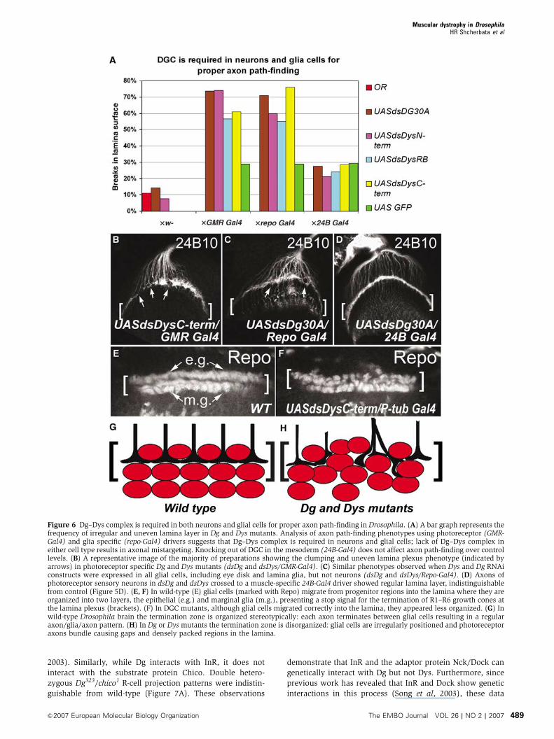
6A and B; Table I, dsDg and dsDys/GMR-Gal4 74 and 61%).
When Dys and Dg RNAi constructs were expressed in
all glial cells, including eye disk and lamina glia, but not
neurons, axons of the photoreceptor sensory neurons also
showed bundling and irregular termination (Figure 6A
and C; Table I, dsDg and dsDys/repo-Gal4 71 and 76%). To
test whether the obtained axon path-finding phenotype is
specific to DGC function in neurons and glia, we knocked-
down Dg and Dys in mesodermal tissue and observed no
effect on the axon termination process above control samples
(Figure 6A and D; dsDg and dsDys/24B-Gal4). To determine
the potential effect of DGC mutations on the development of
laminal glial cells, we stained the third-instar optic lobe using
a monoclonal antibody that recognizes the glial-specific
nuclear protein Repo (Perez and Steller, 1996; Poeck et al,
2001). In wild type (Figure 6E and G), differentiating glial
cells migrate into the lamina forming two clearly separated
layers of glial cells (i.e., epithelial and marginal glia), which
in turn present a stop signal for terminating R1–R6 growth
cones in the lamina (Poeck et al, 2001). In Dg and Dys
mutants, although glial cells migrated correctly into the
lamina, they appeared less organized lacking the clear sepa-
ration of epithelial and marginal glial layers (Figure 6F and
H). We also used the MARCM technique in order to generate
marked photoreceptor neurons and/or glial cells mutant for
Dg323 (elav-Gal4 hsFLP;FRT42B tubGal80/FRT42BDg323;UAS
GFP actoCD2oGal4). The termination zone observed for
mutant photoreceptors was irregular; clumping of axons at
the lamina and lamina breaks were associated with the
presence of Dg mutant glial cells as well as mutant photo-
receptor axons (Supplementary Figure 5A and B). In contrast
to the wild-type regular axon/glia/axon pattern, in Dg mutant
lamina the gaps were occupied by mislocalized glial cells
(Supplementary Figure 5B). These data suggest that Dg acts
autonomously and non-autonomously for correct axon path-
finding; Dg–Dys complex is required both in neurons and in
glial cells for proper neuron axonal growth and targeting.
As discussed, several congenital muscular dystrophies
exhibit neuronal migration disorders (Michele et al, 2002;
Moore et al, 2002). The mediations of axon path-finding and
neuronal migration require similar processes including sup-
portive glial cells (Bloch-Gallego et al, 2005). In the verte-
brate brain, Dg is required for granule neuron migration
(Michele et al, 2002; Moore et al, 2002; Qu and Smith,
2004). It will be interesting to see in the future if similar to
Drosophila axon path-finding, the Dg–Dys complex in verte-
brates acts both in neurons and glial cells for this process.
Indeed, Dg function has been demonstrated in a support cell
type in peripheral nervous system, Schwann cells for neuro-
nal connectivity (Saito et al, 2003).
Dg interacts with Nck/Dock SH2/SH3 adaptor protein
and InR to regulate axon guidance in Drosophila brain
The phenotypes we have observed in Dg and Dys mutant
photoreceptor axon path-finding are reminiscent of pheno-
types observed before with Nck/Dock SH2/SH3 adaptor
protein (Garrity et al, 1996) and InR (Song et al, 2003)
mutants. To test whether Dock and InR might act in concert
with Dg and Dys in this process, we analyzed whether they
genetically interact with Dys and Dg. Importantly, Dg shows
a strong interaction with InR and Dock, while Dys does not;
Dg, Dys, InR and Dock heterozygous mutants (Dg323/þ ,
DysKX43/þ , Dys8-2/þ , InRex52.1/þ , InR34/þ DockP1/þ ,
DockP2/þ ) and double heterozygous animals Dys/InR
and Dys/Dock (DysKX43/InRex52.1, DysKX43/InR34, Dys8-2/
InRex52.1, Dys8-2/InR34, DockP1/þ ; DysKX43/þ , DockP2/þ ;
DysKX43/þ , DockP1/þ ; Dys8-2/þ , DockP2/þ ; Dys8-2/þ )
mostly had regular termination zone in the lamina plexus,
while Dg323/DockP1, Dg323/DockP2, Dg323/þ ; InRex52.1/þ and
Dg323/þ ; InR34/þ double transheterozygous mutants
showed a significantly increased frequency of axon projection
defects (Figure 7A). Previous genetic and biochemical work
showed that InR can function as a guidance receptor for
Dock. However, this InR function is independent of Chico, the
Drosophila insulin receptor substrate homolog (Song et al,
Muscular dystrophy in DrosophilaHR Shcherbata et al
The EMBO Journal VOL 26 | NO 2 | 2007 &2007 European Molecular Biology Organization488
2003). Similarly, while Dg interacts with InR, it does not
interact with the substrate protein Chico. Double hetero-
zygous Dg323/chico1 R-cell projection patterns were indistin-
guishable from wild-type (Figure 7A). These observations
demonstrate that InR and the adaptor protein Nck/Dock can
genetically interact with Dg but not Dys. Furthermore, since
previous work has revealed that InR and Dock show genetic
interactions in this process (Song et al, 2003), these data
Figure 6 Dg–Dys complex is required in both neurons and glial cells for proper axon path-finding in Drosophila. (A) A bar graph represents thefrequency of irregular and uneven lamina layer in Dg and Dys mutants. Analysis of axon path-finding phenotypes using photoreceptor (GMR-Gal4) and glia specific (repo-Gal4) drivers suggests that Dg–Dys complex is required in neurons and glial cells; lack of Dg–Dys complex ineither cell type results in axonal mistargeting. Knocking out of DGC in the mesoderm (24B-Gal4) does not affect axon path-finding over controllevels. (B) A representative image of the majority of preparations showing the clumping and uneven lamina plexus phenotype (indicated byarrows) in photoreceptor specific Dg and Dys mutants (dsDg and dsDys/GMR-Gal4). (C) Similar phenotypes observed when Dys and Dg RNAiconstructs were expressed in all glial cells, including eye disk and lamina glia, but not neurons (dsDg and dsDys/Repo-Gal4). (D) Axons ofphotoreceptor sensory neurons in dsDg and dsDys crossed to a muscle-specific 24B-Gal4 driver showed regular lamina layer, indistinguishablefrom control (Figure 5D). (E, F) In wild-type (E) glial cells (marked with Repo) migrate from progenitor regions into the lamina where they areorganized into two layers, the epithelial (e.g.) and marginal glia (m.g.), presenting a stop signal for the termination of R1–R6 growth cones atthe lamina plexus (brackets). (F) In DGC mutants, although glial cells migrated correctly into the lamina, they appeared less organized. (G) Inwild-type Drosophila brain the termination zone is organized stereotypically: each axon terminates between glial cells resulting in a regularaxon/glia/axon pattern. (H) In Dg or Dys mutants the termination zone is disorganized: glial cells are irregularly positioned and photoreceptoraxons bundle causing gaps and densely packed regions in the lamina.
Muscular dystrophy in DrosophilaHR Shcherbata et al
&2007 European Molecular Biology Organization The EMBO Journal VOL 26 | NO 2 | 2007 489
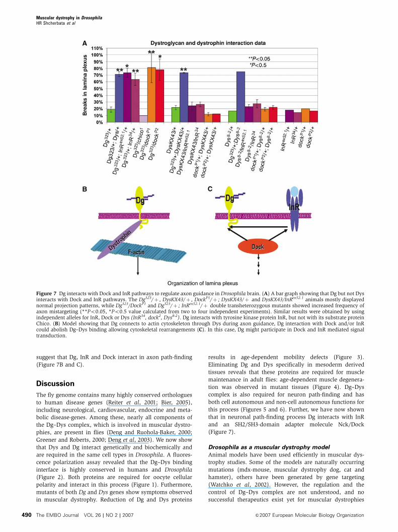
suggest that Dg, InR and Dock interact in axon path-finding
(Figure 7B and C).
Discussion
The fly genome contains many highly conserved orthologues
to human disease genes (Reiter et al, 2001; Bier, 2005),
including neurological, cardiovascular, endocrine and meta-
bolic disease-genes. Among these, nearly all components of
the Dg–Dys complex, which is involved in muscular dystro-
phies, are present in flies (Deng and Ruohola-Baker, 2000;
Greener and Roberts, 2000; Deng et al, 2003). We now show
that Dys and Dg interact genetically and biochemically and
are required in the same cell types in Drosophila. A fluores-
cence polarization assay revealed that the Dg–Dys binding
interface is highly conserved in humans and Drosophila
(Figure 2). Both proteins are required for oocyte cellular
polarity and interact in this process (Figure 1). Futhermore,
mutants of both Dg and Dys genes show symptoms observed
in muscular dystrophy. Reduction of Dg and Dys proteins
results in age-dependent mobility defects (Figure 3).
Eliminating Dg and Dys specifically in mesoderm derived
tissues reveals that these proteins are required for muscle
maintenance in adult flies: age-dependent muscle degenera-
tion was observed in mutant tissues (Figure 4). Dg–Dys
complex is also required for neuron path-finding and has
both cell autonomous and non-cell autonomous functions for
this process (Figures 5 and 6). Further, we have now shown
that in neuronal path-finding process Dg interacts with InR
and an SH2/SH3-domain adapter molecule Nck/Dock
(Figure 7).
Drosophila as a muscular dystrophy model
Animal models have been used efficiently in muscular dys-
trophy studies. Some of the models are naturally occurring
mutations (mdx-mouse, muscular dystrophy dog, cat and
hamster), others have been generated by gene targeting
(Watchko et al, 2002). However, the regulation and the
control of Dg–Dys complex are not understood, and no
successful therapeutics exist yet for muscular dystrophies
Figure 7 Dg interacts with Dock and InR pathways to regulate axon guidance in Drosophila brain. (A) A bar graph showing that Dg but not Dysinteracts with Dock and InR pathways. The Dg323/þ , DysKX43/þ , DockP1/þ ; DysKX43/þ and DysKX43/InRex52.1 animals mostly displayednormal projection patterns, while Dg323/DockP1 and Dg323/þ ; InRex52.1/þ double transheterozygous mutants showed increased frequency ofaxon mistargeting (**Po0.05, *Po0.5 value calculated from two to four independent experiments). Similar results were obtained by usingindependent alleles for InR, Dock or Dys (InR34, dock2, Dys8-2). Dg interacts with tyrosine kinase protein InR, but not with its substrate proteinChico. (B) Model showing that Dg connects to actin cytoskeleton through Dys during axon guidance, Dg interaction with Dock and/or InRcould abolish Dg–Dys binding allowing cytoskeletal rearrangements (C). In this case, Dg might participate in Dock and InR mediated signaltransduction.
Muscular dystrophy in DrosophilaHR Shcherbata et al
The EMBO Journal VOL 26 | NO 2 | 2007 &2007 European Molecular Biology Organization490
(however, systemic delivery-studies using adeno-associated
viral vectors show promise (Gregorevic et al, 2004)). Studies
in new model organisms with easy-to-manipulate genetics
might reveal the mode of regulation of the complex by
identifying key regulatory components through suppressor
screens. In addition, careful functional analysis of the com-
plex in different cell types in model organisms might result in
a unifying theme that will reveal its molecular mechanism of
function. Such recently developed models for muscular dys-
trophy exist in C. elegans and zebrafish (Gieseler et al, 2000;
Parsons et al, 2002; Bassett and Currie, 2003). In C. elegans
Dys mutant, the transporter snf-6 that normally participates
in eliminating acetylcholine from the cholinergic synapses, is
not properly localized, resulting in an increased acetylcholine
concentration at the neuromuscular junction and muscle
wasting (Kim et al, 2004). The function of Dys in neuro-
muscular junctions has also been recently addressed in
Drosophila (van der Plas et al, 2006). These results bring
up the possibility that muscular dystrophies in humans might
also at least partly be attributed to the altered kinetics of
acetylcholine transmission through neuromuscular junctions.
We have now shown that Drosophila melanogaster acts as
a remarkably good model for age-dependent progression of
muscular dystrophy. Dg and Dys reduction in Drosophila
show age-dependent muscle degeneration and lack of climb-
ing ability. It is tempting to speculate that the common
denominator between different defects observed in Dg–Dys
mutants in Drosophila and C. elegans is defective cellular
polarity. The defects observed in C. elegans could be due to
a defect in polarization of a cell, which will generate a
neuromuscular junction that leads to miss-targeted snf-6.
Similarly, we have shown that Drosophila Dg–Dys complex
is required for cellular polarity in the oocyte. In addition,
neural defects observed are plausibly due to polarity defects
in the growing axon.
Dg–Dys complex in axon path-finding
Similar to neuronal defects observed in human muscular
dystrophy patients, neuronal defects were also found in
Drosophila Dg and Dys mutant brains. In vertebrate brains,
Dg affects neuronal migration (Montanaro and Carbonetto,
2003; Qu and Smith, 2004) possibly through interaction of
neurons with their glial guides. The neuronal migration and
process outgrowth have been shown to require supportive
input from glial cells and involve the formation of adhesion
junctions along the length of the soma. Also, the outgrowth
of the leading process involves rapid extension and contrac-
tion over the length of the glial fiber (Rivas and Hatten, 1995;
Shaham, 2005). Disruption of the cytoskeletal organization
within the neuron, either of actin filaments (Rivas and
Hatten, 1995) or microtubule interactions (Vallee et al,
2000), has been shown to inhibit glial-mediated neuronal
migration. The glial function in this process is less well
studied.
Drosophila photoreceptor path-finding provides an excel-
lent system for genetic dissection of neuronal outgrowth and
target recognition (Dickson, 2002). During the formation
of the nervous system, newly born neurons send out axons
to find their targets. Each axon is led by a growth cone that
responds to extracellular axon guidance cues and chooses
between different extracellular substrates upon which to
migrate. Recent work has also identified a variety of intra-
cellular signaling pathways by which these cues induce
cytoskeletal rearrangements (Guan et al, 1996; Rao, 2005),
but the proteins connecting signals from cell surface recep-
tors to actin cytoskeleton have not been clearly determined.
Dg is a good candidate for linking receptor signaling to the
remodeling of the actin cytoskeleton and thereby polarizing
the growth cone. We have now shown that perturbation of
Dg–Dys complex causes phenotypes that resemble Nck/
Dock-Pak-Trio axon path-finding phenotypes (Figure 5)
(Rao, 2005), suggesting that Dg may be one of the key players
in Nck/Dock signaling pathway for axon guidance and target
recognition in Drosophila.
Interestingly, Insulin receptor-tyrosine kinase (InR)
mutants also show similar phenotypes to those of Nck/Dock
signaling in photoreceptor axon path-finding and these two
proteins show genetic and biochemical interactions (Song
et al, 2003). These data have led to speculations of mamma-
lian InR acting in conjunction with Nck/Dock pathway in
learning, memory and eating behavior (Dickson, 2003; Song
et al, 2003). Our data now add Dg–Dys complex to this
pathway; similar to what is seen in the case of Dg and Dys
photoreceptor mutants, InR mutants show no obvious defects
in patterning of the photoreceptors. However, the guidance of
photoreceptor cell axons from the retina to the brain is
aberrant (Song et al, 2003; Figures 5 and 6). Furthermore,
genetic and biochemical evidence suggests that InR function
in axon guidance involves the Dock-Pak pathway rather
than the PI3K-Akt/PKB pathway. Independently, biochemical
interaction between Nck/Dock and Dg has been reported
(Sotgia et al, 2001) supporting the hypothesis that InR, Dg
and Nck/Dock interaction regulates Dg–Dys complex.
Furthermore, we have now shown that Dg interacts geneti-
cally with InR and Dock in photoreceptor axon path-finding.
Since Dys interacts with Dg but not with InR and Dock, it is
tempting to speculate that Dg can selectively interact with
either Dys or InR and Dock (Figure 7). One possibility is that
the tyrosine kinase activity of InR could regulate the Dg–Dys
interaction by tyrosine phosphorylation in the Dg–Dys bind-
ing interphase (Figure 2). This tyrosine phosphorylation
could prohibit the Dg–Dys interaction and thereby result
in rearrangements in the actin cytoskeleton. Alternatively,
other components observed in Dg–Dys complex might be
involved in this regulation (Zhan et al, 2005). However, it
is also possible that potential polarity defects in the Dg
mutant axons result in defective InR membrane localization.
Interestingly, in another cell type, the Drosophila oocyte, InR,
Dg and Dys also show similar phenotypes (Deng et al, 2003;
LaFever and Drummond-Barbosa, 2005; Figure 1). In addi-
tion, insulin-like growth factors (IGF) and InR are important
in maintaining muscle mass in vertebrates (Singleton and
Feldman, 2001). Further connection of InR to Dg–Dys com-
plex comes from experiments showing that muscle specific
expression of IGF counters muscle decline in mdx-mice
(Barton et al, 2002; Shavlakadze et al, 2004; Dobrowolny
et al, 2005). The work presented in this study is the first
demonstration of genetic interaction between Dg and InR.
Future biochemical studies should unravel the molecular
mechanism of this interaction.
Furthermore, we have now shown that Dg–Dys complex is
required both in neural and in targeting glial cells for correct
neuronal axon path-finding in Drosophila brain. These data
reveal that Dg–Dys complex also has a non-cell autonomous
Muscular dystrophy in DrosophilaHR Shcherbata et al
&2007 European Molecular Biology Organization The EMBO Journal VOL 26 | NO 2 | 2007 491
effect on axon path-finding and suggest that Dg–Dys-con-
trolled ECM both from neuron and glial cells regulate neuro-
nal axon path-finding. Further experiments are required
to reveal whether long-range Laminin fibers are involved in
this process, as has been shown in epithelial planar polarity
(Bateman et al, 2001; Deng et al, 2003), or whether glial
processes are observed in close proximity to the neural
growth cone (Georges-Labouesse et al, 1998). Interestingly,
similar phenotypes are observed with Integrin mutants
(Tanaka and Sabry, 1995; Campos, 2005; Curtin et al,
2005), suggesting that, as in planar polarity (Bateman et al,
2001; Deng et al, 2003), Integrin and Dg–Dys complex might
act in concert to regulate the process of ECM organization
that will regulate the cytoskeleton of the cells involved.
Taken together, the phenotypes caused by Drosophila Dg
and Dys mutations are remarkably similar to phenotypes
observed in human muscular dystrophy patients, and there-
fore suggest that functional dissection of Dg–Dys complex in
Drosophila should provide new insights into the origin and
potential treatment of these fatal neuromuscular diseases.
As a proof of principle, using Drosophila as a model we have
now identified InR as a signaling pathway that genetically
interacts with Dg. Future studies are directed to unravel the
molecular mechanism of Dg and InR–Dock interactions in
invertebrates as well as vertebrates.
Materials and methods
Fly stocksFRT42D Dg323/Cyo and FRT42B Dg323/CyO (Dg null allele), UASdsDg(dsDg30A and dsDg33A (Deng et al, 2003), Df(3R)Dl-X43 (referredas DysKX43), EP(3)3397(Dys) (Bloomington Stock Center), thedeletion mutant Dys8-2 in Dys gene that was generated by inducing
transposition of the EP(3)3397 P-element insertion (http://engels.genetics.wisc.edu//Pelements/index.html), Dyse6 deletion mutant(van der Plas et al, 2006), three dsRNA constructs were created toknock out the different Dys transcripts: UASdsDysN-term (dsDysN-term) knocks out the three long forms (DLPs), UASdsDysRB(dsDys RB) the short form (Dp186), and UASdsDysC-term (dsDysC-term) targets the common C-terminus, thereby knocking downall transcripts (see Supplementary Materials and Methods),yw;FRT82BpM88C InR34/TM6, FRT82B InRex52.1/TM6 (gifts fromB Edgar), dockP1FRT40A/CyOGFP, yw;eyFlpgl-lacZ;Trio1FRT80B/TM6, yw;eyFlpgl-lacZ; Pak14FRT82B/TM6 (gifts from N Harden),hsFLP; FRT42DUbi-GFP/Cyo and eyFLPGMR-lacZ; FRT42D l(2)cl-R111/CyO, Gal4-elav hsFLP; FRT42B tubGal80/CyO3; act-GFP andP-tub-Gal4 (ubiquitous expression), w;MatTub-Gal4/CyO (germlineexpression), GMR-Gal4 (eye expression), w�;;24B-Gal4 (mesoderm,muscle expression), w�;;repo-Gal4/TM3,Sb (glial expression) fromBloomington Stock Center.
Supplementary dataSupplementary data are available at The EMBO Journal Online(http://www.embojournal.org).
Acknowledgements
We thank Drs Leslie Pick and Paul Garrity for reagents and advice,Alex Whitworth for help with mobility assay, Glenda Froelick forhelp with histological sections, Volodymyr Shcherbatyy for injec-tions to produce the transgenic lines, Christian Walker-Richards andLuis Tulloch for generating Dys8-2 deletion line, Merle Gilbert forhelp with initial experiments, Greg Martin for confocal microscopyinstructions, Junlin Qi for help with qRT–PCR, Arul Subramanianand Amir Sapir for helping in the preparation of the RNAi flies,Michael Eck and Florence Poy for the human Dystrophin clone,Martina Schneider and Ellen Ward for help with embryonic analy-sis, and Mario Pantoja for critical reading. This work was supportedby an AHA fellowship for HRS, CRDF for ASY, HRS and HR-B, CIHRfor VDS, MDA, AFM grants and The Israel Science Foundation forUN and DY, MDA for HR-B and by the grants from the NationalInstitute of Health for DB and HR-B.
References
Barton ER, Morris L, Musaro A, Rosenthal N, Sweeney HL (2002)Muscle-specific expression of insulin-like growth factor I countersmuscle decline in mdx mice. J Cell Biol 157: 137–148
Bassett DI, Currie PD (2003) The zebrafish as a model for musculardystrophy and congenital myopathy. Hum Mol Genet 12 (Spec No2): R265–R270
Bateman J, Reddy RS, Saito H, Van Vactor D (2001) The receptortyrosine phosphatase Dlar and integrins organize actin filamentsin the Drosophila follicular epithelium. Curr Biol 11: 1317–1327
Benzer S (1967) Behavioral mutants of Drosophila isolated bycountercurrent distribution. Proc Natl Acad Sci USA 58: 1112–1119
Bier E (2005) Drosophila, the golden bug, emerges as a tool forhuman genetics. Nat Rev Genet 6: 9–23
Bloch-Gallego E, Causeret F, Ezan F, Backer S, Hidalgo-Sanchez M(2005) Development of precerebellar nuclei: instructive factorsand intracellular mediators in neuronal migration, survival andaxon pathfinding. Brain Res Brain Res Rev 49: 253–266
Campbell KP (1995) Three muscular dystrophies: loss of cytoskele-ton-extracellular matrix linkage. Cell 80: 675–679
Campos LS (2005) Beta1 integrins and neural stem cells: makingsense of the extracellular environment. Bioessays 27: 698–707
Clandinin TR, Zipursky SL (2002) Making connections in the flyvisual system. Neuron 35: 827–841
Cohn RD (2005) Dystroglycan: important player in skeletal muscleand beyond. Neuromuscul Disord 15: 207–217
Cohn RD, Campbell KP (2000) Molecular basis of muscular dystro-phies. Muscle Nerve 23: 1456–1471
Curtin KD, Meinertzhagen IA, Wyman RJ (2005) Basigin(EMMPRIN/CD147) interacts with integrin to affect cellular ar-chitecture. J Cell Sci 118: 2649–2660
Dekkers LC, van der Plas MC, van Loenen PB, den Dunnen JT,van Ommen GJ, Fradkin LG, Noordermeer JN (2004) Embryonic
expression patterns of the Drosophila dystrophin-associated gly-coprotein complex orthologs. Gene Expr Patterns 4: 153–159
Deng WM, Ruohola-Baker H (2000) Laminin A is required forfollicle cell-oocyte signaling that leads to establishment of theanterior–posterior axis in Drosophila. Curr Biol 10: 683–686
Deng WM, Schneider M, Frock R, Castillejo-Lopez C, Gaman EA,Baumgartner S, Ruohola-Baker H (2003) Dystroglycan is requiredfor polarizing the epithelial cells and the oocyte in Drosophila.Development 130: 173–184
Dickson BJ (2002) Molecular mechanisms of axon guidance. Science298: 1959–1964
Dickson BJ (2003) Development. Wiring the brain with insulin.Science 300: 440–441
Dobrowolny G, Giacinti C, Pelosi L, Nicoletti C, Winn N, Barberi L,Molinaro M, Rosenthal N, Musaro A (2005) Muscle expression ofa local Igf-1 isoform protects motor neurons in an ALS mousemodel. J Cell Biol 168: 193–199
Durbeej M, Campbell KP (2002) Muscular dystrophies involving thedystrophin–glycoprotein complex: an overview of current mousemodels. Curr Opin Genet Dev 12: 349–361
Garrity PA, Rao Y, Salecker I, McGlade J, Pawson T, Zipursky SL(1996) Drosophila photoreceptor axon guidance and targetingrequires the dreadlocks SH2/SH3 adapter protein. Cell 85:639–650
Georges-Labouesse E, Mark M, Messaddeq N, Gansmuller A (1998)Essential role of alpha 6 integrins in cortical and retinal lamina-tion. Curr Biol 8: 983–986
Gieseler K, Grisoni K, Segalat L (2000) Genetic suppression ofphenotypes arising from mutations in dystrophin-related genesin Caenorhabditis elegans. Curr Biol 10: 1092–1097
Greener MJ, Roberts RG (2000) Conservation of components of thedystrophin complex in Drosophila. FEBS Lett 482: 13–18
Muscular dystrophy in DrosophilaHR Shcherbata et al
The EMBO Journal VOL 26 | NO 2 | 2007 &2007 European Molecular Biology Organization492
Gregorevic P, Blankinship MJ, Allen JM, Crawford RW, Meuse L,Miller DG, Russell DW, Chamberlain JS (2004) Systemic deliveryof genes to striated muscles using adeno-associated viral vectors.Nat Med 10: 828–834
Guan B, Hartmann B, Kho YH, Gorczyca M, Budnik V (1996) TheDrosophila tumor suppressor gene, dlg, is involved in structuralplasticity at a glutamatergic synapse. Curr Biol 6: 695–706
Hoffman EP, Brown Jr RH, Kunkel LM (1987) Dystrophin: theprotein product of the Duchenne muscular dystrophy locus. Cell51: 919–928
Huang X, Poy F, Zhang R, Joachimiak A, Sudol M, Eck MJ (2000)Structure of a WW domain containing fragment of dystrophin incomplex with beta-dystroglycan. Nat Struct Biol 7: 634–638
Izaddoost S, Nam SC, Bhat MA, Bellen HJ, Choi KW (2002)Drosophila crumbs is a positional cue in photoreceptor adherensjunctions and rhabdomeres. Nature 416: 178–183
Kato Y, Ito M, Kawai K, Nagata K, Tanokura M (2002) Determinantsof ligand specificity in groups I and IV WW domains as studied bysurface plasmon resonance and model building. J Biol Chem 277:10173–10177
Kim H, Rogers MJ, Richmond JE, McIntire SL (2004) SNF-6 is anacetylcholine transporter interacting with the dystrophin complexin Caenorhabditis elegans. Nature 430: 891–896
Koenig M, Hoffman EP, Bertelson CJ, Monaco AP, Feener C, KunkelLM (1987) Complete cloning of the Duchenne muscular dystro-phy (DMD) cDNA and preliminary genomic organization of theDMD gene in normal and affected individuals. Cell 50: 509–517
LaFever L, Drummond-Barbosa D (2005) Direct control of germlinestem cell division and cyst growth by neural insulin inDrosophila. Science 309: 1071–1073
Michele DE, Barresi R, Kanagawa M, Saito F, Cohn RD, Satz JS,Dollar J, Nishino I, Kelley RI, Somer H, Straub V, Mathews KD,Moore SA, Campbell KP (2002) Post-translational disruption ofdystroglycan–ligand interactions in congenital muscular dystro-phies. Nature 418: 417–422
Montanaro F, Carbonetto S (2003) Targeting dystroglycan in thebrain. Neuron 37: 193–196
Moore SA, Saito F, Chen J, Michele DE, Henry MD, Messing A, CohnRD, Ross-Barta SE, Westra S, Williamson RA, Hoshi T, CampbellKP (2002) Deletion of brain dystroglycan recapitulates aspects ofcongenital muscular dystrophy. Nature 418: 422–425
Muntoni F, Brockington M, Blake DJ, Torelli S, Brown SC(2002) Defective glycosylation in muscular dystrophy. Lancet360: 1419–1421
Neuman S, Kaban A, Volk T, Yaffe D, Nudel U (2001) The dystro-phin/utrophin homologues in Drosophila and in sea urchin. Gene263: 17–29
Neuman S, Kovalio M, Yaffe D, Nudel U (2005) The Drosophilahomologue of the dystrophin gene—introns containing promotersare major contributors to the large size of the gene. FEBS Lett 579:5365–5371
Parsons MJ, Campos I, Hirst EM, Stemple DL (2002) Removal ofdystroglycan causes severe muscular dystrophy in zebrafishembryos. Development 129: 3505–3512
Perez SE, Steller H (1996) Migration of glial cells into retinalaxon target field in Drosophila melanogaster. J Neurobiol 30:359–373
Pesah Y, Pham T, Burgess H, Middlebrooks B, Verstreken P, Zhou Y,Harding M, Bellen H, Mardon G (2004) Drosophila parkin mu-tants have decreased mass and cell size and increased sensitivityto oxygen radical stress. Development 131: 2183–2194
Poeck B, Fischer S, Gunning D, Zipursky SL, Salecker I (2001) Glialcells mediate target layer selection of retinal axons in the devel-oping visual system of Drosophila. Neuron 29: 99–113
Qu Q, Smith FI (2004) Alpha-dystroglycan interactions affect cere-bellar granule neuron migration. J Neurosci Res 76: 771–782
Rao Y (2005) Dissecting Nck/Dock signaling pathways in Droso-phila visual system. Int J Biol Sci 1: 80–86
Reiter LT, Potocki L, Chien S, Gribskov M, Bier E (2001) A systema-tic analysis of human disease-associated gene sequences inDrosophila melanogaster. Genome Res 11: 1114–1125
Rivas RJ, Hatten ME (1995) Motility and cytoskeletal organizationof migrating cerebellar granule neurons. J Neurosci 15: 981–989
Ruan W, Long H, Vuong DH, Rao Y (2002) Bifocal is a downstreamtarget of the Ste20-like serine/threonine kinase misshapen inregulating photoreceptor growth cone targeting in Drosophila.Neuron 36: 831–842
Saito F, Moore SA, Barresi R, Henry MD, Messing A, Ross-Barta SE,Cohn RD, Williamson RA, Sluka KA, Sherman DL, Brophy PJ,Schmelzer JD, Low PA, Wrabetz L, Feltri ML, Campbell KP (2003)Unique role of dystroglycan in peripheral nerve myelination,nodal structure, and sodium channel stabilization. Neuron 38:747–758
Shaham S (2005) Glia–neuron interactions in nervous system func-tion and development. Curr Top Dev Biol 69: 39–66
Shavlakadze T, White J, Hoh JF, Rosenthal N, Grounds MD (2004)Targeted expression of insulin-like growth factor-I reduces earlymyofiber necrosis in dystrophic mdx mice. Mol Ther 10: 829–843
Singleton JR, Feldman EL (2001) Insulin-like growth factor-I inmuscle metabolism and myotherapies. Neurobiol Dis 8: 541–554
Song J, Wu L, Chen Z, Kohanski RA, Pick L (2003) Axons guidedby insulin receptor in Drosophila visual system. Science 300:502–505
Sotgia F, Lee H, Bedford MT, Petrucci T, Sudol M, Lisanti MP (2001)Tyrosine phosphorylation of beta-dystroglycan at its WW domainbinding motif, PPxY, recruits SH2 domain containing proteins.Biochemistry 40: 14585–14592
Spence HJ, Chen YJ, Batchelor CL, Higginson JR, Suila H, Carpen O,Winder SJ (2004a) Ezrin-dependent regulation of the actin cyto-skeleton by beta-dystroglycan. Hum Mol Genet 13: 1657–1668
Spence HJ, Chen YJ, Winder SJ (2002) Muscular dystrophies, thecytoskeleton and cell adhesion. Bioessays 24: 542–552
Spence HJ, Dhillon AS, James M, Winder SJ (2004b) Dystroglycan,a scaffold for the ERK-MAP kinase cascade. EMBO Rep 5: 484–489
Tanaka E, Sabry J (1995) Making the connection: cytoskeletalrearrangements during growth cone guidance. Cell 83: 171–176
Tessier-Lavigne M, Goodman CS (1996) The molecular biology ofaxon guidance. Science 274: 1123–1133
Vallee RB, Faulkner NE, Tai CY (2000) The role of cytoplasmicdynein in the human brain developmental disease lissencephaly.Biochim Biophys Acta 1496: 89–98
van der Plas MC, Pilgram GS, Plomp JJ, de Jong A, Fradkin LG,Noordermeer JN (2006) Dystrophin is required for appropriateretrograde control of neurotransmitter release at the Drosophilaneuromuscular junction. J Neurosci 26: 333–344
Watchko JF, O’Day TL, Hoffman EP (2002) Functional character-istics of dystrophic skeletal muscle: insights from animal models.J Appl Physiol 93: 407–417
Winder SJ (2001) The complexities of dystroglycan. Trends BiochemSci 26: 118–124
Yang B, Jung D, Motto D, Meyer J, Koretzky G, Campbell KP (1995)SH3 domain-mediated interaction of dystroglycan and Grb2.J Biol Chem 270: 11711–11714
Yang Y, Gehrke S, Imai Y, Huang Z, Ouyang Y, Wang JW, Yang L,Beal MF, Vogel H, Lu B (2006) Mitochondrial pathology andmuscle and dopaminergic neuron degeneration caused by inacti-vation of Drosophila Pink1 is rescued by Parkin. Proc Natl AcadSci USA 103: 10793–10798
Yatsenko AS, Gray EE, Shcherbata HR, Patterson LB, Sood VD,Kucherenko MM, Baker D, Ruohola-Baker H (2006) A putativeSH3-domain binding motif but not the C-terminal DystrophinWW-domain binding motif is required for Dystroglycan functionin cellular polarity in Drosophila. J Biol Chem (submitted)
Zhan Y, Tremblay MR, Melian N, Carbonetto S (2005) Evidence thatdystroglycan is associated with dynamin and regulates endo-cytosis. J Biol Chem 280: 18015–18024
Muscular dystrophy in DrosophilaHR Shcherbata et al
&2007 European Molecular Biology Organization The EMBO Journal VOL 26 | NO 2 | 2007 493