VBP15, a novel anti‐inflammatory and membrane‐stabilizer, improves muscular dystrophy without...
17
VBP15, a novel anti‐inflammatory and membrane‐stabilizer, improves muscular dystrophy without side effects Christopher R. Heier 1 , Jesse M. Damsker 2 , Qing Yu 1 , Blythe C. Dillingham 1,2 , Tony Huynh 1,3 , Jack H. Van der Meulen 1 , Arpana Sali 1 , Brittany K. Miller 1 , Aditi Phadke 1 , Luana Scheffer 1 , James Quinn 1 , Kathleen Tatem 1 , Sarah Jordan 1 , Sherry Dadgar 1,7 , Olga C. Rodriguez 4,5 , Chris Albanese 4,5 , Michael Calhoun 6 , Heather Gordish-Dressman 1,7 , Jyoti K. Jaiswal 1,7 , Edward M. Connor 1,2,8 , John M. McCall 2,9 , Eric P. Hoffman 1,2,7 , Erica K. M. Reeves 2 , Kanneboyina Nagaraju 1,2,7 * Keywords: anti-inflammatory; dystrophy; mdx; membrane injury; muscle DOI 10.1002/emmm.201302621 Received February 08, 2013 Revised July 30, 2013 Accepted August 02, 2013 Absence of dystrophin makes skeletal muscle more susceptible to injury, resulting in breaches of the plasma membrane and chronic inflammation in Duchenne muscular dystrophy (DMD). Current management by glucocorticoids has unclear molecular benefits and harsh side effects. It is uncertain whether therapies that avoid hormonal stunting of growth and development, and/or immunosuppression, would be more or less beneficial. Here, we discover an oral drug with mechanisms that provide efficacy through anti-inflammatory signaling and membrane-stabilizing pathways, independent of hormonal or immunosuppressive effects. We find VBP15 protects and promotes efficient repair of skeletal muscle cells upon laser injury, in opposition to prednisolone. Potent inhibition of NF-kB is mediated through protein interactions of the glucocorticoid receptor, however VBP15 shows significantly reduced hormonal receptor transcriptional activity. The translation of these drug mechanisms into DMD model mice improves muscle strength, live-imaging and pathology through both preventive and post-onset intervention regimens. These data demonstrate successful improvement of dystrophy independent of hormonal, growth, or immunosuppressive effects, indicating VBP15 merits clinical investigation for DMD and would benefit other chronic inflammatory diseases. INTRODUCTION Contraction‐induced myofibre injury and inflammation are characteristic features of Duchenne muscular dystrophy (DMD), a fatal genetic muscle disease. We and others have demonstrated that the pro‐inflammatory transcription factor NF‐ kB is active in dystrophin deficient muscle before symptom onset (Chen et al, 2005; Porter et al, 2002, 2003). Pharmacological glucocorticoids (prednisone, deflazacort) are standard of care in DMD, and we hypothesize their primary mechanism of action to be through anti‐inflammatory activities via NF‐kB pathways (Wissink et al, 1997). However, their harsh side effects in children greatly reduce patient adherence to glucocorticoid regimens and limit their therapeutic window. More general (1) Center for Genetic Medicine Research, Children’s National Medical Center, Washington, DC, USA (2) ReveraGen BioPharma, Rockville, MD, USA (3) Endocrine Research Unit, The Canberra Hospital, Garran, ACT, Australia (4) Lombardi Comprehensive Cancer Center and Department of Oncology, Georgetown University Medical Center, Washington, DC, USA (5) Department of Pathology, Georgetown University Medical Center, Washington, DC, USA (6) Sinq Systems, Silver Spring, MD, USA (7) Department of Integrative Systems Biology, George Washington Univer- sity School of Medicine and Health Sciences, Washington, DC, USA (8) Center for Translational Science, Children’s National Medical Center, Washington, DC, USA (9) PharMac LLC, Boca Grande, FL, USA *Corresponding author: Tel: þ1 202 476 6220; Fax: þ1 202 476 6014; E-mail: [email protected] Research Article OPEN ACCESS VBP15 improves muscular dystrophy ß 2013 The Authors. Published by John Wiley and Sons, Ltd on behalf of EMBO This is an open access article under the terms of the Creative Commons Attribution License, which permits use, distribution and reproduction in any medium, provided the original work is properly cited. EMBO Mol Med (2013) 5, 1569–1585 1569
-
Upload
childrensnational -
Category
Documents
-
view
0 -
download
0
Transcript of VBP15, a novel anti‐inflammatory and membrane‐stabilizer, improves muscular dystrophy without...

Research Article OPENACCESSVBP15 improves muscular dystrophy
VBP15, a novel anti‐inflammatory andmembrane‐stabilizer, improves musculardystrophy without side effects
Christopher R. Heier1, Jesse M. Damsker2, Qing Yu1, Blythe C. Dillingham1,2, Tony Huynh1,3,Jack H. Van der Meulen1, Arpana Sali1, Brittany K. Miller1, Aditi Phadke1, Luana Scheffer1,James Quinn1, Kathleen Tatem1, Sarah Jordan1, Sherry Dadgar1,7, Olga C. Rodriguez4,5,Chris Albanese4,5, Michael Calhoun6, Heather Gordish-Dressman1,7, Jyoti K. Jaiswal1,7,Edward M. Connor1,2,8, John M. McCall2,9, Eric P. Hoffman1,2,7, Erica K. M. Reeves2,Kanneboyina Nagaraju1,2,7*
Keywords: anti-inflammatory; dystrophy;
mdx; membrane injury; muscle
DOI 10.1002/emmm.201302621
Received February 08, 2013
Revised July 30, 2013
Accepted August 02, 2013
(1) Center for GeneticMedicine Research, Children’s Nat
Washington, DC, USA
(2) ReveraGen BioPharma, Rockville, MD, USA
(3) Endocrine Research Unit, The Canberra Hospital, Ga
(4) Lombardi Comprehensive Cancer Center and Depa
Georgetown University Medical Center, Washington
(5) Department of Pathology, Georgetown Universi
Washington, DC, USA
(6) Sinq Systems, Silver Spring, MD, USA
(7) Department of Integrative Systems Biology, George
sity School of Medicine and Health Sciences, Washi
(8) Center for Translational Science, Children’s Natio
Washington, DC, USA
(9) PharMac LLC, Boca Grande, FL, USA
*Corresponding author: Tel: þ1 202 476 6220; Fax: þE-mail: [email protected]
� 2013 The Authors. Published by John Wiley and Sons,under the terms of the Creative Commons Attribution Licein any medium, provided the original work is properly cite
Absence of dystrophin makes skeletal muscle more susceptible to injury,
resulting in breaches of the plasma membrane and chronic inflammation in
Duchenne muscular dystrophy (DMD). Current management by glucocorticoids
has unclear molecular benefits and harsh side effects. It is uncertain whether
therapies that avoid hormonal stunting of growth and development, and/or
immunosuppression, would be more or less beneficial. Here, we discover an oral
drug with mechanisms that provide efficacy through anti-inflammatory
signaling and membrane-stabilizing pathways, independent of hormonal or
immunosuppressive effects. We find VBP15 protects and promotes efficient
repair of skeletal muscle cells upon laser injury, in opposition to prednisolone.
Potent inhibition of NF-kB is mediated through protein interactions of the
glucocorticoid receptor, however VBP15 shows significantly reduced hormonal
receptor transcriptional activity. The translation of these drug mechanisms into
DMDmodel mice improves muscle strength, live-imaging and pathology through
both preventive and post-onset intervention regimens. These data demonstrate
successful improvement of dystrophy independent of hormonal, growth, or
immunosuppressive effects, indicating VBP15 merits clinical investigation for
DMD and would benefit other chronic inflammatory diseases.
ional Medical Center,
rran, ACT, Australia
rtment of Oncology,
, DC, USA
ty Medical Center,
Washington Univer-
ngton, DC, USA
nal Medical Center,
1 202 476 6014;
Ltd on behalf of EMBO This is an open access articlense, which permits use, distribution and reproductiond. EMBO Mol Med (
INTRODUCTION
Contraction‐induced myofibre injury and inflammation arecharacteristic features of Duchenne muscular dystrophy(DMD), a fatal genetic muscle disease. We and others havedemonstrated that the pro‐inflammatory transcription factor NF‐kB is active in dystrophin deficient muscle before symptom onset(Chen et al, 2005; Porter et al, 2002, 2003). Pharmacologicalglucocorticoids (prednisone, deflazacort) are standard of care inDMD, and we hypothesize their primary mechanism of action tobe through anti‐inflammatory activities via NF‐kB pathways(Wissink et al, 1997). However, their harsh side effects inchildren greatly reduce patient adherence to glucocorticoidregimens and limit their therapeutic window. More general
2013) 5, 1569–1585 1569

Research Article www.embomolmed.orgVBP15 improves muscular dystrophy
1570
immunosuppressive compounds reduce inflammation in DMDbut fail to increase patient strength in the same manner asglucocorticoids (Griggs et al, 1993; Kissel et al, 1993), whilespecific targeting of NF‐kB increases strength in animal models(Grounds& Torrisi, 2004; Peterson et al, 2011). These data suggestthat the specific mechanism by which glucocorticoids inhibitNF‐kB is of particular importance to DMD treatment efficacy.Therapeutics that target this pathway in the absence of side effectsmay provide a substantial improvement in the treatment of DMD.
At the cellular level, dystrophin deficient muscles showincreased susceptibility to stretch‐mediated membrane instabil-ity and calcium dependent hyper‐contracture (Bertoriniet al, 1982; Yasuda et al, 2005), as well as increased oxidativestress (Disatnik et al, 1998; Rando et al, 1998). Glucocorticoidsand other steroidal compounds are multi‐mechanistic; inaddition to binding hormonal receptors they can interact withthe plasma membrane to exert rapid and specific physico-chemical effects (Buttgereit et al, 1999; Lipworth, 2000; Rhenet al, 2003; Shivaji & Jagannadham, 1992). These effects can altermembrane fluidity, vesicular fusion (Shivaji & Jagannadham,1992) and ionic flux (Buttgereit et al, 1999), which are importantfor resistance to and repair of membrane injury. Recently,membrane stabilizing compounds such as poloxamer 188(Spurney et al, 2011; Townsend et al, 2010), Mitsugumin 53(Burkin & Wuebbles, 2012; Weisleder et al, 2012) and cromolynsodium (Granchelli et al, 1996; Marques et al, 2008) haveshown improvements to pathology, myofibre tension andcardiopulmonary function in dystrophin‐deficient mice anddogs. The effects of glucocorticoids on membrane stability,however, have not been reported.
Because glucocorticoids act through multiple mechanisms,it has been unclear and controversial which molecularpathways provide efficacy in DMD and which are simplyresponsible for detrimental effects. For example, impairedgrowth is a glucocorticoid side effect for children with asthma(Avioli, 1993; Wolthers & Pedersen, 1990), but has beenproposed as a pathway of efficacy in DMD by limiting muscleworkload and delaying muscle maturation (Grounds &Shavlakadze, 2011). Further, immunotoxic effects contributeto reduced chronic inflammation, but recent evidence suggestsant‐inflammatory NF‐kB inhibition may be sufficient for efficacy(Peterson et al, 2011). It is clear, however, that detrimentaleffects of glucocorticoids currently limit their application; inDMD neonatal screening is not performed, and glucocorticoidregimens are delayed years until after the onset of fairlyadvanced symptoms. In other forms of muscular dystrophy,glucocorticoids are avoided altogether because the net balance ofpositive and negative effects is unclear. By investigating themolecular mechanisms of glucocorticoids, we have developedVBP15 as a novel oral drug. This compound is optimized forNF‐kB inhibition, membrane insertion and glucocorticoidreceptor (GR) specificity. Medicinal chemistry, however, botheliminates key glucocorticoid pathways and provides novelproperties. Here, we present the discovery and mechanisms ofthis drug, then extensively examine efficacy and side effects inmdxmuscular dystrophy model mice. We find VBP15 has novelmembrane‐stabilizing and immunological properties, and shows
� 2013 The Authors. Published by John Wiley and Sons, Ltd on behalf of EMBO.
potent NF‐kB inhibition and substantially reduced hormonaleffects. To capitalize on this mechanism profile, which targetsmultiple pre‐symptomatic defects, we adopt a prophylacticregimen, beginning dosing before mdx symptom onset in ablinded pre‐clinical trial. This strategy would be analogous to aneonatal screening, preventive regimen in the clinic. Anotherintervention experiment in post‐onset adult mdx mice showsrepeatable efficacy in a different stage of disease. We find dose–response improvements with successful ablation of growth,bone and immunological toxicities seen with traditionalglucocorticoids. These data provide new insights into biologicalmechanisms of efficacy versus side effects in DMD, identifyVBP15 as a novel entity that warrants clinical investigation forDMD, and show therapeutic potential for other disorders ofchronic inflammation and membrane instability.
RESULTS
In vitro characterization of VBP15VBP15 was selected as our lead compound for clinicaldevelopment from a screening program focused on D‐9,11compounds. This D‐9,11 class is differentiated from glucocorti-coids by the key conversion of a hydroxyl group to a carbon‐carbon double bond (Fig 1). Preliminary studies suggested thesedrugs had potential anti‐inflammatory effects (Baudy et al, 2012)but lacked activation of a synthetic GR reporter. Throughextensive medicinal chemistry probing the R1–R3 groups of theD‐ring in this steroidal structure to generate a compound library,followed by multiple lines of screening studies focused on 20candidates, VBP15 was subsequently identified as our leadcompound. Selection was based upon its superior profile in an invitro assay for NF‐kB inhibition in myogenic cells, in addition toligand‐induced nuclear translocation of the GR, cytotoxicity,metabolite and pharmacokinetic properties (Reeves et al, 2013).To further screen candidate compounds for target receptorspecificity, we performed competitive nuclear hormone receptorbinding assays (Fig 1E–H). In these assays, we found that VBP15shows increased specificity for GR binding in comparison toother D‐9,11 compounds. For example, VBP15 exhibited anapproximately 50‐fold greater affinity for the GR than VBP3, anda 64‐fold lower affinity for the mineralocorticoid receptor (MR).VBP15 also showed only very low affinity for the androgenreceptor (Fig 1G), over 500‐fold lower than the controlmethyltrienolone, and lacked any detectable binding to theoestrogen (Fig 1H) or progesterone (data not shown) receptorsin these in vitro assays. From these screening, biochemicaland specificity data, VBP15 presented a superior profile fortherapeutic development.
Our studies here are benchmarked against prednisolone, theactive form of prednisone. Both VBP15 and prednisoloneinhibited TNFa‐induced pro‐inflammatory NF‐kB signaling atsimilar levels in NF‐kB reporter assays in C2C12 muscle cells at1 nM or more (Fig 2A). To confirm effects on NF‐kB target genes,several inflammatory transcripts known to be induced by TNFawere assayed by qPCR in VBP15‐ and prednisolone‐treatedH2K myotubes. We found VBP15 inhibited the TNFa‐induced
EMBO Mol Med (2013) 5, 1569–1585

Figure 1. VBP15 structure differentiates it from glucocorticoids and improves GR specificity.
A–D. The chemical structure of VBP15 is provided (A). VBP15 was selected for clinical development by having the optimal profile from a library of D-9,11
compounds, whose general structure is provided in (B). Compound diversity for this library was generated by medicinal chemistry probing of the R1–R3
groups. These compounds are structurally related to glucocorticoids (C), but contain an essential D-9,11 double bond modification to the steroid C-ring, at
the location depicted by the red box. This modification produces novel properties and clear differences in sub-activity profiles of these compounds.
Prednisolone (D) is the active form of prednisone, a current glucocorticoid standard of care for DMD.
E–H. Receptor specificity was determined through competitive binding assays. Here, radiolabeled high-affinity ligands were incubated with extracted steroid
receptors and increasing concentrations of unlabeled competitor (high-affinity control, VBP15, VBP1 or VBP3). Best-fit curves are provided. VBP15 showed
increased GR specificity through increased binding to the (E) GR and decreased binding to the (F) MR in comparison to other D-9,11 compounds (VBP1 and
VBP3). VBP15 also showed low (H) androgen receptor binding and no detectable binding to the (G) oestrogen receptor. (Triam, triamcinolone; Spiro,
spironolactone; R1881, methyltrienolone; Estradiol, 17b-estradiol).
Research Articlewww.embomolmed.orgChristopher R. Heier et al.
inflammatory transcripts Cox2, Irf1 and Nos2 (p< 0.005) atpotencies similar to prednisolone (Fig 2B).
Both prednisolone and VBP15 are hydrophobic compoundsthat are expected to have physicochemical effects on lipidbilayers. We compared the effects of VBP15 and prednisolone onmembrane injury and repair in live cells using an establishedlaser injury assay (Sharma et al, 2012). Skeletal muscle cellstreated with VBP15 showed reduced impact of the injury andenhanced repair in a dose dependent fashion (Fig 2C and D).Cells treated with prednisolone, however, showed greaterimpact from injury with elevated dye uptake. In this livesingle‐cell injury model, prednisolone exacerbated, while VBP15protected, injury to the plasma membrane.
GR mediates VBP15 anti‐inflammatory effects withoutinducing classical steroid transactivationTo investigate whether NF‐kB inhibition by VBP15 is mediatedby the same pathways as glucocorticoids, we examined theeffects of the steroidal receptor antagonist, RU‐486, on NF‐kBinhibition. Increasing concentrations of RU‐486 from 1nM to10mM ablated NF‐kB inhibition by VBP15 in a dose dependentmanner, similar to results seen with prednisolone anddexamethasone (Fig 3A). This shows that the anti‐inflammatoryeffects of VBP15, prednisolone and dexamethasone are allmediated through shared steroidal pathways.
EMBO Mol Med (2013) 5, 1569–1585 �
A sub‐activity of pharmacologic glucocorticoids that is largelyseparable from NF‐kB inhibitor activities is the translocation ofligand‐GR complexes to the nucleus where they directly mediatetranscriptional pathways via glucocorticoid response elements(GRE) (e.g. classical steroid receptor transactivation or hormon-al properties). Both positive‐ and negative‐acting GRE‐mediatedtranscriptional regulation has been described, and both formsof hormonal activities are more often associated with gluco-corticoid side effects rather than efficacy, with some of thesemediated by the pituitary (Diamond et al, 1990; Drouinet al, 1993; Itani et al, 2002; Meijsing et al, 2009; Yoshiuchiet al, 1998). In AtT‐20 pituitary cells, we examined genesregulated by positive and negative GREs. Sgk1, a key mediator offibrosis, is activated by a positive GRE. Both prednisone anddexamethasone (0.1mM) showed a greater than 13‐foldinduction of Sgk1 gene transcription, whereas VBP15 showedno such GRE‐mediated transcriptional activity at the sameconcentration (Fig 3B). At 1.0 and 10mM, VBP15 began to showsome evidence of Sgk1 transcriptional induction, but to a lowerdegree than traditional glucocorticoids. Adrenocorticotropichormone (ACTH), the stimulatory hormone in adrenal steroido-genesis, is negatively regulated by a ligand/GR‐GRE interaction(Drouin et al, 1993). Treatment with dexamethasone orprednisolone reduced ACTH secretion in AtT‐20 cells toapproximately 20% of untreated at all concentrations tested
2013 The Authors. Published by John Wiley and Sons, Ltd on behalf of EMBO. 1571
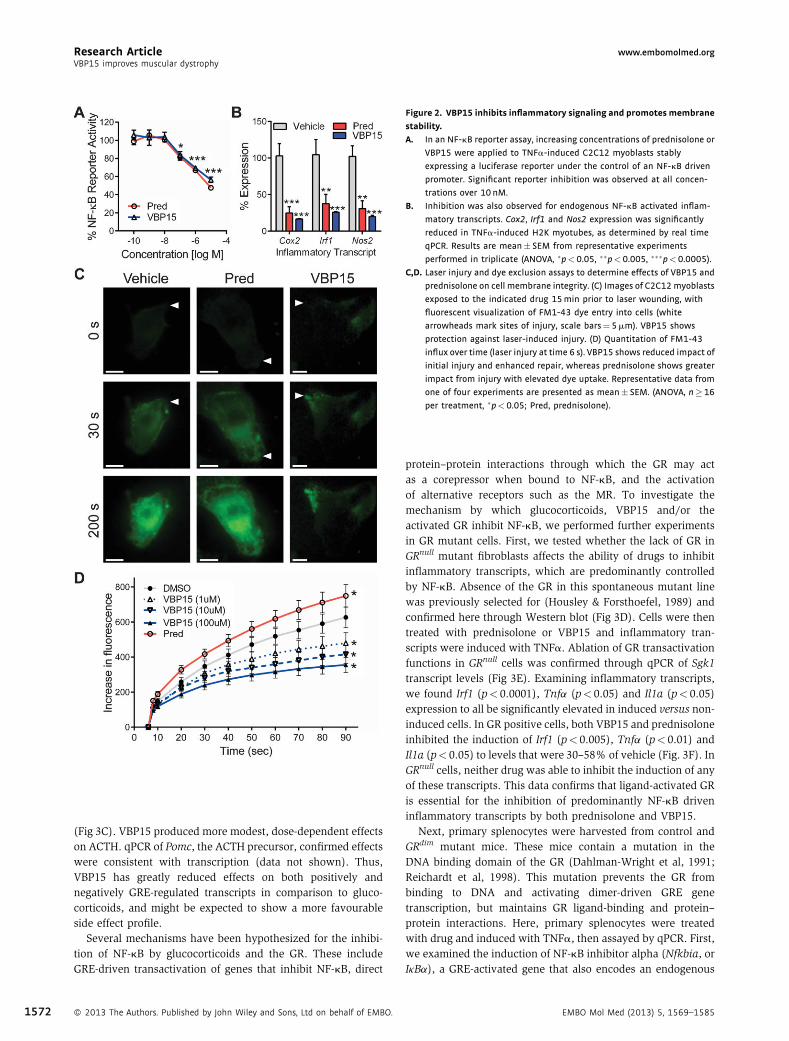
Figure 2. VBP15 inhibits inflammatory signaling and promotes membrane
stability.
A. In an NF-kB reporter assay, increasing concentrations of prednisolone or
VBP15 were applied to TNFa-induced C2C12 myoblasts stably
expressing a luciferase reporter under the control of an NF-kB driven
promoter. Significant reporter inhibition was observed at all concen-
trations over 10 nM.
B. Inhibition was also observed for endogenous NF-kB activated inflam-
matory transcripts. Cox2, Irf1 and Nos2 expression was significantly
reduced in TNFa-induced H2K myotubes, as determined by real time
qPCR. Results are mean� SEM from representative experiments
performed in triplicate (ANOVA, �p<0.05, ��p<0.005, ���p<0.0005).
C,D. Laser injury and dye exclusion assays to determine effects of VBP15 and
prednisolone on cell membrane integrity. (C) Images of C2C12myoblasts
exposed to the indicated drug 15min prior to laser wounding, with
fluorescent visualization of FM1-43 dye entry into cells (white
arrowheads mark sites of injury, scale bars¼5mm). VBP15 shows
protection against laser-induced injury. (D) Quantitation of FM1-43
influx over time (laser injury at time 6 s). VBP15 shows reduced impact of
initial injury and enhanced repair, whereas prednisolone shows greater
impact from injury with elevated dye uptake. Representative data from
one of four experiments are presented as mean� SEM. (ANOVA, n�16
per treatment, �p<0.05; Pred, prednisolone).
Research Article www.embomolmed.orgVBP15 improves muscular dystrophy
1572
(Fig 3C). VBP15 produced more modest, dose‐dependent effectson ACTH. qPCR of Pomc, the ACTH precursor, confirmed effectswere consistent with transcription (data not shown). Thus,VBP15 has greatly reduced effects on both positively andnegatively GRE‐regulated transcripts in comparison to gluco-corticoids, and might be expected to show a more favourableside effect profile.
Several mechanisms have been hypothesized for the inhibi-tion of NF‐kB by glucocorticoids and the GR. These includeGRE‐driven transactivation of genes that inhibit NF‐kB, direct
� 2013 The Authors. Published by John Wiley and Sons, Ltd on behalf of EMBO.
protein–protein interactions through which the GR may actas a corepressor when bound to NF‐kB, and the activationof alternative receptors such as the MR. To investigate themechanism by which glucocorticoids, VBP15 and/or theactivated GR inhibit NF‐kB, we performed further experimentsin GR mutant cells. First, we tested whether the lack of GR inGRnull mutant fibroblasts affects the ability of drugs to inhibitinflammatory transcripts, which are predominantly controlledby NF‐kB. Absence of the GR in this spontaneous mutant linewas previously selected for (Housley & Forsthoefel, 1989) andconfirmed here through Western blot (Fig 3D). Cells were thentreated with prednisolone or VBP15 and inflammatory tran-scripts were induced with TNFa. Ablation of GR transactivationfunctions in GRnull cells was confirmed through qPCR of Sgk1transcript levels (Fig 3E). Examining inflammatory transcripts,we found Irf1 (p< 0.0001), Tnfa (p< 0.05) and Il1a (p< 0.05)expression to all be significantly elevated in induced versus non‐induced cells. In GR positive cells, both VBP15 and prednisoloneinhibited the induction of Irf1 (p< 0.005), Tnfa (p< 0.01) andIl1a (p< 0.05) to levels that were 30–58% of vehicle (Fig. 3F). InGRnull cells, neither drug was able to inhibit the induction of anyof these transcripts. This data confirms that ligand‐activated GRis essential for the inhibition of predominantly NF‐kB driveninflammatory transcripts by both prednisolone and VBP15.
Next, primary splenocytes were harvested from control andGRdim mutant mice. These mice contain a mutation in theDNA binding domain of the GR (Dahlman‐Wright et al, 1991;Reichardt et al, 1998). This mutation prevents the GR frombinding to DNA and activating dimer‐driven GRE genetranscription, but maintains GR ligand‐binding and protein–protein interactions. Here, primary splenocytes were treatedwith drug and induced with TNFa, then assayed by qPCR. First,we examined the induction of NF‐kB inhibitor alpha (Nfkbia, orIkBa), a GRE‐activated gene that also encodes an endogenous
EMBO Mol Med (2013) 5, 1569–1585
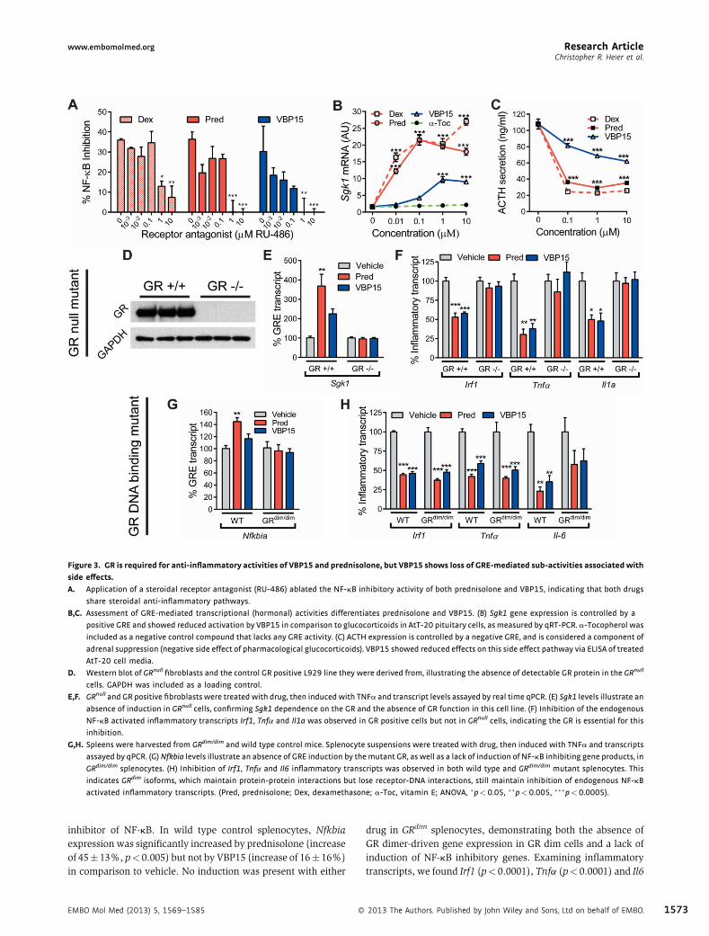
Figure 3. GR is required for anti‐inflammatory activities of VBP15 and prednisolone, but VBP15 shows loss of GRE‐mediated sub‐activities associatedwith
side effects.
A. Application of a steroidal receptor antagonist (RU-486) ablated the NF-kB inhibitory activity of both prednisolone and VBP15, indicating that both drugs
share steroidal anti-inflammatory pathways.
B,C. Assessment of GRE-mediated transcriptional (hormonal) activities differentiates prednisolone and VBP15. (B) Sgk1 gene expression is controlled by a
positive GRE and showed reduced activation by VBP15 in comparison to glucocorticoids in AtT-20 pituitary cells, asmeasured by qRT-PCR. a-Tocopherol was
included as a negative control compound that lacks any GRE activity. (C) ACTH expression is controlled by a negative GRE, and is considered a component of
adrenal suppression (negative side effect of pharmacological glucocorticoids). VBP15 showed reduced effects on this side effect pathway via ELISA of treated
AtT-20 cell media.
D. Western blot of GRnull fibroblasts and the control GR positive L929 line they were derived from, illustrating the absence of detectable GR protein in the GRnull
cells. GAPDH was included as a loading control.
E,F. GRnull and GR positive fibroblasts were treated with drug, then induced with TNFa and transcript levels assayed by real time qPCR. (E) Sgk1 levels illustrate an
absence of induction in GRnull cells, confirming Sgk1 dependence on the GR and the absence of GR function in this cell line. (F) Inhibition of the endogenous
NF-kB activated inflammatory transcripts Irf1, Tnfa and Il1a was observed in GR positive cells but not in GRnull cells, indicating the GR is essential for this
inhibition.
G,H. Spleens were harvested from GRdim/dim and wild type control mice. Splenocyte suspensions were treated with drug, then induced with TNFa and transcripts
assayed by qPCR. (G)Nfkbia levels illustrate an absence of GRE induction by themutant GR, as well as a lack of induction of NF-kB inhibiting gene products, in
GRdim/dim splenocytes. (H) Inhibition of Irf1, Tnfa and Il6 inflammatory transcripts was observed in both wild type and GRdim/dim mutant splenocytes. This
indicates GRdim isoforms, which maintain protein-protein interactions but lose receptor-DNA interactions, still maintain inhibition of endogenous NF-kB
activated inflammatory transcripts. (Pred, prednisolone; Dex, dexamethasone; a-Toc, vitamin E; ANOVA, �p<0.05, ��p<0.005, ���p<0.0005).
Research Articlewww.embomolmed.orgChristopher R. Heier et al.
inhibitor of NF‐kB. In wild type control splenocytes, Nfkbiaexpression was significantly increased by prednisolone (increaseof 45� 13%, p< 0.005) but not by VBP15 (increase of 16� 16%)in comparison to vehicle. No induction was present with either
EMBO Mol Med (2013) 5, 1569–1585 �
drug in GRdim splenocytes, demonstrating both the absence ofGR dimer‐driven gene expression in GR dim cells and a lack ofinduction of NF‐kB inhibitory genes. Examining inflammatorytranscripts, we found Irf1 (p< 0.0001), Tnfa (p< 0.0001) and Il6
2013 The Authors. Published by John Wiley and Sons, Ltd on behalf of EMBO. 1573

Research Article www.embomolmed.orgVBP15 improves muscular dystrophy
1574
(p< 0.05) were significantly elevated within induced versusnon‐induced primary splenocytes. Consistent with GR positivefibroblasts and H2K myotubes, treatment of wild typesplenocytes with both VBP15 and prednisolone successfullyinhibited the induction of all three inflammatory transcripts(p< 0.001) to levels that were roughly half those of vehicle. Incontrast to GRnull genotype and GRE transcript experiments, wefound that inhibition of all three inflammatory transcripts wasmaintained in the GRdim mutant cells. Together, these experi-ments show that both prednisolone and VBP15 activate the GR toefficiently inhibit inflammatory transcription programs throughprotein–protein interactions, independent of DNA binding ortransactivation of inhibitory genes.
VBP15 improves dystrophic phenotypes in mice treated beforethe onset of early necrosisThe mdx mouse model of DMD shows staged histopathology,with little evidence of dystrophy from 0 to 3 weeks of age, thenwide‐spread necrosis from 3 to 6 weeks, followed by successfulregeneration and a milder, more stable histological picture. Wetested efficacy of VBP15 in the mdx model with treatmentbeginning prior to the 3 weeks onset of widespread pathology(prophylactic strategy).We carried out a blinded pre‐clinical trialof pre‐symptomatic mice with VBP15 (5, 15 or 30mg/kg),prednisolone (5mg/kg), or vehicle beginning at postnatal day 15(PND15), following guidelines for robust pre‐clinical trialsand international SOPs (Landis et al, 2012; Nagaraju &Willmann, 2009; Spurney et al, 2009). These doses were chosenon the basis of favourable bioavailability, ADME and metaboliteprofiles (Reeves et al, 2013), as well as early safety studies inwild type mice by independent groups, which suggest the 28 dayno‐observable adverse effect level (NOAEL) of daily oral VBP15in mice is at least 100mg/kg. This dose range was chosen tobetter define the therapeutic window within this mouse diseasemodel. The prednisolone dose was chosen based on ourextensive pre‐clinical experience with this drug in the mdxmouse model.
Both VBP15 and prednisolone increased mdx forelimb andhindlimb normalized grip strength in comparison to vehicle(Fig 4A and B). Significant increases in VBP15 groups followed adose‐dependent pattern from 14% at 5mg/kg (p< 0.05) to 20%at 30mg/kg (p< 0.0005). For maximal force exerted, we againsaw a dose dependent increase in forelimb strength upon VBP15treatment, while prednisolone actually showed a reduction inmaximal forelimb strength (Fig 4C). The discrepancy betweenprednisolone’s effects on maximal and normalized forcemeasures was due to the marked retardation of mouse growthinduced by prednisolone, but not by VBP15 (see below). Thisindicates VBP15 increases functional mouse limb strength.
Evaluating muscle strength of isolated muscles ex vivo,extensor digitorum longus (EDL) muscles showed a reduction inspecific force for mdx compared to WT (Fig 4D). Whileprednisolone showed no increase, specific force increased withVBP15 at both 15 and 30mg/kg by an average of 12%. Followinglengthening contractions, smaller drops in force for mdx EDLsafter 10 contractions were observed for mice treated withprednisolone (7%) and VBP15 (11% at 15mg/kg, p< 0.05), in
� 2013 The Authors. Published by John Wiley and Sons, Ltd on behalf of EMBO.
comparison to vehicle (Supporting Information Fig 1A). Thesedata suggest functional benefits to isolated dystrophic muscles.
Optical imaging of live animals was used to monitor muscleinflammation. ProSense 680, a substrate cleaved by cathepsinproteases upregulated in DMD (Kar & Pearson, 1978; Takedaet al, 1992), was injected as previously reported (Baudyet al, 2011). Cathepsin activity was elevated in mdx mice(Fig 4E, Supporting Information Fig 1B and C). VBP15 andprednisolone decreased cathepsin activity towards WT levels.Decreases in VBP15 groups followed a dose‐dependent pattern,from a 22% decrease in comparison to vehicle at 5mg/kg to a41% decrease at 30mg/kg in hindlimbs. This suggests VBP15reduces muscle inflammatory disease in vivo.
In histopathology studies, quantitative H&E analysis of mdxdiaphragms revealed a clear inflammatory phenotype, with16‐fold higher inflammatory cell counts compared to WT(Fig 4F). Mice treated with VBP15 at 15 and 30mg/kg displayed38 and 30% reductions in inflammatory foci compared to vehicle.VBP15 also reduced calcified fibres (Supporting InformationFig 1D). These data are evidence that VBP15 reduces inflamma-tion and improves inflammatory muscle pathology.
VBP15 improves dystrophic phenotypes in adult mdx micetreated after symptom onsetIn a separate trial, exercised adult mdx mice were treated for4 months. In agreement with the pre‐symptomatic trial above,ProSense680 live animal imaging exhibited a 20% and 13%decrease in muscle inflammation upon VBP15 treatment at 15and 45mg/kg (Fig 4G). Isolated EDLs showed a 16% increase inspecific force upon treatment with VBP15 at 15mg/kg (Fig 4H).H&E histology revealed VBP15 significantly decreased dia-phragm inflammation (Fig 4I). These data reinforce VBP15efficacy and indicate both pre‐symptomatic and post‐onsettreatment regimens can benefit disease.
VBP15 does not display immunotoxicity seen withprednisolonePharmacologic glucocorticoids show immunosuppressive andimmunotoxic properties that limit therapeutic windows andlong‐term prescription. We benchmarked VBP15 againstprednisolone to determine if similar sub‐activities were seen.Untreated mdx mice showed enlarged spleens and increasednumbers of peripheral blood leucocytes (PBLs) compared to WTmice (Supporting Information Fig 2A and B). VBP15 treatmentreduced spleen mass and PBL counts in a dose‐dependentmanner to levels resembling WT. Prednisolone reduced thesemeasures below WT, suggesting immunosuppressive and/orimmunotoxic properties. Further, prednisolone significantlydecreased viable splenocytes per gram of tissue (p< 0.005),while this was not observed for any VBP15 dose (Fig 5A).
We next examined effects of VBP15 on B and T lymphocytesisolated from mdx spleens at the trial conclusion. Both Blymphocytes and CD4þ T lymphocytes were depleted byprednisolone but not VBP15, as measured by percent B220þ
and CD4þ positive splenocytes, respectively (Fig 5B and C). CD4þ
T cell activation was assayed by stimulation of splenocytes withconcanavalin A (ConA). Prednisolone treatment significantly
EMBO Mol Med (2013) 5, 1569–1585
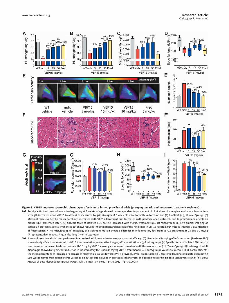
Figure 4. VBP15 improves dystrophic phenotypes of mdx mice in two pre‐clinical trials (pre‐symptomatic and post‐onset treatment regimens).
A–F. Prophylactic treatment of mdx mice beginning at 2 weeks of age showed dose-dependent improvement of clinical and histological endpoints. Mouse limb
strength increased upon VBP15 treatment as measured by grip strength of 6 week old mice for both (A) forelimb and (B) hindlimb (n�12 mice/group). (C)
Maximal force exerted by mouse forelimbs increased with VBP15 treatment but decreased with prednisolone treatment, due to prednisolone effects on
mouse size (presented later). (D) Specific force of isolated EDL muscle increased with VBP15 treatment (n¼10 mice/group). (E) Live-animal imaging of
cathepsin protease activity (ProSense680) shows reduced inflammation and necrosis of the hindlimbs in VBP15-treatedmdxmice (E images; E0 quantitationof fluorescence; n�6 mice/group). (F) Histology of diaphragm muscle shows a decrease in inflammatory foci from VBP15 treatment at 15 and 30mg/kg
(F representative images, F0 quantitation; n¼6 mice/group).
G–I. A second pre-clinical trial was performed in exercised adultmdx mice to assay post-onset efficacy. (G) Live-animal imaging of inflammation (ProSense680)
showed a significant decrease with VBP15 treatment (G representative images, (G0) quantitation; n�6mice/group). (H) Specific force of isolated EDLmuscle
wasmeasured ex vivo at trial conclusion with 15mg/kg VBP15 showing an increase consistent with the neonate trial (n�7mice/group). (I) Histology of adult
diaphragm showed a significant reduction in inflammatory foci upon 45mg/kg VBP15 treatment (n¼6mice/group). Values aremean� SEM. For treatments,
the mean percentage of increase or decrease ofmdx vehicle values towards WT is provided. (Pred, prednisolone; FL, forelimb; HL, hindlimb; data exceeding 2
SD’s was removed from specific force values as an outlier but included in all statistical analyses; one-tailed t-test of single dose versus vehiclemdx yp<0.05;
ANOVA of dose-dependence groups versus vehicle mdx �p<0.05, ��p<0.005, ���p<0.0005).
Research Articlewww.embomolmed.orgChristopher R. Heier et al.
EMBO Mol Med (2013) 5, 1569–1585 � 2013 The Authors. Published by John Wiley and Sons, Ltd on behalf of EMBO. 1575

Figure 5. VBP15 does not show immunosuppressive activities shown by
prednisolone.
A. Prednisolone significantly reduced the number of viable splenocytes per
gram of spleen tissue, whereas VBP15 did not at any dose.
B. The percentage of B lymphocytes, as measured by FACS analysis of B220
positive cells, was reduced in spleens from prednisolone treatedmdxmice,
while VBP15 showed no decrease in B cells.
C. Spleen CD4þ T cell numbers were significantly decreased in prednisolone
treated mdx spleens, but not by VBP15 treatment.
D. Activation of mdx splenocyte T cells by concavalin A was impaired
by prednisolone, but not impaired by VBP15 treatment. Values are
mean� SEM. (Pred, prednisolone; (A) n�12, (B–D) n¼3–5; �p�0.05,��p<0.005).
Research Article www.embomolmed.orgVBP15 improves muscular dystrophy
1576
reduced activated CD4þCD25þ cells (p¼ 0.01), while VBP15 didnot. Taken together, these findings suggest VBP15 modulatesinflamed mdx immune systems towards a WT state, whileprednisolone treatment leads towards an immunocompromisedstate.
VBP15 shows a superior side effect profile compared topharmacological glucocorticoidsStunted growth is a significant side effect of chronic prednisoneuse in children (Avioli, 1993; Wolthers & Pedersen, 1990). In ourpre‐symptomaticmdx study, prednisolone treatment significant-ly stunted the growth of young mice (Fig 6A). After 5 weeks oftreatment, mdx mice receiving prednisolone were significantlyshorter (8.6� 0.4 cm) than vehicle (9.1� 0.3 cm, p< 0.001). Nosignificant reduction in body length was observed for any VBP15dose.
Chronic treatment with glucocorticoids negatively affectsbone growth and development, and can cause osteoporosis(Bircan et al, 1997; Manolagas & Weinstein, 1999). Tibia lengthwas measured to determine if VBP15 inhibited bone growth(Fig 6B). Vehicle mdx mice had tibia lengths of 15.9� 0.3mm,while prednisolone significantly decreased this to 14.8� 0.5mm
� 2013 The Authors. Published by John Wiley and Sons, Ltd on behalf of EMBO.
(p< 0.005). VBP15, however, did not affect tibia length at anyconcentration. MicroCT was performed on femurs to examinebone density and structure (Fig 6C). Comparison of vehicle,prednisolone and the highest VBP15 dose showed prednisoloneto significantly reduce trabecular thickness (p< 0.005) com-pared to vehicle, while VBP15 did not. Prednisolone thusdemonstrated side effects to bone not observed with VBP15treatment.
We have previously reported deleterious effects of prednisoneon increased fibrosis in mdx hearts (Sali et al, 2012). In bothpre‐clinical trials (pre‐symptomatic and adult), we examinedcardiac and skeletal muscle for measures of fibrosis. In the pre‐symptomatic trial (Fig 6D–F), prednisolone caused a significantelevation of heart mass ratios over vehicle (5.9� 0.6 vs.5.4� 0.4, p< 0.05), indicative of cardiac hypertrophy. Noincrease was present in VBP15 groups. Histologically, clearfibrosis was evident in 50% of young (8 weeks) prednisolone‐treated hearts compared to 0% of all other groups. Histologicalanalyses of skeletal muscle (gastrocnemius) also showedincreased fibrosis in prednisolone‐treated mice (8.1� 2.2%,p< 0.05) compared to vehicle‐treated (4.2� 1.8%), VBP15‐treated (3.5� 1.2% at 30mg/kg), and WT (2.0� 0.5%) mice(Supporting Information Fig 2C–E). In the adult trial, cardiacfindings were consistent with the pre‐symptomatic trial (Fig 6Gand H). Here as well, prednisolone treatment increased fibrosisand mass ratios of mdx hearts, while VBP15 did not.
DISCUSSION
Development of mechanisms to improve muscular dystrophy inthe absence of detrimental hormonal effects will substantiallyimprove DMD patient medical care, could provide a therapy fordystrophies with no current treatment, and could improve careof diverse chronic inflammatory disorders. Here, we describe thedevelopment, mechanisms and effects of a novel drug thatdissects and optimizes several sub‐activities of classic gluco-corticoids (Fig 7), demonstrating it is possible to treat musculardystrophy in the absence of growth, hormonal and immuno-suppressive side effects. For one sub‐activity, we show VBP15has protective physicochemical effects on the plasma mem-brane, protecting cells from injury and promoting membranerepair. This sub‐activity is likely to be particularly important inDMD where disease pathogenesis is clearly linked to membraneinstability and myofibre injury. For another, we show that a keyanti‐inflammatory activity, inhibition of TNFa‐induced NF‐kB,is retained by VBP15. We further show that this mechanismoccurs through protein–protein interactions of the VBP15ligand‐activated GR, independently of DNA binding, GREactivation, or upregulation of inhibitory transcripts. We havepreviously shown that NF‐kB activation is among the earliesthistological features of DMD neonates (Chen et al, 2005;Porter et al, 2002, 2003), years before symptoms appear. This,coupled with the results of our blinded mdx pre‐clinical datahere, suggests that very early treatment of DMD patientswith VBP15 may prevent or delay the onset of some clinicalsymptoms. Finally, the well‐documented and extensive side
EMBO Mol Med (2013) 5, 1569–1585

Figure 6. VBP15 lacks the side effects of current glucocorticoid regimens in vivo.
A. Prednisolone treatment stunted the growth of developing mice in comparison to both vehicle and VBP15 groups. Representative photographs (A) and
quantitation of body length (A0) are provided.
B. Bone lengths were reduced upon prednisolone treatment. X-rays (B) of mouse tibias illustrate size differences (scale bars¼2mm). Quantitation shows a
significant decrease in tibia length (B0).C. MicroCT imaging analysis of femur revealed a significant decrease in trabecular thickness (C0) for prednisolone treated mice.
D–F. Increases in cardiac fibrosis and heart mass were detected in prednisolone treated mice, suggestive of cardiac damage as a side effect lacking for VBP15.
Sirius red staining of cardiac muscle shows increased fibrosis in prednisolone-treated mice, but not VBP15 mice. Representative images (D) and digital
quantitation of fibrosis (E) are provided. To the right of the image panel is a higher magnification image from the area outlined in box. (F) Heart mass ratios
were increased by prednisolone but not by VBP15.
G,H. In adultmdxmice as well, increases in cardiac fibrosis (G) and heart mass (G) were observed with prednisolone treatment but not VBP15 treatment. In adult
mdx vehicle mice, an expected disease- and age-related increase in fibrosis over WT is seen. Values are mean� SEM. (n�12 per group for (A,B,E,F); n�5 for
(C,G,H); �p<0.05, ��p<0.005, ���p<0.0005).
Research Articlewww.embomolmed.orgChristopher R. Heier et al.
effect profiles of glucocorticoids, inclusive of immunotoxicity,growth stunting and effects on pituitary function, were notseen with VBP15 at doses up to nine times prednisolonedosing. These properties provide us with a new mechanisticprofile with which to approach both patient therapy andscientific questions regarding inflammation, signaling anddisease mechanisms.
EMBO Mol Med (2013) 5, 1569–1585 �
Steroidal compounds are multi‐mechanistic by nature anddisplay physicochemical effects on the plasma membrane (Rhenet al, 2003; Shivaji & Jagannadham, 1992). We find VBP15 andprednisolone differ in their effects on membranes, with VBP15treatment protecting live cells from laser‐induced injury.Membrane‐stabilization is a property that is analogous topoloxamer 188, Mitsugumin 53 or cromolyn sodium, which
2013 The Authors. Published by John Wiley and Sons, Ltd on behalf of EMBO. 1577

Figure 7. Working model of VBP15 and prednisone drug mechanism sub‐activity profiles. Steroidal compounds such as glucocorticoids (prednisone) and
D-9,11 compounds (VBP15) are multi-potent drugs. Through dissecting the sub-activities of these compounds, we find that: (1) VBP15 reduces inflammation but
does not show the immunosuppressive impairment of lymphocyte viability and function observed for prednisone. (2) Within an environment of plasma
membrane disruption, VBP15 helps to promote resistance to and repair of injuries, while prednisone can exacerbate membrane injury. (3) Inside cells, both
compounds bind to and activate the GR to potently inhibit inflammatory NF-kB signaling through protein–protein interactions. (4) Though they both bind to the
GR, prednisone causes strong induction of hormonal GRE controlled promoter elements, while VBP15 eliminates or greatly reduces these effects.
Research Article www.embomolmed.orgVBP15 improves muscular dystrophy
1578
operate through varying mechanisms and show beneficialeffects on dystrophin deficient dog and mouse muscle in vivo(Marques et al, 2008; Townsend et al, 2010; Weislederet al, 2012). Membrane‐stabilizing effects of VBP15, but notprednisolone, are consistent with increases in specific forceobserved for VBP15 but not for prednisolone. VBP15 effects onmembrane stability could be explained by altered compressionof phospholipid head groups within the membrane, altered ionbalances (Howard et al, 2011), altered membrane or vesicularfusion (Shivaji & Jagannadham, 1992), or altered oxidativestress at the plasma membrane (Howard et al, 2011; Kavanagh& Kam, 2001; Marques et al, 2008; Saija et al, 2001). Withmembrane integrity and repair becoming of increasing impor-tance in muscle (Bansal et al, 2003; Jaiswal et al, 2007),cardiovascular (Chase et al, 2009), neurodegenerative (Bazanet al, 2005) and airway (Gajic et al, 2003) disorders,physicochemical properties of VBP15 will be an intriguing areaof investigation moving forward.
Chronic treatment with glucocorticoids (prednisone, defla-zacort) is the current standard of care for DMD, yetglucocorticoids are well‐known to induce muscle atrophy
� 2013 The Authors. Published by John Wiley and Sons, Ltd on behalf of EMBO.
pathways via FOXO1, stunt the growth of paediatric patients,and can suppress the immune system which plays an importantrole in myofibre repair cycles. Thus, clinical improvements inDMD patients treated with glucocorticoids may be the sumbalance of beneficial anti‐inflammatory effects and deleteriouspathways. Both in vitro and in vivo data presented hereare consistent with this model. In mdx mice, we find the netbalance of prednisolone treatment increases normalizedstrength, however at the same time it stunts the growth ofmice resulting in lower maximal strength, is immunosuppres-sive, and increases the presence of muscle damage. We, as wellas others in recent reports (Bauer et al, 2009), also find thatprednisolone increases cardiac fibrosis inmdxmice. Comparableexamination of cardiac fibrosis in glucocorticoid treated DMDpatients has not been examined directly in the literature,however recent anecdotal cardiac MRI reports show substantialfibrosis, suggesting this may be an intriguing area of investiga-tion moving forward, with a possibility to develop more “hearthealthy” treatments. VBP15 however does not stunt the growthof mice, shows no evidence of splenocyte immunotoxicity, anddoes not increase muscle fibrosis in skeletal or cardiac muscle. In
EMBO Mol Med (2013) 5, 1569–1585

Research Articlewww.embomolmed.orgChristopher R. Heier et al.
the absence of these side effects, VBP15 increases strength,increases absolute and specific force measures and decreasesmuscle inflammation. A comparison of VBP15 and prednisolonemechanistic profiles in the context of these results has severalimplications. One, stunted growth appears to be a side effect ofglucocorticoid treatment in DMD as opposed to a mechanism ofefficacy, as has been logically proposed in the past (Grounds &Shavlakadze, 2011) by limiting body size to reduce muscleworkload. Here, we see dose‐dependent increases in mdxstrength in the absence of stunted growth, suggesting thepotential to increase patient strength without overt effects togrowth and development. Two, immunotoxicity is a potentialside effect of glucocorticoid treatment – not the primary cause ofefficacy. This is supported by the failure of general immunosup-pression to increase DMD patient strength (Griggs et al, 1993;Kissel et al, 1993). Three, NF‐kB inhibition is shared by bothdrugs, supporting our hypothesis that this is a shared pathway ofefficacy. In further support of this, peptides or antibodiestargeting NF‐kB pathways benefit mdx phenotypes (Grounds &Torrisi, 2004; Peterson et al, 2011), while in contrast,constitutive activation of NF‐kB causes severe muscle wasting(Cai et al, 2004; Mourkioti et al, 2006). Finally, GRE trans-activation appears to be disposable to efficacy in DMD. As GRE‐regulated genes have been implicated in a number ofglucocorticoid side effects, this is particularly exciting becauseit provides a clear avenue by which to reduce harsh side effectprofiles currently limiting the use of these widely applicabledrugs. Indeed, along with a reduction in GRE activity forVBP15, we also see an absence of glucocorticoid side effectsin mdx mice. Importantly, efficacy is maintained in the absenceof immunosuppression and overt hormonal effects to growthand development, providing insight into mechanisms ofglucocorticoids in muscular dystrophy and demonstrating asuccessful separation of pathways into dystrophy efficacy andside effects.
Exon skipping represents another promising line of therapeu-tic development for DMD (Hoffman et al, 2011). Short of genereplacement, this may offer the greatest potential to alleviateDMD because it aims to restore expression of disease‐causingdystrophin deficiencies. However, isoforms induced by exonskipping, as well as the mini‐gene dystrophin constructsenvisioned in gene therapy, are also expressed in Beckermuscular dystrophy (alleles of dystrophinopathy leading tomilder disease). In other words, both exon skipping and genetherapy are expected to mitigate but not cure disease. We andothers find exon skipping partially restores specific force deficitsinmdxmuscle, with ex vivo force contractions typically showingapproximately 20% increases over mdx controls (Aokiet al, 2010; Dumonceaux et al, 2010). This likely representsan upper limit to therapeutic strength increases, short of genereplacement. We find VBP15 treatment increases EDL specificforce as well, with 15mg/kg producing 12 and 16% increases inthe two trials presented here. Currently, patients with Becker orother milder muscle dystrophies are not routinely administeredprednisone (Johnsen, 2001) due to the unclear net balance ofdetrimental versus beneficial effects it would provide. Evidencehere suggests VBP15 could provide a novel therapy for Becker’s
EMBO Mol Med (2013) 5, 1569–1585 �
and other milder dystrophies, or serve as a valuable combinationtherapy used with exon skipping to provide efficacy throughindependent mechanisms.
Currently, glucocorticoid regimens for DMD delay treatmentto avoid serious detriment, and many patients eventuallydiscontinue treatment as a result of side effects. A drug lackingsuch harsh effects has the potential to change physicians’treatment approaches since it would be more amenable to achronic treatment regimen, and would enable treatment duringpre‐symptomatic or late stages when many patients are nottaking prednisone. This rationale prompted us to change ourapproach to mdx preclinical trial design for VBP15, and indeedwe saw clear efficacy with an ablation of side effects to mdxgrowth, bone and muscle. Intriguingly, by enabling treatment ofDMD at pre‐symptomatic ages, a strong rationale for neonatalscreening could be built to move forward towards a preventativemedicine approach to treatment, thereby improving the way wediagnose and treat DMD patients.
International consensus has established themdxmouse as themodel of choice for preclinical and proof‐of‐concept studiesbecause they represent the exact monogenic biochemical defectpresent in DMD (Nagaraju & Willmann, 2009; Willmannet al, 2009, 2012). However, mdx mice present a milder diseasethan DMD, with peak severity from approximately 3–8 weeksof age after which they recover substantially until advancedages. This prompts various strategies to exacerbate the mdxphenotype. One strategy is to introduce additional mutationswhich exacerbate disease onto the mdx background, examplesof which include the mdx:utrophin�/� (Deconinck et al, 1997;Grady et al, 1997b), mdx:adbn�/� (Grady et al, 1999), mdx:a7 integrin�/� (Guo et al, 2006), mdx:PV�/� (Raymackerset al, 2003) and mdx:MyoD�/� (Megeney et al, 1999) doubleknockout models. These provide advantages through increaseddisease severity and a differing array of symptoms, which allowfor more efficient trials utilizing smaller sample sizes withoutthe added need of forced exercise protocols. Several groupshave thus utilized mdx:utrophin�/� double transgenic miceto successfully detect therapeutic efficacy (Delfin et al, 2011;Gehrig et al, 2012; Goyenvalle et al, 2010; Wakefield et al, 2000).It is possible for a second mutation to introduce underlyingbiochemical or biological differences however, for exampleutrophin�/� single transgenic mice have an increased suscepti-bility to seizures (Knuesel et al, 2002), along with alteredneuromuscular junction folding and altered acetyl cholinereceptor density (Grady et al, 1997a), which could feasiblyaffect neuromuscular disease outside of a direct consequence ofdystrophin deficiency (Willmann et al, 2009). Here, we chose touse larger sample sizes of monogenic mdx mice to ensure thatthe efficacy parameters we measured were from phenotypesdirectly resulting from dystrophin deficiency. To optimize ourtrial designs, we adopted two strategies to measure mdxphenotypes at points of increased severity, in one trial byassaying young mice during natural peaks in disease severity,and in the other by using forced exercise protocols in adult miceto exacerbate disease. Through both strategies, we consistentlydetect significant mdx phenotypes and VBP15 efficacy throughan improvement of mdx phenotypes towards wild type.
2013 The Authors. Published by John Wiley and Sons, Ltd on behalf of EMBO. 1579

Research Article www.embomolmed.orgVBP15 improves muscular dystrophy
1580
Extension of VBP15 to other clinical disorders of membraneinstability and chronic inflammation will require further studiesand clinical development. Studies of VBP15 in animal models ofarthritis, asthma, multiple sclerosis and inflammatory boweldiseases are currently underway. Through collaborationwith theMuscular Dystrophy Association Venture Philanthropy, and theNational Institutes of Health Therapeutics for Rare andNeglected Disease (TRND) program, VBP15 is being activelydeveloped for DMD as the initial indication. Participation inthese programs has provided repeatability, efficacy and safetythrough independent trials both in vitro and in vivo. VBP15shows favourable pharmacokinetic, ADME and metaboliteprofiles (Reeves et al, 2013). Early safety studies by independentgroups suggest a single dose tolerance of at least 500mg/kg inmice, and a 28 day NOAEL of at least 100mg/kg inmice, which ismore than twice the highest doses (30 and 45mg/kg) used hereto show both efficacy and a clear reduction in side effects incomparison to prednisolone.
Our results demonstrate the successful separation of path-ways providing efficacy from side effects in muscular dystrophy.The translation of these into model mice by treatment with anovel, orally available drug, indicates that strength andpathology phenotypes can be improved by treatment withoutovert hormonal, growth or immunosuppressive effects. VBP15merits further investigation for efficacy in clinical DMD trials,and is relevant to a diverse group of disorders through sharedinflammation or membrane injury molecular pathways. Byfocusing on DMD as an initial indication, we benefit from having(i) models reproducing the ubiquitous molecular deficit presentin all patients, and (ii) a homogenous patient population withstrong foundations providing national clinical trial support.Movement into the clinic would improve treatment of humandisease, provide further mechanism insight and provide atemplate for future drug development. With a molecular profilerelevant to diverse disorders and DMD amenable to neonatalscreening, VBP15 may provide an excellent opportunity todevelop an Orphan disease therapy in a way that helps largergroups of more complex disorders.
MATERIALS AND METHODS
NF‐kB inhibitionC2C12 cells stably expressing an NF‐kB luciferase reporter were cultured
and assayed as described previously (Baudy et al, 2009). For GR antagonist
experiments, cells were treated with a constant concentration of drug
(1mM) and increasing RU‐486 (Sigma) concentrations. In both experi-
ments, cells were pretreated with drug for 1h, stimulated with TNFa
(10ng/ml) and assayed for luciferase activity 3h later. H2K myoblasts
were cultured with gamma‐interferon at 33°C, and differentiated into
myotubes in six‐well plates with Matrigel at 37°C. Cells were plated 1E5
per well, treated with drug after 4 days of differentiation, induced with
TNFa 24h later, and RNA harvested the following day.
Pituitary cell assaysFor pituitary cell line experiments, AtT‐20/D16v‐F2 cells (ATCC) were
maintained at 37°C, 5% humidity in DMEM with 10% FBS. In Sgk1
� 2013 The Authors. Published by John Wiley and Sons, Ltd on behalf of EMBO.
studies, cells were plated at 6E5 per well overnight, then serum starved
in six‐well plates. After 48 h, cells were drug treated for 6 h then lysed
for RNA. For ACTH studies, cells were plated at 1.3E6 per T25 flask and
treated with drug. Media and drug were changed daily for 5 days, then
cells were counted and replated in six‐well plates. Twenty‐four hours
later, media was collected and cells lysed for RNA. ACTH secretion was
assessed by lumELISA (Calbiotech).
GR mutant assaysGRnull cells and the parental L929 fibroblast line they were derived from
(Housley & Forsthoefel, 1989) were cultured in DMEM at 37°C. Protein
lysates were obtained from untreated cells using RIPA buffer, separated
on 4–15% PAGE gels, and transferred to nitrocellulose membranes,
which were immunoblotted with rabbit polyclonal anti‐GR (Santa
Cruz) and rabbit monoclonal anti‐GAPDH (Cell Signaling Technology),
followed by HRP‐secondary (Bio‐Rad). For assays of GRE and
inflammatory transcripts, cells were treated with drug for 24 h, then
stimulated with TNFa (1 ng/ml) for an additional 24 h, lysed for RNA,
and assayed by qPCR.
GRdim/dim mice (Reichardt et al, 1998) were obtained from the
Deutsches Krebsforschungszentrum (German Cancer Research Center).
GRdim/dim and wild type control (C57BL/6) mice were maintained in an
animal facility within IACUC guidelines under approved protocols.
Spleens were isolated and single cell suspensions generated through
homogenization and lysis of red blood cells using ACK lysis buffer (Lonza).
Splenocytes were treated with drug for 24h, then stimulated with TNFa
(10ng/ml) for another 24h. RNA was extracted from splenocytes, with
analysis of GRE and inflammatory transcripts performed by qPCR.
Real‐time qPCRcDNA was produced using the High Capacity cDNA Reverse Transcription
Kit (ABI).Transcript levels were analysed via TaqMan qPCR assays (LifeTech).
The following assays were used: Sgk1, Mm00441380_m1; Pomc,
Mm00435874_m1; Irf1, Mm01288580_m1; Cox2, Mm03294838_
g1; Nos2, Mm00440502_m1; Tnfa, Mm00443258_m1; Il1a,
Mm00439620_m1; Nfkbia, Mm00477800_g1; Il6, Mm00446190_m1.
qPCR was performed using TaqMan gene expression master mix and
18s rRNA as a normalization control (ABI).
Laser‐mediated wounding of live cellsC2C12 myoblasts were pretreated with drug in growth media for
15min. Immediately following this, cells were wounded in imaging
media (Hank’s Balanced Salts, 10mM HEPES, pH 7.4) containing drug
or equivalent vehicle, 2mM Ca2þ and 2mg/ml FM1‐43 dye (Molecular
Probes Inc.) at 37°C. Injuries were performed with a pulsed one‐photon
laser (‘Ablate’, Intelligent Imaging Innovations Inc.) and a custom
built Olympus IX81 microscope (Olympus America). Wounding was
performed with ablation power 116 in a 2�2mm2 for all injuries.
Cells were imaged at 2 s intervals. Initial fluorescence intensity was
measured and used to normalize subsequent time points. Fluorescence
intensity over time was measured within cell borders using SlideBook
5.0 (Intelligent Imaging Innovations Inc.).
Receptor binding assaysThe various steroid receptors (GR, MR, ER, AR and PR) were extracted
and incubated with a constant concentration of radiolabeled, high‐
affinity ligand. Increasing concentrations of unlabeled VBP1, VBP3,
EMBO Mol Med (2013) 5, 1569–1585

Research Articlewww.embomolmed.orgChristopher R. Heier et al.
VBP15 or high‐affinity ligand controls (triamcinolone, spironolactone,
17‐b‐Estradiol, methyltrienolone or Promegestone) were added and
the percent binding of radiolabeled ligands determined to gauge the
affinity of the unlabeled competitors for the steroid receptors.
Animal care and drug dosingTwo separatemdx trials were performed to provide repeatability as well
as contrasting treatment and phenotyping regimens. The larger “pre‐
symptomatic”mdx trial (78mice total) is presented here as the primary
trial. WT (C57BL/10ScSnJ) and mdx (C57BL/10ScSn‐Dmd<mdx>/J)
mice were obtained from Jackson Laboratory (Bar Harbor, ME). All
experiments were conducted within IACUC guidelines under approved
protocols. PND15 was chosen as the trial start point because it was the
earliest age prednisolone could confidently be safely administered
(Heine & Rowitch, 2009; Pinsky & Digeorge, 1965). At this point, mice
were divided into groups of equally matched body mass, which were
then blinded to both drug and genotype for subsequent phenotyping
and histology experiments. Treatment groups (n¼12–14 per group)
consisted of WT vehicle, mdx vehicle, mdx VBP15 (5, 15 or 30mg/kg),
and prednisolone (5mg/kg). Mice received daily AM dosing via cherry
syrup vehicle at 1ml per 1 g body weight. One mouse suffered a head
injury during phenotyping and was removed from subsequent
experiments. No adverse effects from drug treatment were observed.
Functional phenotyping was performed in 5‐week‐old mice. In vivo
imaging was performed in 6–7 week old mice. At 8 weeks of age,
terminal assays were performed and tissues harvested.
A separate ‘adult’ mdx trial (48 mice total) was performed during
lead compound identification according to established standard
operating procedures. In this smaller, open‐label study, WT and mdx
mice (n¼8 per group) received daily PM oral syrup vehicle,
prednisolone (5mg/kg) or VBP15 (5, 15 or 45mg/kg). All mice were
subjected to 30‐min run on horizontal treadmills at 12m/min, twice a
week except during data collection to unmask the mild phenotype of
mdx mice. One death was recorded at VBP15 45mg/kg body weight.
Mice were administered drug for 4 months starting at 6 weeks of age.
Motor functionAt 5 weeks of age, mice in the neonate trial were assayed for motor
function via grip strength measurement. Strength was assessed daily
AM for 5 days using a grip strength meter (Columbus Instruments).
Data was interpreted as maximum daily values for each of five testing
days and averaged over the 5 days. Animals were acclimated for 1 week
prior to data collection.
Live imagingMice were anaesthetized with isoflurane, and cathepsin caged near‐
infrared imaging was performed on 6–8 mice per group as described
previously (Baudy et al, 2011). Briefly, mice received intraperitoneal (IP)
injections of ProSense 680 (Perkin–Elmer) in PBS 24 h prior to imaging
within an Optix MX2 Imager (ART). Scans of uninjected mice were
performed to obtain baseline optical intensity measurements. Forelimb
and hindlimb measurements were made at 0.5mm resolution and
analysed using Optiview software.
Ex vivo force contractionsAt trial endpoint, EDL muscle was isolated from live anaesthetized mice
and placed in Ringer’s solution (137mM NaCl, 24mM NaHCO3, 1mM
EMBO Mol Med (2013) 5, 1569–1585 �
glucose, 5mM KCl, 2mM CaCl2, 1mM MgSO4, 1mM NaH2PO4 and
0.025mM tubocurarine chloride) at 25°C bubbled with 95% O2 and
5% CO2. Contractile properties were measured ex vivo according to
establishedmethods (Brooks & Faulkner, 1988) using a force apparatus
(model 305B, Aurora Scientific). Drop in force was measured after 10
lengthening contractions where each muscle was stretched over 10%
of its length.
Immunotoxicity studiesPeripheral blood was obtained via retro‐orbital bleed. Following
sacrifice, spleens and thymuses were harvested, weighed, and
processed to generate single cell suspensions of splenocytes and
thymocytes, respectively. Red blood cells in splenocytes and peripheral
blood were lysed with 3% acetic acidþmethylene blue (Stem Cell
Technologies). All leucocytes were quantified via haemocytometer. For
lympho‐phenotyping studies, splenocytes were stained for FACS with
FITC‐conjugated anti‐mouse CD4, PE‐conjugated anti‐mouse CD8, or
APC‐conjugated anti‐mouse B220 monoclonal antibodies (eBio-
science). For CD4þ cell activation studies, splenocytes (5E5 per well)
were stimulated in RPMI 1640þ 10% FBSwith 5mg/ml concanavalin A
(Sigma–Aldrich) in 48‐well plates for 72 h at 37°C. Following
stimulation, cells were stained with FITC‐conjugated anti‐mouse
CD4 and APC‐conjugated anti‐mouse CD25 monoclonal antibodies
(eBioscience). All FACS analyses were conducted using a FACSCalibur
(BD Biosciences).
HistologyParaffin cross‐sections were made of gastrocnemius, heart and
diaphragm muscles and stained with H&E. For gastrocnemius, images
were analysed in Image J software (NIH) according to previously
established methods (Spurney et al, 2009). For diaphragm, full tissue
sections were scored for inflammation by a trained veterinary
immunologist blinded to drug and genotype.
To assay fibrosis, paraffin embedded muscles were cross‐sectioned
and stained with Sirius Red. Tissue was imaged with a 4� objective,
digital captures were made with Olympus software, and fibrotic signal
quantified using Image J (NIH). Blood and background were removed
from blinded images to prevent false detection of tissue and percent
signals when threshold measurements were made during ImageJ
quantitative analysis. The percentage fibrotic tissue was calculated
as area reaching Sirius Red positive thresholds divided by total tissue
area of the section.
X‐ray and microCT analysis of boneSkeletons were harvested at trial endpoint and stored in 10% formalin.
X‐rays of tibias were obtained using a Cabinet X‐Ray System (Faxitron
Model 43855) with exposure at 50 kVp for 1.5min. Magnification error
was calculated to be�0.02mm. Images were scanned and tibia lengths
measured in Adobe Illustrator (v6.0) at 2400% zoom. Measurements of
the opposite tibia were also obtained physically with digital calipers
during dissection, with results in agreement betweenmethods. MicroCT
analysis was performed on harvested femurs using a SkyScan 1172
MicroCT (Bruker, Belgium). Imaging was performed at 40 kV source
voltage, 250 uA source current, 295ms exposure time, and 0.4° rotation
step, with a 0.5mm aluminum filter. The imaging resolution size was
6.2 um. Three‐dimensional reconstructions were performed with
Skyscan NRecon and Dataviewer software. Trabecular bone was
2013 The Authors. Published by John Wiley and Sons, Ltd on behalf of EMBO. 1581

The paper explained
PROBLEM:
Glucocorticoids have been a mainstay in medicine since their
discovery over 60 years ago. They are powerful anti-inflamma-
tory drugs used to treat a variety of conditions. However, due to a
complex mechanism profile, glucocorticoids also cause harsh
side effects such as brittle bones, muscle wasting, stunted
growth, adrenal suppression and weight gain. Patients and
doctors must therefore manage their net positive and negative
effects. This is of particular importance in some chronic or
paediatric disorders, where lifelong treatment is required and
patients must live with serious side effects. DMD is a lethal
genetic muscle disease for which glucocorticoids are the current
standard of care. Though glucocorticoids produce established
improvements in DMD patient outcome measures, their harsh
side effects dramatically affect patients’ quality of life. As a
result, physicians typically delay treatment in young children
until well after disease onset, and many families choose to stop
treatment even though there is no alternative currently available
in the clinic.
RESULTS:
The discovery that glucocorticoids possess several distinct sub-
activities provides an intriguing opportunity to produce drugs
that stimulate some of these activities while avoiding others. We
discover VBP15 as a novel, orally administered compound that
shares specific anti-inflammatory effects with glucocorticoids
and also acts to stabilize cell membranes. Importantly, we also
find that VBP15 avoids specific activities established to cause
glucocorticoid side effects. Translating these findings into mice
with muscular dystrophy, we find that both preventive and
therapeutic regimens improve muscle strength and disease
pathology. Further, this efficacy is displayed in the absence of
hormonal, immunological and growth side effects seen in
glucocorticoid treated mice.
IMPACT:
There is a clear need for improved treatments in chronic
inflammatory diseases such as DMD, where safer drugs would
improve quality of life and provide justification for neonatal
screening. Data here confirms that small molecules can be
produced which separate the sub-activities of glucocorticoids
towards fulfilling this need. VBP15 is identified as the lead
compound, which is actively being developed towards the clinic.
Excitingly, proof-of-principle data shows that this compound
provides efficacy in mice with muscular dystrophy while
successfully eliminating important side effects. This provides
new insight into glucocorticoid sub-activities, and demonstrates
the potential to replace glucocorticoids as the standard of care
for DMD as well as other chronic inflammatory diseases.
Research Article www.embomolmed.orgVBP15 improves muscular dystrophy
1582
selected for analysis by a polygonal region of interest within the centre
of femur, starting at 70 slices (0.43mm) proximal from the growth plate
and extending proximally 200 slices (1.23mm) further. Trabecular
measurements were obtained from 3D analysis of the selected bone
using Skyscan CT‐analyzer software.
Statistical analyses for animal trialsUnless otherwise noted, normality of eachmeasurement was tested via
Shapiro–Wilk normality test and normally distributed measurements
were compared betweenmdx treatment groups using one‐way ANOVA.
Measurements that were not normally distributedwere comparedwith
a non‐parametric test. For efficacy studies where mdx treatment
comparisons showed a significant overall p‐value, post hoc linear tests
between each VBP15 dosage group and vehicle only were performed
and resulting p‐value adjusted for multiple testing by Sidak method. In
side effect assays where comparisons showed a significant overall
p‐value, a Student’s t‐test was included between prednisolone and
vehicle groups for normally distributed measurements.
Author contributionsCRH designed, performed, and managed in vitro experimentsand the in vivo pre‐symptomatic trial, analysed data from bothtrials and wrote the paper. JMD designed and performed
� 2013 The Authors. Published by John Wiley and Sons, Ltd on behalf of EMBO.
immunology and other in vivo experiments, analysed/inter-preted data, and is actively participating in preclinical VBPcompound development. QY performed several specializedmouse imaging experiments. BCD performed dissection andimmunology experiments and is actively participating inpreclinical development. TH contributed to the design andinterpretation of experiments, and helped to perform in vivoexperiments. JHVM performed specialized in vivo musclephysiology experiments. AS, BKM, and AP performed blinded,in vivo experiments in the neonate trial. LS performed initial invitro injury experiments, provided training, and helped tointerpret data. JQ and KT performed blinded histopathologyexperiments. SJ and SD designed and performed in vivo adultpreclinical trial experiments. OCR and CA designed and providedX‐ray imaging, services, instruction and data processing forX‐ray experiments. MC contributed to histology experimentdesign and provided automated imaging, blinding and randomi-zation. HGD performed statistical analyses for the two in vivotrials. JKJ developed the live cell laser injury technology atCNMC, provided the core equipment/services, provided fund-ing, and participated in experimental design and analysis. EMCand JMM are responsible for identifying and developing theVBP compounds towards clinical development. EPH providedmentorship, funding, input into experimental design, and helped
EMBO Mol Med (2013) 5, 1569–1585

Research Articlewww.embomolmed.orgChristopher R. Heier et al.
to write the paper. EKMR helped to identify VBP15, is activelyinvolved in preclinical development, contributed to dataanalysis/interpretation, provided mentorship and participatedin trial and experimental design. KN contributed greatly toexperimental designs, provided mentorship, funding, dataanalysis/interpretation, facilitated the in vivo preclinical trials,and helped to write the paper.
AcknowledgementsThe authors would like to thank Brenda Klaunberg and DanielleDonahue along with the NIH Mouse Imaging Facility and Drs.Carsten Bonnemann and Jachinta Rooney for a microCTcollaboration. We also thank Dr. Gregory Cox and JacksonLaboratories for assistance with mdx mice. GRnull fibroblastswere generously donated to us by Dr. Paul Housley. Drs. AlysonFiorillo and Aurelia Defour, along with Amanda Mullen, RanaShehata and Beryl Ampong, provided training, technical supportand/or supportive efforts for work in the manuscript. X‐rayimaging was performed in the Georgetown‐Lombardi PreclinicalImaging Research Laboratory. Sinq systems provided automatedhistology imaging and randomization services. Caliper LifeSciences performed receptor binding assays. These studies werefunded in part by the United States Department of DefenseCDMRP grants (W81XWH‐05‐1‐0616, W81XWH‐09‐1‐0218,W81XWH‐11‐1‐0754), the Foundation to Eradicate Duchenne,the Muscular Dystrophy Association USA (MDA‐VP program),and the National Institutes of Health (R01‐AR050478,1U54HD053177‐01A1 [Wellstone Muscular Dystrophy Center],RO1AR055686, and NCATS TRND program). Core support wasreceived from NIH P50AR060836 (Center of Research Transla-tion), 2R24HD050846‐06 (Center for Medical Rehabilitation) andP30HD040677 (Intellectual and Developmental DisabilitiesResearch Center). CH is funded by a T32 postdoctoral traininggrant in the Genetics and Genomics of Muscle (5T32AR056993‐02). KN is also supported by NIH K26OD011171, the MDA(translational grant), the US Department of Defense (W81XWH‐05‐1‐0659, W81XWH‐11‐1‐0782), and a pilot grant from ParentProject Muscular Dystrophy. Funders had no role in studydesign, data collection and analysis, decision to publish orpreparation of the manuscript.
Supporting Information is available at EMBO MolecularMedicine Online.
Conflict of interest statement: ReveraGen Biopharma ownsmethod of use intellectual property relating to use of D‐9,11compounds to treat disease. EMC is CEO of ReveraGen. JMM,EMC, EPH and KN are co‐founders of ReveraGen with sharesin the company. EKMR, JMD and BCD are employees ofReveraGen.
For more information
M
uscular Dystrophy Association:http://mda.org/EMBO Mol Med (2013) 5, 1569–1585 �
Fo
undation to Eradicate Duchenne:http://duchennemd.org/Pa
rent Project Muscular Dystrophy:http://www.parentprojectmd.org/Co
operative International Neuromuscular Research Group:http://www.cinrgresearch.org/On
preclinical DMD models:http://www.treat‐nmd.eu/research/preclinical/dmd‐models/On
standardized protocols for DMD model research:http://www.treat‐nmd.eu/research/preclinical/dmd‐sops/ReferencesAoki Y, Nakamura A, Yokota T, Saito T, Okazawa H, Nagata T, Takeda S (2010)
In-frame dystrophin following exon 51-skipping improves muscle
pathology and function in the exon 52-deficient mdx mouse. Mol Ther 18:
1995-2005
Avioli LV (1993) Glucocorticoid effects on statural growth. Br J Rheumatol 32:
27-30
Bansal D, Miyake K, Vogel SS, Groh S, Chen CC, Williamson R, McNeil PL,
Campbell KP (2003) Defective membrane repair in dysferlin-deficient
muscular dystrophy. Nature 423: 168-172
Baudy AR, Saxena N, Gordish H, Hoffman EP, Nagaraju K (2009) A robust in
vitro screening assay to identify NF-kappaB inhibitors for inflammatory
muscle diseases. Int Immunopharmacol 9: 1209-1214
Baudy AR, Sali A, Jordan S, Kesari A, Johnston HK, Hoffman EP, Nagaraju K
(2011) Non-invasive optical imaging of muscle pathology in mdx mice
using cathepsin caged near-infrared imaging. Mol Imaging Biol 13: 462-
470
Baudy AR, Reeves EK, Damsker JM, Heier C, Garvin LM, Dillingham BC, McCall J,
Rayavarapu S, Wang Z, Vandermeulen JH, et al (2012) Delta-9,11
modification of glucocorticoids dissociates nuclear factor-kappaB inhibi-
tory efficacy from glucocorticoid response element-associated side effects.
J Pharmacol Exp Ther 343: 225-232
Bauer R, Straub V, Blain A, Bushby K, MacGowan GA (2009) Contrasting effects
of steroids and angiotensin-converting-enzyme inhibitors in a mouse
model of dystrophin-deficient cardiomyopathy. Eur J Heart Fail 11: 463-
471
Bazan NG, Marcheselli VL, Cole-Edwards K (2005) Brain response to injury and
neurodegeneration: endogenous neuroprotective signaling. Ann N Y Acad
Sci 1053: 137-147
Bertorini TE, Bhattacharya SK, Palmieri GM, Chesney CM, Pifer D, Baker B
(1982) Muscle calcium and magnesium content in Duchenne muscular
dystrophy. Neurology 32: 1088-1092
Bircan Z, Soran M, Yildirim I, Dogan M, Sahin A, Bilici A, Danaci M (1997)
The effect of alternate-day low dose prednisolone on bone age in
children with steroid dependent nephrotic syndrome. Int Urol Nephrol 29:
357-361
Brooks SV, Faulkner JA (1988) Contractile properties of skeletal muscles from
young, adult and aged mice. J Physiol 404: 71-82
Burkin DJ, Wuebbles RD (2012) A molecular bandage for diseased muscle. Sci
Transl Med 4: 139fs119
Buttgereit F, Brand MD, Burmester GR (1999) Equivalent doses and relative
drug potencies for non-genomic glucocorticoid effects: a novel glucocor-
ticoid hierarchy. Biochem Pharmacol 58: 363-368
Cai D, Frantz JD, Tawa NE, Jr, Melendez PA, Oh BC, Lidov HG, Hasselgren PO,
Frontera WR, Lee J, Glass DJ, et al (2004) IKKbeta/NF-kappaB activation
causes severe muscle wasting in mice. Cell 119: 285-298
Chase TH, Cox GA, Burzenski L, Foreman O, Shultz LD (2009) Dysferlin
deficiency and the development of cardiomyopathy in a mouse model of
limb-girdle muscular dystrophy 2B. Am J Pathol 175: 2299-2308
Chen YW, Nagaraju K, BakayM,McIntyre O, Rawat R, Shi R, Hoffman EP (2005)
Early onset of inflammation and later involvement of TGFbeta in Duchenne
muscular dystrophy. Neurology 65: 826-834
2013 The Authors. Published by John Wiley and Sons, Ltd on behalf of EMBO. 1583

Research Article www.embomolmed.orgVBP15 improves muscular dystrophy
1584
Dahlman-Wright K, Wright A, Gustafsson JA, Carlstedt-Duke J (1991)
Interaction of the glucocorticoid receptor DNA-binding domain with DNA
as a dimer is mediated by a short segment of five amino acids. J Biol Chem
266: 3107-3112
Deconinck AE, Rafael JA, Skinner JA, Brown SC, Potter AC, Metzinger L,
Watt DJ, Dickson JG, Tinsley JM, Davies KE (1997) Utrophin-dystrophin-
deficient mice as a model for Duchenne muscular dystrophy. Cell 90: 717-
727
Delfin DA, Xu Y, Peterson JM, Guttridge DC, Rafael-Fortney JA, Janssen PM
(2011) Improvement of cardiac contractile function by peptide-based
inhibition of NF-kappaB in the utrophin/dystrophin-deficient murinemodel
of muscular dystrophy. J Transl Med 9: 68
Diamond MI, Miner JN, Yoshinaga SK, Yamamoto KR (1990) Transcription
factor interactions: selectors of positive or negative regulation from a single
DNA element. Science 249: 1266-1272
Disatnik MH, Dhawan J, Yu Y, Beal MF, Whirl MM, Franco AA, Rando TA (1998)
Evidence of oxidative stress in mdx mouse muscle: studies of the pre-
necrotic state. J Neurol Sci 161: 77-84
Drouin J, Sun YL, Chamberland M, Gauthier Y, De Lean A, Nemer M, Schmidt TJ
(1993) Novel glucocorticoid receptor complex with DNA element of the
hormone-repressed POMC gene. EMBO J 12: 145-156
Dumonceaux J, Marie S, Beley C, Trollet C, Vignaud A, Ferry A, Butler-Browne G,
Garcia L (2010) Combination of myostatin pathway interference and
dystrophin rescue enhances tetanic and specific force in dystrophic mdx
mice. Mol Ther 18: 881-887
Gajic O, Lee J, Doerr CH, Berrios JC, Myers JL, Hubmayr RD (2003) Ventilator-
induced cell wounding and repair in the intact lung. Am J Respir Crit Care
Med 167: 1057-1063
Gehrig SM, van der Poel C, Sayer TA, Schertzer JD, Henstridge DC, Church JE,
Lamon S, Russell AP, Davies KE, Febbraio MA, et al (2012) Hsp72 preserves
muscle function and slows progression of severe muscular dystrophy.
Nature 484: 394-398
Goyenvalle A, Babbs A, Powell D, Kole R, Fletcher S, Wilton SD, Davies KE
(2010) Prevention of dystrophic pathology in severely affected dystrophin/
utrophin-deficient mice by morpholino-oligomer-mediated exon-skipping.
Mol Ther 18: 198-205
Grady RM, Merlie JP, Sanes JR (1997a) Subtle neuromuscular defects in
utrophin-deficient mice. J Cell Biol 136: 871-882
Grady RM, Teng H, Nichol MC, Cunningham JC, Wilkinson RS, Sanes JR (1997b)
Skeletal and cardiac myopathies in mice lacking utrophin and dystrophin: a
model for Duchenne muscular dystrophy. Cell 90: 729-738
Grady RM, Grange RW, Lau KS, Maimone MM, Nichol MC, Stull JT, Sanes JR
(1999) Role for alpha-dystrobrevin in the pathogenesis of dystrophin-
dependent muscular dystrophies. Nat Cell Biol 1: 215-220
Granchelli JA, Avosso DL, Hudecki MS, Pollina C (1996) Cromolyn increases
strength in exercised mdx mice. Res Commun Mol Pathol Pharmacol 91:
287-296
Griggs RC, Moxley RT, III, Mendell JR, Fenichel GM, Brooke MH, Pestronk A,
Miller JP, Cwik VA, Pandya S, Robison J, et al (1993) Duchenne dystrophy:
randomized, controlled trial of prednisone (18 months) and azathioprine
(12 months). Neurology 43: 520-527
Grounds MD, Shavlakadze T (2011) Growing muscle has different sarco-
lemmal properties from adult muscle: a proposal with scientific and clinical
implications: reasons to reassess skeletal muscle molecular dynamics,
cellular responses and suitability of experimental models of muscle
disorders. Bioessays 33: 458-468
Grounds MD, Torrisi J (2004) Anti-TNFalpha (Remicade) therapy protects
dystrophic skeletal muscle from necrosis. FASEB J 18: 676-682
Guo C, Willem M, Werner A, Raivich G, Emerson M, Neyses L, Mayer U (2006)
Absence of alpha 7 integrin in dystrophin-deficient mice causes a
myopathy similar to Duchenne muscular dystrophy. Hum Mol Genet 15:
989-998
Heine VM, Rowitch DH (2009) Hedgehog signaling has a protective effect in
glucocorticoid-induced mouse neonatal brain injury through an
11betaHSD2-dependent mechanism. J Clin Invest 119: 267-277
� 2013 The Authors. Published by John Wiley and Sons, Ltd on behalf of EMBO.
Hoffman EP, Bronson A, Levin AA, Takeda S, Yokota T, Baudy AR, Connor EM
(2011) Restoring dystrophin expression in duchenne muscular dystrophy
muscle progress in exon skipping and stop codon read through. Am J Pathol
179: 12-22
Housley PR, Forsthoefel AM (1989) Isolation and characterization of a mouse
L cell variant deficient in glucocorticoid receptors. Biochem Biophys Res
Commun 164: 480-487
Howard AC, McNeil AK, McNeil PL (2011) Promotion of plasma membrane
repair by vitamin E. Nat Commun 2: 597
Itani OA, Liu KZ, Cornish KL, Campbell JR, Thomas CP (2002) Glucocorticoids
stimulate human sgk1 gene expression by activation of a GRE in its
50-flanking region. Am J Physiol Endocrinol Metab 283: E971-E979
Jaiswal JK, Marlow G, Summerill G, Mahjneh I, Mueller S, Hill M, Miyake K,
Haase H, Anderson LV, Richard I, et al (2007) Patients with a non-dysferlin
Miyoshi myopathy have a novel membrane repair defect. Traffic 8: 77-88
Johnsen SD (2001) Prednisone therapy in Becker’s muscular dystrophy. J Child
Neurol 16: 870-871
Kar NC, Pearson CM (1978) Muscular dystrophy and activation of proteinases.
Muscle Nerve 1: 308-313
Kavanagh RJ, Kam PC (2001) Lazaroids: efficacy and mechanism of action of
the 21-aminosteroids in neuroprotection. Br J Anaesth 86: 110-119
Kissel JT, Lynn DJ, Rammohan KW, Klein JP, Griggs RC, Moxley RT, III, Cwik VA,
BrookeMH, Mendell JR (1993) Mononuclear cell analysis of muscle biopsies
in prednisone- and azathioprine-treated Duchenne muscular dystrophy.
Neurology 43: 532-536
Knuesel I, Riban V, Zuellig RA, Schaub MC, Grady RM, Sanes JR, Fritschy JM
(2002) Increased vulnerability to kainate-induced seizures in utrophin-
knockout mice. Eur J Neurosci 15: 1474-1484
Landis SC, Amara SG, Asadullah K, Austin CP, Blumenstein R, Bradley EW,
Crystal RG, Darnell RB, Ferrante RJ, Fillit H, et al (2012) A call for transparent
reporting to optimize the predictive value of preclinical research. Nature
490: 187-191
Lipworth BJ (2000) Therapeutic implications of non-genomic glucocorticoid
activity. Lancet 356: 87-89
Manolagas SC, Weinstein RS (1999) New developments in the pathogenesis
and treatment of steroid-induced osteoporosis. J Bone Miner Res 14: 1061-
1066
Marques MJ, Ventura Machado R, Minatel E, Santo Neto H (2008) Disodium
cromoglycate protects dystrophin-deficient muscle fibers from leakiness.
Muscle Nerve 37: 61-67
Megeney LA, Kablar B, Perry RL, Ying C, May L, Rudnicki MA (1999) Severe
cardiomyopathy in mice lacking dystrophin and MyoD. Proc Natl Acad Sci
USA 96: 220-225
Meijsing SH, Pufall MA, So AY, Bates DL, Chen L, Yamamoto KR (2009) DNA
binding site sequence directs glucocorticoid receptor structure and activity.
Science 324: 407-410
Mourkioti F, Kratsios P, Luedde T, Song YH, Delafontaine P, Adami R, Parente V,
Bottinelli R, Pasparakis M, Rosenthal N (2006) Targeted ablation of IKK2
improves skeletal muscle strength, maintains mass, and promotes
regeneration. J Clin Invest 116: 2945-2954
Nagaraju K, Willmann R (2009) Developing standard procedures for murine
and canine efficacy studies of DMD therapeutics: report of two expert
workshops on “Pre-clinical testing for Duchenne dystrophy”: Washington
DC, October 27th–28th 2007 and Zurich, June 30th–July 1st 2008.
Neuromuscul Disord 19: 502-506
Peterson JM, Kline W, Canan BD, Ricca DJ, Kaspar B, Delfin DA, DiRienzo K,
Clemens PR, Robbins PD, Baldwin AS, et al (2011) Peptide-based inhibition
of NF-kappaB rescues diaphragm muscle contractile dysfunction in a
murine model of Duchenne muscular dystrophy. Mol Med 17: 508-515
Pinsky L, Digeorge AM (1965) Cleft palate in the mouse: a teratogenic index of
glucocorticoid potency. Science 147: 402-403
Porter JD, Khanna S, Kaminski HJ, Rao JS, Merriam AP, Richmonds CR, Leahy P,
Li J, GuoW, Andrade FH (2002) A chronic inflammatory response dominates
the skeletal muscle molecular signature in dystrophin-deficient mdx mice.
Hum Mol Genet 11: 263-272
EMBO Mol Med (2013) 5, 1569–1585
Research Articlewww.embomolmed.orgChristopher R. Heier et al.
Porter JD, Merriam AP, Leahy P, Gong B, Khanna S (2003) Dissection of
temporal gene expression signatures of affected and spared muscle groups
in dystrophin-deficient (mdx) mice. Hum Mol Genet 12: 1813-1821
Rando TA, Disatnik MH, Yu Y, Franco A (1998) Muscle cells from mdx mice
have an increased susceptibility to oxidative stress. Neuromuscul Disord 8:
14-21
Raymackers JM, Debaix H, Colson-Van Schoor M, De Backer F, Tajeddine N,
Schwaller B, Gailly P, Gillis JM (2003) Consequence of parvalbumin
deficiency in the mdx mouse: histological, biochemical and mechanical
phenotype of a new double mutant. Neuromuscul Disord 13: 376-387
Reeves EK, Hoffman EP, Nagaraju K, Damsker JM, McCall JM (2013) VBP15:
preclinical characterization of a novel anti-inflammatory delta 9,11 steroid.
Bioorg Med Chem 21: 2241-2249
Reichardt HM, Kaestner KH, Tuckermann J, Kretz O, Wessely O, Bock R, Gass P,
Schmid W, Herrlich P, Angel P, et al (1998) DNA binding of the
glucocorticoid receptor is not essential for survival. Cell 93: 531-541
Rhen T, Grissom S, Afshari C, Cidlowski JA (2003) Dexamethasone blocks the
rapid biological effects of 17beta-estradiol in the rat uterus without
antagonizing its global genomic actions. FASEB J 17: 1849-1870
Saija A, Tomaino A, Pellegrino ML, Giuffrida N, Trombetta D, Castelli F (2001)
In vitro evaluation of the antioxidant activity and biomembrane interaction
of the lazaroid U-74389G. Life Sci 68: 1351-1366
Sali A, Guerron AD, Gordish-Dressman H, Spurney CF, Iantorno M, Hoffman EP,
Nagaraju K (2012) Glucocorticoid-treated mice are an inappropriate
positive control for long-term preclinical studies in the mdx mouse. PLoS
ONE 7: e34204
Sharma N, Medikayala S, Defour A, Rayavarapu S, Brown KJ, Hathout Y, Jaiswal
JK (2012) Use of quantitative membrane proteomics identifies a novel role
of mitochondria in healing injured muscles. J Biol Chem 287: 30455-30467
Shivaji S, Jagannadham MV (1992) Steroid-induced perturbations of
membranes and its relevance to sperm acrosome reaction. Biochim Biophys
Acta 1108: 99-109
Spurney CF, Gordish-Dressman H, Guerron AD, Sali A, Pandey GS, Rawat R, Van
Der Meulen JH, Cha HJ, Pistilli EE, Partridge TA, et al (2009) Preclinical drug
trials in the mdx mouse: assessment of reliable and sensitive outcome
measures. Muscle Nerve 39: 591-602
Spurney CF, Guerron AD, Yu Q, Sali A, van der Meulen JH, Hoffman EP,
Nagaraju K (2011) Membrane sealant Poloxamer P188 protects against
EMBO Mol Med (2013) 5, 1569–1585 �
isoproterenol induced cardiomyopathy in dystrophin deficient mice. BMC
Cardiovasc Disord 11: 20
Takeda A, Jimi T, Wakayama Y, Misugi N, Miyake S, Kumagai T (1992)
Demonstration of cathepsins B, H and L in xenografts of normal and
Duchenne-muscular-dystrophy muscles transplanted into nude mice.
Biochem J 288: 643-648
Townsend D, Turner I, Yasuda S, Martindale J, Davis J, Shillingford M, Kornegay
JN, Metzger JM (2010) Chronic administration of membrane sealant
prevents severe cardiac injury and ventricular dilatation in dystrophic dogs.
J Clin Invest 120: 1140-1150
Wakefield PM, Tinsley JM, Wood MJ, Gilbert R, Karpati G, Davies KE (2000)
Prevention of the dystrophic phenotype in dystrophin/utrophin-deficient
muscle following adenovirus-mediated transfer of a utrophin minigene.
Gene Ther 7: 201-204
Weisleder N, Takizawa N, Lin P, Wang X, Cao C, Zhang Y, Tan T, Ferrante C, Zhu
H, Chen PJ, et al (2012) Recombinant MG53 protein modulates therapeutic
cell membrane repair in treatment of muscular dystrophy. Sci Transl Med 4:
139ra185
Willmann R, Possekel S, Dubach-Powell J, Meier T, Ruegg MA (2009)
Mammalian animal models for Duchenne muscular dystrophy. Neuro-
muscul Disord 19: 241-249
Willmann R, De Luca A, Benatar M, Grounds M, Dubach J, Raymackers JM,
Nagaraju K (2012) Enhancing translation: guidelines for standard pre-
clinical experiments in mdx mice. Neuromuscul Disord 22: 43-449
Wissink S, van Heerde EC, Schmitz ML, Kalkhoven E, van der Burg B,
Baeuerle PA, van der Saag PT (1997) Distinct domains of the RelA
NF-kappaB subunit are required for negative cross-talk and direct
interaction with the glucocorticoid receptor. J Biol Chem 272: 22278-
22284
Wolthers OD, Pedersen S (1990) Short term linear growth in asthmatic
children during treatment with prednisolone. BMJ 301: 145-148
Yasuda S, Townsend D, Michele DE, Favre EG, Day SM, Metzger JM (2005)
Dystrophic heart failure blocked by membrane sealant poloxamer. Nature
436: 1025-1029
Yoshiuchi I, Shingu R, Nakajima H, Hamaguchi T, Horikawa Y, Yamasaki T, Oue
T, Ono A, Miyagawa JI, Namba M, et al (1998) Mutation/polymorphism
scanning of glucose-6-phosphatase gene promoter in noninsulin-depen-
dent diabetes mellitus patients. J Clin Endocrinol Metab 83: 1016-1019
2013 The Authors. Published by John Wiley and Sons, Ltd on behalf of EMBO. 1585