DMD Trp3X nonsense mutation associated with a founder effect in North American families with mild...
14
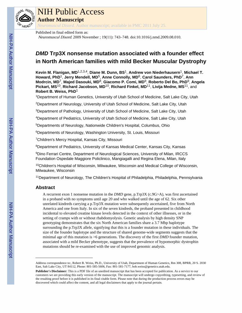
DMD Trp3X nonsense mutation associated with a founder effect in North American families with mild Becker Muscular Dystrophy Kevin M. Flanigan, MD 1,2,3,4 , Diane M. Dunn, BS 1 , Andrew von Niederhausern 1 , Michael T. Howard, PhD 1 , Jerry Mendell, MD 5 , Anne Connolly, MD 6 , Carol Saunders, PhD 7 , Ann Modrcin, MD 7 , Majed Dasouki, MD 8 , Giacomo P. Comi, MD 9 , Roberto Del Bo, PhD 9 , Angela Pickart, MS 10 , Richard Jacobson, MD 10 , Richard Finkel, MD 11 , Livija Medne, MS 11 , and Robert B. Weiss, PhD 1 1 Department of Human Genetics, University of Utah School of Medicine, Salt Lake City, Utah 2 Department of Neurology, University of Utah School of Medicine, Salt Lake City, Utah 3 Department of Pathology, University of Utah School of Medicine, Salt Lake City, Utah 4 Department of Pediatrics, University of Utah School of Medicine, Salt Lake City, Utah 5 Departments of Neurology, Nationwide Children's Hospital, Columbus, Ohio 6 Departments of Neurology, Washington University, St. Louis, Missouri 7 Children's Mercy Hospital, Kansas City, Missouri 8 Department of Pediatrics, University of Kansas Medical Center, Kansas City, Kansas 9 Dino Ferrari Centre, Department of Neurological Sciences, University of Milan, IRCCS Foundation Ospedale Maggiore Policlinico, Mangiagalli and Regina Elena, Milan, Italy 10 Children's Hospital of Wisconsin, Milwaukee, Wisconsin and Medical College of Wisconsin, Milwaukee, Wisconsin 11 Department of Neurology, The Children's Hospital of Philadelphia, Philadelphia, Pennsylvania Abstract A recurrent exon 1 nonsense mutation in the DMD gene, p.Trp3X (c.9G>A), was first ascertained in a proband with no symptoms until age 20 and who walked until the age of 62. Six other unrelated kindreds carrying a p.Trp3X mutation were subsequently ascertained, five from North America and one from Italy. In six of the seven kindreds, the proband presented in childhood incidental to elevated creatine kinase levels detected in the context of other illnesses, or in the setting of cramps with or without rhabdomyolysis. Genetic analysis by high density SNP genotyping demonstrates that the six North American families share a 3.7 Mbp haplotype surrounding the p.Trp3X allele, signifying that this is a founder mutation in these individuals. The size of the founder haplotype and the structure of shared genome-wide segments suggests that the minimal age of this mutation is >6 generations. The discovery of the first DMD founder mutation, associated with a mild Becker phenotype, suggests that the prevalence of hypomorphic dystrophin mutations should be re-examined with the use of improved genomic analysis. Address correspondence to:, Robert B. Weiss, Ph.D., University of Utah, Department of Human Genetics, Rm 308, BPRB, 20 S. 2030 East, Salt Lake City, UT 84112, Phone: 801-585-5606, Fax: 801-581-7177, [email protected]. Publisher's Disclaimer: This is a PDF file of an unedited manuscript that has been accepted for publication. As a service to our customers we are providing this early version of the manuscript. The manuscript will undergo copyediting, typesetting, and review of the resulting proof before it is published in its final citable form. Please note that during the production process errors may be discovered which could affect the content, and all legal disclaimers that apply to the journal pertain. NIH Public Access Author Manuscript Neuromuscul Disord. Author manuscript; available in PMC 2011 July 25. Published in final edited form as: Neuromuscul Disord. 2009 November ; 19(11): 743–748. doi:10.1016/j.nmd.2009.08.010. NIH-PA Author Manuscript NIH-PA Author Manuscript NIH-PA Author Manuscript
Transcript of DMD Trp3X nonsense mutation associated with a founder effect in North American families with mild...
DMD Trp3X nonsense mutation associated with a founder effectin North American families with mild Becker Muscular Dystrophy
Kevin M. Flanigan, MD1,2,3,4, Diane M. Dunn, BS1, Andrew von Niederhausern1, Michael T.Howard, PhD1, Jerry Mendell, MD5, Anne Connolly, MD6, Carol Saunders, PhD7, AnnModrcin, MD7, Majed Dasouki, MD8, Giacomo P. Comi, MD9, Roberto Del Bo, PhD9, AngelaPickart, MS10, Richard Jacobson, MD10, Richard Finkel, MD11, Livija Medne, MS11, andRobert B. Weiss, PhD1
1Department of Human Genetics, University of Utah School of Medicine, Salt Lake City, Utah2Department of Neurology, University of Utah School of Medicine, Salt Lake City, Utah3Department of Pathology, University of Utah School of Medicine, Salt Lake City, Utah4Department of Pediatrics, University of Utah School of Medicine, Salt Lake City, Utah5Departments of Neurology, Nationwide Children's Hospital, Columbus, Ohio6Departments of Neurology, Washington University, St. Louis, Missouri7Children's Mercy Hospital, Kansas City, Missouri8Department of Pediatrics, University of Kansas Medical Center, Kansas City, Kansas9Dino Ferrari Centre, Department of Neurological Sciences, University of Milan, IRCCSFoundation Ospedale Maggiore Policlinico, Mangiagalli and Regina Elena, Milan, Italy10Children's Hospital of Wisconsin, Milwaukee, Wisconsin and Medical College of Wisconsin,Milwaukee, Wisconsin11Department of Neurology, The Children's Hospital of Philadelphia, Philadelphia, Pennsylvania
AbstractA recurrent exon 1 nonsense mutation in the DMD gene, p.Trp3X (c.9G>A), was first ascertainedin a proband with no symptoms until age 20 and who walked until the age of 62. Six otherunrelated kindreds carrying a p.Trp3X mutation were subsequently ascertained, five from NorthAmerica and one from Italy. In six of the seven kindreds, the proband presented in childhoodincidental to elevated creatine kinase levels detected in the context of other illnesses, or in thesetting of cramps with or without rhabdomyolysis. Genetic analysis by high density SNPgenotyping demonstrates that the six North American families share a 3.7 Mbp haplotypesurrounding the p.Trp3X allele, signifying that this is a founder mutation in these individuals. Thesize of the founder haplotype and the structure of shared genome-wide segments suggests that theminimal age of this mutation is >6 generations. The discovery of the first DMD founder mutation,associated with a mild Becker phenotype, suggests that the prevalence of hypomorphic dystrophinmutations should be re-examined with the use of improved genomic analysis.
Address correspondence to:, Robert B. Weiss, Ph.D., University of Utah, Department of Human Genetics, Rm 308, BPRB, 20 S. 2030East, Salt Lake City, UT 84112, Phone: 801-585-5606, Fax: 801-581-7177, [email protected]'s Disclaimer: This is a PDF file of an unedited manuscript that has been accepted for publication. As a service to ourcustomers we are providing this early version of the manuscript. The manuscript will undergo copyediting, typesetting, and review ofthe resulting proof before it is published in its final citable form. Please note that during the production process errors may bediscovered which could affect the content, and all legal disclaimers that apply to the journal pertain.
NIH Public AccessAuthor ManuscriptNeuromuscul Disord. Author manuscript; available in PMC 2011 July 25.
Published in final edited form as:Neuromuscul Disord. 2009 November ; 19(11): 743–748. doi:10.1016/j.nmd.2009.08.010.
NIH
-PA Author Manuscript
NIH
-PA Author Manuscript
NIH
-PA Author Manuscript

KeywordsDMD; Duchenne muscular dystrophy; Becker muscular dystrophy; founder allele
1. IntroductionThe Duchenne (DMD [MIM 310200]) and Becker (BMD [MIM 300376]) forms ofmuscular dystrophy are the most common inherited disorders of muscle. These allelic X-linked recessive disorders are both caused by loss-of-function mutations in the 79 exon, 2.2Mbp dystrophin gene (MIM 300377). DMD is a severe childhood myopathy with anestimated incidence of 1:3,500 male births, from which patients suffer loss of ambulationbefore the age of 12 years and often die in their early 20's of cardiac or respiratory failure.BMD is a clinically similar but less severe form affecting 1:12,000 male births [1], and theseverity of BMD patients is more variable than that of DMD patients, ranging from patientswho are wheelchair-dependent at the age of 16 years, to those who remain asymptomaticuntil the fifth or sixth decade of life [2]. Asymptomatic X-linked hyperCKemia has alsobeen described as a dystrophinopathy phenotype [3], although it can be viewed as themildest form of a BMD phenotype. The high prevalence and clinical variability of bothDMD and BMD reflect the mutation rate of the dystrophin gene due to its extreme size andthe correlation between loss-of-function alleles, residual dystophin protein levels andclinical phenotypes. In DMD patients, the dystrophin Dp427m protein isoform is typicallyabsent in muscle, while in BMD patients, dystrophin protein is present but abnormal inquantity or size [4,5]. This difference in residual protein levels is a consequence of the DMDmutational spectrum, which is characterized by a near complete absence of missensemutations, despite the large target size (Dp427m, 3685 amino acids) versus an abundance ofdeletion mutations of one or more exons (60-65% DMD phenotypes), truncating pointmutations (25-30% DMD phenotypes) and duplications of one or more exons (5-10% DMDphenotypes) [6].
The BMD mutation spectrum is less well characterized, but generally conforms to the‘reading-frame rule’ hypothesis where this class of mutations leads to the generation ofinternally deleted, but partly functional dystrophins [7]. The internally deleted dystrophinsproduced from ‘in-frame’ deletion mutations in patients with BMD conforming to thereading-frame rule lack portions of the middle rod domain formed by 24 spectrin-likerepeats. BMD patients with exceptions to this reading-frame rule include those with ‘out-of-frame’ deletions in the 5′ region of the dystrophin gene, such as exon 3-7 deletions. Patientswith this mutation show the entire range of clinical severity from BMD to DMD [8-11].Numerous molecular mechanisms, such as cryptic promoter utilization, ribosomalframeshifting, and translation reinitiation, have been suggested as having a role in restoringthe reading frame for these exceptional classes [12,13].
We have previously reported a patient with BMD (42970) with a muscle isoform exon 1nonsense mutation (c.9G>A, p.Trp3X) who was ascertained from a large cohort screen forpoint mutations by DNA sequencing [14]. The molecular mechanism underlying residualdystrophin expression in patients with this mutation (and other exon 1 mutations that disruptthe reading frame) has been shown to result from initiation of translation at two exon 6 AUGcodons [15], located between the tandem calponin-homology sub-domains (CH1 and CH2)within the N-terminal actin-binding domain (ABD, residues 1-246). These results mayexplain how mutations in the first exons of the DMD gene are ameliorated by internalinitiation, and suggest that dystrophin with a single N-terminal CH2 domain has significantfunction. Here, we investigate whether the recurrent c.9G>A, p.Trp3X mutations ascertained
Flanigan et al. Page 2
Neuromuscul Disord. Author manuscript; available in PMC 2011 July 25.
NIH
-PA Author Manuscript
NIH
-PA Author Manuscript
NIH
-PA Author Manuscript
independently in seven unrelated families are due to independent mutations or are the resultof a single founder mutation derived from a recent common ancestor.
2. Patients and Methods2.1 Families included for study
The probands of families I, II, and III were enrolled within the United DystrophinopathyProject (UDP), a large multi-center natural history and genotype/phenotype study(http://dystrophy.genetics.utah.edu). Families IV, V and VI were originally identified byprimary clinicians and subsequently enrolled in the UDP, and family VII was identified atthe Dino Ferrari Centre, Department of Neurological Sciences, University of Milan. Thisstudy was approved by the Institutional Review Board of the University of Utah and by theI.R.S. of the “I.R.C.C.S. Foundation Ospedale Maggiore Policlinico, Mangiagalli andRegina Elena, Milan, Italy”. All DNA samples were purified from peripheral blood drawnafter written informed consent had been obtained.
2.2 Genetic AnalysisThe c.9G>A, p.Trp3X (NM_004006) mutation was identified or confirmed in each patientby DNA sequencing using an established sequencing protocol [14]. Genome-wide SNPanalysis used standard protocols for the Affymetrix GeneChip® Human Mapping 250K Nspassay (Santa Clara, CA, USA), which has been optimized to detect 262,000 SNPs in eachsample. Genomic DNA from each male patient I-2 (41934), II-1 (43043), II-2 (43640), III-1(43800), IV-1 (43676), V-2 (43738), and V-3 (43741), VI-1 (43889), and VII (DR0007) wasdigested with the restriction enzyme Nsp I. Adaptor ligation, single-primer PCRamplification, fragmentation, labeling and hybridization were performed according to themanufacturers' protocols, and the genotypes were called from probe intensities using theBRLMM algorithm. Genomic DNA from III-1 (43800), VI-1 (43889) and VII (DR0007)were also genotyped on the Affymetrix Genome-Wide Human SNP Array 6.0 whichincludes more than 906,600 SNPs.
PLINK [16] was used to filter the SNP dataset for missing genotypes and to find extendedstretches of homozygosity in the X chromosome data and in the whole genome allowing fora certain amount of missing genotypes and occasional heterozygote calls. We used thePLINK method of moments approach to estimate the probability of sharing 0, 1 or 2 allelesIBD for any two individuals. The HHanalysis program [17] was modified to include the Xchromosome along with the default analysis of autosomes. The type A and the type B falsepositive rate was calculated using the patients' genotype data and regions with conservedhomozygosity haplotypes (RCHH) cutoff value of 6.0 cM was chosen as optimal, althoughthe results were robust for RCHH values from 3.0 to 8.0 cM. The control pool forcalculating P values for segmental homozygous haplotype sharing consisted of 53 unrelatedEuropean-American samples.
3. Results3.1 p.Trp3X kindreds
The clinical features of the probands and affected male relatives (n=12) from all sevenkindreds are summarized in Table 1, and more detailed versions of the clinical histories arecontained in the Supplemental Data. As noted in Table 1, in six of the seven families(families II – VII) the probands ranged from age 4 to age 16 when they were last examined;all had presented with elevated serum CK (often incidentally detected), post-exertionalmyalgias, or a combination of the two. Six of the nine males examined at age 16 or younger
Flanigan et al. Page 3
Neuromuscul Disord. Author manuscript; available in PMC 2011 July 25.
NIH
-PA Author Manuscript
NIH
-PA Author Manuscript
NIH
-PA Author Manuscript

(ages 2 – 16) showed no weakness; the remaining three showed only trace to mild proximalweakness.
Three older patients suggest that the clinical course associated with the mutation remainsmild. The proband in family I (patient 42970; ascertained in Utah) is now 67 years old. Hefirst noted weakness at age 20 years, when he was running an obstacle course in the Navy,and was unable to do it in the allotted time. By age 28, he was unable to walk up a flight ofstairs while carrying a box of supplies. His weakness progressed through adulthood;although he walked with assistive devices, he never used a wheelchair until the age of 62years, when he became functionally non-ambulant. His younger brother (patient 43194) wasexamined by one of us (KMF) at age 58 years. At that time, although he noted occasionalnocturnal post-exertional muscle cramps, his examination revealed only trace hip flexor andextensor weakness; he has never used a wheelchair. Similarly, the maternal grandfather ofthe proband in family VI (patient 43910) also carries the mutation, and at age 73 denies anycurrent symptoms but recalls severe post-exertional myalgias in childhood; his examinationshows a mild Trendelenberg gait but no weakness to manual muscle testing.Echocardiogram was performed in patient 42970 and patient 43910; both are normal.
3.2 X chromosome allele sharingWe were unable to establish a genealogical relationship between any of the families, whoare all of European-American or European heritage. Although not definitive, more extensivegenealogic research carried out by members of families I and II demonstrated no commonancestors between them. To determine whether the p.Trp3X mutation represents a founderallele, we used Affymetrix 250K NspI mapping arrays to detect the pattern of SNP markersharing throughout the genome and at the DMD gene. If the p.Trp3X mutation resulted froma single event in a common ancestor of the seven families, we would expect identical bydescent (IBD) allele sharing of the surrounding chromosomal region. The fine structure ofSNP allele sharing in the DMD region is shown in Figure 1, where the 5′ end of the DMDgene overlaps the telomeric end of a shared IBD segment in 6 out of 7 families. This sharedsegment begins in intron 2 of the Dp427m transcript isoform and extends 3.7 Mbp towardsthe centromere. The c.9G>A, p.Trp3X mutation is contained within this IBD block and themost parsimonious conclusion is that this mutation is derived from a single event in a distantcommon ancestor of the North American families I through VI, while family VII of Italianorigin does not share the haplotype block surrounding the mutation (Figure 1) and thereforerepresents an independent event.
The number of shared contiguous SNP blocks on the X chromosome that are identical bystate (IBS) or identical by descent (IBD) between individuals from the six North Americanfamilies is 1,351 (average block size = 67 kb), indicating that recombination has reduced thesegmental sharing to the range expected for common “haplotype blocks” separated byrecombination hotspots [18]. One of the two largest IBS/IBD blocks overlaps the DMD gene(3.7 Mbp, 195 SNP markers) and the other spans the centromere (8.9 Mbp, 66 SNPmarkers). The actual size of centromere-spanning block contains significant uncertainty dueto sparse SNP density surrounding this region and the padding of a 3.0 Mbp centromeric gapof “unfinished” sequence. Conversion of distance units from megabases to centimorgansusing sex-averaged recombination rates from the genetic map [19] reverses the apparentblock size (3.7 Mbp = 6.7 cM versus 8.8 Mbp = 0.9 cM); thus, the p.Trp3X IBD blockoverlapping the DMD gene is the largest euchromatic X chromosomal segment shared bythe six North American families.
Flanigan et al. Page 4
Neuromuscul Disord. Author manuscript; available in PMC 2011 July 25.
NIH
-PA Author Manuscript
NIH
-PA Author Manuscript
NIH
-PA Author Manuscript
3.3 Empirical detection of ancestryInformation on the age of origin for the p.Trp3X mutation can be derived from the length ofthe chromosomal region in linkage disequilibrium (LD) and the strength of the LD.However, applying direct methods for estimating allele age, such as those described byRisch et al. [20] or Stephens et al. [21], on the small number of p.Trp3X chromosomesascertained here without a wider estimate of the population frequency of p.Trp3X wouldplace a wide error margin on these estimates. These methods also rely on estimates of theintensity of past selection, presumably weakly negative. It has been suggested that even withdetailed knowledge of genetic and demographic parameters, precise estimates of allele ageand selection intensity cannot be accurately calculated because of the stochastic nature ofrecombination and genetic drift [22].
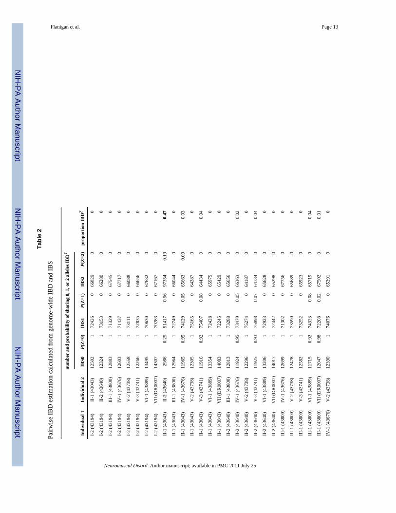
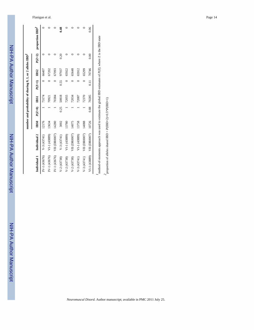
As an alternative to estimating the allele age by LD methods, we used genome-wide metricsto evaluate the genetic relatedness among the seven patients. The size distribution of sharedX chromosome IBD segments and the length of the p.Trp3X DMD IBD block are consistentwith an allele age >6 generations old, leading to a testable prediction of distant geneticrelatedness by estimation of genome-wide identity-by-state and identity-by-descentprobabilities for all pairs of study participants. We estimated the cryptic relatedness betweenstudy participants based on sharing of genotypes measured with Affymetrix 250K NspIarrays by estimating identity by descent (IBD) using the PLINK software [16]. Table 2shows the genome-wide probabilities of sharing 0, 1, or 2 alleles IBD for any two patientsfrom the seven p.Trp3X families, including the non-founder family VII of Italian origin. Asexpected, the two pairs of full sibs (family II: 43043 vs. 43640 and family V: 43738 vs.43741) show the estimated proportion of alleles shared IBD at ∼0.5. That only several pairsof individual from different families show values ranging from 0.01 to 0.06 for theproportion of alleles shared IBD supports the conclusion that these families are onlydistantly related.
A more direct genome-wide test that searches for shared chromosomal segments derivedfrom a common ancestor is homozygosity haplotype (HH) analysis. This method hassuccessfully been applied in mapping single disease gene locations using high density SNPdata [17] from a small number of patients when certain conditions are met. Application ofthis technique to the p.Trp3X mutation relies on the assumption that a common ancestorbrought the disease gene into this patient population within the last several hundred years (6to 30 generations ago) and the technique is also robust to an admixture of founder and non-founder alleles in the patient pool. Figure 2 shows the result of HH analysis usingAffymetrix 250K NspI mapping data from one member of each of the seven p.Trp3Xfamilies versus 53 unrelated controls drawn from the same study population. HH analysisrobustly detected the correct location of the DMD gene and the p.Trp3X mutation with a−log10(P) value of 10.01. Removing the non-founder Trp3X patient from family VIIincreases the −log10(P) value to 11.01 supporting robustness to admixture in the patientpool. Given that HH analysis works optimally when 6 ≤ m + n ≤ 30, where m and n are thegenerations away from the common ancestor, this genome-wide analysis suggests that thep.Trp3X has been inherited from a distant common ancestor.
4. DiscussionOur report highlights the fact that BMD founder mutations in the DMD gene may exist inlarge and diverse populations. This is the first such founder mutation reported in the DMDgene, but analogous recent founder mutations have been reported in autosomal dominantforms of hereditary colon cancer syndromes. Of note is a founder mutation in MSH2 thatdates back to the 1700's and has been observed in 9 independently ascertained Americanfamilies across the USA with hereditary nonpolyposis colorectal cancer (HNPCC). The
Flanigan et al. Page 5
Neuromuscul Disord. Author manuscript; available in PMC 2011 July 25.
NIH
-PA Author Manuscript
NIH
-PA Author Manuscript
NIH
-PA Author Manuscript

geographic distribution and ascertainment frequency suggests that this mutation mayaccount for a significant proportion of HNPCC cases in the white USA population [23].Also, a recent founder mutation embedded in a 6.7 Mbp haplotype surrounding the APCgene has been found in attenuated form of familial adenomatous polyposis [24]. Throughgenealogical analysis of two large kindreds, this APC mutation has been traced back to afounding couple in the early 1600s, and the mutation has been independently ascertained in13 other families across the US population. The conserved haplotype surrounding thep.Trp3X mutation and the independent ascertainment of the six BMD families analyzed heresuggest that this founder mutation may be similar in frequency and demographic distributionto those found in the late onset colorectal cancer syndromes.
The availability of comprehensive mutation analysis of the dystrophin gene from leukocyte-derived DNA samples has improved the diagnosis and genetic counseling ofdystrophinopathies. For Duchenne muscular dystrophy, the observation that 1/3 of sporadicmutations occur in the maternal germ line or ovum supports the lethality of the phenotype asexpected from Haldane's theory of the balance between selection and mutation in X-linkedlethals. Even deleterious X-linked mutations persist for only a small number of generationsand affect only a small number of people [25]. Our finding of a founder mutation for mildBecker muscular dystrophy in the US population suggests that the observed frequency maybe due to genetic drift and demographic effects, such as population expansion. Using ‘long-range haplotype’ methods, it has been recently demonstrated that a signal of positiveselection resides near DMD intron 12 (SNP rs808540) in the HapMap African sample [26].Prior evidence from a sampling a 2.4 kb segment in DMD intron 7 indicated positiveselection in a small number of individuals from Africa [27]. Although the findings presentedhere are the result of ancestral haplotype sharing surrounding a nonsense mutation resultingin a mild BMD phenotype, it is possible that the N-terminal region of human dystrophin isstill experiencing positive selection based on its actin-binding properties, perhaps through aheterozygote advantage. Internal initiation at exon 6 AUG124 leads to a single N-terminalCH2 domain instead of the typical tandem CH1-CH2 high-affinity actin-binding domain,perhaps contributing to alternate modes of actin-binding activity by dystrophin.
The p.Trp3X mutation is one example of the general class of dystrophin mutations that leadto partial activity through residual dystrophin protein levels. Further characterization of thephenotypes and population prevalence of mutations exemplified by p.Trp3X may helpfurther elucidate the function of dystrophin in muscular and neuronal tissues. With improvedmutation surveillance, we can anticipate that similar alleles may be found at appreciablepopulation frequencies and may be important in the overall burden of skeletal and cardiacphenotypes associated with the dystrophinopathies.
Supplementary MaterialRefer to Web version on PubMed Central for supplementary material.
AcknowledgmentsThe authors wish to thank O. Gurvich and T. Tuohy for discussions about the mechanism of p.Trp3X amelioration,A. Bringard and J. Tyce for administrative assistance, and to acknowledge the study coordinator assistance of K.Hart, C. Moural, and K. Hak and the technical assistance of E. Meenan, A. Aoyagi, B. Duval, C. Hamil and M.Mahmoud. This work is supported by the National Institute of Neurologic Diseases and Stroke (R01 NS043264[KMF, MTH, RBW]; the National Center for Research Resources (M01-RR00064, to the University of Utah, Dr. L.Betz, P.I.); by the Association Francaise Contre les Myopathies (KMF); by the Jett Foundation; and by the ParentProject Muscular Dystrophy.
Flanigan et al. Page 6
Neuromuscul Disord. Author manuscript; available in PMC 2011 July 25.
NIH
-PA Author Manuscript
NIH
-PA Author Manuscript
NIH
-PA Author Manuscript
References1. Emery AE. The muscular dystrophies. Lancet. 2002; 359(9307):687–95. [PubMed: 11879882]2. Bushby KM, Gardner-Medwin D. The clinical, genetic and dystrophin characteristics of Becker
muscular dystrophy. I. Natural history. J Neurol. 1993; 240(2):98–104. [PubMed: 8437027]3. Saengpattrachai M, Ray PN, Hawkins CE, Berzen A, Banwell BL. Grandpa and I have
dystrophinopathy?: approach to asymptomatic hyperCKemia. Pediatr Neurol. 2006; 35(2):145–9.[PubMed: 16876015]
4. Tuffery-Giraud S, Saquet C, Thorel D, Disset A, Rivier F, Malcolm S, et al. Mutation spectrumleading to an attenuated phenotype in dystrophinopathies. Eur J Hum Genet. 2005; 13(12):1254–60.[PubMed: 16077730]
5. Deburgrave N, Daoud F, Llense S, Barbot JC, Recan D, Peccate C, et al. Protein-and mRNA-basedphenotype-genotype correlations in DMD/BMD with point mutations and molecular basis for BMDwith nonsense and frameshift mutations in the DMD gene. Hum Mutat. 2007; 28(2):183–95.[PubMed: 17041906]
6. Dent KM, Dunn DM, von Niederhausern AC, Aoyagi AT, Kerr L, Bromberg MB, et al. Improvedmolecular diagnosis of dystrophinopathies in an unselected clinical cohort. Am J Med Genet A.2005; 134(3):295–8. [PubMed: 15723292]
7. Monaco AP, Bertelson CJ, Liechti-Gallati S, Moser H, Kunkel LM. An explanation for thephenotypic differences between patients bearing partial deletions of the DMD locus. Genomics.1988; 2(1):90–5. [PubMed: 3384440]
8. Malhotra SB, Hart KA, Klamut HJ, Thomas NS, Bodrug SE, Burghes AH, et al. Frame-shiftdeletions in patients with Duchenne and Becker muscular dystrophy. Science. 1988; 242(4879):755–9. [PubMed: 3055295]
9. Chelly J, Gilgenkrantz H, Lambert M, Hamard G, Chafey P, Recan D, et al. Effect of dystrophingene deletions on mRNA levels and processing in Duchenne and Becker muscular dystrophies.Cell. 1990; 63(6):1239–48. [PubMed: 2261642]
10. Winnard AV, Klein CJ, Coovert DD, Prior T, Papp A, Snyder P, et al. Characterization oftranslational frame exception patients in Duchenne/Becker muscular dystrophy. Hum Mol Genet.1993; 2(6):737–44. [PubMed: 8353493]
11. Muntoni F, Gobbi P, Sewry C, Sherratt T, Taylor J, Sandhu SK, et al. Deletions in the 5′ region ofdystrophin and resulting phenotypes. J Med Genet. 1994; 31(11):843–7. [PubMed: 7853367]
12. Gangopadhyay SB, Sherratt TG, Heckmatt JZ, Dubowitz V, Miller G, Shokeir M, et al. Dystrophinin frameshift deletion patients with Becker muscular dystrophy. Am J Hum Genet. 1992; 51(3):562–70. [PubMed: 1496988]
13. Winnard AV, Mendell JR, Prior TW, Florence J, Burghes AH. Frameshift deletions of exons 3-7and revertant fibers in Duchenne muscular dystrophy: mechanisms of dystrophin production. Am JHum Genet. 1995; 56(1):158–66. [PubMed: 7825572]
14. Flanigan KM, von Niederhausern A, Dunn DM, Alder J, Mendell JR, Weiss RB. Rapid directsequence analysis of the dystrophin gene. Am J Hum Genet. 2003; 72(4):931–9. [PubMed:12632325]
15. Gurvich OL, Maiti B, Weiss RB, Aggarwal G, Howard MT, Flanigan KM. DMD exon 1 truncatingpoint mutations: amelioration of phenotype by alternative translation initiation in exon 6. HumMutat. 2009; 30(4):633–40. [PubMed: 19206170]
16. Purcell S, Neale B, Todd-Brown K, Thomas L, Ferreira MA, Bender D, et al. PLINK: a tool set forwhole-genome association and population-based linkage analyses. Am J Hum Genet. 2007; 81(3):559–75. [PubMed: 17701901]
17. Miyazawa H, Kato M, Awata T, Kohda M, Iwasa H, Koyama N, et al. Homozygosity haplotypeallows a genomewide search for the autosomal segments shared among patients. Am J Hum Genet.2007; 80(6):1090–102. [PubMed: 17503327]
18. Gabriel SB, Schaffner SF, Nguyen H, Moore JM, Roy J, Blumenstiel B, et al. The structure ofhaplotype blocks in the human genome. Science. 2002; 296(5576):2225–9. [PubMed: 12029063]
Flanigan et al. Page 7
Neuromuscul Disord. Author manuscript; available in PMC 2011 July 25.
NIH
-PA Author Manuscript
NIH
-PA Author Manuscript
NIH
-PA Author Manuscript

19. Kong A, Gudbjartsson DF, Sainz J, Jonsdottir GM, Gudjonsson SA, Richardsson B, et al. A high-resolution recombination map of the human genome. Nat Genet. 2002; 31(3):241–7. [PubMed:12053178]
20. Risch N, de Leon D, Ozelius L, Kramer P, Almasy L, Singer B, et al. Genetic analysis ofidiopathic torsion dystonia in Ashkenazi Jews and their recent descent from a small founderpopulation. Nat Genet. 1995; 9(2):152–9. [PubMed: 7719342]
21. Stephens JC, Reich DE, Goldstein DB, Shin HD, Smith MW, Carrington M, et al. Dating theorigin of the CCR5-Delta32 AIDS-resistance allele by the coalescence of haplotypes. Am J HumGenet. 1998; 62(6):1507–15. [PubMed: 9585595]
22. Slatkin M. A Bayesian method for jointly estimating allele age and selection intensity. Genet Res.2008; 90(1):129–37.
23. Lynch HT, Coronel SM, Okimoto R, Hampel H, Sweet K, Lynch JF, et al. A founder mutation ofthe MSH2 gene and hereditary nonpolyposis colorectal cancer in the United States. Jama. 2004;291(6):718–24. [PubMed: 14871915]
24. Neklason DW, Stevens J, Boucher KM, Kerber RA, Matsunami N, Barlow J, et al. Americanfounder mutation for attenuated familial adenomatous polyposis. Clin Gastroenterol Hepatol.2008; 6(1):46–52. [PubMed: 18063416]
25. Lange K, Gladstien K, Zatz M. Effects of reproductive compensation and genetic drift on X-linkedlethals. Am J Hum Genet. 1978; 30(2):180–9. [PubMed: 655165]
26. Sabeti PC, Varilly P, Fry B, Lohmueller J, Hostetter E, Cotsapas C, et al. Genome-wide detectionand characterization of positive selection in human populations. Nature. 2007; 449(7164):913–8.[PubMed: 17943131]
27. Nachman MW, Crowell SL. Contrasting evolutionary histories of two introns of the duchennemuscular dystrophy gene, DMD, in humans. Genetics. 2000; 155(4):1855–64. [PubMed:10924480]
Flanigan et al. Page 8
Neuromuscul Disord. Author manuscript; available in PMC 2011 July 25.
NIH
-PA Author Manuscript
NIH
-PA Author Manuscript
NIH
-PA Author Manuscript
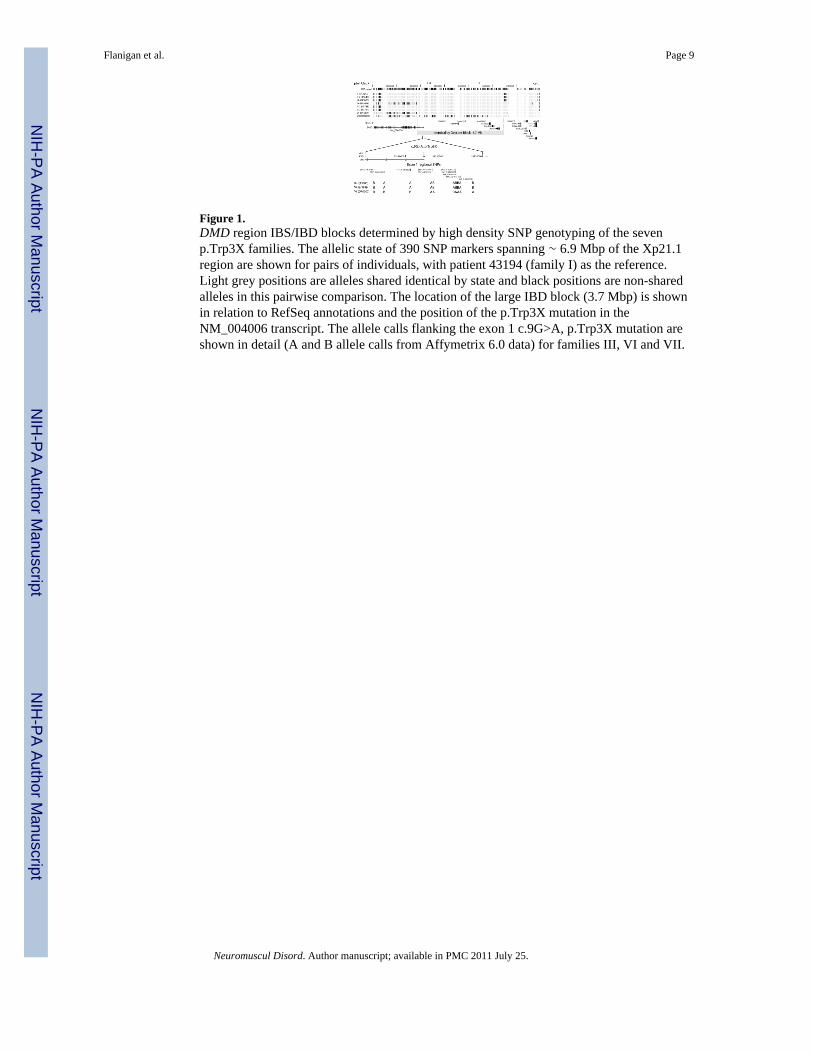
Figure 1.DMD region IBS/IBD blocks determined by high density SNP genotyping of the sevenp.Trp3X families. The allelic state of 390 SNP markers spanning ∼ 6.9 Mbp of the Xp21.1region are shown for pairs of individuals, with patient 43194 (family I) as the reference.Light grey positions are alleles shared identical by state and black positions are non-sharedalleles in this pairwise comparison. The location of the large IBD block (3.7 Mbp) is shownin relation to RefSeq annotations and the position of the p.Trp3X mutation in theNM_004006 transcript. The allele calls flanking the exon 1 c.9G>A, p.Trp3X mutation areshown in detail (A and B allele calls from Affymetrix 6.0 data) for families III, VI and VII.
Flanigan et al. Page 9
Neuromuscul Disord. Author manuscript; available in PMC 2011 July 25.
NIH
-PA Author Manuscript
NIH
-PA Author Manuscript
NIH
-PA Author Manuscript
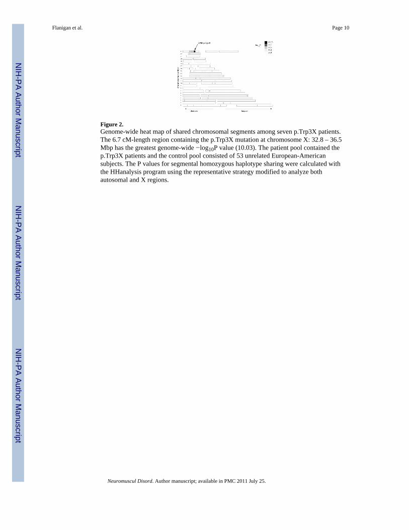
Figure 2.Genome-wide heat map of shared chromosomal segments among seven p.Trp3X patients.The 6.7 cM-length region containing the p.Trp3X mutation at chromosome X: 32.8 – 36.5Mbp has the greatest genome-wide −log10P value (10.03). The patient pool contained thep.Trp3X patients and the control pool consisted of 53 unrelated European-Americansubjects. The P values for segmental homozygous haplotype sharing were calculated withthe HHanalysis program using the representative strategy modified to analyze bothautosomal and X regions.
Flanigan et al. Page 10
Neuromuscul Disord. Author manuscript; available in PMC 2011 July 25.
NIH
-PA Author Manuscript
NIH
-PA Author Manuscript
NIH
-PA Author Manuscript

NIH
-PA Author Manuscript
NIH
-PA Author Manuscript
NIH
-PA Author Manuscript
Flanigan et al. Page 11
Tabl
e 1
Sum
mar
y of
Trp
3X p
atie
nt d
ata
in th
e se
ven
affe
cted
fam
ilies
.
Patie
nts,
fam
ilyPr
oban
d/re
lativ
eG
eogr
aphi
cal o
rigi
nA
ge a
t pre
sent
atio
nPr
esen
ting
com
plai
nt
Cur
rent
degr
ee o
fw
eakn
ess
(age
at
exam
, yrs
)D
ystr
ophi
n im
mun
o-st
aini
ngC
K le
vel (
IU/L
)M
ater
nal g
enot
ype
I-1
(429
70)
prob
and
Uta
h20
Wea
knes
s
prof
ound
limb-
gird
lew
eakn
ess
(67)
[Los
s of
ambu
latio
nat
age
62]
n.d.
n.d.
n.d.
I-2
(431
94)
brot
her
Uta
hn/
an/
a
trace
pel
vic
gird
lew
eakn
ess
(age
58)
;de
nies
sym
ptom
svi
a ph
one
(age
61)
n.d.
n.d.
n.d.
II-1
(430
43)
prob
and
Mic
higa
n4
inci
dent
al h
yper
CK
emia
none
(4)
dim
inis
hed
523 ↑
carr
ier
II-2
(436
40)
brot
her
Mic
higa
n2
inci
dent
al h
yper
CK
emia
none
(2)
n.d.
5080
↑ca
rrie
r
III-
1(4
3800
)pr
oban
dM
isso
uri
7B
ilate
ral c
alf p
ain,
pos
t-ex
erci
se; e
leva
ted
CK
trace
delto
idw
eakn
ess
(7)
dim
inis
hed
8000
↑n.
d.
IV-1
(436
76)
prob
and
Mis
sour
i4
inci
dent
al h
yper
CK
emia
Min
imal
delto
idw
eakn
ess,
min
imal
heel
cor
dco
ntra
ctur
es(4
)oc
casi
onal
fibe
rs se
vere
lyre
duce
d45
58 ↑
n.d.
4383
1, V
-1pr
oban
dW
isco
nsin
“chi
ldho
od”
mya
lgia
s, el
evat
ed C
K(c
hild
hood
);rh
abdo
myo
lysi
s (ag
e14
)no
ne (1
4)di
min
ishe
d19
,189
↑ca
rrie
r
V-2
(437
38)
mat
erna
l cou
sin
Wis
cons
in12
post
exer
tiona
l lim
bcr
ampi
ng a
nd“s
tiffn
ess”
none
(16)
n.d.
n.d.
carr
ier
V-3
(437
41)
mat
erna
l cou
sin
Wis
cons
in2.
5in
cide
ntal
hyp
erC
Kem
iano
ne (4
.5)
n.d.
“ele
vate
d”ca
rrie
r
Neuromuscul Disord. Author manuscript; available in PMC 2011 July 25.

NIH
-PA Author Manuscript
NIH
-PA Author Manuscript
NIH
-PA Author Manuscript
Flanigan et al. Page 12
Patie
nts,
fam
ilyPr
oban
d/re
lativ
eG
eogr
aphi
cal o
rigi
nA
ge a
t pre
sent
atio
nPr
esen
ting
com
plai
nt
Cur
rent
degr
ee o
fw
eakn
ess
(age
at
exam
, yrs
)D
ystr
ophi
n im
mun
o-st
aini
ngC
K le
vel (
IU/L
)M
ater
nal g
enot
ype
VI-
1(4
3889
)pr
oban
dPe
nnsy
lvan
ia13
inci
dent
alhy
perC
Kem
ia,
post
exer
tiona
l mya
lgia
none
(14)
n.d.
712 ↑
carr
ier
VI-
2(4
3910
)M
ater
nal g
rand
fath
erPe
nnsy
lvan
ia“c
hild
hood
”se
vere
pos
t-exe
rtion
alm
yalg
ias i
n ch
ildho
odno
ne (7
3)n.
d.n.
d.n.
d.
VII
DR
0007
prob
and
Mila
n, It
aly
6
inci
dent
alhy
perC
Kem
ia; r
unni
ngdi
ffic
ultie
s, fr
eque
ntfa
lling
mild
prox
imal
wea
knes
s(1
2)di
min
ishe
d29
32 ↑
carr
ier
Neuromuscul Disord. Author manuscript; available in PMC 2011 July 25.
NIH
-PA Author Manuscript
NIH
-PA Author Manuscript
NIH
-PA Author Manuscript
Flanigan et al. Page 13
Tabl
e 2
Pairw
ise
IBD
est
imat
ion
calc
ulat
ed fr
om g
enom
e-w
ide
IBD
and
IBS
num
ber
and
prob
abili
ty o
f sha
ring
0, 1
, or
2 al
lele
s IB
D1
Indi
vidu
al 1
Indi
vidu
al 2
IBS0
P(Z
=0)
IBS1
P(Z
=1)
IBS2
P(Z
=2)
prop
ortio
n IB
D2
I-2
(431
94)
II-1
(430
43)
1250
21
7242
60
6682
90
0
I-2
(431
94)
II-2
(436
40)
1232
41
7315
30
6628
00
0
I-2
(431
94)
III-
1 (4
3800
)12
883
171
329
067
545
00
I-2
(431
94)
IV-1
(436
76)
1260
31
7143
70
6771
70
0
I-2
(431
94)
V-2
(437
38)
1255
81
7311
10
6608
80
0
I-2
(431
94)
V-3
(437
41)
1226
61
7283
50
6665
60
0
I-2
(431
94)
VI-
1 (4
3889
)13
495
170
630
067
632
00
I-2
(431
94)
VII
(DR
0007
)14
307
170
283
067
167
00
II-1
(430
43)
II-2
(436
40)
2986
0.25
5141
70.
5697
354
0.19
0.47
II-1
(430
43)
III-
1 (4
3800
)12
964
172
749
066
044
00
II-1
(430
43)
IV-1
(436
76)
1196
50.
9574
129
0.05
6566
30.
000.
03
II-1
(430
43)
V-2
(437
38)
1230
51
7516
50
6428
70
0
II-1
(430
43)
V-3
(437
41)
1191
60.
9275
407
0.08
6443
40
0.04
II-1
(430
43)
VI-
1 (4
3889
)13
354
172
428
065
975
00
II-1
(430
43)
VII
(DR
0007
)14
083
172
245
065
429
00
II-2
(436
40)
III-
1 (4
3800
)12
813
173
288
065
656
00
II-2
(436
40)
IV-1
(436
76)
1192
40.
9573
470
0.05
6636
30
0.02
II-2
(436
40)
V-2
(437
38)
1229
61
7527
40
6418
70
0
II-2
(436
40)
V-3
(437
41)
1192
50.
9375
098
0.07
6473
40
0.04
II-2
(436
40)
VI-
1 (4
3889
)13
206
172
923
065
628
00
II-2
(436
40)
VII
(DR
0007
)14
017
172
442
065
298
00
III-
1 (4
3800
)IV
-1 (4
3676
)12
699
171
302
067
756
00
III-
1 (4
3800
)V
-2 (4
3738
)12
478
173
590
065
689
00
III-
1 (4
3800
)V
-3 (4
3741
)12
582
173
252
065
923
00
III-
1 (4
3800
)V
I-1
(438
89)
1171
50.
9274
323
0.08
6571
90
0.04
III-
1 (4
3800
)V
II (D
R00
07)
1204
70.
9872
208
0.02
6750
20
0.01
IV-1
(436
76)
V-2
(437
38)
1239
01
7407
60
6529
10
0
Neuromuscul Disord. Author manuscript; available in PMC 2011 July 25.
NIH
-PA Author Manuscript
NIH
-PA Author Manuscript
NIH
-PA Author Manuscript
Flanigan et al. Page 14
num
ber
and
prob
abili
ty o
f sha
ring
0, 1
, or
2 al
lele
s IB
D1
Indi
vidu
al 1
Indi
vidu
al 2
IBS0
P(Z
=0)
IBS1
P(Z
=1)
IBS2
P(Z
=2)
prop
ortio
n IB
D2
IV-1
(436
76)
V-3
(437
41)
1217
61
7317
40
6640
70
0
IV-1
(436
76)
VI-
1 (4
3889
)13
634
170
921
067
202
00
IV-1
(436
76)
VII
(DR
0007
)14
280
170
384
067
093
00
V-2
(437
38)
V-3
(437
41)
3002
0.25
5081
80.
5597
937
0.20
0.48
V-2
(437
38)
VI-
1 (4
3889
)13
780
172
055
065
922
00
V-2
(437
38)
VII
(DR
0007
)14
075
172
034
065
648
00
V-3
(437
41)
VI-
1 (4
3889
)13
758
172
087
065
912
00
V-3
(437
41)
VII
(DR
0007
)14
088
172
370
065
299
00
VI-
1 (4
3889
)V
II (D
R00
07)
1072
60.
8870
285
0.11
7074
60.
000.
06
1 met
hod-
of-m
omen
ts a
ppro
ach
was
use
d to
est
imat
e th
e gl
obal
IBD
est
imat
es o
f P(Z
), w
here
Z is
the
IBD
stat
e
2 prop
ortio
n of
alle
les s
hare
d IB
D =
P(I
BD
=2)+
0.5*
P(IB
D=1
)
Neuromuscul Disord. Author manuscript; available in PMC 2011 July 25.