Assessing the informational value of parameter estimates in cognitive models
Upload
khangminh22Category
view
1download
0
Assessing the Value of Fusidic Acid in
Dermatology
Proceedings from LEO Pharma Satellite Symposium, European Academy of Dermatology and Venereology (EADV)
Vienna, 18 May 2007
Guest Editor: Donald Leung

© 2008 Acta Dermato-Venereologica. ISSN 0365-8341 Acta Derm Venereol Suppl 216
Acta Derm Venereol 2008; Suppl 216: 1–39
List of contributors 4
Chairman’s Introduction: Assessing the value of fusidic acid in dermatologyDonald Y. M. Leung 5
Chapter 1. Fusidic acid: a valuable agent for controlling Staphylococcus aureus skin infectionsDimitris Rigopoulos and Georgios Larios 7
Chapter 2. Fusidic acid in skin and soft-tissue infectionsBarry H. Long 14
Chapter 3. The role of Staphylococcus aureus in atopic eczema Donald Y. M. Leung 21
Chapter 4. Antibacterial/steroid combination therapy in infected eczemaAnthony C. Chu 28
Chapter 5. Treatment success factors: diagnostic and treatment choices, and patient educationThomas L. Diepgen 35
Contents

© 2008 Acta Dermato-Venereologica. ISSN 0365-8341Acta Derm Venereol Suppl 216
Acta Derm Venereol 2008; Suppl 216: 4
Dr ANTHONY C. CHU, Department of Dermatology, Hammersmith Hospitals Trust, Hammersmith Campus, Faculty of Medicine, Imperial College, London, W12 0NN, UK. E-mail: [email protected]
Professor THOMAS L. DIEPGEN, Department of Social Medicine, Occupational & Environmental Dermatology, University of Heidelberg, Thibautstrasse. 3, DE-69115 Heidelberg, Germany. E-mail: [email protected] heidelberg.de
Dr GEOrGIOS LArIOS, Department of Dermatology, Andreas Sygros University Hospital, 5 Ionos Dragoumi Str, Gr-16121 Athens, Greece.
Professor DONALD Y. M. LEUNG, National Jewish Medical and research Center, 1400 Jackson Street, room K926, Denver, CO 80206, USA. E-mail: [email protected]
Dr BArrY H. LONG, Division of Dermatology, Women’s College Hospital, 76 Grenville Street, Toronto, ON, Canada M5S 1B2. E-mail: [email protected]
Dr DIMITrIS rIGOPOULOS, Department of Dermatology, Andreas Sygros University Hospital, 5 Ionos Dragoumi Str, Gr-16121 Athens, Greece. E-mail: [email protected]
List of contributors
Speakers at the LEO Symposium (left to right): Dr Tony Chu, Dr Dimitris rigopoulos, Professor Donald Leung, Professor Thomas Diepgen, Dr Barry Long.

© 2008 Acta Dermato-Venereologica. ISSN 0365-8341 Acta Derm Venereol Suppl 216
Acta Derm Venereol 2008; Suppl 216: 5
doi: 10.2340/00015555-0385
This supplement covers the contents of a symposium that was held in Vienna, Austria, on 17 May 2007 at the 16th Congress of the European Academy of Dermatology and Venereology.
Fusidic acid has been a mainstay in the treatment of dermatological infections, particularly those caused by Staphylococcus aureus, for many years. The aim of the symposium was to present a comprehensive account of the characteristics, clinical effectiveness and potential for resistance development of fusidic acid, placed in context with other drugs also used in dermatology. This compilation adds the results of newer studies, not previously re-viewed, to those of older ones. It is timely, in light of the recent launch of a new fusidic acid/steroid combination formulation, namely fusidic acid 2%/betamethasone valerate 0.1% lipid cream (Fucicort® Lipid).
The first article, by Dr D. Rigopoulos, presents a brief overview of the value of fusidic acid for controlling S. aureus. This is followed by a review of the evidence for the clinical efficacy of plain fusidic acid formulations, by Dr B. Long. I then discuss the role of S. aureus in atopic eczema, highlighting the effects of superantigens released by the bacterium. The article by Dr T. Chu reviews the use of antibacterial/steroid combination therapy to combat the “vicious cycle” of dryness of the skin, inflammation and infection that is seen in infected eczema. Finally, Dr T. Diepgen’s paper reports on an interactive session in which case histories formed the basis for a discussion of treatment success factors and the importance of patient education.
The costs of this publication have been covered by a grant from LEO Pharma, who also covered the travel costs of the participants. None of the participating doctors was working as a consultant for LEO Pharma at the time of the symposium. The articles were written by the participants, with editorial assistance from Watermeadow Medical.
September 2007Donald Y. M. Leung
Edelstein Family Chair of Pediatric Allergy-ImmunologyNational Jewish Medical and research Center
Denver, Colorado, USA
Chairman’s Introduction: Assessing the value of fusidic acid in dermatology

Acta Derm Venereol Suppl 216
Key wordsStaphylococcus aureus; fusidic acid; antibiotics; skin infections; treatment choice; impetigo; atopic dermatitis; eczema; antimicrobial peptides; superantigens; infected eczema; antibacterial; inflammation; antibacterial/steroid combination therapy; eczema; eczema school; anti-infective; compliance; patient education.
© 2008 Acta Dermato-Venereologica. ISSN 0365-8341 Acta Derm Venereol Suppl 216
Acta Derm Venereol 2008; Suppl 216: 7–13
doi: 10.2340/00015555-0386
Staphylococcus aureus is a key pathogen in skin and soft-tissue infections, and controlling it is crucial in treating these conditions. The principal antibiotics used for ambulatory treatment of S. aureus skin infections are beta-lactams, macrolides, aminoglycosides, tetra-cyclines, mupirocin and fusidic acid. In choosing an antibiotic, ideally the following characteristics should be met: adequate antibacterial activity and limited spectrum of activity; minimal resistance concerns; attainment of sufficiently high local concentration; minimal side-effects and risk of sensitization; and a choice of different for-mulations. Compared with the other classes, fusidic acid shows exceptionally good skin penetration through both intact and damaged skin, enabling it to reach antibacte-rial concentrations at the site of infection; the incidence of adverse events and allergic reactions to fusidic acid is low, and it is available in a wide choice of formulations. Thus, fusidic acid offers all the properties of an ideal agent to control S. aureus in skin infections.
INTrODUCTION
The genus Staphylococcus includes more than 30 species and subspecies of Gram-positive bacteria. In dermatology, a significant member of this genus is the coagulase-positive S. aureus (from the Latin aurum; “gold”), discovered in Aberdeen, UK, in 1880 by the surgeon Sir Alexander Ogston in pus from surgical abscesses (1). S. aureus is acknowledged as one of the most important bacterial pathogens of humans, causing a variety of syndromes, including superficial and deep pyogenic infections as well as systemic infections.
Skin carriage of coagulase-negative staphylococci is nearly universal, but this is not the case for S. aureus, which is carried in the anterior nares by about 35% of normal individuals. Other sites of resident carriage are the perineum (20%), axillae (10%) and toe-webs (5%) (2). Isolation of S. aureus from these sites does not always indicate infection and therefore does not always require treatment. On normal skin a small number of S. aureus cells might be found, usually considered as a contami-nant derived from a resident carrier site. On the other hand, colonization is commonly observed in more than 90% of patients with atopic eczema (3).
Skin infections with S. aureus can be classified as primary or secondary. The main primary cutaneous infections (on apparently normal skin) are folliculitis, abscesses, furuncles, carbuncles, impetigo and acute paronychia. Folliculitis most often represents endo-
genous infection with S. aureus from a carrier site on the patient’s own body. Furuncles are acute circumscribed, pus-filled nodules that evolve from folliculitis due to S. aureus. Carbuncles are deeper infections, composed of interconnecting abscesses. Impetigo is normally an exogenous infection and may occur in epidemics, as reflected in the term “impetigo contagiosa”. In second-ary cutaneous infections (arising in pre-existing skin disease) such as infected eczema, the causative staphylo-cocci are most often endogenous.
Staphylococci produce disease through their ability to multiply and spread widely in tissues, or through producing extracellular substances such as exotoxins or enzymes. S. aureus produces a range of potential virulence/pathogenicity factors. Some of these, such as the haemolysins, esterases, proteases, protein A and cell wall aggressins, are non-specific. It is probably a cocktail of these that is responsible for the spectrum of furunculosis. Inside abscesses, coagulase is produ-ced from S. aureus and forms a fibrin wall around the lesion that limits the spread. Within the centre of the lesion, liquefaction of necrotic tissue occurs, and the abscess spreads in the direction of least resistance (4). In bullous impetigo, specific serine proteases, such as the exfoliative toxins A and B, cause skin splitting at the stratum granulosum, which results in blisters (5). recently, attention has been focused on the Panton– Valentine leukocidin, which has been epidemiologically associated with severe cutaneous infections (6). Exo-toxins produced by S. aureus, such as superantigens, can cause polyclonal T-cell activation by binding directly to antigen-presenting cells (7). Normal skin presents no barrier to superantigens, which can initiate or exacerbate pre-existing, eczematous lesions (2). Consequently, S. aureus has been shown to be implicated in the patho-genesis of various inflammatory skin diseases, such as atopic eczema (8).
Clearly, controlling S. aureus is crucial in the treat-ment of skin infections in clinical practice. In choosing an antibiotic, adequate antibacterial activity is a pre-requisite. In addition, ideally the following characteristics should be met: a narrow spectrum of activity, minimal resistance concerns, attainment of a sufficiently high local concentration, minimal side-effects (including risk of sensitization), and a choice of different formulations in order to improve compliance.
The principal antibiotics used for ambulatory treat-ment of S. aureus skin infections are the beta-lactams, macrolides, aminoglycosides, tetracyclines, mupirocin
1. Fusidic acid: a valuable agent for controlling Staphylococcus aureus skin infectionsDimitris rIGOPOULOS and Georgios LArIOS
8 D. Rigopoulos and G. Larios
and fusidic acid. This article briefly describes their mechanism of action and reviews each class according to the characteristics mentioned above. Treatment of the rare cases of severe infections is not included in this article, nor is a discussion of clinical efficacy; the efficacy of fusidic acid is covered in other articles in this supplement.
MECHANISM OF ACTION
The beta-lactam antibiotics comprise 3 groups of clini-cally important therapeutic agents: penicillinase-stable penicillins (e.g. flucloxacillin, dicloxacillin, ampicil-lin), cephalosporins and carbapenems. They act by inhibiting bacterial peptidoglycan cell wall synthesis.
The macrolides (e.g. erythromycin, clarithromycin, azithromycin), lincosamides (e.g. clindamycin), and streptogramins (e.g. pristinamycin) all inhibit bacterial protein synthesis by binding to 50S ribosomal subunits of sensitive micro-organisms. These antibiotic classes are often grouped together.
The aminoglycosides (e.g. gentamicin, streptomycin, neomycin), which are generally bactericidal, include agents that bind irreversibly to the 30S ribosomal sub-unit and inhibit bacterial protein synthesis.
Tetracyclines (e.g. oxytetracycline, doxycycline, minocycline) are agents that disrupt the function of 30S ribosomal subunits and interfere with aminoacyl t-rNA, binding to an acceptor site on the messenger rNA ribo-somal complex, to reversibly inhibit protein synthesis.
Mupirocin, a topical antibiotic produced by fermen-tation of Pseudomonas fluorescens inhibits bacterial protein synthesis by reversible binding and inhibition of isoleucyl transfer-rNA synthetase (9).
Fusidic acid belongs to a group of its own, the fusidanes, isolated from culture media of the fungus Fusidium coccineum (10). The molecule has a steroid-like structure, but does not possess any steroid activity (11). Fusidic acid inhibits bacterial protein synthesis by interfering with elongation factor G in the translocation step, the process by which the ribosome moves relative to mrNA (12, 13).
ANTIBACTErIAL SPECTrUM AND BACTErIAL rESISTANCE
When treating cutaneous infections, it is important to choose an agent with an adequate spectrum. However, the spectrum should not be too broad, as the host’s own resident commensal flora of the genitourinary and gastrointestinal tracts could then be affected, possibly leading to superinfection. Also, the development of antibiotic-resistant strains is increased when antibiotics of unnecessarily broad spectrum are used. If the identity of the pathogen is suspected, an appropriate narrow-
spectrum drug with high activity against the pathogen should be preferred as first-line treatment.
Development of resistance towards commonly used antibiotics is an issue of growing concern. As mentioned above, when choosing antibiotics in dermatology, it is important to target the specific bacteria involved in skin le-sions, thus reducing the risk of developing resistance (11). The likelihood of spread and the fitness/competitive advan-tage of the resistant mutant vs. the original strain will vary depending on the resistance mechanism (plasmid-mediated or chromosomal), and therefore the concerns associated with the emergence of resistance will vary too.
Beta-lactams
The beta-lactams are a large group that includes anti-biotics with narrow or broad spectrums of activity, active against Gram-positive or both Gram-positive and Gram-negative bacteria. As about 80% of all S. aureus strains produce penicillinase (14), the penicil-linase-resistant penicillins and cephalosporins are the antibiotics most commonly used to treat methicillin-susceptible S. aureus (MSSA) infections. Methicillin-resistant S. aureus (MrSA) is an increasing problem in many countries. In dermatology outpatients, MrSA is most likely to pose a risk in closed societies (prisons, football teams, etc.) (15). Bacterial resistance against the beta-lactam antibiotics continues to increase at a dramatic rate. Mechanisms of resistance include not only production of beta-lactamases that destroy the antibiotic, but also alterations in or acquisition of novel penicillin-binding proteins and decreased entry and/or active efflux of the antibiotic (16).
Carbapenems (imipenem, meropenem) are extre-mely broad-spectrum, expensive, parenteral beta-lactam antibiotics with good activity against many bacterial species, including MSSA (17), but should be reserved for treatment of life-threatening infections.
Macrolides
The antibacterial spectrum of activity of the macro-lides includes Gram-positive and Gram-negative cocci, chlamydia and mycoplasmas. Surveillance data from a study testing resistance to erythromycin of S. aureus associated with skin and soft tissue infections (SSTI) in the USA and Europe show resistance of approximately 20% in outpatients (14). Due to the common binding site to 50S ribosomal subunits for macrolides, linco-samide and stretogramins, there can be cross-resistance between these 3 classes.
Aminoglycosides
The aminoglycosides are active primarily against Gram-negative aerobic bacteria, but also have some
Acta Derm Venereol Suppl 216

9Fusidic acid for controlling S. aureus skin infections
activity against Gram-positive organisms. Aminogly-cosides used topically in the treatment of SSTI are gen-tamicin and neomycin. Neomycin has only moderate activity against S. aureus and is therefore sometimes used in combination with other agents. There are con-cerns about the development of cross-resistance with other aminoglycosides, including valuable systemic agents used for severe infections (e.g. tobramycin and amikacin).
Tetracyclines
The tetracyclines have a broad spectrum of action, displaying good activity against a wide range of Gram-positive and Gram-negative bacteria, chlamydiae, and rickettsiae. However, their general usefulness has been reduced owing to increasing bacterial resistance. They are not used widely against S. aureus, but, as there is relatively low resistance to these agents among MrSA causing SSTI, they may be used in some countries for this purpose (18).
Mupirocin
Mupirocin has a narrow bacterial spectrum against Gram-positive bacteria such as staphylococci and strepto-cocci. Mupirocin has proven efficient in eradicating nasal colonization in MrSA patients (19). Proper use of topical mupirocin over a long period of time in a hospital setting has not been found to be associated with increased resistance to the drug (20). On the other hand, various outbreaks of clonal and plasmid-medi-ated spread of high-level mupirocin resistance have been reported after prolonged use in patients (21–23). resistance rates averaged 28% in New Zealand in 1999 (23), and a rate of 20% was reported at a US hospital in 2000 (24).
Fusidic acid
Fusidic acid has a narrow antibacterial spectrum, mainly against Gram-positive bacteria; in particular, it has high activity against S. aureus, and has been shown to be active against both MSSA and MrSA isolates (25–27).
No cross-resistance with other antibiotics has been observed, probably due to the unique structure of fusidic acid (26). Surveillance data for fusidic acid resistance across the world are scarce, but the risk of resistance is generally low, although, as for other antibiotics, the resistance level reported depends strongly on the patient population and the geographical area. In Europe, Scandinavia and the UK resistance levels have been above 10% (28–30). This is primarily due the spread of a clone in impetigo patients. Data for Sweden show that the prevalence of the resistant clone peaked in 2002 and has since declined (31). The rest of Europe and
Canada have levels of resistance below 10% (32–35). Several studies have shown that resistance does not develop when fusidic acid is used for up to 2 weeks at a time (36–38).
SKIN PENETrATION/CONCENTrATION AT THE SITE OF INFECTION
The effectiveness of an antibiotic depends on its abi-lity to achieve the minimum inhibitory or bactericidal concentration at the site of the infection. One of the main advantages with topical treatment is the ability to achieve a high local tissue concentration at the specific site of infection, with minimal side-effects, compared with systemic treatment (39, 40).
Beta-lactams are used only systemically and not topically. Systemic administration results in low con-centrations at the site of skin infections, as shown in a study by Vaillant et al. (41). After oral administration, the concentration of oxacillin in suction blister fluid was very low (0.98 mg/l, vs. 45.5 mg/l for fusidic acid) (41).
Macrolides such as erythromycin are relatively large molecules and do not readily penetrate normal intact stratum corneum. They can, however, penetrate hair fol-licles, and are therefore used topically in the treatment of acne, but not other SSTI.
The aminoglycosides, such as neomycin and genta-micin, are not absorbed through intact skin. If they are applied to large areas of damaged skin, there is a risk of systemic toxicity (42). Penetration of intact skin by the tetracyclines is also poor, as shown with tetracycline (43), and the only indications where these drugs may be used topically are acne and rosacea.
Mupirocin penetrates intact skin to a limited extent. One study showed a penetration rate of 0.24% after 24 h of occlusion, and another showed rates of 0.06–0.32% across cadaver skin after 7 days (42). Because it is likely to be used on damaged skin or diseased skin, greater penetration is expected in clinical practice (44).
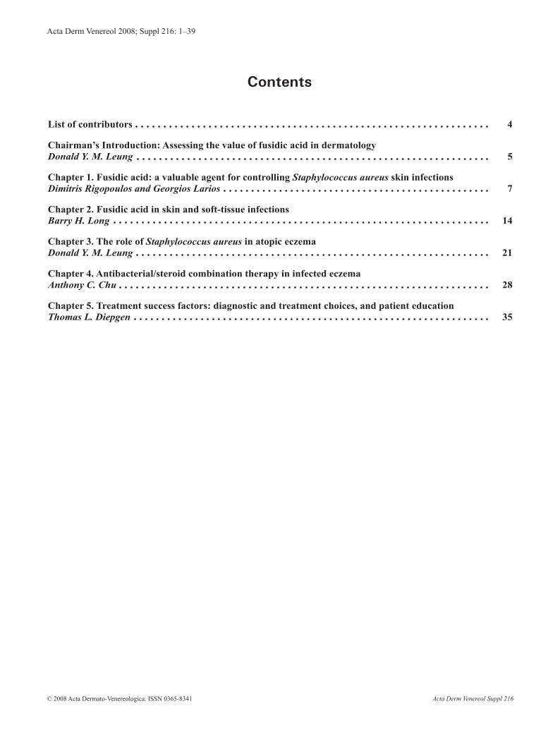
In contrast to the antibiotics described above, fusidic acid has a remarkable ability to penetrate both intact and damaged human skin (42). Skin penetration is similar to that of topical steroids, as was demonstrated in early studies by Vickers (45) and Knight et al. (43). A later study by Stuttgen & Bauer (46) showed that fusidic acid ointment and cream both penetrated intact skin, and that in damaged skin, both the ointment and cream achieved antimicrobial concentrations in the dermis (Fig. 1) (46). This makes it useful in the treatment of deeper infec-tions such as paronychia or boils. Furthermore, Vaillant et al. (47) showed that administration of oral fusidic acid (250 mg or 500 mg twice daily) achieves effective penetration into skin blister fluid (SBF). With the 250 mg tablets, a mean maximum SBF level of 21 mg/l was achieved – about 100 times greater than the MIC90 of fusidic acid for S. aureus (typically 0.25 mg/l) (25). A
Acta Derm Venereol Suppl 216

10 D. Rigopoulos and G. Larios
recent study also showed high in vitro skin permeability of fusidic acid (48).
SIDE-EFFECTS AND SENSITIZATION
Treatment with anti-infective agents can cause side-effects, such as toxic effects arising from direct cell and tissue damage (e.g. as with the aminoglycosides), allergic reactions (e.g. with penicillin), or biological side-effects (e.g. a change in or elimination of normal flora) (49). The goal of therapy is to minimize unwanted side-effects without losing clinical efficacy.
In general, oral formulations of antibiotics cause more side-effects than topical formulations, including in particular gastrointestinal effects. Topical antibiotics
have the advantage of being applied only where needed, thereby minimizing risk of systemic adverse effects. Side-effects of topical agents are often limited to local irritation or allergic contact sensitization; the capacity of an agent to induce the latter unwanted effect is an important consideration.
With beta-lactams, gastrointestinal side-effects are common. There is a risk of allergic response to peni-cillins and cephalosporins (50). Gastrointestinal side- effects are also seen with the macrolides, but the risk of sensitization is low (50).
Among topical drugs, the aminoglycosides have been identified as the most important contact allergens (51). A study by Morris et al. (52) compared the frequency of patch test reactions in successive patients attending a dermatology clinic, for 3 antibiotics (in petrolatum vehicle): neomycin (20%); clioquinol (5%) and fusidic acid (2%). Of the 1119 patients that were involved in the study, only 3 (0.3%) patients experienced positive reac-tions to fusidic acid. reactions to neomycin occurred 10 times more often than to fusidic acid (3.6%, p < 0.05), and 0.7% of patients showed an allergy towards clio-quinol. A more recent study estimated the prevalence of positive reactions to patch tests in the general German population as 2.2% for neomycin, 3.2% for gentamicin and 0.8% for fusidic acid (51).
With the tetracyclines, the risk of sensitization is low. Minocycline has better gastrointestinal absorption than tetracycline and may be less photosensitizing than either tetracycline or doxycycline. Side-effects of minocycline include dizziness and drug-induced lupus erythematosus (53). With all the tetracyclines there is a possibility of staining of the tissues (bone, teeth, skin) and of clothes staining yellow.
No sensitization to mupirocin has been reported. Local irritancy may be due to the polyethylene glycol base of the ointment. Caution is required when mupi-rocin ointment is used in renal failure patients or on extensive open wounds or burns, due to the risk of ab-sorption of polyethylene glycol and possible resulting nephrotoxicity (54).
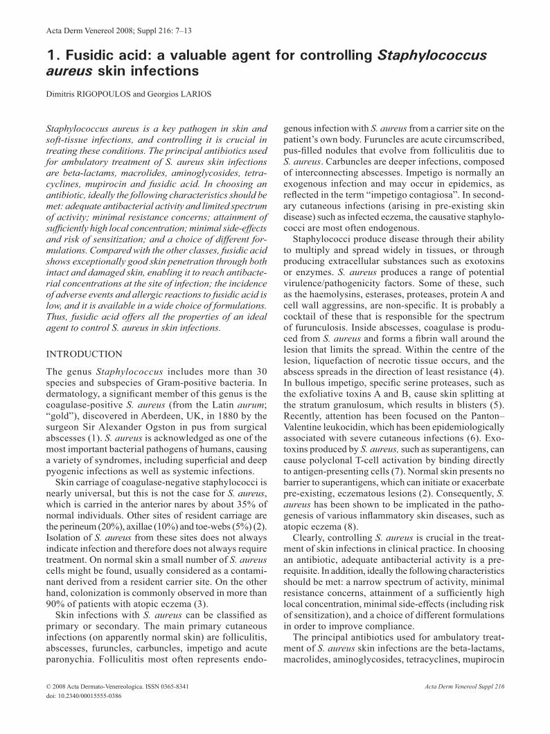
Local irritancy to fusidic acid is uncommon, and the incidence of allergic reactions is low (55–57). In addi-tion, no cross-allergy has been seen. Despite a marked increase in the use of fusidic acid, the frequency of hypersensitivity to the agent did not increase from 1982 to 1999 (Fig. 2) (52).
CHOICE OF FOrMULATIONS
With any drug, in order to maximize the chances of treatment success, it is important to prescribe the for-mulation that is most suitable for the individual patient. In some conditions, such as deep-seated or systemic infection, systemic antibiotics are mandatory. With the topical agents, in addition to any requirements arising
Fig. 1. Penetration through (a) damaged and (b) normal skin of fusidic acid cream, ointment and gel in an in vitro study. Even in intact skin, fusidic acid ointment reached a concentration of about 1 µg/ml at a depth of 800 µm. In damaged skin, fusidic acid ointment reached a concentration of about 10 µg/ml at this depth. © 1988 Editio Cantor Verlag, reproduced with permission from: Stuttgen G & Bauer E. Arzneimittelforschung 1988; 38: 730–735 (46).
Acta Derm Venereol Suppl 216

11Fusidic acid for controlling S. aureus skin infections
from the condition itself, personal preferences for factors such as lipid content and emollient properties must also be considered, as meeting these preferences is likely to increase adherence to treatment and thus improve the outcome.
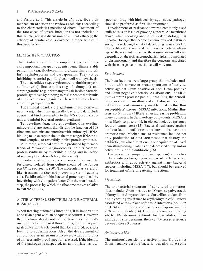
Available formulations of the antibiotic classes described above are shown in Table I. Among the anti-biotics that are available as topical preparations, fusidic acid offers the widest choice of formulations.
CONCLUSION
An ideal antibiotic for controlling S. aureus in SSTI should have high activity against S. aureus, a limited
spectrum of activity against other organisms, minimal concerns about resistance development, the ability to attain a sufficiently high concentration in the affected tissues, minimal side-effects and risk of sensitization, and a choice of different formulations. Fusidic acid fulfils all of these criteria.
rEFErENCES
1. Lyell A. Alexander Ogston, micrococci, and Joseph Lister. J Am Acad Dermatol 1989; 20: 302–310.
2. Noble WC. Skin bacteriology and the role of Staphylococ-cus aureus in infection. Br J Dermatol 1998; 139 Suppl 53: 9–12.
3. Bibel DJ, Greenberg JH, Cook JL. Staphylococcus au-reus and the microbial ecology of atopic dermatitis. Can J Microbiol 1977; 23: 1062–1068.
4. Prescott LM, Harley JP, Klein DA. Staphylococcal diseases. In: Prescott LM, Harley JP, Klein DA, editors. Microbio-logy, 5th edn. New York: McGraw Hill, 2002: 919–923.
5. Amagai M, Matsuyoshi N, Wang ZH, Andl C, Stanley Jr. Toxin in bullous impetigo and staphylococcal scalded-skin syndrome targets desmoglein 1. Nat Med 2000; 6: 1275–1277.
6. Iwatsuki K, Yamasaki O, Morizane S, Oono T. Staphylococ-cal cutaneous infections: invasion, evasion and aggression. J Dermatol Sci 2006; 42: 203–214.
7. Veien NK. The clinician’s choice of antibiotics in the treat-ment of bacterial skin infection. Br J Dermatol 1998; 139 Suppl 53: 30–36.
8. Strange P, Skov L, Lisby S, Nielsen PL, Baadsgaard O. Staphylococcal enterotoxin B applied on intact normal and intact atopic skin induces dermatitis. Arch Dermatol 1996; 132: 27–33.
9. Parenti MA, Hatfield SM, Leyden JJ. Mupirocin: a topical antibiotic with a unique structure and mechanism of action. Clin Pharm 1987; 6: 761–770.
10. Collignon P, Turnidge J. Fusidic acid in vitro activity. Int J Antimicrob Agents 1999; 12 Suppl 2: S45–S58.
11. Wilkinson JD. Fusidic acid in dermatology. Br J Dermatol 1998; 139 (suppl 53): 37–40.
12. Burns K, Cannon M, Cundliffe E. A resolution of conflicting reports concerning the mode of action of fusidic acid. FEBS Lett 1974; 40: 219–223.
13. riber D, Venkataramana M, Sanyal S, Duvold T. Synthesis and biological evaluation of photoaffinity labeled fusidic acid analogues. J Med Chem 2006; 49: 1503–1505.
14. Jones ME, Karlowsky JA, Draghi DC, Thornsberry C, Sahm DF, Nathwani D. Epidemiology and antibiotic susceptibility of bacteria causing skin and soft tissue infections in the USA and Europe: a guide to appropriate antimicrobial therapy. Int J Antimicrob Agents 2003; 22: 406–419.
15. Naimi TS, LeDell KH, Como-Sabetti K, Borchardt SM, Boxrud DJ, Etienne J, et al. Comparison of community- and health care-associated methicillin-resistant Staphylococcus aureus infection. JAMA 2003; 290: 2976–2984.
16. Chambers HF. General principles of antimicrobial therapy. In: Brunton L, Lazo J, Parker K, editors. Goodman & Gilman’s the pharmacological basis of therapeutics, 11th edn. New York: McGraw Hill, 2006: 1095–1111.
17. Jones rN, Barry AL, Thornsberry C. In-vitro studies of meropenem. J Antimicrob Chemother 1989; 24 Suppl A: 9–29.
18. Khawcharoenporn T, Alan T. Oral antibiotic treatment for methicillin-resistant Staphylococcus aureus skin and soft
Table I. Availability of different formulations for the antibiotics used to control S. aureus in dermatology
Antibiotic Formulations
Beta-lactams Oral only
Macrolides OralTopical: cream, ointment or gel (erythromycin), combination with zinc acetate designed for acne
Aminoglycosides Parenteral Topical: cream, ointment (gentamicin) Combinations of neomycin or gentamicin cream with betamethasone
Tetracyclines Oral Topical: ointment, cream (usually mixed with polymyxin B), solution used in acne
Mupirocin Topical: ointment and creamNasal ointment (for methicillin-resistant S. aureus eradication only)
Fusidic acid Oral: tablets and suspension Topical: ointment, cream, combinations with corticosteroids (cream and lipid cream) for infected atopic dermatitis
Fig. 2. Frequency of allergic reactions to fusidic acid among 3307 patients who were patch tested from 1980 to 2000 (triangles). The frequency of allergic reactions has remained low despite increasing use of fusidic acid in the UK over the same period (squares). © 2002 Blackwell Publishing, reproduced with permission from: Morris SD, et al. Br J Dermatol 2002; 146: 1047–1051 (52).
Acta Derm Venereol Suppl 216

12 D. Rigopoulos and G. Larios
tissue infections: review of the literature. Hawaii Med J 2006; 65: 290–293.
19. Sandri AM, Dalarosa MG, ruschel de Alcantara L, da Silva Elias L, Zavascki AP. reduction in incidence of nosoco-mial methicillin-resistant Staphylococcus aureus (MrSA) infection in an intensive care unit: role of treatment with mupirocin ointment and chlorhexidine baths for nasal car-riers of MrSA. Infect Control Hosp Epidemiol 2006; 27: 185–187.
20. Dupeyron C, Campillo B, richardet JP, Soussy CJ. Long-term efficacy of mupirocin in the prevention of infections with methicillin-resistant Staphylococcus aureus in a gastro-enterology unit. J Hosp Infect 2006; 63: 385–392.
21. Walker ES, Vasquez JE, Dula r, Bullock H, Sarubbi FA. Mupirocin-resistant, methicillin-resistant Staphylococcus aureus: does mupirocin remain effective? Infect Control Hosp Epidemiol 2003; 24: 342–346.
22. Perez-roth E, Lopez-Aguilar C, Alcoba-Florez J, Mendez-Alvarez S. High-level mupirocin resistance within methi-cillin-resistant Staphylococcus aureus pandemic lineages. Antimicrob Agents Chemother 2006; 50: 3207–3211.
23. Upton A, Lang S, Heffernan H. Mupirocin and Staphylo-coccus aureus: a recent paradigm of emerging antibiotic resistance. J Antimicrob Chemother 2003; 51: 613–617.
24. Vasquez JE, Walker ES, Franzus BW, Overbay BK, reagan Dr, Sarubbi FA. The epidemiology of mupirocin resistance among methicillin-resistant Staphylococcus aureus at a Veterans’ Affairs hospital. Infect Control Hosp Epidemiol 2000; 21: 459–464.
25. Bogdanovich T, Ednie LM, Shapiro S, Appelbaum PC. Antistaphylococcal activity of ceftobiprole, a new broad-spectrum cephalosporin. Antimicrob Agents Chemother 2005; 49: 4210–4219.
26. Verbist L. The antimicrobial activity of fusidic acid. J Antimicrob Chemother 1990; 25 Suppl B: 1–5.
27. Bishop EJ, Howden BP. Treatment of Staphylococcus aureus infections: new issues, emerging therapies and future directions. Expert Opin Emerg Drugs 2007; 12: 1–22.
28. ravenscroft JC, Layton A, Barnham M. Observations on high levels of fusidic acid resistant Staphylococcus aureus in Harrogate, North Yorkshire, UK. Clin Exp Dermatol 2000; 25: 327–330.
29. Tveten Y, Jenkins A, Kristiansen BE. A fusidic acid-resistant clone of Staphylococcus aureus associated with impetigo bullosa is spreading in Norway. J Antimicrob Chemother 2002; 50: 873–876.
30. Osterlund A, Eden T, Olsson-Liljequist B, Haeggman S, Kahlmeter G; Swedish Study. Group on Fusidic Acid-resis-tant Staphylococcus aureus. Clonal spread among Swedish children of a Staphylococcus aureus strain resistant to fusidic acid. Scand J Infect Dis 2002; 34: 729–734.
31. Osterlund A, Kahlmeter G, Haeggman S, Olsson-Liljequist B; Swedish Study Group On Fusidic Acid resistant S. Aureus. Staphylococcus aureus resistant to fusidic acid among Swedish children: a follow-up study. Scand J Infect Dis 2006; 38: 332–334.
32. Lorette G, Beaulieu P, Bismuth r, Duru G, Guihard W, Lemaitre M, et al. Infections cutanées communautaires: Bactéries en cause et sensibilité aux antibiotiques. Ann Dermatol Venereol 2003; 130: 723–728.
33. Hoeger PH. Antimicrobial susceptibility of skin-colonizing S. aureus strains in children with atopic dermatitis. Pediatr Allergy Immunol 2004; 15: 474–477.
34. rennie rP. Susceptibility of Staphylococcus aureus to fusidic acid: Canadian data. J Cutan Med Surg 2006; 10: 277–280.
35. Bernard P, Jarlier V, Santerre-Henriksen A. Sensibilité
aux antibiotiques des souches de S. aureus responsables d’infections cutanées communautaires. Ann Dermatol Venereol 2008 (in press) .
36. Menday AP, Noble WC. Topical betamethasone/fusidic acid in eczema: efficacy against and emergence of resistance in Staphylococcus aureus. J Dermatolog Treat 2000; 11: 143–149.
37. ravenscroft JC, Layton AM, Eady EA, Murtagh MS, Coates P, Walker M, Cove JH. Short-term effects of topical fusidic acid or mupirocin on the prevalence of fusidic acid resistant (Fusr) Staphylococcus aureus in atopic eczema. Br J Dermatol 2003; 148: 1010–1017.
38. Schultz Larsen FS, Simonsen L, Melgaard A, Wendicke K, Henriksen AS. An efficient new formulation of fusidic acid and betamethasone 17-valerate (Fucicort® Lipid cream) for treatment of clinically infected atopic dermatitis. Acta Derm Venereol 2007; 87: 62–68.
39. Stringel G, Bawdon r, Savrich M, Guertin L, Horton J. Topical and systemic antibiotics in the prevention of wound infection. J Pediatr Surg 1989; 24: 1003–1006.
40. George A, rubin G. A systematic review and meta- analysis of treatments for impetigo. Br J Gen Pract 2003; 53: 480–487.
41. Vaillant L, Le Guellec C, Jehl F, Barruet r, Sorensen H, roiron r, et al. Diffusions comparées de l’acide fusidique, e l’oxacilline et de la pristinamycine ans le liquide interstitiel dermique près administration orale répétée. Ann Dermatol Venereol 2000; 127: 33–39.
42. Winkelman W, Gratton D. Topical antibacterials. Clin Der-matol 1989; 7: 156–162.
43. Knight AG, Vickers CF, Percival P. The percutaneous ab-sorption of antibacterial substances. Br J Dermatol 1969; 81: 88–91.
44. Baines PJ, Jackson D, Mellows G, Swaisland AJ, Tasker TCG. Mupirocin: its chemistry and metabolism. In: Wilkinson DS, Price JD, editors. Mupirocin – a novel topical antibiotic. London (UK): royal Society of Medicine, 1984: 13–20.
45. Vickers CFH. Percutaneous absorption of sodium fusidate and fusidic acid. Br J Dermatol 1969; 81: 902–908.
46. Stuttgen G, Bauer E. Penetration and permeation into hu-man skin of fusidic acid in different galenical formulation. Arzneimittelforschung 1988; 38: 730–735.
47. Vaillant L, Machet L, Taburet AM, Sorensen H, Lorette G. Levels of fusidic acid in skin blister fluid and serum after repeated administration of two dosages (250 and 500 mg). Br J Dermatol 1992; 126: 591–595.
48. Simonsen L, Fullerton A. Development of an in vitro skin permeation model simulating atopic dermatitis skin for the evaluation of dermatological products. Skin Pharmacol Physiol 2007; 20: 230–236.
49. Kayser FH, Kurt A, Bienz KA, Eckert J, Zinkernagel rM, editors. The principles of antibiotic therapy. In: Medical Microbiology. Stuttgart, Germany: Thieme, 2005: 187–206.
50. Prescott LM, Harley JP, Klein DA. Antimicrobial chemo-therapy. In: Prescott LM, Harley JP, Klein DA, editors. Microbiology, 5th edn. New York: McGraw Hill, 2002: 806–821.
51. De Padua CA, Uter W, Schnuch A. Contact allergy to to-pical drugs: prevalence in a clinical setting and estimation of frequency at the population level. Pharmacoepidemiol Drug Saf 2007; 16: 377–384.
52. Morris SD, rycroft rJ, White Ir, Wakelin SH, McFadden JP. Comparative frequency of patch test reactions to topical antibiotics. Br J Dermatol 2002; 146: 1047–1051.
53. Fox LP, Merk HF, Bickers Dr. Dermatological pharmaco-logy. In: Brunton L, Lazo J, Parker K, editors. Goodman & Gilman’s the pharmacological basis of therapeutics, 11th
Acta Derm Venereol Suppl 216

13Fusidic acid for controlling S. aureus skin infections
edn. New York: McGraw Hill, 2006: 1679–1707.54. Infectious Diseases and Immunization Committee, Canadian
Paediatric Society. Mupirocin in the treatment of impetigo. CMAJ 1990; 142: 543–544.
55. Jappe U, Schnuch A, Uter W. Frequency of sensitization to antimicrobials in patients with atopic eczema compared with nonatopic individuals: analysis of multicentre surveil-lance data, 1995–1999. Br J Dermatol 2003; 149: 87–93.
56. Karup C. Safety review of fusidic acid cream and ointment for the treatment of infected dermatoses. 16th Congress of the European Academy of Dermatology and Venereology, Vienna, Austria, 16–20 May 2007: Poster P110.
57. Karup C. Safety review of fusidic acid/steroid combinations for the treatment of infected dermatoses. 16th Congress of the European Academy of Dermatology and Venereology, Vienna, Austria, 16–20 May 2007: Poster P63.
Acta Derm Venereol Suppl 216

© 2008 Acta Dermato-Venereologica. ISSN 0365-8341Acta Derm Venereol Suppl 216
Acta Derm Venereol 2008; Suppl 216: 14–20
doi: 10.2340/00015555-0387
2. Fusidic acid in skin and soft-tissue infectionsBarry H. LONG
Table I. Examples of topical antibiotics commonly used for superficial skin and soft tissue infections
Generic name Class Mechanism of action
Fusidic acid Fusidanes Inhibits protein synthesisMupirocin Unique Inhibits protein synthesisNeomycin Aminoglycoside Inhibits protein synthesisGentamicin Aminoglycoside Inhibits protein synthesisBacitracin Cyclic polypeptide Inhibits cell wall synthesisPolymyxin B Cyclic lipopeptide Increases cell membrane
permeabilitySulfacetamide sodium Sulfonamide Inhibits folic acid synthesisSilver sulfadiazine Sulfonamide Inhibits folic acid synthesis
Silver – inhibits cell wall synthesis
Erythromycin Macrolide Inhibits protein synthesisClindamycin Lincosamide Inhibits protein synthesisretapamulin Pleuromutilin Inhibits protein synthesis
Topical antibacterial therapy is an important component in managing skin and soft-tissue infections (SSTIs). Fusidic acid, a narrow-spectrum antibiotic active against Staphylococcus aureus, has shown good skin permeabi-lity and low allergenic potential. The resistance rate in S. aureus remains low, as shown in a study of Canadian hospitals from 1999 to 2005. In treating primary skin infections, including impetigo, fusidic acid cream and ointment provided similar response rates and equal/better tolerability compared with other topical and oral anti-biotics. Fusidic acid and mupirocin are equally or more efficacious than oral treatment in localized impetigo, and may be similarly efficacious in extensive impetigo, according to a recent Cochrane review. In clinical prac-tice, mupirocin is often reserved for methicillin-resistant S. aureus infections. Studies of oral fusidic acid forms in SSTI have shown that: tablets are as effective as compara-tor antibiotics; they have fewer side-effects; a suspension achieves high cure rates, and is suitable for paediatric use. Fusidic acid, both topical and systemic, is an effective treatment for SSTI with few adverse reactions.
INTrODUCTION
Superficial skin and soft tissue infections (SSTIs) are common presentations in clinical practice. These may manifest either as primary infections or as secondary to some other cutaneous problem. Primary SSTIs, such as impetigo contagiosa, bullous impetigo, folliculitis, furuncles, carbuncles and cellulitis, are frequent occur-rences, in addition to secondary SSTIs, for example, secondarily infected wounds or secondarily infected dermatoses of different types such as atopic dermatitis, contact dermatitis, prurigo and neurodermatitis.
The majority of primary and secondary skin infections are caused by either S. aureus or Streptococcus pyo-genes. Primary skin infections caused by Gram-negative organisms are infrequent but may occur in patients who are immunocompromised or diabetic. Chronic wound infections are more likely to be colonized by Gram- negative organisms, although initial colonization is usually by Gram-positive organisms.
Topical antibacterial therapy is an important com-ponent of therapeutic management. There are various classes of topical antibacterial therapy, both antibiotic and non-antibiotic, which may have beneficial results on the overall therapeutic outcome. Culture should ideally be carried out and a microbiological diagnosis obtained before instituting any form of therapy, but this may not be possible in a given clinical situation. Antibiotic
treatment may subsequently require modification once the culture results become available.
Topical antibacterials have a distinct advantage over systemic agents, in that they can be applied to the af-fected area and therefore high local concentrations of the agent may be achieved. With selection of the appropriate agent, interaction with normal flora can be avoided. The ideal topical antibiotic should: •have a selective effect on one (or at least very few)
organisms of the same class, therefore minimizing the development of cross-resistance to other orga-nisms;
•not cause allergic reactions or potential cross-allergic reactions with other medications of the same class or individual components of these, such as preservati-ves;
•be safe, efficacious and ideally penetrate the skin in sufficiently high concentrations to kill bacteria effic-iently;
•be available in different formulations in order to meet patients' preferences and needs, as this will increase compliance with treatment and thus improve thera-peutic outcomes. The obvious limitation to topical antibacterial therapy
is that the infections must be limited or localized in area and must, for the most part, be superficial.
Classes of topical antibiotics used for superficial SSTIs are shown in Table I. Fusidic acid is an antibio-tic that has all of the features listed above for an ideal topical antibacterial treatment. This article reviews the clinical evidence on the efficacy and safety of fusidic acid in primary skin infections. A review of the use of fusidic acid in secondary skin infections appears elsewhere in this supplement (1).

15Fusidic acid in skin infections
WHY USE FUSIDIC ACID?
Fusidic acid is available in different topical formu-lations: fusidic acid (Fucidin® cream; LEO Pharma A/S, Ballerup, Denmark) and sodium fusidate (Fuci-din® ointment; LEO Pharma A/S). There are also oral formulations in the form of tablets and a suspension. Following absorption, fusidic acid and sodium fusi-date ionize into the same molecule, fusidate; thus, in this article the term fusidic acid will be used to refer to the therapeutic agents in all Fucidin® formulations. Combinations of fusidic acid with corticosteroids are covered elsewhere in this supplement (1).
Fusidic acid has a steroid-like structure but no steroid side-effects (2). In topical form, its penetration is time-related and is comparable to glucocorticoids in diseased skin (3, 4). The normal skin horny layer offers marked resistance to outside agents unless it is damaged or re-moved, but fusidic acid does still penetrate intact skin to some extent (3, 5). Because of its significant absorption qualities, topical administration of fusidic acid results in much higher local concentrations than can be achieved with systemic administration, even at deeper layers of the epidermis or dermis (6). It is indicated for use in the treatment of mild to moderately severe primary and secondary skin infections caused by sensitive strains of S. aureus, Streptococcus species and Corynebacterium minutissimum. Fusidic acid has some activity against other corynebacteria and strains of Clostridium. It is vir-tually inactive against Gram-negative bacteria because of a difference in cell wall permeability; however, it has demonstrated good in vitro activity against strains of Neisseria and Bacteroides.
Policies designed to limit the development of antibio-tic resistance recommend that, in any therapeutic situa-tion, the optimal antibiotic with the narrowest spectrum should be used. As fusidic acid targets the common pathogens in skin infection, a broader-spectrum anti-biotic should not be necessary. This therefore limits the development of antibiotic resistance, cross-resistance and cross-allergic reactions with other medications.
Clinical disease states that would be expected to respond to the topical use of fusidic acid are impetigo contagiosa, bullous impetigo, folliculitis, sycosis bar-bae, furuncles, carbuncles, ecthyma, acute paronychia, erythrasma, infected wound and burns, and secondarily infected dermatoses such as eczema.
CLINICAL STUDIES ON TOPICAL FUSIDIC ACID
A number of studies have examined the use of fusidic acid cream and ointment in the treatment of superficial skin infections (Table II) (7–19). These studies varied in design with regard to randomization, blinding and use of comparator. Nearly all studies included children. These will be looked at with respect to speed of action,
efficacy, safety and outcome compared with other topical therapies and systemic antibiotics in various disease states.
Comparison of fusidic acid cream and ointment
Two studies have compared fusidic acid cream and ointment (Table II) (7–8). In a study by Pakrooh (7), the use of these 2 formulations was compared in 101 patients with SSTI, specifically abscess/boil, paro-nychia and infected wounds. Each preparation was applied 2 or 3 times a day or once daily if a dressing was applied. S. aureus was the most frequently isolated pathogen. Both preparations were effective treatments, with mean healing times being similar: 7.7 days for the ointment and 7.9 days for the cream. Both preparations were well tolerated and there were no complaints of side-effects.
A larger multicentre study by Baldwin & Cranfield (8), involving 487 patients with skin infections (ab-scess/boil, impetigo, paronychia, wounds and burns), compared the use of these 2 formulations applied 3 times daily or once daily with a dressing. An excellent or good response to treatment was observed in over 90% of pa-tients, with mean healing times of 7.1 days for patients treated with the ointment and 7.7 days for those using the cream. Both preparations were well tolerated: only one patient complained of a mild skin reaction with the ointment, which was not severe enough to discontinue treatment. Subsequent treatment with fusidic acid cream elicited no reaction.
Skin infections
Further studies using either fusidic acid cream or oint-ment have shown that there is fast and effective healing of SSTIs (Table II) (9–14). Studies in mainly primary skin infections, such as impetigo, abscesses/boils, folliculitis and paronychia, and including a few cases of infected wounds and other secondary infections (9, 10, 12–14), have demonstrated response rates of between 86% and 100%, with treatment duration or mean healing time varying between 4 and 7.1 days. Adverse events have been infrequent, with most related to application site irritation.
A study by Pakrooh (10) examined the clinical effi-cacy of topical fusidic acid ointment applied once daily compared with that of 3 oral antibiotics given for 5 days: 150 mg clindamycin, 250 mg flucloxacillin or 250 mg of erythromycin 4 times daily plus placebo ointment. A total of 90 patients suffering from SSTIs, including infected wounds, paronychia and abscesses/boils, were included. The mean healing time in patients receiving oral antibiotics was grouped and compared with that in patients using fusidic acid ointment. A significantly more rapid healing time in soft tissue infections was
Acta Derm Venereol Suppl 216

16 B. H. Long
Table II. Studies of topical fusidic acid in skin infections in general, and impetigo. The studies shown under ”Skin infections” were mainly of primary skin infections (including impetigo), but some infected wounds and other secondary infections were included
Fucidin® formulation Comparator
reference n
response ratea (%)Mean healing time or treatment duration (days) n
response ratea (%)Mean healing time or treatment duration (days)
Skin infectionsPakrooh, 1980 (7) Ointment
n = 5191%7.7
Fusidic acid creamn = 50
98%7.9
Baldwin & Cranfield, 1981 (8) Ointmentn = 249
90%7.1
Fusidic acid creamn = 238
92%7.7
Jackson et al., 1966 (9) Ointmentn = 101
93%6.8
Oral/i.m. penicillinn = 58
96% (oral), 94% (i.m.)5.3 (oral)4.9 (i.m.)
Pakrooh, 1977 (10) Ointmentn = 49
100%7.1
Oral antibioticsb
n = 4183%9.7
Zelvelder, 1984 (11) Ointmentn = 30
Nr4–7d
Oral amoxicillinn = 30
Nr4–7d
Morley & Munot, 1988 (12) Ointmentn = 191
86%7e
Mupirocinn = 163
86%7e
Langdon & Mahapatra, 1990 (13) Creamn = 104
95%7e
Mupirocinn = 102
98%7e
Jasuja et al., 2001 (14) Ointmentn = 50
84%7e
Mupirocinn = 50
90%7e
ImpetigoJackson et al., 1966 (9) Ointment
n = 32100%5.9
None –
Cassels-Brown, 1981 (15) Ointmentn = 52
100%7e
Neomycin/bacitracinn = 58
90%7e
Morley & Munot, 1988 (12) Creamn = 51
88%7e
Mupirocinn = 38
84%7e
Sutton, 1992 (16) Ointmentn = 93
97%7e
Mupirocinn = 84
98%7e
Christensen & Anehus, 1994 (17) Creamn = 128
82%Up to 3 weekse
Hydrogen peroxide creamn = 128
72%Up to 3 weekse
Koning et al., 2002 (18) Cream + povidone-iodinen = 78
87%7
Placebo cream + povidone-iodinen = 82
59%7
Oranje et al., 2007 (19) Ointmentn = 172
90%7
retapamulinn = 345
95%5
aAs defined in each study, to include cure or cure/improvement.bClindamycin, erythromycin, or flucloxacillin.cStudy included a fusidic acid/amoxicillin combination arm, not reported here.dreported time to improvement or healing.eDuration of treatment (healing time not stated).i.m.: intramuscular; Nr: overall rate not reported.
shown for fusidic acid ointment compared with the oral antibiotics (7.1 days vs. 9.7 days; p < 0.0002). There were no adverse events in the fusidic acid ointment treatment group, whereas gastrointestinal events were reported in the oral antibiotic group.
A double-blind 3-arm comparative study by Zelvelder (11) compared the effects of fusidic acid ointment plus placebo amoxicillin, placebo fusidic acid ointment plus amoxicillin, or fusidic acid ointment plus amoxicillin in 90 patients in the treatment of furuncles, carbuncles, impetigo and infected wounds. Fusidic acid ointment was as effective as amoxicillin, and there was no further improvement in clinical outcome when the treatments were used in combination.
Fusidic acid ointment is as effective as mupirocin ointment but has superior patient acceptability. In a study by Morley & Munot (12), 354 patients with pri-mary or secondary skin infections were randomized to receive either medication 3 times daily for up to 7 days. There was no difference between the two preparations in outcome in either primary or secondary infections. However, adverse events were reported in 1.0% of the fusidic acid ointment group, compared with 7.4% of those using mupirocin ointment. The greasy, messy or sticky nature of mupirocin ointment accounted for the majority of complaints. A study by Langdon & Maha-patra (13) obtained similar results, while comparing fusidic acid cream and mupirocin ointment.
Acta Derm Venereol Suppl 216
17Fusidic acid in skin infections
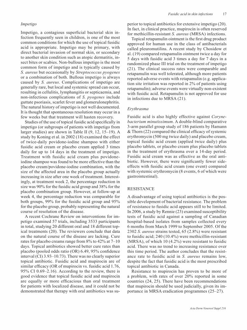
Impetigo
Impetigo, a contagious superficial bacterial skin in-fection frequently seen in children, is one of the most common conditions for which the use of topical fusidic acid is appropriate. Impetigo may be primary, with direct bacterial invasion of normal skin, or secondary to another skin condition such as atopic dermatitis, in-sect bites or scabies. Non-bullous impetigo is the most common form of impetigo and is typically caused by S. aureus but occasionally by Streptococcus pyogenes or a combination of both. Bullous impetigo is always caused by S. aureus. Complications of impetigo are generally rare, but local and systemic spread can occur, resulting in cellulitis, lymphangitis or septicaemia, and non-infectious complications of S. pyogenes include guttate psoriasis, scarlet fever and glomerulonephritis. The natural history of impetigo is not well documented. It is thought that spontaneous resolution may occur in a few weeks but that treatment will hasten recovery.
Studies of the use of topical fusidic acid specifically in impetigo (or subgroups of patients with impetigo from larger studies) are shown in Table II (9, 12, 15–19). A study by Koning et al. in 2002 (18) examined the effect of twice-daily povidone-iodine shampoo with either fusidic acid cream or placebo cream applied 3 times daily for up to 14 days in the treatment of impetigo. Treatment with fusidic acid cream plus povidone- iodine shampoo was found to be more effective than the placebo cream/povidone-iodine combination, with the size of the affected area in the placebo group actually increasing in size after one week of treatment. Interest-ingly, at treatment week 2, the percentage reduction in size was 90% for the fusidic acid group and 38% for the placebo combination group. However, at follow-up at week 4, the percentage reduction was comparable for both groups, 99% for the fusidic acid group and 95% for the placebo group, probably representing the natural course of resolution of the disease.
A recent Cochrane review on interventions for im-petigo examined 57 trials, including 3533 participants in total, studying 20 different oral and 18 different top-ical treatments (20). The reviewers conclude that data on the natural course of the disease are lacking. Cure rates for placebo creams range from 8% to 42% at 7–10 days. Topical antibiotics showed better cure rates than placebo (pooled odds ratio (OR) 6.49, 95% confidence interval (CI) 3.93–10.73). There was no clearly superior topical antibiotic. Fusidic acid and mupirocin are of similar efficacy (OR of mupirocin vs. fusidic acid 1.76, 95% CI 0.69–2.16). According to the review, there is good evidence that topical fusidic acid and mupirocin are equally or more efficacious than oral treatment for patients with localized disease, and it could not be demonstrated that therapy with oral antibiotics was su-
perior to topical antibiotics for extensive impetigo (20). In fact, in clinical practice, mupirocin is often reserved for methicillin-resistant S. aureus (MrSA) infections.
Topical retapamulin ointment is the first drug product approved for human use in the class of antibacterials called pleuromutilins. A recent study by Chosidow et al. (19) compared retapamulin ointment twice a day for 5 days with fusidic acid 3 times a day for 7 days in a randomized phase III trial on the treatment of impetigo (21). The clinical success rates were comparable and retapamulin was well tolerated, although more patients reported adverse events with retapamulin (e.g. applica-tion site irritation was reported in 2% of patients using retapamulin); adverse events were virtually non-existent with fusidic acid. retapamulin is not approved for use in infections due to MrSA (21).
Erythrasma
Fusidic acid is also highly effective against Coryne-bacterium minutissimum. A double-blind comparative 3-arm parallel group study of 186 patients by Hamann & Thorn (22) compared the clinical efficacy of systemic erythromycin (500 mg twice daily) and placebo cream, topical fusidic acid cream (applied twice daily) plus placebo tablets, or placebo cream plus placebo tablets in the treatment of erythrasma over a 14-day period. Fusidic acid cream was as effective as the oral anti-biotic. However, there were significantly fewer side-effects with fusidic acid cream (one event) compared with systemic erythromycin (8 events, 6 of which were gastrointestinal).
rESISTANCE
A disadvantage of using topical antibiotics is the pos-sible development of bacterial resistance. The problem of resistance to fusidic acid appears still to be limited. In 2006, a study by rennie (23) examined susceptibility tests of fusidic acid against a sampling of Canadian hospital-based isolates from samples collected every 6 months from March 1999 to September 2005. Of the 2302 S. aureus strains tested, 65 (2.8%) were resistant to fusidic acid; 240 (10.4%) were methicillin-resistant (MrSA), of which 10 (4.2%) were resistant to fusidic acid. There was no trend to increasing resistance over this time period. The author concludes that the resist-ance rate to fusidic acid in S. aureus remains low, despite the fact that fusidic acid is the most prescribed topical antibiotic in Canada.
resistance to mupirocin has proven to be more of a problem, with rates of over 20% reported in some countries (24, 25). There have been recommendations that mupirocin should be used judicially, given its im-portance in MrSA eradication programmes (25–27).
Acta Derm Venereol Suppl 216

18 B. H. Long
ALLErGENIC POTENTIAL
A further potential disadvantage of the use of topical antibiotics is the development of hypersensitivity or allergic contact dermatitis to a component of the for-mulation. This is more common with certain antibiotics such as gentamicin, bacitracin and neomycin. Adverse events with topical antibiotics are frequently irritant in nature, with complaints of burning or stinging.
In 2002, a study by Morris et al. (28) involved patch testing 1119 patients over 1 year to neomycin, clioquinol and fusidic acid. Positive patch test reactions to neomy-cin were recorded in 40 patients (3.6%), to clioquinol in 8 patients (0.7%) and to fusidic acid in 3 patients (0.3%). The authors also reviewed positive patch test reactions to fusidic acid over a 20-year period, and found that the frequency of allergic reactions to fusidic acid had decreased since the early 1980s, despite increasing use. recently, the prevalence of positive reactions to patch tests in the general German population was estimated as 2.2% for neomycin, 3.2% for gentamicin and 0.8% for fusidic acid, based on data from a network of allergy de-partments (29). Post-marketing safety surveillance has shown a low rate of spontaneous reporting of adverse events for fusidic acid (30). The majority of reported events are similar to those noted in clinical studies: mild localized skin reactions at the site of application. Only 34 reports of allergic reactions have been received after up to 40 years of clinical use. Worldwide experience has shown that there is no significant difference in the safety of fusidic acid cream compared with the ointment.
SYSTEMIC ANTIBIOTIC TrEATMENT
Systemic antibiotic treatment of SSTI is normally reserved for those patients having more extensive disease, deeper infections, with evidence of systemic spread of infection or septicaemia, or those who are immunocompromised or have ophthalmic-orbital or intranasal disease.
There are two oral forms of fusidic acid: a tablet (250 mg) and a suspension formula (50 mg/ml). The accumulation of systemic antibiotic in skin crust or avascular tissue may prevent bacterial invasion; orally administered fusidic acid has been shown to achieve concentrations in skin blister fluid that are above the mini-mal inhibitory concentration (MIC) of both staphylo- cocci and streptococci (31). For an antibiotic to be ef-fective, it must also have adequate tissue penetration and interstitial concentrations higher than MIC90 for the offending organism. In a recent study, concentrations of oxacillin, fusidic acid (given as fusidic acid tablets) and pristinamycin were measured in suction blisters in healthy volunteers at day 5 of a 6-day cycle of antibiotic therapy (32). After a rest period, this was repeated twice so that all volunteers had received each antibiotic. The
mean antibiotic concentration in interstitial fluid was highest for fusidic acid, with Cmax values much greater than the MIC90 of S. aureus, indicating that fusidic acid tablets would potentially be more active than the com-parator antibiotics against all staphylococci.
A randomized double-blind study by Carr et al. (33) using 3 doses of fusidic acid tablets (500 mg 3 times a day, 500 mg twice a day and 250 mg twice a day) demon-strated that a dose of 250 mg twice a day was sufficient to improve and cure SSTI, and there was no significant difference in improvement with higher dosing. Further-more, an obvious advantage of the lower dose was the occurrence of fewer gastrointestinal side-effects.
Another randomized double-blind trial by Nordin & Mobacken (34) compared the efficacy of 2 fusidic acid regimens (250 mg and 500 mg both twice a day) with flucloxacillin (500 mg 3 times a day) in 532 patients. Patients with SSTIs such as abscesses/furuncles, acute paronychia and superficial wound infections were includ-ed and were given an initial 5 days therapy followed by an additional 5 days if necessary. Significantly more patients were cured at the end of 5 days with fusidic acid 250 mg twice a day (32.2%) compared with flu-cloxacillin (21.1%, p < 0.05), but all 3 regimens had high comparable cure rates by the end of treatment. Side-effects were significantly less in the fusidic acid 250 mg group, the most common adverse event being diarrhoea.
Other studies comparing fusidic acid with pristina-mycin (35), ciprofloxacin (36), flucloxacillin (37), or erythromycin (38) have all shown equal efficacy for fusidic acid, with comparable or fewer side-effects.
The suspension formulation of fusidic acid is parti-cularly suitable for paediatric use. Two regimens of the suspension, 20 mg/kg/day twice a day vs. 50 mg/kg/day 3 times a day, were compared in 411 children aged 1–12 years with SSTI (39). Patients were treated for 5 days and for a further 5 days if the condition remained un-cured. At the end of treatment, 91% of the 20 mg group and 89% of the 50 mg group were cured. Bacterio-logical cure, with elimination of fusidic acid-susceptible S. aureus and/or beta-haemolytic streptococci, was achieved in 100% and 99% of children, respectively. The lower-dose regimen had significantly better tolerability (p = 0.025), due to fewer gastrointestinal side-effects.
CONCLUSION
It has been well established that topical antibiotics are extremely important in the management of SSTIs, most of which are due to S. aureus and Streptococcus species. Fusidic acid (in both topical and systemic forms) has been demonstrated to be an effective treatment with a low incidence of adverse reactions when studied alone or in comparison with other topical and systemic anti-bacterial therapies.
Acta Derm Venereol Suppl 216
19Fusidic acid in skin infections
rEFErENCES
1. Chu AC. Antibacterial/steroid combination therapy in infected eczema. Acta Derm Venereol 2008; Suppl 216: 28–34.
2. Winkelman W, Gratton D. Topical antibacterials. Clin Der-matol 1989; 7: 156–162.
3. Vickers CFH. Percutaneous absorption of sodium fusidate and fusidic acid. Br J Dermatol 1969; 81: 902–908.
4. Knight AG, Vickers CF, Percival P. The percutaneous ab-sorption of antibacterial substances. Br J Dermatol 1969; 81: 88–91.
5. Simonsen L, Fullerton A. Development of an in vitro skin permeation model simulating atopic dermatitis skin for the evaluation of dermatological products. Skin Pharmacol Physiol 2007; 20: 230–236.
6. Stuttgen G, Bauer E. Penetration and permeation into hu-man skin of fusidic acid in different galenical formulation. Arzneimittelforschung 1988; 38: 730–735.
7. Pakrooh H. Comparative trial of Fucidin ointment and Fucidin cream in skin sepsis. J Int Med res 1980; 8: 425–429.
8. Baldwin RJT, Cranfield R. A multi-centre general practice trial comparing Fucidin ointment and Fucidin cream. Br J Clin Pract 1981; 35: 157–160.
9. Jackson N, Verling W, Deasy D, MacMahon JJ, Sherry TB. Treatment of cutaneous infections with Fucidin ointment. Clin Trials J 1966; 3: 591–595.
10. Pakrooh H. A comparison of sodium fusidate ointment (Fucidin) alone versus oral antibiotic therapy in soft-tissue infections. Curr Med res Opin 1977; 5: 289–294.
11. Zelvelder WG. A double-blind comparative study of sodium fusidate (topical), amoxycillin (oral) and the combination of both drugs in skin infections. Tijdschr Geneesmiddele-nonderz 1984; 9: 87–92.
12. Morley PAr, Munot LD. A comparison of sodium fusidate ointment and mupirocin ointment in superficial skin sepsis. Curr Med res Opin 1988; 11: 142–148.
13. Langdon CG, Mahapatra KS. Efficacy and acceptability of fusidic acid cream and mupirocin ointment in acute skin sepsis. Current Ther research 1990; 48: 174–179.
14. Jasuja K, Gupta S, Arora D, Gupta V. Bacteriology of primary pyodermas and comparative efficacy of topical application of mupirocin and sodium fusidate ointments in their treatment. Indian J Dermatol Venereol Leprol 2001; 67: 132–134.
15. Cassels-Brown G. A comparative study of Fucidin ointment and Cicatrin cream in the treatment of impetigo. Br J Clin Pract 1981; 35: 153–155.
16. Sutton JB. Efficacy and acceptability of fusidic acid cream and mupirocin ointment in facial impetigo. Curr Ther res 1992; 51: 673–678.
17. Christensen OB, Anehus S. Hydrogen peroxide cream: an alternative to topical antibiotics in the treatment of impetigo contagiosa. Acta Derm Venereol 1994; 74: 460–462.
18. Koning S, van Suijlekom-Smit L, Nouwen J, Verduin CM, Bernsen rM, Oranje AP, et al. Fusidic acid cream in the treatment of impetigo in general practice: double-blind randomised placebo-controlled trial. BMJ 2002; 324: 203–207.
19. Oranje AP, Chosidow O, Sacchidanand S, Todd G, Singh K, Scangarella N, et al. Topical retapamulin ointment, 1%, versus sodium fusidate ointment, 2%, for impetigo: a randomized, observer-blinded, noninferiority study. Dermatology 2007; 215: 331–340.
20. Koning S, Verhagen AP, van Suijlekom-Smit L, Morris A, Butler CC, van der Wouden JC. Interventions for im-
petigo (review). Cochrane Database Sys rev 2004; (2): CD003261.
21. FDA. Medical review, Application number: 22-055 (Altabax), April 2007. Available from: http: //www.fda.gov/cder/foi/nda/2007/022055s000_Medr.pdf [accessed 12 June 2007].
22. Hamann K, Thorn P. Systemic or local treatment of erythrasma? A comparison between erythromycin tablets and Fucidin cream in general practice. Scand J Prim Health Care 1991; 9: 35–39.
23. rennie rP. Susceptibility of Staphylococcus aureus to fusidic acid: Canadian data. J Cutan Med Surg 2006; 10: 277–280.
24. Vasquez JE, Walker ES, Franzus BW, Overbay BK, reagan Dr, Sarubbi FA. The epidemiology of mupirocin resistance among methicillin-resistant Staphylococcus aureus at a Veterans’ Affairs hospital. Infect Control Hosp Epidemiol 2000; 2: 459–464.
25. Upton A, Lang S, Heffernan H. Mupirocin and Staphylo-coccus aureus: a recent paradigm of emerging antibiotic resistance. J Antimicrob Chemother 2003; 51: 613–617.
26. Tschachler E, Brockmeyer N, Effendy I, Geiss HK, Harder S, Hartmann M, et al. Streptococcal infections of the skin and mucous membranes. J Dtsch Dermatol Ges 2007; 5: 527–532.
27. Cookson BD. The emergence of mupirocin resistance: a challenge to infection control and antibiotic prescribing practice. J Antimicrob Chemother 1998; 41: 11–18.
28. Morris SD, rycroft rJ, White Ir, Wakelin SH, McFadden JP. Comparative frequency of patch test reactions to topical antibiotics. Br J Dermatol 2002; 146: 1047–1051.
29. De Padua CA, Uter W, Schnuch A. Contact allergy to top-ical drugs: prevalence in a clinical setting and estimation of frequency at the population level. Pharmacoepidemiol Drug Saf 2007; 16: 377–384.
30. Karup C. Safety review of fusidic acid cream and ointment for the treatment of infected dermatoses. 16th Congress of the European Academy of Dermatology and Venereology, Vienna, Austria, 16–20 May 2007: Poster P110.
31. Vaillant L, Machet L, Taburet AM, Sorensen H, Lorette G. Levels of fusidic acid in skin blister fluid and serum after repeated administration of two dosages (250 and 500 mg). Br J Dermatol 1992; 126: 591–595.
32. Vaillant L, Le Guellec C, Jehl F, Barruet r, Sorensen H, roiron r, et al. Comparative diffusion of fusidic acid, oxacillin, and pristinamycin in interstitial dermal fluid after repeated oral administration. Ann Dermatol Venereol 2000; 127: 33–39.
33. Carr WD, Wall A, Georgala-Zervogiani S, Stratigos J, Gouriotou K. Fusidic acid tablets in patients with skin and soft tissue infections: a dose finding study. Eur J Clin Res 1994; 5: 87–95.
34. Nordin P, Mobacken H. A comparison of fusidic acid and flucloxacillin in the treatment of skin and soft-tissue infec-tion. Eur J Clin res 1994; 5: 97–106.
35. Claudy A; Groupe Francais d’Etude. Pyodermites superficielles nécessitant une antibiothérapie orale – Acide fusidique versus pristinamycine. Presse Med 2001; 30: 364–368.
36. Newby MR. Comparative efficacy of fusidic acid and ciprofloxacin in skin and soft tissue infection. J Clin Res 1999; 2: 77–84.
37. Morris CDE, Talbot DT. A comparison of fusidic acid and flucloxacillin capsules in the treatment of skin and soft- tissue infection. J Clin res 2000; 3: 1–14.
38. Wall ArJ, Menday AP. Fusidic acid and erythromycin in the
Acta Derm Venereol Suppl 216

20 B. H. Long
treatment of skin and soft tissue infection: a double blind study. J Clin res 2000; 3: 12–28.
39. Török E, Somogyi T, rutkai K, Iglesias L, Bielsa I. Fusidic acid suspension twice daily: a new treatment schedule for skin and soft tissue infection in children with improved tolerability. J Dermatolog Treat 2004; 15: 158–163.
DISCUSSION
Q: Is it beneficial to combine oral and topical therapy, or two different antibiotics?
Long: No. Clearly if there is evidence of systemic infection, or if the person is developing septicaemia, a systemic antibiotic should be used. But the studies of topical fusidic acid have shown that it works well in mild-to-moderate infections and even in some severe infections. As mentioned earlier, fusidic acid penetrates the skin very well and achieves high local concentra-tions – greater concentrations than those achieved with systemic antibiotics. This is an advantage of topical agents. I would only use a systemic antibiotic if there is evidence of systemic or severe infection.
Acta Derm Venereol Suppl 216

© 2008 Acta Dermato-Venereologica. ISSN 0365-8341 Acta Derm Venereol Suppl 216
Acta Derm Venereol 2008; Suppl 216: 21–27
doi: 10.2340/00015555-0388
3. The role of Staphylococcus aureus in atopic eczemaDonald Y. M. LEUNG
Staphylococcus aureus infection plays an important role in atopic eczema (AE) because of its ability to produce virulence factors such as superantigens. Epicutaneous application of superantigens induces eczema. Super-antigens also induce corticosteroid resistance, and subvert T-regulatory cell activity, thereby increasing AE severity. Increased binding of S. aureus to skin is driven by underlying AE skin inflammation. This is supported by studies demonstrating that treatment with topical corticosteroids reduces S. aureus counts on atopic skin.
AE has also been found to be deficient in antimicrobial peptides needed for host defence against bacteria. The reduced production of antimicrobial peptides in AE appear to be an acquired defect resulting from increased T-helper type 2 cell (Th2) cytokine production. A vicious cycle of skin barrier dysfunction, skin infection and Th2 cell immune activation therefore occurs in AE. Effective strategies for controlling AE require combination therapy that reduces skin inflammation and controls S. aureus colonization and infection.
INTrODUCTION
Atopic eczema (AE), also referred to as atopic derma-titis (AD), is a chronic inflammatory skin disease com-monly presenting in infants and young children, with a point prevalence of 10–20% of the population (1). Pruritic skin lesions evolve from complex interactions between IgE-bearing antigen-presenting cells, T-cell activation, mast cell degranulation, keratinocytes, and eosinophils that can be triggered by irritants, foods, aeroallergens and infection (2, 3). recent studies demonstrating that AE is associated with a defective skin barrier provide evidence of a genetic basis to the disease. Patients are predisposed to selective skin in-flammation via enhanced permeability of allergens and microbes, resulting in high-level allergen sensitization and the atopic march leading to respiratory allergy (4, 5). This review focuses on the role of S. aureus in the pathogenesis of AE. An understanding of the mecha-nisms underlying enhanced S. aureus colonization and infection in AE, and identification of the molecules involved in triggering atopic skin inflammation, has important implications in our current approach to the management of AE.
S. AuREuS IN ATOPIC ECZEMA
S. aureus colonizes the skin of most patients with AE (6). The number of S. aureus on atopic skin depends
on the type of skin lesion: S. aureus can be isolated from 55–75% of unaffected AE skin, 85–91% of chronic lichenified lesions and 80–100% of acute exudative skin lesions. The density of S. aureus can reach 107 organisms per cm2 on acute exudative AE skin lesions. Thus, atopic skin provides a favourable environment for the colonization and proliferation of S. aureus. Secondarily infected patients show greater clinical improvement to combined treatment with anti-staphylococcal antibiotics and topical corticosteroids, compared with topical corticosteroids alone, supporting the concept that S. aureus contributes to skin inflam-mation in AE (7, 8).
MECHANISM(S) LEADING TO S. AuREuS COLONIZATION
The mechanism(s) leading to increased S. aureus colo-nization in AE are an active area of investigation. The increased S. aureus colonization probably results from a combination of processes. These include, in addition to defective skin barrier function, the loss of certain innate anti-bacterial activities as a result of changes in antimicrobial peptide (AMP) levels or reduced immune responses necessary for defence against bacteria. There has also been much interest in the potential role of lipid deficiencies, since lipids have antimicrobial effects (9), and reduced lipid content in AE skin leads to increased transepidermal water loss as well as dry, cracked, brittle skin, which predisposes to S. aureus colonization (3, 4). These factors are not mutually exclusive. Indeed, all probably play a role in S. aureus colonization of AE skin, varying according to the patient’s genetic predisposition and environment.
Increased S. aureus adherence
The initial step in colonization or infection requires attachment of S. aureus to skin surfaces. The skin of patients with AE has been demonstrated to have increased adherence for S. aureus (Fig. 1). The reason for increased binding of S. aureus to AE skin is proba-bly related to the underlying skin atopic inflammation (Table I).
This concept is supported by the following studies. First, acute AE skin lesions are colonized with greater numbers of S. aureus than chronic skin lesions, unaffect-ed atopic skin or normal non-atopic skin (6). Secondly, it has been found that treatment with anti-inflammatory medications such as topical corticosteroids or calci-

22 D. Y. M. Leung
Table I. Factors contributing to S. aureus colonization/infection in atopic eczema
• Impaired skin barrier function• reduced skin lipid content in atopic eczema• Increased skin adherence to S. aureus due to increased fibronectin and
fibrinogen • Decreased production of endogenous antimicrobial peptides (beta-
defensins, LL-37) by keratinocytes
neurin inhibitors significantly reduces the numbers of S. aureus found on atopic skin (10–12). Thirdly, bacterial binding was found to be significantly greater at mouse skin sites with T-helper type 2 cell (Th2)-mediated inflammation than at skin sites with T-helper type 1 cell (Th1)-mediated inflammation (13). This increased bacterial binding did not occur in interleukin (IL)-4 gene knockout mice, suggesting that IL-4 plays a critical role in the enhancement of S. aureus binding to skin. In contrast, when normal skin was incubated with IL-4 or with interferon-g, increased S. aureus binding occurred only to skin explants treated with IL-4.
Staphylococcal cell surface molecules termed “ad-hesins”, which are responsible for the adherence of S. aureus to the skin, have been identified. These include fibronectin-binding proteins A and B, fibrinogen- binding proteins, and collagen adhesins (14, 15). Relevant to atopic inflammation, IL-4, but not inter-feron-g, is known to induce fibronectin production by skin fibroblasts (16). Recently, we found that fibronectin and fibrinogen are involved in the binding of S. aureus to Th2-induced inflammatory skin lesions (17). Thus, IL-4 induced fibronectin synthesis, in combination with plasma exudation of fibrinogen, could provide a mecha-nism by which the atopic/inflammatory environment mediates enhanced S. aureus attachment to the skin.
Decreased innate immune response
The density of S. aureus on acutely inflamed AE lesions is generally more than 1000-fold higher than on non-lesional AE skin. As increased S. aureus adherence can account only for a several-fold increase in S. aureus on AE skin, other local host defence mechanisms must also be defective. Using electron microscopy, Morishita et al. (18) found colonies of S. aureus distributed on the surface of the epidermis as well as growing between layers of keratinocytes in the absence of an active anti-microbial response. This observation suggests that an exponential increase in S. aureus could result from fail-ure of the innate immune response to restrict the growth of microorganisms. Indeed, a direct comparison of AE and psoriasis showed that about 30% of patients with AE suffered from clinical infections, whereas only 6.7% of patients with psoriasis had this complication (19),
despite the fact that both skin diseases have defective skin barrier function (20). It is thought that the reduced prevalence of infections in psoriasis may be associated with the increased production of AMPs (21).
Two major classes of AMPs have been found in mammalian skin: beta-defensins (22, 23) and catheli-cidins (LL-37) (24, 25). They have been shown to have antimicrobial activities against bacterial, fungal and viral pathogens (26). In the skin, keratinocytes are the primary producer of these peptides. We have compared the expression of AMPs in AE vs. psoriasis to determine if the increased susceptibility to infection in AE is due to a deficiency in AMPs (27, 28). We found that there was abundant LL-37, human beta-defensin (HBD)-2 and HBD-3 in the skin of all patients with psoriasis. In AE lesions, however, immunostaining of LL-37, HBD-2, and HBD-3 was significantly decreased. HBD-2 and LL-37 mrNA was also lower in AE lesions than pso-riasis lesions. The combination of LL-37 and HBD-2
Fig. 1. Atopic skin, compared with normal skin is associated with increased adherence of S. aureus. © 2001 Elsevier, reproduced with permission from: Cho S-H, et al. J Allergy Clin Immunol 2001; 108: 269–274 (17).
Acta Derm Venereol Suppl 216

23S. aureus in atopic eczema
showed synergistic antimicrobial activity by effectively killing S. aureus more than either AMP alone. Thus, a deficiency in AMP expression could account for the ability of S. aureus readily to infect skin from patients with AE.
To examine the potential mechanism for this defect, we examined the ability of cultured AE keratinocytes to produce AMP. We found that after the keratinocytes were removed from the inflammatory milieu of AE skin, they produced normal levels of AMP, suggesting that the defect was acquired (29). As acute AE skin lesions are associated with marked overexpression of IL-4 and IL-13, we studied the effects of IL-4 and IL-13 on tumour necrosis factor-alpha (TNF-a-induced HBD-2 and HBD-3 expression in keratinocytes). IL-4 alone or in combination with IL-13 significantly suppressed TNF-a-induced expression of HBD-2 and HBD-3 in keratinocytes (30). This data suggest that the low ex-pression of AMP expression in AE may be acquired as the result of allergic immune responses (31–33).
Skin inflammation induced by S. aureus
The exact mechanisms by which S. aureus induces skin inflammatory responses in AE are being investigated. A number of staphylococcal products, including pro-tein A, lipoteichoic acid and various toxins have been observed to induce activation of cells involved in the pathogenesis of AE including mast cells, T cells, kera-tinocytes and macrophages (3). An important strategy by which S. aureus induces skin inflammation in AE is by secreting a group of toxins known as superantigens (Fig. 2).
Superantigens bind directly to constitutively expres-sed human leukocyte antigen D-related (HLA-Dr) molecules on professional antigen-presenting cells such as macrophages or dendritic cells, and to gamma inter-feron-induced HLA-Dr molecules on non-professional antigen-presenting cells such as keratinocytes (34). This results in the release of pro-inflammatory cytokines by these HLA-Dr+ cells, or via the subsequent activation of T cells. The stimulation of T cells by superantigens results in the activation of lymphocytes expressing specific T-cell receptor V-beta regions (35).
A variety of observations support a role for super-antigens in triggering AE (Table II). First, the majority of patients with AE have S. aureus cultured from their skin that secrete superantigens such as enterotoxins A (SEA), B (SEB) and toxic shock syndrome toxin-1 (TSST-1) (33, 36, 37). Analysis of the peripheral blood skin-homing T cells expressing cutaneous lymphoid antigen (CLA) from these patients as well as their skin lesions reveals that they have undergone a T-cell receptor V-beta expansion within both their CD4+ T cells and their CD8+ T cells, indicative of superanti-gen stimulation (38, 39).
Secondly, most patients with AE make specific IgE antibodies directed against superantigens found on their skin (36, 37). Basophils from patients with IgE to superantigens release histamine on exposure to the relevant superantigen, but not in response to super-antigens to which they make no specific IgE. These data suggest that superantigens induce specific IgE in AE and chronic mast cell degranulation in vivo when the superantigens penetrate their impaired skin barrier. This promotes the itch–scratch cycle, thereby contributing to
Fig. 2. Immune actions of staphylococcal superantigens. © 2000 Elsevier, reproduced with permission from: Leung DYM. J Allergy Clin Immunol 2000; 105: 860–876 (34).
Acta Derm Venereol Suppl 216
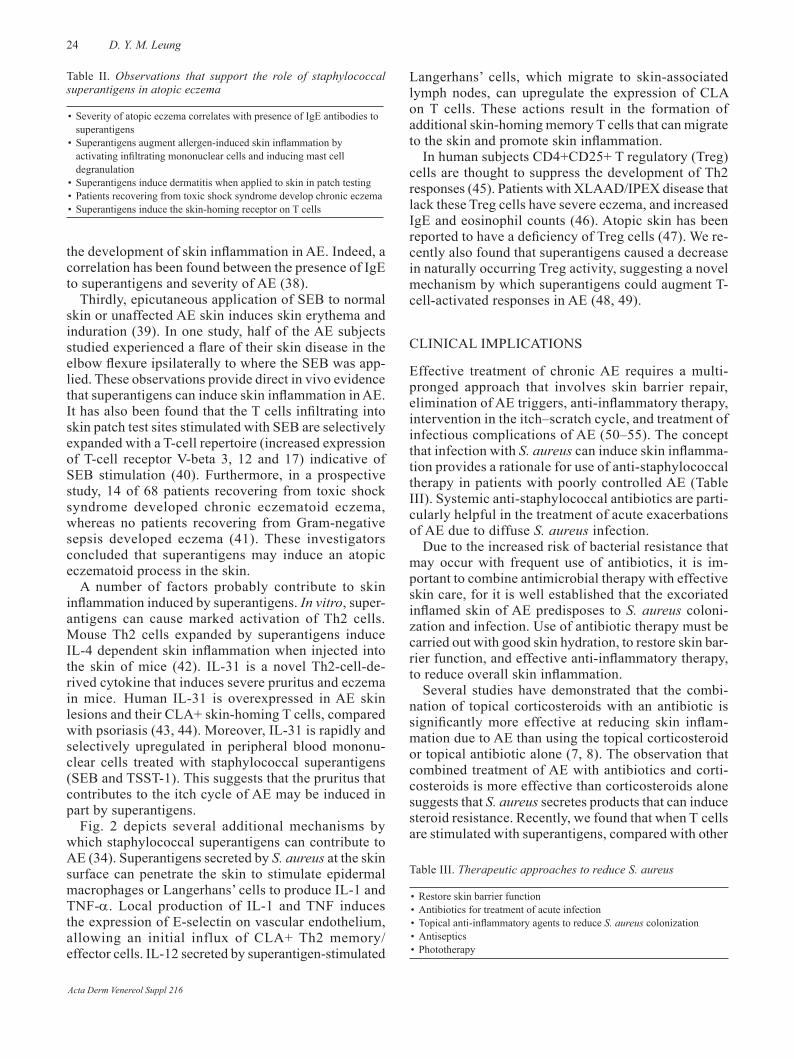
24 D. Y. M. Leung
Table II. Observations that support the role of staphylococcal superantigens in atopic eczema
• Severity of atopic eczema correlates with presence of IgE antibodies to superantigens
• Superantigens augment allergen-induced skin inflammation by activating infiltrating mononuclear cells and inducing mast cell degranulation
• Superantigens induce dermatitis when applied to skin in patch testing• Patients recovering from toxic shock syndrome develop chronic eczema• Superantigens induce the skin-homing receptor on T cells
Table III. Therapeutic approaches to reduce S. aureus
• restore skin barrier function• Antibiotics for treatment of acute infection• Topical anti-inflammatory agents to reduce S. aureus colonization• Antiseptics• Phototherapy
the development of skin inflammation in AE. Indeed, a correlation has been found between the presence of IgE to superantigens and severity of AE (38).
Thirdly, epicutaneous application of SEB to normal skin or unaffected AE skin induces skin erythema and induration (39). In one study, half of the AE subjects studied experienced a flare of their skin disease in the elbow flexure ipsilaterally to where the SEB was app-lied. These observations provide direct in vivo evidence that superantigens can induce skin inflammation in AE. It has also been found that the T cells infiltrating into skin patch test sites stimulated with SEB are selectively expanded with a T-cell repertoire (increased expression of T-cell receptor V-beta 3, 12 and 17) indicative of SEB stimulation (40). Furthermore, in a prospective study, 14 of 68 patients recovering from toxic shock syndrome developed chronic eczematoid eczema, whereas no patients recovering from Gram-negative sepsis developed eczema (41). These investigators concluded that superantigens may induce an atopic eczematoid process in the skin.
A number of factors probably contribute to skin inflammation induced by superantigens. In vitro, super-antigens can cause marked activation of Th2 cells. Mouse Th2 cells expanded by superantigens induce IL-4 dependent skin inflammation when injected into the skin of mice (42). IL-31 is a novel Th2-cell-de-rived cytokine that induces severe pruritus and eczema in mice. Human IL-31 is overexpressed in AE skin lesions and their CLA+ skin-homing T cells, compared with psoriasis (43, 44). Moreover, IL-31 is rapidly and selectively upregulated in peripheral blood mononu-clear cells treated with staphylococcal superantigens (SEB and TSST-1). This suggests that the pruritus that contributes to the itch cycle of AE may be induced in part by superantigens.
Fig. 2 depicts several additional mechanisms by which staphylococcal superantigens can contribute to AE (34). Superantigens secreted by S. aureus at the skin surface can penetrate the skin to stimulate epidermal macrophages or Langerhans’ cells to produce IL-1 and TNF-a. Local production of IL-1 and TNF induces the expression of E-selectin on vascular endothelium, allowing an initial influx of CLA+ Th2 memory/ effector cells. IL-12 secreted by superantigen-stimulated
Langerhans’ cells, which migrate to skin-associated lymph nodes, can upregulate the expression of CLA on T cells. These actions result in the formation of additional skin-homing memory T cells that can migrate to the skin and promote skin inflammation.
In human subjects CD4+CD25+ T regulatory (Treg) cells are thought to suppress the development of Th2 responses (45). Patients with XLAAD/IPEX disease that lack these Treg cells have severe eczema, and increased IgE and eosinophil counts (46). Atopic skin has been reported to have a deficiency of Treg cells (47). We re-cently also found that superantigens caused a decrease in naturally occurring Treg activity, suggesting a novel mechanism by which superantigens could augment T-cell-activated responses in AE (48, 49).
CLINICAL IMPLICATIONS
Effective treatment of chronic AE requires a multi-pronged approach that involves skin barrier repair, elimination of AE triggers, anti-inflammatory therapy, intervention in the itch–scratch cycle, and treatment of infectious complications of AE (50–55). The concept that infection with S. aureus can induce skin inflamma-tion provides a rationale for use of anti-staphylococcal therapy in patients with poorly controlled AE (Table III). Systemic anti-staphylococcal antibiotics are parti-cularly helpful in the treatment of acute exacerbations of AE due to diffuse S. aureus infection.
Due to the increased risk of bacterial resistance that may occur with frequent use of antibiotics, it is im-portant to combine antimicrobial therapy with effective skin care, for it is well established that the excoriated inflamed skin of AE predisposes to S. aureus coloni-zation and infection. Use of antibiotic therapy must be carried out with good skin hydration, to restore skin bar-rier function, and effective anti-inflammatory therapy, to reduce overall skin inflammation.
Several studies have demonstrated that the combi-nation of topical corticosteroids with an antibiotic is significantly more effective at reducing skin inflam-mation due to AE than using the topical corticosteroid or topical antibiotic alone (7, 8). The observation that combined treatment of AE with antibiotics and corti-costeroids is more effective than corticosteroids alone suggests that S. aureus secretes products that can induce steroid resistance. recently, we found that when T cells are stimulated with superantigens, compared with other
Acta Derm Venereol Suppl 216
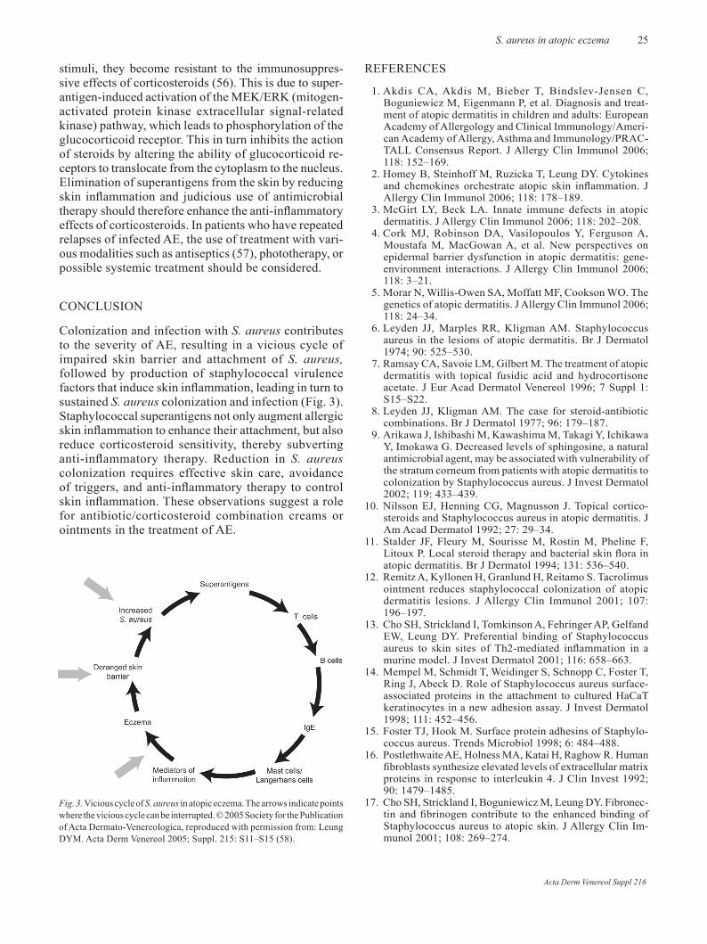
25S. aureus in atopic eczema
Fig. 3. Vicious cycle of S. aureus in atopic eczema. The arrows indicate points where the vicious cycle can be interrupted. © 2005 Society for the Publication of Acta Dermato-Venereologica, reproduced with permission from: Leung DYM. Acta Derm Venereol 2005; Suppl. 215: S11–S15 (58).
stimuli, they become resistant to the immunosuppres-sive effects of corticosteroids (56). This is due to super- antigen-induced activation of the MEK/ErK (mitogen-activated protein kinase extracellular signal-related kinase) pathway, which leads to phosphorylation of the glucocorticoid receptor. This in turn inhibits the action of steroids by altering the ability of glucocorticoid re-ceptors to translocate from the cytoplasm to the nucleus. Elimination of superantigens from the skin by reducing skin inflammation and judicious use of antimicrobial therapy should therefore enhance the anti-inflammatory effects of corticosteroids. In patients who have repeated relapses of infected AE, the use of treatment with vari-ous modalities such as antiseptics (57), phototherapy, or possible systemic treatment should be considered.
CONCLUSION
Colonization and infection with S. aureus contributes to the severity of AE, resulting in a vicious cycle of impaired skin barrier and attachment of S. aureus, followed by production of staphylococcal virulence factors that induce skin inflammation, leading in turn to sustained S. aureus colonization and infection (Fig. 3). Staphylococcal superantigens not only augment allergic skin inflammation to enhance their attachment, but also reduce corticosteroid sensitivity, thereby subverting anti-inflammatory therapy. Reduction in S. aureus colonization requires effective skin care, avoidance of triggers, and anti-inflammatory therapy to control skin inflammation. These observations suggest a role for antibiotic/corticosteroid combination creams or ointments in the treatment of AE.
rEFErENCES
1. Akdis CA, Akdis M, Bieber T, Bindslev-Jensen C, Boguniewicz M, Eigenmann P, et al. Diagnosis and treat-ment of atopic dermatitis in children and adults: European Academy of Allergology and Clinical Immunology/Ameri-can Academy of Allergy, Asthma and Immunology/PrAC-TALL Consensus report. J Allergy Clin Immunol 2006; 118: 152–169.
2. Homey B, Steinhoff M, ruzicka T, Leung DY. Cytokines and chemokines orchestrate atopic skin inflammation. J Allergy Clin Immunol 2006; 118: 178–189.
3. McGirt LY, Beck LA. Innate immune defects in atopic dermatitis. J Allergy Clin Immunol 2006; 118: 202–208.
4. Cork MJ, robinson DA, Vasilopoulos Y, Ferguson A, Moustafa M, MacGowan A, et al. New perspectives on epidermal barrier dysfunction in atopic dermatitis: gene-environment interactions. J Allergy Clin Immunol 2006; 118: 3–21.
5. Morar N, Willis-Owen SA, Moffatt MF, Cookson WO. The genetics of atopic dermatitis. J Allergy Clin Immunol 2006; 118: 24–34.
6. Leyden JJ, Marples rr, Kligman AM. Staphylococcus aureus in the lesions of atopic dermatitis. Br J Dermatol 1974; 90: 525–530.
7. ramsay CA, Savoie LM, Gilbert M. The treatment of atopic dermatitis with topical fusidic acid and hydrocortisone acetate. J Eur Acad Dermatol Venereol 1996; 7 Suppl 1: S15–S22.
8. Leyden JJ, Kligman AM. The case for steroid-antibiotic combinations. Br J Dermatol 1977; 96: 179–187.
9. Arikawa J, Ishibashi M, Kawashima M, Takagi Y, Ichikawa Y, Imokawa G. Decreased levels of sphingosine, a natural antimicrobial agent, may be associated with vulnerability of the stratum corneum from patients with atopic dermatitis to colonization by Staphylococcus aureus. J Invest Dermatol 2002; 119: 433–439.
10. Nilsson EJ, Henning CG, Magnusson J. Topical cortico-steroids and Staphylococcus aureus in atopic dermatitis. J Am Acad Dermatol 1992; 27: 29–34.
11. Stalder JF, Fleury M, Sourisse M, rostin M, Pheline F, Litoux P. Local steroid therapy and bacterial skin flora in atopic dermatitis. Br J Dermatol 1994; 131: 536–540.
12. remitz A, Kyllonen H, Granlund H, reitamo S. Tacrolimus ointment reduces staphylococcal colonization of atopic dermatitis lesions. J Allergy Clin Immunol 2001; 107: 196–197.
13. Cho SH, Strickland I, Tomkinson A, Fehringer AP, Gelfand EW, Leung DY. Preferential binding of Staphylococcus aureus to skin sites of Th2-mediated inflammation in a murine model. J Invest Dermatol 2001; 116: 658–663.
14. Mempel M, Schmidt T, Weidinger S, Schnopp C, Foster T, ring J, Abeck D. role of Staphylococcus aureus surface-associated proteins in the attachment to cultured HaCaT keratinocytes in a new adhesion assay. J Invest Dermatol 1998; 111: 452–456.
15. Foster TJ, Hook M. Surface protein adhesins of Staphylo-coccus aureus. Trends Microbiol 1998; 6: 484–488.
16. Postlethwaite AE, Holness MA, Katai H, raghow r. Human fibroblasts synthesize elevated levels of extracellular matrix proteins in response to interleukin 4. J Clin Invest 1992; 90: 1479–1485.
17. Cho SH, Strickland I, Boguniewicz M, Leung DY. Fibronec-tin and fibrinogen contribute to the enhanced binding of Staphylococcus aureus to atopic skin. J Allergy Clin Im-munol 2001; 108: 269–274.
Acta Derm Venereol Suppl 216

26 D. Y. M. Leung
18. Morishita Y, Tada J, Sato A, Toi Y, Kanzaki H, Akiyama H, Arata J. Possible influences of Staphylococcus aureus on atopic dermatitis – the colonizing features and the effects of staphylococcal enterotoxins. Clin Exp Allergy 1999; 29: 1110–1117.
19. Christophers E, Henseler T. Contrasting disease patterns in psoriasis and atopic dermatitis. Arch Dermatol res 1987; 279 (suppl): S48–S51.
20. Grice K, Sattar H, Baker H, Sharratt M. The relationship of transepidermal water loss to skin temperature in psoriasis and eczema. J Invest Dermatol 1975; 64: 313–315.
21. Fulton C, Anderson GM, Zasloff M, Bull r, Quinn AG. Expression of natural peptide antibiotics in human skin. Lancet 1997; 350: 1750–1751.
22. Harder J, Bartels J, Christophers E, Schroder JM. A peptide antibiotic from human skin. Nature 1997; 387: 861.
23. Stolzenberg ED, Anderson GM, Ackermann Mr, Whitlock rH, Zasloff M. Epithelial antibiotic induced in states of disease. Proc Natl Acad Sci USA 1997; 94: 8686–8690.
24. Gallo rL, Ono M, Povsic T, Page C, Eriksson E, Klagsbrun M, Bernfield M. Syndecans, cell surface heparan sulfate proteoglycans, are induced by a proline-rich antimicrobial peptide from wounds. Proc Natl Acad Sci USA 1994; 91: 11035–11039.
25. Frohm M, Agerberth B, Ahangari G, Stahle-Backdahl M, Liden S, Wigzell H, Gudmundsson GH. The expression of the gene coding for the antibacterial peptide LL-37 is indu-ced in human keratinocytes during inflammatory disorders. J Biol Chem 1997; 272: 15258–15263.
26. Gropp r, Frye M, Wagner TO, Bargon J. Epithelial defen-sins impair adenoviral infection: implication for adeno-virus-mediated gene therapy. Hum Gene Ther 1999; 10: 957–964.
27. Gallo rL, Murakami M, Ohtake T, Zaiou M. Biology and clinical relevance of naturally occurring antimicrobial peptides. J Allergy Clin Immunol 2002; 110: 823–831.
28. Nizet V, Ohtake T, Lauth X, Trowbridge J, rudisill J, Dorschner rA, et al. Innate antimicrobial peptide protects the skin from invasive bacterial infection. Nature 2001; 414: 454–457.
29. Ong PY, Ohtake T, Brandt C, Strickland I, Boguniewicz M, Ganz T, et al. Endogenous antimicrobial peptides and skin infections in atopic dermatitis. N Engl J Med 2002; 347: 1151–1160.
30. Nomura I, Goleva E, Howell MD, Hamid QA, Ong PY, Hall CF, et al. Cytokine milieu of atopic dermatitis, as compared to psoriasis, skin prevents induction of innate immune response genes. J Immunol 2003; 171: 3262–3269.
31. Howell MD, Novak N, Bieber T, Pastore S, Girolomoni G, Boguniewicz M, et al. Interleukin-10 downregulates anti-microbial peptide expression in atopic dermatitis. J Invest Dermatol 2005; 125: 738–745.
32. Fiset PO, Leung DY, Hamid Q. Immunopathology of atopic dermatitis. J Allergy Clin Immunol 2006; 118: 287–290.
33. Boguniewicz M, Leung DY. Atopic dermatitis. J Allergy Clin Immunol 2006; 117: S475–S480.
34. Leung DY. Atopic dermatitis: new insights and opportunities for therapeutic intervention. J Allergy Clin Immunol 2000; 105: 860–876.
35. Kotzin BL, Leung DY, Kappler J, Marrack P. Superantigens and their potential role in human disease. Adv Immunol 1993; 54: 99–166.
36. Leung DY, Harbeck r, Bina P, reiser rF, Yang E, Norris DA, et al. Presence of IgE antibodies to staphylococcal exotoxins on the skin of patients with atopic dermatitis. Evidence for a new group of allergens. J Clin Invest 1993; 92: 1374–1380.
37. Bunikowski r, Mielke M, Skarabis H, Herz U, Bergmann rL, Wahn U, renz H. Prevalence and role of serum IgE antibodies to the Staphylococcus aureus-derived super-antigens SEA and SEB in children with atopic dermatitis. J Allergy Clin Immunol 1999; 103: 119–124.
38. Bunikowski r, Mielke ME, Skarabis H, Worm M, Anagnostopoulos I, Kolde G, et al. Evidence for a disease-promoting effect of Staphylococcus aureus-derived exo-toxins in atopic dermatitis. J Allergy Clin Immunol 2000; 105: 814–819.
39. Strange P, Skov L, Lisby S, Nielsen PL, Baadsgaard O. Staphylococcal enterotoxin B applied on intact normal and intact atopic skin induces dermatitis. Arch Dermatol 1996; 132: 27–33.
40. Skov L, Olsen JV, Giorno r, Schlievert PM, Baadsgaard O, Leung DY. Application of Staphylococcal enterotoxin B on normal and atopic skin induces up-regulation of T cells by a superantigen-mediated mechanism. J Allergy Clin Immunol 2000; 105: 820–826.
41. Michie CA, Davis T. Atopic dermatitis and staphylococcal superantigens. Lancet 1996; 347: 324.
42. Müller KM, Jaunin F, Masouye I, Saurat JH, Hauser C. Th2 cells mediate IL-4-dependent local tissue inflammation. J Immunol 1993; 150: 5576–5584.
43. Sonkoly E, Muller A, Lauerma AI, Pivarcsi A, Soto H, Kemeny L, et al. IL-31: a new link between T cells and pruritus in atopic skin inflammation. J Allergy Clin Im-munol 2006; 117: 411–417.
44. Bilsborough J, Leung DY, Maurer M, Howell M, Boguniewicz M, Yao L, et al. IL-31 is associated with cu-taneous lymphocyte antigen-positive skin homing T cells in patients with atopic dermatitis. J Allergy Clin Immunol 2006; 117: 418–425.
45. Akdis M, Blaser K, Akdis CA. T regulatory cells in allergy: novel concepts in the pathogenesis, prevention, and treat-ment of allergic diseases. J Allergy Clin Immunol 2005; 116: 961–968.
46. Chatila TA. role of regulatory T cells in human diseases. J Allergy Clin Immunol 2005; 116: 949–959.
47. Verhagen J, Akdis M, Traidl-Hoffmann C, Schmid- Grendelmeier P, Hijnen D, Knol EF, et al. Absence of T-regulatory cell expression and function in atopic dermatitis skin. J Allergy Clin Immunol 2006; 117: 176–183.
48. Cardona ID, Goleva E, Ou LS, Leung DY. Staphylococcal enterotoxin B inhibits regulatory T cells by inducing glu-cocorticoid-induced TNF receptor-related protein ligand on monocytes. J Allergy Clin Immunol 2006; 117: 688–695.
49. Goleva E, Cardona ID, Ou LS, Leung DY. Factors that regulate naturally occurring T regulatory cell-mediated sup-pression. J Allergy Clin Immunol 2005; 116: 1094–1100.
50. Boguniewicz M, Schmid-Grendelmeier P, Leung DY. Atopic dermatitis. J Allergy Clin Immunol 2006; 118: 40–43.
51. Leung DYM. New insights into the complex gene-environ-ment interactions evolving into atopic dermatitis. J Allergy Clin Immunol 2006; 118: 37–39.
52. Bender BG, Leung DY. Sleep disorders in patients with asthma, atopic dermatitis, and allergic rhinitis. J Allergy Clin Immunol 2005; 116: 1200–1201.
53. Howell MD, Wollenberg A, Gallo rL, Flaig M, Streib JE, Wong C, et al. Cathelicidin deficiency predisposes to eczema herpeticum. J Allergy Clin Immunol 2006; 117: 836–841.
54. Kim HY, Kim HS. Upregulation of MIP-2 (CXCL2) ex-pression by 15-deoxy-Delta (12,14)-prostaglandin J (2) in mouse peritoneal macrophages. Immunol Cell Biol 2007; 85: 60–67.
55. Salt BH, Boguniewicz M, Leung DY. Severe refractory
Acta Derm Venereol Suppl 216
27S. aureus in atopic eczema
atopic dermatitis in adults is highly atopic. J Allergy Clin Immunol 2007; 119: 508–509.
56. Li LB, Goleva E, Hall CF, Ou LS, Leung DY. Super-antigen-induced corticosteroid resistance of human T cells occurs through activation of the mitogen-activated protein kinase kinase/extracellular signal-regulated kinase (MEK-ErK) pathway. J Allergy Clin Immunol 2004; 114: 1059–1069.
57. Stalder JF, Fleury M, Sourisse M, Allavoine T, Chalamet C, Brosset P, Litoux P. Comparative effects of two topical antiseptics (chlorhexidine vs KMn04) on bacterial skin flora in atopic dermatitis. Acta Derm Venereol 1992; Suppl 176: 132–134.
58. Leung D. Superantigens, steroid insensitivity and innate immunity in atopic eczema. Acta Derm Venereol 2005; Suppl 215: 11–15.
Acta Derm Venereol Suppl 216

© 2008 Acta Dermato-Venereologica. ISSN 0365-8341Acta Derm Venereol Suppl 216
Acta Derm Venereol 2008; Suppl 216: 28–34
doi: 10.2340/00015555-0389
Infection with Staphylococcus aureus is common in all forms of eczema. Production of superantigens by S. aureus increases skin inflammation in eczema; antibacterial treatment is thus pivotal. Poor patient compliance is a major cause of treatment failure; combination prepara-tions that contain an antibacterial and a topical steroid and that work quickly can improve compliance and thus treatment outcome. Fusidic acid has advantages over other available topical antibacterial agents – neomycin, gentamicin, clioquinol, chlortetracycline, and the anti-fungal agent miconazole. The clinical efficacy, antibac-terial activity and cosmetic acceptability of fusidic acid/corticosteroid combinations are similar to or better than those of comparator combinations. Fusidic acid/steroid combinations work quickly with observable improvement within the first week. Studies have shown that short-term (2 weeks) use of fusidic acid/corticosteroid combinations does not increase the development of resistance. A new formulation of fusidic acid with betamethasone valerate in a lipid cream also addresses xerosis in eczema.
INTrODUCTION
Staphylococcus aureus is often implicated in different forms of eczema. It has been shown to produce super-antigens that exacerbate the inflammatory response in eczema (1–3) and induce corticosteroid insensitivity (4). Anti-staphylococcal agents are thus pivotal agents in our treatment of eczema. A number of topical anti-bacterial agents are commercially available. A combi-nation product that contains both an antibacterial agent and a topical steroid in one preparation has obvious advantages over 2 different products each containing one active agent, as the combination preparation will increase patient compliance and thus improve thera-peutic outcome.
This paper briefly describes the importance of S. aureus in eczema, examines the rationale for the use of combination antibacterial/steroid preparations, compares the characteristics of those available, and suggests management strategies for the optimal use of these agents.
S. AuREuS AND ECZEMA
S. aureus is commonly found in all types of eczema (5). This may manifest as obvious infection with impe-tiginization or cellulitis, but may also be more cryptic, presenting as excoriations, increased erythema, or fissuring of the skin. Even when overt infection is not
present, the use of anti-staphylococcal agents with top-ical corticosteroids has been shown to produce greater clinical improvement than topical corticosteroids alone (6, 7). These findings are in keeping with the demon-stration that S. aureus can be isolated from more than 90% of atopic eczema skin lesions (8); in one study, it was isolated from 100% of lesional skin and 79% of normal skin in patients with atopic eczema (9).
We observed similar rates of infection in a prospective audit at the Hammersmith Hospital, in which all new patients referred with atopic eczema were evaluated. In a 2-month period, 30 patients were referred (22 children and 8 adults). The reason given by the primary health physician for referral in 29 was failure to respond to prescribed treatment, and one patient was referred be-cause the parents wanted a consultant opinion. In 90% of the patients there was clinical evidence of infection; in 87% swabs from lesional skin were highly positive for S. aureus; and 93% showed marked improvement within one week of treatment with topical fusidic acid/corticosteroid combinations and emollients, with or without a systemic antibiotic.
These findings are compelling arguments for the gene-ral use of anti-staphylococcal agents in the management of all patients with eczema.
IS TrEATMENT OF S. AuREuS SUFFICIENT TO TrEAT ECZEMA?
S. aureus is very important in the pathophysiology of eczema, but is its eradication sufficient to control eczema? In a randomized, double-blind, prospective, parallel-group study, ramsay et al. (10) compared topical 2% fusidic acid, 1% hydrocortisone, or a com-bination of 2% fusidic acid with 1% hydrocortisone in the treatment of atopic eczema. One group of patients was treated with hydrocortisone or the fusidic acid/ hydrocortisone combination, and the second with fusidic acid or the fusidic acid/hydrocortisone combination. As expected, the fusidic acid-containing preparations were superior in eradicating S. aureus and beta haemolytic streptococci, with eradication rates of 100% for fusidic acid cream and 98% for fusidic acid/hydrocortisone cream vs. 53% for the hydrocortisone cream (Fig. 1). When the results of all the patients were pooled, the 3 preparations were found to be statistically significantly different in achieving > 50% improvement in total signs and symptoms, and in reduction of sign and symptom scores after 2 weeks’ treatment. Fusidic acid/hydrocor-tisone cream gave the best results, followed by hydro-cortisone cream and then fusidic acid cream.
4. Antibacterial/steroid combination therapy in infected eczemaAnthony C. CHU

29Antibacterial/steroid combination therapy
This study demonstrates that eradication of pathogenic bacteria from eczema is not sufficient to treat eczema. An important finding, however, was that the combi-nation of an antibacterial with a topical corticosteroid improved the outcome compared with the topical cortico-steroid by itself. This shows the importance of also using antibacterial therapy when treating eczema.
TrEATMENT FAILUrE IN ECZEMA
To treat eczema effectively, the 3 principal problems need to be targeted: dryness of the skin (xerosis); inflammation; and infection (see Fig. 3 by Leung in this supplement, p. 25) (3). These can be individually targeted by the use of, respectively, emollients and moisturizers; appropriate strength topical steroid or topical immune response modifiers; and topical or systemic antibacterial agents.
Treatment failure is complex, but the major cause is failure to adhere to therapy. There are a number of pos-sible reasons for this, including: lack of understanding of the topical agents prescribed, complex regimens comprising a number of different topical agents, fear of real or imaginary side-effects of topical agents, slow response to treatment, under-prescribing by the physi-cian, failure to renew prescriptions, child refusal of topical agents, adult unwillingness to use the treatment prescribed, and poor cosmetic acceptability (11, 12). The ideal treatment in eczema is one that addresses all the problems in a single preparation, works quickly, is
free of side-effects, and is cosmetically acceptable. Few preparations match this ideal.
It is of major importance to discuss with patients the treatments prescribed to ensure appropriate usage. The importance of providing patients or carers with informa-tion is illustrated by a study that showed parents’ lack of knowledge and incorrect perceptions concerning com-monly prescribed topical corticosteroids (11). Among the parents or carers of 100 children attending paediatric outpatient clinics, 44% of those who had been prescribed 1% hydrocortisone for their children’s eczema graded it as moderately potent; 42% of those who had used beta-methasone valerate 0.1% did not grade it as potent; and the fusidic acid 2%/hydrocortisone 1% combination was graded as moderately potent by 56% and potent by 32%. Such misunderstanding increases the anxiety of patients about the risks of using steroids and leads to avoidance where use would help to control the eczema.
TOPICAL ANTIBACTErIAL/STErOID COMBINATIONS
A number of antibacterial/steroid topical combinations are commercially available. These include combinations with 1% hydrocortisone or with a potent topical cortico-steroid such as betamethasone valerate 0.1% (Table I).
Fusidic acid has major advantages over other available topical antimicrobial agents. It shows very good penetra-tion into the skin (13, 14). High in vitro skin permeabil-ity to both the fusidic acid and betamethasone valerate components of the combination product formulation has also been documented (15). Furthermore, fusidic acid has high anti-staphylococcal activity even against methicil-lin-resistant S. aureus (MrSA) (16). Unlike neomycin and gentamicin, it has a very low potential to sensitize and induce contact allergic dermatitis (17). It also has very good cosmetic acceptability, unlike clioquinol and chlortetracycline, which can mark clothes and bedding.
Sensitization by topical antibacterials
The major advantage of using topical antibiotics is the ability to achieve a high concentration of the antibiotic in the skin where it is needed, without the side-effects inherent with the use of systemic antibiotics. When used on eczematous skin, where the skin barrier function is impaired, there is an increased risk of cutaneous sensi-tization. Antibiotics such as neomycin are contained in over-the-counter products in Europe, some deodorants, and a number of topical prescription drugs used on the skin and in the eyes. Sensitization to neomycin is a well recognized problem and many dermatologists avoid topical neomycin for this reason.
Two recent studies have examined patch test results from Departments of Dermatology participating in the Information Network of Departments of Dermatology in
Fig. 1. results of treating patients with infected atopic eczema with topical 2% fusidic acid (FA), 1% hydrocortisone (HC), or a combination of 2% fusidic acid with 1% hydrocortisone (FA/HC) for 2 weeks (data from ramsay et al. (10)). The graph shows bacteriological response (eradication of bacteria) and anti-eczema effect (expressed as a percentage of patients who did not fail treatment) for patients with bacteria present at baseline. results are combined results for 2 parallel studies comparing hydrocortisone with the combination (n = 73), or fusidic acid with the combination (n = 32).
Acta Derm Venereol Suppl 216

30 A. C. Chu
Germany (18, 19). In the first study, 8532 patients with atopic eczema were subjected to aimed testing for suspect-ed allergic contact dermatitis over a 4-year period. Among those tested, 2.1% were sensitive to neomycin (n = 7619 tested), 2.11% were sensitive to gentamicin (n = 1635), and 0.31% were sensitive to clioquinol (n = 1177), compared with 0% sensitive to fusidic acid (n = 48) (18). The second study estimated the incidence of contact allergy to topical drugs in the overall German population, based on patch tests performed during 2000 to 2004, as 2.2% for neomy-cin, 3.2% for gentamicin, and 0.8% for fusidic acid (19).
Cosmetic acceptability
The majority of topical antibiotics available in com-bination with topical corticosteroids are cosmetically acceptable. Notable exceptions are chlortetracycline, which is yellow and can mark clothing, and clioquinol. Clioquinol is initially colourless, but when exposed to air it turns yellow, and if applied to clothing it will turn brown: this often discourages use.
Efficacy of fusidic acid/steroid combination products
A number of randomized clinical trials have compared the efficacy of different topical antimicrobial/cortico-steroid preparations with fusidic acid/corticosteroid preparations in infected eczema (Table II) (5, 20–24).
Fusidic acid 2%/hydrocortisone 1% cream vs. micona-zole 2%/hydrocortisone 1% cream. In this study, fusidic acid/hydrocortisone cream (Fucidin® H; LEO Pharma A/S, Ballerup, Denmark) was compared with a com-bination of the antifungal compound miconazole with hydrocortisone (20). Both treatments were equally ef-fective in treating clinically infected eczema, but heal-ing was more rapid with the fusidic acid/hydrocortisone cream (p = 0.04 in favour of fusidic acid/hydrocortisone after 1 week of treatment).
Fusidic acid 2%/betamethasone 0.1% cream vs. neo-mycin 0.5%/betamethasone 0.1% cream. Two clinical trials compared fusidic acid/betamethasone cream (Fucicort®, Fucibet®; LEO Pharma A/S) with neomy-
Table I. Antimicrobial agents available as combination preparations with topical corticosteroids, showing some of their characteristics
Agent Available in combination with Sensitization potential Formulationa Cosmetic acceptability
Fusidic acid Hydrocortisone 1%Betamethasone valerate 0.1%
Low Cream, ointmentCream, lipid cream
Good
Neomycin Betamethasone valerate 0.1% High Cream, ointment GoodGentamicin Betamethasone valerate 0.05% High Cream, ointment GoodClioquinol Betamethasone valerate 0.1% Medium Cream, ointment PoorChlortetracycline Triamcinolone acetonide 0.1% Low Ointment PoorMiconazoleb Hydrocortisone 1% Low Cream, ointment GoodaAvailability of formulations varies by country.bAlthough an antifungal compound, miconazole, is included here, as it has been used in the treatment of atopic eczema.
Table II. Comparative trials of fusidic acid/corticosteroid combination preparations
reference Fusidic acid combination Comparator Trial design Condition
Poyner & Dass, 1996 (20) Fusidic acid 2%/hydrocortisone 1% cream (n = 95)
Miconazole 2%/hydrocortisone 1% cream (n = 102)
Open Mild to moderate infected eczema of the trunk or limbs
Wilkinson et al., 1985 (5) Fusidic acid 2%/betamethasone 0.1% cream (n = 45)
Neomycin 0.5%/betamethasone 0.1% cream (n = 46)
Double-blind Infected or potentially infected eczema
Javier et al., 1986 (21) Fusidic acid 2%/betamethasone 0.1% cream (n = 27)
Neomycin 0.5%/betamethasone 0.1% cream (n = 32)
Double-blind Infected or potentially infected eczema
Strategos, 1986 (22) Fusidic acid 2%/betamethasone valerate 0.1% cream (n = 50)
Gentamicin 0.1%/betamethasone valerate 0.1% cream (n = 49)
Open Infected eczema
Hill et al., 1998 (23) Fusidic acid 2%/betamethasone 0.1% cream (n = 58)
Clioquinol 3%/betamethasone 0.1% cream (n = 62)
Open Infected hand eczema
Schultz Larsen et al., 2007 (24)
Fusidic acid 2%/betamethasone 0.1% cream (n = 275) and Fusidic acid 2%/betamethasone 0.1% lipid cream (n = 258)
Lipid cream vehicle (n = 88)
Double-blind Infected atopic eczema
Acta Derm Venereol Suppl 216

31Antibacterial/steroid combination therapy
cin/betamethasone cream (5, 21). The two preparations showed similar high clinical efficacy. In the Wilkinson study, at 2 weeks 90% and 95% of patients using the neomycin or fusidic acid combination creams, respec-tively, considered their treatment beneficial, and both treatments were equally effective at eradicating S. aureus (5). In the study by Javier et al. (21), at 7–10 days both preparations were equally effective, with a satisfactory clinical response seen in 81% and 85% of patients using the neomycin or fusidic acid combination creams, respectively. Both preparations were equally effective in eradicating bacterial pathogens.
Fusidic acid 2%/betamethasone valerate 0.1% cream vs. gentamicin 0.1%/betamethasone valerate 0.1% cream. In this study, after 7–12 days of treatment, 74% of the fusidic acid group achieved an excellent response compared with 55% of the gentamicin group (p = 0.03). The two treatments were equally effective in eradicating skin pathogens (22).
Fusidic acid 2%/betamethasone 0.1% cream vs. clio-quinol 3%/betamethasone 0.1% cream. The study by Hill et al. (23) compared the two preparations used twice daily in the treatment of infected hand eczema for a period of up to 4 weeks. The overall clinical response was similar in both groups, with 54.8% of patients achieving a good or excellent response in the fusidic acid group and 53.4% in the clioquinol group. Overall cosmetic acceptability, however, was significantly different in the two groups: 29.6% of the clioquinol group and 90.6% of the fusidic acid group found the cosmetic acceptability of their treatment good (p < 0.0001). Fusidic acid was also superior in bacteriological efficacy, eradicating S. aureus in 92.3% of patients, whereas clioquinol eradicated S. aureus in only 55.2% of patients (p = 0.004).
New fusidic acid 2%/betamethasone valerate 0.1% lipid cream. It is important to relieve the dryness of eczematous skin, and the use of corticosteroid cream without emollients and moisturizers may lead to further problems with dryness and subsequent itching in some patients. A new formulation of fusidic acid and betame-thasone in a lipid cream has recently been developed to provide an alternative treatment for patients with infec-ted eczema in whom the existing combination cream does not provide an adequate moisturizing effect. The efficacy of this new lipid cream was compared with that of fusidic acid/betamethasone cream in a double-blind, randomized controlled study (24). In this study, 630 pa-tients aged 6 years or older with infected atopic eczema received treatment with fusidic acid/betamethasone lipid cream, fusidic acid/betamethasone cream, or the lipid cream vehicle. At the end of 2 weeks’ treatment, total severity scores were reduced by 82.9% in the lipid cream group, 82.7% in the cream group, and 33.0% in
the vehicle group. Successful bacteriological response was seen in 89.7%, 89.6% and 25.0% of patients, re-spectively, and adverse events of pruritus or a burning sensation in 2.6%, 1.6% and 13.6%, respectively.
The new fusidic acid/betamethasone lipid cream has thus been shown to be as effective and well tolerated as fusidic acid/betamethasone cream. It provides patients and doctors with an alternative, so that patients’ indi-vidual needs and preferences for emollient treatment can be better met.
Efficacy of fusidic acid/steroid combination products: Comment
The unifying theme of all these comparative studies was the efficacy of the fusidic acid/corticosteroid prepara-tions. These were as effective as or more effective than the comparator preparations in terms of clinical efficacy, antibacterial activity and cosmetic acceptability.
DEVELOPMENT OF rESISTANCE
A major concern in using topical antibiotics is the emer-gence of antibiotic drug resistance. This is of particular importance with an antibiotic such as fusidic acid, which has a major medical role against methicillin- resistant staphylococci. Since the launch of topical fusidic acid, resistance levels to this antibiotic have remained low (25). resistance to fusidic acid has been reported in closed environments, such as hospital wards, where the risk of cross-infection is high (26). Increased levels of resistance have also been reported in dermatology departments where fusidic acid/cor-ticosteroid use has been high (27). It is possible that the way these preparations were used was responsible, as a retrospective review of 8 previously conducted clinical trials using fusidic acid/betamethasone to treat infected or potentially infected eczema showed that the emergence of fusidic acid-resistant strains was observed in only 2.8% of patients given fusidic acid-containing cream, compared with 2.5% given the comparator cream (28). These authors concluded that fusidic acid/betamethasone, when given for short periods, leads to little selective pressure for the de-velopment of resistance to fusidic acid. Furthermore, in the recent prospective study of the new fusidic acid/ betamethasone lipid cream, selection of S. aureus isolates resistant to fusidic acid was seen in 2.3% of the patients who applied fusidic acid, and 1.9% of those given the vehicle only, which again suggests that short-term use of fusidic acid/betamethasone does not increase resistance (24).
This conclusion has been supported by two studies looking at the emergence of drug resistance to fusidic acid in patients with eczema treated for short, 2-week periods with topical preparations containing fusidic
Acta Derm Venereol Suppl 216

32 A. C. Chu
acid. The first was a case-controlled study to assess the effect of short-term use of fusidic acid/betamethasone cream in clinically infected eczema on the emergence of fusidic acid-resistant strains of S. aureus (Fusr S. aureus) (29). Forty-six patients were randomized to receive either the fusidic acid/betamethasone cream or topical 2% mupirocin ointment plus betamethasone cream used twice daily for 2 weeks. Both groups showed a similar significant improvement in clinical severity of the eczema at the end of the study. Microbiologically, no patients developed fusidic acid resistance during the study. Baseline samples from the site of worst eczema showed Fusr S. aureus in 26% of patients, with no significant difference between treatment groups. After 2 weeks, there was a reduction in prevalence and popula-tion density of S. aureus (sensitive and resistant) at the worst eczema site (p < 0.0001), but no significant change in the prevalence of carriage or population density of Fusr S. aureus, although there was a downward trend in both groups. The prevalence of carriage of either S. aureus (sensitive and resistant) or Fusr S. aureus in the nares did not change between baseline and 2 weeks. The authors concluded that the use of topical fusidic acid containing preparations for a 2-week period does not promote resistance to fusidic acid in the skin or nares.
The second study was an open study to examine the efficacy in atopic eczema of cyclical therapy, alternating fusidic acid/hydrocortisone cream or 1% hydrocortisone cream each for 2 weeks in children, and fusidic acid/betamethasone cream or betamethasone 0.1% cream in adults, and to determine the occurrence of fusidic acid drug resistance using this regime (Chu AC, poster presentation at American Academy of Dermatology Meeting, 2001). Of 24 patients recruited into the study, 18 were children and 6 adults. Prior to starting the study, all patients had been using a topical corticosteroid and emollient, and one patient had been using fusidic acid/hydrocortisone cream for 8 months. Seventeen patients were poorly controlled, with frequent exacerbations of their eczema often requiring a course of systemic antibiotics. Swabs of lesional skin grew S. aureus in 22 patients: 21 were sensitive to fusidic acid and one (the child using long-term fusidic acid/hydrocortisone) was resistant to fusidic acid. Patients responded well to cyclical therapy, with most patients being well con-trolled. The mother of one patient, an 8-year-old boy, kept a detailed diary before and after treatment. In the 12 months prior to the study he had required 2 hospital admissions and 4 courses of oral antibiotics for infected atopic eczema. Following the start of the trial, no further oral antibiotics were required (Fig. 2).
Patients were reviewed every month for at least 2 months (3 patients were reviewed for 12 months) and swabs were taken at each visit. As shown in Table III, the prevalence of carriage of S. aureus decreased over time, and no new cases of Fusr S. aureus were observed. This
study demonstrates that, even with prolonged treatment of up to one year, as long as the fusidic acid preparation is only used for 2 weeks each month, control of eczema is good and there is no selective pressure on S. aureus to develop fusidic acid resistance.
CONCLUSION
Eczema of all types frequently becomes infected with S. aureus, and infection may exacerbate the eczema, making it less responsive to topical corticosteroids. The short-term use of a fusidic acid corticosteroid combina-tion preparation effectively controls infection without risk of drug resistance developing. In the author’s clinic, all patients referred with eczema are treated with daily baths, emollients, moisturizers and cyclical 2-week treatments with a fusidic acid/corticosteroid preparation of suitable strength alternating with cortico-steroid alone. Where xerosis is a particular problem, the new formulation of fusidic acid/betamethasone in a lipid cream would be indicated.
Fig. 2. Eczema diary kept by the mother of an 8-year-old child. The curve shows severity as assessed by the mother. The child received fusidic acid together with hydrocortisone cream for several months in the first year, but suffered a severe exacerbation when this was discontinued. After initiation of cyclical therapy with fusidic acid/hydrocortisone cream or 1% hydrocortisone cream every 2 weeks, severity decreased markedly, and there were no further flare-ups requiring oral antibiotics or hospitalization. OA: oral antibiotics.
Table III. Culture results for 24 patients with atopic eczema treated with cyclical therapy, alternating fusidic acid/hydrocortisone cream or 1% hydrocortisone cream every 2 weeks in children (n = 18), and fusidic acid/betamethasone cream or betamethasone 0.1% cream in adults (n = 6)
Time point
Patients with fusidic acid sensitive S. aureusn (%)
Patients with fusidic acid resistant S. aureusn (%)
recruitment (n = 24) 21 (88) 1 (4.1)2 months (n = 24) 17 (71) 06 months (n = 16) 9 (57) 09 months (n = 7) 4 (56) 012 months (n = 3) 1 (33) 0
Acta Derm Venereol Suppl 216

33Antibacterial/steroid combination therapy
rEFErENCES
1. Homey B, Steinhoff M, ruzicka T, Leung DY. Cytokines and chemokines orchestrate atopic skin inflammation. J Allergy Clin Immunol 2006; 118: 178–189.
2. McGirt LY, Beck LA. Innate immune defects in atopic dermatitis. J Allergy Clin Immunol 2006; 118: 202–208.
3. Leung D. The role of Staphylococcus aureus in atopic ec-zema. Acta Derm Venereol 2008; Suppl 216: 21–27.
4. Hauk PJ, Hamid QA, Chrousos GPO, Leung DYM. In-duction of corticosteroid insensitivity in human PBMCs by microbial superantigens. Allergy Clin Immunol 2000; 105: 782–787.
5. Wilkinson JD, Leigh DA, Menday AP. Comparative effi-cacy of betamethasone and either fusidic acid or neomycin in infected or potentially infected eczema. Curr Ther res 1985; 38: 177–182.
6. Lever R, Hadfley K, Downey D, Makie R. Staphylococcal colonisation in atopic dermatitis and the effects of topical mupirocin therapy. Br J Dermatol 1988; 119: 189–198.
7. Nilsson EJ, Henning CG, Magnusson J. Topical cortico-steroids and Staphylococcus aureus in atopic dermatitis. J Am Acad Dermatol 1992; 27: 29–34.
8. Leung DY. Atopic dermatitis: new insights and opportunities for therapeutic intervention. J Allergy Clin Immunol 2000; 105: 860–876.
9. Hauser C, Wuethrich B, Matter L, Wilhelm JA, Sonnabend W, Schopfer K. Staphylococcus aureus skin colonisation in atopic dermatitis. Dermatologica 1985; 170: 35–39.
10. ramsay CA, Savoie JM, Gilbert M, Gidon M, Kidson P. The treatment of atopic dermatitis with topical fusidic acid and hydrocortisone acetate. J Eur Acad Dermatol Venereol 1996; 7 Suppl 1: S15–S22.
11. Beattie PE, Lewis-Jones MS. Parental knowledge of topical therapies in the treatment of childhood atopic eczema. Clin Exp Dermatol 2003; 28: 549–553.
12. Thestrup-Pedersen K. Treatment strategies and compliance for the adult patient with atopic eczema. Acta Derm Vene-reol 2005; Suppl. 215: 36–40.
13. Vickers CFH. Percutaneous absorption of sodium fusidate and fusidic acid. Br J Dermatol 1969; 81: 902–908.
14. Stüttgen G, Bauer E. Penetration and permeation into hu-man skin of fusidic acid in different galenical formulation. Arzneim Forsch 1988; 38: 730–735.
15. Simonsen L, Fullerton A. Development of an in vitro skin permeation model simulating atopic dermatitis skin for the evaluation of dermatological products. Skin Pharmacol Physiol 2007; 20: 230–236.
16. Bogdanovich T, Ednie LM, Shapiro S, Appelbaum PC. Antistaphylococcal activity of ceftobiprole, a new broad-spectrum cephalosporin. Antimicrob Agents Chemother 2005; 49: 4210–4219.
17. Morris SD, rycroft rJ, White Ir, Wakelin SH, McFadden JP. Comparative frequency of patch test reactions to topical antibiotics. Br J Dermatol 2002; 146: 1047–1051.
18. Jappe U, Schnuch A, Uter W. Frequency of sensitisation to antimicrobials in patients with atopic eczema compared with non-atopic individuals: analysis of multicentre surveil-lance data, 1995–1999. Br J Dermatol 2003; 149: 87–93.
19. De Padua CA, Uter W, Schnuch A. Contact allergy to to-pical drugs: prevalence in a clinical setting and estimation of frequency at the population level. Pharmacoepidemiol Drug Saf 2007; 16: 377–384.
20. Poyner TF, Dass BK. Comparative efficacy and tolerability of fusidic acid/hydrocortisone cream (Fucidin H cream) and miconazole/hydrocortisone cream (Daktacort cream)
in infected eczema. J Eur Acad Dermatol Venereol 1996; 7 Suppl 1: S23–S30.
21. Javier Pr, Ortiz M, Torralaba L, Montinola FL, Lim M, Canette r. Fusidic acid/betamethasone in infected dermato-ses – a double blind comparison with neomycin/betametha-sone. Br J Clin Pract 1986; 40: 235–238.
22. Strategos I. Fusidic acid-betamethasone combination in infected eczema: an open, randomised comparison with gentamicin-betamethasone combination. Pharmacothera-peutica 1986; 4: 601–606.
23. Hill VA, Wong E, Corbett MF, Menday AP. Comparative efficacy of betamethasone/clioquinol (Betnovate-C) cream and betamethasone/fusidic acid (Fucibet) cream in the treatment of infected hand eczema. J Dermatol Treat 1998; 9: 15–19.
24. Schultz Larsen F, Simonsen L, Melgaard A, Wendicke K, Henriksen AS. An efficient new formulation of fusidic acid and betamethasone 17-valerate (Fucicort Lipid cream) for treatment of clinically infected atopic dermatitis. Acta Derm Venereol 2007; 87: 62–68.
25. Andrews J, Ashby J, Jevons G, Lines N, Wise r. Antibiotic resistance levels in Gram positive pathogens isolated in the UK between October 1996 and January 1997. J Antimicrob Chemother 1999; 43: 689–698.
26. Shanson DC. Clinical relevance of resistance to fusidic acid in Staphylococcus aureus. J Antimicrob Chemother 1990; 25 Suppl B: 15–21.
27. reed J, Lyons M, Waghorn D, Wilkinson J. Fusidic acid resistance rates in South Buckinghamshire. Br J Dermatol 1999; 141 Suppl 55: 57.
28. Menday AP, Noble WC. Topical betamethasone/fusidic acid in eczema: efficacy against and emergence to resistance in Staphylococcus aureus. J Dermatolog Treat 2000; 11: 143–149.
29. ravenscroft JC, Layton AM, Eady EA, Murtagh MS, Coates P, Walker M, Cove JH. Short-term effects of topical fusidic acid or mupirocin on the prevalence of fusidic acid resist-ant Staphylococcus aureus in infected atopic eczema. Br J Dermatol 2003; 148: 1010–1017.
DISCUSSION
Q: Would you use fusidic acid for all patients with acute atopic eczema, or are there certain criteria, e.g. impetigi-nization? Even patients with no visible impetiginization could still be heavily colonized with S. aureus.
Chu: This is a good question. We do not get the re-sults of swabs back for several days so we have to go with our clinical instinct. My clinical instinct is that if there are signs such as exacerbation of the eczema, ery-thema, or broken skin, then infection is present. These patients invariably do very well on fusidic acid/steroid combinations. Furthermore, there is no risk in using these combinations: as we have heard, development of resistance is very low, fusidic acid is not allergenic, and it is well tolerated. Therefore, in this scenario I always use a fusidic acid/steroid combination, for up to 2 weeks at a time. Fusidic acid should not be used continuously for more than 2 weeks.
Q: Could you comment on the use of silver in under-garments to decrease bacterial burden?
Acta Derm Venereol Suppl 216

34 A. C. Chu
Chu: Silver has a good antibacterial effect, but I did not include it in this presentation because it is not available in combination with a steroid. It is useful in situations where secondary infection is a concern, such as second-degree burns, and it is now a component in many of the preparations used for leg ulcers.
Q: How do you treat patients with atopic eczema who have methicillin-resistant S. aureus (MrSA)?
Chu: MrSA creates problems as the patients have to be isolated and seen in a separate room. We very rarely see MrSA in our atopic eczema outpatients – very oc-casionally it is seen in patients who have been admitted to hospital. If this occurs, Hammersmith Hospital has special eradication procedures that have to be fol-lowed, including the use of mupirocin and systemic antibiotics.
Q: Can fusidic acid be used in such cases?
Chu: I am bound by hospital policy, and at Hammer-smith the policy is to use mupirocin.
Q: How do you treat resistant atopic eczema of the eyelids?
Chu: Atopic eczema frequently occurs on the face among both children and adults. The eyelids are very sensitive and often become infected. In these cases, I see no problem in using the fusidic acid/hydrocortisone combination for short-term treatment. If the problem persists, as the skin is so thin I would use a topical im-munomodulatory, such as pimecrolimus. This can be used in combination with cyclical fusidic acid cream (2 weeks on and 2 weeks off) to keep infection under control.
Q: With long-term use of steroids, do you see problems such as tachyphylaxis?
Chu: I have not encountered any tachyphylaxis. I al-ways give my patients diaries: in any one month, they use fusidic acid/steroid combination therapy for the first 2 weeks, and steroids only for the next 2 weeks. If the eczema is under control they can stop using steroids – thus they do have intermittent breaks. When the in-fection is brought under control, the skin condition and dryness often improve markedly. Because the “vicious cycle”, described by Dr Leung, has been interrupted, it is much easier to achieve good results using only emollients, and the patients seem more responsive to steroids when they do use them.
Acta Derm Venereol Suppl 216

© 2008 Acta Dermato-Venereologica. ISSN 0365-8341 Acta Derm Venereol Suppl 216
Acta Derm Venereol 2008; Suppl 216: 35–39
doi: 10.2340/00015555-0390
In eczema, many factors influence treatment success. This interactive session included discussion of two case histo-ries, illustrating the range of diagnostic procedures and treatment options available in eczema, and the variety of solutions that individual clinicians might choose. When in-flammation and infection are both present, a topical treat-ment that combines anti-inflammatory and anti-infectious actions is an excellent choice. Even if the correct diagnosis is made and the correct therapy prescribed, poor comp-liance with treatment will result in failure. A structured standardized atopic eczema education programme used in Germany, which has been shown to improve compliance and outcomes, is described. A combination of the doctor’s skills, the use of evidence-based medicine and patient education all contribute to treatment success.
INTrODUCTION
All clinicians want to combine good medical skills with the best and most current information available when mak-ing treatment decisions for their patients. Because there are so many treatment options available, we must carefully consider the factors that define treatment success. These include outcome measures such as scores, as recorded in clinical trials; quality of life; adverse events; economic impact; disease management; and compliance.
Quality of life and adverse events are self-explana-tory, but studies have shown that there is often a poor correlation between the patient’s and physician’s as-sessments of how successful the treatment has been. In many countries, economic implications are an increas-ingly important factor in choice of therapy, particularly for disease management in chronic conditions such as atopic eczema. Finally, patient compliance is a key determinant of whether treatment will succeed.
This article describes an interactive session during which two patient case histories were presented and audience members voted on their choice of diagnosis and therapy. Following these practical illustrations of how different clinicians may arrive at different treatment decisions, the article briefly describes a standardized atopic eczema education programme that we have de-veloped in Germany, and the trial that we performed to determine its effectiveness.
KEYPAD VOTING
During the interactive part of the session, audience members voted anonymously from multiple-choice
alternatives using wireless keypads. The results were displayed on a screen immediately after each vote.
Seventy audience members participated in the keypad voting. The results showed the following regional splits: 39% from Northern Europe (including the UK and Ire-land); 23% from Central Europe (including Germany, Switzerland, Austria and the Benelux countries); 21% from Southern Europe; 7% from the Middle East; 3% from North America; and 7% from the rest of the world. Although some geographic patterns in the results are mentioned below, it should be understood that the actual numbers representing some regions were very small.
The breakdown of audience responders by profession was as follows: dermatologists 73%; paediatricians 1%; other doctors 6%; nurses 1%; scientists 10%; and other professions 9%. Twenty-nine percent of audience responders worked in private practice; 46% in hospitals; 22% in industry; and 3% elsewhere.
CASE HISTOrY 1: OCCUPATIONALLY-rELATED HAND ECZEMA
JH, a 54-year-old drilling machine operator, had an occupational accident 16 years ago that required grafting a flap of skin from his groin onto his hand. He was able to continue working for 15 years following the accident, but last year developed occupationally- related eczema in the area of the skin graft (Fig. 1a). This eczema, which was chronically relapsing, was getting worse after exposure to skin hazards at the workplace. Audience members were asked to vote on diagnosis, diagnostic procedures and treatment: the results are described in Table I.
Approximately 33% of audience responders diag-nosed this case as irritant contact dermatitis, and 41% diagnosed it as allergic contact dermatitis. Participants from Northern Europe were more likely to diagnose irri-tant contact dermatitis, while those from North America diagnosed irritant or allergic contact dermatitis at equal rates and those from other countries were more likely to choose allergic contact dermatitis. In fact, we diagnosed both allergic and contact dermatitis at our clinic.
All of the suggested options for testing attracted votes (including no testing at all), but the highest percentage of audience responders were in favour of mandatory patch testing plus bacteriology (25%) or patch testing after treatment, plus bacteriology (24%). We performed patch testing at our clinic using standard and other work-related series, and also tested substances from
5. Treatment success factors: diagnostic and treatment choices and patient educationThomas L. DIEPGEN

36 T. L. Diepgen
the patient’s workplace. The test results showed that JH had positive Type IV sensitizations against thiuram mix, tetramethyl thiuram disulfide, tetramethyl thiuram monosulfide, tetraethyl thiuram disulfide (disulfiram), dipentamethylene thiuram disulfide and nickel. Some of these products are present in rubber. The condition had started as irritant contact dermatitis due to skin contact with the rubber inside the gloves that JH wore at work, and had developed into an allergic dermatitis. The fact that the reaction was limited to the area of the skin graft suggests that the barrier function of that skin had been compromised.
The most popular audience choices for therapy in this case were mild topical corticosteroids plus topical anti-infective treatment (47%) or potent topical cor-ticosteroids (29%). Mild topical corticosteroids plus topical anti-infective treatment was the most popular choice in all regions except for the Middle East. It is not surprising that so many participants included an anti-infective component in their choice of therapy, since Staphylococcus aureus is commonly found in all types of eczema (1–2). In fact, JH had already been unsuccessfully treated using potent topical cortico-steroids alone for several weeks. We therefore prescribed an initial course of fusidic acid together with a mild steroid, in order to address the patient’s superinfected mild eczema and to supplement the anti-inflammatory properties of the corticosteroids with an additional topical anti-infective treatment. A treatment with cor-ticosteroids alone would not have been as effective as combination therapy in this case.
Treatment was continued with cream psoralen plus ultraviolet light A (PUVA) therapy and emollients. Within 10 days, the patient’s eczema had cleared (Fig. 1b).
CASE HISTOrY 2: ATOPIC ECZEMA
GW, a 27-year-old man had had typical atopic eczema as a child, but had been free of it as an adult until he acquired chronic relapsing hand dermatitis 2 years ago. GW also suffered from hyperhidrosis of the hands and feet and from itchy, dry skin. Upon examination, his hands showed signs of inflammation and there was eczema on his wrists (Fig. 2). I always interpret eczema on the wrists as an indicator that the condition is atopic eczema of the hands.
Tests showed that GW had a serum IgE level of 10.5 kU/l. He responded positively to the Phadiatop test for a specific birch pollen (CAP class 1) and to prick tests for birch, hazel and alder pollen. He also had a positive patch test to epoxy resin, though this played no
Table I. Diagnosis and treatment of occupationally-related hand eczema: results of audience polling for case history 1
Questions and answersVotes (%)
1. What is the correct diagnosis?Irritant contact dermatitis 32.8Allergic contact dermatitis 41.0Infected skin flap 14.8Tinea manuum 3.3Other 8.2
2. What diagnostic procedures are needed?
Patch testing is mandatory 4.8Patch testing is mandatory, plus bacteriology 25.4Patch testing after treatment 19.0Patch testing after treatment, plus bacteriology 23.8Patch testing after treatment, plus bacteriology and mycology 19.0No diagnostic procedures needed 7.9
3. What treatment would you prescribe?Potent topical corticosteroids 29.4Mild topical corticosteroids 2.9Potent topical corticosteroids plus UV phototherapy 0.0Mild topical corticosteroids plus UV phototherapy 0.0Topical anti-infective treatment plus UV phototherapy plus
emollients11.8
UV phototherapy plus emollients 0.0Mild topical corticosteroids plus topical anti-infective treatment 47.1Others 8.8
Fig. 1. Case history 1: hand of patient JH, showing (a) eczema localized to the area of grafted skin, and (b) appearance after 10 days of treatment.
a
ba
Acta Derm Venereol Suppl 216

37Treatment success factors
role in his current eczema. Audience members’ choices for first-line treatment and additional recommended measures are described in Table II.
A wide range of treatment options is available. As can be seen, most audience responders chose either anti-inflammatory plus topical anti-infective treatment (52%) or anti-inflammatory treatment with topical cor-ticosteroids (23%). The former was the most popular choice in all regions except North America, where most responders chose either anti-inflammatory treatment with topical corticosteroids or topical immunomodula-
tors (TIMs). TIMs, or combination therapies including TIMs, were chosen by very few European audience members, and were favoured only by those from the Middle East and North America. Typically, either corticosteroids or TIMs can be used depending on the severity and extent of eczema; however, since S. aureus is commonly found in atopic eczema, the inclusion of an anti-infective component will accelerate the healing process and support the anti-inflammatory treatment.
The patient was treated successfully at our clinic with a combination product containing betamethasone and fusidic acid, applied topically. This combination provides a high, rapid rate of healing in infected eczema, as has been demonstrated in several studies (2). Our own clinical experience has shown that the chosen combination is very effective and can significantly shorten time to healing.
Audience responders had a wide range of opinions on other possible recommended measures for GW. Nearly two-thirds of the audience recommended an eczema school (i.e. standardized eczema education pro-grammes – see below), either alone or in combination with other treatment. However, very few audience members from the Middle East would recommend eczema schools, suggesting that such schools are not yet an established concept there. UV phototherapy is an effective treatment for eczema, particularly hand eczema. If hyperhidrosis is a significant cofactor of hand eczema, tap water iontophore-sis should be considered in addition to topical treatment.
Tap water iontophoresis was used successfully at our clinic to improve GW’s hyperhidrosis, and we also encouraged him to attend an eczema school. This last
27 yrs old
Fig. 2. Case history 2: atopic eczema.
Table II. Treatment of atopic eczema: results of audience polling for case history 2
Questions and answersVotes (%)
1. What would be your first-line treatment?Anti-inflammatory treatment with topical corticosteroids 23.3Anti-inflammatory plus topical anti-infective treatment 51.7TIMs 6.7Anti-inflammatory treatment with topical corticosteroids plus TIMs 5.0Anti-inflammatory plus topical anti-infective treatment plus TIMs 11.7Other 1.7
2. In addition, would you recommendEczema school 36.1UV phototherapy 16.4Tap water iontophoresis 3.3Eczema school plus tap water iontophoresis 9.8UV phototherapy plus tap water iontophoresis 4.9Eczema school plus UV phototherapy plus tap water iontophoresis 19.7Other 9.8
TIMs: topical immunomodulators.
Acta Derm Venereol Suppl 216

38 T. L. Diepgen
measure is very important, because correct diagnosis and choice of therapy do not ensure success unless the patient complies with treatment.
COMMENT ON THE CASE HISTOrY VOTING
Most questions attracted a wide range of answers, and apart from the regional variations described above, this wide range appeared across all regions. A wide variety of answers was also seen among dermatologists, who comprised the largest proportion (73%) of audience members. It is reasonable to assume that all respondents would want to provide their patients with the best pos-sible care, and that they all have an interest in keeping up with the evidence, demonstrated by the fact that they were attending a scientific congress. While the audience did not have as full a clinical picture as they would have had in real life, the variety of responses illustrates how several doctors faced with a similar case may choose different treatment solutions.
CONTENTS OF AN ECZEMA SCHOOL COUrSE
Compliance is a key factor in achieving treatment suc-cess. As a result, “eczema schools”, or standardized eczema education programmes, have been developed in several countries, with the goal of increasing patients’ understanding of their condition and its treatment and improving their subsequent compliance. The rest of this article briefly describes a programme of this type that we have developed in Germany.
Our standardized atopic eczema education program-me, which has been proven effective in a multicentre clinical trial (3), consists of 6 once-weekly 2-h sessions. Although several educational interventions have been developed for adult patients with atopic dermatitis over the years (4), the German Atopic Dermatitis Intervention Study (GADIS) is the first to demonstrate the efficacy of educational intervention in a large, randomized, controlled clinical trial. The trial covered only courses directed at patients aged up to 18 years, but in Germany there are also eczema schools for adults.
Each 2-h session is led by members of a multi-professional team that may include a dermatologist, paediatrician, psychologist, dietician and nurse (Table III). Each team member receives 40 h of training before participating, and a detailed training manual specifies the content of each lesson. The lessons are age-related and may be attended by the parents of children aged 3 months to 7 years; by children aged 8–12 years with parents attending separate sessions; by adolescents aged 13–18 years; or by adult patients. This strategy maximizes patient and parent education, and can com-plement a symptom-oriented therapeutic approach. It
is particularly appropriate for atopic eczema, where consideration of psychological and nutritional aspects and topical and systemic therapy combinations may be required to address the underlying multi-factorial pathophysiology of this chronic disease. In addition to treating the symptoms of atopic eczema in childhood and adolescence, providing educational support for pa-rents can be an important factor in achieving a positive long-term outcome.
The programme covers medical, nutritional and psy-chological issues. The first session provides introductory information on atopic eczema and teaches relaxation techniques, which can help patients to cope with their disease. An important topic in the fourth session is “stage-related treatment of symptoms”, which discusses how long patients should self-treat before seeing their physicians. A plan for self-management is included in the sixth session. Taken as a whole, the programme helps patients to take control of their disease by teaching them to manage it effectively (5). It also reassures patients that treatments are safe and effective, and helps to alleviate the “steroid phobia” exhibited by some parents. Patients are taught to apply topical treatments properly, and are motivated to continue their treatment (including appropriate modified treatment when the eczema is in remission).
Participants are encouraged to share personal ex-periences and exercise newly learned skills in all programme sessions. The course does not specify a par-ticular treatment regimen; individual therapy remains the responsibility of patients’ doctors.
Table III. Structure and contents of the standardized atopic eczema education programme
Session Team Topics
1 Dermatologist/Paediatrician + Psychologist
IntroductionBasic medical information about atopic eczemarelaxation techniques
2 Psychologist Stress managementDealing with itching and scratchingSleep disturbance
3 Nurse recognition and avoidance of triggering factorsDaily skin care
4 Dermatologist/Paediatrician Stage-related treatment of symptomsAlternative therapies
5 Dietician General child nutritionFood allergies in atopic eczemaDifferent forms of diets
6 Dermatologist/Paediatrician + Psychologist
Issues of copingSelf-management planProblems in transfer to daily routine
Acta Derm Venereol Suppl 216

39Treatment success factors
EFFECTIVENESS OF THE STrUCTUrED EDUCATION PrOGrAMME
A multicentre, randomized controlled trial conducted at 7 hospitals in Germany examined whether attendance at the education programme had an effect on the long-term outcome of atopic eczema (3, 5, 6). Information was col-lected from 992 families with children (aged 3 months to 18 years) who had moderate to severe atopic eczema. The children were split into 3 groups according to age, were randomized to attend the 6-week education course or to receive no education, and were followed up at one year. Outcome measures included severity of eczema ac-cording to the atopic dermatitis scale (7), and subjective severity according to standardized questionnaires.
At one year, severity of eczema and subjective seve-rity had improved significantly in the groups that had received education compared with the control groups (Table IV), so that the benefits of education were shown to be long-lasting. In terms of the goal of achieving bet-ter disease management and health behaviour, these are very promising results.
The results also showed that poor compliance, which is a major cause of treatment failure, can be improved through education. Patients/parents need access to clear, consistent and informed advice about their disease and the benefits and proper use of treatment.
CONCLUSION
A combination of the doctor’s skills, use of evidence-based medicine and patient education all contribute to treatment success.
Table IV. Differences in changes in severity of eczema at one-year follow-up between an intervention group who attended an eczema school, and a control group who did not. Data from Staab et al. (3)
Age groupDifference in change of score (95% CI): Control group – intervention group p-value
3 months to 7 years (Intervention: n = 274; Control: n = 244)
Total severity scorea –5.2 (–8.2 to –2.2) 0.0002Objective severity scorea –4.2 (–6.8 to –1.7) 0.0009Subjective severity –1.1 (–1.9 to –0.3) < 0.001
8–12 years(Intervention: n = 102; Control: n = 83)
Total severity scorea –8.2 (–13.6 to –2.8) 0.003Objective severity scorea –6.7 (–11.2 to –2.1) 0.005Subjective severity –2.1 (–3.4 to –0.8) < 0.001
13–18 years(Intervention: n = 70; Control: n = 50)
Total severity scorea –14.5 (–21.2 to –7.9) < 0.0001Objective severity scorea –9.9 (–15.5 to –4.3) < 0.0001Subjective severity –2.1 (–3.5 to –0.7) < 0.0022
aScoring of atopic dermatitis scale.
rEFErENCES
1. rigopoulos D, Larios G. Fusidic acid: a valuable agent for controlling Staphylococcus aureus skin infections. Acta Derm Venereol 2008; Suppl 216: 7–13.
2. Chu AC. Antibacterial/steroid combination therapy in infec-ted eczema. Acta Derm Venereol 2008; Suppl 216: 28–34.
3. Staab D, Diepgen TL, Fartasch M, Kupfer J, Lob-Corzilius T, ring J, et al. Age-related, structured educational program-mes for the management of atopic dermatitis in children and adolescents: multicentre, randomised controlled trial. BMJ 2006; 332: 933–938.
4. Broberg A, Kalimo K, Lindblad B, Swanbeck G. Parental education in the treatment of childhood atopic eczema. Acta Derm Venereol 1990; 70: 495–499.
5. Lapsley P. The double benefits of educational programmes for patients with eczema. BMJ 2006; 332: 936.
6. Williams HC. Educational programmes for young people with eczema. BMJ 2006; 332: 923–924.
7. The European Task Force on Atopic Dermatitis. Severity scoring of atopic dermatitis: the SCOrAD index. Consensus report of the European Task Force on Atopic Dermatitis. Dermatology 1993; 186: 23–31.
DISCUSSION
Q: Is the efficacy of fusidic acid in atopic eczema due to its antimicrobial effect or due to a possible anti- inflammatory effect?
Leung: This is a good question, but, to my knowledge, fusidic acid does not have intrinsic anti-inflammatory effects, and this is borne out by the results of the stud-ies that examined fusidic acid and steroids as single components or in combination.
Acta Derm Venereol Suppl 216

Copyright © 2022 FDOKUMEN