Protective effect of aminoguanidine against oxidative stress and bladder injury in...
7
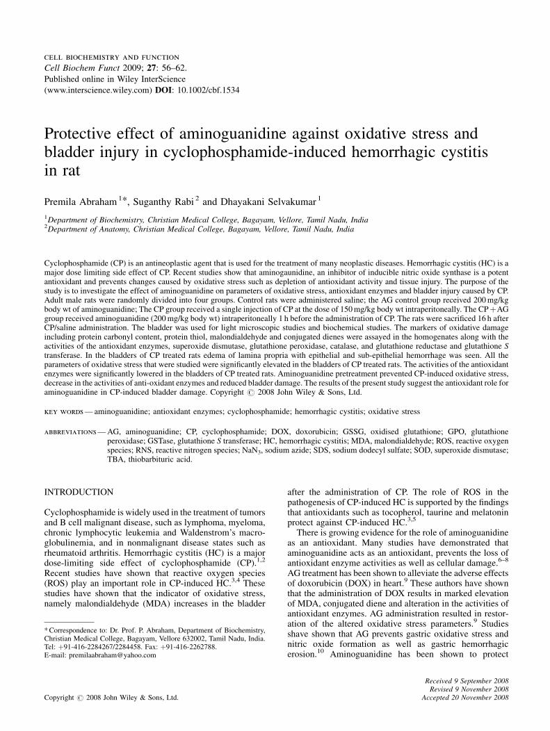
Protective effect of aminoguanidine against oxidative stress and bladder injury in cyclophosphamide-induced hemorrhagic cystitis in rat Premila Abraham 1 * , Suganthy Rabi 2 and Dhayakani Selvakumar 1 1 Department of Biochemistry, Christian Medical College, Bagayam, Vellore, Tamil Nadu, India 2 Department of Anatomy, Christian Medical College, Bagayam, Vellore, Tamil Nadu, India Cyclophosphamide (CP) is an antineoplastic agent that is used for the treatment of many neoplastic diseases. Hemorrhagic cystitis (HC) is a major dose limiting side effect of CP. Recent studies show that aminogaunidine, an inhibitor of inducible nitric oxide synthase is a potent antioxidant and prevents changes caused by oxidative stress such as depletion of antioxidant activity and tissue injury. The purpose of the study is to investigate the effect of aminoguanidine on parameters of oxidative stress, antioxidant enzymes and bladder injury caused by CP. Adult male rats were randomly divided into four groups. Control rats were administered saline; the AG control group received 200 mg/kg body wt of aminoguanidine; The CP group received a single injection of CP at the dose of 150 mg/kg body wt intraperitoneally. The CP þAG group received aminoguanidine (200 mg/kg body wt) intraperitoneally 1 h before the administration of CP. The rats were sacrificed 16 h after CP/saline administration. The bladder was used for light microscopic studies and biochemical studies. The markers of oxidative damage including protein carbonyl content, protein thiol, malondialdehyde and conjugated dienes were assayed in the homogenates along with the activities of the antioxidant enzymes, superoxide dismutase, glutathione peroxidase, catalase, and glutathione reductase and glutathione S transferase. In the bladders of CP treated rats edema of lamina propria with epithelial and sub-epithelial hemorrhage was seen. All the parameters of oxidative stress that were studied were significantly elevated in the bladders of CP treated rats. The activities of the antioxidant enzymes were significantly lowered in the bladders of CP treated rats. Aminoguanidine pretreatment prevented CP-induced oxidative stress, decrease in the activities of anti-oxidant enzymes and reduced bladder damage. The results of the present study suggest the antioxidant role for aminoguanidine in CP-induced bladder damage. Copyright # 2008 John Wiley & Sons, Ltd. key words — aminoguanidine; antioxidant enzymes; cyclophosphamide; hemorrhagic cystitis; oxidative stress abbreviations —AG, aminoguanidine; CP, cyclophosphamide; DOX, doxorubicin; GSSG, oxidised glutathione; GPO, glutathione peroxidase; GSTase, glutathione S transferase; HC, hemorrhagic cystitis; MDA, malondialdehyde; ROS, reactive oxygen species; RNS, reactive nitrogen species; NaN 3 , sodium azide; SDS, sodium dodecyl sulfate; SOD, superoxide dismutase; TBA, thiobarbituric acid. INTRODUCTION Cyclophosphamide is widely used in the treatment of tumors and B cell malignant disease, such as lymphoma, myeloma, chronic lymphocytic leukemia and Waldenstrom’s macro- globulinemia, and in nonmalignant disease states such as rheumatoid arthritis. Hemorrhagic cystitis (HC) is a major dose-limiting side effect of cyclophosphamide (CP). 1,2 Recent studies have shown that reactive oxygen species (ROS) play an important role in CP-induced HC. 3,4 These studies have shown that the indicator of oxidative stress, namely malondialdehyde (MDA) increases in the bladder after the administration of CP. The role of ROS in the pathogenesis of CP-induced HC is supported by the findings that antioxidants such as tocopherol, taurine and melatonin protect against CP-induced HC. 3,5 There is growing evidence for the role of aminoguanidine as an antioxidant. Many studies have demonstrated that aminoguanidine acts as an antioxidant, prevents the loss of antioxidant enzyme activities as well as cellular damage. 6–8 AG treatment has been shown to alleviate the adverse effects of doxorubicin (DOX) in heart. 9 These authors have shown that the administration of DOX results in marked elevation of MDA, conjugated diene and alteration in the activities of antioxidant enzymes. AG administration resulted in restor- ation of the altered oxidative stress parameters. 9 Studies shave shown that AG prevents gastric oxidative stress and nitric oxide formation as well as gastric hemorrhagic erosion. 10 Aminoguanidine has been shown to protect cell biochemistry and function Cell Biochem Funct 2009; 27: 56–62. Published online in Wiley InterScience (www.interscience.wiley.com) DOI: 10.1002/cbf.1534 *Correspondence to: Dr. Prof. P. Abraham, Department of Biochemistry, Christian Medical College, Bagayam, Vellore 632002, Tamil Nadu, India. Tel: þ91-416-2284267/2284458. Fax: þ91-416-2262788. E-mail: [email protected] Copyright # 2008 John Wiley & Sons, Ltd. Received 9 September 2008 Revised 9 November 2008 Accepted 20 November 2008
Transcript of Protective effect of aminoguanidine against oxidative stress and bladder injury in...
cell biochemistry and function
Cell Biochem Funct 2009; 27: 56–62.
Published online in Wiley InterScience
(www.interscience.wiley.com) DOI: 10.1002/cbf.1534
Protective effect of aminoguanidine against oxidative stress andbladder injury in cyclophosphamide-induced hemorrhagic cystitisin rat
Premila Abraham1*, Suganthy Rabi 2 and Dhayakani Selvakumar 1
1Department of Biochemistry, Christian Medical College, Bagayam, Vellore, Tamil Nadu, India2Department of Anatomy, Christian Medical College, Bagayam, Vellore, Tamil Nadu, India
Cyclophosphamide (CP) is an antineoplastic agent that is used for the treatment of many neoplastic diseases. Hemorrhagic cystitis (HC) is amajor dose limiting side effect of CP. Recent studies show that aminogaunidine, an inhibitor of inducible nitric oxide synthase is a potentantioxidant and prevents changes caused by oxidative stress such as depletion of antioxidant activity and tissue injury. The purpose of thestudy is to investigate the effect of aminoguanidine on parameters of oxidative stress, antioxidant enzymes and bladder injury caused by CP.Adult male rats were randomly divided into four groups. Control rats were administered saline; the AG control group received 200mg/kgbody wt of aminoguanidine; The CP group received a single injection of CP at the dose of 150mg/kg body wt intraperitoneally. The CPþAGgroup received aminoguanidine (200mg/kg body wt) intraperitoneally 1 h before the administration of CP. The rats were sacrificed 16 h afterCP/saline administration. The bladder was used for light microscopic studies and biochemical studies. The markers of oxidative damageincluding protein carbonyl content, protein thiol, malondialdehyde and conjugated dienes were assayed in the homogenates along with theactivities of the antioxidant enzymes, superoxide dismutase, glutathione peroxidase, catalase, and glutathione reductase and glutathione Stransferase. In the bladders of CP treated rats edema of lamina propria with epithelial and sub-epithelial hemorrhage was seen. All theparameters of oxidative stress that were studied were significantly elevated in the bladders of CP treated rats. The activities of the antioxidantenzymes were significantly lowered in the bladders of CP treated rats. Aminoguanidine pretreatment prevented CP-induced oxidative stress,decrease in the activities of anti-oxidant enzymes and reduced bladder damage. The results of the present study suggest the antioxidant role foraminoguanidine in CP-induced bladder damage. Copyright # 2008 John Wiley & Sons, Ltd.
key words—aminoguanidine; antioxidant enzymes; cyclophosphamide; hemorrhagic cystitis; oxidative stress
abbreviations—AG, aminoguanidine; CP, cyclophosphamide; DOX, doxorubicin; GSSG, oxidised glutathione; GPO, glutathioneperoxidase; GSTase, glutathione S transferase; HC, hemorrhagic cystitis; MDA, malondialdehyde; ROS, reactive oxygenspecies; RNS, reactive nitrogen species; NaN3, sodium azide; SDS, sodium dodecyl sulfate; SOD, superoxide dismutase;TBA, thiobarbituric acid.
INTRODUCTION
Cyclophosphamide is widely used in the treatment of tumorsand B cell malignant disease, such as lymphoma, myeloma,chronic lymphocytic leukemia and Waldenstrom’s macro-globulinemia, and in nonmalignant disease states such asrheumatoid arthritis. Hemorrhagic cystitis (HC) is a majordose-limiting side effect of cyclophosphamide (CP).1,2
Recent studies have shown that reactive oxygen species(ROS) play an important role in CP-induced HC.3,4 Thesestudies have shown that the indicator of oxidative stress,namely malondialdehyde (MDA) increases in the bladder
*Correspondence to: Dr. Prof. P. Abraham, Department of Biochemistry,Christian Medical College, Bagayam, Vellore 632002, Tamil Nadu, India.Tel: þ91-416-2284267/2284458. Fax: þ91-416-2262788.E-mail: [email protected]
Copyright # 2008 John Wiley & Sons, Ltd.
after the administration of CP. The role of ROS in thepathogenesis of CP-induced HC is supported by the findingsthat antioxidants such as tocopherol, taurine and melatoninprotect against CP-induced HC.3,5
There is growing evidence for the role of aminoguanidineas an antioxidant. Many studies have demonstrated thataminoguanidine acts as an antioxidant, prevents the loss ofantioxidant enzyme activities as well as cellular damage.6–8
AG treatment has been shown to alleviate the adverse effectsof doxorubicin (DOX) in heart.9 These authors have shownthat the administration of DOX results in marked elevationof MDA, conjugated diene and alteration in the activities ofantioxidant enzymes. AG administration resulted in restor-ation of the altered oxidative stress parameters.9 Studiesshave shown that AG prevents gastric oxidative stress andnitric oxide formation as well as gastric hemorrhagicerosion.10 Aminoguanidine has been shown to protect
Received 9 September 2008Revised 9 November 2008
Accepted 20 November 2008

AG AMELIORATES CP-INDUCED OXIDATIVE STRESS AND BLADDER DAMAGE 57
against the tissue damage associated with periodontitis byreducing nitric oxide production and oxidative stress, andincreasing glutathione content.11 In a very recent study,12
aminoguanidine has been shown to reduce cisplatin inducednephrotoxicity due to its antioxidant effect. In another recentstudy,13 aminoguanidine has been shown to increase theendogenous antioxidant defence mechanism in rats andprotect the animals from radiation-induced lung toxicity.Abdel-Zaher et al.14 have shown that AG markedly inhibitsacetaminophen -induced hepatic and renal depletion ofantioxidants as well as nitric oxide overproduction. Thesestudies demonstrate that AG acts as a potent antioxidant.
Reactive oxygen species is reported to play an importantrole in CP-induced HC. However, there are no studies on theantioxidant effect of AG in CP-induced HC, to the best ofour knowledge. Therefore, in the present study, theantioxidant effect of aminoguanidine on CP-induced HCis investigated.
MATERIALS AND METHODS
Animals
Adult male Wistar rats (200–250 g) were used for theexperiments. The study was approved by animal ethicsCommittee for the Purpose of Control and Supervision ofExperimentation on Animals (CPCSEA), Government ofIndia. The guidelines were followed. Dosage and routeof administration of cyclophosphamide (CP) were deter-mined from that described in literature.15
Animal treatment
The rats were divided into four groups and were treated asfollows.
Group I: This group of rats received saline alone.Group II: This group of rats received AG (200mg/kg bodywt, i.p.).Group III: The rats in this group received a single intraper-itoneal injection of CP in saline at the dose of 150mg/kgbody weight.Group IV: The rats in this group aminoguanidine(200mg/kg body wt, i.p.), 1 h before the administration ofCP.
All the rats (six in each group) were killed 16 h after theadministration of CP/saline.
Tissue procurement
Rats were killed by exsanguination. The urinary bladder wasremoved and blotted dry before weighing. A part of thebladder was used for biochemical assays and another part forhistological assessment.
Histology
The tissues were fixed overnight in 10% buffered neutralformalin, processed to paraffin wax, sectioned at 5mm, and
Copyright # 2008 John Wiley & Sons, Ltd.
stained with hematoxylin and eosin (H & E) for examinationby light microscopy.
Biochemical assays
Bladder tissue was weighed and homogenized in appropriatebuffers and used for the following assays.
Malondialdehyde. Malonaldehyde content was measured asdescribed by Ohkawa et al.16. The mixture consisted of0.8ml of sample (1mg), 0.2ml of 8.1% SDS, 1.5ml of 20%glacial acetic acid adjusted to pH 3.5, and 1.5ml of 0.8%aqueous solution of TBA. The mixture was made up to 4mlwith distilled water and heated at 95C for 60min using aglass ball as condenser. After cooling with tap water, 1mldistilled water and 5ml n-butanol and pyridine mixture(15:1) were added and the solution was shaken vigorously.After centrifugation at 2000g for 10min the absorbance ofthe organic layer was measured at 532 nm. Amount ofthiobarbituric reacting substances formed is calculated fromstandard curve prepared using 1,10,3,30-tetramethoxy pro-pane and the values expressed as nmol per mg protein.
Conjugated diene. Total lipids were extracted as describedby Chan and Levett,17 and evaporated to dryness undernitrogen. This was dissolved in 1ml heptane and theabsorbance was measured at 233 nm. The amount ofconjugated diene formed in the sample is calculated usinga molar absorption coefficient of 2.52� 104.
Protein carbonyl content. Protein carbonyl content wasmeasured using DNPH as described by Sohal et al.18 To0.5ml of sample (1–2mg), an equal volume of 10mMDNPH in 2N HCl was added and incubated for 1 h shakingintermittently at room temperature. Corresponding blankwas carried out by adding only 2N HCL to the sample. Afterincubation, the mixture was precipitated with 10% TCA(final concentration) and centrifuged. The precipitate waswashed twice with ethanol:ethylacetate (1:1) and finallydissolved in 1ml of 6M guanidine HCl, centrifuged at lowspeed and the supernatant was read at 366 nm. Thedifference in absorbance between the DNPH treated andHCl treated sample is determined and expressed as nmol ofcarbonyl groups per mg of protein, using extinctioncoefficient of 22mM�1 cm�1.
Protein thiol groups. Thiol groups were measured asdescribed by Sedlak and Lindsay.19 To 1ml of the samplesuspension (1mg protein/ml), 1ml of 10% TCA containing1mM EDTA was added. The protein precipitate wasseparated by high-speed centrifugation for 10min. For totalthiol estimation the sample was taken directly with outprecipitation. To this, 1ml of solution I and 0.5% SDS wereadded followed by 2ml of solution II and 30ml of DTNB.The tubes were mixed well and kept in the dark for 15min atroom temperature. The intense yellow color of thenitromercapto benzoate anion formed from the DTNB
Cell Biochem Funct 2009; 27: 56–62.
DOI: 10.1002/cbf

Figure 1. Bladder weights of control rats and experimental rats, n¼ 6 rats,��p< 0.01 versus control, #p< 0.01 versus CP
58 p. abraham ET AL.
reaction with the thiol was read at 412 nm which has a molarabsorption of 13 600mM�1 cm�1.
Assay of anti-oxidant enzyme activitiesSuperoxide dismutase. Superoxide dismutase (SOD) was
measured as described by Ohkuma et al.20 The assaymixture consisted of 100ml of phosphate buffer, 10ml ofBSC, 50ml of Triton X-100, 5ml of EDTA, 5ml of xanthineoxidase, 50ml of xanthine. To this 150ml MTT and sample(50–150mg protein) were added and, the volume was madeup to 1ml with water. The mixture was incubated for 5minat room temperature (308C) and the reaction was terminatedwith the addition of 1ml of stop buffer. The optical densitywas read at 540 nm. Amount of superoxide formed iscalculated using the molar extinction coefficient of MTTformazan of 17 000M�1 cm�1 at pH 7.4 to 10.5. Thepercentage of inhibition by the presence of SOD wascalculated 540 from the reduction of the MTT colorformation as compared to the MTT formazan formed in theabsence of SOD, which is taken as 100%. One unit of SODactivity is defined as the amount of protein required to inhibitMTT reduction by 50%.
Catalase. Catalase activity was estimated by measuringthe change in absorption at 240 nm using H2O2 assubstrate.21 To 1ml of 30mM buffered H2O2, the enzyme(sample) was added to start the reaction. The final volumewas made up to 2ml with 0.05M phosphate buffer pH 7.0.Change in OD was observed for 2min at 240 nm. One unit isthe activity that disproportionates H2O2 at the rateof 10�3 absorbance/s.
Glutathione S transferase (GSTase). The activity ofGSTase was measured spectrophotometrically using thesubstrate 1-chloro-2,4-dinitrobenzene (CDNB).22 To 0.1mlof 1M potassium phosphate buffer pH 6.5, followingreagents were added: 0.1ml of 10mM GSH, 0.05ml 20mMCDNB and water and made up the volume to 1ml. Thereaction was started by adding the enzyme and change in ODat 340 nm is measured for 1–2min. One unit of enzyme is theamount required to conjugate 1mmol of substrate withglutathione in 1min.
Glutathione reductase. In the presence of enzyme,hydrogen is transferred from NADPH to GSSG and thereaction can be measured at 340 nm.23 To the reactionmixture containing 0.05ml of 1M phosphate buffer pH 7.6,0.15ml of 10mM EDTA, 0.1ml of 1mM NADPH, and0.1ml 10mM GSSG, the enzyme was added. The volumewas made up to 1ml and the decrease in OD at 340 nm wasmeasured for 2–3min. One unit is the amount of enzymeneeded to oxidize 1mmol of NADPH/min.
Glutathione peroxidase. Total peroxidase was determinedby following the oxidation of NADPH at 340 nm usinghydrogen peroxide.24 To 0.25ml of 0.4M phosphate buffer,0.2ml of 4mM EDTA, 0.2ml of 10mM GSH, 0.2ml of
Copyright # 2008 John Wiley & Sons, Ltd.
NaN3, 0.2ml of 1.6mM NADPH, 0.03ml glutathionereductase (one unit) and the enzyme (sample) was added.Total volume was made up to 2ml with water. Reaction wasstarted by adding 0.2ml of H2O2 and change in OD at340 nm was followed. Extinction coefficient of 6.1mm�1
was used for the calculation. One unit of glutathioneperoxidase activity is defined as the amount of enzymeneeded to oxidize 1 nmol of NADPH/min.
Assay of nitrite and nitrate. Total nitrate (nitrite and nitrate)was estimated in the bladder homogenates as described bySastry et al.25 In this method, copper-cadmium alloyreduces nitrate to nitrite which reacts with Griess reagent(sulphanilamide and N-naphthalenediamine) in acidicmedium to give purple color.Protein content in the homogenates was measured by the
method of Lowry et al.26
STATISTICAL ANALYSIS
The results are expressed as mean� SD. Comparisonbetween groups was done using ANOVA. Student’s t-testwith Bonferroni correction was used to compare individualmeans in the case of a significant F.
RESULTS
Gross findings
CP injection resulted in severe cystitis with macroscopichemorrhage (hematuria). The bladder weights of CP treatedrats were significantly higher than that of control rats(Figure 1). The bladder weights of AG pretreated rats werecomparable to that of control rats.
Histology
In control animal, the urinary bladder had the urotheliumformed by tightly packed cells with little intercellular space.The basement membrane that separates the epithelium fromthe underlying lamina propria was intact. There was nobreach in it. The connective tissue that constituted thelamina propria was dense with normal vascular supply. Themucosa was thrown into folds and there were sub-epithelialcrypts. The smooth muscle coat underlying the lamina
Cell Biochem Funct 2009; 27: 56–62.
DOI: 10.1002/cbf

Figure 2. Light microscopy of urinary bladder of control rats and experimental rats. (a) Saline control. Normal morphology. Arrow points to folded mucosawith epithelium showing tight packing of cells and dense connective tissue in lamina propria. Sub-epithelial crypts are also seen in this figure. There is nohemorrhage. Magnification 100�. (b) Aminoguanidine control. Normal epithelium and lamina propria. Magnification 100�. (c) Cyclophosphamide treatedbladder shows edema of the epithelium and it appears that epithelium is breached. Vascular changes in the lamina propria are apparent. Magnification 100�.(d) CPþAG showed almost normal morphology. There is no edema or hemorrhage. The lamina propria appears normal. Magnification 100�
AG AMELIORATES CP-INDUCED OXIDATIVE STRESS AND BLADDER DAMAGE 59
propria displayed circular and longitudinal muscle coat withminimal connective tissue packing. There were no exudatesin the lumen (Figure 2a).
Contrary to the normal picture presented above, thebladder wall in CP treated animals showed damages. Themucosa became edematous and the cells of the urotheliumwere not compact. There seemed to be cellular exudates inthe lumen. Mucosal content formed follicular cystitis(Figure 2b). Edema of lamina propria with epithelial andsub-epithelial hemorrhage was also seen (arrows inFigure 2c).
Table 1. Oxidative stress parameters, anti-oxidant enzyme activities, and nitrate lwith or without pretreatment with aminoguanidine
Parameter Control (n¼ 6)
Malondialdehyde (nmol/mg protein) 53� 12Conjugated diene (nmol/mg protein) 17� 1.2Protein carbonyl content (pmol/mg protein) 79� 9Protein thiol (nmol/mg protein) 0.74� 0.04Glutathione peroxidase activity (mU/mg protein) 2.12� 0.2SOD activity (mU/mg protein) 41� 4GSTase activity (mU/mg protein) 0.61� 0.09Glutathione reductase Activity (mU/mg protein) 3.3� 0.3Catalase activity (mU/mg protein) 19� 5Total nitrate (mmol/mg protein) 0.31� 0.03
Data represent mean� SD. n¼ number of rats.�p< 0.05.��p< 0.01.���p< 0.001 as compared with control.xp< 0.05.#p< 0.01 as compared with CP.
Copyright # 2008 John Wiley & Sons, Ltd.
Pretreatment with AG reduced this damage significantlyas shown in Figure 2. Symptoms of HC such as edema,inflammation and hemorrhage were significantly reduced.
Biochemical parameters
The results are shown in Table 1. The markers of lipidperoxidation, namely malondialdehye (MDA) and conju-gated diene were increased significantly in bladders of CPtreated rats 16 h after treatment as compared with control(p< 0.01). Protein carbonyl content, an early and sensitive
evels in the urinary bladder of control rats and cyclophosphamide treated rats
AG (n¼ 6) CP (n¼ 6) CPþAG (n¼ 6)
47� 10 111� 14�� 38� 7#
16� 1.7 69� 7.9�� 18� 1.7#
65� 12 120� 22�� 70� 7#
0.77� 0.25 0.44� 0.08� 0.77� 0.02x
1.93� 0.4 1.04� 0.1��� 2.1� 0.3#
43� 12 19� 3��� 40� 4#
0.63� 0.08 0.41� 0.02� 0.66� 0.22x
2.8� 0.9 4.3� 0.4� 4� 1.5x
18� 3 12� 3� 17� 2x
0.32� 0.04 0.61� 0.07�� 0.35� 0.03x
Cell Biochem Funct 2009; 27: 56–62.
DOI: 10.1002/cbf

60 p. abraham ET AL.
indicator of oxidative damage to proteins was increased by52% 16 h after treatment with CP as compared with control,p< 0.01. Protein thiol, one of the important antioxidants andindicators of oxidative damage to proteins was decreased42% as compared with control, p< 0.05.With regard to the antioxidant enzymes, a significant
decrease in glutathione peroxidase (GPO) activity (51%)was observed 16 h after treatment with CP as compared withcontrol, p< 0.01. With respect to SOD activity, a 50%decrease was observed after treatment with CP as comparedwith control, p< 0.001. The activity of the drug detoxifyingenzyme, GSTase was decreased by 65% after treatment withCP respectively as compared with control, p< 0.05.Glutathione reductase activity was increased by 24% aftertreatment with CP as compared with control, p< 0.05. Theactivity of catalase was decreased in the bladders of CPtreated rats as compared with control. CP treatment resultedin twofold increase in nitrate levels in the bladder ascompared with control (p< 0.01).Pretreatment with AG markedly reduced CP-induced
elevation in MDA, conjugated diene, and protein carbonylcontent (p< 0.01, p< 0.01, and p< 0.001 respectively ascompared with CP). Protein thiol levels in the AG pretreatedrats were restored to those comparable with the control rats(p< 0.05 compared with CP). Besides, AG pretreatmentrestored the activities of the antioxidant enzymes catalase,GSTase, glutathione peroxidase and superoxide dismutase inthe bladders of CP treated rats (p< 0.05, p< 0.02,p< 0.001, p< 0.01, respectively), AG pretreatment resultedin the prevention of elevation of nitrate levels (p< 0.01 ascompared with CP).Histologically also AG pretreatment ameliorated CP-
induced cystitis. Edema of lamina propria and epithelial andsub-epithelial hemorrhage that was noted in the CP treatedrats were not seen in the AG pretreated rats.
DISCUSSION
Urotoxicity is one of the major dose limiting side effects ofCP. Studies have shown that mechanisms other thanproduction of acrolein such as reactive oxygen speciesand reactive nitrogen species are involved in CP-inducedHC. Oxidative stress is known to play an important role inCP-induced HC.3,4 Accordingly, in the present study all theparameters of oxidative stress, that is, conjugated dienes,malondialdehyde and protein carbonyl content were foundto be markedly increased in the bladders of CP treated ratssuggesting that CP treatment caused oxidative damage to thelipids and proteins of the bladder. AG pretreatmentprevented CP-induced elevation in all the oxidative stressparameters studied.Antioxidant enzymes protect tissue against oxidative
injury. Tissue damage is reported to occur after theantioxidants are depleted. In the present study adecrease in the activities of the important antioxidantenzymes namely SOD, GPO, GSTase was observed in thebladders of CP treated rats. AG pretreatment restored theactivities of the antioxidant enzymes significantly. AG
Copyright # 2008 John Wiley & Sons, Ltd.
pretreatment minimized CP-induced bladder damage aswell.The protective effect of AG in CP-induced HC may
be achieved by two mechanisms. Firstly, AG reduces theformation of potent oxidant peroxynitrite. Most of thedeleterious effects of nitric oxide is believed to be producedby peroxynitrite, a potent oxidant that is produced by thereaction of nitric oxide with superoxide anion.27,28
Mitochondria in vivo produce superoxide and in pathologi-cal situations the amount of superoxide produced increases;therefore, in the presence of nitric oxide, mitochondria willbe a major site of peroxynitrite formation.29 Peroxynitrite isa potent oxidant that depletes the antioxidant defencesincluding glutathione, protein thiol, and antioxidantenzymes.30–32 Insufficiency of the protective antioxidantsystem results in enhanced lipid peroxidation and tissueinjury. When the production of nitric oxide is decreased bythe administration of nitric oxide synthase inhibitoraminoguanidine, the source of peroxynitrite is reduced.Thus, less peroxynitrite is produced, the oxidative damage tothe lipids and proteins is decreased and the cellularantioxidant systems are spared. Accordingly, in the presentstudy we observed that AG pretreatment reduced CP-induced oxidative stress and restored the antioxidant activityin the bladder.SOD provides the first line of defense against superoxide
generated in mitochondria. SOD competes with nitric oxidefor reaction with superoxide and prevents generation ofperoxynitrite, a potent oxidant that can modify proteins toform 3-nitrotyrosine.33 Thus, sufficient amounts of cataly-tically competent SOD are required to prevent tissuedamage. SOD is a target of tyrosine nitration and nitrationinactivates the enzyme.34 Nitration and inactivation of SODcould lead to self-amplification of oxidative stress in thetissues progressively enhancing peroxynitrite productionand secondary damage. It has been already demonstratedthat CP-induced bladder damage involves peroxynitriteformation and protein nitration.35,36 It is noteworthy tomention that in the present study, CP treatment resulted inmarked decrease in the activity of SOD, and pretreatmentwith AG almost completely prevented the loss of activity aswell as reduced bladder damage.Secondly, the protective effect of AG may result from
direct antioxidant effects of aminoguanidine. There isgrowing evidence for the role of AG as an antioxidant. AGhas been reported to be an effective hydroxyl radicalscavenger.37 AG exhibits a significant dose-dependent effectagainst free radical damage. Aminoguanidine has beenshown to exhibit trapping activity toward lipid-derivedaldehydes such as MDA and 4-hydroxynonenal.38 Severalstudies have shown that AG acts as an antioxidant and canrestore the antioxidants in the tissues. Aminoguanidine hasbeen reported to prevent gentamicin induced renal damageby acting as a potent free radical scavenger and prevents thedepletion of cellular antioxidants.39 A study has shown thatthe protective effect of aminoguanidine against nephrotoxi-city induced by cisplatin in normal rats is by its capacity toinhibit lipid peroxidation.40 Kedziora-kornatowska et al.41
Cell Biochem Funct 2009; 27: 56–62.
DOI: 10.1002/cbf

AG AMELIORATES CP-INDUCED OXIDATIVE STRESS AND BLADDER DAMAGE 61
have demonstrated that AG can prevent oxidative stress inthe peripheral blood granulocytes of rats with streptozoto-cin-induced diabetes. Besides, AG prevents impairment ofblood antioxidant systems in insulin dependent diabeticrats.42 A recent study shows that AG improves antioxidantstatus and attenuates lipid peroxidation in the brain of ratsexposed to chronic stress.43 Another recent study demon-strates that aminoguanidine can reduce the oxidativedamage to proteins and hence protein carbonyl content.44
The role of aminoguanidine as an inhibitor of nitric oxideproduction is well known. The results of the present studyshow for the first time that AG acts as an antioxidant, reducesCP-induced oxidative stress and ameliorates bladderdamage. These results suggest that AG has a protectiveeffect on urotoxicity induced by CP and it may thereforeimprove the therapeutic index of CP.
ACKNOWLEDGEMENTS
The study was supported by Department of Science andTechnology (DST) New Delhi, India. The authors thankDr. Indirani Kanakasabapathy, Professor in Anatomy for herassistance in light microscopic studies and Miss. Preethi K.for her technical assistance in biochemical studies.
REFERENCES
1. Levine AL, Richie PJ. Urological complications of cyclophosphamide.J Urol 1989; 141: 1063–1069.
2. West NJ. Prevention and treatment of hemorrhagic cystitis. Pharma-cotherapy 1997; 4: 696–706.
3. Sadir S, Deveci S, Korkmaz A. Alpha tocopherol, beta carotene, andmelatonin administration protects cyclophosphamide induced oxidativedamage to bladder tissue in rats. Cell Biochem Funct 2007; 25: 521–552.
4. Topal P, Oztas Y, Korkmaz A. Melatonin ameliorates bladder damageinduced by cyclophosphamide in rats. J Pineal Res 2005; 38: 272–277.
5. Abd-Allah AR, Gado AM, Al-Majed AA, et al. Protective effect oftaurine against cyclophosphamide-induced urinary bladder toxicity inrats. Clin Exp Pharmacol Physiol 2005; 32: 167–172.
6. Kedziora-Kornatowska KZ, Luciak M, Blaszczyk J, Pawlak W. Effectof aminoguanidine on erythrocyte lipid peroxidation and activities ofantioxidant enzymes in experimental diabetes. Clin Chem Lab Med1998; 36: 771–775.
7. Di Paola R, Marzocco S, Mazzon E, et al. Effect of aminoguanidine inligature-induced periodontitis in rats. J Dent Res 2004; 83: 343–348.
8. Baynes JW, Thorpe SR. Glycoxidation and lipoxidation in atherogen-esis. Free Radic Biol Med 2000; 28: 1708–1716.
9. Abd El-Gawad HM, El-Sawalhi MM. Nitric oxide and oxidative stressin brain and heart of normal rats treated with doxorubicin: role ofaminoguanidine. J Biochem Mol Toxicol 2004; 18: 69–77.
10. Hung CR. Role of gastric oxidative stress and nitric oxide in formationof hemorrhagic erosion in rats with ischemic brain. World J Gastro-enterol 2006; 12: 574–581.
11. Ara C, Karabulut AB, Kirimlioglu H, et al. Protective effect ofaminoguanidine against oxidative stress in an experimental peritonealadhesion model in rats. Cell Biochem Funct 2006; 24: 443–448.
12. Atasayar S, Gurer-Orhan H, Orhan H, Gurel B, Girgin G, Ozgunes H.Preventive effect of amino guanidine compared to vitamin E and C oncisplatin-induced nephrotoxicity in rats. Exp Toxicol Pathol 2008; Aug4 [Epub ahead of print].
13. Eroglu C, Yildiz OG, Saraymen R, Soyuer S, Kilic E, Ozcan S.Aminoguanidine ameliorates radiation-induced oxidative lung damagein rats. Clin Invest Med 2008; 31: E182–E188.
Copyright # 2008 John Wiley & Sons, Ltd.
14. Abdel-Zaher AO, Abdel-RahmanMM, HafezMM, Omran FM. Role ofnitric oxide and reduced glutathione in the protective effects ofaminoguanidine, gadolinium chloride and oleanolic acid against acet-aminophen-induced hepatic and renal damage. Toxicology 2007; 234:124–134.
15. Ahluwalia A, Maggi CA, Santicioli P, Lecci A, Giuliani S. Charac-terization of the capsaicin-sensitive component of cyclophosphamide-induced inflammation in the rat urinary bladder. Br J Pharmacol 1994;111: 1017–1022.
16. Ohkawa H, Ohishi N, Yagi K. Assay for lipid peroxides in animaltissues by thiobarbituric acid reaction. Anal Biochem 1979; 95: 351–358.
17. Chan HW, Levett G. Autoxidation of methyl linoleate. Separation andanalysis of isomeric mixtures of methyl linoleate hydroperoxides andmethyl hydroxylinoleates. Lipids 1977; 12: 99–104.
18. Sohal RS, Agarwal S, Dubey A, Orr WC. Protein oxidative damage isassociated with life expectancy of houseflies. Proc Natl Acad Sci USA1993; 90: 7255–7259.
19. Sedlak J, Lindsay RH. Estimation of total, protein-bound, and non-protein sulfhydryl groups in tissuewith Ellman’s reagent.Anal Biochem1968; 25: 192–205.
20. OhkumaN, Matsuo S, Tutsui M, Ohkawara A. Superoxide dismutase inthe epidermis (author’s transl). Nippon Hifuka Gakkai Zasshi 1982; 92:583–590.
21. Aebi IH. Catalase in vitro. Methods Enzymol 1984; 105: 121–126.22. Awasthi YC, Dao DD, Saneto RP. Interrelationship between anionic
and cationic forms of glutathione S-transferases of human liver.Biochem J 1980; 191: 1–10.
23. Racker E. Glutathione reductase from bakers’ yeast and beef liver.J Biol Chem 1955; 217: 855–865.
24. Nakamura W, Hosada S. Purification and properties of rat liverglutathione peroxidase. Biochim Biophys Acta 1974; 358: 251–261.
25. Sastry KV,Moudgal RP,Mohan J. Spectrophotometric determination ofserum nitrite and nitrate by copper-cadmium alloy. Anal Biochem 2002;306: 79–82.
26. Lowry OH, Rosebrough NJ, Farr AL, Randall RJ. Protein measurementwith the Folin phenol reagent. J Biol Chem 1951; 93: 265–275.
27. Radi R, Beckman JS, Bush KM. Peroxynitrite-induced membrane lipidperoxidation: the cytotoxic potential of superoxide and nitric oxide.Arch Biochem Biophys 1991; 288: 481–485.
28. Radi R. Nitric oxide, oxidants, and protein tyrosine nitration: Proc.NatlAcad Sci USA 2004; 101: 4003–4008.
29. Radi R, Cassina A, Hodara R, Quijano C, Castro L. Peroxynitritereactions and formation in mitochondria. Free Radic Biol Med 2002;33: 1451–1464.
30. Olas B, Nowak P, Wachowicz B. Resveratrol protects against perox-ynitrite-induced thiol oxidation in blood platelets. Cell Mol Biol Lett2004; 9: 577–587.
31. Varma SD, Hegde KR. Lens thiol depletion by peroxynitrite. Protectiveeffect of pyruvate. Mol Cell Biochem 2007; 298: 199–204.
32. ReinartzM, Ding Z, Flogel U, Godecke A, Schrader J. Nitrosative stressleads to protein glutathiolation, increased s-nitrosation, and up-regu-lation of peroxiredoxins in the heart. J Biol Chem 2008; 283: 17440–17449.
33. Johnson F, Giulivi C. Superoxide dismutases and their impact uponhuman health. Mol Aspects Med 2005; 26: 340–352.
34. Bayir H, Kagan VE, Clark RS, Janesko-Feldman K, Rafikov R, HuangZ, Zhang X, Vagni V, Billiar TR, Kochanek PM. Neuronal NOS-mediated nitration and inactivation of manganese superoxide dismutasein brain after experimental and human brain injury. J Neurochem 2007;101: 168–181.
35. Korkmaz A, Topal T, Oter S. Pathophysiological aspects of cyclopho-sphamide and ifosfamide induced hemorrhagic cystitis; implication ofreactive oxygen and nitrogen species as well as PARP activation. CellBiol Toxicol 2007; 23: 303–312.
36. Yildrim I, Korkmaz A, Oter S. Contribution of antioxidants to pre-ventive effect of mesna in cyclophosphamide-induced hemorrhagiccystitis in rats. Cancer Chemother Pharmacol 2004; 54: 469–473.
Cell Biochem Funct 2009; 27: 56–62.
DOI: 10.1002/cbf

62 p. abraham ET AL.
37. Courderot-Masuyer C, Dalloz F, Maupoil V, Rochette L. Antioxidantproperties of aminoguanidine. Fundam Clin Pharmacol 1999; 13: 535–540.
38. Burcham PC, Kaminskas LM, Fontaine FR, Petersen DR, Pyke SM.Aldehyde-sequestering drugs: tools for studying protein damage bylipid peroxidation products. Toxicology 2002; 181–182: 229–236.
39. Polat A, Parlakpinar H, Tasdemir S, et al. Protective role of amino-guanidine on gentamicin-induced acute renal failure in rats. ActaHistochem 2006; 108: 365–371.
40. Mansour MA, Mostafa AM, Nagi MN, Khattab MM, Al-Shabanah OA.Protective effect of aminoguanidine against nephrotoxicity induced bycisplatin in normal rats. Comp Biochem Physiol Toxicol Pharmacol2002; 132: 123–128.
Copyright # 2008 John Wiley & Sons, Ltd.
41. Kedziora-Kornatowska KZ, Luciak M, Błaszczyk J, Pawlak W. Effectof aminoguanidine on the generation of superoxide anion and nitricoxide by peripheral blood granulocytes of rats with streptozotocin-induced diabetes. Clin Chim Acta 1998; 278: 45–53.
42. Stoppa GR, Cesquini M, Roman EA, Ogo SH, Torsoni MA. Amino-guanidine prevented impairment of blood antioxidant system in insulin-dependent diabetic rats. Life Sci 2006; 78: 1352–1361.
43. Akpinar D, Yargicoglu P, Derin N, Aslan M, Agar A. Effect ofaminoguanidine on visual evoked potentials (VEPs), antioxidant statusand lipid peroxidation in rats exposed to chronic restraint stress. BrainRes 2007; 1186: 87–94.
44. Brodiak IV, Sybirna NO. Effect of aminoguanidine on oxidativemodification of proteins in experimental diabetes mellitus in rats[Article in Ukrainian]. Ukr Biokhim Zh 2006; 78: 114–119.
Cell Biochem Funct 2009; 27: 56–62.
DOI: 10.1002/cbf